Embed Size (px)
Citation preview
Harran Üniversitesi Tıp Fakültesi Dergisi (Journal of Harran University Medical Faculty) Cilt 8. Sayı 1, 201114
Abstract
Background: The human TP53 gene, also known as p53, encodes for the tumor protein 53 (p53), regulates
the cell cycle and hence functions as a tumor suppressor. This study aimed to investigate some properties of
the TP53 gene and its products, such as the homologous protein sequences in different species, the common
transcription factor binding sites on their promoters, their phylogenetic relationship, conserved domains, and
their expression profiles by in silico biology approach.
Methods: We investigated the homology, conserved domain, promoter and expression profiles of the TP53
gene in various species using bioinformatics approaches.
Results: Our results revealed that which investigated p53 molecules among all organisms are conserved.
They have three conserved domains (p53_TAD, p53 DNA_binding, and p53 tetramerization motif), some of
which have full and truncated sub-domains. Human p53 proteins is similar to those of Pan troglodytes,
Macaca mulatta, Macaca fascicularis and Chlorocebus aethiops. In contrast, t Monodelphis domestica
protein is the most diverse of human p53. With the multiple alignment strategy, protein and domain
sequences of Equus asinus, Muntiacus muntjak vaginalis and Monodelphis domestica are predicted to have a
truncation. The comparative screening of the promoters demonstrated that TP53 genes do not seem to have
any common conserved transcription factor binding sites.
Conclusion: This study demonstrated that, p53 molecules from various species are well conserved in the
process of evolution. Comparative screening of the promoter sequences of the human p53 and its
homologues found in human and NCBI database revealed that there was no any common transcription factor
binding sites. Phylogenetic trees constructed using the neighbor-joining method (NJ) revealed a close
evolutionary relationship of p53 in various species.
Key words: TP53, bioinformatics, comparative genomics, phylogenetics, in silico biology
Özet
Amaç: p53 olarak bilinen insan TP53 geni, hücre siklusunu düzenleyen ve tümör baskılayıcı olarak görev
gören tumor protein 53'ü (p53) kodlar. Bu çalışmada, çeşitli türlerde TP53 geni ve onun ürünleri olan
homolog protein dizileri, promotorlarda genel transkripsiyon faktör bağlanma alanları, filogenetik
akrabalıkları, korunan alanları (p53) ve ekspresyon profilleri gibi özellikleri in silico biyoloji yaklaşımı ile
araştırılması amaçlandı.
Metodlar: Biyoinformatik uygulamaları kullanarak çeşitli türlerde TP53 genlerinin homolojisini,
korunmuş alanlarını, promotorlarını ve ekspresyon profillerini araştırdık.
Bulgular: Sonuçlarımız, p53 moleküllerinin araştırılan tüm organizmalar arasında korunduğunu
göstermiştir. Bunların üç korunan alanı (p53_TAD, p53 DNA_binding ve p53 tetramerization motif) vardır,
Analysis of TP53 Gene Using Bioinformatics Tools
1 2 3 3Fuat Dilmec , Mete Köksal , Abdullah Özgönül , Ali Uzunköy
1 2Harran University, Medicine Faculty, Departments of Medical Biology , Histology and Embryology , and General
3Surgery , Sanliurfa, Turkey
Correspondence: Fuat DILMEC, Harran University, Medicine Faculty, Department of Medical Biology, 63300
Sanliurfa-Turkey, Tel: 0(414) 3183028, Fax: 0(414) 318 31 92, E-mail: [email protected]
Original Article
Tp53 Geninin Biyoinformatik Araçlarla Analizi
Harran Üniversitesi Tıp Fakültesi Dergisi (Journal of Harran University Medical Faculty) Cilt 8. Sayı 1, 2011
Analysis of TP53 gene
15
Introduction
The TP53 gene is a key regulator of the cellular
response to stress and plays a critical role in
preventing cancer progression. The activation of
p53 in response to DNA damage or cellular stress
leads to cell cycle arrest, apoptosis, or senescence,
depending on the cellular context (1).
The human TP53 gene, also known as p53,
localized on chromosome 17 (17p13.1), comprises
11 exons encoding a 393 amino-acid protein with a
molecular weight of 53 kDa. p53 protein acts as a
transcription factor and serves as a key regulator of
the cell cycle (2).
The human p53 protein has four domains, N-
terminal domain, core domain (DNA-binding
domain), oligomerization domain and nuclear
localization domain (1). The C-terminus of p53
tumor suppressor contains a DNA binding motif.
The oncogenic activity of p53 C-terminus required
both the DNA damage recognition motif and the
repair enzyme-associating domain (3).
Human p53 molecule shows strong homology to
several organisms, Spalax and mouse of several
organisms, including those of Spalax and mouse.
At the amino acid level, Spalax p53, with a 391-aa
protein, an identity of 85.4% to human, and 81.9%
to mouse p53 proteins is observed. Besides, in the
p53 DNA-binding domain region, there is a
nucleotide sequence homology of 88.1% to humans
and 86.1% to mice, whereas amino acid homology is
95.8% for humans and 89.5% for mice (4).
It has been shown that the relative levels of mRNA specific for the mouse p53 cellular tumor antigen
were determined in various normal adult tissues,
embryos, and tumors. All tumors studied contained concentrations of TP53 mRNA well above those
present in most normal tissues. In most normal
tissues, the levels of these transcripts were very low,
but spleen cells contained much higher quantities of
TP53 mRNA. Nevertheless, the spleen did not
overproduce p53, owing to the exceptionally rapid
turnover of the protein in this organ (5).
In this study, we aimed to analyze the TP53 genes in
different species in silico biology. Specifically, their
p53 domains, the transcription factor binding sites on
their promoters, the tissue expression profile,
homology level and phylogenetic tree among
mammalian TP53 genes using bioinformatics tools.
Materials and Methods
Homology search
The search for homologous protein sequence to
human p53 was carried out using the BLASTp
bazılarının tam ve bazılarının kesikli korunan alt bölgeye sahip oldukları belirlendi. İnsan p53 proteini, Pan
troglodytes, Macaca mulatta, Macaca fascicularis ve Chlorocebus aethiops'dakilerine en yakındır. Tam
tersine, insan p53, Monodelphis domestica proteinine en uzaktır. Çoklu dizileme stratejisine göre, Equus
asinus, Muntiacus muntjak vaginalis ve Monodelphis domestica'nın protein ve domain dizilerinin kesintili
olduğu gösterilmiştir. Promotorların karşılıklı taramaları, TP53 genlerinin herhangi bir korunmuş genel
transkripsiyon faktor bağlama bölgelerinin olmadığını göstermiştir.
Sonuçlar: Bu çalışma, çeşitli türler arasında p53 moleküllerinin, evrim sürecinde iyi korunduğunu
göstermektedir. İnsan p53 ve NCBI databazında bulunan homologlarının promotor dizilerinin
karşılaştırmalı taramaları, genel transkripsiyon faktör bağlama bölgelerinin olmadığını göstermiştir.
Neighbor-joining metodu (NJ) kullanılarak filogenetik ağaçlar, çeşitli türlerde p53'ün yakın bir evrimsel
akrabalığının olduğunu açığa çıkarmıştır.
Anahtar kelimeler: TP53, biyoinformatik, karşılaştırmalı genomik, filogenetik, in silico biyoloji
Harran Üniversitesi Tıp Fakültesi Dergisi (Journal of Harran University Medical Faculty) Cilt 8. Sayı 1, 2011
Analysis of TP53 gene
16
program (6,7) at NCBI (http://www.ncbi.
nlm.nih.gov) using human p53 amino acid
sequence (GI: 8400737) as query against the
SwissProt protein databases. Full protein and
tumor protein p53 domains sequences of human
and other species were downloaded and then
aligned using the ClustalW (8) program at EBI
(http://www.ebi.ac.uk).
Promoter Analysis
We used Genomatix software (http://www.
genomatix.de) for analysis of TP53 gene
promoters in various species. These nucleotide
sequences were downloaded and then were
aligned using the ClustalW program. Then
common transcription factor binding sites were
searched with the Dialign TF program in
Genomatix software for all of TP53 promoters
present in the database.
Evolutionary Analysis
We used amino acid sequences of p53 proteins to
construct phylogenetic trees using the neighbor-
joining method (NJ) with Jones-Taylor-Thomton
(JTT) distances. NJ searches were conducted by
using MEGA5 (9) and 500 bootstrap replicates
were assessed for the reliability of internal
branches; sites with gaps were ignored in this
analysis.
In silico Expression Analysis
The DigiNorthern database (10) was used to
analyze the expression of TP53 mRNAs based on
EST data. The DigiNorthern collects all ESTs for a
query gene and categorizes these ESTs based on
the types of tissues and their histological status.
Pairwise comparisons of relative frequencies were
performed with the Fisher's exact test using SPSS
11.0 for Windows.
Results
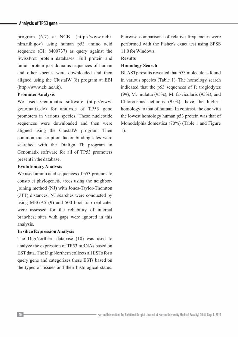
Homology Search
BLASTp results revealed that p53 molecule is found
in various species (Table 1). The homology search
indicated that the p53 sequences of P. troglodytes
(99), M. mulatta (95%), M. fascicularis (95%), and
Chlorocebus aethiops (95%), have the highest
homology to that of human. In contrast, the one with
the lowest homology human p53 protein was that of
Monodelphis domestica (70%) (Table 1 and Figure
1).
Harran Üniversitesi Tıp Fakültesi Dergisi (Journal of Harran University Medical Faculty) Cilt 8. Sayı 1, 2011
Analysis of TP53 gene
17
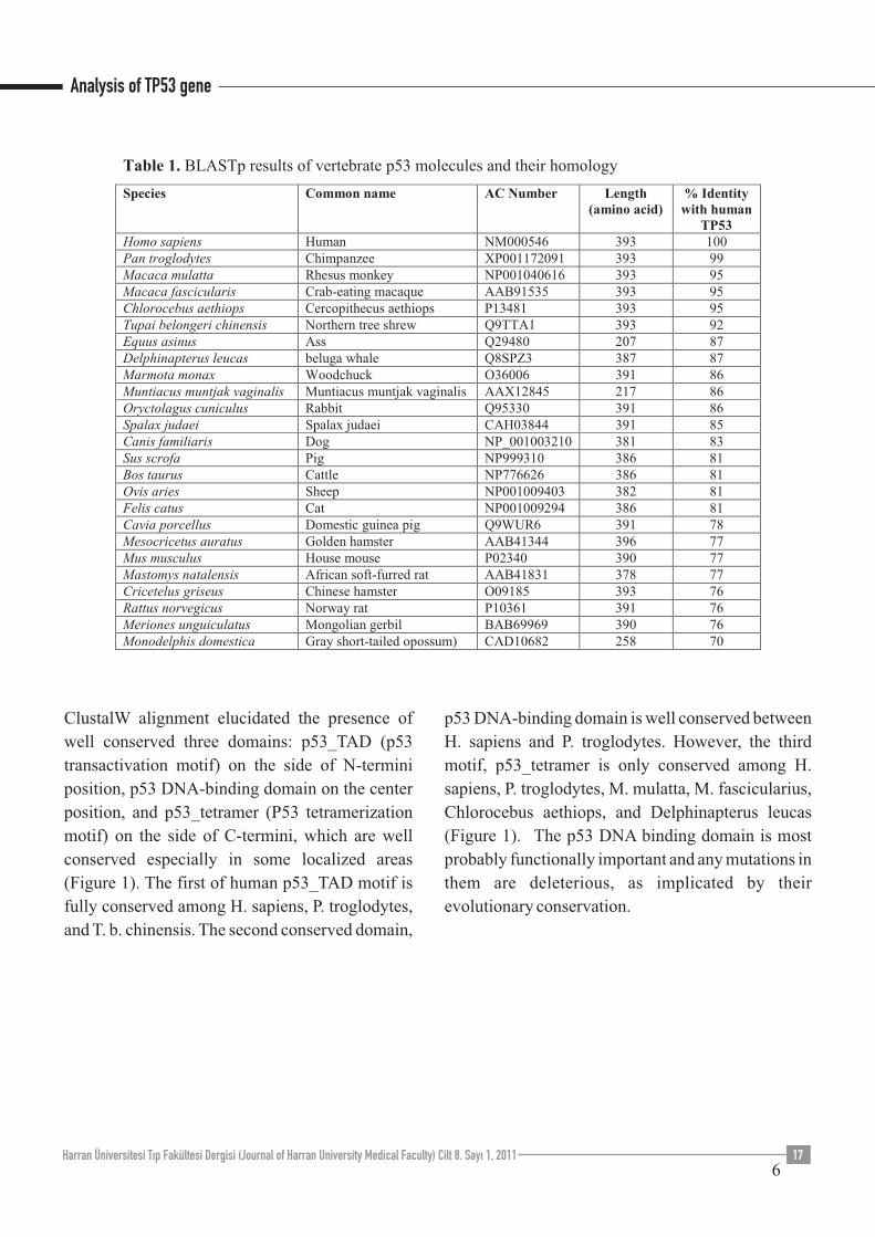
ClustalW alignment elucidated the presence of
well conserved three domains: p53_TAD (p53
transactivation motif) on the side of N-termini
position, p53 DNA-binding domain on the center
position, and p53_tetramer (P53 tetramerization
motif) on the side of C-termini, which are well
conserved especially in some localized areas
(Figure 1). The first of human p53_TAD motif is
fully conserved among H. sapiens, P. troglodytes,
and T. b. chinensis. The second conserved domain,
p53 DNA-binding domain is well conserved between
H. sapiens and P. troglodytes. However, the third
motif, p53_tetramer is only conserved among H.
sapiens, P. troglodytes, M. mulatta, M. fascicularius,
Chlorocebus aethiops, and Delphinapterus leucas
(Figure 1). The p53 DNA binding domain is most
probably functionally important and any mutations in
them are deleterious, as implicated by their
evolutionary conservation.
Harran Üniversitesi Tıp Fakültesi Dergisi (Journal of Harran University Medical Faculty) Cilt 8. Sayı 1, 2011
Analysis of TP53 gene
18
Harran Üniversitesi Tıp Fakültesi Dergisi (Journal of Harran University Medical Faculty) Cilt 8. Sayı 1, 2011
Analysis of TP53 gene
19
Multiple alignment results of human p53 and its
homologous revealed that this molecule is yet
uncharacterized in five species, which M.
natalensis (N-termini), and E. asinus, M. muntjak
vaginalis, M. domestica (N-, and C- termini), and
O. aries (C-termini), probably due their possible
truncation (Figure 1).
Promoter Analysis
We found by the database search that the
orthologous p53 gene promoters do not include
any common transcription factor binding sites
(TFBs) among H. sapiens, M. mulatta, R.
norvegicus, C. familiaris, D. rerio in Database of
Genomatix software. However, we observed that the
similarity (value 1.000) and the number of identical
nucleic acids (in % of short sequence segments) was
28% between the TP53 promoters of H. sapiens and
M. mulatta for each pairwise alignment; however,
this does not necessarily mean that the two sequences
are identical.
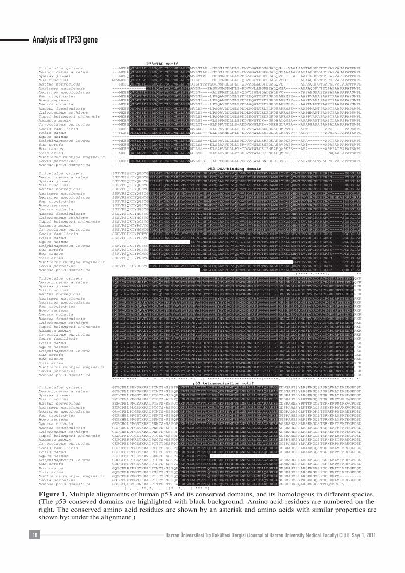
Evolutionary Analysis
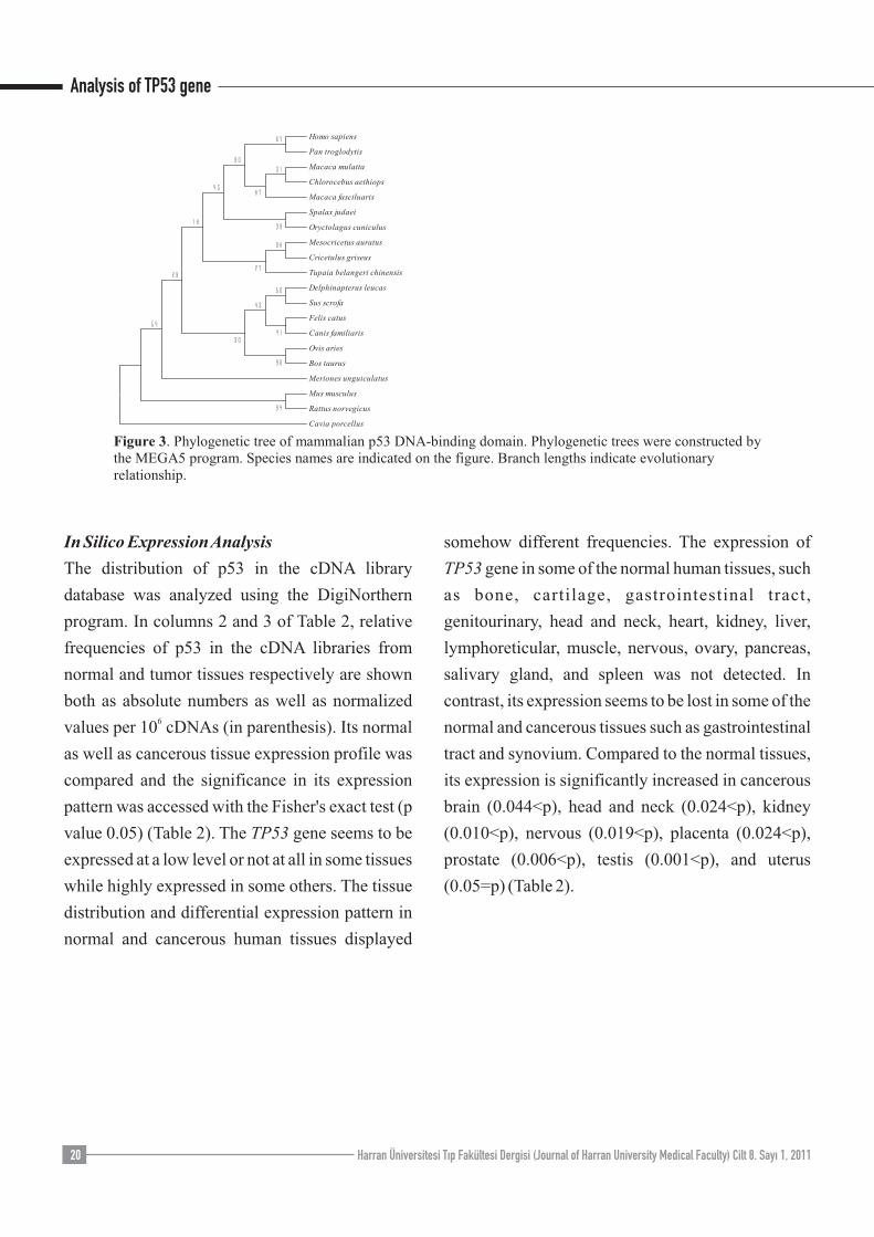
From the phylogenetic trees constructed by MEGA5
we found that p53 molecules are more closely
grouped among H. sapiens, P.troglodytes, C.
aethiops, M. mulatta, and M. fascicularius species
(Figure 2).
Multiple alignment results of human p53 and its
homologous revealed that this molecule is yet
uncharacterized in four species, which M.
natalensis, E. asinus, M. m. vaginalis, and M.
domestica, probably due their possible truncation.
Ignoring the molecules of these four foregoing
species, the p53 DNA-binding domains of the other
species are very well conserved between H. sapiens
and P. troglodytes through evolution (Figure 3).
Analysis of TP53 gene
Harran Üniversitesi Tıp Fakültesi Dergisi (Journal of Harran University Medical Faculty) Cilt 8. Sayı 1, 201120
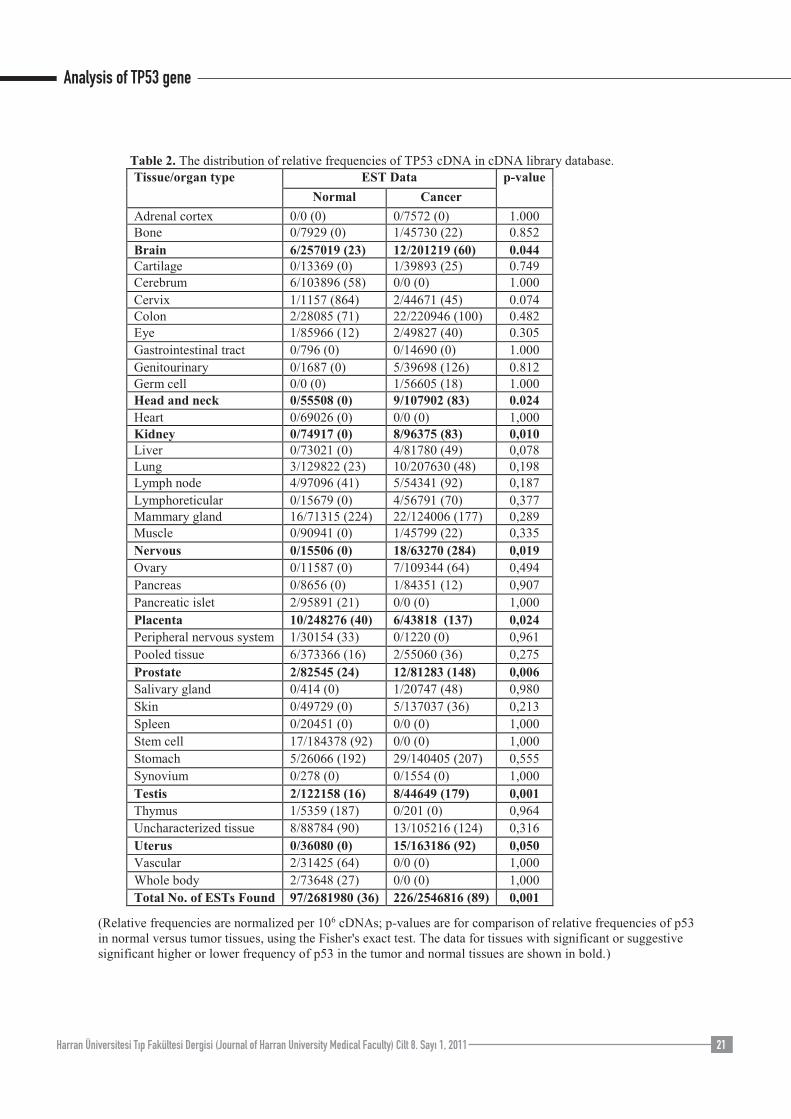
In Silico Expression Analysis
The distribution of p53 in the cDNA library
database was analyzed using the DigiNorthern
program. In columns 2 and 3 of Table 2, relative
frequencies of p53 in the cDNA libraries from
normal and tumor tissues respectively are shown
both as absolute numbers as well as normalized 6
values per 10 cDNAs (in parenthesis). Its normal
as well as cancerous tissue expression profile was
compared and the significance in its expression
pattern was accessed with the Fisher's exact test (p
value 0.05) (Table 2). The TP53 gene seems to be
expressed at a low level or not at all in some tissues
while highly expressed in some others. The tissue
distribution and differential expression pattern in
normal and cancerous human tissues displayed
somehow different frequencies. The expression of
TP53 gene in some of the normal human tissues, such
as bone, cartilage, gastrointestinal tract,
genitourinary, head and neck, heart, kidney, liver,
lymphoreticular, muscle, nervous, ovary, pancreas,
salivary gland, and spleen was not detected. In
contrast, its expression seems to be lost in some of the
normal and cancerous tissues such as gastrointestinal
tract and synovium. Compared to the normal tissues,
its expression is significantly increased in cancerous
brain (0.044<p), head and neck (0.024<p), kidney
(0.010<p), nervous (0.019<p), placenta (0.024<p),
prostate (0.006<p), testis (0.001<p), and uterus
(0.05=p) (Table 2).
Analysis of TP53 gene
Harran Üniversitesi Tıp Fakültesi Dergisi (Journal of Harran University Medical Faculty) Cilt 8. Sayı 1, 2011 21
Analysis of TP53 gene
Harran Üniversitesi Tıp Fakültesi Dergisi (Journal of Harran University Medical Faculty) Cilt 8. Sayı 1, 201120
Discussion
Human TP53 gene (also called p53) is located on
chromosome 17p13.1 and recently identified as a
tumor suppressor gene in which its mutation can lead to
Li-Fraumeni syndrome-1 (LPS), characterized by
autosomal dominant inheritance and early onset of
tumors, multiple tumors within an individual, and
multiple affected family members (11). The open
reading frame of p53 is 393 amino acids long, with the
central region containing the p53 DNA-binding
domain. This proteolysis-resistant core is flanked by a
C-terminal end mediating oligomerization, and an N-
terminal end containing a strong transcription
activation signal also identified a Drosophila sp. p53
homolog and demonstrated that it can activate
transcription from a promoter containing binding sites
for human p53 (12, 13). We found in this study that the
human p53 protein has three important regions:
p53_TAD motif on N-terminal side (at position 5-29
amino acides), core domain (p53 DNA-binding
domain) on the center (at position 95-289), and the
p53_tetramerization motif on the C-terminal side (at
position 324-359 amino asids). Both the motifs on the
N- and the C-terminal sides are not conserved among
all investigated species, but p53 DNA_binding domain
is well conserved between H.sapiens and P.troglodytes.
This domain is important, and a proline-rich domain
that mediates p53 response to DNA damage through
apoptosis. It is where most of the TP53 mutations are
found on p53 DNA binding domain. Mutation in the
core domain disrupts the p53 DNA-binding capability
and hence causes p53 to lose its function as
transcription factor (1).
In this study, the p53 molecules among human,
monkey, and Cercopithecus aethiops are very similar
(95-99% homology). Direct comparison of human and
chimpanzee cancer genes indicates that they are highly
conserved, showing 99.38% identities at the protein
level, and 99.19% at the nucleotide level, what is
similar to the average amino acid identities between
both organisms (99.38%) (14). Likewise, our BLASTp
results indicate that p53 is found in various species of
vertebrates and these molecules have 70-99%
conservation degree in the total amino acid sequences
(Table 1). The human p53 molecule has highest homology
to those of P. troglodytes (99%), M. mulatta (95%), and
Chlorocebus aethiops (95%), and lowest homology to that
of Monodelphis domestica (70%). So, these results
indicate that the TP53 gene has been evolutionary well
conserved (Table 1). Additionally, we also examined the
phylogenetic trees of p53 in different species using
MEGA5 program. We observed that human p53 shows
closest homology to those of P. trogylodytes, and then M.
mulatta, and M. fascicularis (Figure 2).
The expression of TP53 in different tissues was analyzed
using the DigiNorthern program. Its expression patterns in
normal and cancer tissues displayed somehow different
frequencies in human. In some normal tissues, such as
adrenal cortex, bone, cartilage, Gastrointestinal tract,
Genitourinary, germ cell, head and neck, heart, kidney,
liver, lymphoreticular, muscle, nervous,ovary, pancreas,
salivary gland, skin, spleen, synovium, and uterus, and in
cancerous tissues, such as adrenal cortex, cerebrum,
gastrointestinal tract, heart, pancreatic islet, peripheral
nervous system, spleen, stem cells, synovisium, thymus,
vascular, and whole body, their expressions seem to be
expressed at a very low level or not at all. In contrast, its
expression is significantly increased in cancerous brain,
head and neck, kidney, nervous, placenta, prostate, testis,
uterus tissues (Table 2). Transcriptional regulation of
TP53 under different conditions is related to vast number
of biological events in response to various cellular stresses
including cell-cycle progression, carcinogenic
stimulation, and so forth (15, 16). At the same time, the
expression level of TP53 varies in the context of different
cell functions (17, 18). Studies have shown that the
differential regulation of TP53 in cell cycle control or cell-
type specific tumorigenesis is reflected by elements of
transcriptional control (16,19). Multiple sequence
alignment and phylogenetic analysis of the human p53
mRNA sequence was performed which showed its
relationship and pattern of variations among different
organisms (20).
We suspect that TP53 the change in its expression pattern
in different tissues may be related to the role for
pathogenesis of some sporadic cancers. The availability of
the comprehensive data generated by high-throughput
functional genomic approaches, mainly expressed
Analysis of TP53 gene
Harran Üniversitesi Tıp Fakültesi Dergisi (Journal of Harran University Medical Faculty) Cilt 8. Sayı 1, 2011 21
References
1.Yang Y, Tantoso E, Chua GH, Yeo ZX, Ng FS, Wong
ST, Chung CW, and Li KB. In silico analysis of p53
us ing the p53 knowledgebase: mutat ions ,
polymorphisms, microRNAs and pathways. In Silico
Biol 2007; 7: 61-75.
2.Kristensen AT, Bjorheim J, and Ekstrom PO.
Detection of mutations in exon 8 of TP53 by temperature
gradient 96-capillary array electrophoresis.
Biotechniques 2002; 33: 650-53.
3.Yamane K KE, Tsuruo T. p53 contains a DNA break-
binding motif similar to the functional part of BRCT-
related region of Rb. Oncogene 2001; 20: 2859-867.
4.Ashur-Fabian O, Avivi A, Trakhtenbrot L, Adamsky
K, Cohen M, Kajakaro G, Joel A, Amariglio N, Nevo E,
and Rechavi G. Evolution of p53 in hypoxia-stressed
Spalax mimics human tumor mutation. Proc Natl Acad
Sci U S A 2004; 101: 12236-2241.
5.Rogel A, Popliker M, Webb CG, and Oren M. p53
cellular tumor antigen: analysis of mRNA levels in
normal adult tissues, embryos, and tumors. Mol Cell
Biol 1985; 5: 2851-855.
6.Altschul SF GW, Miller W, Myers EW, Lipman DJ.
Basic local alignment search tool. J Mol Biol 1990; 215:
403-10.
7.Gissen P JC, Gentle D. Comparative evolutionary
analysis of VPS33 homologues: genetic and functional
insights. Hum Mol Genet 2005; 14: 1261-270.
8.Thompson JD HD, Gibson TJ. CLUSTALW:
improving the sensitivity of progressive multiple
sequence alignment through sequence weighting,
position-specific gap penalties and weight matrix
choice. Nucleic Acids Res 1994; 22: 4673-80.
9.Tamura K DJ, Nei M, Kumar S. MEGA4: Molecular
Evolutionary Genetics Analysis (MEGA) Software
Version 4.0. Mol. Biol. Evol 2007; 24: 1598-599.
10.Wang J LP. DigiNorthern, digital expression analysis
of query genes based on ESTs. Bioinformatics 2003; 19:
653-54.
11.Shete S AC, Hwang SJ, Strong LC. Individual-
specific liability groups in genetic linkage, with
applications to kindreds with Li-Fraumeni syndrome.
Am. J. Hum. Genet 2002; 70: 813-17.
12.Vogelstein B KK. X-rays strike p53 again. Nature
1994; 370: 174-75.
13.Brodsky MH NW, Tsang G, Kwan E, Rubin GM,
Abrams JM. Drosophila p53 binds a damage response
element at the reaper locus. Cell 2000; 101: 103-13.
14.Mikkelsen TS HL, Eichler EE, Zody MC, Jaffe DB,
Yang S, Enard W, Hellmann I, Lindblad-Toh K, Altheide
TK, Archidiacono N, Bork P, Butler J, Chang JL, Cheng
Z, Chinwalla AT, deJong P, Delehaunty KD, Fronick CC,
Fulton LL, Gilad Y, Glusman G, Gnerre S, Graves TA,
Hayakawa T, Hayden KE, Huang X, Ji H, Kent WJ, King
MC, KulbokasIII EJ, Lee MK, Liu G, López-Otín C,
Makova KD, Man O, Mardis ER, Mauceli E, Miner TL,
Nash WE, Nelson JO, Pääbo S, Patterson NJ, Pohl CS,
Pollard KS, Prüfer K, Puente XS, Reich D, Rocchi M,
Rosenbloom K, Ruvolo M, Richter DJ, Schaffner SF,
Smit AFA, Smith SM, Suyama M, Taylor T, Torrents D,
Tuzun E, Varki A, Velasco G, Ventura M, Wallis JW,
Wendl MC, Wilson RK, Lander ES, Waterston RH: .
Initial sequence of the chimpanzee genome and
comparison with the human genome. Nature 2005; 437.
15.Abbas T WD, Hui L, Yoshida K, Foster DA,
Bargonetti J. Inhibition of human p53 basal transcription
by down-regulation of protein kinase Cδ. J. Biol. Chem
2004; 279: 9970-977.
16.Boggs K RD. Increased p53 transcription prior to
DNA synthesis is regulated through a novel regulatory
element within the p53 promoter. Oncogene 2006; 25:
555-65.
17.Kawauchi J ZC, Nobori K, Hashimoto Y, Adachi MT,
Noda A, Sunamori M, Kitajima S. Transcriptional
repressor activating transcription factor 3 protects human
umbilical vein endothelial cells from tumor necrosis
factor-α-induced apoptosis through downregulation of
p53 transcription. Biol. Chem 2002; 227: 39025-9034.
18.Rowland BD BR, Peeper DS. The KLF4 tumour
suppressor is a transcriptional repressor of p53 that acts
as a context-dependent oncogene. Nat. Cell Biol 2005; 7:
1074-82.
19.Strudwick S CL, Stagg T, Lazarus P. Differential
transcription-coupled translational inhibition of human
p53 expression: a potentially important mechanism of
regulating p53 expression in normal versus tumor tissue.
Mol Cancer Res 2003; 1: 463-74.
20.Khan MH RH, Mir A. Phylogenetic analysis of human
Tp53 gene using computational approach. African Journal
of Biotechnology 2011; 10: 344-49.
21.Lash AE TC, Wagner L et al. SAGEmap: a public gene
expression resource. Genome Res 2000; 10: 1051-60.
22.Giaccia AJ KM. The complexity of p53 modulation:
emerging patterns from divergent signals. Genes Dev
1998; 12: 2973-983.
23.Pao SY LW, Hwang MJ. In silico identification and
comparative analysis of differentially expressed genes in
human and mouse tissues. BMC Genomics 2006; 7: 1-11.
24.Hu Z CK, Wang L, Yao Q. Identification and
characterization of Bombyx mori eIF5A gene through
bioinformatics approaches. In Silico Biol 2005; 4: 573-80.
25.Varisli L CO. Identification and Characterization of Rat
GMDS Gene by Using Bioinformatics Tools. Turk J
Biochem 2005; 30: 306-9.
Yazarlarla ilgili bildirilmesi gereken konular (Conflict of interest statement) : Yok (None)
sequence tag (EST) and serial analysis of gene
expression (SAGE), provides the feasibility to study
gene expression through in silico analysis (21). In
normal tissues, p53 levels are low, but p53 protein
accumulates after exposure to DNA-damaging agents
or during the onset of various physiological processes
(22). Comparative analysis showed that, although
highly tissue-specific genes tend to exhibit similar
expression profiles in human and mouse, there are
significant exceptions, indicating that orthologous
genes, while sharing basic genomic properties, could
result in distinct phenotypes (23).
Since the completion of the human genome, cataloging
transcription factor binding sites (TFBSs) has been
critical for understanding gene regulation. We used
Dialign TF program in Genomatix software for
predicting transcription factor binding sites
(transcriptional elements) of all orthologous TP53
promoters that present in the database. Dialign TF
results revealed that TP53 orthologous promoters had
no common conserved transcriptional elements. Our
results indicate that the binding sites of different
transcription factors might have located on different
parts of the promoter or promoter vicinity in various
species.
Basic bioinformatics techniques are powerful tools in
terms of leading to the discoveries and analysis of
novel genes (24). Recently we identified and further
characterized two novel genes using bioinformatics
tools (25). Even though the results from bioinformatics
studies are very helpful in directing and designing the
experiments, they need to be supported and confirmed
by further experimentation. By choosing suitable
bioinformatics analysis on these data, more
discoveries will be made. Bu çalışma 2007 yılında
Adana'da “III. Çukurova Kolo-Proktoloji ve Stoma-
terapi sempozyumu”nda poster bildirisi olarak
sunulmuştur.