Embed Size (px)
Citation preview
Int J Clin Exp Pathol 2013;6(7):1380-1391www.ijcep.com /ISSN:1936-2625/IJCEP1304034
Original Article An associated classification of triple negative breast cancer: the risk of relapse and the response to chemotherapy
Jing Zhang1, Yahong Wang2, Quangui Yin3, Wei Zhang1, Tongxian Zhang1, Yun Niu1
1Key Laboratory of Breast Cancer Prevention and Therapy, Tianjin Medical University, Ministry of Education and Key Laboratory of Cancer Prevention and Therapy of Tianjin, Tianjin Medical University Cancer Institute and Hos-pital, West Huanhu Road, Ti Yuan Bei, Hexi District, Tianjin 300060, China; 2Department of radiotherapy, Tianjin Huanhu Hospital, No. 122, Qixiangtai Road, Hexi District, Tianjin, P. R. 300060, China; 3Department of Internal Medicine, WuQing Hospital, Tianjin 301700, China. *These authors contributed equally to this work.
Received April 25, 2013; Accepted May 20, 2013; Epub June 15, 2013; Published July 1, 2013
Abstract: Background: Triple negative breast cancer (TNBC) is heterogeneous and considered as an aggressive tumor. This study was to evaluate the associated classification and its correlations with prognosis and the response to chemotherapy in Chinese women. Methods: Four hundred and twenty-eight cases of invasive TNBC were involved in this study. The expression of estrogen receptor (ER), progesterone receptor (PR), human epidermal growth fac-tor receptor 2 (HER2), epidermal growth factor receptor (EGFR), and cytokeratin 5/6 (CK5/6), Ki67 and p53 were analyzed by immunohistochemistry and compared with patient outcome, and its implications and chemotherapy re-sponse were evaluated in four subgroups: typical medullary carcinoma (TMC), atypical medullary carcinoma (AMC), non-specific invasive ductal carcinoma (IDC) and other types. Results: The factors of tumor grade, tumor stage, lymph node status, EGFR/CK5/6 status and p53 labeling index were different among the groups. TMC tumors had the lowest rate of relapse (5.8%), while AMC, IDC and other types were associated with an increased risk of relapse (19.1%, 26.7% and 38.2% respectively). Many factors were risk predictors of relapse for TNBC and IDC, while only positive lymph node was for AMC. For MC tumors, adjunctive chemotherapy decreased the risk of relapse in lymph node positive subgroup (36.8% and 66.7%), while not significant in lymph node negative one (8.1% and 10.0%). Conclusion: The classification based on histologic and IHC findings may be a significant improvement in predicting outcome in TNBC. The different chemotherapy response in subgroups may contribute to guiding the treatment of TNBC.
Keywords: Triple negative breast cancer, typical medullary carcinoma, atypical medullary carcinoma, relapse, chemotherapy
Introduction
Molecular and genetic studies demonstrated that breast cancer was a heterogeneous dis-ease [1], and had been proposed to be classi-fied into subgroups according to different immunohistochemical biomarkers [2, 3]. Of which estrogen receptor (ER), progesterone receptor (PR), and human epidermal growth factor receptor 2 (HER2) were the most impor-tant biomarkers. In the 2007 St. Gallen Consensus Meeting made a decision about adjuvant therapies (chemotherapy, endocrine therapy, and trastuzumab), operable primary breast cancers were recommended to be cate-
gorized based on the status of ER, PR, and HER2 [4]. TNBC was defined as a subtype of breast cancers that were negative for ER, PR and HER2. TNBC was generally considered as the most difficult subtype to treat among these newly proposed subtypes of breast cancer because of the aggressive clinical behavior and the lack of current availability of specific target-ed therapy such as selective ER modulators, aromatase inhibitors, trastuzumab, and lapa-tinib [3].
TNBC accounted for approximately 10–20% of the whole breast cancer and was correlated with relatively early clinical relapse within 3
The prognosis and the response to chemotherapy of TNCBC
1381 Int J Clin Exp Pathol 2013;6(7):1380-1391
years, with frequent progression to distant metastasis, particularly, visceral metastasis. Although the metastatic potential in TNBC was similar to that of other breast cancer subtypes, these tumors were associated with a shorter median time to relapse and death [5]. DNA microarray analyses proved that TNBCs were composed of basal-like subtype and normal-like (or unclassified) subtype. Basal-like sub-type was characterized by the expression of myoepithelial/basal markers (EGFR and/or CK5/6) and molecular changes including p53 gene mutation and many chromosomal altera-tions which were correlated with an aggressive clinical course. Histological types of TNBC were reported to be similar with those of basal-like subtype, comprising high-grade non-specific IDC, AMC, TMC, and other types of carcinomas, of which TMC was of particular interest for being high grade carcinoma but favorable prog-nosis [6-8]. At present, chemotherapy remains the main treatment of TNBC despite of many limitations that need to be overcome. There is still not a clear, proven effective agent that tar-gets a definite vulnerability in TNBC.
TNBC was a clearly distinct subtype of the whole breast cancer and had usually been divided into subgroups by the expression of myoepithelial/basal markers [2, 3]. However, further subclassification with different progno-sis is needed. In this study, we retrospectively studied the histological types, traditional patho-logic indices and the description of CK5/6, EGFR, Ki67 and p53 on 428 Chinese women with TNBC in predicting subclassifications and investigating the risk of relapse and the response of chemotherapy in TNBC.
Materials and methods
Patient characteristics
The cohort used for this study was derived from the archival paraffin-embedded breast cancer samples collected at the Cancer Hospital of Tianjin Medical University between January 2002 and December 2004, and all tumors were primary, operable early breast cancer. The age of patients ranged from 25 to 76 years old, with the median age of 50.3 years. All patients underwent preoperative mammography and ultrasound of the breast and abdomen, X-ray, or computed tomography (CT) scan of the tho-rax. If there were any signs of metastasis to the
bone or brain, bone scan or brain CT was per-formed as a standard procedure. All patients involved in this study were stages I, II, or III and were diagnosed as invasive carcinoma based on either the core-biopsy before operation or frozen biopsy intra-operation and were given diagnosis on paraffin-embedded tissue sec-tions after operation by two pathologists. The sections were reviewed in double blind by dif-ferent pathologist. All patients underwent local and/or systemic treatments. Local treatment included surgery and radiotherapy. Surgical procedures consisted of mastectomy and breast-conserving surgery. Patients who under-went breast-conserving surgery had received adjuvant radiotherapy as a routine. The main systemic treatment in our study was chemo-therapy guided by National Comprehensive Cancer Network (NCCN). The exclusion criteria were: the patients treated with neoadjuvant chemotherapy, the patients without complete follow-up, and non-tumor-mediated mortality. A total of 428 cases were identified and contrib-uted to this study. We retrospectively reviewed 70.6-month follow-up data. The follow-up con-tacts were carried out at 3-month intervals dur-ing the first year, 6-month intervals during the second year and 12-month intervals thereafter. The medical work-up consisted of regular physi-cal checkups, imaging tests such as chest X-ray, bone scan and/or ultrasound, and to look for recurrences, second primary breast can-cers, or metastatic disease. Relapse was defined as radiographic or pathological evi-dence of regional tumor recurrence or distant metastasis at any time after initial therapy. The study protocol was approved by the Hospital Human Ethical Committee. Informed consent was obtained from all patients before their sur-gery and the examination of the specimens.
Clinicopathological evaluation
We retrospectively evaluated conventional clini-copathological factors, including age at diagno-sis, tumor size, tumor grade, lymph node sta-tus, tumor stage, menopausal status, family history (family history of breast cancer within first and second-degree relatives). The patho-logical tumor stage was assessed according to the criteria established by the 6th edition of the American Joint Committee on Cancer (AJCC) staging manual. Histological grade of the tumors were classified into grades I–III accord-ing to the Nottingham combined histological
The prognosis and the response to chemotherapy of TNCBC
1382 Int J Clin Exp Pathol 2013;6(7):1380-1391
grade. Histologic type and pertinent histologic features included tumor border, glandular for-mation, mitotic index, degree of stromal lym-phocytic infiltration, lymphovascular invasion, axillary lymph node (ALN) status, and results of IHC assess. The histologic type was assigned according to the 2003 World Health Organization criteria, and notably, diagnosis of TMC was made when the tumor showed pre-dominantly (greater than 75%) syncytial archi-tecture, absence of glandular structure, diffuse lymphoplasmacytic infiltrate (moderate to marked), nuclear pleomorphism (moderate to marked), and complete histological circum-scription. Diagnosis of AMC was made when the tumor showed predominantly (greater than 75%) syncytial architecture with only two or three of the preceding four criteria. Tumor bor-der was graded as circumscribed or infiltrative. Glandular formation was absent when there was no tubule formation; Mitotic index was graded as high (grade 3) when 20 or more mitotic figures were seen within 10 high-power fields, and as intermediate (grade 2) when the mitotic count was between 10 and 19, and low (grade 1) when less than 10 per 10 high power fields respectively. The degree of stromal lym-phocytic infiltrate was graded as strong when more than two-thirds of the stroma of tumor mass had lymphocytic infiltration and weak when less than two-thirds of the stroma had lymphocytic infiltration. Metaplastic carcino-mas were known to be a rare but characteristic subgroup of TNBC. They were aggressive, che-moresistant tumors characterized by concur-rence of a high-grade carcinoma component (poorly differentiated ductal carcinoma) and extensive metaplastic component comprising
retrieval was necessary for EGFR. All the anti-bodies were used for IHC studies on serial tis-sue sections from each case; Primary antibod-ies used in this study included ER (SP1, 1:200 dilution; Zymed), PR (SP2, 1:200 dilution; Zymed), HER2 (CB11, 1:600 dilution; Zymed), EGFR (31G7, 1:100 dilution; Zymed), CK5/6 (D5/16B4, 1:200 dilution; Zymed), p53 (BP53.12 1:100 dilution; Zymed) and Ki67 (K-2, 1:100 dilution; Zymed). The immunostain-ing was scored in double blind by two different pathologists, who were blinded to patients’ clinicopathologic characteristics and out-comes. For each antibody, the location of immunoreactivity, percentage of stained cells, and intensity were determined. The evaluation of each protein expression was determined from the mean of the individual cases. ER and PR stains were assessed using Allred scores, with positive scores ranging from 2 to 8 [9]. CK5/6 and EGFR stains were considered posi-tive if any cytoplasmic and/or membranous staining was observed, whereas HER2+ was defined as the whole membrane strong staining in >30% of the tumor cells, and Ki67 status was expressed in terms of percentage of posi-tive cells, with a threshold of 14% of positive cells [2]. p53 was defined as positive when more than 10% of tumor cells were positive for nuclear staining [10].
Statisticcal analysis
All statistical analyses were carried out using SPSS software (Version 17.0 for Windows). The χ2 test and Fisher exact test were performed for group comparisons. For univariable survival analysis, Overall survival (OS) and Relapse-free
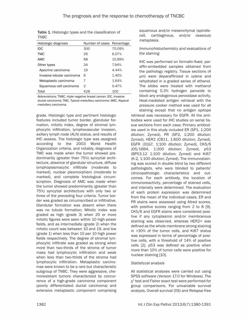
Table 1. Histologic types and the classification of TNBC Histologic diagnosis Number of cases PercentageIDC 300 70.09%TMC 26 6.07%AMC 68 15.89%Other types 34 7.94% Apocrine carcinoma 19 4.44% Invasive lobular carcinoma 6 1.40% Metaplastic carcinoma 7 1.64% Squamous cell carcinoma 2 0.47%Total 428 100Abbreviations: TNBC, triple negative breast cancer; IDC, Invasive ductal carcinoma; TMC, Typical medullary carcinoma; AMC, Atypical medullary carcinoma.
squamous and/or mesenchymal (spindle-cell, cartilaginous, and/or osseous) metaplasia.
Immunohistochemistry and evaluations of the staining
IHC was performed on formalin-fixed, par-affin-embedded samples obtained from the pathology registry. Tissue sections (4 µm) were deparaffinized in xylene and rehydrated in a graded series of ethanol. The slides were treated with methanol containing 0.3% hydrogen peroxide to block any endogenous peroxidase activity. Heat-mediated antigen retrieval with the pressure cooker method was used for all staining except that no antigen epitope
The prognosis and the response to chemotherapy of TNCBC
1383 Int J Clin Exp Pathol 2013;6(7):1380-1391
survival (RFS) were conducted using the Kaplan-Meier curves. The log-rank test was used to compare survival differences among the subtypes. Cox proportional hazards models were used to calculate relative risk accounting for covariates. A 2-sided P<0.05 was consid-ered statistically significant in all the analyses.
Results
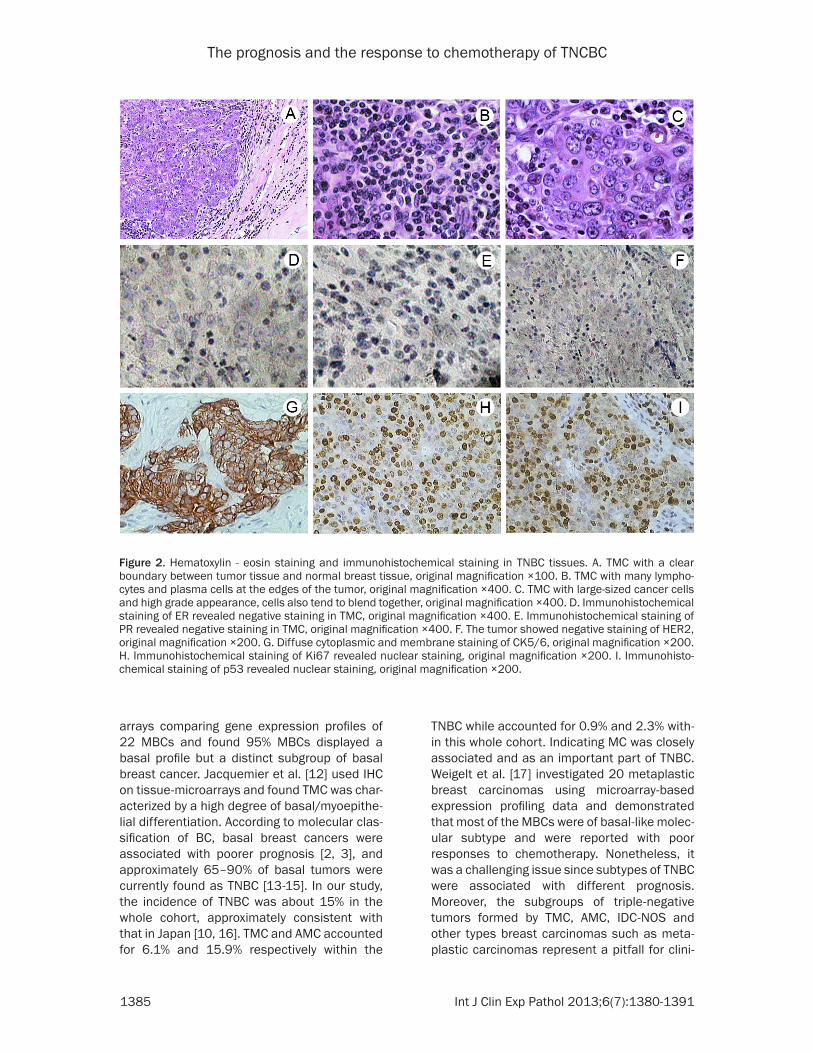
The intrinsic histologic types were illustrated in (Figure 2). Three hundred (70.09%) patients
were diagnosed as non-specific IDC, 26 (6.07%) were TMC, 68 (15.89%) were AMC and 34 (7.94%) were other types carcinoma including apocrine carcinoma (4.44%), invasive lobular carcinoma (1.40%), metaplastic carcinoma (1.64%) and squamous cell carcinoma (0.47%) (Table 1). The distribution of clinicopathological characteristics among the subtypes were listed in Table 2, MC was associated with small tumor size, lower percentage of node positivity, but higher tumor grade and higher family history of
Table 2. Distribution of clinicopathological characteristics among the subtypes of TNBC
CharacteristicsTMC n=26 AMC n=68 IDC n=300 Other types n=34
χ2 P-valuesNo. % No. % No. % No. %
Age at diagnosis, years 8.125 0.229 <40 5 19.2 8 11.8 40 13.3 6 17.6 40-55 17 65.4 52 76.5 185 61.7 21 61.8 >55 4 15.4 8 11.8 75 25.0 7 20.6Tumor size, cm 6.313 0.389 ≤2 13 50.0 20 29.4 89 29.7 9 26.5 2-5 11 42.3 44 64.7 193 64.3 24 70.6 >5 2 7.7 4 5.9 18 6.0 1 2.9Tumor grade NA 32.471 0.000 1-2 6 8.8 121 40.3 19 55.9 3 62 91.2 179 59.7 15 44.1Tumor stage 15.551 0.007 AJCC stage I/II 25 96.2 56 82.4 215 71.7 19 55.9 AJCC stage III 1 3.8 12 17.6 85 28.3 15 44.1Lymph node status 32.696 0.000 Negative 24 92.3 48 70.6 142 47.3 12 35.3 Positive 2 7.7 20 29.4 158 52.7 22 64.7Menopausal status 1.230 0.746 Premenopausal 15 57.7 34 50.0 144 48.0 15 44.1 Postmenopausal 11 42.3 34 50.0 156 52.0 19 55.9Family history 2.855 0.415 Yes 6 23.1 11 16.2 39 13.0 4 11.8 No 20 76.9 57 83.8 261 87.0 30 88.2EGFR/CK5/6 10.633 0.014 Positive 23 88.5 62 91.2 222 74.0 27 79.4 Negative 3 11.5 6 8.8 78 26.0 7 20.6Ki-67 labeling index 5.087 0.166 ≥14% 19 73.1 33 48.5 155 51.7 18 52.9 <14% 7 26.9 35 51.5 145 48.3 16 47.1p53 labeling index 57,871 0.000 Positive 23 88.5 45 66.2 102 34.0 5 14.7 Negative 3 11.5 23 33.8 198 66.0 29 85.3Abbreviations: TNBC, triple negative breast cancer; TMC, typical medullary carcinoma; AMC, atypical medullary carcinoma; IDC, invasive ductal carcinoma; NA, not applicable; AJCC, American Joint Committee on Cancer. P-values were calculated to compare the four groups.
The prognosis and the response to chemotherapy of TNCBC
1384 Int J Clin Exp Pathol 2013;6(7):1380-1391
BC. The differences in tumor grade, tumor stage, lymph node status, EGFR/CK5/6 status and p53 labeling index among the subtypes were significant (P<0.05).
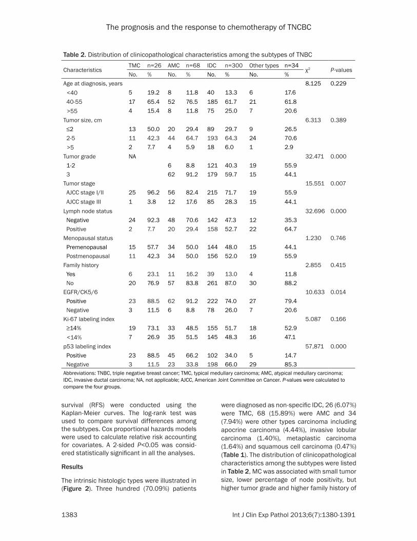
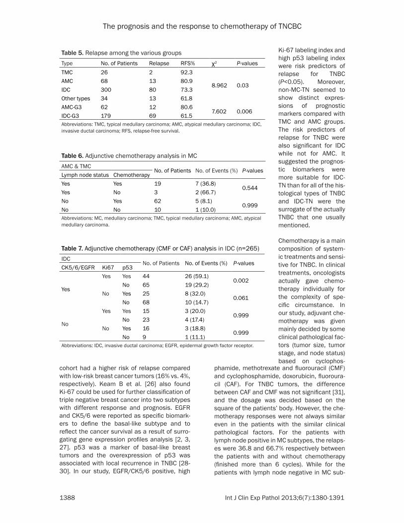
Of the 428 cases involved in this study, 88.87% received chemotherapy, with a follow-up period of 70.6 months, the actuarial OS of TMC, AMC, IDC and other types were 92.3%, 85.3%, 82.0% and 67.6% respectively (Figure 1A). The RFS of TMC, AMC, IDC and other types were 92.3%, 80.9%, 73.3% and 61.8% respectively (Figure 1B, Table 5). Patients with TMC and AMC tumors had favorable prognosis, with relapse rates of 5.8% and 19.1%, conversely, the IDC and other types exhibited high rates of relapse (26.7% vs. 38.2%). The difference between these types was significant (P=0.030) (Figure 1B, Table 5).
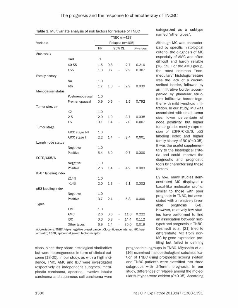
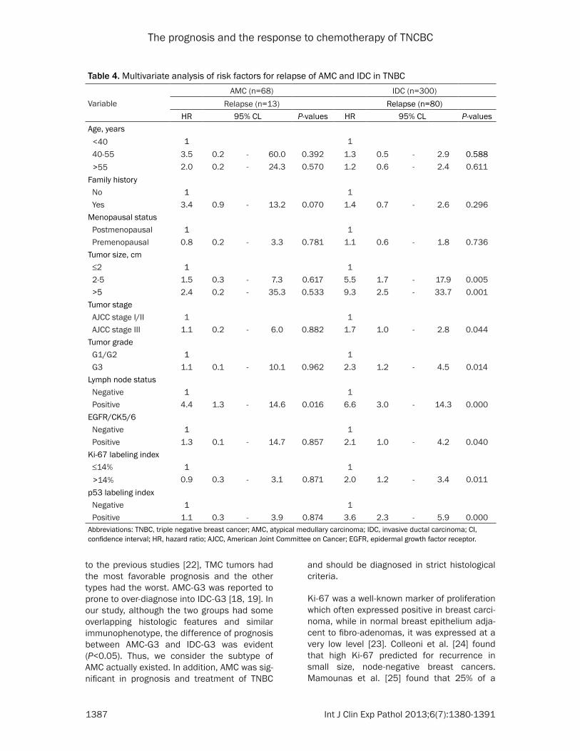
Multivariable Cox analysis revealed that family history positive, large tumor size, lymph node positive, tumor stage III, EGFR/CK5/6 positive, high Ki-67 labeling index and high p53 labeling index were risk predictors of relapse for TNBC (P<0.05). The differences were also significant for IDC while only lymph node positive was risk predictor for AMC (P=0.016) (Tables 3, 4). The relapse hazard ratios (HR) of the subtypes were: 2.6 (AMC), 3.3 (IDC), 6.9 (other types) (Table 3). Although AMC-G3 have many overlap-ping histologic features with IDC-G3, the relapse of the two groups were significantly dif-ferent (P=0.006) (Table 5).
For MC tumors, lymph node status played an important role. When compared given adjunc-tive chemotherapy or not, in lymph node posi-tive groups the relapse percentages were 36.8% and 66.7% respectively. However, in lymph node negative groups, the relapse per-centages were 8.1% and 10.0% respectively (Table 6). For the 265 patients with IDC tumors who had finished adjunctive chemotherapy in our study, the expression of CK5/6/EGFR, Ki67 and p53 markers assayed by IHC were consid-ered having associations with increased risk of relapse: patients with positive expression of all the CK5/6/EGFR, Ki67 and p53 markers exhib-ited the highest relapse (59.1%). Conversely, patients with negative expression of the mark-ers had the most favorable prognosis, with the relapse rate of only 11.1%. Patients with one or two positive expression of the markers had the middle relapses: 29.2%, 32.0%, 14.7%, 20.0%, 17.4% and 18.8% respectively (Table 7).
Discussion
TNBC is generally considered to be associated with aggressive clinical behavior. However, by now a limited number of studies have investi-gated the prevalence of the subclassifications of TNBC in the yellow race. As known, histologic type is one of the most important and clinically assessed prognostic factors in BCs. MC is par-ticular for its features of aggressiveness but favorable prognosis. Bertucci et al. [11] obtained whole-genome oligonucleotide micro-
Figure 1. Survival curves: (A) The OS curve of the groups in TNBC (B) The RFS curve of the groups in TNBC.
The prognosis and the response to chemotherapy of TNCBC
1385 Int J Clin Exp Pathol 2013;6(7):1380-1391
TNBC while accounted for 0.9% and 2.3% with-in this whole cohort. Indicating MC was closely associated and as an important part of TNBC. Weigelt et al. [17] investigated 20 metaplastic breast carcinomas using microarray-based expression profiling data and demonstrated that most of the MBCs were of basal-like molec-ular subtype and were reported with poor responses to chemotherapy. Nonetheless, it was a challenging issue since subtypes of TNBC were associated with different prognosis. Moreover, the subgroups of triple-negative tumors formed by TMC, AMC, IDC-NOS and other types breast carcinomas such as meta-plastic carcinomas represent a pitfall for clini-
arrays comparing gene expression profiles of 22 MBCs and found 95% MBCs displayed a basal profile but a distinct subgroup of basal breast cancer. Jacquemier et al. [12] used IHC on tissue-microarrays and found TMC was char-acterized by a high degree of basal/myoepithe-lial differentiation. According to molecular clas-sification of BC, basal breast cancers were associated with poorer prognosis [2, 3], and approximately 65–90% of basal tumors were currently found as TNBC [13-15]. In our study, the incidence of TNBC was about 15% in the whole cohort, approximately consistent with that in Japan [10, 16]. TMC and AMC accounted for 6.1% and 15.9% respectively within the
Figure 2. Hematoxylin - eosin staining and immunohistochemical staining in TNBC tissues. A. TMC with a clear boundary between tumor tissue and normal breast tissue, original magnification ×100. B. TMC with many lympho-cytes and plasma cells at the edges of the tumor, original magnification ×400. C. TMC with large-sized cancer cells and high grade appearance, cells also tend to blend together, original magnification ×400. D. Immunohistochemical staining of ER revealed negative staining in TMC, original magnification ×400. E. Immunohistochemical staining of PR revealed negative staining in TMC, original magnification ×400. F. The tumor showed negative staining of HER2, original magnification ×200. G. Diffuse cytoplasmic and membrane staining of CK5/6, original magnification ×200. H. Immunohistochemical staining of Ki67 revealed nuclear staining, original magnification ×200. I. Immunohisto-chemical staining of p53 revealed nuclear staining, original magnification ×200.
The prognosis and the response to chemotherapy of TNCBC
1386 Int J Clin Exp Pathol 2013;6(7):1380-1391
prognostic subgroups in TNBC. Miyashita et al. [16] examined histopathological subclassifica-tion of TNBC using prognostic scoring system and TNBC patients were classified into three subgroups with different prognosis. In our study, differences of relapse among the molec-ular subtypes were evident (P<0.05). According
cians, since they share histological similarities but were heterogeneous in term of clinical out-come [18-20]. In our study, as with a high inci-dence, TMC, AMC and IDC were investigated respectively as independent subtypes, meta-plastic carcinoma, apocrine, invasive lobular carcinoma and squamous cell carcinoma were
Table 3. Multivariate analysis of risk factors for relapse of TNBC
VariableTNBC (n=428)
Relapse (n=108)HR 95% CL P-values
Age, years<40 1
40-55 1.5 0.8 - 2.7 0.216>55 1.3 0.7 - 2.9 0.307
Family historyNo 1.0Yes 1.7 1.0 - 2.9 0.039
Menopausal statusPostmenopausal 1.0Premenopausal 0.9 0.6 - 1.5 0.792
Tumor size, cm≤2 1.02-5 2.0 1.0 - 3.7 0.038>5 3.1 1.4 - 7.0 0.007
Tumor stageAJCC stage I/II 1.0AJCC stage III 2.2 1.4 - 3.4 0.001
Lymph node statusNegative 1.0Positive 5.4 3.0 - 9.7 0.000
EGFR/CK5/6Negative 1.0Positive 2.6 1.4 - 4.9 0.003
Ki-67 labeling index≤14% 1.0>14% 2.0 1.3 - 3.1 0.002
p53 labeling indexNegative 1.0Positive 3.7 2.4 - 5.8 0.000
TypesTMC 1.0AMC 2.6 0.6 - 11.6 0.222IDC 3.3 0.8 - 14.4 0.112Other types 6.9 1.4 - 35.0 0.019
Abbreviations: TNBC, triple negative breast cancer; CI, confidence interval; HR, haz-ard ratio; EGFR, epidermal growth factor receptor.
categorized as a subtype named “other types”.
Although MC was character-ized by specific histological criteria, the diagnosis of MC especially of AMC was often difficult and hardly reliable [18, 19]. For the AMC group, the most common ‘‘non-medullary’’ histologic feature was the lack of a circum-scribed border, followed by an infiltrative border accom-panied by glandular struc-ture; infiltrative border toge- ther with mild lymphoid infil-tration. In our study, MC was associated with small tumor size, lower percentage of node positivity, but higher tumor grade, mostly expres-sion of EGFR/CK5/6, p53 labeling index and higher family history of BC (P<0.05). It was the useful supplemen-tary to the histological crite-ria and could improve the diagnostic and prognostic tools by characterising these factors.
By now, many studies dem-onstrated MC displayed a basal-like molecular profile, similar to those with poor prognosis in TNBC, but asso-ciated with a relatively favor-able prognosis [6-8]. However, relatively few stud-ies have performed to find an association between sub-types and prognosis in TNBC. Desmedt et al. [21] tried to differentiate MC from non-MC by gene expression pro-filing but failed in defining
The prognosis and the response to chemotherapy of TNCBC
1387 Int J Clin Exp Pathol 2013;6(7):1380-1391
and should be diagnosed in strict histological criteria.
Ki-67 was a well-known marker of proliferation which often expressed positive in breast carci-noma, while in normal breast epithelium adja-cent to fibro-adenomas, it was expressed at a very low level [23]. Colleoni et al. [24] found that high Ki-67 predicted for recurrence in small size, node-negative breast cancers. Mamounas et al. [25] found that 25% of a
to the previous studies [22], TMC tumors had the most favorable prognosis and the other types had the worst. AMC-G3 was reported to prone to over-diagnose into IDC-G3 [18, 19]. In our study, although the two groups had some overlapping histologic features and similar immunophenotype, the difference of prognosis between AMC-G3 and IDC-G3 was evident (P<0.05). Thus, we consider the subtype of AMC actually existed. In addition, AMC was sig-nificant in prognosis and treatment of TNBC
Table 4. Multivariate analysis of risk factors for relapse of AMC and IDC in TNBC
VariableAMC (n=68) IDC (n=300)
Relapse (n=13) Relapse (n=80)HR 95% CL P-values HR 95% CL P-values
Age, years <40 1 1 40-55 3.5 0.2 - 60.0 0.392 1.3 0.5 - 2.9 0.588 >55 2.0 0.2 - 24.3 0.570 1.2 0.6 - 2.4 0.611Family history No 1 1 Yes 3.4 0.9 - 13.2 0.070 1.4 0.7 - 2.6 0.296Menopausal status Postmenopausal 1 1 Premenopausal 0.8 0.2 - 3.3 0.781 1.1 0.6 - 1.8 0.736Tumor size, cm ≤2 1 1 2-5 1.5 0.3 - 7.3 0.617 5.5 1.7 - 17.9 0.005 >5 2.4 0.2 - 35.3 0.533 9.3 2.5 - 33.7 0.001Tumor stage AJCC stage I/II 1 1 AJCC stage III 1.1 0.2 - 6.0 0.882 1.7 1.0 - 2.8 0.044Tumor grade G1/G2 1 1 G3 1.1 0.1 - 10.1 0.962 2.3 1.2 - 4.5 0.014Lymph node status Negative 1 1 Positive 4.4 1.3 - 14.6 0.016 6.6 3.0 - 14.3 0.000EGFR/CK5/6 Negative 1 1 Positive 1.3 0.1 - 14.7 0.857 2.1 1.0 - 4.2 0.040Ki-67 labeling index ≤14% 1 1 >14% 0.9 0.3 - 3.1 0.871 2.0 1.2 - 3.4 0.011p53 labeling index Negative 1 1 Positive 1.1 0.3 - 3.9 0.874 3.6 2.3 - 5.9 0.000Abbreviations: TNBC, triple negative breast cancer; AMC, atypical medullary carcinoma; IDC, invasive ductal carcinoma; CI, confidence interval; HR, hazard ratio; AJCC, American Joint Committee on Cancer; EGFR, epidermal growth factor receptor.
The prognosis and the response to chemotherapy of TNCBC
1388 Int J Clin Exp Pathol 2013;6(7):1380-1391
phamide, methotrexate and fluorouracil (CMF) and cyclophosphamide, doxorubicin, fluoroura-cil (CAF). For TNBC tumors, the difference between CAF and CMF was not significant [31], and the dosage was decided based on the square of the patients’ body. However, the che-motherapy responses were not always similar even in the patients with the similar clinical pathological factors. For the patients with lymph node positive in MC subtypes, the relaps-es were 36.8 and 66.7% respectively between the patients with and without chemotherapy (finished more than 6 cycles). While for the patients with lymph node negative in MC sub-
cohort had a higher risk of relapse compared with low-risk breast cancer tumors (16% vs. 4%, respectively). Keam B et al. [26] also found Ki-67 could be used for further classification of triple negative breast cancer into two subtypes with different response and prognosis. EGFR and CK5/6 were reported as specific biomark-ers to define the basal-like subtype and to reflect the cancer survival as a result of surro-gating gene expression profiles analysis [2, 3, 27]. p53 was a marker of basal-like breast tumors and the overexpression of p53 was associated with local recurrence in TNBC [28-30]. In our study, EGFR/CK5/6 positive, high
Table 5. Relapse among the various groups Type No. of Patients Relapse RFS% χ2 P-valuesTMC 26 2 92.3
8.962 0.03AMC 68 13 80.9 IDC 300 80 73.3 Other types 34 13 61.8 AMC-G3 62 12 80.6
7.602 0.006IDC-G3 179 69 61.5 Abbreviations: TMC, typical medullary carcinoma; AMC, atypical medullary carcinoma; IDC, invasive ductal carcinoma; RFS, relapse-free survival.
Table 6. Adjunctive chemotherapy analysis in MC AMC & TMC
No. of Patients No. of Events (%) P-valuesLymph node status ChemotherapyYes Yes 19 7 (36.8)
0.544Yes No 3 2 (66.7)No Yes 62 5 (8.1)
0.999No No 10 1 (10.0)Abbreviations: MC, medullary carcinoma; TMC, typical medullary carcinoma; AMC, atypical medullary carcinoma.
Table 7. Adjunctive chemotherapy (CMF or CAF) analysis in IDC (n=265)IDC
No. of Patients No. of Events (%) P-valuesCK5/6/EGFR Ki67 p53
Yes
Yes Yes 44 26 (59.1)0.002
No 65 19 (29.2)No Yes 25 8 (32.0)
0.061No 68 10 (14.7)
No
Yes Yes 15 3 (20.0)0.999
No 23 4 (17.4)No Yes 16 3 (18.8)
0.999No 9 1 (11.1)
Abbreviations: IDC, invasive ductal carcinoma; EGFR, epidermal growth factor receptor.
Ki-67 labeling index and high p53 labeling index were risk predictors of relapse for TNBC (P<0.05). Moreover, non-MC-TN seemed to show distinct expres-sions of prognostic markers compared with TMC and AMC groups. The risk predictors of relapse for TNBC were also significant for IDC while not for AMC. It suggested the prognos-tic biomarkers were more suitable for IDC-TN than for all of the his-tological types of TNBC and IDC-TN were the surrogate of the actually TNBC that one usually mentioned.
Chemotherapy is a main composition of system-ic treatments and sensi-tive for TNBC. In clinical treatments, oncologists actually gave chemo-therapy individually for the complexity of spe-cific circumstance. In our study, adjuvant che-motherapy was given mainly decided by some clinical pathological fac-tors (tumor size, tumor stage, and node status) based on cyclophos-
The prognosis and the response to chemotherapy of TNCBC
1389 Int J Clin Exp Pathol 2013;6(7):1380-1391
Abbreviations
NCCN, National Comprehensive Cancer Network; AJCC, American Joint Committee on Cancer; ER, Estrogen Receptor; PR, Progesterone Receptor; HER2, Human Epidermal growth factor receptor 2; EGFR, Epidermal growth factor receptor; TNBC, Triple negative breast cancer; IDC, Invasive ductal carcinoma; MC, Medullary Carcinoma; TMC, Typical medullary carcinoma; AMC, Atypical medullary carcinoma; HR, Hazard ratio; OS, Overall survival; RFS, Relapse-free survival.
Address correspondence to: Dr. Yun Niu, Key Laboratory of Breast Cancer Prevention and Therapy, Ministry of Education and Key Laboratory of Cancer Prevention and Therapy of Tianjin, Tianjin Medical University Cancer Institute and Hospital, West Huanhu Road, Ti Yuan Bei, Hexi District, Tianjin 300060, China. Tel: +86-22-23340123-6006; Fax: +86-22-23340123-6026; E-mail: [email protected]
References
[1] Perou CM, Sorlie T, Eisen MB, van de Rijn M, Jeffrey SS, Rees CA, Pollack JR, Ross DT, John-sen H, Akslen LA, Fluge O, Pergamenschikov A, Williams C, Zhu SX, Lonning PE, Borresen-Dale AL, Brown PO and Botstein D. Molecular por-traits of human breast tumors. Nature 2000; 406: 747-752.
[2] Voduc KD, Cheang MC, Gelmon K, Nielsen TO and Kennecke H. Breast cancer subtypes and the risk of local and regional relapse. J Clin On-col 2010; 28: 1684-1691.
[3] Wang Y, Yin Q, Yu Q, Zhang J, Liu Z, Wang S, Lv S and Niu Y. A retrospective study of breast cancer subtypes: the risk of relapse and the relations with treatments. Breast Cancer Res Treat 2010; 130: 489-498.
[4] Goldhirsch A, Wood WC, Gelber RD, Coates AS, Thurlimann B and Senn HJ. Progress and promise: highlights of the international expert consensus on the primary therapy of early breast cancer. Ann Oncol 2007; 18: 1133-1144.
[5] Hudis CA and Gianni L. Triple-negative breast cancer: an unmet medical need. Oncologist 2011; 16: 1-11.
[6] Fulford LG, Reis-Filho JS, Ryder K, Jones C, Gil-lett CE, Hanby A, Easton D and Lakhani SR. Basal-like grade III invasive ductal carcinoma of the breast: patterns of metastasis and long-term survival. Breast Cancer Res 2007; 9: R4.
[7] Rakha EA, Reis-Filho JS and Ellis IO. Basal-like breast cancer: a critical review. J Clin Oncol 2008; 26: 2568-2581.
types, compared the patients treated with che-motherapy and those without chemotherapy, the relapses were similar (8.1% vs. 10.0%), indi-cating those with lymph node positive had a higher relapse but can benefit more from adju-vant chemotherapy than those with lymph node negative in MC. In IDC subtype for the patients given CAF/CMF chemotherapy, those with the expression of CK5/6/EGFR, Ki67 and p53 posi-tive had the highest relapse (59.1%), far higher than those without or with some of the expres-sions of the three markers. It suggested addi-tional studies should be required to identify more effective adjuvant chemotherapy to address greater risks of relapse.
Conclusions
Of the subtypes based on histologic and IHC findings in TNBC, We demonstrated that the histologic types combined with a small panel of IHC markers can identify patients at different risk of relapse and some of different response on CMF/CAF. However, the biology underlying these observations remains poorly understood. This work suggests that we could identify bio-logic mechanisms associated with tumor aggressiveness, nodal metastasis, and treat-ment response. Additional studies will be required to identify the most effective treat-ment modality including more extensive sur-gery, radiotherapy and systemic therapy to address a greater risk of relapse; Because effective treatment modalities could exist for the local control of breast cancer, further inves-tigation into breast cancer biomarkers, molecu-lar subtypes, and the associated risk of relapse may profoundly affect the treatment of breast cancer. We expect the differences of relapse among the subtypes of TNBC will likely be diminished after modern systemic therapy.
Acknowledgments
This work was financially supported by National Science Foundation of China (30872519); Scientific and Technological Development Fund (09JCYBJC10100) of Tianjin Scientific and Technological Committee; Program for Chang-jiang Scholars and Innovative Research Team in University (TRT0743). The authors gratefully acknowledge Mrs Xiumin Ding and Ying Wang for technical assistance.
Disclosure of conflict of interest
The authors declare no conflicts of interest.
The prognosis and the response to chemotherapy of TNCBC
1390 Int J Clin Exp Pathol 2013;6(7):1380-1391
[19] Böcker W. WHO classification of breast tumors and tumors of the female genital organs: pa-thology and genetics. Verh Dtsch Ges Pathol 2002; 86: 116-119.
[20] Sloane JP, Amendoeira I, Apostolikas N, Bel-locq JP, Bianchi S, Boecker W, Bussolati G, Coleman D, Connolly CE, Eusebi V, De Miguel C, Dervan P, Drijkoningen R, Elston CW, Faverly D, Gad A, Jacquemier J, Lacerda M, Martinez-Penuela J, Munt C, Peterse JL, Rank F, Sylvan M, Tsakraklides V and Zafrani B. Consistency achieved by 23 European pathologists from 12 countries in diagnosing breast disease and re-porting prognostic features of carcinomas. Eu-ropean Commission Working Group on Breast Screening Pathology. Virchows Arch 1999; 434: 3-10.
[21] Desmedt C, Haibe-Kains B, Wirapati P, Buyse M, Larsimont D, Bontempi G, Delorenzi M, Pic-cart M and Sotiriou C. Biological processes as-sociated with breast cancer clinical outcome depend on the molecular subtypes. Clin Can-cer Res 2008; 14: 5158-5165.
[22] Martinez SR, Beal SH, Canter RJ, Chen SL, Khatri VP and Bold RJ. Medullary carcinoma of the breast: a population-based perspective. Med Oncol 2011; 28: 738-744.
[23] Urruticoechea A, Smith IE and Dowsett M. Pro-liferation marker Ki-67 in early breast cancer. J Clin Oncol 2005; 23: 7212-7220.
[24] Colleoni M, Rotmensz N, Peruzzotti G, Maison-neuve P, Viale G, Renne G, Casadio C, Veronesi P, Intra M, Torrisi R and Goldhirsch A. Minimal and small size invasive breast cancer with no axillary lymph node involvement: The need for tailored adjuvant therapies. Ann Oncol 2004; 15: 1633-1639.
[25] Mamounas EP, Tang G, Fisher B, Paik S, Shak S, Costantino JP, Watson D, Geyer CE Jr, Wick-erham DL and Wolmark N. Association be-tween the 21-gene recurrence score assay and risk of locoregional recurrence in node-nega-tive, estrogen receptor-positive breast cancer: results from NSABP B-14 and NSABP B-20. J Clin Oncol 2010; 28: 1677-1683.
[26] Im SA, Lee KH, Han SW, Oh DY, Kim JH, Lee SH, Han W, Kim DW, Kim TY, Park IA, Noh DY, Heo DS, Bang YJ and Keam B. Ki-67 can be used for further classification of triple negative breast cancer into two subtypes with different response and prognosis. Breast Cancer Res 2011; 13: R22.
[27] Shin BK, Lee Y, Lee JB, Kim HK, Lee JB, Cho SJ and Kim A. Breast carcinomas expressing bas-al markers have poor clinical outcome regard-less of estrogen receptor status. Oncol Rep 2008; 19: 617-625.
[28] de Roos MA, de Bock GH, de Vries J, van der Vegt B and Wesseling J. P53 overexpression is
[8] Reis-Filho JS and Tutt AN. Triple negative tu-mors: a critical review. Histopathology 2008; 52: 108-118.
[9] Harvey JM, Clark GM, Osborne CK, Allred DC.Estrogen receptor status by immunohisto-chemistry is superior to the ligand-uvant end-binding assay for predicting response to adjo-crine therapy in breast cancer. J Clin Oncol 1999; 17: 1474-1481.
[10] Ishikawa Y, Horiguchi J, Toya H, Nakajima H, Hayashi M, Tagaya N, Takeyoshi I and Oyama T. Triple-negative breast cancer: Histological sub-types and immunohistochemical and clinico-pathological features. Cancer Sci 2011; 102: 656-662.
[11] Bertucci F, Finetti P, Cervera N, Maraninchi D, Viens P and Birnbaum D. Gene expression pro-filing shows medullary breast cancer is a sub-group of basal breast cancers. Cancer Res 2006; 66: 4636-4644.
[12] Jacquemiert J, Padovani L, Rabayrol L, Lakhani SR, Penault-Llorca F, Denoux Y, Fiche M, Figueiro P, Maisongrosse V, Ledoussal V, Mari-nez Penuela J, Udvarhely N, El Makdissi G, Gin-estier C, Geneix J, Charafe-Jauffret E, Xerri L, Eisinger F, Birnbaum D and Sobol H. Typical medullary breast carcinomas have a basal/myoepithelial phenotype. J Pathol 2005; 207: 260-268.
[13] Bidard FC, Conforti R, Boulet T, Michiels S, Delaloge S and Andre F. Does triple-negative phenotype accurately identify basal-like tu-mour? An immunohistochemical analysis based on 143 ‘triple-negative’ breast cancers. Ann Oncol 2007; 18: 1285-1286.
[14] Kreike B, van Kouwenhove M, Horlings H, Wei-gelt B, Peterse H, Bartelink H and van de Vijver MJ. Gene expression profiling and histopatho-logical characterization of triple-negative/bas-al-like breast carcinomas. Breast Cancer Res 2007; 9: R65.
[15] Bertucci F, Finetti P, Cervera N, Esterni B, Her-mitte F, Viens P and Birnbaum D. How basal are triple-negative breast cancers? Int J Can-cer 2008; 123: 236-240.
[16] Miyashita M, Ishida T, Ishida K, Tamaki K, Am-ari M, Watanabe M, Ohuchi N and Sasano H. Histopathological subclassification of triple negative breast cancer using prognostic scor-ing system: five variables as candidates. Vir-chows Arch 2011; 458: 65-72.
[17] Weigelt B, Kreike B and Reis-Filho JS. Meta-plastic breast carcinomas are basal-like breast cancers: a genomic profiling analysis. Breast Cancer Res Treat 2009; 117: 273-280.
[18] Ridolfi RL, Rosen PP and Port A. Medullary car-cinoma of the breast: a clinicopathologic study with 10 year follow-up. Cancer 1977; 40: 1365-1385.
The prognosis and the response to chemotherapy of TNCBC
1391 Int J Clin Exp Pathol 2013;6(7):1380-1391
RM. Prognostic value of p53 for local failure in mastectomy-treated breast cancer patients. J Clin Oncol 2000; 18: 1906-1913.
[31] Kao KJ, Chang KM, Hsu HC, Huang AT. Correla-tion of microarray-based breast cancer molec-ular subtypes and clinical outcomes: implica-tions for treatment optimization. BMC Cancer 2011; 11: 143.
a predictor of local recurrence after treatment for both in situ and invasive ductal carcinoma of the breast. J Surg Res 2007; 140: 109-114.
[29] Koukourakis MI, Giatromanolaki A, Galazios G and Sivridis E. Molecular analysis of local re-lapse in high risk breast cancer patients: Can radiotherapy fractionation and time factors make a difference? Br J Cancer 2003; 88: 711-717.
[30] Zellars RC, Hilsenbeck SG, Clark GM, Allred DC, Herman TS, Chamness GC and Elledge


![Platinum-based neoadjuvant chemotherapy in triple … Triple-negative breast cancer (TNBC) accounts for 10%–20% of all breast tumors [ 1]. Although TNBC is characterized by aggres-sive](https://img.pdfslide.us/doc/110x75/5b4b954d7f8b9a403d8cfb6e/platinum-based-neoadjuvant-chemotherapy-in-triple-triple-negative-breast-cancer.jpg)