Embed Size (px)
Citation preview
Int J Clin Exp Med 2017;10(1):1502-1512www.ijcem.com /ISSN:1940-5901/IJCEM0019636
Original ArticleA meta-analysis of ultrasound imaging in diagnosis of endoleak among patients after endovascular abdominal aortic aneurysm repair
Chen Sun1, Shu Lin2, Lin Zhao2, Shijie Xin1
1Department of Vascular Surgery, 1st Hospital of China Medical University, Shenyang 110000, Liaoning Province, China; 2Department of Pharmacology, China Medical University, China
Received November 12, 2015; Accepted January 23, 2016; Epub January 15, 2017; Published January 30, 2017
Abstract: Background: Due to the high expenditure and the harmfulness of ion radiation of tradition computed tomography (CT) in endoleak diagnosis with patients after endovascular abdominal aortic aneurysm repair (EVAR), the alternative diagnostic methods with lower cost and more safety were being investigated. Although ultrasound (US) imaging had great prospect, its clinical sensitivity and specificity remained uncertain. Methods: All the relative publications were searched through PubMed and Embase, irrespective of the language restriction. And studies, that compared one or more US with CT in diagnosis of endoleak among patients after EVAR, were included. The data of true positive, false positive, true negative and false negative in each study were extracted, based on which, the sensitivity and specificity with their 95% confidence intervals (CI) were figured out as outcome. Then a meta-analysis was performed, in view of the significant heterogeneity among different types of US, we also performed the sub-group analysis of color-Doppler ultrasound (CDUS), power color-Doppler ultrasound (PDUS) and contrast enhanced ultrasonography (CEUS). Besides, the summary receiver operating characteristic (SROC) curve was plotted and its area under the curve (AUC) was calculated, which was convenient for latter comparison. Results: Based on results of meta-analysis, CEUS seemed the optimal choice with the largest AUC of 0.936, and its sensitivity was 0.889 with 95% CI from 0.813 to 0.936. And the secondary was CDUS, AUC of which was 0.822, but had the best performance in specificity of 0.887 with 95% CI from 0.826 to 0.929. No overlap in 95% CI of sensitivity indicated the significant difference between CDUS and CEUS, while there was less discrepancy in specificity. In terms of PDUS, it was insuf-ficient in both items with AUC of 0.803. Besides, CEUS had higher positive ratio in general, when compared with CTA. Conclusion: CEUS with good comprehensive performance in sensitivity and specificity of diagnosing endoleak was recommended to be an alternate of CT. Because of the good performance, especially in specificity, CDUS also can be popularized as a primary or assisted diagnosis. The efficacy of PDUS remained uncertain. And more high-quality studies should be conducted for further research to reach a more powerful conclusion, so the present clinical diag-nosis selection needed to take an adequate consideration of patients’ situation.
Keywords: Ultrasound, color-Doppler ultrasound, power color-Doppler ultrasound, contrast enhanced ultrasonog-raphy, endovascular abdominal aortic aneurysm repair, endoleak, meta-analysis
Introduction
Abdominal aortic aneurysms (AAA) is an enlargement of abdominal artery with the diam-eter more than 50% larger than normal, which is a relatively common disease, especially in elderly male [1]. Usually, AAA is asymptomatic, difficult to detect on physical examination, and silent until discovered during radiologic testing for other reasons, like abdominal, back or leg pain [2]. One of the therapeutic approaches is endovascular aneurysm repair (EVAR) which
was first described in 1991 and has been wide-ly used to replace old open aneurysm repair [3]. EVAR avoids aneurysm rupture caused by AAA and relieve general pressure by exclusion of the aneurysmic sac from the systemic circulation [4]. Besides, EVAR has been demonstrated to achieve low perioperative mortality during treat-ment of AAA [5]. The patients taking EVAR require a shorter hospital stay. However, there are still unknown questions surrounding the long-term durability of EVAR [6, 7]. Many poten-tial complications, including endoleak, graft
Ultrasound imaging, diagnosis, endoleak, meta-analysis
1503 Int J Clin Exp Med 2017;10(1):1502-1512
migration, graft thrombosis [8], occur in one quarter or more of all patients, and what’s worse, the rate of complications does not di- minish with time [9]. The- refore, patients after EVAR have no choice but to take a lifelong surveillance for pos-sible potential complica-tions. If any complications occur, re-intervention need to be applied to prevent aor-tic rupture [10].
Among the clinically compli-cations mentioned after EVAR, a common and impor-tant complication is the oc- currence of endoleak which is defined as the flow of blood within the aneurysm sac but outside the endo-vascular graft. Half of all patients who need re-inter-vention after EVAR to detect endoleak [7]. It may result in continued aneurysm growth and, rupture eventually.
According to the report of EUROSTAR registry, among different diagnosis methods of endoleak during post-operation follow-ups, con-trast enhanced computed tomography (CT) was in 84%, angiography was in 4%, magnetic residence angiography was in 3%, and 8% was Doppler ultrasound examination [11]. Therefore, at present, CT is widely accepted as the most appro-priate method for detection of endoleak, which is also considered as the gold stan-dard in the efficacy analysis of endoleak diagnosis. Ho- wever, given the policy of lifelong surveillance of en- doleak, CT carries consider-able cost. About 65% of postoperative EVAR costs
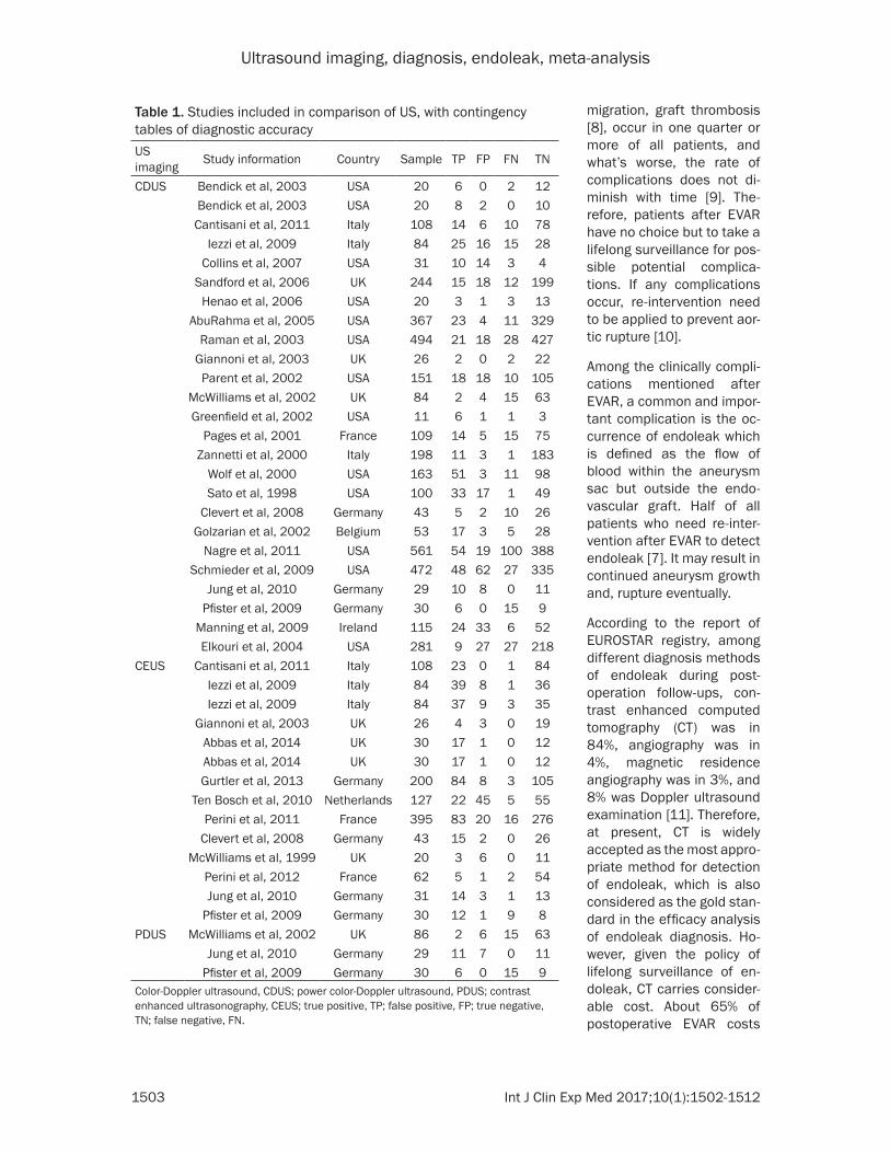
Table 1. Studies included in comparison of US, with contingency tables of diagnostic accuracyUS imaging Study information Country Sample TP FP FN TN
CDUS Bendick et al, 2003 USA 20 6 0 2 12Bendick et al, 2003 USA 20 8 2 0 10Cantisani et al, 2011 Italy 108 14 6 10 78
Iezzi et al, 2009 Italy 84 25 16 15 28Collins et al, 2007 USA 31 10 14 3 4
Sandford et al, 2006 UK 244 15 18 12 199Henao et al, 2006 USA 20 3 1 3 13
AbuRahma et al, 2005 USA 367 23 4 11 329Raman et al, 2003 USA 494 21 18 28 427
Giannoni et al, 2003 UK 26 2 0 2 22Parent et al, 2002 USA 151 18 18 10 105
McWilliams et al, 2002 UK 84 2 4 15 63Greenfield et al, 2002 USA 11 6 1 1 3
Pages et al, 2001 France 109 14 5 15 75Zannetti et al, 2000 Italy 198 11 3 1 183
Wolf et al, 2000 USA 163 51 3 11 98Sato et al, 1998 USA 100 33 17 1 49
Clevert et al, 2008 Germany 43 5 2 10 26Golzarian et al, 2002 Belgium 53 17 3 5 28
Nagre et al, 2011 USA 561 54 19 100 388Schmieder et al, 2009 USA 472 48 62 27 335
Jung et al, 2010 Germany 29 10 8 0 11Pfister et al, 2009 Germany 30 6 0 15 9
Manning et al, 2009 Ireland 115 24 33 6 52Elkouri et al, 2004 USA 281 9 27 27 218
CEUS Cantisani et al, 2011 Italy 108 23 0 1 84Iezzi et al, 2009 Italy 84 39 8 1 36Iezzi et al, 2009 Italy 84 37 9 3 35
Giannoni et al, 2003 UK 26 4 3 0 19Abbas et al, 2014 UK 30 17 1 0 12Abbas et al, 2014 UK 30 17 1 0 12Gurtler et al, 2013 Germany 200 84 8 3 105
Ten Bosch et al, 2010 Netherlands 127 22 45 5 55Perini et al, 2011 France 395 83 20 16 276
Clevert et al, 2008 Germany 43 15 2 0 26McWilliams et al, 1999 UK 20 3 6 0 11
Perini et al, 2012 France 62 5 1 2 54Jung et al, 2010 Germany 31 14 3 1 13
Pfister et al, 2009 Germany 30 12 1 9 8PDUS McWilliams et al, 2002 UK 86 2 6 15 63
Jung et al, 2010 Germany 29 11 7 0 11Pfister et al, 2009 Germany 30 6 0 15 9
Color-Doppler ultrasound, CDUS; power color-Doppler ultrasound, PDUS; contrast enhanced ultrasonography, CEUS; true positive, TP; false positive, FP; true negative, TN; false negative, FN.
Ultrasound imaging, diagnosis, endoleak, meta-analysis
1504 Int J Clin Exp Med 2017;10(1):1502-1512
are due to CT [12]. Also it is known that CT sometimes fails to detect the presence of endoleak [13]. Therefore, other endoleak detec-tion methods are required to be as an assist or an alternative if possible. US methods have been investigated for many years as a potential alternative to CT for the follow-up of EVAR patients, since it usually carries less cost and lowers the risk of ionizing radiation.
Although lots of studies had compared useful-ness of a certain single US method in endoleak detection to that with CT, many discrepancies in existing studies made it difficult to reach a powerful conclusion. With the same method, computed tomographic angiography (CTA), as the gold standard, the conclusion of Ten Bosch
less of language restriction. The following searching terms as “ultrasonic imaging diagno-sis”, “endovascular abdominal aortic aneurysm repair”, “endoleak” and their synonyms were jointly used. Additionally, all the reference lists of relative literatures were searched and exam-ined manually, for fear of any omission. Then, two reviewers retrieved all the potential litera-tures respectively.
Inclusion criteria
The eligible study need to meet the following criteria: (i) study subjects must have undergone the EVAR surgery, without limitation of aneu-rysm type, and were clinical suspected endole-ak patients; (ii) study must involve at least one
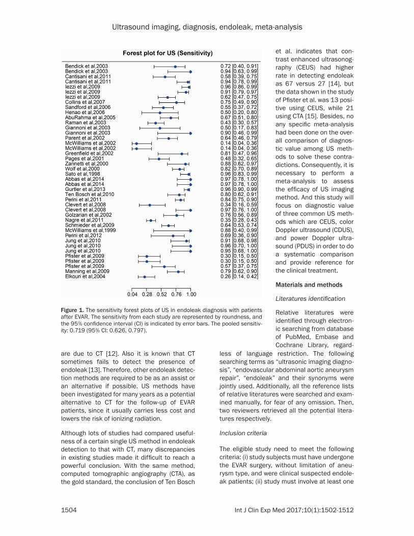
Figure 1. The sensitivity forest plots of US in endoleak diagnosis with patients after EVAR. The sensitivity from each study are represented by roundness, and the 95% confidence interval (CI) is indicated by error bars. The pooled sensitiv-ity: 0.719 (95% CI: 0.626, 0.797).
et al. indicates that con-trast enhanced ultrasonog-raphy (CEUS) had higher rate in detecting endoleak as 67 versus 27 [14], but the data shown in the study of Pfister et al. was 13 posi-tive using CEUS, while 21 using CTA [15]. Besides, no any specific meta-analysis had been done on the over-all comparison of diagnos-tic value among US meth-ods to solve these contra-dictions. Consequently, it is necessary to perform a meta-analysis to assess the efficacy of US imaging method. And this study will focus on diagnostic value of three common US meth-ods which are CEUS, color Doppler ultrasound (CDUS), and power Doppler ultra-sound (PDUS) in order to do a systematic comparison and provide reference for the clinical treatment.
Materials and methods
Literatures identification
Relative literatures were identified through electron-ic searching from database of PubMed, Embase and Cochrane Library, regard-
Ultrasound imaging, diagnosis, endoleak, meta-analysis
1505 Int J Clin Exp Med 2017;10(1):1502-1512
Statistical analysis
According to the data in both true- and false-positives and -negatives contingency tables, sensibility and specificity of each index test was calculated as percentages. To test if the data can be pooled together, the heterogeneity among selected studies was tested through Cochran’s Q. Usually, when P < 0.01, there was significant heterogeneity, and subgroup analy-ses were necessary.
Next, meta-analysis model involved total ultra-sonic imaging methods and subgroup meta-analyses were fitted for sensibility and specific-ity, with the help of R® version 3.2.1 (MathSoft, Cambridge, Massachusetts). The sensibility and the specificity of each diagnosis method
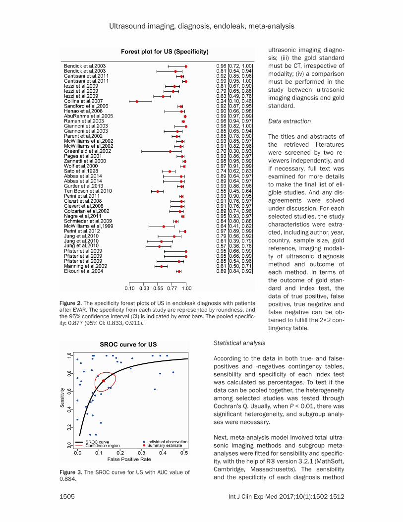
Figure 2. The specificity forest plots of US in endoleak diagnosis with patients after EVAR. The specificity from each study are represented by roundness, and the 95% confidence interval (CI) is indicated by error bars. The pooled specific-ity: 0.877 (95% CI: 0.833, 0.911).
ultrasonic imaging diagno-sis; (iii) the gold standard must be CT, irrespective of modality; (iv) a comparison must be performed in the study between ultrasonic imaging diagnosis and gold standard.
Data extraction
The titles and abstracts of the retrieved literatures were screened by two re- viewers independently, and if necessary, full text was examined for more details to make the final list of eli-gible studies. And any dis-agreements were solved under discussion. For each selected studies, the study characteristics were extra- cted, including author, year, country, sample size, gold reference, imaging modali-ty of ultrasonic diagnosis method and outcome of each method. In terms of the outcome of gold stan-dard and index test, the data of true positive, false positive, true negative and false negative can be ob- tained to fulfill the 2×2 con-tingency table.
Figure 3. The SROC curve for US with AUC value of 0.884.
Ultrasound imaging, diagnosis, endoleak, meta-analysis
1506 Int J Clin Exp Med 2017;10(1):1502-1512
compared with gold standard were expressed in forest plots as pooled point estimates with
95% confidence intervals (CI). Based on these data, a summary receiver operat-ing characteristic (SROC) curve was performed, and the area under the SROC curve (AUC) was computed through the relative inte-gral formula to reflect the sensibility and specificity of different imaging diagnosis methods. The AUC values were in the range of 0 to 1, and if a diagnosis method is certainly the best, its AUC would be 1; contrarily, AUC of the worst was 0. Besides, to make our SROC curves more visible, we used false positive rate (FPR), instead of specificity, as abscissa. And the con-version between them is: FPR=1-SPE.
Results
Included studies
A total of 42 studies from 30 articles [14-43] with 5,229 subjects were in- cluded (Table 1), as they met the criteria mentioned above, out of 105 initial retrieved articles. 34 full texts were reviewed, which included 58 studies. Am- ong them, 16 studies were excluded, since they tested non-ultrasound methods, or one study embodied three sub-studies with dif-ferent follow-ups, in which cases final sub-study with the longest follow-up was included.
Characteristics of included studies
The ultrasonic imaging methods of these 42 stud-ies involved CDUS, CEUS,
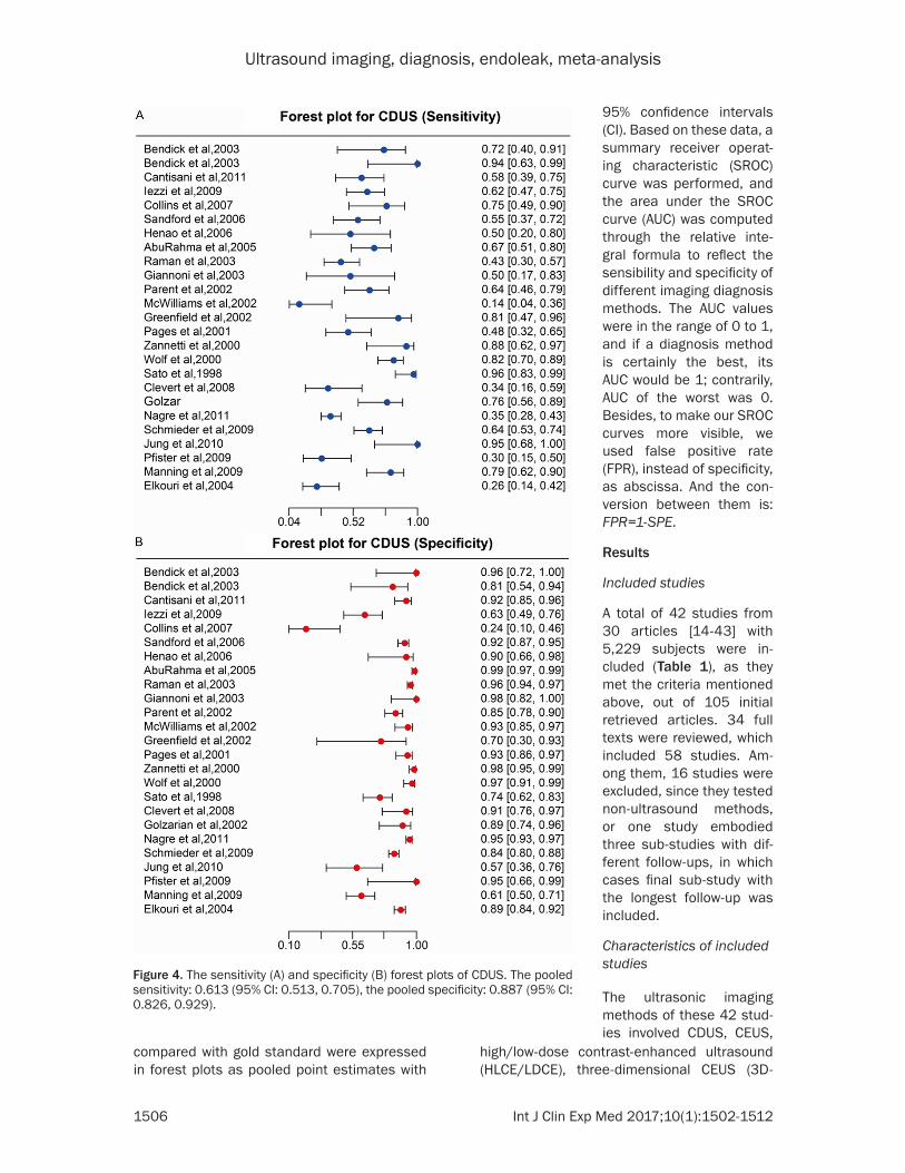
Figure 4. The sensitivity (A) and specificity (B) forest plots of CDUS. The pooled sensitivity: 0.613 (95% CI: 0.513, 0.705), the pooled specificity: 0.887 (95% CI: 0.826, 0.929).
high/low-dose contrast-enhanced ultrasound (HLCE/LDCE), three-dimensional CEUS (3D-
Ultrasound imaging, diagnosis, endoleak, meta-analysis
1507 Int J Clin Exp Med 2017;10(1):1502-1512
CEUS) and PDUS. And all the selected studies compared the diagnosis outcome of these ultrasonic methods with traditional CT, some cases were supplement by magnetic resonance imaging (MRI), as gold standard, in patients after EVAR.
Total ultrasound imaging
All the 42 studies were aimed to make a com-parison between different US imaging and CT. Pooling the whole data together, it can be sta-tistic that the its sensitivity was 0.719 with 95% CI ranged from 0.626 to 0.797 (see Figure 1), while its specificity was 0.877 with 95% CI from 0.911 to 0.833 (see Figure 2). Both values were lower than 1.000, which accounted for its less insufficient to traditional CT with normal sensitivity and good specificity. Additionally, based on the SROC curve for US shown in Figure 3, we can attain its AUC was 0.884.
Meanwhile, heterogeneity among all the ultra-sonic diagnosis methods was calculated indi-vidually, as to sensibility and specificity. And both P values were less than 0.01, which could be deemed as existing significant heterogene-ity. In consideration of the probable differences generated by the various imaging modality, the subgroup analysis was performed. All the ultra-sonic diagnosis methods were divided into three subgroups, as CDUS, CEUS and PDUS, which means HDCE, LDCE and 3D-CEUS were all categorized in CEUS.
Color-Doppler ultrasound (CDUS)
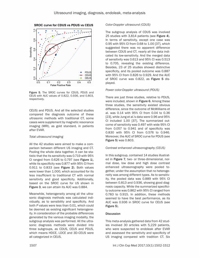
The subgroup analysis of CDUS was involved 25 studies with 3,814 patients (see Figure 4). In terms of sensitivity, except one case was 0.95 with 95% CI from 0.68 to 1.00 [37], which suggested there was no apparent difference between CDUS and CT, nearly all the data indi-cated its low-sensitivity. And the merged data of sensitivity was 0.613 and 95% CI was 0.513 to 0.705, revealing the existing difference. Besides, 23 of 25 studies showed distinctive specificity, and its pooled outcome was 0.887 with 95% CI from 0.826 to 0.929. And the AUC of SROC curve was 0.822, as Figure 5 dis- played.
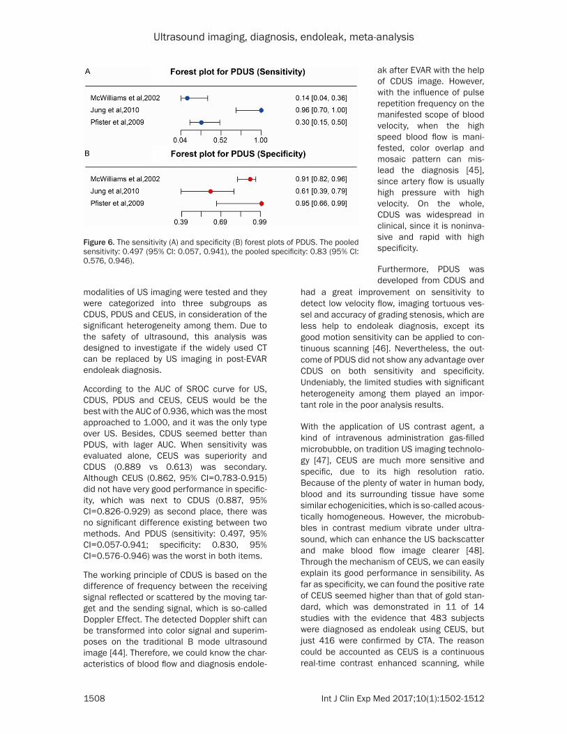
Power color-Doppler ultrasound (PDUS)
There are just three studies, relative to PDUS, were included, shown in Figure 6. Among these three studies, the sensitivity existed obvious difference, since the outcome of McWilliams et al. was 0.14 with 95% CI from 0.04 to 0.36 [23], while Jung et al.’s data were 0.96 and 95% CI included 1.00 [37]. The summarized out-come of sensitivity was 0.497 with wide 95% CI from 0.057 to 0.941 and of specificity was 0.830 with 95% CI from 0.576 to 0.946. Moreover, the AUC of SROC curve for PDUS (see Figure 5) was 0.803.
Contrast enhanced ultrasonography (CEUS)
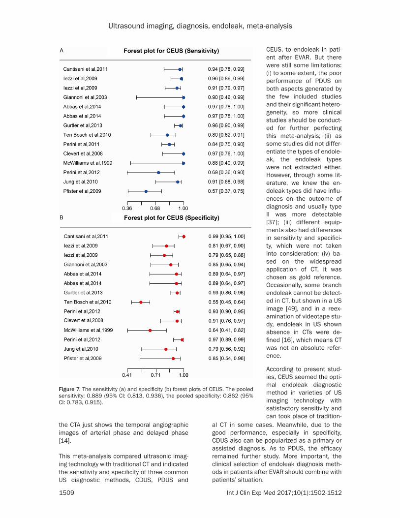
In this subgroup, contained 14 studies illustrat-ed in Figure 7, two- or three-dimensional, nor-mal dose, low dose and high dose contrast enhanced ultrasonography were pooled to- gether, under the assumption that no heteroge-neity was among different types. As to sensitiv-ity, the pooled data was 0.889 with 95% CI between 0.813 and 0.936, showing good diag-nosis capacity. While the summarized specifici-ty outcome was 0.862 with 95% CI ranged from 0.783 to 0.915. In addition, these methods seemed to have the best performance, as its AUC was 0.936 in SROC curve for CEUS (see Figure 5).
Discussion
This meta-analysis gathered data from 42 stud-ies involved 30 articles with 5,229 patients, who were suspected to endoleak after EVAR and assessed the sensitivity and specificity of US imaging compared with tradition CT. Six
Figure 5. The SROC curves for CDUS, PDUS and CEUS with AUC values of 0.822, 0.936, and 0.803, respectively.
Ultrasound imaging, diagnosis, endoleak, meta-analysis
1508 Int J Clin Exp Med 2017;10(1):1502-1512
modalities of US imaging were tested and they were categorized into three subgroups as CDUS, PDUS and CEUS, in consideration of the significant heterogeneity among them. Due to the safety of ultrasound, this analysis was designed to investigate if the widely used CT can be replaced by US imaging in post-EVAR endoleak diagnosis.
According to the AUC of SROC curve for US, CDUS, PDUS and CEUS, CEUS would be the best with the AUC of 0.936, which was the most approached to 1.000, and it was the only type over US. Besides, CDUS seemed better than PDUS, with lager AUC. When sensitivity was evaluated alone, CEUS was superiority and CDUS (0.889 vs 0.613) was secondary. Although CEUS (0.862, 95% CI=0.783-0.915) did not have very good performance in specific-ity, which was next to CDUS (0.887, 95% CI=0.826-0.929) as second place, there was no significant difference existing between two methods. And PDUS (sensitivity: 0.497, 95% CI=0.057-0.941; specificity: 0.830, 95% CI=0.576-0.946) was the worst in both items.
The working principle of CDUS is based on the difference of frequency between the receiving signal reflected or scattered by the moving tar-get and the sending signal, which is so-called Doppler Effect. The detected Doppler shift can be transformed into color signal and superim-poses on the traditional B mode ultrasound image [44]. Therefore, we could know the char-acteristics of blood flow and diagnosis endole-
had a great improvement on sensitivity to detect low velocity flow, imaging tortuous ves-sel and accuracy of grading stenosis, which are less help to endoleak diagnosis, except its good motion sensitivity can be applied to con-tinuous scanning [46]. Nevertheless, the out-come of PDUS did not show any advantage over CDUS on both sensitivity and specificity. Undeniably, the limited studies with significant heterogeneity among them played an impor-tant role in the poor analysis results.
With the application of US contrast agent, a kind of intravenous administration gas-filled microbubble, on tradition US imaging technolo-gy [47], CEUS are much more sensitive and specific, due to its high resolution ratio. Because of the plenty of water in human body, blood and its surrounding tissue have some similar echogenicities, which is so-called acous-tically homogeneous. However, the microbub-bles in contrast medium vibrate under ultra-sound, which can enhance the US backscatter and make blood flow image clearer [48]. Through the mechanism of CEUS, we can easily explain its good performance in sensibility. As far as specificity, we can found the positive rate of CEUS seemed higher than that of gold stan-dard, which was demonstrated in 11 of 14 studies with the evidence that 483 subjects were diagnosed as endoleak using CEUS, but just 416 were confirmed by CTA. The reason could be accounted as CEUS is a continuous real-time contrast enhanced scanning, while
Figure 6. The sensitivity (A) and specificity (B) forest plots of PDUS. The pooled sensitivity: 0.497 (95% CI: 0.057, 0.941), the pooled specificity: 0.83 (95% CI: 0.576, 0.946).
ak after EVAR with the help of CDUS image. However, with the influence of pulse repetition frequency on the manifested scope of blood velocity, when the high speed blood flow is mani-fested, color overlap and mosaic pattern can mis-lead the diagnosis [45], since artery flow is usually high pressure with high velocity. On the whole, CDUS was widespread in clinical, since it is noninva-sive and rapid with high specificity.
Furthermore, PDUS was developed from CDUS and
Ultrasound imaging, diagnosis, endoleak, meta-analysis
1509 Int J Clin Exp Med 2017;10(1):1502-1512
the CTA just shows the temporal angiographic images of arterial phase and delayed phase [14].
This meta-analysis compared ultrasonic imag-ing technology with traditional CT and indicated the sensitivity and specificity of three common US diagnostic methods, CDUS, PDUS and
al CT in some cases. Meanwhile, due to the good performance, especially in specificity, CDUS also can be popularized as a primary or assisted diagnosis. As to PDUS, the efficacy remained further study. More important, the clinical selection of endoleak diagnosis meth-ods in patients after EVAR should combine with patients’ situation.
Figure 7. The sensitivity (a) and specificity (b) forest plots of CEUS. The pooled sensitivity: 0.889 (95% CI: 0.813, 0.936), the pooled specificity: 0.862 (95% CI: 0.783, 0.915).
CEUS, to endoleak in pati- ent after EVAR. But there were still some limitations: (i) to some extent, the poor performance of PDUS on both aspects generated by the few included studies and their significant hetero-geneity, so more clinical studies should be conduct-ed for further perfecting this meta-analysis; (ii) as some studies did not differ-entiate the types of endole-ak, the endoleak types were not extracted either. However, through some lit-erature, we knew the en- doleak types did have influ-ences on the outcome of diagnosis and usually type II was more detectable [37]; (iii) different equip-ments also had differences in sensitivity and specifici-ty, which were not taken into consideration; (iv) ba- sed on the widespread application of CT, it was chosen as gold reference. Occasionally, some branch endoleak cannot be detect-ed in CT, but shown in a US image [49], and in a reex-amination of videotape stu- dy, endoleak in US shown absence in CTs were de- fined [16], which means CT was not an absolute refer- ence.
According to present stud-ies, CEUS seemed the opti-mal endoleak diagnostic method in varieties of US imaging technology with satisfactory sensitivity and can took place of tradition-
Ultrasound imaging, diagnosis, endoleak, meta-analysis
1510 Int J Clin Exp Med 2017;10(1):1502-1512
Acknowledgements
We acknowledge all authors whose publica-tions could be included in our meta-analysis.
Disclosure of conflict of interest
None.
Address correspondence to: Shijie Xin, Department of Vascular Surgery, 1st Hospital of China Medical University, Nanjing North Street 155, Heping District, Shenyang 110000, Liaoning Province, China. Tel: +86 024-83283288; E-mail: [email protected]
References
[1] Kent KC. Clinical practice. Abdominal aortic aneurysms. N Engl J Med 2014; 371: 2101-2108.
[2] Upchurch GR Jr and Schaub TA. Abdominal aortic aneurysm. Am Fam Physician 2006; 73: 1198-1204.
[3] Parodi JC, Palmaz JC and Barone HD. Transfemoral intraluminal graft implantation for abdominal aortic aneurysms. Ann Vasc Surg 1991; 5: 491-499.
[4] Habets J, Zandvoort HJ, Reitsma JB, Bartels LW, Moll FL, Leiner T and van Herwaarden JA. Magnetic resonance imaging is more sensitive than computed tomography angiography for the detection of endoleaks after endovascular abdominal aortic aneurysm repair: a system-atic review. Eur J Vasc Endovasc Surg 2013; 45: 340-350.
[5] Greenhalgh RM, Brown LC, Powell JT, Thompson SG, Epstein D and Sculpher MJ. Endovascular versus open repair of abdominal aortic aneurysm. N Engl J Med 2010; 362: 1863-1871.
[6] De Bruin JL, Baas AF, Buth J, Prinssen M, Verhoeven EL, Cuypers PW, van Sambeek MR, Balm R, Grobbee DE and Blankensteijn JD. Long-term outcome of open or endovascular repair of abdominal aortic aneurysm. N Engl J Med 2010; 362: 1881-1889.
[7] Nordon IM, Karthikesalingam A, Hinchliffe RJ, Holt PJ, Loftus IM and Thompson MM. Secondary interventions following endovascu-lar aneurysm repair (EVAR) and the enduring value of graft surveillance. Eur J Vasc Endovasc Surg 2010; 39: 547-554.
[8] Mita T, Arita T, Matsunaga N, Furukawa M, Zempo N, Esato K and Matsuzaki M. Complications of endovascular repair for tho-racic and abdominal aortic aneurysm: an im-aging spectrum. Radiographics 2000; 20: 1263-1278.
[9] Sampram ES, Karafa MT, Mascha EJ, Clair DG, Greenberg RK, Lyden SP, O’Hara PJ, Sarac TP, Srivastava SD, Butler B and Ouriel K. Nature, frequency, and predictors of secondary proce-dures after endovascular repair of abdominal aortic aneurysm. J Vasc Surg 2003; 37: 930-937.
[10] Karthikesalingam A, Al-Jundi W, Jackson D, Boyle JR, Beard JD, Holt PJ and Thompson MM. Systematic review and meta-analysis of duplex ultrasonography, contrast-enhanced ultraso-nography or computed tomography for surveil-lance after endovascular aneurysm repair. Br J Surg 2012; 99: 1514-1523.
[11] Buth J, Harris PL, van Marrewijk C and Fransen G. The significance and management of differ-ent types of endoleaks. Semin Vasc Surg 2003; 16: 95-102.
[12] Prinssen M, Wixon CL, Buskens E and Blankensteijn JD. Surveillance after endovas-cular aneurysm repair: diagnostics, complica-tions, and associated costs. Ann Vasc Surg 2004; 18: 421-427.
[13] Rozenblit AM, Patlas M, Rosenbaum AT, Okhi T, Veith FJ, Laks MP and Ricci ZJ. Detection of en-doleaks after endovascular repair of abdomi-nal aortic aneurysm: value of unenhanced and delayed helical CT acquisitions. Radiology 2003; 227: 426-433.
[14] Ten Bosch JA, Rouwet EV, Peters CT, Jansen L, Verhagen HJ, Prins MH and Teijink JA. Contrast-enhanced ultrasound versus computed tomo-graphic angiography for surveillance of endo-vascular abdominal aortic aneurysm repair. J Vasc Interv Radiol 2010; 21: 638-643.
[15] Pfister K, Rennert J, Uller W, Schnitzbauer AA, Stehr A, Jung W, Hofstetter P, Zorger N, Kasprzak PM and Jung EM. Contrast harmonic imaging ultrasound and perfusion imaging for surveillance after endovascular abdominal an-eurysm repair regarding detection and charac-terization of suspected endoleaks. Clin Hemorheol Microcirc 2009; 43: 119-128.
[16] Sato DT, Goff CD, Gregory RT, Robinson KD, Carter KA, Herts BR, Vilsack HB, Gayle RG, Parent FN 3rd, DeMasi RJ and Meier GH. Endoleak after aortic stent graft repair: diag-nosis by color duplex ultrasound scan versus computed tomography scan. J Vasc Surg 1998; 28: 657-663.
[17] McWilliams RG, Martin J, White D, Gould DA, Harris PL, Fear SC, Brennan J, Gilling-Smith GL, Bakran A and Rowlands PC. Use of con-trast-enhanced ultrasound in follow-up after endovascular aortic aneurysm repair. J Vasc Interv Radiol 1999; 10: 1107-1114.
[18] Wolf YG, Johnson BL, Hill BB, Rubin GD, Fogarty TJ and Zarins CK. Duplex ultrasound scanning versus computed tomographic angiography for

Ultrasound imaging, diagnosis, endoleak, meta-analysis
1511 Int J Clin Exp Med 2017;10(1):1502-1512
postoperative evaluation of endovascular ab-dominal aortic aneurysm repair. J Vasc Surg 2000; 32: 1142-1148.
[19] Zannetti S, De Rango P, Parente B, Parlani G, Verzini F, Maselli A, Nardelli L and Cao P. Role of duplex scan in endoleak detection after en-doluminal abdominal aortic aneurysm repair. Eur J Vasc Endovasc Surg 2000; 19: 531-535.
[20] Pages S, Favre JP, Cerisier A, Pyneeandee S, Boissier C and Veyret C. Comparison of color duplex ultrasound and computed tomography scan for surveillance after aortic endografting. Ann Vasc Surg 2001; 15: 155-162.
[21] Greenfield AL, Halpern EJ, Bonn J, Wechsler RJ, Kahn MB. Application of Duplex US for Characterization of Endoleaks in Abdominal Aortic Stent-Grafts: Report of Five Cases. Radiology 2002; 225: 845-51.
[22] Golzarian J, Murgo S, Dussaussois L, Guyot S, Said KA, Wautrecht JC and Struyven J. Evaluation of abdominal aortic aneurysm after endoluminal treatment: comparison of color Doppler sonography with biphasic helical CT. AJR Am J Roentgenol 2002; 178: 623-628.
[23] McWilliams RG, Martin J, White D, Gould DA, Rowlands PC, Haycox A, Brennan J, Gilling-Smith GL and Harris PL. Detection of endoleak with enhanced ultrasound imaging: compari-son with biphasic computed tomography. J Endovasc Ther 2002; 9: 170-179.
[24] Parent FN, Meier GH, Godziachvili V, LeSar CJ, Parker FM, Carter KA, Gayle RG, DeMasi RJ, Marcinczyk MJ and Gregory RT. The incidence and natural history of type I and II endoleak: A 5-year follow-up assessment with color duplex ultrasound scan. J Vasc Surg 2002; 35: 474-481.
[25] Bendick PJ, Bove PG, Long GW, Zelenock GB, Brown OW and Shanley CJ. Efficacy of ultra-sound scan contrast agents in the noninvasive follow-up of aortic stent grafts. J Vasc Surg 2003; 37: 381-385.
[26] Giannoni MF, Palombo G, Sbarigia E, Speziale F, Zaccaria A, Fiorani P. Contrast-Enhanced Ultrasound Imaging for Aortic Stent-Graft Surveillance. J Endovasc Ther 2003; 10: 208-17.
[27] Raman KG, Missig-Carroll N, Richardson T, Muluk SC and Makaroun MS. Color-flow duplex ultrasound scan versus computed tomograph-ic scan in the surveillance of endovascular an-eurysm repair. J Vasc Surg 2003; 38: 645-651.
[28] Elkouri S, Panneton JM, Andrews JC, Lewis BD, McKusick MA, Noel AA, Rowland CM, Bower TC, Cherry KJ Jr and Gloviczki P. Computed to-mography and ultrasound in follow-up of pa-tients after endovascular repair of abdominal aortic aneurysm. Ann Vasc Surg 2004; 18: 271-279.
[29] AbuRahma AF, Welch CA, Mullins BB and Dyer B. Computed tomography versus color duplex ultrasound for surveillance of abdominal aortic stent-grafts. J Endovasc Ther 2005; 12: 568-573.
[30] Henao EA, Hodge MD, Felkai DD, McCollum CH, Noon GP, Lin PH, Lumsden AB and Bush RL. Contrast-enhanced Duplex surveillance af-ter endovascular abdominal aortic aneurysm repair: improved efficacy using a continuous infusion technique. J Vasc Surg 2006; 43: 259-264; discussion 264.
[31] Sandford RM, Bown MJ, Fishwick G, Murphy F, Naylor M, Sensier Y, Sharpe R, Walker J, Hartshorn T, London NJ and Sayers RD. Duplex ultrasound scanning is reliable in the detection of endoleak following endovascular aneurysm repair. Eur J Vasc Endovasc Surg 2006; 32: 537-541.
[32] Collins JT, Boros MJ and Combs K. Ultrasound surveillance of endovascular aneurysm repair: a safe modality versus computed tomography. Ann Vasc Surg 2007; 21: 671-5.
[33] Clevert DA, Minaifar N, Weckbach S, Kopp R, Meimarakis G, Clevert DA and Reiser M. Color duplex ultrasound and contrast-enhanced ul-trasound in comparison to MS-CT in the detec-tion of endoleak following endovascular aneu-rysm repair. Clin Hemorheol Microcirc 2008; 39: 121-132.
[34] Iezzi R, Basilico R, Giancristofaro D, Pascali D, Cotroneo AR and Storto ML. Contrast-enhanced ultrasound versus color duplex ul-trasound imaging in the follow-up of patients after endovascular abdominal aortic aneu-rysm repair. J Vasc Surg 2009; 49: 552-560.
[35] Manning BJ, O’Neill SM, Haider SN, Colgan MP, Madhavan P and Moore DJ. Duplex ultrasound in aneurysm surveillance following endovascu-lar aneurysm repair: a comparison with com-puted tomography aortography. J Vasc Surg 2009; 49: 60-65.
[36] Schmieder GC, Stout CL, Stokes GK, Parent FN and Panneton JM. Endoleak after endovascu-lar aneurysm repair: duplex ultrasound imag-ing is better than computed tomography at determining the need for intervention. J Vasc Surg 2009; 50: 1012-1017; discussion 1017-1018.
[37] Jung EM, Rennert J, Fellner C, Uller W, Jung W, Schreyer A, Heiss P, Hoffstetter P, Feuerbach S, Kasparzk P, Zorger N and Pfister K. Detection and characterization of endoleaks following endovascular treatment of abdominal aortic aneurysms using contrast harmonic imaging (CHI) with quantitative perfusion analysis (TIC) compared to CT angiography (CTA). Ultraschall Med 2010; 31: 564-570.
[38] Cantisani V, Ricci P, Grazhdani H, Napoli A, Fanelli F, Catalano C, Galati G, D’Andrea V,
Ultrasound imaging, diagnosis, endoleak, meta-analysis
1512 Int J Clin Exp Med 2017;10(1):1502-1512
Biancari F and Passariello R. Prospective com-parative analysis of colour-Doppler ultrasound, contrast-enhanced ultrasound, computed to-mography and magnetic resonance in detect-ing endoleak after endovascular abdominal aortic aneurysm repair. Eur J Vasc Endovasc Surg 2011; 41: 186-192.
[39] Nagre SB, Taylor SM, Passman MA, Patterson MA, Combs BR, Lowman BG and Jordan WD Jr. Evaluating outcomes of endoleak discrepan-cies between computed tomography scan and ultrasound imaging after endovascular ab-dominal aneurysm repair. Ann Vasc Surg 2011; 25: 94-100.
[40] Perini P, Sediri I, Midulla M, Delsart P, Mouton S, Gautier C, Pruvo JP and Haulon S. Single-centre prospective comparison between con-trast-enhanced ultrasound and computed to-mography angiography after EVAR. Eur J Vasc Endovasc Surg 2011; 42: 797-802.
[41] Perini P, Sediri I, Midulla M, Delsart P, Gautier C and Haulon S. Contrast-enhanced ultra-sound vs CT angiography in fenestrated EVAR surveillance: a single-center comparison. J Endovasc Ther 2012; 19: 648-655.
[42] Gurtler VM, Sommer WH, Meimarakis G, Kopp R, Weidenhagen R, Reiser MF and Clevert DA. A comparison between contrast-enhanced ul-trasound imaging and multislice computed to-mography in detecting and classifying endole-aks in the follow-up after endovascular aneu-rysm repair. J Vasc Surg 2013; 58: 340-345.
[43] Abbas A, Hansrani V, Sedgwick N, Ghosh J and McCollum CN. 3D contrast enhanced ultra-sound for detecting endoleak following endo-vascular aneurysm repair (EVAR). Eur J Vasc Endovasc Surg 2014; 47: 487-492.
[44] Mitchell DG. Color Doppler imaging: principles, limitations, and artifacts. Radiology 1990; 177: 1-10.
[45] Millington SJ and Arntfield RT. Advanced Point-of-Care Cardiac Ultrasound Examination: Doppler Applications, Valvular Assessment, and Advanced Right Heart Examination. Glob Heart 2013; 8: 305-312.
[46] Martinoli C, Derchi LE, Rizzatto G and Solbiati L. Power Doppler sonography: general princi-ples, clinical applications, and future pros-pects. Eur Radiol 1998; 8: 1224-1235.
[47] Lindner JR. Microbubbles in medical imaging: current applications and future directions. Nat Rev Drug Discov 2004; 3: 527-532.
[48] McCulloch M, Gresser C, Moos S, Odabashian J, Jasper S, Bednarz J, Burgess P, Carney D, Moore V, Sisk E, Waggoner A, Witt S and Adams D. Ultrasound contrast physics: A series on contrast echocardiography, article 3. J Am Soc Echocardiogr 2000; 13: 959-967.
[49] Heilberger P, Schunn C, Ritter W, Weber S and Raithel D. Postoperative color flow duplex scanning in aortic endografting. J Endovasc Surg 1997; 4: 262-271.


![Karl Pearson's meta-analysis revisitedstatweb.stanford.edu/~owen/reports/AOS697.pdf · Recent research in genomics [Zahn et al. (2007)] and functional magnetic reso-nance imaging](https://img.pdfslide.us/doc/110x75/5ed5700711be98291d0426e0/karl-pearsons-meta-analysis-owenreportsaos697pdf-recent-research-in-genomics.jpg)