Embed Size (px)
Citation preview
Louisiana State UniversityLSU Digital Commons
LSU Master's Theses Graduate School
2010
Origin of Pyrite Nodules At the Top of the NantuoDiamictites, Southern ChinaChangjie LiuLouisiana State University and Agricultural and Mechanical College, [email protected]
Follow this and additional works at: https://digitalcommons.lsu.edu/gradschool_theses
Part of the Earth Sciences Commons
This Thesis is brought to you for free and open access by the Graduate School at LSU Digital Commons. It has been accepted for inclusion in LSUMaster's Theses by an authorized graduate school editor of LSU Digital Commons. For more information, please contact [email protected].
Recommended CitationLiu, Changjie, "Origin of Pyrite Nodules At the Top of the Nantuo Diamictites, Southern China" (2010). LSU Master's Theses. 3804.https://digitalcommons.lsu.edu/gradschool_theses/3804
ORIGIN OF PYRITE NODULES AT THE TOP OF THE NANTUO DIAMICTITES,
SOUTHERN CHINA
A Thesis
Submitted to Graduate Faculty of the
Louisiana State University and
Agricultural and Mechanical College
In partial fulfillment of the
requirements for the degree of
Master of Science
in
Department of Geology and Geophysics
by
Changjie Liu
B.S., University of Science and Technology of China, 2009
May, 2012

ii
ACKNOWLEDGEMENTS
I would like to thank my major advisor, Dr. Huiming Bao. With his guidance, I
conducted a set of geochemical and stable isotope experiments and research projects; some of
them reflect the usual journey of trial and error and are not written here in this thesis. I have
learned a lot from Dr. Bao in the past two and a half years. He taught me not only the
knowledge, but more importantly, what scientific research is and what it takes to be a scientist. I
also thank my committee members, Dr. Annette Engel, Dr. Brooks Ellwood, Dr. Bryan Fry, who
have helped me along the way. Also, I am grateful to my fellow labmates and friends, Tao Sun,
Yongbo Peng, Issaku Kohl, Bryan Killingsworth, Justin Hayles, Xiaoqian Li, Dian He, Chang
Liu, Hongjiao Yu and Xuan Feng. I appreciate their help and support.
The measurement of sulfur isotope composition was assisted by Dr. Yongbo Peng at the
University of Maryland. Financial support for this research was provided by LSU Dean of
Science and the National Science Foundation.
iii
TABLE OF CONTENTS
ACKNOWLEDGEMENTS……………………………………………………………………....ii
LIST OF TABLES………………………………………………………………………………..iv
LIST OF FIGURES……………………………………………………………………………….v
ABSTRACT……………………………………………………………………………………...vi
INTRODUCTION………………………………………………………………………………...1
GEOLOGY AND FIELD OCCURRENCE OF PYRITE NODULES……………………………4
APPROACH……………………………………………………………………………………....9
METHODS………………………………………………………………………………………13
Petrography………………………………………………………………………………13
Stable Isotope Analysis…………………………………………………………………..13
RESULTS………………………………………………………………………………………..18
Petrographic Observation………………………………………………………………...18
XRD Analysis……………………………………………………………………………18
Stable Isotope Composition……………………………………………………………18
DISCUSSION……………………………………………………………………………………34
Pyrite Formation…………………………………………………………………………34
The Pyrite Formation Scenario……………..……………………………………………35
Formation Model for Pyrite Nodules in the Nantuo Diamictite………..………...……...35
FUTURE WORK NEEDED..........................................................................................................38
CONCLUSIONS………………………………………………………………………………...39
REFFERENCES…………………………………………………………………………………41
APPENDIX:XRD PATTERNS OF ALL THE SAMPLES…………………………………...44
VITA……………………………………………………………………………………………..48

iv
LISTS OF TABLES
1. Mineral composition and estimated weight percentage of samples ……..…………………..27
2. Sulfur isotope composition of pyrite nodules in Taoying, Guizhou, southern China………..28
v
LISTS OF FIGURES
1. Geographic location of Taoying, Guizhou Province, southern China…………………………5
2. Paleogeography of the research area…………………………………………………………..5
3. Field photos of pyrite nodules of the lower Doushantuo Formation in Taoying, Guizhou
Province, southern China………………………………………………………………………6
4. Scheme showing the pyrite concretion formation ……………………………………………11
5. Stratigraphic column showing the distribution of the analyzed pyrite nodules …………….12
6. Sampling positions within individual pyrite nodules ………………………………………..14
7. Drilling hole positions in each pyrite nodule samples ………………………………..........15
8. Photomicrographs (reflected light) for thin sections of bulk diamictite samples……..........19
9. Photomicrographs (reflected light) for thin sections of pyrite nodules ………………………22
10. Photomicrographs (transmitted, polarized light) for thin sections of bulk diamictite
samples…………………………………………………………………………………………...25
11. XRD pattern of sample ZB11-12…………………………………………………………….26
12. Results of the sulfur isotope analysis in centimeter scale…………………………………..29
13. Results of the analyzed pyrite nodules in stratigraphic column……………………...........30
14. Millimeter-scale sampling of pyrite nodules with their δ34
S displayed ……………………..31
15. Schematic diagram showing the process of sedimentary pyrite formation ………………....36
vi
ABSTRACT
Pyrite nodules up to 20 cm in diameter are found at the top of the Marinoan (~635 Ma)
Nantuo glacial diamictite as well as in the cap dolostones and shale/siltstones in the lower
Doushantuo Formation in eastern Guizhou, southern China. Earlier studies on the occurrence and
stable sulfur and triple oxygen isotope composition of barite in the cap dolostones concluded that
seawater sulfate concentrations in shallow oceans in the South China Block were low during the
deposition of the cap dolostones. Therefore, the occurrence of pyrite nodules suggests two
scenarios: 1) Formation before the precipitation of the cap dolostone, when seawater sulfate
concentration was high enough to result in pyrite formation in sediments, either via direct
precipitation from a euxinic water column or through in-sediment sulfate reduction; or 2)
Diagenetic formation via sulfate reduction the precipitation of the cap dolostone when seawater
sulfate content became high enough to diffuse into the organic-rich cap dolostone and the
underlying diamictite. Scenario 1 would predict large and irregular variations of δ34
S value for
pyrite nodules from different vertical horizons, whereas scenario 2 predicts a gradual increase of
pyrite δ34
S with increasing depth, at least from the top of the diamictite. Field occurrences,
petrography, and stable sulfur isotopic compositions of pyrite nodules were studied from a
section at Taoying, eastern Guizhou, China. Pyrite δ34
S values from different nodules varied
from 7.3‰ to 60.5‰ at different stratigraphic levels. No stratigraphic trend existed for the δ34
S,
supporting scenario 1. Pyrite δ34
S values were also homogeneous within individual nodules at a
0.3 to 1 cm sampling scales, but were heterogeneous at a 2 mm sampling scale. Homogeneity
was not expected from the particular model for pyrite nodule formation in a largely closed or
semi-closed environment. Therefore, pyrite formation likely occurred prior to cap dolostone
deposition, when seawater sulfate rose appreciably to support extensive sulfate reduction in
sediments. Differential cementation and compaction of the pyrite-bearing sediments may have

vii
produced the nodular shape of the pyrite deposit. Future work needs to test this alternative model
for pyrite nodule formation at multiple Marinoan sections in South China.
1
INTRODUCTION
Sulfate (SO42-
) in modern seawater is 0.2% by weight, and is second only to chloride (Cl-)
in concentration. Seawater sulfate concentration has varied over geological history. While
periods of dramatic changes did occur, seawater sulfate concentration has generally increased
over time. One of the extreme shifts in sulfate concentration was expected to have occurred at
the aftermath of Marinoan global glaciations at ~635Ma. Sulfate concentration is believed to be
exceedingly low at the onset of deglaciation in the oceans. Peng et al. (2011) studied the
occurrence of non-mass-dependently 17
O depleted barite deposits in cap carbonates that drape the
Nantuo diamictite, South China Block. They concluded that sulfate concentration in seawater
was low or nearly absent during the deposition of the Marinoan cap carbonates and the sulfate
concentration in the oceans only rose after the deposition of cap dolostones, as evident from the
first barite crystal fans being precipitated only at the top of reworked cap dolostones. Initially,
shallow ocean sulfate had a significant riverine sulfate component, as supported by distinct
negative Δ17
O values (a measure of the δ17
O deviation from what is expected from a mass-
dependent relationship between the δ17
O and δ18
O) in these barite sulfates. The barium was
supplied episodically to shallow oceans through the upwelling of deep Ba2+
-rich water. This
conclusion is echoed by the sequence of events occurring at the aftermath of Marinoan meltdown
in the entire South China Block (Zhou et al., 2010).
In many shallow platform, shelf, and basinal facies of the South China Block, pyrite
nodules of different sizes (up to 20 cm in diameter) occur at the top 0 to 2 meters of the Nantuo
diamictite, and occasionally within the cap dolostone of the basal Doushantuo Formation. Pyrite
is usually precipitated through the reaction of dissolved sulfide produced by microbial sulfate
reduction with Fe2+
derived from detrital iron-bearing minerals in anoxic marine sediments
2
(Berner, 1970; Raiswell and Canfield, 1998). Pyrite precipitation can occur diagenetically in
shallow sediments where both organic matter and sulfate are present in pore fluids, so that
microbial sulfate reduction can produce sulfides (HS– and H2S) to be precipitated as insoluble
FeS. The initial FeS is later transformed to the more stable mineral pyrite (FeS2), the common
sulfide minerals seen in the rock record (Rickard, 1975; Rickard and Luther III, 1997). Pyrite can
also form in the water column. In a euxinic water column, dissolved sulfide reacts with free Fe2+
to form small FeS aggregates. Once the aggregates are larger than a critical size, they settle to
bottom of the water column and are later transformed to pyrite (Boesen and Postma, 1988;
Wilkin and Barnes, 1997).
A scenario supporting the conclusion reached in Zhou et al. (2010) and Peng et al. (2011)
would, therefore, predict that the basal Doushantuo pyrite nodules were formed in pore fluids
after the deposition and disruption of the cap dolostones. By then, the ocean sulfate concentration
had risen to a level that enough of it could diffuse into the pore fluids within the underlying
sediments. Considering that the source of sulfate would be exclusively derived from the water
column after the deposition of the cap dolostones and the Nantuo diamictite, this scenario
predicts that the pyrite δ34
S value would increase with depth, starting at the top of the diamictite.
Another possible scenario is that seawater sulfate concentration had risen to a sufficiently
high level so that widespread pyrite formation in sediments could occur before the precipitation
of the cap dolostone, either via direct precipitation of pyrite from a euxinic water column or
through in-sediment sulfate reduction. This scenario predicts that the many horizons of pyrite
nodules at the top of the Nantuo diamictite would have large variability in their δ34
S value and
that the variation should have no relationship with depth. Although this scenario is consistent
with the low sulfate ocean concluded by the earlier studies (Zhou et al., 2010; Peng et al., 2011),

3
it does require that we re-examine the sulfur-sulfate cycle at the waning stage of the diamictite
deposition. This time window has been largely neglected so far.
My thesis work evaluates these scenarios to explain the occurrence of the nodules in the
South China Block. Although pyrite nodules have been observed in many facies in the Marinoan
South China, I focused my study on samples from a well-exposed field section in Taoying,
Tongling, eastern Guizhou (109˚1'4.9"E, 27˚50'1.4"N; Fig.1). In summary, I examined the field
occurrences, petrographic features, and stable sulfur isotope compositions (the δ34
S) of pyrite
nodules, together with a few pyrite lenses and beddings in the overlying Doushantuo shale and
siltstones for comparison.
4
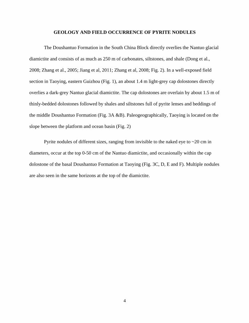
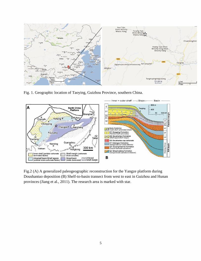
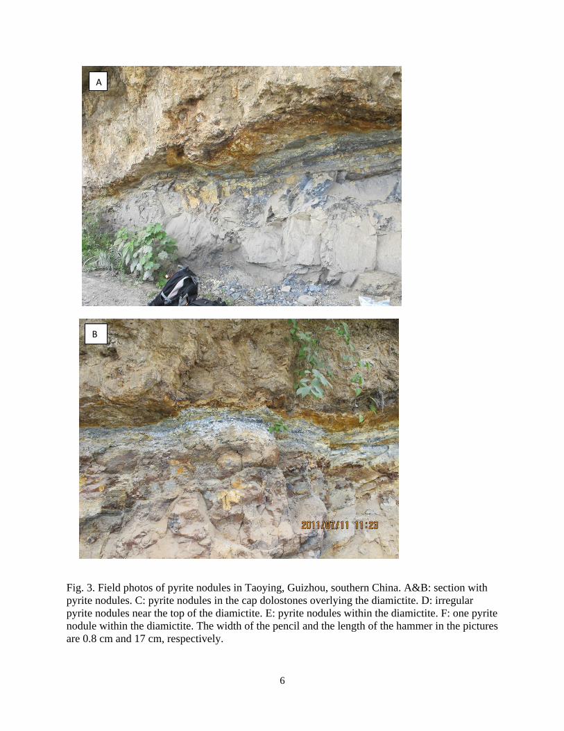
GEOLOGY AND FIELD OCCURRENCE OF PYRITE NODULES
The Doushantuo Formation in the South China Block directly overlies the Nantuo glacial
diamictite and consists of as much as 250 m of carbonates, siltstones, and shale (Dong et al.,
2008; Zhang et al., 2005; Jiang et al, 2011; Zhang et al, 2008; Fig. 2). In a well-exposed field
section in Taoying, eastern Guizhou (Fig. 1), an about 1.4 m light-grey cap dolostones directly
overlies a dark-grey Nantuo glacial diamictite. The cap dolostones are overlain by about 1.5 m of
thinly-bedded dolostones followed by shales and siltstones full of pyrite lenses and beddings of
the middle Doushantuo Formation (Fig. 3A &B). Paleogeographically, Taoying is located on the
slope between the platform and ocean basin (Fig. 2)
Pyrite nodules of different sizes, ranging from invisible to the naked eye to ~20 cm in
diameters, occur at the top 0-50 cm of the Nantuo diamictite, and occasionally within the cap
dolostone of the basal Doushantuo Formation at Taoying (Fig. 3C, D, E and F). Multiple nodules
are also seen in the same horizons at the top of the diamictite.
5
Fig. 1. Geographic location of Taoying, Guizhou Province, southern China.
Fig.2 (A) A generalized paleogeographic reconstruction for the Yangze platform during
Doushantuo deposition (B) Shelf-to-basin transect from west to east in Guizhou and Hunan
provinces (Jiang et al., 2011). The research area is marked with star.
6
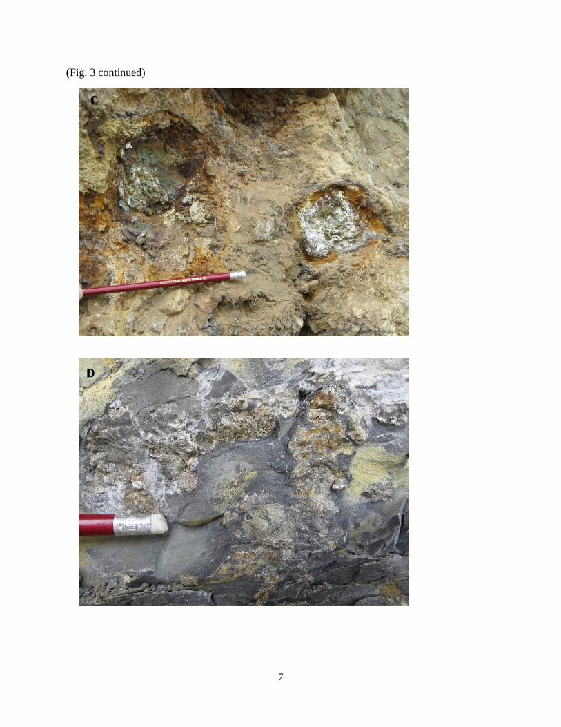
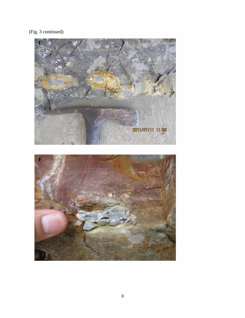
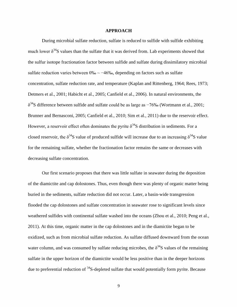
Fig. 3. Field photos of pyrite nodules in Taoying, Guizhou, southern China. A&B: section with
pyrite nodules. C: pyrite nodules in the cap dolostones overlying the diamictite. D: irregular
pyrite nodules near the top of the diamictite. E: pyrite nodules within the diamictite. F: one pyrite
nodule within the diamictite. The width of the pencil and the length of the hammer in the pictures
are 0.8 cm and 17 cm, respectively.
A
B
7
(Fig. 3 continued)
8
(Fig. 3 continued)
9
APPROACH
During microbial sulfate reduction, sulfate is reduced to sulfide with sulfide exhibiting
much lower δ34
S values than the sulfate that it was derived from. Lab experiments showed that
the sulfur isotope fractionation factor between sulfide and sulfate during dissimilatory microbial
sulfate reduction varies between 0‰ ~ −46‰, depending on factors such as sulfate
concentration, sulfate reduction rate, and temperature (Kaplan and Rittenberg, 1964; Rees, 1973;
Detmers et al., 2001; Habicht et al., 2005; Canfield et al., 2006). In natural environments, the
δ34
S difference between sulfide and sulfate could be as large as −76‰ (Wortmann et al., 2001;
Brunner and Bernasconi, 2005; Canfield et al., 2010; Sim et al., 2011) due to the reservoir effect.
However, a reservoir effect often dominates the pyrite δ34
S distribution in sediments. For a
closed reservoir, the δ34
S value of produced sulfide will increase due to an increasing δ34
S value
for the remaining sulfate, whether the fractionation factor remains the same or decreases with
decreasing sulfate concentration.
Our first scenario proposes that there was little sulfate in seawater during the deposition
of the diamictite and cap dolostones. Thus, even though there was plenty of organic matter being
buried in the sediments, sulfate reduction did not occur. Later, a basin-wide transgression
flooded the cap dolostones and sulfate concentration in seawater rose to significant levels since
weathered sulfides with continental sulfate washed into the oceans (Zhou et al., 2010; Peng et al.,
2011). At this time, organic matter in the cap dolostones and in the diamictite began to be
oxidized, such as from microbial sulfate reduction. As sulfate diffused downward from the ocean
water column, and was consumed by sulfate reducing microbes, the δ34
S values of the remaining
sulfate in the upper horizon of the diamictite would be less positive than in the deeper horizons
due to preferential reduction of 34
S-depleted sulfate that would potentially form pyrite. Because
10
little to no sulfate reduction occurred during the deposition of the diamictite, all sulfate came
from the top. This scenario predicts that the pyrite δ34
S value would increase with increasing
depth in the diamictite. Due to the widespread occurrence of fractures in the cap dolostones, the
sulfate reservoir would be less constrained than the more compacted diamictite. Thus, such a
depth-δ34
S trend may not be expected in the cap dolostones.
However, if seawater sulfate during the waning stage of diamictite deposition was low,
but not to a level that concurrent sulfate reduction could occur, then pyrite could form in
sediments via direct precipitation in a euxinic water column or via in-sediment sulfate reduction.
In this second scenario, pyrite formation in the diamictite would have occurred continuously at
different times and at different depths. The highly variable sedimentation rate (Zhou et al., 2007),
sulfate concentration, organic content, sediment type, and microbial activity in this scenario
would result in highly variable pyrite δ34
S values from horizon to horizon with no correlation
with stratigraphic depth.
Although the two different pyrite formation scenarios can be differentiated by the
proposed stable sulfur isotope ratio analysis, the nodular form of the pyrite occurrence in the cap
dolostones and the diamictite needs to be explained. Berner (1969) proposed a model for the
formation of at least one type of pyrite concretions. In his model, a small mass of organic matter
was deposited in sediments of otherwise generally low organic content in a reducing micro-
environment and with a high concentration of iron. When the sulfide ions diffuse radially out
from the organic source during sulfate reduction, the ions would be trapped close to the organic
source by reactive iron, e.g. Fe2+
. The dissolved iron could then diffuse radially towards the
organic center and precipitate at organic source boundaries through the precipitation FeS.
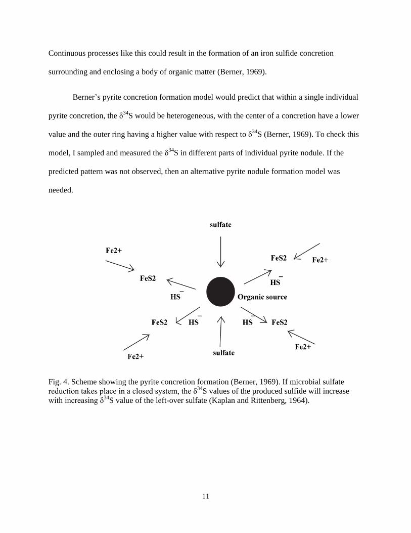
11
Continuous processes like this could result in the formation of an iron sulfide concretion
surrounding and enclosing a body of organic matter (Berner, 1969).
Berner’s pyrite concretion formation model would predict that within a single individual
pyrite concretion, the δ34
S would be heterogeneous, with the center of a concretion have a lower
value and the outer ring having a higher value with respect to δ34
S (Berner, 1969). To check this
model, I sampled and measured the δ34
S in different parts of individual pyrite nodule. If the
predicted pattern was not observed, then an alternative pyrite nodule formation model was
needed.
Fig. 4. Scheme showing the pyrite concretion formation (Berner, 1969). If microbial sulfate
reduction takes place in a closed system, the δ34
S values of the produced sulfide will increase
with increasing δ34
S value of the left-over sulfate (Kaplan and Rittenberg, 1964).
12
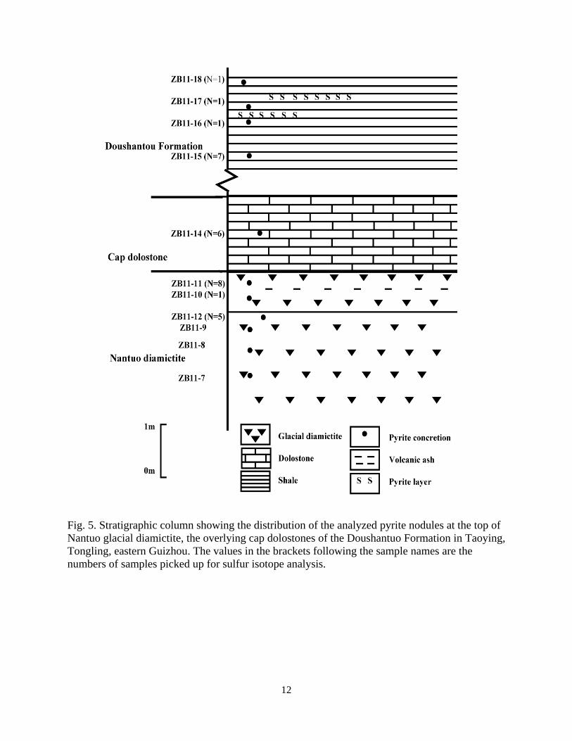
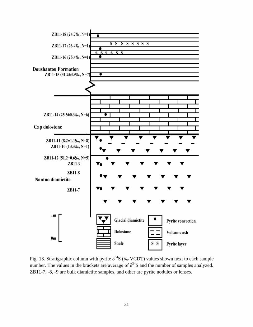
Fig. 5. Stratigraphic column showing the distribution of the analyzed pyrite nodules at the top of
Nantuo glacial diamictite, the overlying cap dolostones of the Doushantuo Formation in Taoying,
Tongling, eastern Guizhou. The values in the brackets following the sample names are the
numbers of samples picked up for sulfur isotope analysis.
13
METHODS
Petrography
Samples ZB11-7, ZB11-8, and ZB11-9 were bulk diamictite samples. Samples ZB11-10,
ZB11-11, and ZB11-12 were pyrite nodules in the diamictite. ZB11-13 and Zb11-14 were pyrite
nodules in the cap dolostones (Fig. 3C and D). ZB11-15a, b, c, d, e were five individual pyrite
nodules in the shale at ~19 m above the top of the diamictite. Going further upward in
stratigraphic level (22 m above the cap dolostones), pyrite nodules, ZB11-16, ZB11-17, and
Zb11-18 were collected. The distribution of pyrite nodules collected at the top of Nantuo glacial
diamictite, cap dolostones, and overlying shale are shown in Fig. 5. Thin sections were made
from the bulk diamictite samples (ZB11-7, ZB11-8, ZB11-9), and pyrite nodules (ZB11-11 and
ZB11-12), and a polished slab was made for the nodule ZB11-14. Sample ZB11-10 was too
small to make a thin section. Photomicrographs were taken for thin sections and polished slabs
using reflected or transmitted light microscopes and a digital camera.
The X-ray Diffraction (XRD) analysis was conducted on eight sample powders, including
both bulk diamictite and pyrite nodules, using a Bruker/Siemens D5000 X-ray
Diffractometer. The machine was set at 40kV and 30 mA. The samples were run from 2o to 70
o
at a rate of 0.02o every 2 seconds. The diffraction pattern data were analyzed using Jade 9.3.3
software to confirm mineral identification from Material Data Incorporated. The quantitative
analysis was obtained from XRDPHil program.
Stable Isotopic Analysis
Field and initial petrographic observations revealed that fine-grained pyrite crystals or
aggregates were common at the top of the diamictite. To examine the spatial heterogeneity of
pyrite sulfur isotope compositions at different stratigraphic levels, among different nodules, or
14
within the same nodule, I sampled both the bulk diamictite and pyrite nodules. For picking a
pyrite sample for δ34
S analysis, only ~ 30 μg of pure pyrite was needed. To sample pyrite
nodules, I broke a piece of a nodule into many smaller pieces and then ground each into a fine
powder. Approximately 30 μg were used per piece. Depending on the size of the overall nodule,
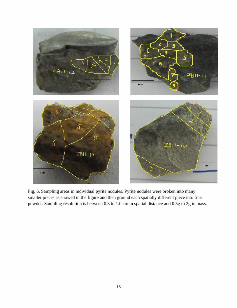
samples were taken from between 0.3 to 1.0 cm. Sample ZB11-10, -12, -14, and -15a had
spatially different sampling of δ34
S for the same nodule (Fig. 6). All other nodules, lenses, or
beds only had one δ34
S measurement for each. The stratigraphic positions for these samples are
shown in Fig. 5. In total, I obtained δ34
S data for 29 samples from 12 pyrite nodules or lenses by
this centimeter sampling resolution.
To further examine the potential spatial heterogeneity of the pyrite δ34
S values within and
between nodules, I sampled pyrite nodules ZB11-11, ZB11-12, ZB11-14, and ZB11-15a using
smaller distances between samples, at approximately 2 mm apart, from polished pieces of the
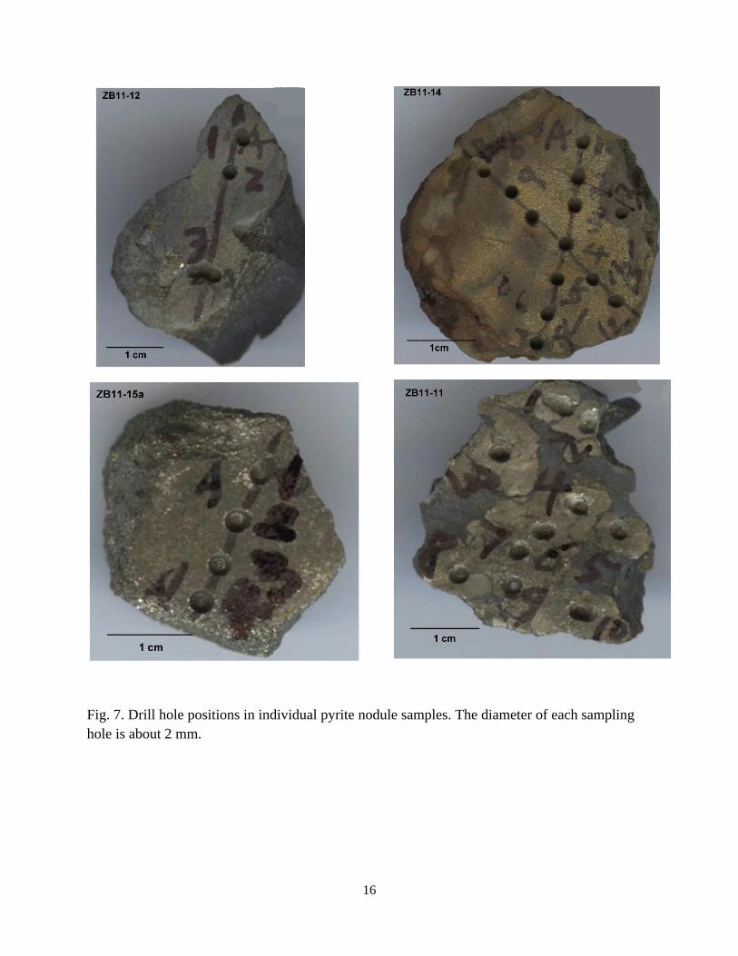
pyrite nodules (Fig.7). Powder was drilled out of each sample, yielding 30 additional data points.
For δ34
S measurements of bulk diamictite samples, samples ZB11-7, ZB11-8, and ZB11-
9 were taken before visual inspection of their corresponding thin-sections. A 10% wt FeS2 was
assumed, and about 300 μg bulk materials were weighed out. Three bulk diamictite samples did
not yield enough signal for the data to be reliable. This was due to an initial overestimation of the
pyrite content in these bulk diamictites. Based on SO2 peak intensities, I determined that an
average of 2.1 mg of diamictite sample was needed for a good sulfur isotope measurement of the
sulfides in the sample. The information will be useful for future sampling.
15
Fig. 6. Sampling areas in individual pyrite nodules. Pyrite nodules were broken into many
smaller pieces as showed in the figure and then ground each spatially different piece into fine
powder. Sampling resolution is between 0.3 to 1.0 cm in spatial distance and 0.5g to 2g in mass.
16
Fig. 7. Drill hole positions in individual pyrite nodule samples. The diameter of each sampling
hole is about 2 mm.
17
Petrographic preparation, microscopic observation, sample milling and weighing were
carried out at Louisiana State University (LSU) and the millimeter sampling and all δ34
S
measurement of sulfide was conducted at University of Maryland, where FeS2 was converted to
SO2 using an Elemental Analyzer (EA) at 1050 °C, and analyzed on a Micromass Isoprime in a
continuous-flow mode. The standard deviation associated with δ34
S measurement was ± 0.2‰.
All δ34
S values are reported with respect to VCDT.
18
RESULTS
Petrographic Observation
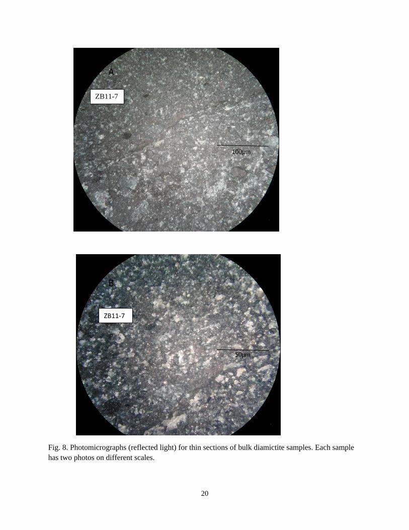
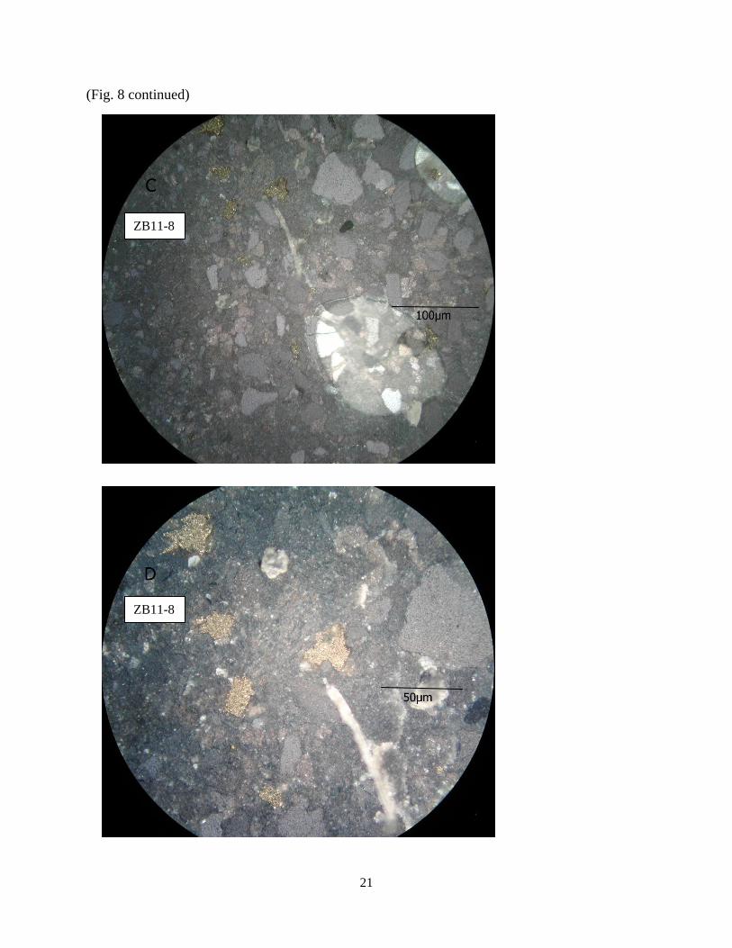
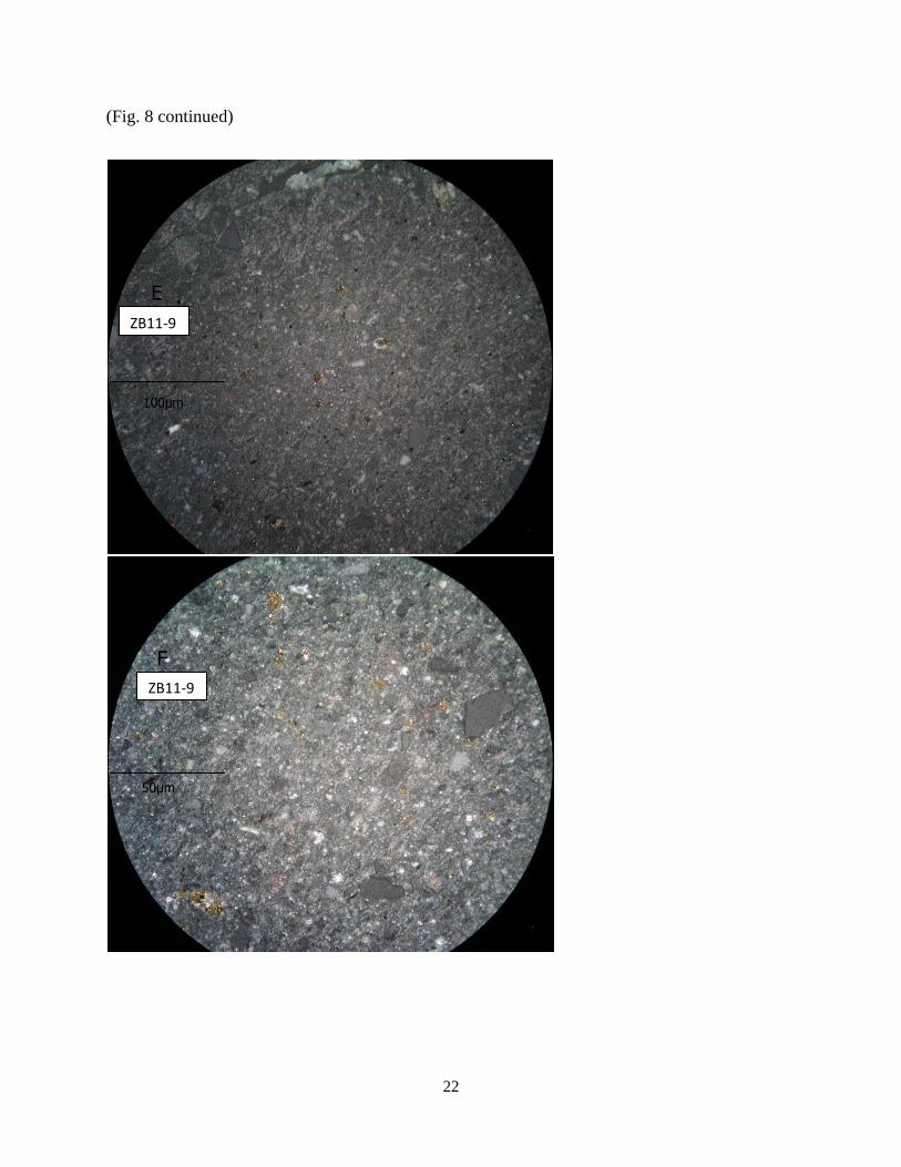
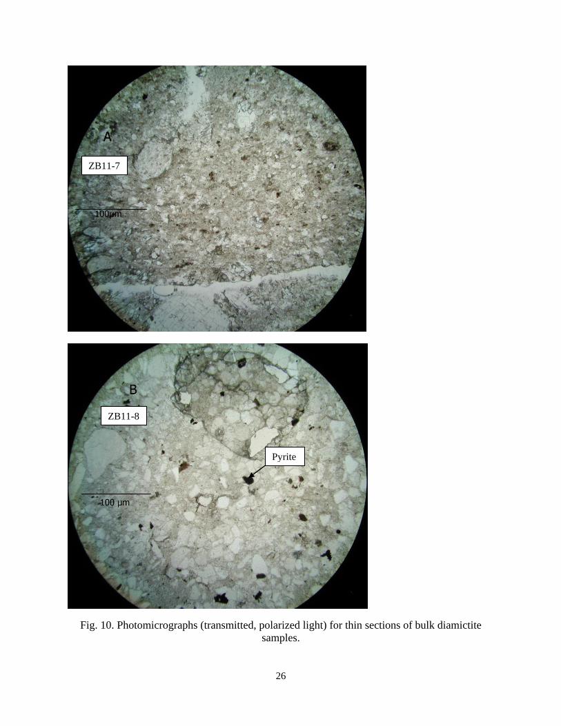
Microscopic observation revealed that disseminated pyrite grains were ubiquitous in the
Nantuo diamictite. Within pyrite nodules, the individual pyrite grains occured as aggregates.
Although pyrite grain distribution density varied between nodules, the distribution was
homogeneous at a millimeter scale. However, uneven distribution of pyrite and surrounding
silicate matrix was observed on the scale of tens of micrometers (not considering some of the
vein fillings). There was a general increase in pyrite abundance towards the top of the diamictite
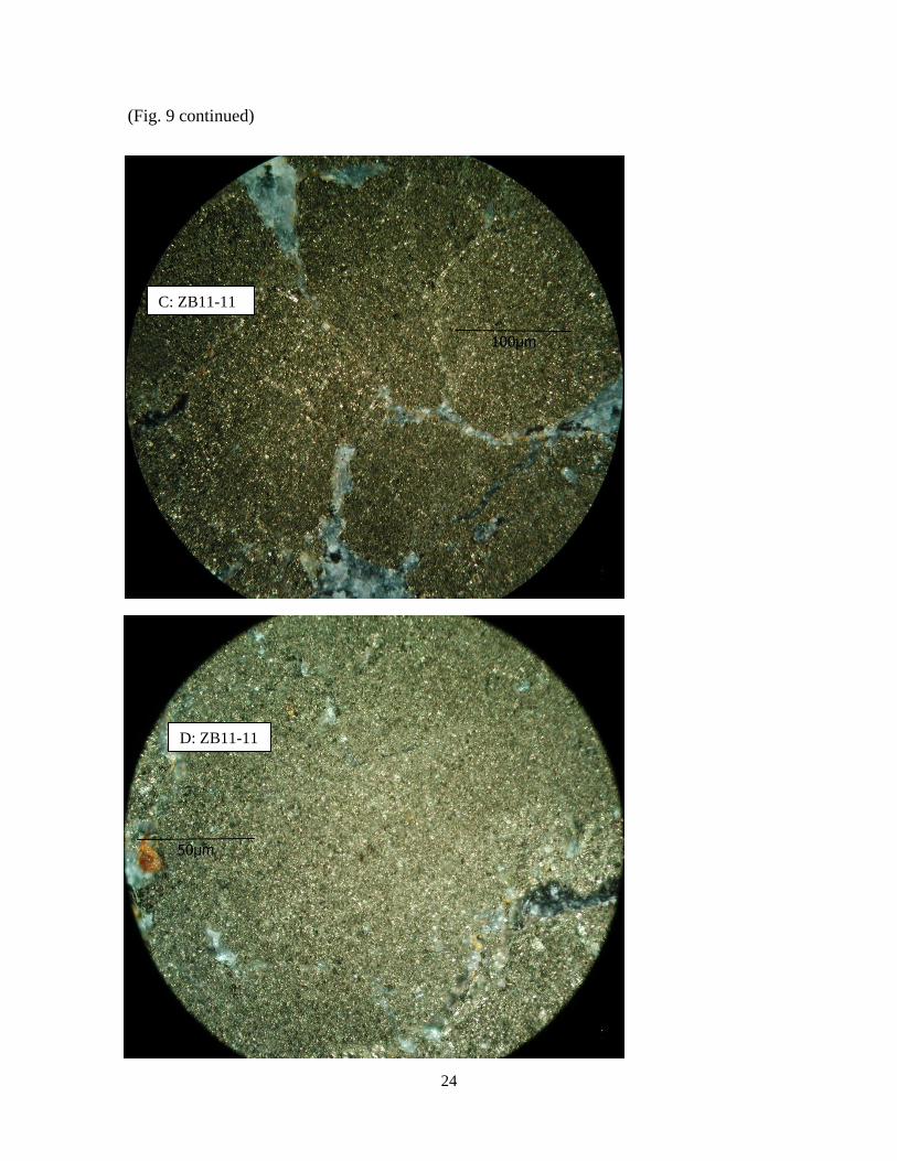
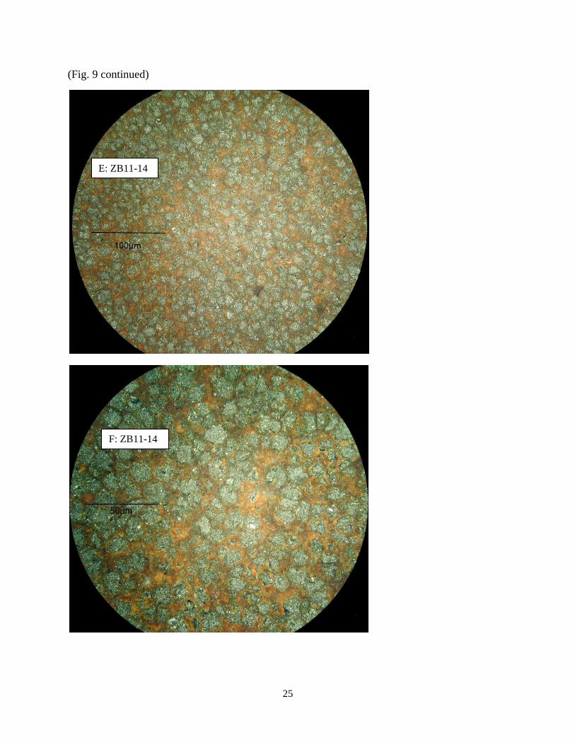
from sample ZB11-7 to sample ZB11-11 (Figs. 8, 9, and 10).
XRD Analysis
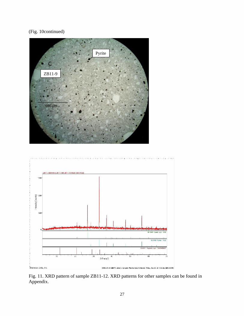
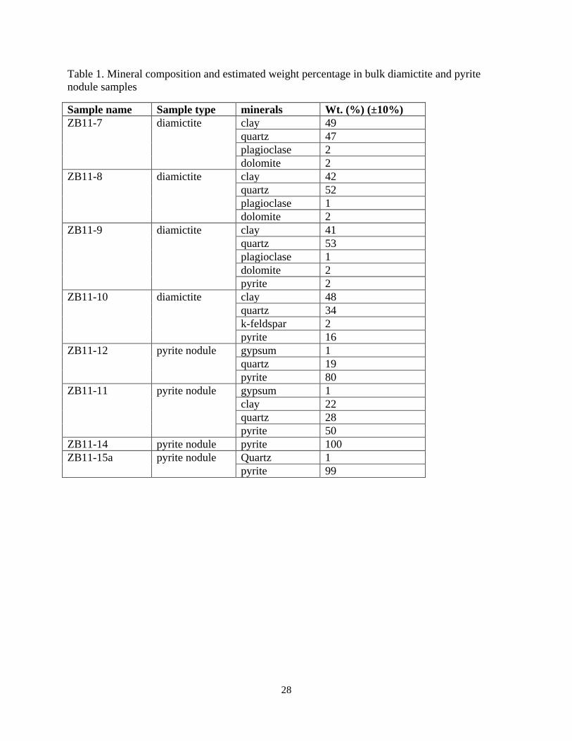
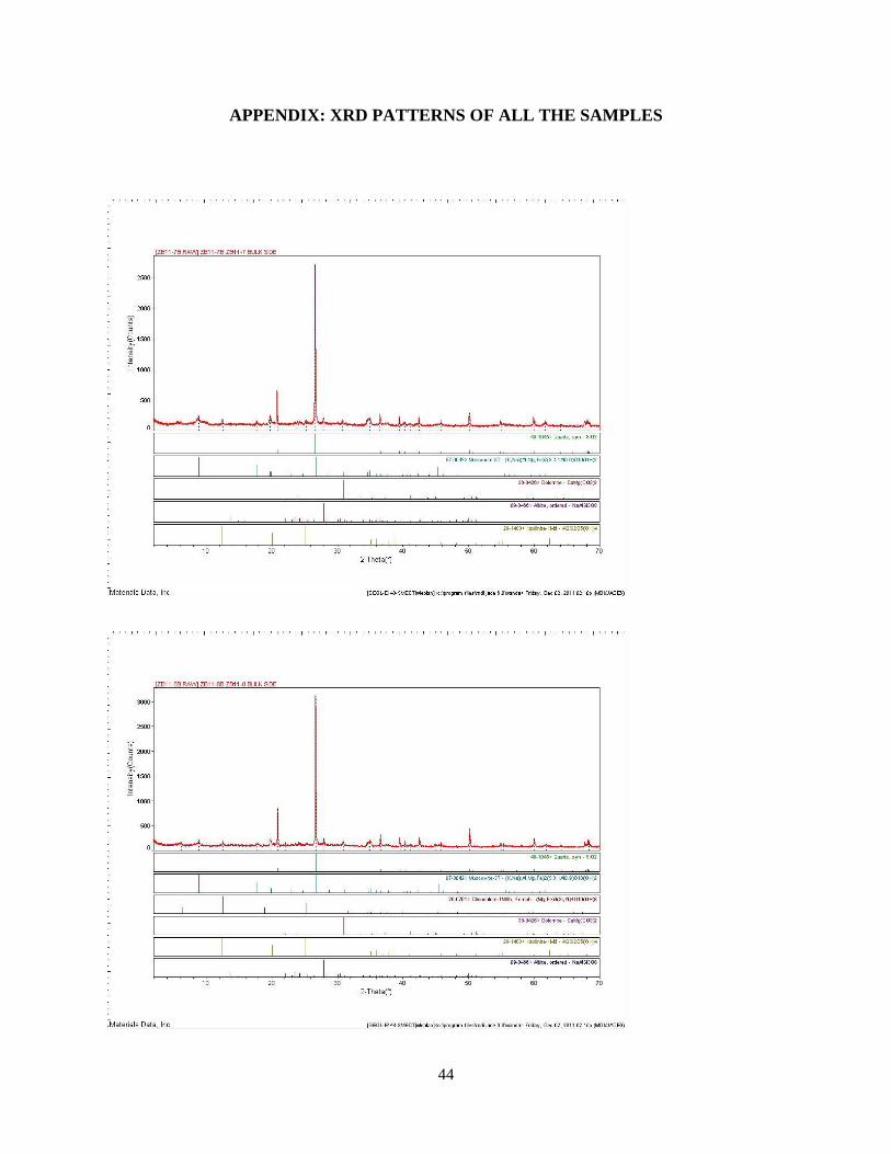
XRD analysis of four bulk diamictite and four pyrite nodule samples confirmed that the
iron sulfide mineral in the nodules was pyrite (Fig. 11; Table 1). Other than pyrite, the significant
minerals in the nodules were quartz and clay. In bulk diamictite, pyrite accounted for less than
2% of the weight, but in nodules the percentageof pyrite was at least more than 50%.
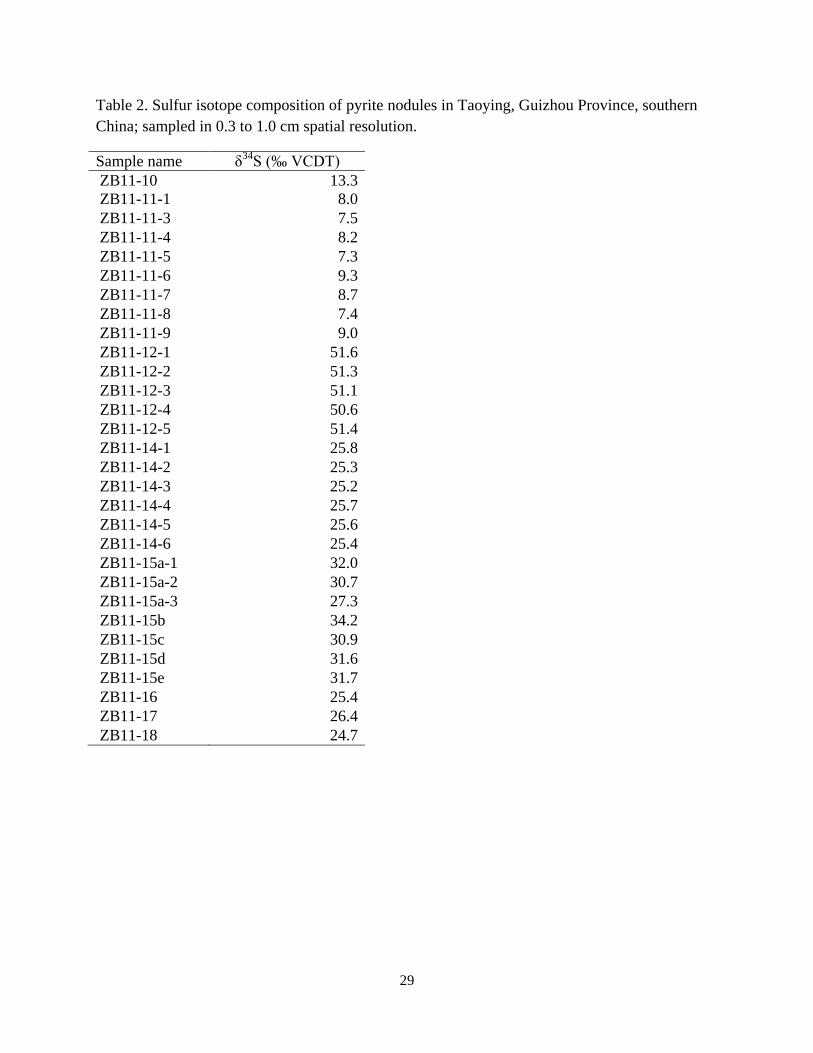
Stable Sulfur Isotope Composition
The δ34
S values for pyrite nodules sampled at a centimeter scale are shown in Fig. 12 and
Table 2. The δ34
S values varied from one pyrite nodule to another (from 7.3‰ to 51.6‰).
However, δ34
S values were homogeneous within the same pyrite nodule at the 0.3-1.0 cm
sampling resolution (Table 2). For the three pyrite nodule samples in the diamictite, δ34
S values
ranged from 7.3‰ to 9.3‰ (average 8.2‰, N=8) for ZB11-11, was 13.3‰ (only one
measurement) for ZB11-10, and ranged from 50.6‰ to 51.6‰ (average 51.2‰, N=5) for sample
ZB11-12. The δ34
S valued for pyrite in the cap dolostones, ZB11-14, ranged from 25.2‰ to
25.8‰, with an average value of 25.5‰ (N=5). The δ34
S values for the 5 individual pyrite
19
lenses collected in the shale overlying the cap dolostones, ZB11-15a, b, c, d and e, ranged from
27.2‰ to 31.7‰. Note that three samples were collected from pyrite lens ZB11-15a with a δ34
S
value range of 27.3‰ to 32.0‰, which was a much larger range than the values for the nodules
from the top of the diamictite and from within the cap dolostones. For the three pyrite lenses in
the overlying shale, ZB11-16, ZB11-17, and ZB11-18, the δ34
S values were 25.4‰, 26.4‰ and
24.7‰, respectively (Table 2).
The results of pyrite sulfur isotope analysis from the 2 mm sampling scale are shown in
Fig. 14. The δ34
S values were more heterogeneous than those obtained with the larger sampling
scale. Sample ZB11-12 values were more or less the same (~51.2‰) at both sampling
resolutions. The δ34
S values from sample ZB11-14 ranged from 24 to 31‰ on the millimeter
scale, which was a larger range than at the larger sampling scale (25.2‰ to 25.8‰). The δ34
S
values for sample ZB11-15a was ~23‰ at the fine sampling scale, which was different than at
the wider interval (~30‰). Sample ZB11-11 was an interesting case. There appeared to be
multiple aggregates in the same individual nodule that had very different δ34
S values, 14‰,
57.8‰, and 60.5‰, as compared to the centimeter-resolution value of ~8‰.
20
Fig. 8. Photomicrographs (reflected light) for thin sections of bulk diamictite samples. Each sample
has two photos on different scales.
ZB11-7
ZB11-7
21
(Fig. 8 continued)
ZB11-8
ZB11-8
22
(Fig. 8 continued)
ZB11-9
ZB11-9
23
Fig. 9. Photomicrographs (reflected light) for thin sections of pyrite nodules. Each sample has
two photos with different magnification.
A: ZB11-12
B: ZB11-12
24
(Fig. 9 continued)
C: ZB11-11
D: ZB11-11
25
(Fig. 9 continued)
F: ZB11-14
E: ZB11-14
26
Fig. 10. Photomicrographs (transmitted, polarized light) for thin sections of bulk diamictite
samples.
ZB11-7
ZB11-8
Pyrite
27
(Fig. 10continued)
Fig. 11. XRD pattern of sample ZB11-12. XRD patterns for other samples can be found in
Appendix.
ZB11-9
Pyrite
28
Table 1. Mineral composition and estimated weight percentage in bulk diamictite and pyrite
nodule samples
Sample name Sample type minerals Wt. (%) (±10%)
ZB11-7 diamictite clay 49
quartz 47
plagioclase 2
dolomite 2
ZB11-8 diamictite clay 42
quartz 52
plagioclase 1
dolomite 2
ZB11-9 diamictite clay 41
quartz 53
plagioclase 1
dolomite 2
pyrite 2
ZB11-10 diamictite clay 48
quartz 34
k-feldspar 2
pyrite 16
ZB11-12 pyrite nodule gypsum 1
quartz 19
pyrite 80
ZB11-11 pyrite nodule gypsum 1
clay 22
quartz 28
pyrite 50
ZB11-14 pyrite nodule pyrite 100
ZB11-15a pyrite nodule Quartz 1
pyrite 99
29
Table 2. Sulfur isotope composition of pyrite nodules in Taoying, Guizhou Province, southern
China; sampled in 0.3 to 1.0 cm spatial resolution.
Sample name Sample name δ34
S (‰ VCDT)
ZB11-10 13.3
ZB11-11-1 8.0
ZB11-11-3 7.5
ZB11-11-4 8.2
ZB11-11-5 7.3
ZB11-11-6 9.3
ZB11-11-7 8.7
ZB11-11-8 7.4
ZB11-11-9 9.0
ZB11-12-1 51.6
ZB11-12-2 51.3
ZB11-12-3 51.1
ZB11-12-4 50.6
ZB11-12-5 51.4
ZB11-14-1 25.8
ZB11-14-2 25.3
ZB11-14-3 25.2
ZB11-14-4 25.7
ZB11-14-5 25.6
ZB11-14-6 25.4
ZB11-15a-1 32.0
ZB11-15a-2 30.7
ZB11-15a-3 27.3
ZB11-15b 34.2
ZB11-15c 30.9
ZB11-15d 31.6
ZB11-15e 31.7
ZB11-16 25.4
ZB11-17 26.4
ZB11-18 24.7
30
Fig. 12. Results of the sulfur isotope analysis at a centimeter resolution. Each numbered region
is shown with their respective δ34
S (‰ VCDT).
31
Fig. 13. Stratigraphic column with pyrite δ34
S (‰ VCDT) values shown next to each sample
number. The values in the brackets are average of δ34
S and the number of samples analyzed.
ZB11-7, -8, -9 are bulk diamictite samples, and other are pyrite nodules or lenses.
32
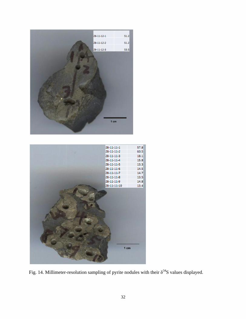
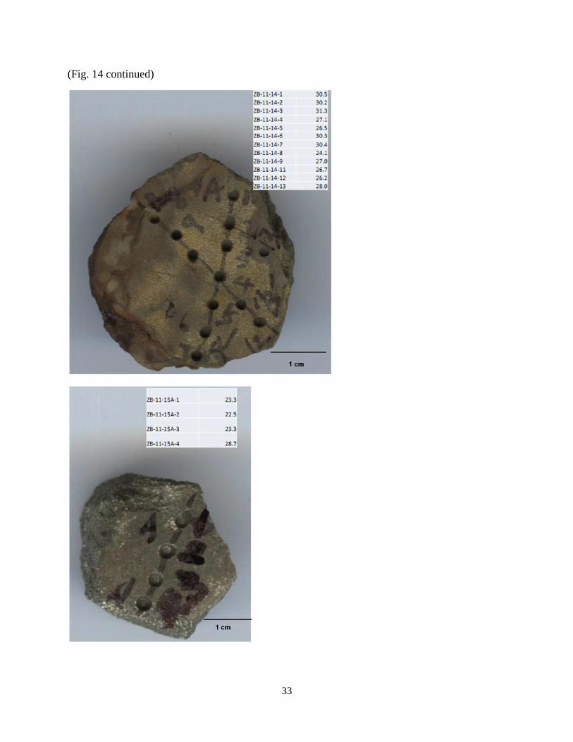
Fig. 14. Millimeter-resolution sampling of pyrite nodules with their δ34
S values displayed.
33
(Fig. 14 continued)
34
DISCUSSION
Pyrite Formation
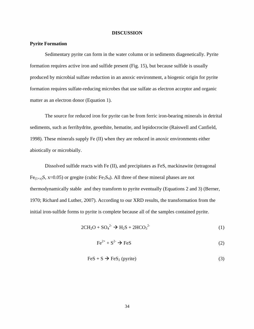
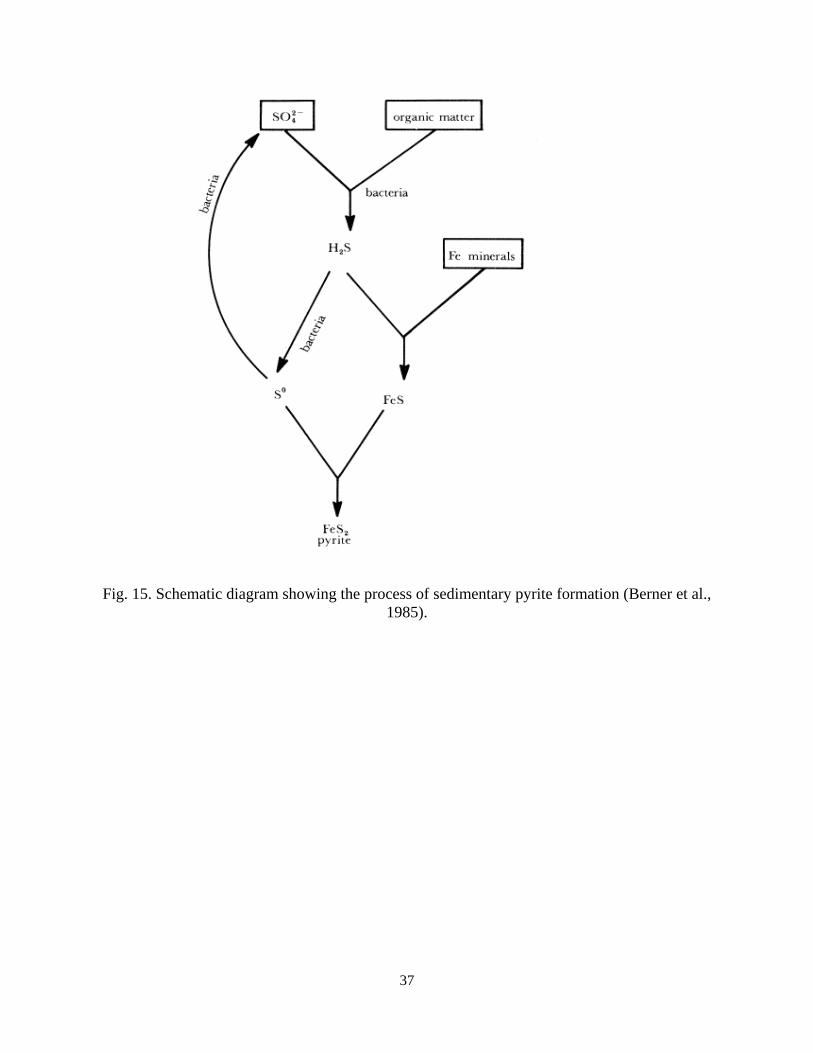
Sedimentary pyrite can form in the water column or in sediments diagenetically. Pyrite
formation requires active iron and sulfide present (Fig. 15), but because sulfide is usually
produced by microbial sulfate reduction in an anoxic environment, a biogenic origin for pyrite
formation requires sulfate-reducing microbes that use sulfate as electron acceptor and organic
matter as an electron donor (Equation 1).
The source for reduced iron for pyrite can be from ferric iron-bearing minerals in detrital
sediments, such as ferrihydrite, geoethite, hematite, and lepidocrocite (Raiswell and Canfield,
1998). These minerals supply Fe (II) when they are reduced in anoxic environments either
abiotically or microbially.
Dissolved sulfide reacts with Fe (II), and precipitates as FeS, mackinawite (tetragonal
Fe(1+x)S, x≈0.05) or gregite (cubic Fe3S4). All three of these mineral phases are not
thermodynamically stable and they transform to pyrite eventually (Equations 2 and 3) (Berner,
1970; Richard and Luther, 2007). According to our XRD results, the transformation from the
initial iron-sulfide forms to pyrite is complete because all of the samples contained pyrite.
2CH2O + SO42-
H2S + 2HCO32-
(1)
Fe2+
+ S2-
FeS (2)
FeS + S FeS2 (pyrite) (3)
35
The Pyrite Formation Scenario
The difference between the two scenarios that I proposed is the timing of pyrite
formation, which is critical to understanding the ending of Marinoan global glaciations and the
recovery of the biosphere at that time. The scenario that pyrite nodules were diagenetically
formed after the precipitation of the cap dolostones predicts a gradual increase in pyrite δ34
S
values with increasing depth into the diamictite. The scenario that pyrite formed continuously at
the waning stage of diamictite deposition predicts a highly variable pyrite δ34
S value from
horizon to horizon with no correlation with depth. Therefore, the vertical δ34
S pattern for pyrite
nodules collected from the diamictite can test which scenario is more likely in our geological
settings. At the centimeter sampling resolution, pyrite δ34
S values seem to increase with depth
into the diamictite - the δ34
S of ZB11-11 averages 8.2‰, the δ34
S of ZB11-10 is 13.3‰, and the
δ34
S of ZB11-12 averages 51.2‰ (Fig. 5). However, this trend is not supported in the millimeter
sampling resolution - the δ34
S of ZB11-11ranges from 13.3‰ to 18.2‰ with two outliers
(57.8‰ and 60.5‰) and the δ34
S of ZB11-12 ranges from 51.2‰ to 53.5‰ (Fig. 14). Thus, I
conclude from these different datasets that pyrite δ34
S values are highly variable from horizon to
horizon with no correlation with depth, implying that seawater sulfate concentration was already
high enough to result in pyrite formation in sediments at the waning stage of diamictite
deposition and before the precipitation of the cap dolostones, either via direct precipitation of
pyrite from a euxinic water column or via in-sediment sulfate reduction.
Formation Model for Pyrite Nodules in the Nantuo Diamictite
Pyrite nodules have been reported in shales from lake and marine sequences (Dell, 1975;
Mathias, 1928; Sass et al., 1965; Jowett et al., 1990). Raiswell (1982) sampled two pyritiferous
carbonate nodules (30 cm and 70 cm in diameter) for sulfur isotope analysis. He took 5 or 6
36
samples from the nodule center to the edge from a slice of the nodules, and found that the δ34
S
values for pyrite increased from the center to the edge for both nodules sampled. These data are
consistent with a restricted sulfate reservoir being progressively depleted by microbial sulfate
reduction.
Consequently, the only pyrite nodule formation model that has been proposed, by Berner
(1969), predicts that δ34
S values from pyrite will increase from center to edge in a pyrite
concretion due to a reservoir effect. This model, however, cannot explain the spatially
homogeneous δ34
S values in the pyrite nodules in my study, at either one of the sampling
intervals. Although millimeter sampling resolution revealed more heterogeneity, the δ34
S values
of pyrite were more or less within ±2‰ of each other in one single aggregate. It is, therefore,
apparent that Berner’s pyrite concretion model does not apply to the pyrite nodules at the top of
the Nantuo diamictite in South China.
It is possible that the pyrite nodules at the top of the Nantuo diamictite were initially
deposited as layers of disseminated pyrite grains or framboidal clusters. Due to differences in
early cementation rates between pyrite layers and surrounding fine silicate muds, sedimentary
compaction can turn the layered pyrite into nodular form of semi-linked and later totally
independent pyrite nodules. The nodule formation model by differential cementation and
compaction of the sediments has been applied to explain carbonate concretion formation (Dong
et al., 2008). Such a pyrite nodule formation model can explain the δ34
S homogeneity within a
nodule.
37
Fig. 15. Schematic diagram showing the process of sedimentary pyrite formation (Berner et al.,
1985).
38
FUTURE WORK NEEDED
Sampling different nodules from the same horizon: The first reason that it needs to be
done is to determine what the pyrite δ34
S patterns in diamictite at different stratigraphic levels
are. Another reason is to identify if pyrite nodules from the same horizon formed through the
same mechanism. More data on the pyrite-rich lenses and bedding in the middle Doushantuo
Formation are also needed, as these pyrite beddings offer good reference pyrite occurrence for
comparison. If indeed the nodules at the top of the diamictite had the same initial occurrence as
those in the middle Doushantuo shale, we expect to see a similar δ34
S trend from layer to layer
and from one nodule to another.
Scanning electron microscope (SEM) analysis of polished slabs: SEM work will provide
further petrographic information on the pyrite formation. High resolution (about 1 μm) SEM
analysis can show the microstructure of pyrite, such as the size distribution of framboidal pyrite,
which can provide some evidence to support different formation environments (i.e. water column
or within sediment) (Butler and Rickard, 2000; Schieber, 2002; Wilkin et al., 1997; Wilkin et al.,
1996).
Study similar nodules in other facies of the Neoproterozoic South China: This is the first
study of these enigmatic pyrite nodules in the Nantuo diamictite. We know of dozens of
localities in the South China Block where similar pyrite nodules occur. If we are to establish a
general formation model for these pyrite nodules, then we need to examine more sites in
different facies.
39
CONCLUSIONS
There are two scenarios to explain when and how the pyrite nodules in the Nantuo
diamictite formed. One scenario is that these pyrite nodules formed diagenetically after the cap
dolostone deposition when the seawater sulfate concentration became high enough to sufficiently
diffuse into the diamictite. Another scenario is that the pyrite formed at the waning stage of
diamictite deposition before the cap dolostone deposition when seawater sulfate concentration
was sufficient to support microbial sulfate reduction. The difference between these two scenarios
is in their predictions about the relationship between the δ34
S of pyrite and depth. Our results
show that pyrite δ34
S values have no correlation with depth in the diamictite. Therefore, I
conclude that the pyrite formed before the deposition of cap dolostones, and at that time the
sulfate concentration was high enough for microbial sulfate reduction. Pyrite could form in
sediments via direct precipitation in a euxinic water column or via in-sediment sulfate reduction.
In any case, sulfate concentrations had to be sufficiently high, at least intermittently, in the
oceans before the deposition of the cap dolostones. This conclusion has important implications in
our understanding of the post-glacial world 635 million years ago. The globally distributed cap
dolostones on top of the Marinoan diamictite has been concluded to be deposited immediately
and continuously following the diamictite (Shen et al., 2005; Shields, 2005). It becomes clear
from this study that there was a time window when sulfur cycling is especially active before the
cap dolostones deposition.
Berner’s pyrite nodule formation model does not apply to the pyrite nodule formation
largely because of the observed pyrite δ34
S homogeneity within a pyrite nodule. I propose that
differential cementation and compaction of the pyrite-bearing sediments formed the rounded

40
shapes of the pyrite nodules and can account for the δ34
S homogeneity within an individual
nodule.
41
REFERENCES
Berner, R.A., 1969, Migration of iron and sulfur within anaerobic sediments during early
diagenesis: American Journal of Science, v. 267, p. 19-42.
Berner, R.A., 1970, Sedimentary pyrite formation: American Journal of Science, v. 268, p. 1-23.
Berner, R. A., Leeuw, J. W., Spiro, B., Murchison, D. G. and Eglinton, G., 1985, Sulfate
reduction, organic matter decomposition and pyrite formation (and discussion),
Philosophical transactions of the royal socirty A, vol., 315, p. 25-38.
Boesen, C., and Postma, D., 1988, Pyrite formation in anoxic environments of the Baltic:
American Journal of Science, v. 288, p. 575-603.
Brunner, B., and Bernasconi, S.M., 2005, A revised isotope fractionation model for dissimilatory
sulfate reduction in sulfate reducing bacteria: Geochimica et Cosmochimica Acta, v. 69,
p. 4759-4771.
Butler, I.B., and Rickard, D., 2000, Framboidal pyrite formation via the oxidation of iron (II)
monosulfide by hydrogen sulphide: Geochimica et Cosmochimica Acta, v. 64, p. 2665-
2672.
Canfield, D.E., Farquhar, J., and Zerkle, A.L., 2010, High isotope fractionations during sulfate
reduction in a low-sulfate euxinic ocean analog: Geology, v. 38, p. 415-418.
Canfield , D.E., Olesen, C.A., and Cox, R.P., 2006, Temperature and its control of isotope
fractionation by a sulfate-reducing bacterium: Geochimica et Cosmochimica Acta, v. 70,
p. 548-561
Dell, C.I., 1975, Pyrite Nodules in Sediment from South Bay, Lake Huron: Canadian Journal of
Earth Sciences, v. 12, p. 1077-1083.
Detmers, J., Bruchert, V., Habicht, K.S., and Kuever, J., 2001, Diversity of sulfur isotope
fractionations by sulfate-reducing prokaryotes: Applied and Environmental
Microbiology, v. 67, p. 888-894.
Dong, J., Zhang, S., Jiang, G., Zhao, Q., Li, H., Shi, X., and Liu, J., 2008, Early diagenetic
growth of carbonate nodules in the upper Doushantuo Formation in South China and their
significance for the assessment of hydrocarbon source rock: Science in China Series D:
Earth Sciences, v. 51, p. 1330-1339.
Habicht, K.S., Salling, L., Thamdrup, B., and Canfield, D.E., 2005, Effect of low sulfate
concentrations on lactate oxidation and isotope fractionation during sulfate reduction by
Archaeoglobus fulgidus Strain Z: Applied and Environmental Microbilogy, v. 71, p.
3770-3777
42
Jiang, G., Shi, X., Zhang, S., Wang, Y., and Xiao, S., 2011, Stratigraphy and paleogeography of
the Ediacaran Doushantuo Formation (ca. 635-551 Ma) in South China: Gondwana
Reaserch, v.9, p.831-849
Jowett, E. C., Roth, T., Rydzewski, A., and Oszczepalski, S., 1990, “Background” d34
S values of
Kupferschiefer sulphides in Poland: pyrite-marcasite nodels, Mineralium Deposita, v. 26,
p. 89-98
Kaplan, I.R., and Rittenberg, S.C., 1964, Microbiological fractionation of sulphur isotopes:
Journal of General Microbiology, v. 34, p. 195-212.
Mathias, H.E., 1928, Syngenetic Origin of Pyrite Nodules in the Pennsylvanian Shales of North-
Central Missouri: The Journal of Geology, v. 36, p. 440-450.
Peng, Y., Bao, H., Zhou, C., and Yuan, X., 2011, 17
O-depleted barite from two Marinoan cap
dolostone sections, South China: Earth and Planetary Science Leeters, v. 305, p. 21-31
Raiswell, R., 1982, Pyrite texture, isotopic composition and the availability of iron: American
Journal of Science, v. 282, p. 1244-1263.
Raiswell, R., and Canfield, D.E., 1998, Sources of iron for pyrite formation in marine sediments:
American Journal of Science, v. 298, p. 219-245.
Rees, C.E., 1973, A steady-state model for sulphur isotope fractionation in bacterial reduction
processes: Geochimica et Cosmochimica Acta, v. 37, p. 1141-1162.
Rickard, D., and Luther III, G.W., 1997, Kinetics of pyrite formation by the H2S oxidation of
iron (II) monosulfide in aqueous solutions between 25 and 125°C: The mechanism:
Geochimica et Cosmochimica Acta, v. 61, p. 135-147.
Rickard, D., and Luther III, G.W., 2007, Chemistry of iron sulfides, Chemistry Review, Vol.,
107, p. 514-562
Rickard, D.T., 1975, Kinetics and mechanism of pyrite formation at low temperatures: American
Journal of Science, v. 275, p. 636-652.
Sass, E., Nathan, Y., and Nissenbaum, A., 1965, mineralogy of certain pyrite nodules from israel
and their alteration products: Mineralogical Magazine and Journal of the Mineralogical
Society, v. 35, p. 84-87.
Schieber, J., 2002, The role of an organic slime matrix in the formation of pyritized burrow trails
and pyrite nodules: Palaios, v. 17, p. 104-109.
Shen, Y., Zhang, T., Chu, X., 2005, C-isotope stratification in a Neoproterozoic postglacial
ocean, Precambrian Research, v. 137, p. 243-251
43
Shields, G. A., 2005, Neoproterozoic cap carbonates: a critical appraisal of existing models and
the plumeworld hypothesis, Terra Nova, v. 17, p. 299-310
Sim, M.S., Bosak, T., and Ono, S., 2011, Large sulfur isotope fractionation does not require
disproportionation: Science, v. 333, p. 74-77.
Wilkin, R.T., Arthur, M.A., and Dean, W.E., 1997, History of water-column anoxia in the Black
Sea indicated by pyrite framboid size distributions: Earth and Planetary Science Letters,
v. 148, p. 517-525.
Wilkin, R.T., and Barnes, H.L., 1997, Pyrite formation in an anoxic estuarine basin: American
Jounal of Science, v. 297, p. 620-650.
Wilkin, R.T., Barnes, H.L., and Brantley, S.L., 1996, The size distribution of framboidal pyrite
in modern sediments: An indicator of redox conditions: Geochimica et Cosmochimica
Acta, v. 60, p. 3897-3912.
Wortmann, U.G., Bernasconi, S.M., and Bottcher, M.E., 2001, Hypersulfidic deep biosphere
indicates extreme sulfur isotope fractionation during single-step microbial sulfate
reduction: Geology, v. 29, p. 647-650.
Zhang, Q.R., Li, X.H., Feng, L.J., Huang, J., and Song, B., 2008, A new age constraint on the
onset of the neoproterozoic glaciations in the Yangtze platform, South China: Journal of
Geology, v. 116, p. 423-429.
Zhang, S., Jiang, G., Zhang, J., Song, B., Kennedy, M.J., and Christie-Blick, N., 2005, U-Pb
sensitive high-resolution ion microprobe ages from the Doushantuo Formation in south
China: Constraints on late Neoproterozoic glaciations: Geology, v. 33, p. 473-476.
Zhou, C., Bao, H., Peng Y., Yuan, X., 2010, Timing the deposition of 17
O-depleted barite at the
aftermath of Nantuo glacial meltdown in South China: Geology, v. 38, p. 903-906
Zhou, C., Xie, G., Mcfadden, K., Xiao, S., and Yuan, X., 2007, The diversification and
extinction of Doushantuo-Pertatataka acritarchs in South China: causes and
biostratigraphic significance, Geological Journal, v.42, p. 229-262.
44
APPENDIX: XRD PATTERNS OF ALL THE SAMPLES
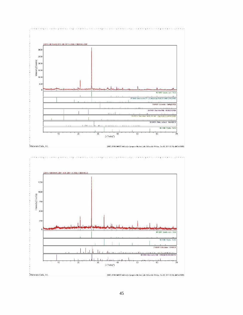
45
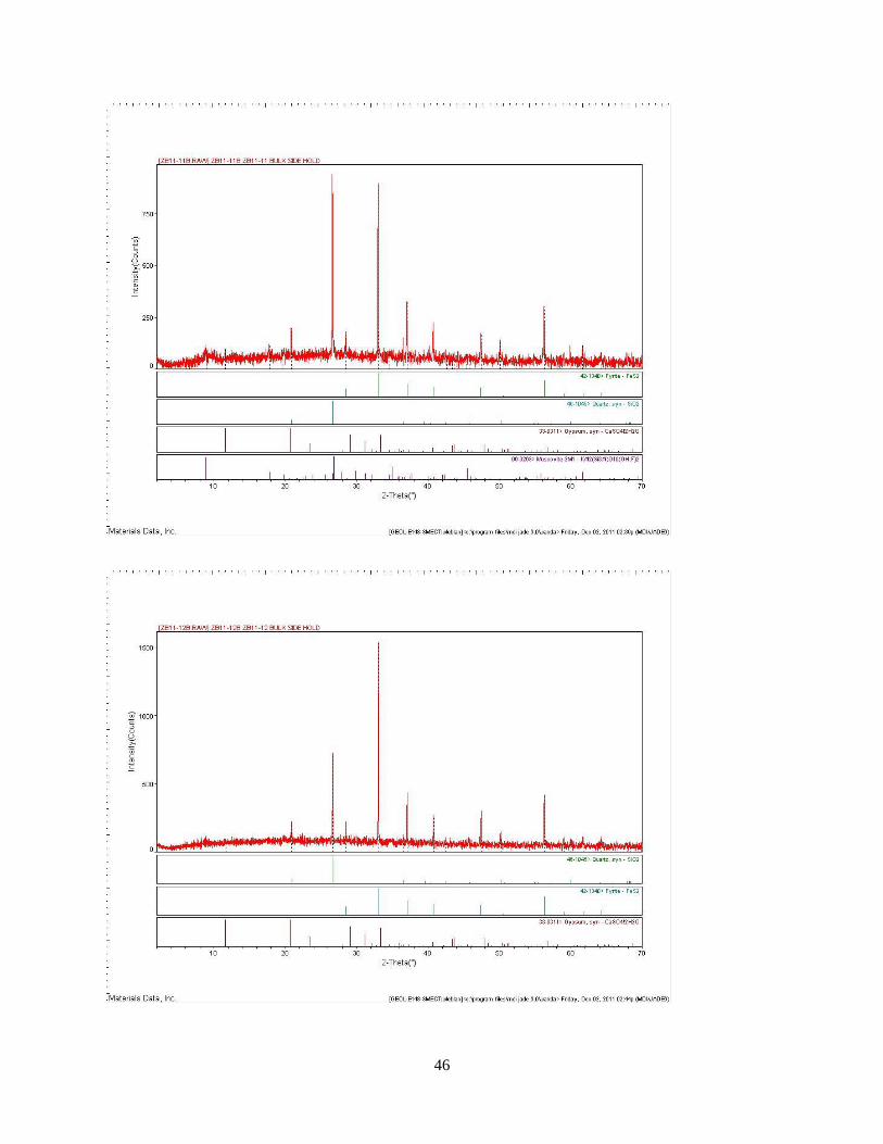
46
47
48
VITA
Changjie Liu was born in a small village in Jiangsu Province in eastern China in 1987.
He entered the University of Science and Technology of China in 2005, and received his
bachelor’s degree in environmental science in 2009. He came to the United States and started his
studies in the Department of Geology and Geophysics at Louisiana State University in 2009.
Under the guidance of Dr. Huiming Bao, he has been working on several research topics that
utilize stable isotopes as a tool to extract paleocmilate information.