Embed Size (px)
DESCRIPTION
Since the discovery of autophagy, the membrane origin of the autophagosomes has been the subject of intense debate. Recent studies employing advanced technologies have confirmed and extended the pioneering ultrastructural observations and have provided some insights on the membrane origin of these unique vesicles. The diverse conclusions of the recent work, however, have not yet provided an unequivocal answer, but rather have raised new questions that now need to be addressed. The work on this topic has only just begun.
Citation preview
The puzzling origin of the autophagosomal membraneMuriel Mari1, Sharon A. Tooze2* and Fulvio Reggiori1*
Addresses: 1Department of Cell Biology and Institute of Biomembranes, University Medical Center Utrecht, Heidelberglaan 100, 3584 CX Utrecht,Netherlands; 2Secretory Pathways Laboratory, London Research institute, Lincoln’s Inn Fields Laboratories, Cancer Research UK, 44 Lincoln’s InnFields, WC2A 3LY, London, UK
*Corresponding authors: Fulvio Reggiori ([email protected]); Sharon A. Tooze ([email protected])
F1000 Biology Reports 2011, 3:25 (doi:10.3410/B3-25)
This is an open-access article distributed under the terms of the Creative Commons Attribution-Non Commercial License(http://creativecommons.org/licenses/by-nc/3.0/legalcode), which permits unrestricted use, distribution, and reproduction in any medium,provided the original work is properly cited. You may not use this work for commercial purposes.
The electronic version of this article is the complete one and can be found at: http://f1000.com/reports/b/3/25
Abstract
Autophagy is one of the newest and fastest emerging research areas in biomedical life sciences.Autophagosomes, large double-membrane vesicles enclosing cytoplasmic components targeted fordegradation, are the hallmark of this catabolic pathway. The origin of the lipid bilayers composing thesetransport carriers has been the central enigma of the field since the discovery of autophagy. A series ofrecent studies has implicated several cellular organelles as the possible source of the autophagosomalmembranes, if anything further clouding our view. In this compendium, we will discuss these apparentlycontradictory results and briefly emphasize the relevance of determining the lipid source used forautophagy for future translational research, for example in drug discovery programs.
Autophagy as a gatekeeper of cellular functionsThe degradation of damaged and excess organelles as wellas the elimination of invading pathogens is essential tomaintain cell homeostasis. Autophagy is the principalcatabolic pathway allowing the cell to survive the stressof these and other intrinsic and extrinsic insults. Becauseof its ability to rapidly eliminate unwanted structures,this conserved eukaryotic pathway plays a central role ina multitude of physiological processes including type IIprogrammed cell death, development, cellular remodel-ling and differentiation [1-3]. In addition, it plays aprotective role against aging, tumorigenesis, neurodegen-eration and infections [4-7]. As a consequence of its crucialrole in cell and organism physiology, impaired autophagyis correlated with various severe pathologies includingcardiovascular and autoimmune diseases, neuro- andmyo-degenerative disorders, and malignancies [8-10].
Autophagy in five stagesDuring autophagy, structures targeted for degradation aresequestered into large double-membrane vesicles calledautophagosomes. Two characteristics make autophago-somes a unique type of cellular transport carrier. First, thecargo is surrounded by two lipid bilayers and second,
these giant vesicles generally have an average diameter ofapproximately 700 nm, which can further expand toaccommodate large structures such as cellular organellesand bacteria [11-13]. Autophagosome biogenesis and con-sumption can be divided in five discrete steps: induction,expansion, vesicle completion, fusion and cargo degrada-tion (Figure 1) [11, 12]. The initial event upon autophagyinduction is the formation of amembranous cistern calledthe phagophore or isolation membrane [14-16]. Thiscompartment appears to be generated fromwhat has beendefined in yeast as the phagophore assembly site or pre-autophagosomal structure [11, 12, 17-19], a putative earlyautophagosomal precursor structure that is formed bythe sequential association of at least a subset of the Atg(autophagy-related) proteins, proteins specificallyinvolved in autophagy. The subsequent expansion of thephagophore through the acquisition of extra lipids permitsthe engulfment of the intracellular material targeted fordestruction (Figure 1). The double-membrane vesicle iscompleted when the inner and outer bilayer fuse to formtwo distinct membranes, one inside the other. Then,completeautophagosomes first fusewithendosomal struc-tures to form amphisomes, an event that appears not tooccur in yeast, and then with the mammalian lysosome or
Page 1 of 9(page number not for citation purposes)
Published: 01 December 2011© 2011 Faculty of 1000 Ltd

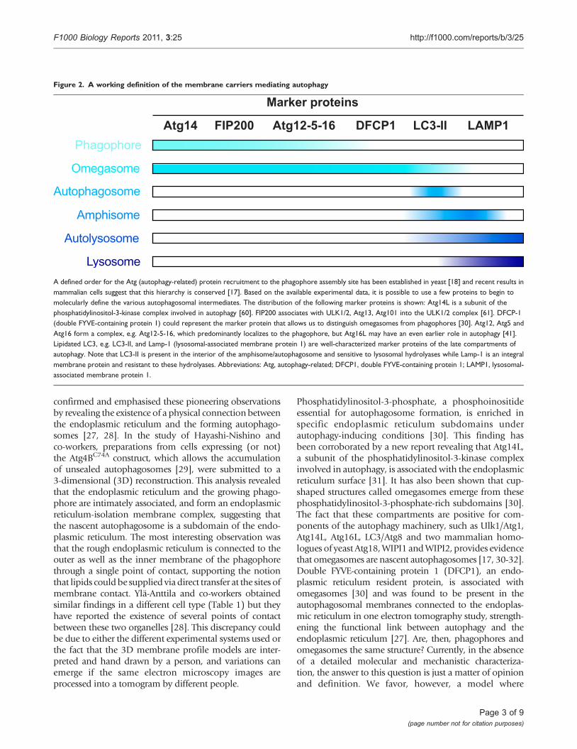
the yeast and plant vacuole, allowing the degradation ofthe inner vesicle and its cargo by acid hydrolases residingin these lytic compartments (Figure 1). The basic meta-bolites generated from this catabolic processing ofbiological macromolecules are finally transported in thecytoplasm, where they are reused as either a source ofenergy or as building blocks for new proteins and lipids.To define the membranes of the autophagy pathwayshown in Figure 1, we propose a working definition basedon which Atg proteins characterize the different inter-mediate structures of autophagy (Figure 2).
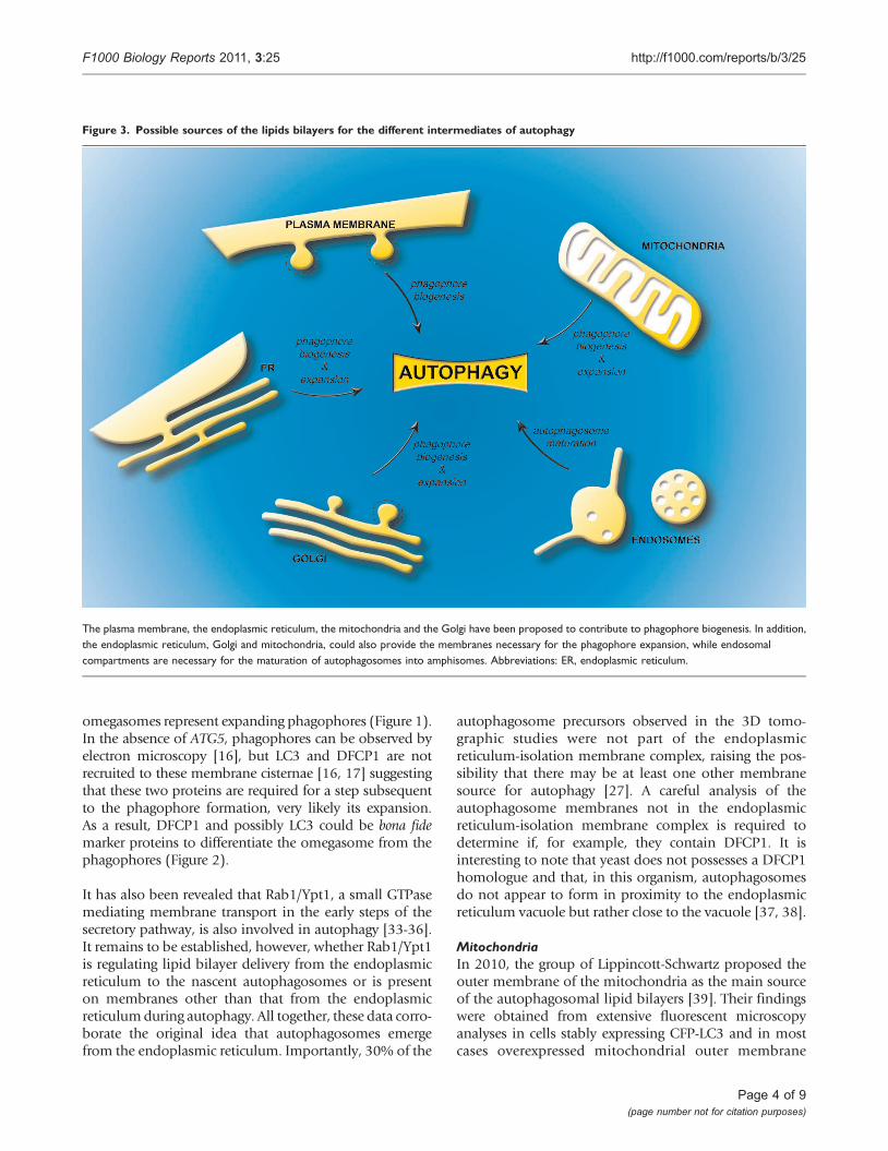
The autophagosomal membranes and theirpromiscuous originDuring the last decade, an immense effort has been inves-ted to try to understand how autophagosomes are formed.One of the key questions in the field that still lacks a clearanswer is the identity of the source of the membranescomposing these large transport carriers (Figure 3) [20,21]. One major difficulty has been that phagophores
as well as autophagosomes can be considered uniqueorganelles because they do not contain any markerproteins of other subcellular compartments [19, 22-24].Recently, several groups have approached this questionusing cutting-edge microscopy techniques combined withbiochemistry approaches. The results of their work arecontroversial because they point to different subcellularorigins of the lipid bilayers composing the phagophoresand/or the autophagosomes.
The endoplasmic reticulumOriginally, the first organelle proposed as the source ofautophagosomal membranes has been the endoplasmicreticulum, through morphological studies performed inthe 60’s [22, 25]. These observations were subsequentlysupported by immuno-ultrastructural observations madebyBill Dunn in1990, inwhich integralmembrane proteinsof the rough endoplasmic reticulum were detected in boththe inner and outermembranes of these vesicles [26].Morerecently, using electron tomography, two groups have
Figure 1. Model for autophagosome biogenesis and cargo degradation
The process of autophagy can be divided in five steps. The induction (1) is elicited by the formation of the phagophore (or isolation membrane), which thenexpands (2) around the material targeted for degradation. The omegasome probably represents a phagophore expansion intermediate. The closure of thegrowing phagophore (3) leads to the formation of a complete autophagosome. The autophagosome then fuses with endosomal structures to become anamphisome, an organelle specific to high eukaryotic cells, where the sequestered material starts to be degraded. Subsequently, the amphisome fuses (4) withlysosomes (vacuoles in plants and yeast) to generate autolysosomes, in the interior of which resident hydrolases break down the internal membrane ofautophagosomes (dashed lines) and the cargo into basic metabolites (5). These metabolites are finally transported in the cytoplasm where they are reused aseither a source of energy or building blocks for new proteins and lipids. Abbreviations: PAS, phagophore assembly site.
Page 2 of 9(page number not for citation purposes)
F1000 Biology Reports 2011, 3:25 http://f1000.com/reports/b/3/25
confirmed and emphasised these pioneering observationsby revealing the existence of a physical connection betweenthe endoplasmic reticulum and the forming autophago-somes [27, 28]. In the study of Hayashi-Nishino andco-workers, preparations from cells expressing (or not)the Atg4BC74A construct, which allows the accumulationof unsealed autophagosomes [29], were submitted to a3-dimensional (3D) reconstruction. This analysis revealedthat the endoplasmic reticulum and the growing phago-phore are intimately associated, and form an endoplasmicreticulum-isolation membrane complex, suggesting thatthe nascent autophagosome is a subdomain of the endo-plasmic reticulum. The most interesting observation wasthat the rough endoplasmic reticulum is connected to theouter as well as the inner membrane of the phagophorethrough a single point of contact, supporting the notionthat lipids could be supplied via direct transfer at the sites ofmembrane contact. Ylä-Anttila and co-workers obtainedsimilar findings in a different cell type (Table 1) but theyhave reported the existence of several points of contactbetween these two organelles [28]. This discrepancy couldbe due to either the different experimental systems used orthe fact that the 3D membrane profile models are inter-preted and hand drawn by a person, and variations canemerge if the same electron microscopy images areprocessed into a tomogram by different people.
Phosphatidylinositol-3-phosphate, a phosphoinositideessential for autophagosome formation, is enriched inspecific endoplasmic reticulum subdomains underautophagy-inducing conditions [30]. This finding hasbeen corroborated by a new report revealing that Atg14L,a subunit of the phosphatidylinositol-3-kinase complexinvolved in autophagy, is associated with the endoplasmicreticulum surface [31]. It has also been shown that cup-shaped structures called omegasomes emerge from thesephosphatidylinositol-3-phosphate-rich subdomains [30].The fact that these compartments are positive for com-ponents of the autophagy machinery, such as Ulk1/Atg1,Atg14L, Atg16L, LC3/Atg8 and two mammalian homo-logues of yeast Atg18,WIPI1 andWIPI2, provides evidencethat omegasomes are nascent autophagosomes [17, 30-32].Double FYVE-containing protein 1 (DFCP1), an endo-plasmic reticulum resident protein, is associated withomegasomes [30] and was found to be present in theautophagosomal membranes connected to the endoplas-mic reticulum in one electron tomography study, strength-ening the functional link between autophagy and theendoplasmic reticulum [27]. Are, then, phagophores andomegasomes the same structure? Currently, in the absenceof a detailed molecular and mechanistic characteriza-tion, the answer to this question is just a matter of opinionand definition. We favor, however, a model where
Figure 2. A working definition of the membrane carriers mediating autophagy
Marker proteins
Atg14 FIP200 Atg12-5-16 DFCP1 LC3-II LAMP1 Phagophore
Autophagosome
Amphisome
Autolysosome
Lysosome
Omegasome
A defined order for the Atg (autophagy-related) protein recruitment to the phagophore assembly site has been established in yeast [18] and recent results inmammalian cells suggest that this hierarchy is conserved [17]. Based on the available experimental data, it is possible to use a few proteins to begin tomolecularly define the various autophagosomal intermediates. The distribution of the following marker proteins is shown: Atg14L is a subunit of thephosphatidylinositol-3-kinase complex involved in autophagy [60]. FIP200 associates with ULK1/2, Atg13, Atg101 into the ULK1/2 complex [61]. DFCP-1(double FYVE-containing protein 1) could represent the marker protein that allows us to distinguish omegasomes from phagophores [30]. Atg12, Atg5 andAtg16 form a complex, e.g. Atg12-5-16, which predominantly localizes to the phagophore, but Atg16L may have an even earlier role in autophagy [41].Lipidated LC3, e.g. LC3-II, and Lamp-1 (lysosomal-associated membrane protein 1) are well-characterized marker proteins of the late compartments ofautophagy. Note that LC3-II is present in the interior of the amphisome/autophagosome and sensitive to lysosomal hydrolyases while Lamp-1 is an integralmembrane protein and resistant to these hydrolyases. Abbreviations: Atg, autophagy-related; DFCP1, double FYVE-containing protein 1; LAMP1, lysosomal-associated membrane protein 1.
Page 3 of 9(page number not for citation purposes)
F1000 Biology Reports 2011, 3:25 http://f1000.com/reports/b/3/25
omegasomes represent expanding phagophores (Figure 1).In the absence of ATG5, phagophores can be observed byelectron microscopy [16], but LC3 and DFCP1 are notrecruited to these membrane cisternae [16, 17] suggestingthat these two proteins are required for a step subsequentto the phagophore formation, very likely its expansion.As a result, DFCP1 and possibly LC3 could be bona fidemarker proteins to differentiate the omegasome from thephagophores (Figure 2).
It has also been revealed that Rab1/Ypt1, a small GTPasemediating membrane transport in the early steps of thesecretory pathway, is also involved in autophagy [33-36].It remains to be established, however, whether Rab1/Ypt1is regulating lipid bilayer delivery from the endoplasmicreticulum to the nascent autophagosomes or is presenton membranes other than that from the endoplasmicreticulumduring autophagy. All together, these data corro-borate the original idea that autophagosomes emergefrom the endoplasmic reticulum. Importantly, 30% of the
autophagosome precursors observed in the 3D tomo-graphic studies were not part of the endoplasmicreticulum-isolation membrane complex, raising the pos-sibility that there may be at least one other membranesource for autophagy [27]. A careful analysis of theautophagosome membranes not in the endoplasmicreticulum-isolation membrane complex is required todetermine if, for example, they contain DFCP1. It isinteresting to note that yeast does not possesses a DFCP1homologue and that, in this organism, autophagosomesdo not appear to form in proximity to the endoplasmicreticulum vacuole but rather close to the vacuole [37, 38].
MitochondriaIn 2010, the group of Lippincott-Schwartz proposed theouter membrane of the mitochondria as the main sourceof the autophagosomal lipid bilayers [39]. Their findingswere obtained from extensive fluorescent microscopyanalyses in cells stably expressing CFP-LC3 and in mostcases overexpressed mitochondrial outer membrane
Figure 3. Possible sources of the lipids bilayers for the different intermediates of autophagy
The plasma membrane, the endoplasmic reticulum, the mitochondria and the Golgi have been proposed to contribute to phagophore biogenesis. In addition,the endoplasmic reticulum, Golgi and mitochondria, could also provide the membranes necessary for the phagophore expansion, while endosomalcompartments are necessary for the maturation of autophagosomes into amphisomes. Abbreviations: ER, endoplasmic reticulum.
Page 4 of 9(page number not for citation purposes)
F1000 Biology Reports 2011, 3:25 http://f1000.com/reports/b/3/25
probes. Under amino acid starvation conditions (designedto induce autophagy), these two sets of marker proteinswere found to co-localize on both mitochondria andautophagosomes suggesting that the membranes of thiscarrier and those of mitochondria are linked. The idea thatthere is a potential direct physical connection betweenthese two organelles was reinforced by the visualization ofgrowing autophagosomes in close proximity tomitochon-dria using live-cell imaging technology and electronmicroscopy. The model that the authors proposed is thatthere is lipid transfer between these two organelles, withthemitochondria supplying the forming autophagosomeswith newly synthesized phospholipids, in particular thephosphatidylethanolamine required for LC3 lipidation[11, 12, 40]. While the co-localization of their mitochon-drial marker proteins with LC3 indicates that mitochon-dria are involved in phagophore expansion, theirexperiments have not clarified whether the mitochondriallipids are also necessary for generation of the phagophore.In the same study the authors also showed that the con-nection between endoplasmic reticulum and mitochon-dria is crucial because in its absence, starvation-inducedautophagosomes are not formed [39]. This observationhas led them to suggest that the endoplasmic reticulumcontribution and the mitochondria could be equally
important asmembrane providers for autophagy. Anotherpossible interpretation, however, is that disruption ofendoplasmic reticulum homeostasis could affect thecontribution of mitochondria (or an organelle connec-ted to it) to phagophore biogenesis and/or expansion.Recently, it has been shown that Salmonella-containingautophagosomal structures are positive for DFCP1 andassociated with the endoplasmic reticulum but nega-tive for the mitochondrial marker protein used by theLippincott-Schwartz group suggesting that mitochond-rial lipids may not be involved in the biogenesis of allautophagosomes [33].
The plasma membraneIn parallel, the laboratory of David Rubinsztein hasreported that lipids of the plasma membrane directlycontribute to forming autophagosomes [41]. In particular,their study shows that the clathrin heavy chain, an elementof the endocytic vesicle coat, binds to Atg16L1, a com-ponent of the Atg autophagy machinery. This associationappears to be crucial for the generation of an earlyautophagosomal precursor, possibly a phagophore or anomegasome, because the formation of autophagosomesis reduced when the interaction between Atg16L1 andthe clathrin heavy chain is disrupted [41]. Additional
Table 1. An overview of organelles, tissues and type of autophagy induction used in different studies on membrane origin ofautophagosome
Organelle implicated inautophagy
Tissue Autophagy induction Starvationperiod
Reference
Endoplasmic reticulum Primary cells isolated from rat livers Fasted animals Overnight [22]Endoplasmic reticulum Primary cells isolated from mice renal
proximal convoluted tubules and hepaticparenchymal cells
Fasted animals Overnight [25]
Endoplasmic reticulum Primary cells from rat livers i) Fasted animalii) Amino acid starvation ofisolated livers
i) 18-24h [26]
Endoplasmic reticulum NIH 3T3 mouse embryonic fibroblastsexpressing (or not) the Atg4BC74A construct
Amino acid, serum andglucose starvation
1h [27]
Endoplasmic reticulum i) Mouse embryonic stem cellsii) Normal rat kidney (NRK) cellsiii) Rat renal proximal tubular (NRK-52E)cells
Amino acid starvation 1h [28]
Mitochondria NRK cells i) Amino acid and serumstarvationii) Serum starvation
1h [39]
Plasma membrane Human cervical cancer (HeLa) cells Amino acid and serumstarvation
6h [41]
Plasma membrane Human cervical cancer (Hela) cells i) Amino acid and serumstarvationii) 1 μg/ml rapamycin
i) 1h to 4hii) 1h
[42]
Plasma membrane/Golgi Yeast Saccharomyces cerevisiae Nitrogen starvation 0.5h to 2h [43]Golgi Yeast Saccharomyces cerevisiae Nitrogen starvation 4h [44]Golgi Rat livers Fasted animals 24h [48]Golgi Yeast Saccharomyces cerevisiae Nitrogen starvation [49]Golgi/endosomal compartments Yeast Saccharomyces cerevisiae Rich medium - [19, 46]trans-Golgi network/endosomalcompartments
Human embryonic kidney 293 (HEK293)cells
Nutrient starvation 2h [47]
Abbreviations: HEK293, human embryonic kidney 293; NRK, normal rat kidney.
Page 5 of 9(page number not for citation purposes)
F1000 Biology Reports 2011, 3:25 http://f1000.com/reports/b/3/25
investigations from the same group have emphasizedthis initial discovery by revealing that maturation of theAtg16L1-positive early autophagosomal precursorsrequires their homotypic fusion through the action of theplasma membrane-soluble N-ethylmaleimide-sensitive-factor attachment receptor (SNARE) protein VAMP7 andits interacting partners [42]. The involvement of plasmamembrane SNAREs in the early steps of autophagy hasalso been highlighted by work performed in yeast [43].Additional support for the plasma membrane in autopha-gosome biogenesis has also indirectly provided throughthe discovery that components of the exocyst, a tetheringcomplex that, in concert with SNAREs,mediates the fusionof post-Golgi vesicles with the plasma membrane,associates with nascent autophagosomes and is essentialfor nutrient starvation-induced autophagy [44, 45].
The GolgiThese last two studies have also raised the idea of the Golgibeing a potential source for autophagosomal membranesbecause the exocyst complex is present at the trans-Golginetwork as well as on post-Golgi vesicles [44, 45]. Also,the work on yeast revealed that plasma membranesSNAREs regulate the organization of Atg9-positive tubulo-vesicular membrane organelles [44], Interestingly, Atg9, atransmembrane Atg protein essential for autophagy,localizes to the trans-Golgi network, post-Golgi and endo-somal compartments [19, 46, 47] and in yeast plays acentral role in generating the phagophore assembly site[19]. In the 90’s, the Golgi was implicated in autophago-some biogenesis because the growing extremities of thephagophores as well as complete autophagosomes weredecorated with lectins that recognize glycans exclusivelypresent in post-Golgi membranes [48]. This concept hasrecently been reinforced by work in yeast [49], where it hasalso been shown that large multi-subunit Golgi tetheringcomplexes, lobe B of the conserved oligomeric Golgicomplex and subunits of TRAPPIII, a complex homologousto the TRAPPI and TRAPPII oligomers, are essential forautophagy [34, 50].
Can all these membrane sources be reconciledin a single model?While studies on the membrane origin of autophago-somes have started to provide some answers about themolecular mechanism and machinery, they have alsocreated some confusion regarding the source of the lipidsforming the membrane carriers mediating autophagy.
This apparent discrepancy between the conclusionsreached by the different laboratories could be in part dueto different experimental approaches and techniques usedin the various laboratories. More importantly, the differentcontributions could vary depending on the tissues and
conditions used to trigger autophagy (Table 1), with cellsable to derive the membranes from the most suitable orexpendable reservoirs.
This hypothesis could explain the reported contradictoryresults as different studies used different cell types, organ-isms and diverse conditions to induce starvation (Table 1).Thus, in a tissue with a defined function, in response toa specific stress stimulus, autophagy would be suppliedwith membranes from the most optimal reservoir: an orga-nelle that could guarantee the delivery of a large amount oflipids, but ideally would not adversely affect the specificfunctions of the tissue. From a cursory look at the currentavailable data, one might conclude that fasting animalsutilize endoplasmic reticulum while nitrogen-starved yeastuse Golgi (Table 1). However, accurate comparative studiesare needed to determine whether, for example, NRK cellsstarved of amino acids indeed use the endoplasmic reticu-lumas the source for autophagosomalmembranes [28] andif they turn to mitochondria when autophagy is inducedby either serum, or serumand amino acid, deprivation [39].Along the same lines, it would be important to study thevarious marker proteins used to implicate the differentorganelles in autophagy [26, 39, 41, 42] in parallel in thesame experimental setup.
Another possibility that should not be discarded a prioriis that autophagosomes could be mosaics of membranesderived from more than one organelle. For example, thephagophore could originate from one compartment andthe additional lipid bilayers required for its expansioncould be acquired from another source (Figure 3). Forexample, the plasma membrane and Atg9-positivemembranes could contribute to the formation of thephagophore while the endoplasmic reticulum, mito-chondria and/or Golgi might be necessary for itsexpansion. In this regard, it is important to note thatthe endosomal system provides the membranes requiredfor the last steps of autophagy, i.e. amphisome andautolysosome formation (Figure 3) [51]. The advantageto the cell of having a spectrum of membrane sources tochoose from is the availability of a large supply of lipidsto sustain the progression of autophagy; autophago-somes are huge carriers and a multitude of them areproduced upon induction of this degradative pathway.One could imagine that a single intracellular organellecould not provide enough membranes, especially duringprolonged period of starvation or stress.
Finally, it still remains to be determined whether thedifferent organelles implicated so far in autophagosomebiogenesis, contribute to non-selective bulk autophagyor to selective forms of autophagy such as mitophagy(selective degradation of mitochondria), or pexophagy
Page 6 of 9(page number not for citation purposes)
F1000 Biology Reports 2011, 3:25 http://f1000.com/reports/b/3/25
(selective degradation of peroxisomes). In this regard,the observation that the endoplasmic reticulum is foundconnected to both the inside and the outside membraneof the phagophore [26-28] could suggest that theendoplasmic reticulum found in the interior of theclosing phagophore is not undergoing the generalprocess of bulk autophagy but rather is specificallydegraded through a seleletive type of autophagy knownas endoplasmic reticulumphagy or reticulophagy [52,53]. A similar explanation could be applicable to theother organelles acting in the phagophore and autopha-gosome biogenesis.
Is this hypothesis, implicating multiple membranesources for autophagosomes, something reconcilable atthe molecular mechanistc level? Potentially yes. Apartfrom the transmembrane protein Atg9, the core Atgmachinery is composed of soluble proteins that tran-siently associate with membranes. Consequently, the cellcould select the membrane source by targeting the signaland/or the molecule initiating the assembly of the Atgmachinery to the organelle of choice.
Autophagosomal membranes and drug therapiesExperimental evidence indicates that autophagosomebiogenesis is probably a very complex process ondifferent levels, including its regulation in response tothe different cellular and environmental cues, and in thechoice of membrane sources.
Is there any therapeutic value in determining, or at the veryleast understanding, the origin of the autophagosomalmembranes? We believe this discovery will not have asignificant therapeutic value per se but it will be a funda-mental step forward in the investigation of autophago-some biogenesis. However, defects in autophagy underlievarious diseases, and the delivery of lipid bilayers fromtheir source to the site of autophagosome formation is ahighly regulated process that involves several of the pro-teins that have been shown to be essential for the pro-gression of autophagy and these could be the target fordrugs modulating autophagy [54]. It is also worth bearinginmind that the task of assigning a function to a protein isgenerally better facilitated when the step of a pathway inwhich it participates is defined.
A good example is the transmembrane protein Atg9.Because of its intrinsic associationwith lipid bilayers, it hasbeen postulated that Atg9 could be involved is supplyingconstituents of themembranes necessary to the biogenesisof autophagosomes. This concept is supported by thefinding that a change in Atg9 trafficking is directly asso-ciated with autophagy induction [19, 47], probablybecause of its key contribution to the phagophore
assembly site/phagophore formation [19]. Not surpris-ingly, Atg9 transport is controlled by signaling cascades[55]. While Atg9 itself has not been to date associated toparticular diseases, alterations of factors regulating thetrafficking of this protein such as the phosphatidylinositol-3-kinase complex involved in autophagy [47, 56] are thedirect cause of tumorigenesis [57, 58]. In this context, thetumor-suppressor Bif-1 has also been linked to Atg9 [59].These observations support the possibility that specificillnesses could be the phenomenological manifestation ofamisregulation of lipid bilayer flux during autophagy. As aresult, the factorsmodulating these pathways would be theoptimal targets for drugs aimed to restore normalmembrane supply and consequently proper progressionof autophagy.
Conclusions and future directionsSince the discovery of autophagy, the membrane originof the autophagosomes has been the subject of intensedebate in the field. The recent studies employingadvanced technologies have confirmed and extendedthe pioneering ultrastructural observations and haveprovided some insights on the membrane origin of theseunique carriers. The diverse conclusions of the recentwork, however, have not given an unequivocal answeryet, but rather they have raised new questions that nowneed to be addressed. The work on this phenomenon hasjust started.
AbbreviationsAtg, autophagy-related; DFCP1, double FYVE-containingprotein 1; SNARE, soluble N-ethylmaleimide-sensitive-factor attachment receptor; 3D, 3-dimensional.
Competing interestsThe authors declare that they have no competing interests.
AcknowledgementsThe authors wish to thank Marc van Peski for thepreparation of the figures. Fulvio Reggiori is supportedby the Netherlands Organization for Health Researchand Development (ZonMW-VIDI-917.76.329), by theNetherlands Organization for Scientific Research (Che-mical Sciences, ECHO grant-700.59.003, and Earth andLife Sciences, Open Program grant-821.02.017). SharonTooze is supported by Cancer Research UK.
References1. Maiuri MC, Zalckvar E, Kimchi A, Kroemer G: Self-eating and self-
killing: crosstalk between autophagy and apoptosis. Nat RevMol Cell Biol 2007, 8:741-52.
2. Kroemer G, Marino G, Levine B: Autophagy and the integratedstress response. Mol Cell 2010, 40:280-93.
3. Mizushima N, Levine B: Autophagy in mammalian developmentand differentiation. Nat Cell Biol 2010, 12:823-30.
Page 7 of 9(page number not for citation purposes)
F1000 Biology Reports 2011, 3:25 http://f1000.com/reports/b/3/25
4. Levine B, Deretic V: Unveiling the roles of autophagy in innateand adaptive immunity. Nat Rev Immunol 2007, 7:767-77.
5. Deretic V, Levine B: Autophagy, immunity, and microbialadaptations. Cell Host Microbe 2009, 5:527-49.
6. Cuervo AM: Autophagy and aging: keeping that old broomworking. Trends Genet 2008, 24:604-12.
7. Levine B: Cell biology: autophagy and cancer. Nature 2007,446:745-7.
8. Levine B, Mizushima N, Virgin HW: Autophagy in immunity andinflammation. Nature 2011, 469:323-35.
9. Levine B, Kroemer G: Autophagy in the pathogenesis of disease.Cell 2008, 132:27-42.
10. Mizushima N, Levine B, Cuervo AM, Klionsky DJ: Autophagy fightsdisease through cellular self-digestion. Nature 2008,451:1069-75.
11. Xie Z, Klionsky DJ: Autophagosome formation: core machin-ery and adaptations. Nat Cell Biol 2007, 9:1102-9.
F1000 Factor 6Evaluated by Muriel Mari and Fulvio Reggiori 15 Nov 2011
12. Yoshimori T, Noda T: Toward unraveling membrane biogenesisin mammalian autophagy. Curr Opin Cell Biol 2008, 20:401-7.
F1000 Factor 6Evaluated by Muriel Mari and Fulvio Reggiori 16 Nov 2011
13. Eskelinen EL:Maturation of autophagic vacuoles in mammaliancells. Autophagy 2005, 1:1-10.
14. Militello RD, Colombo MI: A membrane is born: origin of theautophagosomal compartment. Curr Mol Med 2011, 11:197-203.
F1000 Factor 6Evaluated by Muriel Mari and Fulvio Reggiori 16 Nov 2011
15. Eskelinen EL, Reggiorri F, Baba M, Kovács AL, Seglen PO: Seeing isbelieving: The impact of electron microscopy on autophagyresearch. Autophagy 2011, 7:935-56.
F1000 Factor 6Evaluated by Muriel Mari and Fulvio Reggiori 16 Nov 2011
16. Mizushima N, Yamamoto A, Hatano M, Kobayashi Y, Kabeya Y,Suzuki K, Tokuhisa T, Ohsumi Y, Yoshimori T: Dissection ofautophagosome formation using Apg5-deficient mouseembryonic stem cells. J Cell Biol 2001, 152:657-68.
F1000 Factor 6Evaluated by Muriel Mari and Fulvio Reggiori 16 Nov 2011
17. Itakura E, Mizushima N: Characterization of autophagosomeformation site by a hierarchical analysis of mammalian Atgproteins. Autophagy 2010, 6:764-76.
F1000 Factor 6Evaluated by Muriel Mari and Fulvio Reggiori 16 Nov 2011
18. Suzuki K, Kubota Y, Sekito T, Ohsumi Y: Hierarchy of Atgproteins in pre-autophagosomal structure organization. GenesCells 2007, 12:209-18.
F1000 Factor 6Evaluated by Muriel Mari and Fulvio Reggiori 16 Nov 2011
19. Mari M, Griffith J, Rieter E, Krishnappa L, Klionsky DJ, Reggiori F: AnAtg9-containing compartment that functions in the earlysteps of autophagosome biogenesis. J Cell Biol 2010, 190:1005-22.
20. Reggiori F, Tooze SA: The EmERgence of autophagosomes. DevCell 2009, 17:747-8.
21. Tooze SA, Yoshimori T: The origin of the autophagosomalmembrane. Nat Cell Biol 2010, 12:831-5.
22. Arstila AU, Trump BF: Studies on cellular autophagocytosis.The formation of autophagic vacuoles in the liver afterglucagon administration. Am J Pathol 1968, 53:687-733.
F1000 Factor 6Evaluated by Muriel Mari and Fulvio Reggiori 16 Nov 2011
23. Stromhaug PE, Berg TO, Fengsrud M, Seglen PO: Purification andcharacterization of autophagosomes from rat hepatocytes.Biochem J 1998, 335:217-24.
F1000 Factor 6Evaluated by Muriel Mari and Fulvio Reggiori 16 Nov 2011
24. Yokota S: Formation of autophagosomes during degradationof excess peroxisomes induced by administration of dioctylphthalate. Eur J Cell Biol 1993, 61:67-80.
25. Ericsson JL: Studies on induced cellular autophagy. I. Electronmicroscopy of cells with in vivo labelled lysosomes. Exp Cell Res1969, 55:95-106.
F1000 Factor 6Evaluated by Muriel Mari and Fulvio Reggiori 16 Nov 2011
26. Dunn WA Jr: Studies on the mechanisms of autophagy:maturation of the autophagic vacuole. J Cell Biol 1990, 110:1923-33.
F1000 Factor 6Evaluated by Muriel Mari and Fulvio Reggiori 16 Nov 2011
27. Hayashi-Nishino M, Fujita N, Noda T, Yamaguchi A, Yoshimori T,Yamamoto A: A subdomain of the endoplasmic reticulumforms a cradle for autophagosome formation. Nat Cell Biol2009, 11:1433-7.
F1000 Factor 9Evaluated by Kerstin Radtke and Michel Desjardins 23 Dec 2009,Muriel Mari and Fulvio Reggiori 16 Nov 2011
28. Yla-Anttila P, Vihinen H, Jokitalo E, Eskelinen EL: 3D tomographyreveals connections between the phagophore and endoplas-mic reticulum. Autophagy 2009, 5:1180-5.
F1000 Factor 8Evaluated by Daniel Klionsky 01 Dec 2009
29. Fujita N, Havashi-Nishino M, Fukumoto H, Omori H, Yamamoto A,Noda T, Yoshimori T: An Atg4B mutant hampers the lipidationof LC3 paralogues and causes defects in autophagosomeclosure. Mol Biol Cell 2008, 19:4651-9.
30. Axe EL, Walker SA, Manifava M, Chandra P, Roderick HL,Habermann A, Griffiths G, Ktistakis NT: Autophagosome forma-tion from membrane compartments enriched in phosphati-dylinositol 3-phosphate and dynamically connected to theendoplasmic reticulum. J Cell Biol 2008, 182:685-701.
F1000 Factor 8Evaluated by Muriel Mari and Fulvio Reggiori 16 Nov 2011
31. Matsunaga K, Morita E, Saitoh T, Akira S, Ktistakis NT, Izumi T,Noda T, Yoshimori T: Autophagy requires endoplasmic reticu-lum targeting of the PI3-kinase complex via Atg14L. J Cell Biol2010, 190:511-21.
F1000 Factor 6Evaluated by Muriel Mari and Fulvio Reggiori 16 Nov 2011
32. Polson HE, de Lartique J, Rigden DJ, Reedijk M, Urbe S, Clague MJ,Tooze SA: Mammalian Atg18 (WIPI2) localizes to omega-some-anchored phagophores and positively regulates LC3lipidation. Autophagy 2010, 6:506-22.
33. Huang J, Birmingham CL, Shahnazari S, Shiu J, Zheng YT, Smith AC,Campellone KG, Heo WD, Gruenheid S, Meyer T, Welch MD,Ktistakis NT, Kim PK, Klionsky DJ, Brumell JH: Antibacterialautophagy occurs at PI(3)P-enriched domains of the endoplas-mic reticulumand requires Rab1GTPase. Autophagy 2011, 7:17-26.
F1000 Factor 6Evaluated by Muriel Mari and Fulvio Reggiori 16 Nov 2011
Page 8 of 9(page number not for citation purposes)
F1000 Biology Reports 2011, 3:25 http://f1000.com/reports/b/3/25
34. Lynch-Day MA, Bhandari D, Menon S, Huang J, Cai H, Bartholomew CR,Brumell JH, Ferro-Novick S, Klionsky DJ: Trs85 directs a Ypt1 GEF,TRAPPIII, to the phagophore to promote autophagy. Proc NatlAcad Sci U S A 2010, 107:7811-6.
35. Winslow AR, Chen CW, Corrochano S, Acevedo-Arozena A,Gordon DE, Peden AA, Lichtenberg M, Menzies FM, Ravikumar B,Imarisio S, Brown S, O’Kane CJ, Rubinsztein DC: alpha-Synucleinimpairsmacroautophagy: implications for Parkinson’s disease. JCell Biol 2010, 190:1023-37.
F1000 Factor 9Evaluated by Vojo Deretic 04 Oct 2010, Muriel Mari and FulvioReggiori 16 Nov 2011
36. Zoppino FC, Militello RD, Slavin I, Alvarez C, Colombo MI: Auto-phagosome formation depends on the small GTPase Rab1 andfunctional ER exit sites. Traffic 2010, 11:1246-61.
F1000 Factor 6Evaluated by Muriel Mari and Fulvio Reggiori 16 Nov 2011
37. Kim J, Huang WP, Stromhaug PE, Klionsky DJ: Convergence ofmultiple autophagy and cytoplasm to vacuole targeting compo-nents to a perivacuolar membrane compartment prior to denovo vesicle formation. J Biol Chem 2002, 277:763-73.
38. Suzuki K, Kirisako T, Kamada Y, Mizushima N, Noda T, Ohsumi Y:The pre-autophagosomal structure organized by concertedfunctions of APG genes is essential for autophagosomeformation. EMBO J 2001, 20:5971-81.
39. Hailey DW, Rambold AS, Satpute-Krishnan P, Mitra K, Sougrat R,Kim PK, Lippincott-Schwartz J: Mitochondria supply membranesfor autophagosome biogenesis during starvation. Cell 2010,141:656-67.
F1000 Factor 16Evaluated by Eric Lau and Ze’ev Ronai 01 Jun 2010, Yi Xiang andYanzhuang Wang 01 Jul 2010, Maria Markaki and NektariosTavernarakis 21 Jul 2010, Roy Gross 21 Oct 2010, Muriel Mariand Fulvio Reggiori 16 Nov 2011
40. Tanida I, Ueno T, Kominami E: LC3 conjugation system inmammalian autophagy. Int J Biochem Cell Biol 2004, 36:2503-18.
41. Ravikumar B, Moreau K, Jahreiss L, Puri C, Rubinsztein DC: Plasmamembrane contributes to the formation of pre-autophago-somal structures. Nat Cell Biol 2010, 12:747-57.
F1000 Factor 6Evaluated by Muriel Mari and Fulvio Reggiori 20 Oct 2010
42. Moreau K, Ravikumar B, Renna M, Puri C, Rubinsztein DC:Autophagosome precursor maturation requires homotypicfusion. Cell 2011, 146:303-17.
F1000 Factor 8Evaluated by Thierry Galli 09 Sep 2011, Vojo Deretic 18 Oct 2011,Muriel Mari and Fulvio Reggiori 16 Nov 2011
43. Nair U, Jotwani A, Geng J, Gammoh N, Richerson D, Yen WL,Griffith J, Nag S, Wang K, Moss T, Baba M, McNew JA, Jiang X,Reggiori F, Melia TJ, Klionsky DJ: SNARE proteins are requiredfor macroautophagy. Cell 2011, 146:290-302.
F1000 Factor 9Evaluated by Sascha Martens 28 Jul 2011, Kerstin Radtke and MichelDesjardins 04 Oct 2011
44. Geng J, Nair U, Yasumura-Yorimitsu K, Klionsky DJ: Post-Golgi Secproteins are required for autophagy in Saccharomycescerevisiae. Mol Biol Cell 2010, 21:2257-69.
F1000 Factor 8Evaluated by Muriel Mari and Fulvio Reggiori 16 Nov 2011
45. Bodemann BO, Orvedahl A, Cheng T, Ram RR, Ou YH,Formstecher E, Maiti M, Hazelett CC, Wauson EM, Balakireva M,Camonis JH, Yeaman C, Levine B, White MA: RalB and the exocyst
mediate the cellular starvation response by direct activationof autophagosome assembly. Cell 2011, 144:253-67.
F1000 Factor 13Evaluated by Sascha Martens 17 Feb 2011, Jianglan Liu and Wei Guo23 Feb 2011, Raphael Valdivia 24 Feb 2011, Muriel Mari and FulvioReggiori 17 May 2011
46. Ohashi Y, Munro S: Membrane delivery to the yeast autopha-gosome from the Golgi-endosomal system. Mol Biol Cell 2010,21:3998-4008.
F1000 Factor 6Evaluated by Muriel Mari and Fulvio Reggiori 16 Nov 2011
47. Young AR, Chan EY, Hu XW, Köchl R, Crawshaw SG, High S,Hailey DW, Lippincott-Schwartz J, Tooze SA: Starvation andULK1-dependent cycling of mammalian Atg9 between theTGN and endosomes. J Cell Sci 2006, 119:3888-900.
48. Yamamoto A, Masaki R, Tashiro Y: Characterization of theisolation membranes and the limiting membranes of autop-hagosomes in rat hepatocytes by lectin cytochemistry. JHistochem Cytochem 1990, 38:573-80.
49. van der Vaart A, Griffith J, Reggiori F: Exit from the Golgi IsRequired for the Expansion of the Autophagosomal Phago-phore in Yeast Saccharomyces cerevisiae. Mol Biol Cell21:2270-84.
50. Yen WL, Shintani T, Nair U, Cao Y, Richardson BC, Li Z, Hughson FM,Baba M, Klionsky DJ:The conserved oligomeric Golgi complex isinvolved in double-membrane vesicle formation duringautophagy. J Cell Biol 2010, 188:101-14.
51. Fader CM, Colombo MI: Autophagy and multivesicular bodies:two closely related partners. Cell Death Differ 2009, 16:70-8.
52. Kincaid MM, Cooper AA: ERADicate ER stress or die trying.Antioxid Redox Signal 2007, 9:2373-87.
53. Kondratyev M, Avezov E, Shenkman M, Groisman B,Lederkremer GZ: PERK-dependent compartmentalization ofERAD and unfolded protein response machineries during ERstress. Exp Cell Res 2007, 313:3395-407.
54. Fleming A, Noda T, Yoshimori T, Rubinsztein DC: Chemicalmodulators of autophagy as biological probes and potentialtherapeutics. Nat Chem Biol 2011, 7:9-17.
55. Webber JL, Tooze SA: Coordinated regulation of autophagy byp38alpha MAPK through mAtg9 and p38IP. EMBO J 2009,29:27-40.
56. Reggiori F, Tucker KA, Stromhaug PE, Klionsky DJ: The Atg1-Atg13complex regulates Atg9 and Atg23 retrieval transport fromthe pre-autophagosomal structure. Dev Cell 2004, 6:79-90.
57. Liang XH, Jackson S, Seaman M, Brown K, Kempkes B, Hibshoosh H,Levine B: Induction of autophagy and inhibition of tumorigen-esis by beclin 1. Nature 1999, 402:672-6.
F1000 Factor 6Evaluated by Muriel Mari and Fulvio Reggiori 16 Nov 2011
58. Takahashi Y, Coppola D, Matsushita N, Cualing HD, Sun M, Sato Y,Liang C, Jung JU, Cheng JQ, Mulé JJ, Pledger WJ, Wang HG: Bif-1interacts with Beclin 1 through UVRAG and regulatesautophagy and tumorigenesis. Nat Cell Biol 2007, 9:1142-51.
F1000 Factor 11Evaluated by Harald Stenmark 22 Oct 2007, Daniel Klionsky 07 Nov2007, Muriel Mari and Fulvio Reggiori 16 Nov 2011
59. Takahashi Y, Meyerkord CL, Wang HG: BARgaining membranesfor autophagosome formation: Regulation of autophagy andtumorigenesis by Bif-1/Endophilin B1. Autophagy 2008, 4:121-4.
60. Itakura E, Kishi C, Inoue K, Mizushima N: Beclin 1 forms twodistinct phosphatidylinositol 3-kinase complexes with mam-malian Atg14 and UVRAG. Mol Biol Cell 2008, 19:5360-72.
61. Hosokawa N, Sasaki T, Iemura S, Natsume T, Hara T, Mizushima N:Atg101, a novel mammalian autophagy protein interactingwith Atg13. Autophagy 2009, 5:973-9.
Page 9 of 9(page number not for citation purposes)
F1000 Biology Reports 2011, 3:25 http://f1000.com/reports/b/3/25

![Lecture 17 Membrane separations - CHERIC · Lecture 17. Membrane Separations [Ch. 14] •Membrane Separation •Membrane Materials •Membrane Modules •Transport in Membranes-Bulk](https://img.pdfslide.us/doc/110x75/5e688f368fbb145949438f76/lecture-17-membrane-separations-cheric-lecture-17-membrane-separations-ch-14.jpg)