Embed Size (px)
Citation preview
Organogenesis in vitro under Altered Auxin Signaling Conditions
by
Tatiana Smirnova
A thesis submitted in conformity with the requirements for the degree of Master of Science
Graduate Department of Cell and System Biology University of Toronto
© Copyright by Tatiana Smirnova 2011
Organogenesis in vitro under Altered Auxin Signaling Conditions
Tatiana Smirnova
Master of Science
Graduate Department of Cell and System Biology University of Toronto
2011
Abstract
The ratio of auxin to cytokinin determines de novo organogenesis in plants. Relatively little is
known about the effect of genetically altered auxin signaling on in vitro organogenesis. Here,
callusogenesis, shoot, and root formation were studied in loss- (LOF) and gain-of-function
(GOF) alleles in two phylogenetically related Auxin Response Factors (ARFs), MONOPTEROS
(MP/ARF5) and NON-PHOTOTROPHIC HYPOCOTYL 4 (NPH4/ARF7). Reduced MP activity
greatly diminished shoot regeneration, and partially diminished callusogenesis and root
formation. LOF in NPH4 strongly decreased callusogenesis, and mildly decreased shoot and root
regeneration in particular categories of explants. By contrast, organogenesis responses were
strongly increased in aerial explants carrying the GOF transgene dMP. Thus, both MP and NPH4
seem to act as positive regulators of certain organogenesis processes and the GOF dMP
transgene may be of interest for stimulating organogenesis in plant species with poor
regeneration properties. Also, organogenesis in vitro may reveal unknown developmental ARF
functions.
ii
Table of Contents Abstract……………………………………………………………………………………..……..ii
Table of Content…………………………………………………………………………..……...iii
List of Figures…………………………………………………………………………………....vii
List of Tables……………………………………………………………………………………..ix
List of Abbreviations……………………………………………………………………………...x
Chapter 1
Introduction………………………………………………………………………………..1
1.1 Plant hormone signals in plant regeneration………………………………………………1
1.1.1 The balance of auxin to cytokinin in a culture medium determines the type of
organogenesis……………………………………………………………………………...2
1.2 The auxin signal transduction pathway…………………………………………………...2
1.2.1 Biosynthesis, transport, and distribution of auxin in planta…...…………….........3
1.2.2 Auxin-mediated regulation of gene expression in Arabidopsis…………………...4
1.2.3 Aux/IAA genes and proteins……………………………………………………....4
1.2.4 ARF genes and proteins…………………………………………………………...7
1.2.5 Auxin response factors MONOPTEROS (MP/ARF5) and NON-PHOTOTROPIC
HYPOCOTYL 4 (NPH4/ARF7)…………………………………………………………..10
1.3 Roles of cytokinins.……………………………………………………………………...12
1.4 Cell proliferation and callusogenesis in plant tissue culture…………………………….13
1.5 De novo organogenesis in plant tissue culture …………………………………………..15
iii
1.5.1 Roles of shoot apical meristem genes during de novo shoot regeneration………18
1.5.2 Mutations affecting shoot regeneration in Arabidopsis…………….....................21
1.5.3 Mutations in auxin signaling genes affecting shoot regeneration……………......21
1.6 Regeneration of adventitious roots in vitro………………………………………………23
1.7 General Research Objectives………………………………………………………….....25
Chapter 2 Materials and Methods...........................................................................................27
2.1 Plant material………………………………………………………………………….....27
2.2 General growth conditions…………………………………………………………….....27
2.3 Hormone stocks………………………………………………………………. ………...30
2.4 Plant DNA extraction………………………………………………………………….....30
2.5 General procedure for polymerase chain reaction (PCR)……………… …………….....31
2.6 Genotyping…………………………………………………………………………….....31
2.7 Microtechniques and microscopy………………………………………………………..32
2.8 Plant tissue culture experiments……………………………………………………….....33
2.8.1 Rooting of dissected mpG12, dMP, nph4-1 and wild type seedlings……………33
2.8.2 Preparation of explants for tissue culture………………………………………...35
2.8.3 Preparation of tissue culture media………………………………………………36
Chapter 3 Callusogenesis under conditions of altered auxin signal transduction…………..37
3.1 Background and rationale…………………………………………………………….....37
3.2 Specific research objectives……………………………………………………………...38
iv
3.3 Results……………………………………………………………………………………39
3.3.1 Types of calluses on CIM………………………………………………………..39
3.3.2 Callusogenesis of cotyledon explants after 12 days on CIM…………………….42
3.3.3 Callusogenesis of leaf explants after 12 days on CIM……………………….......42
3.3.4 Callusogenesis of petiole explants after 12 days on CIM……………………......43
3.3.5 Callusogenesis of hypocotyl explants after 12 days on CIM…………………….45
3.3.6 Callusogenesis of adventitious root explants after 12 days on CIM…………......45
3.3.7 Callus proliferation in aerial explants after 6 days on CIM……………………...50
3.4 Discussion and Conclusions……………………………………………………………..51
Chapter 4 Shoot regeneration under conditions of altered auxin signal transduction………54
4.1 Background and rationale………………………………………………………………..54
4.2 Specific research objectives……………………………………………………………...55
4.3 Results……………………………………………………………………………………56
4.3.1 Shoot regeneration in cotyledon derived calluses………………………………..56
4.3.2 Shoot regeneration in leaf derived calluses………………………………………56
4.3.3 Shoot regeneration in calluses derived from petiole explants……………………59
4.3.4 Shoot regeneration in calluses derived from hypocotyl explants……………......61
4.3.5 Shoot regeneration in calluses derived from adventitious root explants………...62
4.3.6. Effect of duration of CIM preculture on shoot regeneration on SIM in calluses
derived from adventitious root explants………………………………………………….64
4.3.7 Greening of calluses derived from adventitious root explants on SIM…… …….68
v
4.4. Discussion and Conclusions………………………………………..…………………...71
Chapter 5 Root regeneration under conditions of altered auxin signal transduction………..74
5.1 Background and rationale………………………………………………………………..74
5.2 Specific research objectives……………………………………………………………...75
5.3 Results……………………………………………………………………………………76
5.3.1 Root regeneration in cotyledon derived calluses on RIM……………………......76
5.3.2 Root regeneration in leaf derived calluses on RIM………………………………80
5.3.3 Root regeneration in calluses derived from adventitious roots on RIM…………80
5.3.4 Root formation in adventitious root explants on SIM…………………………...82
5.4 Discussion and Conclusions……………………………………………………………..86
Chapter 6 General Discussion and Conclusions………………………………………….....89
6.1 Effects of changed MP/ARF5 and NPH4/ARF7 levels on callusogenesis, de novo shoot
and root regeneration……………………………………………………………..……...89
6.1.1 NPH4/ARF7 in auxin and cytokinin mediated developmental processes in tissue
culture…………………………………………………………………………………....89
6.1.2 MP/ARF5 in auxin and cytokinin mediated developmental processes…………..92
6.1.3 Enhancement of de novo organogenesis in the dMP genotype………………......94
6.2 General Conclusion………………………………………………………………………94
References………………………………………………………………………………………..96
vi
List of Figures 1.1 Model for the regulation of auxin signaling by auxin levels and auxin-induced
Aux/IAA protein degradation……………………………………………………………..5
1.2 MP/ARF5 and dMP/dARF5 protein structures and functions…………………………….8
1.3 Phases of organogenesis in Arabidopsis tissue culture…………………………………..17
2.1 Seedlings and inflorescence phenotypes in LOF mutants nph4-1, mpG12, and GOF
transgene line dMP……………………….………………………………………………29
2.2 Scheme of the tissue culture experiments and scored traits…………………………...…34
3.1 Fresh weight of calluses and frequency of the “whole” type of calluses after 12 days on
CIM……………………………………………………………………………………....40
3.2 Callus initiation and callus proliferation in cotyledon and leaf explants after 6 and 12
days on CIM……………………………………………………………………………...41
3.3 Callus initiation and callus proliferation in petiole and hypocotyl explants after 6 and 12
days on CIM……………………………………………………………………………...44
3.4 Callus initiation and callus proliferation in adventitious root explants
after 6 and 12 days on CIM………………………………………………………………46
3.5 Cotyledon and leaf explants after 12 days on CIM……………………………………....47
3.6 Petiole and hypocotyl explants after 12 days on CIM…………………………………...48
3.7 Adventitious root explants after 12 days on CIM………………………………………..49
4.1 Frequency of shoot regenerating calluses, and mean number of shoots per shoot
regenerating callus after 21 days on SIM………………………………………………...57
4.2 Calluses derived from cotyledon and leaf explants after 21 days on SIM……………….58
4.3 Calluses derived from petiole and hypocotyl explants after 21 days on SIM…………....60
vii
4.4 Calluses derived from adventitious root explants after 21 days on SIM………………...63
4.5 Frequency of shoot regenerating calluses and mean number of shoots per shoot
regenerating callus in calluses derived from adventitious root explants after 21 days
on SIM depending on the time on CIM (6, 9, 12 and 16 days)…………………………..65
4.6 Greening of calluses and root outgrowth in calluses derived from adventitious
root explants on SIM after 8, and 13 days on SIM………………………………………69
4.7 Frequency of green versus whitish calluses after 8 days on SIM in adventitious
root derived calluses ……………………………………………………………………..70
5.1 Frequency of root forming calluses, and the mean numbers of roots per root
forming callus in calluses derived from cotyledon, leaf and adventitious root
explants after 21 days on RIM…………………………………………………………...77
5.2 Cotyledon and leaf derived calluses after 21 days on RIM. …………………………….78
5.3 Root initiation in cotyledon explants after 6 days on RIM……………………………....79
5.4 Calluses derived from adventitious root explants after 21 days on RIM………………...81
5.5 Calluses derived from adventitious root explants after 12 and 21 days on RIM...............83
5.6 Frequency of root regenerating calluses, and the mean numbers of roots per
root regenerating callus in calluses derived from adventitious root explants after
21 days on SIM…………………………………………………………………………..84
5.7 Root outgrowth in calluses derived from adventitious root explants after 8 and 13
days on SIM……………………………………………………………………………...85
viii
List of Tables 2.1 Main characteristics of the mutant genotypes, mpG12, dMP,
and nph4-1…………………………………………………………………...…………………..28
2.2 Types of explants, genotypes, approximate sizes, and DAG at the moment
of dissection and introduction in tissue culture…………………………………………..35
2.3 The hormone composition of tissue culture media: CIM, SIM, and RIM……………….36
ix
List of Abbreviations 2,4-D 2,4-dichlorophenoxyacetic acid
2-iP gamma, gamma-Dimethylallylamino purine
A adenine
ARF AUXIN RESPONSE FACTOR
AuxRE auxin response element
B5 Gamborg B5 media
Bar Basta resistance gene
Basta glufosinate-ammonium
bp base pair
C cytosine
°C degrees Centigrade
CIM callus initiation medium
cm centimeter
Col-0 Columbia-0
DAG days after germination
DMSO methylsulfoxide
DNA deoxyribonucleic acid
dNTP deoxyribonucleotide triphosphate
E Einstein
EDTA ethylenediaminetetraacetic acid
f forward
g gram
G guanine
GFP GREEN FLUORESCENT PROTEIN
GOF gain-of-function
GUS β-glucuronidase
HCL hydrochloric acid
IAA 3-indoleacetic acid
IBA indole-3-butyric acid
KOH potassium hydroxide
x
xi
L litre
LB left border
LOF loss-of-function
m meter
M molar
min minute
MES 2-(4-Morpholino)ethanesulfonic acid
MP MONOPTEROS
MS Murashige and Skoog media
NaCL sodium chloride
NPH4 NON-PHOTOTROPIC HYPOCOTYL 4
dNTP deoxyribonucleotide triphosphate
ORF open reading frame
PCR polymerase chain reaction
r reverse
RB right border
RFLP restriction fragment length polymorphism
RIM root initiation medium
RNA ribonucleic acid
rpm revolutions per minute
RT room temperature
s second
SAM shoot apical meristem
SE standard error of the mean
SDS sodium dodecyl sulfate
SIM shoot initiation medium
T thymine
Tris Tris (hydroxymethyl) aminomethane
UTR untranslated region
v/v volume per volume
w/v weight per volume
wt wild type
Chapter 1
Introduction
1.1 Plant hormone signals in plant regeneration
Plant tissue culture and molecular genetics are core techniques for the genetic engineering of
plants (Li and Gray, 2005). The methodology is based on the unique ability of plants to
regenerate into complete plants from their parts (organs, tissues, or cells) (Schwarz et al., 2005).
Plant tissue culture is also an increasingly important tool for studies in plant development,
especially when integrated with techniques of molecular biology and genetics (Zuo et al., 2002;
Zhang and Lemaux, 2004; Caponetti et al., 2005; Gray, 2005; Mordhorst et al., 2005; Zhang and
Lemaux, 2005).
In 1957, Skoog and Miller demonstrated that the ratio of auxin to cytokinin in a culture medium
determines the outcome of developmental processes in tissue culture, and results in callus, shoot,
or root formation (Skoog and Miller, 1957). During the last two decades, the main foci of tissue
culture research were on cytokine signaling and shoot meristem genes, in light of their effect on
de novo shoot meristem formation (Howell, 2003; Zhang and Lemaux, 2004; Zhang and
Lemaux, 2005). However, auxin response genes may also play a crucial role in organogenesis
and shoot regeneration processes in vitro, given their key role in most developmental processes
in planta (Berleth et al., 2004; Teale et al., 2006), and given the requirement for auxin and
cytokinin balance in the culture medium in vitro (Skoog and Miller, 1957). Thus, the study of
auxin response genes may provide insight into the molecular basis of de novo organogenesis in
tissue culture.
Auxin signaling operates through a family of transcription factors called auxin response factors
(ARFs), which regulate expression of the primary auxin responsive genes (Berleth et al., 2004;
Teale et al., 2006). Two ARFs with partially overlapping functions in embryo shoot meristem
patterning, ARF5 (MONOPTEROS) (MP/ARF5) and ARF7 (NONPHOTOTROPHIC
HYPOCOTYL 4) (NPH4/ARF7) (Hardtke et al., 2004), have been tested in this research to
determine their roles during callus formation and organogenesis in vitro.
1
1.1.1 The balance of auxin to cytokinin in a culture medium determines the
type of organogenesis
Various types of developmental responses and organogenesis in vitro can be triggered by
particular ratios of auxin and cytokinin in a culture medium (Skoog and Miller, 1957). Generally,
high cytokinin to auxin levels promote de novo shoot regeneration; while auxin alone, or auxin in
combination with low cytokinin levels, triggers de novo root formation. An intermediate ratio of
auxin to cytokinin is required for callus proliferation (Skoog and Miller, 1957; Gaba, 2005).
However, it is still unclear, how the ratios of these hormones direct the variations of
organogenesis responses (Howell, 2003; Zhang and Lemaux, 2004; Zhang and Lemaux, 2005).
Yet, it is clear that a molecular understanding of the signal transduction pathways of both
hormones will aid in the research on plant regeneration properties.
1.2 The auxin signal transduction pathway
Physiological responses which are directed by auxin are central to plant structure and
functioning. The fate of developing tissues may be determined by their sensitivity to auxin, to the
concentration of active auxin, and to the relative concentrations of other phytohormones
(reviewed in Teale et al., 2006). Natural auxin (mainly indole-3-acetic acid, IAA) is a key
regulator of remarkably diverse developmental processes in plants, including root initiation,
embryo and fruit development, organogenesis, vascular tissue differentiation, root and embryo
patterning, photo- and gravitropism, apical dominance, and phyllotaxy. On the cellular level,
auxin mediates cell division, elongation, and differentiation, although how exactly it is involved
in each process is not completely understood (reviewed in Hagen and Guilfoyle, 2002; Berleth et
al., 2004; Davies, 2004; Paciorek and Friml, 2006; Teale et al., 2006).
Auxin plays a central role within the plant transduction network, frequently acting in conjunction
with other signals and signaling transduction pathways to regulate complex developmental
processes. There is evidences of auxin ‘cross-talk’ with other pathways: cytokinin, ethylene,
ABA (abscisic acid), GA (gibberellic acid), brassinosteroids, jasmonic acid, and with the light
signaling system (reviewed in Swarup et al., 2002). That is why many “auxin-response” mutants
exhibit pleiotropic signaling defects: they are a reflection of the existence of wide ‘cross-talk’
among signaling pathways (Hobbie and Estelle, 1994; Swarup et al., 2002).
2
The complex and diverse processes mediated by auxin operate through the signaling cascades
that recruit specific transcriptional factors to regulate executor genes, the ones which carry out
the required responses. The specificity of the processes is provided by the diversity of the
transcription factors and by the auxin distribution. Both the auxin distribution and the expression
profiles of certain classes of transcription factors contribute to the specificity of various auxin
responses (Vogler and Kuhlemeier, 2003).
1.2.1 Biosynthesis, transport, and distribution of auxin in planta
Auxin is actively transported basally from the sites of its biosynthesis in the aerial parts to the
roots (Davis, 2004). According to Ljung et al. (2001), all plant organs can synthesize auxin, with
the highest rate of auxin synthesis in young leaves. Patterns of auxin distribution are not only
dependent on auxin synthesis and catabolism, but also on auxin transport, processes which in
turn are regulated through the cells’ capacity for auxin influx and efflux. Auxin active transport
and redistribution involve many proteins. Among them, the family of polarly localized plasma-
membrane proteins, PIN-FORMED (PIN) proteins, which mediate auxin efflux (Petrasek et al.,
2006; Teale et al., 2006; Vieten et al., 2007; Vanneste and Friml, 2009), is intensely studied.
Several other protein families involved in auxin transport are known, among them, the presumed
influx carrier (AUX1), and two more proteins mediating auxin transport (reviewed in Leyser,
2006; Vieten et al., 2007; Vanneste and Friml, 2009).
Several reporters were created to visualize the spatial patterns of auxin responses as an indication
for the distribution of auxin. Among them, the DR5 reporter is the one most commonly used
(Ulmasov et al., 1997; Benkova et al., 2003). DR5 is a composite, synthetic auxin responsive
element (AuxRE) which consists of 5-7 tandem repeats of the auxin-responsive TGTCTC element
(Ulmasov et al., 1997). To visualize auxin responses, the DR5 element can be fused to either the
β-glucurunidase (GUS) reporter gene (Ulmasov et al., 1997), or to the green fluorescent protein
(GFP) gene sequence (Friml et al., 2003). Routs of auxin transport are typically monitored
through a translational fusion of the PIN1 auxin efflux carrier protein to GFP (PIN1::PIN1-GFP
transgene construct) (Benkova et al., 2003; Friml et al., 2003; Heisler et al., 2005; Scarpella et
al., 2006; Gordon et al., 2007).
3
Several reporters were created to visualize auxin distribution and routes of auxin flow. One of
the most commonly used reporters to monitor sites of auxin accumulation is DR5::GUS/GFP, a
composite of 5-7 repeats of the auxin response element (AuxRe) TGTCTC, found in promoters
of auxin response genes (Ulmasov et al., 1997).
1.2.2 Auxin-mediated regulation of gene expression in Arabidopsis
Active auxin transport by PIN proteins influences the cellular auxin concentration that then
triggers the transcriptional control of the expression of auxin dependent genes. The gene
regulatory auxin responses in Arabidopsis are mediated by two major protein families: Aux/IAA
proteins, and auxin response factors (ARFs) (reviewed in Berleth et al., 2004; Teale, 2006;
Guilfoyle and Hagen, 2007; Vanneste and Friml, 2009). A key feature of the auxin response
system is the ubiquitin-mediated proteolysis of Aux/IAA proteins (reviewed in Berleth et al.,
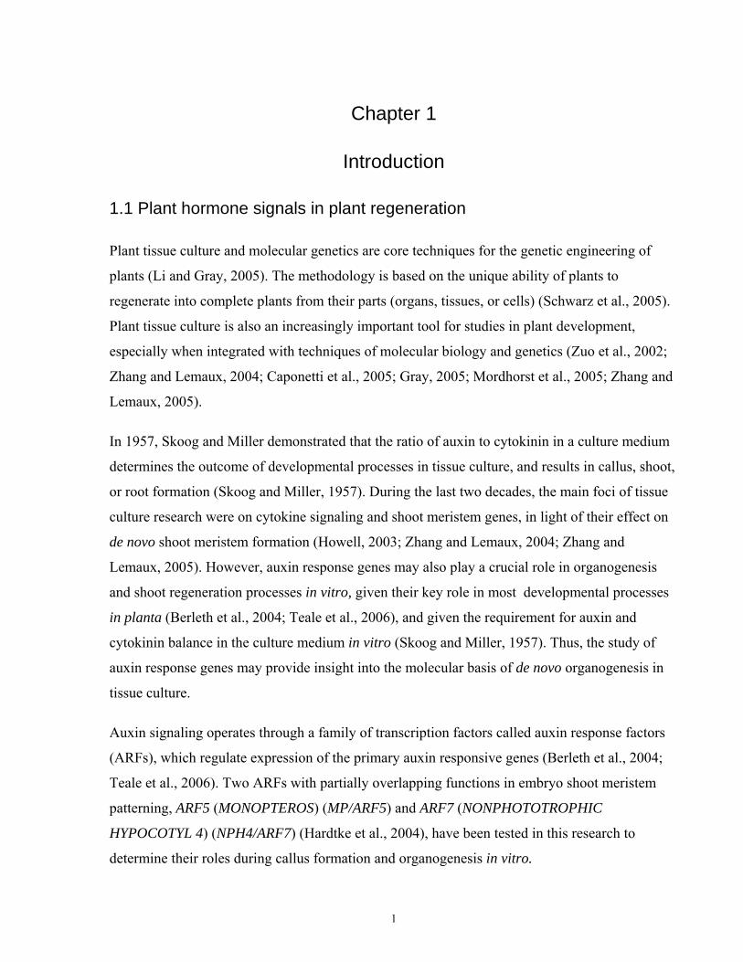
2004; Gray 2004; Weijers et al., 2005; Vanneste and Friml, 2009). A summary of auxin
signaling is represented in Fig. 1.1.
1.2.3 Aux/IAA genes and proteins
The 29 members of the Aux/IAA protein family are small, short-lived nuclear proteins, which
are transcriptional regulators of early auxin-response genes. Aux/IAA proteins are encoded by
the primary auxin response genes. Aux/IAA proteins consist of four domains (I-IV). Domain I
repress the transcription factors to which it becomes attached. Domain II contains a “degron”
sequence, through which AUX/IAA proteins are targeted towards the SCF/TIR ubiquitin ligase.
Interaction between AUX/IAA proteins and SCF/TIR is enhanced by auxin. Through this
mechanism, auxin promotes the ubiquination and degradation of Aux/IAA proteins (Gray et al.,
2001; Berleth et al., 2004; Vanneste and Friml, 2009). Domains III and IV play an important role
in homo- and hetero-dimerization with other Aux/IAA proteins and ARFs (Ulmasov et al.,
1999b; Berleth et al., 2004; Vanneste and Friml, 2009). The functions of Aux/IAA dimers are
not clear (Paciorek and Friml, 2006). Heterodimerization of Aux/IAA proteins with ARFs results
in the ARFs’ repression (Ulmasov et al., 1999b; Berleth et al., 2004; Vanneste and Friml, 2009).
When the concentration of auxin in a cell is low, Aux/IAA proteins are available to bind to the
corresponding ARFs, resulting in the inhibition of ARF. When the auxin concentration in a cell
increases, the Aux/IAA proteins are targeted for degradation, which results in the ARFs’ de-
4
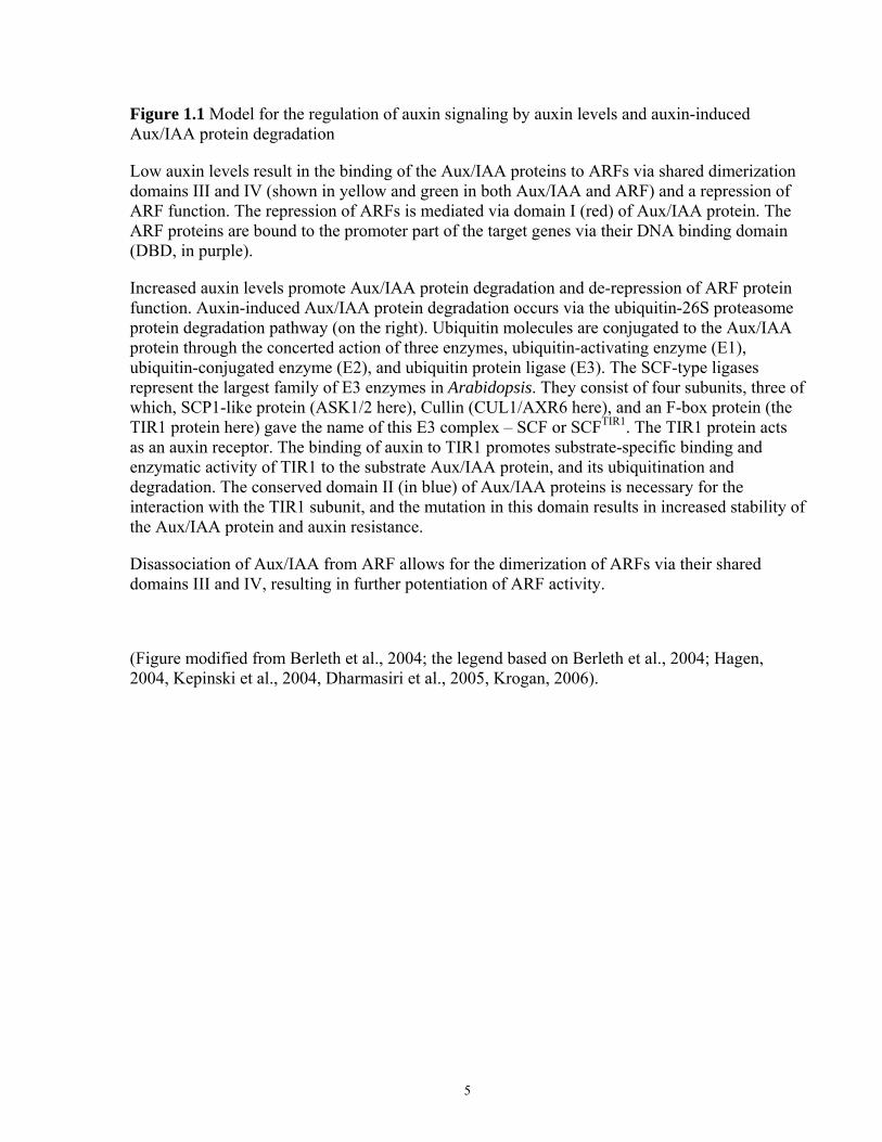
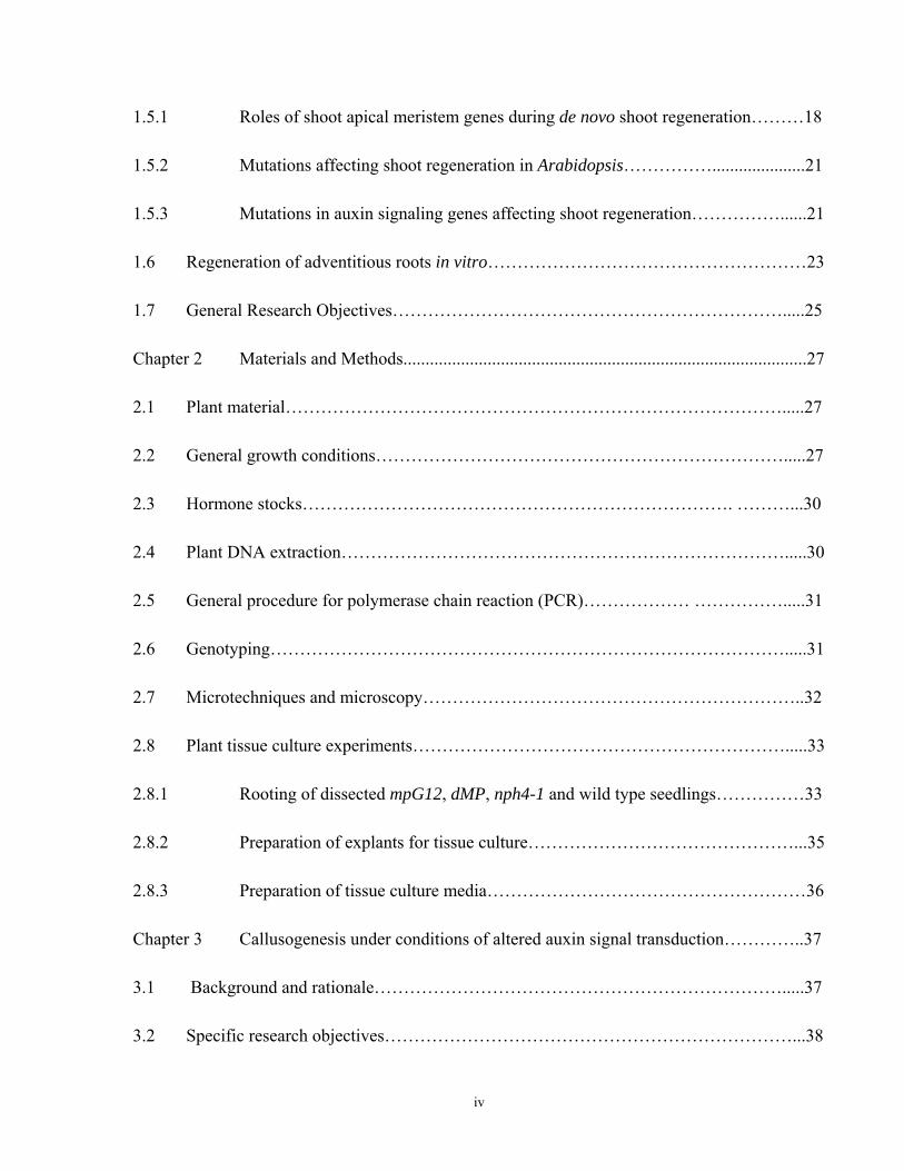
Figure 1.1 Model for the regulation of auxin signaling by auxin levels and auxin-induced Aux/IAA protein degradation
Low auxin levels result in the binding of the Aux/IAA proteins to ARFs via shared dimerization domains III and IV (shown in yellow and green in both Aux/IAA and ARF) and a repression of ARF function. The repression of ARFs is mediated via domain I (red) of Aux/IAA protein. The ARF proteins are bound to the promoter part of the target genes via their DNA binding domain (DBD, in purple).
Increased auxin levels promote Aux/IAA protein degradation and de-repression of ARF protein function. Auxin-induced Aux/IAA protein degradation occurs via the ubiquitin-26S proteasome protein degradation pathway (on the right). Ubiquitin molecules are conjugated to the Aux/IAA protein through the concerted action of three enzymes, ubiquitin-activating enzyme (E1), ubiquitin-conjugated enzyme (E2), and ubiquitin protein ligase (E3). The SCF-type ligases represent the largest family of E3 enzymes in Arabidopsis. They consist of four subunits, three of which, SCP1-like protein (ASK1/2 here), Cullin (CUL1/AXR6 here), and an F-box protein (the TIR1 protein here) gave the name of this E3 complex – SCF or SCFTIR1. The TIR1 protein acts as an auxin receptor. The binding of auxin to TIR1 promotes substrate-specific binding and enzymatic activity of TIR1 to the substrate Aux/IAA protein, and its ubiquitination and degradation. The conserved domain II (in blue) of Aux/IAA proteins is necessary for the interaction with the TIR1 subunit, and the mutation in this domain results in increased stability of the Aux/IAA protein and auxin resistance.
Disassociation of Aux/IAA from ARF allows for the dimerization of ARFs via their shared domains III and IV, resulting in further potentiation of ARF activity.
(Figure modified from Berleth et al., 2004; the legend based on Berleth et al., 2004; Hagen, 2004, Kepinski et al., 2004, Dharmasiri et al., 2005, Krogan, 2006).
5
IAA
REPRESSON
ACTIVATION
POTENTIATION
LOW AUXIN
HIGH AUXIN
E1
E2
E3 SCF complex
TIR1
6
repression and their activation as transcriptional factors. As long as the auxin concentration
remains high, Aux/IAA proteins turn over at a high rate due to the targeted proteolysis (Hagen et
al., 2004). The rapid, auxin-dependent turnover of Aux/IAA proteins makes them highly
responsive to changes in auxin signaling (Gray and Estelle, 2000).
Some AUX/IAA protein interactions with ARFs have been investigated. IAA28 interacts with
ARF5, 6, 7, 8 and 19 (Rybel et al., 2010); IAA14/SLR and IAA13/SHY2 interact with
NPH4/ARF7 and ARF19 (Peret et al., 2009, and references therein). IAA12/BDL interact with
MP/ARF5 (Hamann et al., 2002; Weijers et al., 2005; Rybel et al., 2010). Gain-of-function
(GOF) mutations in AUX/IAA proteins typically affect the “degron” in domain II and lead to
stable AUX/IAA, whose abundance is no longer affected by auxin (Gray et al., 2001; Weijers et
al., 2005). GOF mutants of iaa12/bdl, and, to a lesser degree, with constitutively repressed
MP/ARF5 function, cause phenotypes similar to those found in mp mutants (Hamann et al.,
2002; Hardtke et al., 2004; Weijers et al., 2005). GOF mutations in iaa28 and iaa14/slr cause a
lack of lateral roots, similar to that found in nph4 arf19 double mutants (Rogg et al., 2001;
Okushima et al., 2005; Peret et al., 2009; Rybel et al., 2010).
1.2.4 ARF genes and proteins
ARFs are transcription factors encoded by a gene family of 23 members. ARFs are characterized
by a conservative amino-terminal DNA binding domain (DBD), which interacts directly with the
auxin response element (AuxRE). AuxREs (TGTCTC) are found in the promoters of most
primary auxin-response genes. ARFs comprise another, only moderately conserved domain,
called the middle domain, which determines the functional properties of an ARF as a
transcriptional activator or repressor. Carboxy-terminal domains III and IV are similar to those in
AUX/IAA proteins, and mediate dimerization with Aux/IAAs or other ARFs (Ulmasov et al.,
1999a, Ulmasov et al., 1999b; Berleth et al., 2004). Recently Shin et al. (2007) showed that ARF
activity can also be modulated by interaction with other factors, such as MYB77, which increase
the combinatorial possibilities in auxin-dependent transcriptional regulation.
Whether through differential expression, differential affinities for target promoters, or both,
individual ARFs have a distinct function in developmental processes (Vogler and Kuhlemeier,
7
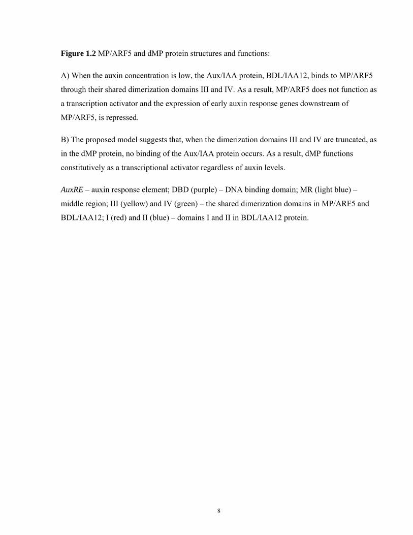
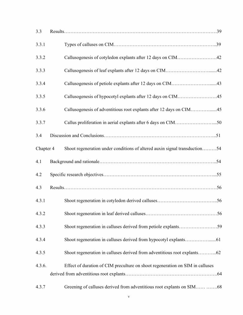
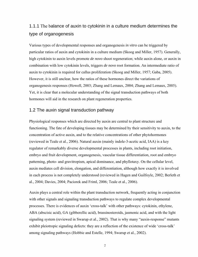
Figure 1.2 MP/ARF5 and dMP protein structures and functions:
A) When the auxin concentration is low, the Aux/IAA protein, BDL/IAA12, binds to MP/ARF5
through their shared dimerization domains III and IV. As a result, MP/ARF5 does not function as
a transcription activator and the expression of early auxin response genes downstream of
MP/ARF5, is repressed.
B) The proposed model suggests that, when the dimerization domains III and IV are truncated, as
in the dMP protein, no binding of the Aux/IAA protein occurs. As a result, dMP functions
constitutively as a transcriptional activator regardless of auxin levels.
AuxRE – auxin response element; DBD (purple) – DNA binding domain; MR (light blue) –
middle region; III (yellow) and IV (green) – the shared dimerization domains in MP/ARF5 and
BDL/IAA12; I (red) and II (blue) – domains I and II in BDL/IAA12 protein.
8
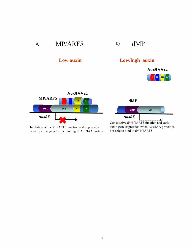
Inhibition of the MP/ARF5 function and expression of early auxin gene by the binding of Aux/IAA protein
AuxRE
I IMP/ARF5
Aux/IAA12
MP/ARF5
Constitutive dMP/dARF5 function and early auxin gene expression when Aux/IAA protein is not able to bind to dMP/dARF5
AuxRE
DBDDBD MRMR
dMP
IVI I II II
Aux/IAA12
I
DBDDBD MRMR IVIVI I II I I
dMPa) b)
Low/high auxin
III IV
Low auxin
9
2003). However, the precise functions and downstream gene targets of most ARFs are still not
known (Okushima et al., 2005; Guilfoyle and Hagen, 2007).
A systematic reverse genetics approach with ARF genes failed to reveal an abnormal phenotype
of most ARF mutations, which suggested that most of the ARFs have largely redundant functions
(Okushima et al., 2005; Teale, 2006). The analyses of ARF1 to ARF10 have shown that their
mRNAs are ubiquitously expressed in most major organs, as well as in cultured undifferentiated
Arabidopsis cells (Ulmasov et al., 1999b). Participation in multiple transduction pathways was
confirmed for several ARFs. For example, ARF19 participates in both auxin and ethylene
signaling (Li et al., 2006), and ARF6 and ARF8 promote jasmonic acid production (Nagpal et
al., 2005). Unlike Aux/IAA genes, ARF genes do not belong to the group of early auxin response
genes. ARFs have been found to be permanently expressed and bound to promoters of the target
genes containing the AuxRE sequence. ARF functions are activated when the auxin
concentration increases and Aux/IAA proteins dissociate from ARFs, allowing a homo- or
hetero-dimerization of ARFs with each other (Berleth et al., 2004; Hagen et al., 2004).
Interestingly, there is a growing body of information on posttranscriptional regulation of ARFs’
mRNA levels by micro-RNAs, and by small interfering RNAs. This posttranscriptional
regulation of ARFs seems to be as important as their transcriptional regulation (Guilfoyle and
Hagen, 2007).
1.2.5 Auxin response factors MONOPTEROS (MP/ARF5) and NON-
PHOTOTROPIC HYPOCOTYL 4 (NPH4/ARF7)
Loss-of-function mutations have been identified by single mutant phenotype in only 4 of 23
ARFs, arf2/hss, arf3/ett, arf5/mp, and arf7/nph4 (Okushima et al., 2005). They include those in
MONOPTEROS (MP/ARF5) and NON-PHOTOTROPIC HYPOCOTYL 4 (NPH4/ARF7). Both
belong to the group of transcriptional activators with corresponding middle domains (Ulmasov et
al., 1999a; Ulmasov et al., 1999b). Both ARFs, MP and NPH4, are capable of homo- and hetero-
dimerization with each other (Hardtke et al., 2004). Especially in their DNA binding domains,
MP and NPH4 have a high degree of sequence similarity, and they have partially redundant
functions (Liscum and Reed, 2002; Hardtke et al., 2004; Remington et al., 2004; De Smet et al.,
2010).
10
Loss of function (LOF) mutations in MP interfere with embryonic patterning, and with the
formation of embryonic hypocotyl and primary roots (Mayer et al., 1991; Berleth and Jurgens,
1993). LOF mutations in MP also severely reduce auxin sensitivity, auxin transport, and vascular
development (Li et al., 2006; Przemeck et al., 1996; Mattsson et al., 2003; Schuetz et al., 2008).
Finally, they result in the formation of “pin-like” inflorescences, which lack leaves and flowers
(Berleth and Jurgens, 1993; Przemeck et al., 1996). The expression of PIN1 (auxin transport) and
DR5 (auxin response) reporters is defective in mp mutant leaves, suggesting that these responses
were diminished in mp mutants (Mattsson et al., 2003; Wenzel et al., 2007).
LOF mutations in NPH4 lead to hypocotyls that no longer bend towards the light, as well to
changes in leaf shapes and in other aspects of post-embryonic morphology (Stowe-Evans et al.,
1998). LOF nph4 mutants exhibit auxin resistance, and severely impaired expression of many
primary auxin-dependent genes (Okushima et al., 2005). Expression of the auxin-responsive
reporter DR5 was also significantly reduced in nph4 mutants (Stowe-Evans et al., 1998; Wang et
al., 2005). Also, Watahiki and Yamamoto (1997) demonstrated that nph4 mutants showed a 15 to
20 fold increase in auxin resistance (2,4-D) in the aerial parts, whereas the root parts were of
normal sensitivity to auxin (Watahiki and Yamamoto, 1997; Okushima et al., 2005). In the
presence of exogenous IAA, the excised nph4 roots formed fewer lateral roots, and the nph4
excised hypocotyls formed fewer adventitious roots than did wild-type roots and hypocotyls
(Wilmoth et al., 2005). However, almost no adventitious or lateral roots were formed in nph4
arf19 double mutants (Okushima et al., 2005; Wilmoth et al., 2005).
The developmental functions of MP and NPH4 are far more related than is suggested by their
single mutant phenotypes. Both MP/ARF5 and NPH4/ARF7 can act in the regulation of
embryonic pattern formation (Hardtke et al., 2004). Although the single mutant phenotype of
NPH4 implicates the gene only in conditional responses during post-embryonic development,
NPH4 is already expressed in embryos, and LOF mutations in NPH4 dramatically enhance the
mp embryo phenotype (Hardtke et al., 2004).
MP is expressed in broad domains in embryos and in emerging organs. The early functions of the
MP gene are related to vascular and body patterning, and to organ initiation (Hardtke and
Berleth, 1998; Hardtke et al., 2004; Schuetz et al., 2008; Cole et al., 2009; De Smet et al., 2010).
However, upon organ maturation, the expression of MP is gradually narrowed to the central
11
domains, and then to the vascular tissues (Hardtke et al., 2004). By contrast, NPH4 is fairly
ubiquitously expressed throughout plant development (Hardtke et al., 2004; Okushima et al.,
2005).
Selective deletion of domains III and IV of MP results in a GOF version of MP that is associated
with the dominant enhancement of auxin-related traits. Therefore, it was named dMP (for
dominant MONOPTEROS) (Krogan et al., 2006). Those traits included increased adventitious
root formation, when dMP cotyledons were rooted in a liquid medium containing IBA (Krogan
et al., 2006). By contrast, adventitious root formation in LOF mp cotyledons was strongly
decreased (Mattsson et al., 2003). Also, MP together with PIN1 is involved in the process of
embryonic shoot meristem formation and patterning, through control of the expression of the
meristem genes CUC2 and STM (Aida et al., 2002). It is also involved in the regulation of
meristem size (Schuetz et al., 2008); and in the regulation of the activity of some A-type
ARABIDOPSIS RESPONSE REGULATORS (A-type ARRs, negative cytokinin response
regulators) in SAM (Zhao et al., 2010). The MP/ARF5 protein interacts with BODENLOS
(IAA12/BDL) resulting in inhibition of the MP/ARF5 function (Fig.1.2 A; Hardtke et al., 2004;
Weijers et al., 2005). However, dMP is partially epistatic over bdl (Krogan et al., 2006).
1.3 Roles of cytokinins
Cytokinins were discovered by their ability to stimulate plant cell division (del Pozo et al., 2005,
and references therein). Later, Skoog and Miller introduced their hypothesis, according to which
plant morphogenesis is under the control of the auxin to cytokinin ratio (Skoog and Miller,
1957). Cytokinins control key aspects of plant growth and development, such as cell division,
shoot meristem initiation, control of the stem cell pool in the shoot apical meristem, leaf and root
differentiation, vascular patterning, chloroplast biogenesis, photomorphogenesis, fertility, seed
development, stress tolerance, and senescence (reviewed in Muller and Sheen, 2007a; Muller and
Sheen, 2007b). Together with auxin, cytokinin can reprogram terminally differentiated cells to
stem cells, and initiate de novo shoot formation in culture (Gaba, 2005; Zhang and Lemaux,
2005). The most common cytokinin in plants is zeatin (Davies, 2004). Although the important
sites of cytokinin biosynthesis seem to be in the root meristems (Davies, 2004; Nordstrom et al.,
2004), it has been shown recently that cytokinin biosynthesis occurs in certain quantities in all
types of aerial organs. The major subcellular compartments of cytokinin biosynthesis are
12
plastids. The major steps in cytokinin signaling are reviewed in detail in Maxwell and Kieber
(2004), and in Muller and Sheen (2007 a, b).
Many studies were done to illustrate the interaction of auxin and cytokinin signaling pathways,
and their mutual control of growth and differentiation in plants (Coenen and Lomax, 1997;
Nordstrom et al., 2004; Moubayidin et al., 2009; Zhao et al., 2010). The existence of complex
and multilevel networks of synergistic, antagonistic, and additive interactions between these two
plant hormones and their signaling systems have been demonstrated in a number of studies (Klee
and Estelle, 1991; Hobbie and Estelle, 1994; Coenen and Lomax, 1997; Nordstrom et al., 2004;
Moubayidin et al., 2009; Zhao et al., 2010). One of the indicators of interaction, between auxin
and cytokinin signaling pathways, is the number of mutants cross resistant to both hormones
(Hobbie and Estelle, 1994; Hobbie et al., 1994; Coenen and Lomax, 1997; Swarup et al., 2002;
Moubayidin et al., 2009). Auxin signaling mutants, such as aux1, axr1, axr2, axr3, iaa28, and
shy2, have demonstrated pleiotropic signaling defects affecting other signaling pathways,
including cytokinin signaling (Hobbie and Estelle, 1994; Swarup et al., 2002). Also, several
mutants, initially characterized as affected in cytokinin perception, have been shown to have
altered auxin responses (Smalle et al., 2002; Tajima et al., 2004; Laxmi et al., 2006). The
molecular basis of auxin-cytokinin cross talk is largely unclear (Gray, 2004; Hartig and Beck,
2006), although important advances in its understanding have been recently made (reviewed in
Moubayidin et al., 2009).
1.4 Cell proliferation and callusogenesis in plant tissue culture
Plant callusogenesis is a process also observed under natural growth conditions, where two types
of triggers can lead to the development of callus tissues – tumor formation in response to
infection by Agrobacterium tumefaciens, and wounding with subsequent regeneration of
damaged tissues (Frank et al., 2000; Gordon et al., 2007). Callus proliferation can be induced in
vitro, when the culture medium contains a certain ratio of auxin to cytokinin (Skoog and Miller,
1957). Cell proliferation during callusogenesis leads to disruption of the ordered morphology of
differentiated tissues (Cary et al., 2002).
The mechanisms leading to callus proliferation are not well understood (Cary et al., 2002; Zhang
and Lemaux, 2005). Generally, cell-cycle genes, such as cyclins and cyclin dependent kinases
(CDKs), play a major role in the mediation of hormonal effects, or in response to wounding
13
signals, leading to cell proliferation (Hemerly et al., 1993; Soni et al., 1995; Shaul et al., 1996;
Riou-Khamlichi et al., 1999; Sugiyama, 1999; Richard et al., 2002; Che et al., 2007). It has been
shown that the level of CDK expression correlates with competence for cell proliferation
(Hemerly et al., 1993; Zhang et al., 2005); and, CDK expression and cell proliferation are
increased upon wounding in tissue culture (Hemerly et al., 1993).
Callus initiation, callus growth on callus inducing medium (CIM), and subsequent shoot
formation on SIM were completely prevented in the Arabidopsis root explants cultured during
the first 1 to 2 days on the CIM supplied with reversible cell cycle inhibitors aphidicolin (APH)
or hydroxyurea (HU), which block G1-S transition (Che et al., 2007). Overexpression of cyclin
D3 (CycD3) resulted in cytokinin-independent callus growth, but these calluses failed to
regenerate shoots (Riou-Khamlichi et al., 1999). Richard et al. (2002) found that, during the
initiation of a cell suspension culture, the different members of the CDK or CYC families were
differentially up-regulated by mitogenic factors, including auxin and cytokinin. Moreover, cis-
acting regulatory elements specific for particular plant hormones, such as AuxREs for auxin,
were found in the promoters of the cell cycle genes (Richard et al., 2002). Auxin responsive
elements (AuxREs) were found in the promoter regions of many cyclin genes, indicating that
they might be primary auxin responsive genes (Perrot-Rechenmann, 2010).
Gordon et al. (2007) studied the dynamics of the auxin and cytokinin responses, and the pattern
of the distribution of hormonal responses during developmental processes in tissue culture.
According to their data, auxin responses, visualized by DR5 and PIN1 driven reporters, were
strongly increased only during the initiation of callus proliferation on CIM, but they were
downregulated after 5 days on CIM. They were not observed within the large callus outgrowths
after 1 week on CIM, and were not detected after 10 days on CIM. Conversely, the expression of
the cytokinin signaling marker pARR5::GFP, and of CUP-SHAPED COTYLEDON 1 and 2
(CUC1 and CUC2), were detected in an already established culture after 8 days on CIM. The
signal expanded throughout the callus after 2 weeks of induction (Gordon et al., 2007). No
expression of WUSCHEL (WUS), SHOOT MERISTEMLESS (STM), CLAVATA3 (CLV3),
FILAMENTOUS FLOWER (FIL), or REVOLUTA (REV) reporters was detected during 2 weeks
on CIM, indicating that expression of these key SAM genes require incubation on SIM (Cary et
al., 2002; Gordon et al., 2007). Interestingly, both the CUC2 and ARR5 reporters were expressed
at the site of callus formation, even on the medium lacking cytokinin, and containing auxin as the
14
sole hormone. By contrast, the culture of explants with the hormone kinetin as the sole hormone
did not lead to callus proliferation and CUC2 reporter expression, suggesting that auxin
containing medium is required for callus proliferation and the CUC2 gene expression (Gordon
et al., 2007).
Global analysis of gene expression during the first 4 days of callus proliferation on CIM
demonstrated that more than 250 genes were significantly upregulated, and about 500 genes were
downregulated, during the early stages of callus formation, usually associated with the stage of
acquisition of competence for organogenesis (Che et al., 2006). A dramatic increase in the
expression of genes involved in hormone responses, particularly Aux/IAA genes, was detected on
the second day of CIM incubation. These genes were downregulated after the transfer of explants
on SIM (Che et al., 2002). When cells proliferated and formed callus tissue (after 10 days of
CIM incubation), the main categories of upregulated genes were stress-related transcription
factors and stress-related proteins (Che et al., 2006). However, the authors concluded that the
genes associated with the acquisition of competence, and those specific for callus formation,
were more difficult to categorize compared to those that are part of the characterization of gene
expression during shoot formation on shoot inducing medium (SIM), or root formation on root
inducing medium (RIM) (Che et al., 2006).
Based on the requirement of a high auxin concentration in CIM (Skoog and Miller, 1957),
mutations in auxin signaling most likely can interfere with callusogenesis processes. Thus,
explants of the auxin-resistant mutant axr1-3 required more auxin for callus induction, and
tolerated more auxin to grow calluses, while sensitivity to kinetin was normal in tissue culture
(Lincoln et al., 1990; Kubo and Kakimoto 2000; Sieberer et al., 2003). Another well known
auxin-resistant mutant, aux1, also required higher auxin concentrations for callus initiation, and
demonstrated a tendency to form roots rather than calluses on CIM (Kakani et al., 2009). Altered
auxin transport in pin1 mutants resulted in the proliferation of callus-like tissues, instead of
lateral roots, when exogenous auxin was applied to the roots (Benkova et al., 2003).
1.5 de novo organogenesis in plant tissue culture
Plant tissues can potentially develop into somatic embryos, shoots, and roots. The specificity of
the regeneration is determined by the hormone composition of the culture medium (Schwarz et
al., 2005, and references therein). Primordia originate de novo from callus cells. The cell, or
15
cells, that can be direct progenitors are stimulated to undergo a number of rapid cell divisions,
resulting in the formation of a meristemoid. A meristemoid represents an aggregate of small,
isodiametric, thin-walled, micro-vacuolated cells. Early in their development, meristemoids are
capable of developing into different types of organ primordia (Schwarz et al., 2005, and
references therein; Atta et al., 2009). The capabilities of plants for de novo shoot and root
organogenesis are among the most important qualities required for successful micropropagation
in vitro (Schwarz et al., 2005).
Organogenesis in vitro can be direct or indirect. In direct organogenesis, explants are fully
competent to respond to inductive hormones, and do not require a callus proliferation phase
(Schwarz et al., 2005). Recently, the capability of Arabidopsis thaliana roots and hypocotyl
explants for direct shoot organogenesis has been described (Atta et al., 2009). In general,
however, the explants of most plants, including Arabidopsis, can undergo organogenesis only
following a callus growth phase on CIM. During this phase, it is hypothesized that genetic or
developmental factors blocking the competence to respond to the inductive signals (such as
inducing shooting or rooting hormone ratio in tissue culture medium) are overcome. As a result,
many efforts to induce organogenesis focus on overcoming these blocks to the competence to
respond to the inductive signals (Cary et al., 2002, and references therein).
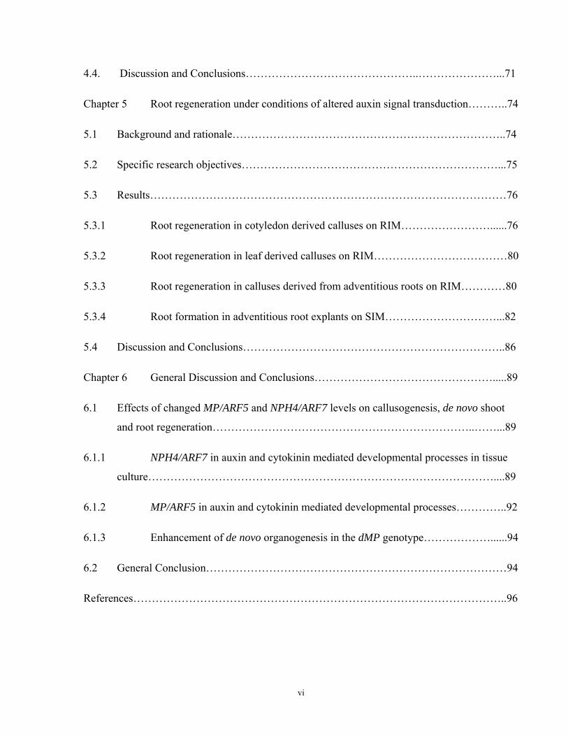
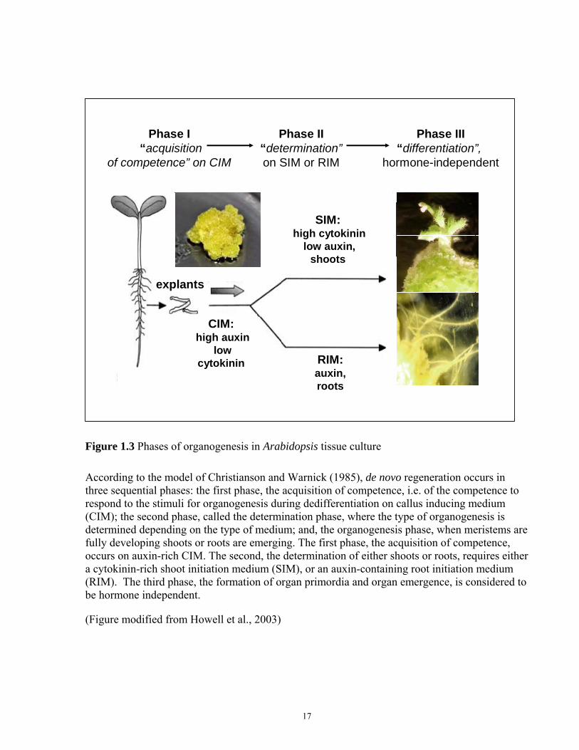
According to the most commonly accepted model (Christianson and Warnick, 1985; Schwarz et
al., 2005), regeneration occurs in three sequential phases (Fig. 1.3): first, when explants are
cultured on CIM, they acquire competence to respond to organogenesis stimuli during the
following stage (on SIM or RIM); second, the determination phase, shortly after calluses are
transferred either to SIM or RIM, when the type of organogenesis (shoot or root regeneration) is
determined, and, third, the organogenesis phase, during which meristems are forming, and
shoots/roots emerging. The third phase, the formation of organ primordia, and organ emergence,
is considered to be hormone independent (Christianson and Warnick, 1985). Ozawa et al. (1998)
argued that “competence for shoot regeneration is assumed to be always acquired additively,
over and above the competence for root redifferentiation”. Shown first in leaf explants of
Convolvulus arvensis (Christianson and Warnick, 1983, 1984, 1985, 1988), the organogenic
phases were widely confirmed for other plant species and cultivars, including pine, tobacco,
apple, Arabidopsis, mangosteen, etc. (Flinn et al., 1988; Attfield and Evans, 1991; Hicks, 1994;
16
Phase I“acquisition
of competence” on CIM
Phase II“determination”on SIM or RIM
Phase III“differentiation”,
hormone-independent
CIM:high auxin
low cytokinin
explants
SIM:high cytokinin
low auxin,shoots
RIM:auxin,roots
Figure 1.3 Phases of organogenesis in Arabidopsis tissue culture
According to the model of Christianson and Warnick (1985), de novo regeneration occurs in three sequential phases: the first phase, the acquisition of competence, i.e. of the competence to respond to the stimuli for organogenesis during dedifferentiation on callus inducing medium (CIM); the second phase, called the determination phase, where the type of organogenesis is determined depending on the type of medium; and, the organogenesis phase, when meristems are fully developing shoots or roots are emerging. The first phase, the acquisition of competence, occurs on auxin-rich CIM. The second, the determination of either shoots or roots, requires either a cytokinin-rich shoot initiation medium (SIM), or an auxin-containing root initiation medium (RIM). The third phase, the formation of organ primordia and organ emergence, is considered to be hormone independent.
(Figure modified from Howell et al., 2003)
17
de Klerk et al., 1995; Lakshmanan et al., 1997; Sugiyama, 1999; Ozawa et al., 1998; Banno et
al., 2001; Cary et al., 2002; Che et al., 2007).
During in vitro organogenesis, a high cytokinin level in SIM initiates and promotes the processes
of meristemoid specification, and the “canalization” of a developmental program for the
formation of shoot primordia (Schwarz et al., 2005). In Arabidopsis, the origin of the shoot
meristem in vitro has recently been shown to be initiated from dividing xylem pericycle cells, or
from lateral root meristem - like protuberances, which are formed during culture on CIM (Che et
al., 2007; Atta et al., 2009; Sugimoto et al., 2010). The pericycle cells remain more meristematic
than the surrounding cells, and retain the expression of cell cycle genes. In particular, the xylem
pericycle cells have a higher capacity to re-enter the cell cycle, and to form new meristems when
compared to the other cells (Atta et al., 2009).
Many authors have indicated that the ability for shoot regeneration differs dramatically among
Arabidopsis ecotypes. In particular, the Columbia (Col) ecotype, widely used in research, has a
low shoot regeneration potential (Akama et al., 1992; Cary et al., 2002; Zhao et al., 2002;
Chatfield and Raizada, 2008). As a rule, Arabidopsis ecotypes more capable of shoot
regeneration, such as C24, and Wasilewskija, need less time on SIM to be committed to
regeneration, and to form green foci and shoots (Cary et al., 2002; Zhao et al., 2002). The
regeneration potential also varies depending on the types of explants, and on explant age - partly
because of the different endogenous hormone concentrations (Akama et al., 1992; Zhao et al.,
2002; Gordon et al., 2007).
1.5.1 Roles of shoot apical meristem genes during de novo shoot
regeneration
Several key genes, many encoding transcription factors, are known to have critical roles in the
initiation and maintenance of a shoot apical meristem (SAM). Some are expressed as early as the
embryonic heart stage, and usually play crucial roles in post-embryonic meristem maintenance
(Aida et al., 1999; Long et al., 1996; Cary et al., 2002). The SHOOT MERISTEMLESS (STM)
gene of Arabidopsis encodes a specific, knotted-like 1 (KN1-like), type homeodomain protein
(Barton and Poethig, 1993; Zhang and Lemaux, 2005). The STM gene is required for SAM
formation and maintenance (Long et al., 1996). It promotes cell division, and suppresses cell
18
differentiation in the SAM (Lenhard et al., 2002). The WUSCHEL (WUS) gene is a member of
another subtype of homeodomain transcription factors, required for specifying and maintaining a
stem cell population in the SAM (Mayer et al., 1998; Zhang and Lemaux, 2005). The WUS and
STM genes are activated independently, but mutations in WUS results in the loss of expression of
STM and vice versa (Lenhard et al., 2002). The CLAVATA3 (CLV3) gene encodes a small
protein, which appears to be a ligand for CLV1 receptor kinase (Fletcher et al., 1999). CLV1 and
CLV3 restrict cell proliferation activity in SAM (Fletcher et al., 1999). The CLV3 gene is
activated by WUS, and functions in a negative feedback loop that antagonizes WUS activity, and
thereby controls the size of the central stem cell population (Gallois et al., 2004, and references
therein). The redundant genes CUP-SHAPED COTYLEDON 1 and 2 (CUC1 and CUC2) encode
transcription factors with similarities to the NO APICAL MERISTEM (NAC) protein. The latter
acts in the development of embryos and flowers in petunias (Aida et al., 1997, and references
therein; Takada et al., 2001). These types of proteins are known to play important roles in
establishing SAM and in separating cotyledons (Aida et al., 1997, and references therein; Takada
et al., 2001). Loss of function mutations in STM, WUS, or CUC1 and CUC 2 (in double mutants)
block SAM formation in planta (Cary et al., 2002; Hibara et al., 2003). All four genes are also
used as reporters of shoot meristem initiation in vitro (Cary et al., 2002; Zhang and Lemaux,
2005; Gordon et al., 2007). De novo shoot organogenesis can be a useful model for the study of
developmental processes in the SAM (Cary et al., 2002; Che et al., 2002; Che et al., 2006;
Gordon et al., 2007).
The dynamics of gene expression of STM, CUC1, CUC2, WUS, and CLV1 during shoot
regeneration have been studied (Cary et al., 2002; Daimon et al., 2003). They showed that CUC1
and CUC2 were strongly expressed at 3 to 6 days on SIM, before the expression of other
markers, and prior to shoot commitment, or any visible green foci or shoot meristem
organization. The increase of the WUS gene expression occurred after 6 days on SIM, and
gradually increased until 15 days on SIM. STM and CLV1 transcript levels rose later than those
of WUS, CUC1, and CUC2, and were significantly higher only after 11 -15 days on SIM, at
about the time of shoot commitment (Cary et al., 2002).
A live imaging approach enabled visualization of the expression of CUC1 and CUC2 via a GFP
fluorescent marker (Cary et al., 2002). This showed that the initially broad expression during
preculture on auxin rich CIM, and the first days on SIM, was progressively localized to the sites
19
of presumptive shoot formation at later stages, finally coinciding with green foci formation and
the development of SAMs (Cary et al., 2002; Gordon et al., 2007). The authors hypothesized that
“restriction of gene expression to the new pattern might be a critical event in shoot commitment”
(Cary et al., 2002). Similar results were obtained by another group, when they analyzed the
expression patterns of the CUC1, CUC2, and STM genes on CIM, and on SIM (Daimon et al.,
2003).
Gordon et al. (2007) studied the dynamics and patterns of expression of several major SAM
genes, such as CUC2, WUS, STM, PIN1, REV, CLV1, and FIL, as well as cytokinin (ARR5) and
auxin (DR5) reporters, during de novo SAM formation in Arabidopsis tissue culture. The authors
demonstrated that CUC2 and PIN1 were expressed within the same domains in the callus tissues
on CIM and in the promeristems on SIM. The distribution of the signals from the hormone
reporters ARR5 and DR5 showed that shoot meristems can be initiated in areas of low auxin and
high cytokinin responses (Gordon et al., 2007). At later stages of meristem development, the
DR5 signal was detected following the PIN1 reporter upregulation at the sites of future leaf
primordia formation. In contrast to DR5, the cytokinin responsive ARR5 reporter was expressed
in the areas of shoot meristem initiation, and within the developing meristems; but, was
downregulated in leaf primordia, and in the areas of callus where no shoots or root meristems
were formed (Gordon et al., 2007). Based on the results, Gordon et al. (2007) suggested that de
novo shoot organogenesis can be broken down into distinct events: callus induction, cytokinin-
induced specification of cell identity within the callus, radial patterning within shoot progenitors,
and meristem morphogenesis. They hypothesized that the non-homogenous distribution of
cytokinin and auxin may be a key factor for the partition of the cell identity within a callus on
SIM. They also hypothesized that the hormone gradient must be gradually reorganized from the
disrupted initial conditions, during shoot induction in culture (Gordon et al., 2007).
In summary, the expression of key meristem genes during de novo SAM formation occurs during
late CIM (4 days) or early SIM (3-6 days) incubation times in Arabidopsis, which appears to be
related to the acquisition of competence on CIM, and to the shoot commitment stage during the
first days on SIM (Che et al., 2002; Che et al., 2006; Christianson and Warnick, 1985; Gordon et
al., 2007).
20
1.5.2 Mutations affecting shoot regeneration in Arabidopsis
Cytokinins mediate de novo shoot regeneration in tissue culture (Skoog and Miller 1957; Gaba,
2005). Accumulating evidence supports the idea of molecular interactions between cytokinin
signaling, the cell cycle, and shoot meristem developmental pathways (Riou-Khamlichi et al.,
1999; Sieberer et al., 2003; Zhang and Lemaux, 2004; Zhang and Lemaux, 2005). Key SAM
genes, and the genes of cytokinin signaling are among the primary candidates for study in tissue
culture to determine their possible effects on the efficiency of shoot regeneration in vitro (Zhang
and Lemaux, 2004; Zhang and Lemaux, 2005). Among the genes involved in meristem initiation
and maintenance, CUC1, CUC2, STM, WUS, ENHANCER OF SHOOT REGENERATION 1 and
2 (ESR1, and ESR2) showed a significant effect on shoot regeneration. Typically, mutations in
these genes affect shoot meristem initiation, but not the callus initiation stages (Barton and
Poethig, 1993; Aida et al., 1997; Banno et al., 2001; Daimon et al., 2003; Gordon et al., 2007).
An elevation of endogenous cytokinin levels generally results in increased shoot regeneration in
cytokinin overproducing mutants, and often in shoot formation on cytokinin-free or hormone-
free media (Chaudhury et al., 1993; Kunkel et al., 1999; Frank et al., 2000; Kakimoto, 2001;
Catterou et al., 2002; Zubko et al., 2002; Sun et al., 2003; Zhang and Lemaux, 2004; Zhang and
Lemaux, 2005). However, genes belonging to other signaling pathways can be implicated in
shoot regeneration as well, and some of them will be discussed in the following sections.
1.5.3 Mutations in auxin signaling genes affecting shoot regeneration
Although SIM contains auxin (Skoog and Muller, 1957), relatively limited information is
available about the direct effects of auxin, and auxin signaling genes on shoot regeneration in
tissue culture. Typically, auxin signaling genes are not among those reviewed in the context of
shoot regeneration in vitro (Zuo et al., 2002b; Howell, 2003; Zhang and Lemaux, 2004; Zhang
and Lemaux, 2005). Nevertheless, auxin response marker levels are highly elevated in shoot
meristems in planta (Kakani et al., 2009). Evidence of the effects of mutations in auxin signaling
genes on shoot regeneration has been demonstrated (Coenen and Lomax, 1998; Chatfield and
Raizada, 2008; Kakani et al., 2009). Also, many auxin signaling genes are implicated in cross
talk with other signaling pathways (Hobbie and Estelle, 1994). In addition, impairment of auxin
genes can affect coordinated interaction among multiple developmental processes (Gordon et al.,
2007; Kakani et al., 2009).
21
For example, the AUXIN RESISTANT1 (AUX1) gene encodes a permease-like membrane protein,
facilitating auxin influx into plant cells. Loss of function aux1 mutants result in resistance to
auxin, ethylene, and cytokinin (Hobbie and Estelle, 1994). The AUX1 gene is highly expressed
within the SAM, in the root tips of plants (Swarup et al., 2004), and in callus culture (Kakani et
al., 2009). Remarkably, aux1 LOF mutants regenerated roots instead of shoots on SIM (Kakani
et al., 2009).
Loss of the PIN1 gene function, encoding the auxin efflux protein and facilitating polar auxin
transport, resulted in a moderate decrease of shoot regeneration (Gordon et al., 2007). However,
the PIN gene family consists of eight members with partly redundant functions. Therefore an
effect on shoot regeneration could be more prominent if multiple PIN mutations were tested in
tissue culture.
The ALTERED AUXIN RESPONSE (AXR1) gene encodes a subunit of the protein RUB1 which
is required for auxin-dependent ubiquitin-mediated degradation of Aux/IAA proteins (Gray et
al., 2001; del Pozo et al., 2002). The AXR1 gene is highly expressed in growing cells throughout
plant organs. Loss of AXR1 function results in decreased auxin responses, auxin resistance in all
plant organs, reduced apical dominance, fewer lateral roots and root hairs, low fertility, impaired
callusogenesis, and abnormal expression of the Aux/IAA and SAUR genes (Lincoln et al., 1990;
del Pozo et al., 2002). Cross-resistance to cytokinin was also reported for axr1 mutants (Hobbie
and Estelle, 1994). Remarkably, the axr1 mutants were dramatically impaired in shoot
regeneration, which was abolished in strong axr1 alleles, and reduced to only 2-3% in weaker
alleles (Chatfield and Raizada, 2008). Similar to aux1 mutants, axr1 mutants also developed
roots rather than shoots on SIM (Chatfield and Raizada, 2008).
Constitutively auxin-overproducing superroot1 (sur1) explants demonstrated auxin-autonomous
growth, proliferation, and root formation on a hormone free medium. Unlike the wild type, they
were capable of shoot regeneration on a medium with cytokinin as the sole hormone (Boerjan et
al., 1995). In contrast, superroot2 (sur2) explants, which also demonstrated elevated auxin
levels, failed to produce shoots on a cytokinin containing medium (Delarue et al., 1998). The
authors discussed the difference between the sur1 and sur2 phenotypes. They proposed that,
while sur1 constantly overproduces auxin, sur2 accumulates auxin as a result of an impaired
mechanism of auxin conjugation, and possibly of impaired auxin transport (Delarue et al., 1998).
22
1.6 Regeneration of adventitious roots in vitro
In tissue culture, regeneration of adventitious roots occurs in response to high auxin and low
cytokinin levels, or when auxin is the sole hormone in the root inducing medium (RIM) (Miller
and Skoog, 1957; Gaba, 2005). Rooting is often a critical step in plant propagation, because
many species are extremely recalcitrant to adventitious root formation (de Klerk, 2002; Schwarz
et al., 2005). Although roots have been routinely induced in response to auxin in plant
propagation (Callis, 2005; Schwarz et al., 2005), the molecular mechanisms of this event are not
completely clear (Schwarz et al., 2005; Rose et al, 2006).
Three temperature-sensitive mutants of Arabidopsis, shoot redifferentiation 1, 2, and 3 (srd1,
srd2 and srd3), defective in shoot regeneration, were isolated as tools for the study of
organogenesis (Yasutani et al., 1994). Genetic analysis indicated that mutations resulted from
single, nuclear, recessive mutations in three different genes located on chromosome 1,
designated SRD1, SRD2, and SRD3. Products of these SRD genes function at different stages of
shoot regeneration (Ozawa et al., 1998). Using srd1, srd2, srd3 mutants, Ozawa et al. (1998)
were able to break down more precisely the time frame for acquiring organogenesis competence
in Arabidopsis calluses. They demonstrated that the competence to form organs is acquired
sequentially: first roots, and then shoots. They also showed that competence to form roots
depends on the type of explant. While hypocotyl explants required CIM preincubation before
transfer to RIM, no CIM stage was required for root explants (Ozawa et al., 1998).
Histological examination of adventitious root formation was performed on leaf explants in
Medicago truncatula (Rose et al., 2006), and in tobacco (Attfield and Evans, 1991). In both
studies, root primordia were formed from procambial cells in veins, which function as
pluripotent stem cells, with the ability to form either root primordia or vascular tissues in
response to added auxin (Attfield and Evans, 1991; Rose et al., 2006). Rose et al. (2006)
concluded that pools of stem cells exist in vascular tissues, which - in combination with auxin
and other factors, such as ethylene and other hormones--drive the diversity of plant
developmental responses.
Cytokinin inhibits root formation in planta (Aloni et al., 2006; Riefler et al., 2006; Kyozuka,
2007), and in vitro (Sugiyama, 1999; Kubo and Kakimoto, 2000). Excessive development of
roots on explants cultured on cytokinin containing media can be attributed either to a low
23

cytokinin to auxin ratio, to an impairment of cytokinin signaling, or to a low cytokinin sensitivity
(Kubo and Kakimoto, 2000; Kakani et al., 2009). When cytokinin responses were elevated,
opposite traits were observed. Such explants typically failed to produce roots, as upon
overexpression of the CYTOKININ-INDEPENDENT1 (CKI1) gene or the
ISOPENTENYLTRANSFERASE (IPT) gene, which mediate cytokinin responses (CKI1) and
cytokinin biosynthesis (IPT) (Kakimoto, 1996; Kunkel et al., 1999; Kakimoto, 2001; Sun et al.,
2003).
Auxin promotes the initiation of root regeneration, but the growth of already formed root
primordia is largely a hormone independent process (Christianson and Warnick, 1985; reviewed
in Schwarz et al., 2005). Auxin overproducing explants of sur1 mutants were capable of root
initiation on a hormone free medium (Boerjan et al., 1995). Also, Frank et al. (2000) isolated
several lines of autotrophic calluses producing roots on a hormone free medium. A molecular
analysis of these lines demonstrated that these rooting calluses had either auxin overproduction,
or had increased levels of Aux/IAA genes, such as IAA1, IAA2, or IAA9 (Frank et al., 2000). The
authors suggested that loss-of-function mutations of auxin response repressors of the AUX/IAA
family can result in a constitutive auxin response, and root overproduction (Frank et al., 2000).
Auxin insensitivity often causes impairment in root regeneration (Sugiyama, 1999, and
references therein). Thus, auxin-tolerant rac mutants in tobacco are defective in primary root
formation, and regeneration of adventitious roots. RAC can be involved in the auxin transduction
pathway, modifying the levels of auxin sensitivity required for adventitious root formation
(Sugiyama, 1999, and references therein).
Che et al. (2006) demonstrated - using the gene profiling method - that molecular signatures for
callus formation and root regeneration are quite similar, because both represent developmental
processes induced by an auxin-rich medium. Competence for root regeneration correlated with
an increased expression of IAA genes on CIM. Many IAA genes were downregulated shortly after
transfer on SIM, at the same time as loss of root commitment occurred (Cary et al., 2002; Che et
al., 2006). The main difference between callusogenesis and root formation was in the expression
of root specific genes associated with the formation of the cell wall, and with vascular
development (Che et al., 2006).
24
The similarity of callus induction and root development pathways was also reflected in auxin
deficient root initiation defective (rid1 and rid2) mutants, which were impaired in both callus
and root formation (Konishi and Sugiyama, 2003). However, one of the mutants, rid3, was
specifically affected only in adventitious root growth, but not callus formation (Konishi and
Sugiyama, 2003). Interestingly, the rid3 mutants were also significantly affected in shoot
initiation, and in the expression of some SAM genes. This demonstrated that the regeneration of
adventitious roots and shoots in culture can also share partly overlapping developmental
pathways, presumably at the stages of acquiring competence, and of promeristemoid formation
(Christian and Warnick, 1985; Schwarz et al., 2005; Tamaki et al., 2009). Recently, it has been
shown that lateral root meristem (LRM)-like structures are formed during CIM incubation; when
calluses are transferred on RIM or SIM, these LRM-like primordia convert to either root or shoot
meristems (Che et al., 2007; Atta et al., 2009; Sugimoto et al., 2010). The enhanced adventitious
root (on RIM) and shoot (on SIM) regeneration in increased organ regeneration 1(ire1) mutants
also supports the model of shared developmental pathways during root and shoot regeneration
(Cary et al., 2001).
1.7 General Research Objectives
In this study, the two related ARFs, MP/ARF5 and NPH4/ARF7, were investigated with regard to
their roles in callusogenesis and de novo organogeneses (shoot and root formation). Mutations in
the MP/ARF5 gene result in the formation of rootless seedlings (Mayer et al., 1991; Berleth and
Jurgens, 1993), and severely reduced auxin sensitivity and auxin transport (Przemeck et al.,
1996; Mattsson et al., 2003; Li et al., 2006; Schuetz et al., 2008). Moreover, MP is upstream of
several meristematic genes, such as CUC and STM, during embryonic meristem formation (Aida
et al., 2002); and, of the ESR1/DRN gene during cotyledon development (Cole et al., 2009).
These genes are also known to be required for shoot regeneration in vitro (Barton and Poethig,
1993; Banno et al., 2001; Daimon et al., 2003; Tamaki et al., 2009). Thus, MP/ARF5 may
possibly be involved in de novo organogenesis. NPH4/ARF7 plays a central role in the
modulation of auxin-dependent differential growth (Stowe-Evans et al., 1998; Watahiki et al.,
1999; Hardtke et al., 2004). nph4 mutants exhibit severely reduced auxin sensitivity, severely
impaired expression of many primary auxin dependent genes, but they have normal responses to
other hormones, including cytokinin (Stowe-Evans et al., 1998; Okushima et al., 2005; Wang et
al., 2005).
25
The general research objectives addressed in this thesis are therefore:
1) To explore the effect of altered auxin signaling on callusogenesis, and shoot and root
regeneration efficiency in vitro;
2) To investigate the impact of explant organ origin on callusogenesis and organogenesis
responses in the genotypes of Arabidopsis thaliana with altered auxin signaling;
Each objective is investigated for both loss- and gain-of-function alleles of specific ARFs:
a) LOF mutations in MP/ARF5 (mpG12) and NPH4/ARF7 (nph4-1);
b) MP GOF transgene dMP, which should no longer be regulated by AUX/IAA proteins (Fig.
1.2; Krogan, 2006)
26
27
Chapter 2
Materials and Methods
2.1 Plant material
All genotypes used in this research were in the Columbia (Col-0) background of Arabidopsis
thaliana. The severe allele mpG12, induced by gamma-ray mutagenesis (Hardtke and Berleth,
1998) was used as a MP LOF mutant. The dMP transgene line, generated by Krogan (2006), was
used in the experiments as a GOF mutation in the MP gene. The dMP line was obtained as a
result of the insertion into the Col-0 background of a truncated copy of the MP gene, which lacks
regulatory and dimerization domains III and IV (Krogan, 2006). For the nph4 LOF allele, I used
the presumed null allele nph4-1, generated by fast neutron mutagenesis (Harper et al., 2000). For
all mutant alleles and the dMP transgene line only homozygous genotypes were used for the
experiments. Some characteristics of the genotypes are represented in Table 2.1; Fig. 2.1.
2.2 General growth conditions
For the experiments and seed propagation, all seeds were dry-sterilized. For this procedure, a
small amount of seeds was placed inside open Eppendorf tubes into an exicator. Sterilization of
the seeds occurred by chlorine gas produced as a result of the reaction when 3 ml of concentrated
hydrochloric acid were added to 100 ml of a 3% bleach solution inside the closed exicator. After
4 hours of sterilization in chlorine gas, the seeds were aseptically transferred into a sterile
laminar box and sown on tissue culture dishes (100x20 mm, Sarstedt, USA) containing 0.5X
Murashige and Skoog (MS) salts (Sigma, USA), 0.5mg/L morpholino ethane sulfonic acid
(MES), 1X Gamborg’s vitamin solution, 1.5% (w/v) sucrose, pH 5.7 (KOH), solidified by 4-5
g/L Bacto-agar (BioShop, Canada) for the purpose of seed propagation, or solidified by 2.4 g/L
Phytogel (Sigma) for the tissue culture experiments. The density of the seeds was about 1-2
seeds per 1 cm2. For seed stratification, plates with seeds were kept in the dark in a refrigerator at
40C for 4 days. After stratification, the seeds were germinated and grown at 24-260C under
constant light (100μEm-2sec-1). “Days after germination” (DAG) are defined as the number of
days the seeds were cultivated in light conditions after stratification.
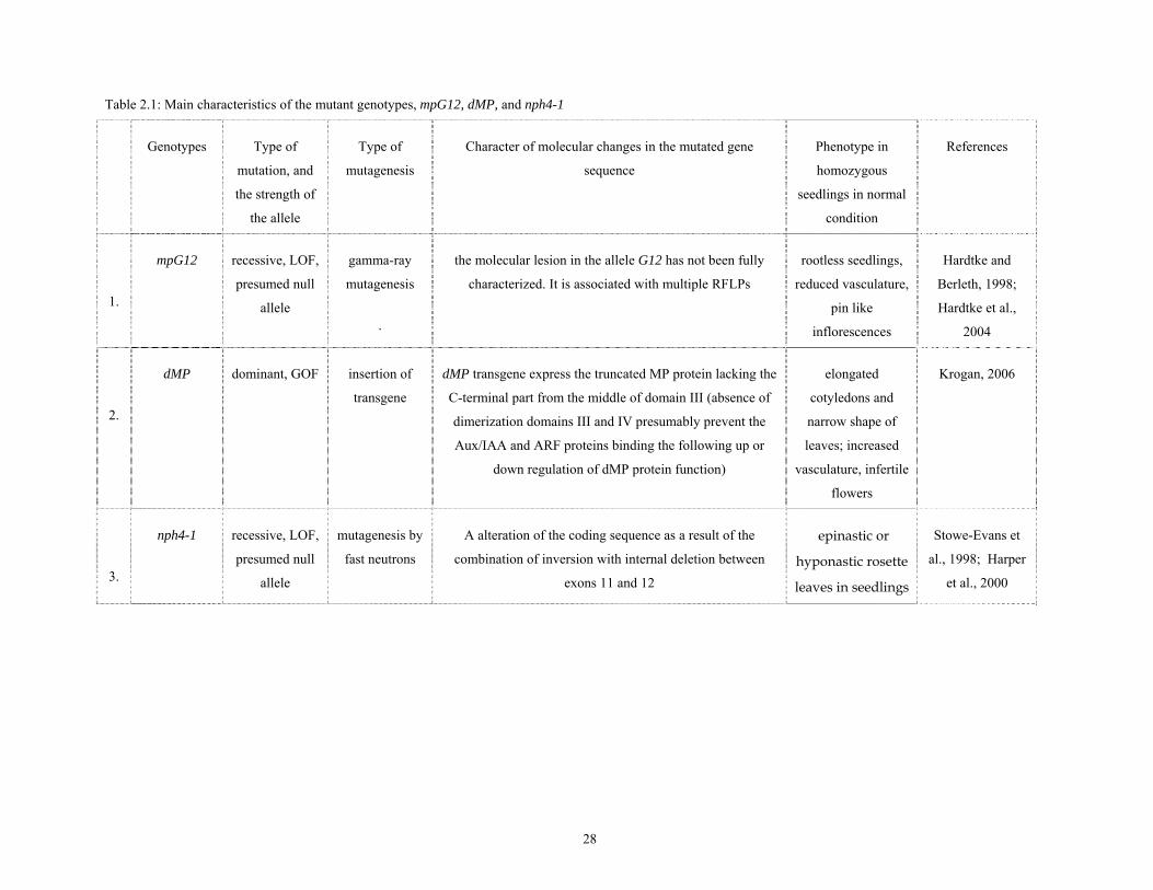
Table 2.1: Main characteristics of the mutant genotypes, mpG12, dMP, and nph4-1
Genotypes Type of
mutation, and
the strength of
the allele
Type of
mutagenesis
Character of molecular changes in the mutated gene
sequence
Phenotype in
homozygous
seedlings in normal
condition
References
1.
mpG12 recessive, LOF,
presumed null
allele
gamma-ray
mutagenesis
.
the molecular lesion in the allele G12 has not been fully
characterized. It is associated with multiple RFLPs
rootless seedlings,
reduced vasculature,
pin like
inflorescences
Hardtke and
Berleth, 1998;
Hardtke et al.,
2004
2.
dMP dominant, GOF insertion of
transgene
dMP transgene express the truncated MP protein lacking the
C-terminal part from the middle of domain III (absence of
dimerization domains III and IV presumably prevent the
Aux/IAA and ARF proteins binding the following up or
down regulation of dMP protein function)
elongated
cotyledons and
narrow shape of
leaves; increased
vasculature, infertile
flowers
Krogan, 2006
3.
nph4-1 recessive, LOF,
presumed null
allele
mutagenesis by
fast neutrons
A alteration of the coding sequence as a result of the
combination of inversion with internal deletion between
exons 11 and 12
epinastic or
hyponastic rosette
leaves in seedlings
Stowe-Evans et
al., 1998; Harper
et al., 2000
28
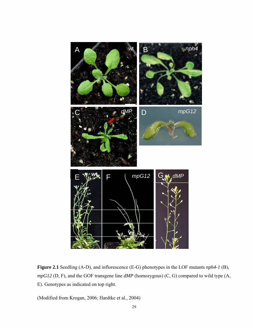
mpG12
A B
C DD
E F G
wt nph4
dMP mpG12
wt mpG12 dMP
Figure 2.1 Seedling (A-D), and inflorescence (E-G) phenotypes in the LOF mutants nph4-1 (B),
mpG12 (D, F), and the GOF transgene line dMP (homozygous) (C, G) compared to wild type (A,
E). Genotypes as indicated on top right.
(Modified from Krogan, 2006; Hardtke et al., 2004)
29
For the propagation of the dMP transgene line, dMP seeds were sown on ½ MS medium without
sucrose, supplemented with 10 μM Basta. This allowed selective growth of homozygous and
heterozygous dMP seedlings, while wild type seedlings were eliminated by Basta. The dMP
homozygous seedlings can be distinguished from heterozygous dMP seedlings based on their
elongated cotyledons and narrowed first leaves (Table 2.1; Fig. 2.1). Only heterozygous dMP
plants are fertile and can be used for propagation; and only homozygous dMP seedlings were
used for the experiments. Seedlings intended for propagation were transferred into Promix BX
soil at 12 DAG, and grown under fluorescent light (100 μE/m2/s2) in an 8-hour dark cycle, at
22°C.
2.3 Hormone stocks
Several auxin and cytokinin plant hormones were used for the experiments. The auxins 2,4-
dichlorophenoxyacetic acid (2,4-D, Sigma), indole-3-butyric acid (IBA, Sigma), and 3-
indoleacetic acid (IAA, Sigma) were dissolved in liquid dimethyl sulfoxide (DMSO) at a
concentration of 50 mg/ml. Cytokinin stock solutions, such as kinetin (Sigma) and 6-
(gamma,gamma-Dimethylallylamino)purine (2-iP, Sigma), were used as commercially prepared
(Sigma) hormone solutions, dissolved in water (sterile filtered, concentration 1 mg/ml). All
stock hormone solutions were stored at -200C.
2.4 Plant DNA extraction
A crude, rapid DNA isolation was done to collect templates for PCR analyses and genotyping.
For crude DNA isolation a small rosette leaf or a small piece of tissue culture was flash-frozen in
liquid nitrogen, and ground in a 1.5 ml Eppendorf tube with a micropestle on ice. Then 500 μL of
extraction buffer [250mM Tris-HCl, 250mM NaCl, 25mM EDTA, 0.5% (w/v) SDS] were added,
and the tube was vortexed for 5 sec. To discharge debris the tube was centrifuged for 1 minute in
a table-top centrifuge at 14000 rpm/RT. The supernatant was transferred into a new Eppendorf
tube and 300 μl of Isopropanol was added to 300 μl of supernatant, vortexed, and left to sit for 2
minutes at RT. Then the mixture was centrifuged at 14000 rpm for 5 minutes at RT, and the
supernatant was discarded. The pellets were washed once with 750 μL of ice-cold 70% (v/v)
ethanol, air-dried, and resuspended with a pipette tip in 30 μL of water. The mix was briefly
centrifuged one more time at 14000 rpm/RT for 1 minute, and the supernatant was carefully
30
removed and stored at -200C. 1μL of crude DNA extract was used as a template per 25μL or
50μL of PCR mix.
2.5 General procedure for polymerase chain reaction (PCR)
PCRs were performed in Biometra UNO-Thermoblock (Germany). The PCR mix (25μL total
volume) contained 1 μl of DNA template, 2.5 μL Taq buffer (supplied with polymerase), 2.5 μL
of MgCl2, 2.5 μL of each primer (Sigma-Genosys), 2.5 μL of dNTPs (Fermentas), and Taq DNA
polymerase 1 unit. Double distilled autoclaved water was added to a total volume of 25 μL was
reached, and then all was overlaid with light mineral oil. The general thermocycling regime was
performed as follows: 94°C for 90s, 40 cycles of (94°C for 30s, 55°C for 30s, 72°C for 2min),
72°C 10min; hold at 4°C.
2.6 Genotyping
Several screening methods can be applied to distinguish between plant samples containing dMP
insertion (homogenous or heterogeneous dMP genotypes) and those of wild type. First, only the
seeds containing the dMP transgene (homogenous or heterogeneous dMP seedlings) can survive
on Basta plates, while wild type seedlings will not germinate, or will die soon after germination
(see Section 2.2, General growth conditions). The homogenous and heterogeneous dMP
seedlings can be distinguished based on a difference in their phenotypes, where homozygous
dMP seedlings had elongated cotyledons and narrowed first leaves (Table 2.1; Fig. 2.1).
To differentiate DNA samples containing dMP (homo or heterozygous insertion) from wild type
the PCR primers for the Basta resistance gene (Bar) were designed. The primers were designed
at the beginning and the end of the Bar gene and gave a PCR product of 403bp. The primer
sequences are:
BST-3-F: 5’-GGTCTGCACCATCGTCAACCACTA-3’
BST-3-R: 5’- CCAGTTCCGTGCTTGAAGCC-3’
The thermocycling regime was performed as follows: 940C for 2min, 35 cycles of (940C for 30s,
620C for 30s, 720C for 1 min), 720C for 5 min; hold at 40C.
31
Homogenous mpG12 seedlings do not form primary roots and hypocotyls (Table 2.1; Fig. 2.1).
This phenotypic trait allows clear distinction of the homogenous mpG12 seedlings from the
heterogeneous (MP/mpG12) or wild type. Additionally, PCR diagnostics were performed on the
genomic DNA to verify a genotypic identity of individual samples (plant or tissue culture
origin). To distinguish between mpG12 homozygous samples and those with at least one wild
type MP copy, primers BS1354-F (5’-GAGATGGCCTGGTTCTAAGTGGC-3’; corresponding
to a central portion of MP ORF) and BS1354-R (5’-GCCAGTTCAACATCTCGGTTATCG-3’;
corresponding to a region of MP 3’-UTR) were used in diagnostic PCR reactions. The presence
of at least one wild type MP gene copy will support amplification of a 2660bp product. In
homozygous mpG12 samples there is no amplification of any PCR products. The thermocycling
regime was performed as follows: 94°C for 90s, 45 cycles of (94°C for 30s, 60°C for 30s, 72°C
for 3min), 72°C 10min; hold at 4°C.
Genotyping of nph4 homozygous individuals was based on PCR diagnostics of plant and tissue
culture samples. To distinguish between nph4-1 homozygous individuals and those with at least
one wild type NPH4 copy, primers NPH4.5 (5’- TCCTGCTGAGTTTGTGGTTCCTT -3’) and
NPH4.6 (5’- GGGGCTTGCTGATTCTGTTTGTTA -3’) were used in diagnostic PCR reactions.
The presence of at least one wild type NPH4 gene copy supports amplification of a 823bp
product, whereas the genomic DNA of nph4-1 homozygous individuals does not support
amplification of any PCR products. The thermocycling regime was performed as follows: 94°C
for 90s, 45 cycles of (94°C for 30s, 63°C for 30s, 72°C for 2min), 72°C 10min; hold at 4°C.
2.7 Microtechniques and microscopy
To introduce explants in tissue culture, seedlings were dissected under a Hund Wetzlar
stereomicroscope (GmbH, Germany) in aseptic conditions in a flowhood. Samples were viewed
under dark field illumination with a Leica MZFLIII stereomicroscope (Leica Microsystem,
Wetzlar, Germany), equipped with a Canon EOS D60 digital camera (Canon inc., Tokyo, Japan).
All images were assembled using Adobe Photoshop 7.0 (Adobe Systems, Mountain View, CA,
USA). The fresh weight of the samples was measured using Sartorius analytical balances
(Sartorius, GmbH, Germany).
32
2.8 Plant tissue culture experiments
Cotyledons, leaves, petioles, hypocotyls, and adventitious roots were used as explants in tissue
culture experiments (Table 2.2). Because mpG12 seedlings lack hypocotyls and primary roots,
no mpG12 hypocotyl explants could be tested. To obtain root explants in mpG12 seedlings, a
special rooting procedure was applied to mpG12 and the rest of the genotypes to obtain
adventitious roots (see Section 2.8.1 for more details). The main steps of the tissue culture
experiments are summarized in Fig. 2.2.
At least three independent replicas of the experiment were taken unless otherwise stated.
Student’s t-test was used to evaluate the significance of the comparison between the mean
responses, such as fresh weight of calluses, number of shoots or roots per callus, etc. For the
purposes of this research, each of the evaluated genotypes was compared against wild type via
Student’s t-test (Microsoft Excel, 2003).
2.8.1 Rooting of dissected mpG12, dMP, nph4-1 and wild type seedlings
Seeds of mpG12, nph4-1, dMP, and wild type were germinated as described above (see for
details Section 2.2, General growth conditions). 5-6 DAG seedlings were dissected under a Hund
Wetzlar stereomicroscope (GmbH, Germany) in aseptic conditions. In rootless mpG12 seedlings,
peg parts (short hypocotyl-like outgrowths below the cotyledon node) were cut with a sharp
scalpel. Seedlings of other genotypes were similarly dissected by cutting off the roots with a part
of the hypocotyl. Dissected seedlings were placed in tissue culture dishes (100x20 mm, Sarstedt,
USA) with root inducing medium (RIM), containing 1 mg/L IBA (Table 2.3), and cultured at 24-
260C under constant light (100μEm-2sec-1). After 7-9 days on RIM, about 5-10% of the mpG12
dissected seedlings, and most of the seedlings of the other genotypes formed adventitious roots.
Rooted seedlings were transferred on hormone-free ½ MS medium, to restore normal root
morphology; and, after 14 days of culture, they were dissected and their adventitious roots were
used for tissue culture experiments.
33

Explants
CIM 6 days
SIM 21 days
Callusogenesis:% callus patterns,
fresh weight
Shoot regeneration:% shoot regeneration,
# shoots per callus
CIM 12 days RIM 21 days
Root regeneration:% root regeneration,
# roots per callus
Figure 2.2 General scheme of the tissue culture experiments and scored traits, unless otherwise
specified in the description of the particular experiment.
34
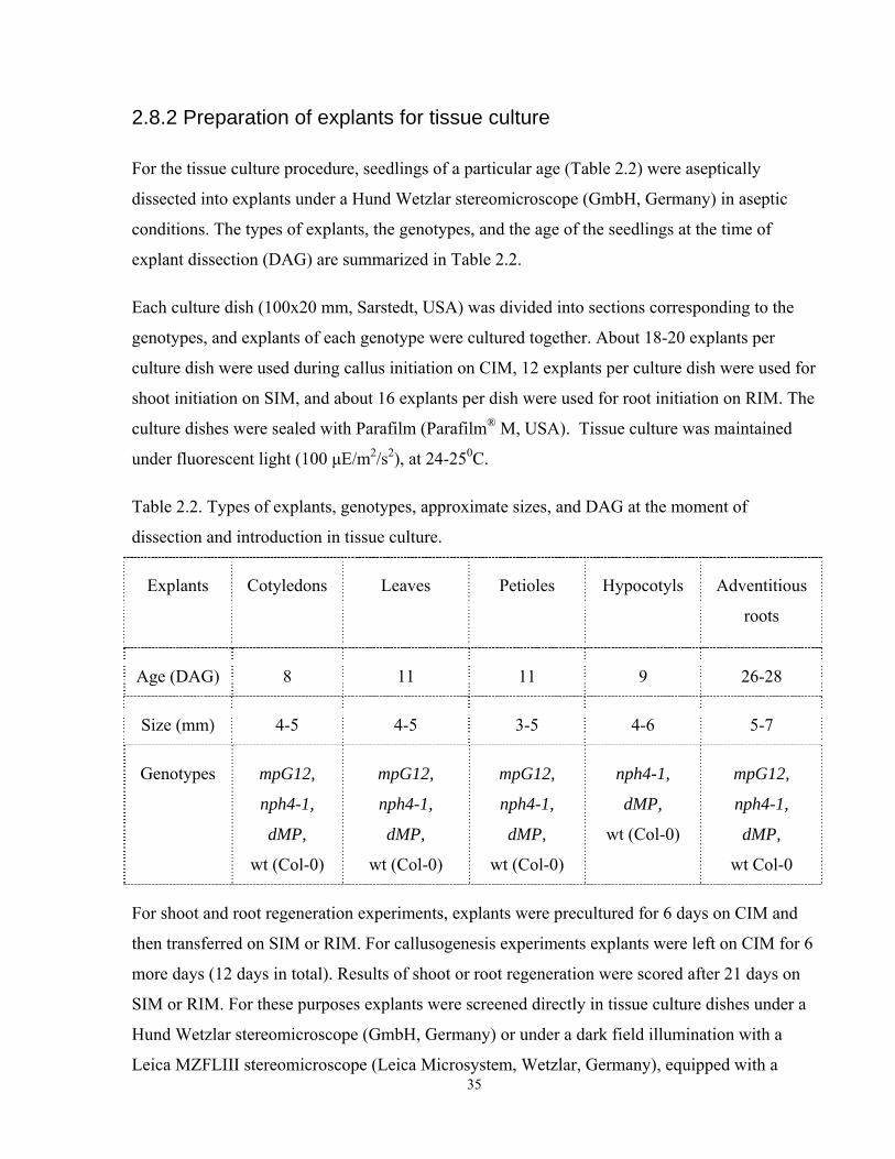
2.8.2 Preparation of explants for tissue culture
For the tissue culture procedure, seedlings of a particular age (Table 2.2) were aseptically
dissected into explants under a Hund Wetzlar stereomicroscope (GmbH, Germany) in aseptic
conditions. The types of explants, the genotypes, and the age of the seedlings at the time of
explant dissection (DAG) are summarized in Table 2.2.
Each culture dish (100x20 mm, Sarstedt, USA) was divided into sections corresponding to the
genotypes, and explants of each genotype were cultured together. About 18-20 explants per
culture dish were used during callus initiation on CIM, 12 explants per culture dish were used for
shoot initiation on SIM, and about 16 explants per dish were used for root initiation on RIM. The
culture dishes were sealed with Parafilm (Parafilm® M, USA). Tissue culture was maintained
under fluorescent light (100 μE/m2/s2), at 24-250C.
Table 2.2. Types of explants, genotypes, approximate sizes, and DAG at the moment of
dissection and introduction in tissue culture.
Explants Cotyledons Leaves Petioles Hypocotyls Adventitious
roots
Age (DAG) 8 11 11 9 26-28
Size (mm) 4-5 4-5 3-5 4-6 5-7
Genotypes mpG12,
nph4-1,
dMP,
wt (Col-0)
mpG12,
nph4-1,
dMP,
wt (Col-0)
mpG12,
nph4-1,
dMP,
wt (Col-0)
nph4-1,
dMP,
wt (Col-0)
mpG12,
nph4-1,
dMP,
wt Col-0
35
For shoot and root regeneration experiments, explants were precultured for 6 days on CIM and
then transferred on SIM or RIM. For callusogenesis experiments explants were left on CIM for 6
more days (12 days in total). Results of shoot or root regeneration were scored after 21 days on
SIM or RIM. For these purposes explants were screened directly in tissue culture dishes under a
Hund Wetzlar stereomicroscope (GmbH, Germany) or under a dark field illumination with a
Leica MZFLIII stereomicroscope (Leica Microsystem, Wetzlar, Germany), equipped with a
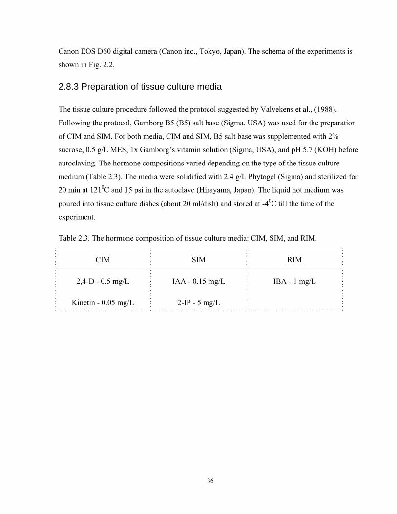
Canon EOS D60 digital camera (Canon inc., Tokyo, Japan). The schema of the experiments is
shown in Fig. 2.2.
2.8.3 Preparation of tissue culture media
The tissue culture procedure followed the protocol suggested by Valvekens et al., (1988).
Following the protocol, Gamborg B5 (B5) salt base (Sigma, USA) was used for the preparation
of CIM and SIM. For both media, CIM and SIM, B5 salt base was supplemented with 2%
sucrose, 0.5 g/L MES, 1x Gamborg’s vitamin solution (Sigma, USA), and pH 5.7 (KOH) before
autoclaving. The hormone compositions varied depending on the type of the tissue culture
medium (Table 2.3). The media were solidified with 2.4 g/L Phytogel (Sigma) and sterilized for
20 min at 1210C and 15 psi in the autoclave (Hirayama, Japan). The liquid hot medium was
poured into tissue culture dishes (about 20 ml/dish) and stored at -40C till the time of the
experiment.
Table 2.3. The hormone composition of tissue culture media: CIM, SIM, and RIM.
CIM SIM RIM
2,4-D - 0.5 mg/L
Kinetin - 0.05 mg/L
IAA - 0.15 mg/L
2-IP - 5 mg/L
IBA - 1 mg/L
36
Chapter 3
Callusogenesis under conditions of altered auxin signal
transduction
3.1 Background and rationale
Callus proliferation is induced in vitro at a certain relative concentration of auxin and cytokinin
in the culture medium (Skoog and Miller 1957). Cell proliferation during callusogenesis leads to
disruption of the ordered morphology of differentiated tissues (Cary et al., 2002). The
mechanisms leading to callus proliferation are not well understood (Cary et al., 2002; Zhang and
Lemaux, 2005). Generally, cell-cycle genes, such as cyclins and cyclin dependent kinases
(CDKs), play a major role in the mediation of hormonal effects, or in the response to wounding
signals, leading to cell proliferation (Hemerly et al., 1993; Soni et al., 1995; Shaul et al., 1996;
Riou-Khamlichi et al., 1999; Sugiyama 1999; Richard et al., 2002; Che et al., 2007). Cis-acting
regulatory elements, specific for the binding of hormone transcription factors, such as AuxREs
for auxin signaling, have been found in the promoters of some cell cycle genes (Richard et al.,
2002; Perrot-Rechenmann, 2010).
Auxin responses, visualized by DR5 and PIN1 driven reporters (reviewed in Chapter 1.2), were
strongly increased during the initiation of callus proliferation. Then they were downregulated
after 5 days on CIM (callus inducing medium). After 10 days on CIM, DR5 and PIN1 reporters
were not detectable in the callus culture (Gordon et al., 2007). Among the other reporters
associated with proliferating callus growth, are the meristem genes CUC1 and CUC2, and the
ARR5 gene, which is involved in cytokinin signaling (reviewed in Chapter 1.4; Cary et al., 2002;
Gordon et al., 2007). The expression of the corresponding reporter genes was observed after 8
days on CIM, and continuously increased during CIM cultivation (Gordon et al., 2007). When
callusogenesis was induced on the medium supplemented with auxin as the sole hormone, CUC2
and ARR5 reporters were still expressed. In contrast, only weak callus proliferation occurred, and
no expression of the callus proliferation reporters, CUC1, CUC2 and ARR5, was observed, when
the explants were cultured on the medium supplemented with cytokinin only (Gordon et al.,
37
2007). This observation emphasizes the importance for callus proliferation of the presence of
auxin, rather than cytokinin, in the culture medium.
As described in Chapter 1 (1.2.5), the auxin response factors MP (ARF5) and NPH4 (ARF7)
belong to the group of the transcriptional activators whose potency is enhanced in the presence
of auxin (Ulmasov et al., 1999a; Ulmasov et al., 1999b; Berleth et al., 2004). Among other
described defects (Hardtke et al., 2004), the loss of MP or NPH4 function severely reduces auxin
sensitivity. The expression of the auxin-responsive reporter DR5 and auxin-inducible genes was
significantly reduced in nph4 and mp mutants (Stowe-Evans et al., 1998; Watahiki et al., 1999;
Hardtke et al., 2004; Li et al. 2006; Przemeck et al., 1996; Mattsson et al., 2003; Okushima et
al., 2005; Wang et al., 2005; Schuetz et al., 2008).
The NPH4 gene is expressed throughout the Arabidopsis life cycle, and NPH4 transcripts are
generally present in all major organs (Hardtke et al., 2004). The MP gene is expressed in broad
domains in embryos and in emerging organs. Later, upon organ maturation, the expression of
MP is gradually narrowed to central domains and then to vascular tissues (Hardtke and Berleth,
1998; Hardtke et al., 2004). The mRNAs of both genes, MP and NPH4, were found to be
ubiquitous and uniformly expressed in Arabidopsis cells in suspension culture (Ulmasov et al.,
1999b; reviewed in Chapter 1.2.4). The genotype dMP generated by the deletion of protein
interaction domains in the MP gene is described in the General Introduction (Chapter 1.2.5; Fig.
1.2; Krogan, 2006). As these interaction domains are required to bind negative regulators of ARF
function, it seems likely that dMP represents a constitutively active MP gene.
In summary, the MP and NPH4 genes play important roles during many auxin-triggered
processes in plant development. Because callus proliferation is triggered by the hormone auxin,
changes in MP and NPH4 function in the loss-of-function mutants may have an impact on
callusogenesis.
3.2 Specific research objectives
The investigation of the possible influence of altered auxin signaling through LOF and GOF
alleles of ARF5/MP and ARF7/NPH4 on callusogenesis was the purpose of this part of the
project. To this end, the loss-of-function mutants mpG12 (Hardtke and Berleth, 1998), and nph4-
1 (Harper et al., 2000), and the gain-of-function transgene line dMP (Krogan, 2006) with 38
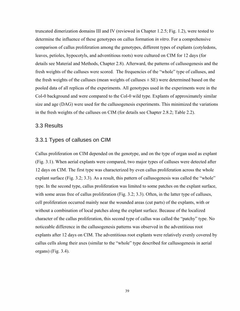
truncated dimerization domains III and IV (reviewed in Chapter 1.2.5; Fig. 1.2), were tested to
determine the influence of these genotypes on callus formation in vitro. For a comprehensive
comparison of callus proliferation among the genotypes, different types of explants (cotyledons,
leaves, petioles, hypocotyls, and adventitious roots) were cultured on CIM for 12 days (for
details see Material and Methods, Chapter 2.8). Afterward, the patterns of callusogenesis and the
fresh weights of the calluses were scored. The frequencies of the “whole” type of calluses, and
the fresh weights of the calluses (mean weights of calluses ± SE) were determined based on the
pooled data of all replicas of the experiments. All genotypes used in the experiments were in the
Col-0 background and were compared to the Col-0 wild type. Explants of approximately similar
size and age (DAG) were used for the callusogenesis experiments. This minimized the variations
in the fresh weights of the calluses on CIM (for details see Chapter 2.8.2; Table 2.2).
3.3 Results
3.3.1 Types of calluses on CIM
Callus proliferation on CIM depended on the genotype, and on the type of organ used as explant
(Fig. 3.1). When aerial explants were compared, two major types of calluses were detected after
12 days on CIM. The first type was characterized by even callus proliferation across the whole
explant surface (Fig. 3.2; 3.3). As a result, this pattern of callusogenesis was called the “whole”
type. In the second type, callus proliferation was limited to some patches on the explant surface,
with some areas free of callus proliferation (Fig. 3.2; 3.3). Often, in the latter type of calluses,
cell proliferation occurred mainly near the wounded areas (cut parts) of the explants, with or
without a combination of local patches along the explant surface. Because of the localized
character of the callus proliferation, this second type of callus was called the “patchy” type. No
noticeable difference in the callusogenesis patterns was observed in the adventitious root
explants after 12 days on CIM. The adventitious root explants were relatively evenly covered by
callus cells along their axes (similar to the “whole” type described for callusogenesis in aerial
organs) (Fig. 3.4).
39
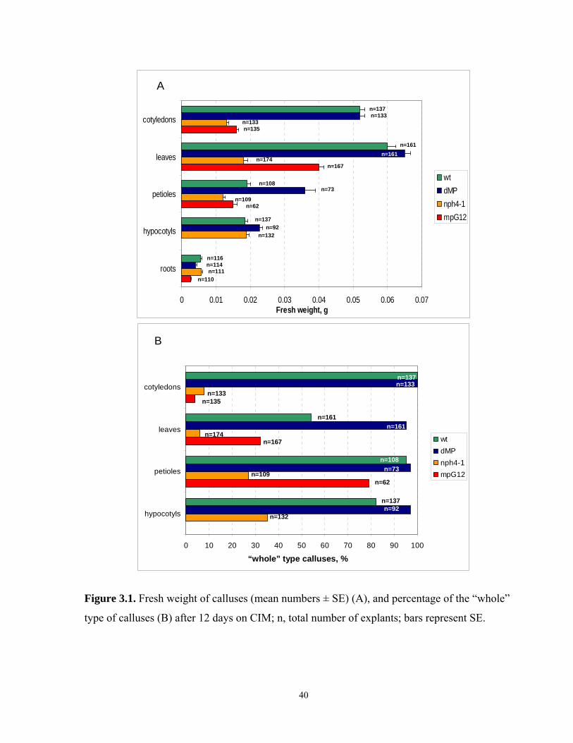
0 0.01 0.02 0.03 0.04 0.05 0.06 0.07
roots
hypocotyls
petioles
leaves
cotyledons
Fresh weight, g
wtdMPnph4-1mpG12
A
n=137n=133
n=133n=135
n=161n=161
n=174n=167
n=108n=73
n=109n=62
n=137n=92
n=132
n=116n=114n=111
n=110
0 10 20 30 40 50 60 70 80 90 100
hypocotyls
petioles
leaves
cotyledons
Frequency of whole type calluses, %
wtdMPnph4-1mpG12
n=137n=133
n=133n=135
n=161n=161
n=174n=167
n=108
n=73
n=73n=109
n=62
n=137n=92
n=132
B
“whole” type calluses, %
Figure 3.1. Fresh weight of calluses (mean numbers ± SE) (A), and percentage of the “whole”
type of calluses (B) after 12 days on CIM; n, total number of explants; bars represent SE.
40
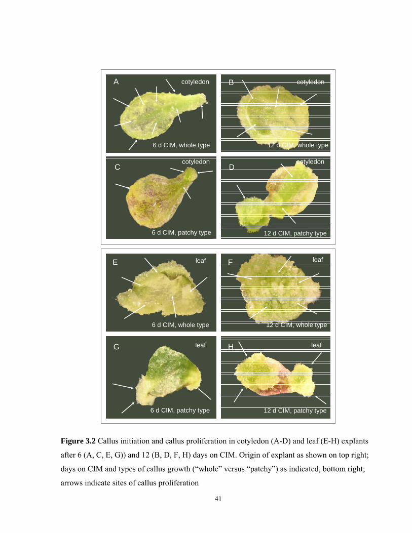
A B
C D
6 d CIM, whole type 12 d CIM, whole type
6 d CIM, patchy type 12 d CIM, patchy type
cotyledon cotyledon
cotyledon cotyledon
6 d CIM, whole type
6 d CIM, patchy type
12 d CIM, whole type
12 d CIM, patchy type
E F
G H
leaf leaf
leaf leaf
Figure 3.2 Callus initiation and callus proliferation in cotyledon (A-D) and leaf (E-H) explants
after 6 (A, C, E, G)) and 12 (B, D, F, H) days on CIM. Origin of explant as shown on top right;
days on CIM and types of callus growth (“whole” versus “patchy”) as indicated, bottom right;
arrows indicate sites of callus proliferation
41
3.3.2 Callusogenesis of cotyledon explants after 12 days on CIM
Callus proliferation, indicated by the fresh weights of the cotyledon derived calluses, differed
among the genotypes. The fresh weights of the dMP calluses were similar to those of the wild
type calluses (the wild type calluses 52±1.4 mg, n=137; dMP 52±1.5 mg, n=133) (Fig. 3.1 A). In
contrast, the fresh weights of the nph4-1 and the mpG12 calluses were significantly less than
those of the wild type calluses (nph4-1 13±6 mg, n=135, P<0.001; and mpG12 16±6 g, n=133,
P<0.001) (Fig. 3.1 A). Thus, there were two distinct phenotype classes: the dMP calluses
growing like the wild type ones versus the strongly impaired callus growth in nph4-1 and mpG12
(Fig. 3.1 A).
The distribution of the callusogenesis types (the “whole” versus the “patchy” types) in the
cotyledon explants also dramatically differed among the genotypes (Fig. 3. 1 B; 3. 5 A). All wild
type (n=137) and all dMP cotyledon explants (n=133) had the “whole” type of callusogenesis
(Fig. 3.1 B). In contrast, mpG12 and nph4-1 showed mostly the “patchy” type of callusogenesis.
The “whole” type calluses were found in only 4% of the mpG12 explants (n=135), and in 8% of
the nph4-1 explants (n=133) (Fig. 3.1 B). The impaired callus proliferation in the arf mutants,
indicated by the lower fresh weights of the calluses, was combined with a strong prevalence of
the “patchy” type calluses in these genotypes. In contrast, the dMP and the wild type calluses,
which defined normal callus growth, were exclusively the “whole” type.
3.3.3 Callusogenesis of leaf explants after 12 days on CIM
The fresh weights of the leaf calluses also differed among the genotypes. As with the cotyledon
explants, the fresh weights of the calluses derived from the leaf explants were similar between
wild type and dMP (wild type 60±2.4 mg, n=161; and dMP 65±1.6 mg, n=161). However, the
fresh weights of the nph4-1 calluses (18±1 mg, n=174; P<0.001), and of the mpG12 calluses
(40±0.6 mg, n=167; P<0.001) were significantly less than those of wild type (Fig. 3.1 A). Also,
the fresh weights of the mutant genotypes, nph4-1 and mpG12, differed significantly: the mean
fresh weight of the nph4-1 calluses was only about half that of mpG12 (nph4-1 18±1 mg, n=174;
mpG12 40±0.6 mg, n=167; P<0.001, for the comparison of the mean responses between nph4-1
and mpG12) (Fig. 3.1 A).
42
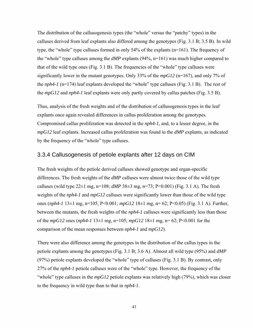
The distribution of the callusogenesis types (the “whole” versus the “patchy” types) in the
calluses derived from leaf explants also differed among the genotypes (Fig. 3.1 B; 3.5 B). In wild
type, the “whole” type calluses formed in only 54% of the explants (n=161). The frequency of
the “whole” type calluses among the dMP explants (94%, n=161) was much higher compared to
that of the wild type ones (Fig. 3.1 B). The frequencies of the “whole” type calluses were
significantly lower in the mutant genotypes. Only 33% of the mpG12 (n=167), and only 7% of
the nph4-1 (n=174) leaf explants developed the “whole” type calluses (Fig. 3.1 B). The rest of
the mpG12 and nph4-1 leaf explants were only partly covered by callus patches (Fig. 3.5 B).
Thus, analysis of the fresh weights and of the distribution of callusogenesis types in the leaf
explants once again revealed differences in callus proliferation among the genotypes.
Compromised callus proliferation was detected in the nph4-1, and, to a lesser degree, in the
mpG12 leaf explants. Increased callus proliferation was found in the dMP explants, as indicated
by the frequency of the “whole” type calluses.
3.3.4 Callusogenesis of petiole explants after 12 days on CIM
The fresh weights of the petiole derived calluses showed genotype and organ-specific
differences. The fresh weights of the dMP calluses were almost twice those of the wild type
calluses (wild type 22±1 mg, n=108; dMP 38±3 mg, n=73; P<0.001) (Fig. 3.1 A). The fresh
weights of the nph4-1 and mpG12 calluses were significantly lower than those of the wild type
ones (nph4-1 13±1 mg, n=105, P<0.001; mpG12 18±1 mg, n= 62; P<0.05) (Fig. 3.1 A). Further,
between the mutants, the fresh weights of the nph4-1 calluses were significantly less than those
of the mpG12 ones (nph4-1 13±1 mg, n=105; mpG12 18±1 mg, n= 62; P<0.001 for the
comparison of the mean responses between nph4-1 and mpG12).
There were also difference among the genotypes in the distribution of the callus types in the
petiole explants among the genotypes (Fig. 3.1 B; 3.6 A). Almost all wild type (95%) and dMP
(97%) petiole explants developed the “whole” type of calluses (Fig. 3.1 B). By contrast, only
27% of the nph4-1 petiole calluses were of the “whole” type. However, the frequency of the
“whole” type calluses in the mpG12 petiole explants was relatively high (79%), which was closer
to the frequency in wild type than to that in nph4-1.
43
A
6 d CIM, the “whole” type
petiole
C
6 d CIM, the “patchy” type
petiole
B
12 d CIM, the “whole” type
petiole
D
12 d CIM, the “patchy” type
petiole
E
6 d CIM, the “whole” type
hypocotyl
G
6 d CIM, the “patchy” type
hypocotyl
F
12 d CIM, the “whole” type
hypocotyl
H
12 d CIM, the “patchy” type
hypocotyl
Figure 3.3 Callus initiation and callus proliferation in petiole (A-D) and hypocotyl (E-H)
explants after 6 and 12 days on CIM. Origin of explant as indicated on top right; days on CIM
44
and types of callus growth (“whole” versus “patchy”) as indicated, bottom right; arrows indicate
sites of callus proliferation.
To summarize, the distribution of the fresh weights and of the types of calluses in the petiole
explants indicated the restricted character of callus proliferation in the nph4-1 explants; and, to a
lesser degree, in the mpG12 petiole explants. By contrast, callus proliferation was elevated in the
dMP explants, as indicated by the fresh weights of the dMP calluses, the vast majority of which
were of the “whole” type.
3.3.5 Callusogenesis of hypocotyl explants after 12 days on CIM
The fresh weights of the hypocotyl calluses also showed genotype related differences. However,
these differences were weaker compared to those of the other organs. The fresh weights of the
dMP calluses were significantly higher than those of the wild type ones (wild type 19±1 mg,
n=137; dMP 23±1 mg, n=92; P<0.001). The hypocotyl explants were the only type of aerial
explants where the fresh weights of the nph4-1 calluses were not less than those in wild type
(nph4-1 19±1 mg, n=132) (Fig. 3.1 B). No mpG12 hypocotyl explants could be tested, because
of the mpG12 seedlings’ lack of a hypocotyl.
The distribution of the callus types in the hypocotyl explants differed among the genotypes (Fig.
3.1 B; 3.6 B). Most of the wild type explants (82%, n=137) and most of the dMP explants (97%,
n=92) had the “whole” type of calluses (Fig. 3.1 B). In contrast, there were only 35% of the
“whole’ type calluses in nph4-1 (n=132) (Fig. 3.1 B).
To summarize, the comparison of the callus growth and type data suggests that the “whole” type
calluses are not just the result of vigorous growth. Specifically, the fresh weights of the nph4-1
calluses were similar to those of the wild type ones in hypocotyl explants. The “patched” calluses
dominated in this group. Among the genotypes, callus proliferation was once again greatest in
dMP calluses.
3.3.6 Callusogenesis of adventitious root explants after 12 days on CIM
Because mpG12 seedlings lack primary roots (Berleth and Jurgens, 1993), the procedure to
initiate adventitious roots in mpG12 and in the other genotypes preceded the callusogenesis
45
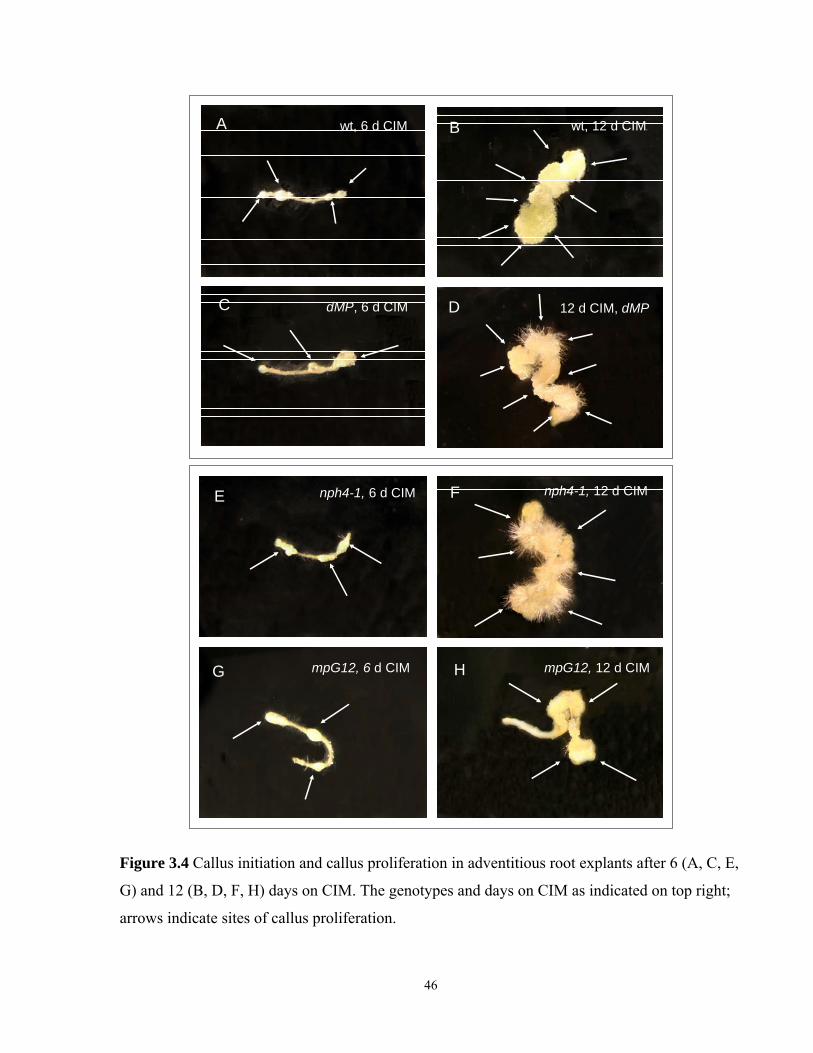
A 6 d CIM, wt
B 6 d CIM, dMP
E 12 d CIM, wt
12 d CIM, dMPD
wt, 6 d CIM wt, 12 d CIM
dMP, 6 d CIM
A B
C
mpG12, 12 d CIMH
nph4-1, 12 d CIMFC 6 d CIM, nph4-1
D 6 d CIM, mpG12
nph4-1, 6 d CIM
mpG12, 6 d CIM
E
G
Figure 3.4 Callus initiation and callus proliferation in adventitious root explants after 6 (A, C, E,
G) and 12 (B, D, F, H) days on CIM. The genotypes and days on CIM as indicated on top right;
arrows indicate sites of callus proliferation.
46
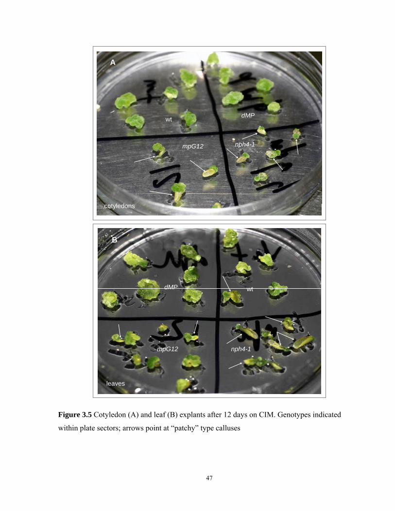
wt
mpG12 nph4-1
dMP
A
cotyledons
dMP
mpG12
wt
nph4-1
B
leaves
Figure 3.5 Cotyledon (A) and leaf (B) explants after 12 days on CIM. Genotypes indicated
within plate sectors; arrows point at “patchy” type calluses
47
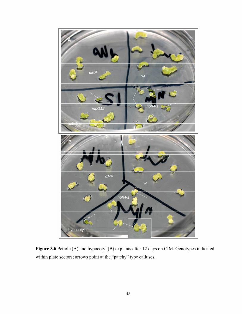
dMP
mpG12
wt
nph4-1
A
petioles
dMP
wt
nph4-1
B
hypocotyls
Figure 3.6 Petiole (A) and hypocotyl (B) explants after 12 days on CIM. Genotypes indicated
within plate sectors; arrows point at the “patchy” type calluses.
48

dMP
nph4-1mpG12
wt
roots
Figure 3.7 Adventitious root explants after 12 days on CIM. Genotypes indicated within plate
sectors.
49
experiments (see Chapter 2.8.1 for details). Seedlings of all the genotypes were dissected by
cutting off the lower part of their hypocotyl (or the low part of the peg in the mpG12 seedlings).
The dissected seedlings were re-rooted on the auxin (IBA) containing medium, and then
transferred to the hormone-free 1/2MS medium to allow the outgrowth of adventitious roots.
These adventitious roots were used as explants in the experiments described below (for details
see Chapter 2.8).
In this category, root explants, the distribution of the fresh weights of the calluses among the
genotypes was different compared to those of the aerial organs (Fig. 3.1A). The fresh weights of
the calluses were: wild type 56±0.4 mg, n=116; the similar nph4-1, 57±0.4 mg, n=111; and, the
significantly lower (by more than half) mpG12, 26±0.2 mg, n=110 (P<0.001). In contrast to the
growth of calluses on all the aerial organs, the growth of the dMP calluses on the root explants
was slightly, but significantly, lower than that of the wild type calluses (dMP 4±0.4 mg, n=114;
P<0.01) (Fig. 3.1 A).
Thus, in this category of root explants, callus growth among the genotypes was quite different
than that in the aerial explants. No genotype related difference in callus types was detected in
this category, and the calluses of all the genotypes looked relatively similar after 12 days on
CIM, resembling the “whole” type calluses of the aerial explant group.
3.3.7 Callus proliferation in aerial explants after 6 days on CIM
The difference in phenotypic classes between genotypes observed after 12 days on CIM in
calluses derived from aerial explants (the “whole’ or the “patchy’’ types of calluses) raises the
question of the origin of this difference. Two distinct possibilities can be considered. First, the
difference in callus types might simply reflect the difference in callus growth rate. In this case,
two callus types could be initiated as local callus patches along the explant surface, and around
the wounded areas. Later, these areas would merge to form the “whole” calluses, when callus
proliferation was strong, or stay “patchy”, when the callus growth rate was low. Alternatively,
the two types could reflect genuinely different growth patterns observable right from the
beginning of callus growth. To distinguish between these two possibilities, the explants were
visually examined after 6 days on CIM.
50
When aerial explants (cotyledons, leaves, petioles and hypocotyls) were examined after 6 days
on CIM, clear differences were detected between explants later developing the “whole” type or
the “patchy” type calluses. Most of the explants (more than 90%), which later developed the
“whole" type of calluses, were getting visibly friable and partly translucent. Their original dense
green color was changing to whitish or yellowish green (Fig. 3.2; 3.3). Callus proliferation in
these explants occurred evenly across the entire explant surface. Further, when cotyledon and
leaf explants were examined, the callus proliferation was denser along the veins and the petiole
parts (Fig. 3.2). All these changes made the explants developing into the “whole” type calluses
look quite distinct from those developing into the “patchy’ type calluses (Fig. 3.2; 3.3).
By contrast, in the majority of the explants (more than 90%) which later developed the “patchy”
type of calluses, relatively large areas of the explants retained their original appearance after 6
days on CIM. In these areas, the tissues did not look friable and translucent. The color of the
explants did not change from the original dense green to whitish or yellowish shades. Initiation
of callus proliferation in these explants was limited to the wounded (cut) areas (in all organ types
of the aerial explants), and to some locally restricted patches of proliferating cells on the explant
surface, when the cotyledon and leaf explants were examined (Fig. 3.2; 3.3). The rest of the
explant tissues stayed “untouched” and mainly preserved their original color and structure (Fig.
3.2; 3.3).
In summary the results indicated that the differences in the callus types (the “whole” type versus
the “patchy” type) were due to the distinctive initiation of callus proliferation in the explants,
rather than to the differences in the callus growth rate, even though there were differences in the
callus growth rates.
3.4 Discussion and Conclusions
The results in this chapter indicate genotype and organ related differences in callus proliferation
responses among the genotypes. These differences affect both the callus growth rate, as well as
the pattern of proliferation.
In the calluses derived from the aerial explants (cotyledons, leaves, petioles, and hypocotyls),
two major phenotypic classes of callus type were observed: the “whole” type, with even callus
proliferation along the entire explant surface; and the “patchy” type, where callus proliferation 51
occurred as isolated patches, often localized to the wounded areas. The callus types and the
callus weights differed among the tested genotypes, falling into two major groups. The first
group was comprised of the dMP and wild type genotypes. Callusogenesis in dMP was either
equal to, or sometimes greater than that in wild type, as indicated by the fresh weights and the
frequencies of the “whole” type calluses. The second group was represented by the arf mutants,
nph4-1 and mpG12. In these, callusogenesis was partly impaired and significantly weaker
compared to that in wild type. This was indicated by the lower fresh weights of the nph4-1 and
the mpG12 calluses, and usually by much lower frequencies of the “whole” type calluses. Also,
of these arf genotypes, callusogenesis compromise was greater in nph4-1 than in mpG12, when
their aerial explants were cultured on CIM. The differences in the callus types (the “whole” type
versus the “patchy” type) were as a result of the distinct initiation of callus proliferation
(throughout the explant surface in the “whole’ type, or in local patches in the “patchy” type of
calluses), rather than as a result of differences in the callus growth rate.
The callusogenesis responses were different when calluses derived from the adventitious root
explants were compared. Although no distinct callus types were detected, genotype related
differences in callusogenesis were indicated by the fresh weights of the calluses. Dissimilar to
calluses derived from the aerial explants, no impairment in callusogenesis was detected in nph4-
1root explants. These data are compatible with previous observations that only the aerial organs,
but not the roots, of nph4 plants were resistant to toxic concentrations of the synthetic auxin 2,4-
D (Watahiki and Yamamoto, 1997). In contrast to nph4-1, callusogenesis in mpG12, as indicated
by the fresh weights of the calluses, was significantly weaker compared to that of wild type.
Also, the callus weights were slightly, but significantly, lower in the dMP calluses. Generally,
however, in this category of root explants, differences in callusogenesis among the genotypes
were more subtle than those in the calluses derived from the aerial explants. Given the artificial
character of the procedure for the induction of adventitious roots (the long exposure to an auxin
containing medium followed by culture on a hormone free medium), the observed differences in
callus weights might reflect the genotype specific response to preculture conditions during
adventitious root induction, rather than a true genotype related difference in callus proliferation.
In nature, callusogenesis is a part of the mechanism of protection facilitating the repair of
wounded or damaged tissues (Frank et al., 2000). This mechanism involves auxin signaling,
which is crucial for callus initiation (Frank et al., 2000; Gordon et al., 2007).Thus, it is plausible 52
that auxin insensitive arf mutants are impaired in callusogenesis. The relative degree of
impairment may reflect the degree to which a given ARF gene is involved in auxin signaling in a
specific organ or tissue. It may also reflect the degree to which a particular ARF gene is related
to the regulation of the cell-cycle genes mediating cell proliferation responses (Hemerly et al.,
1993; Richard et al., 2002; Che et al., 2007; Perrot-Rechenmann, 2010).
The dMP transgene represents a gain-of-function allele of MP, possibly a constitutively active
allele (Krogan, 2006). Callusogenesis was strong in the dMP transgene line in aerial, but not in
root explants. In this case, one may speculate that this reflects the enhanced auxin signal
transduction features of dMP tissue (Krogan, 2006).
To summarize, loss of MP and particularly NPH4 functions in mpG12 and nph4-1 mutants
resulted in the impairment of callus proliferation in particular categories of explants. The
presumably elevated MP function in the dMP genotype may increase callus proliferation in aerial
explants, and decrease the same in adventitious root explants which needs further evaluation.
53
Chapter 4
Shoot regeneration under conditions of altered auxin signal
transduction
4.1 Background and rationale
Shoot regeneration in vitro is triggered by a high cytokinin to auxin ratio in the tissue culture
medium (Skoog and Muller, 1957; Gaba, 2005). Although the phytohormone auxin is always
used in the composition of a shoot inducing medium (SIM), relatively little is known about the
effect of auxin on shoot regeneration in tissue culture. Typically, the role of auxin in this process
is only discussed in the context of the ratio of auxin to cytokinin (Howell 2003; Zhang and
Lemaux, 2004; Gaba, 2005; Zhang and Lemaux, 2005). However, several factors can contribute
to the effect of auxin signaling on shoot regeneration. First, auxin plays a key role in shoot
meristem patterning (Reinhardt et al., 2000); auxin levels are high in shoot meristems in planta
(Kakani et al., 2009). Therefore, proper auxin signaling might be required for de novo shoot
meristem assembly (Coenen and Lomax, 1998; Gordon et al., 2007; Chatfield and Raizada,
2008; Kakani et al., 2009). Second, many genes linked to auxin signaling may also be implicated
in other hormone pathways, due to existing cross talk between signaling pathways (Hobbie and
Estelle, 1994; Swarup et al., 2002). As a result, auxin gene impairment may affect coordinated
interactions among multiple developmental processes (Gordon et al., 2007; Kakani et al., 2009).
Third, shoot regeneration is severely impaired in a number of auxin signaling mutants, such as
aux1 (Kakani et al., 2009) and axr1 (Chatfield and Raizada, 2008), and mildly impaired in pin1
(Gordon et al., 2007). Thus, genes involved in auxin signaling, auxin homeostasis, and auxin
transport may affect processes of de novo shoot regeneration in vitro (reviewed in Chapter 1.7.5).
The auxin response factors MP (ARF5) and NPH4 (ARF7) interact with each other and have
partly overlapping functions. They are both transcriptional activators (Ulmasov et al., 1999a;
Ulmasov et al., 1999b, Berleth et al., 2004; Hardtke et al., 2004). Mutations in the MP gene
affect the formation of the embryonic root meristem and result in rootless seedlings (Berleth and
Jurgens, 1993), and in severely reduced auxin sensitivity, auxin signaling and auxin transport (Li
et al. 2006, Przemeck et al., 1996, Mattsson et al., 2003, Schuetz et al., 2008). Moreover, in
54
planta, MP is involved in the regulation of several meristematic genes, such as the CUC genes
and STM, during embryonic meristem formation (Aida et al., 2002). MP is also linked to the
ESR1/DRN gene during cotyledon development (Cole et al., 2009). Because all these genes are
known to be crucial for de novo shoot meristem formation (Zhang and Lemaux, 2005), the MP
gene may have a significant effect on shoot regeneration.
Regarding nph4 mutants, they exhibit auxin resistance in the aerial parts of plants, indicated by
their resistance to toxic concentrations of auxin, and by the low signal of the DR5 auxin response
reporter. Further, the levels of many primary auxin response genes are severely affected in nph4
mutants (Stowe-Evans et al., 1998, Wang et al., 2005). Notably, nph4 mutants represent a case of
auxin-resistant mutants that are not cross-resistant to other plant hormones, including cytokinin
(Watahiki and Yamamoto, 1997, Wang et al., 2005).
The genotype dMP, generated by the deletion of protein interaction domains in the MP gene has
been described (Krogan, 2006; see also Chapter 1.2.5; Fig. 1.2). As these interaction domains are
required for the binding of regulatory proteins, it seems likely that dMP represents a
constitutively active MP gene.
In this chapter, the roles of the MP and NPH4 genes in de novo shoot regeneration are explored.
The extent to which shoot regeneration in each of the investigated genotypes is further
influenced by the origin of the tissue sample is also explored.
4.2 Specific research objectives
Loss-of-function mutants mpG12 (Hardtke and Berleth, 1998), and nph4-1 (Harper et al., 2000),
and the gain-of -function transgene line dMP with truncated dimerization domains III and IV
(reviewed in Chapter 1.2.5; Fig. 1.2; Krogan, 2006) were tested to determine the influence of
three genotypes on shoot regeneration in vitro. All genotypes used in the experiments were in the
Col-0 background and were compared to the Col-0 wild type. Explants of different organs
(cotyledons, leaves, petioles, hypocotyls, and adventitious roots) were first cultured on CIM for 6
days and then transferred to SIM (Chapter 2.8). The frequency of shoot regeneration was based
on the pooled data of all replicas of the experiments, and the mean number of shoots per shoot
regenerating callus (mean ± SE) was compared among the genotypes after 21 days on SIM.
55
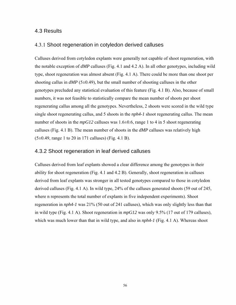
4.3 Results
4.3.1 Shoot regeneration in cotyledon derived calluses
Calluses derived from cotyledon explants were generally not capable of shoot regeneration, with
the notable exception of dMP calluses (Fig. 4.1 and 4.2 A). In all other genotypes, including wild
type, shoot regeneration was almost absent (Fig. 4.1 A). There could be more than one shoot per
shooting callus in dMP (5±0.49), but the small number of shooting calluses in the other
genotypes precluded any statistical evaluation of this feature (Fig. 4.1 B). Also, because of small
numbers, it was not feasible to statistically compare the mean number of shoots per shoot
regenerating callus among all the genotypes. Nevertheless, 2 shoots were scored in the wild type
single shoot regenerating callus, and 5 shoots in the nph4-1 shoot regenerating callus. The mean
number of shoots in the mpG12 calluses was 1.6±0.6, range 1 to 4 in 5 shoot regenerating
calluses (Fig. 4.1 B). The mean number of shoots in the dMP calluses was relatively high
(5±0.49, range 1 to 20 in 171 calluses) (Fig. 4.1 B).
4.3.2 Shoot regeneration in leaf derived calluses
Calluses derived from leaf explants showed a clear difference among the genotypes in their
ability for shoot regeneration (Fig. 4.1 and 4.2 B). Generally, shoot regeneration in calluses
derived from leaf explants was stronger in all tested genotypes compared to those in cotyledon
derived calluses (Fig. 4.1 A). In wild type, 24% of the calluses generated shoots (59 out of 245,
where n represents the total number of explants in five independent experiments). Shoot
regeneration in nph4-1 was 21% (50 out of 241 calluses), which was only slightly less than that
in wild type (Fig. 4.1 A). Shoot regeneration in mpG12 was only 9.5% (17 out of 179 calluses),
which was much lower than that in wild type, and also in nph4-1 (Fig. 4.1 A). Whereas shoot
56
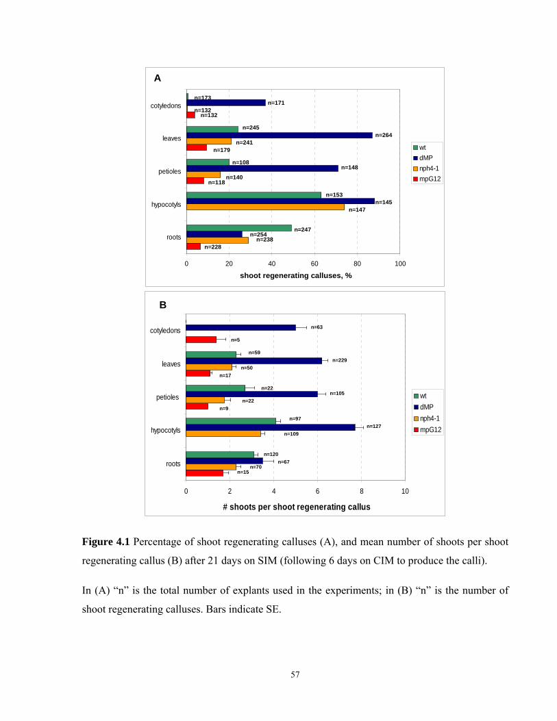
0 20 40 60 80 100
roots
hypocotyls
petioles
leaves
cotyledons
Frequency of shoot regenerating calluses, %
wtdMPnph4-1mpG12
A
n=173n=171
n=132n=132
n=245n=264
n=241n=179
n=108n=148
n=140n=118
n=153n=145
n=147
n=247n=254
n=238n=228
shoot regenerating calluses, %
0 2 4 6 8 10
roots
hypocotyls
petioles
leaves
cotyledons
# shoots per shoot regenerating callus
wtdMPnph4-1mpG12
B
n=63
n=5
n=59n=229
n=50n=17
n=22n=105
n=22n=9
n=97n=127
n=109
n=120n=67
n=70n=15
Figure 4.1 Percentage of shoot regenerating calluses (A), and mean number of shoots per shoot
regenerating callus (B) after 21 days on SIM (following 6 days on CIM to produce the calli).
In (A) “n” is the total number of explants used in the experiments; in (B) “n” is the number of
shoot regenerating calluses. Bars indicate SE.
57
A
dMP
mpG12
wt
nph4-1
cotyledons
B
dMPwt
nph4-1mpG12
leaves
Figure 4.2 Calluses derived from cotyledon (A) and leaf (B) explants after 21 days on SIM
(following 6 days on CIM to produce the calli). Genotypes indicated within plate sectors; arrows
indicate shoot regenerating calluses.
58
regeneration in mpG12 was reduced, it was very high in dMP (87%, or 229 out of 264 calluses)
(Fig. 4.1 A).
The number of shoots per shoot regenerating callus also showed differences in shoot
regeneration among the genotypes (Fig 4.1 B). In wild type, the mean number of shoots per
shoot regenerating callus was 2.2±0.22, range 1 to 8 in 59 calluses. The mean number of shoots
was different in nph4-1 and in mpG12. In nph4-1 calluses, the mean number of shoots per shoot
regenerating callus was 2.1±0.21, range 1 to 7 in 50 calluses. This was comparable to that in wild
type (Fig. 4.1 B). However, mpG12 calluses had only 1.1±0.08 shoots per callus, range 1 to 2 in
17 mpG12 calluses (P<0.001 for the comparison of the mean responses between mpG12 and wild
type, and between mpG12 and nph4-1 calluses) (Fig. 4.1 B). In contrast, in dMP calluses, the
mean number of shoots was much higher than that in wild type (6.2±0.27, range 1 to 24 in 229
calluses, P<0.001) (Fig. 4.1 B).
Collectively, these data indicate a strongly elevated shoot regeneration capability in dMP
calluses derived from leaf explants compared to that in wild type. This was indicated by a
difference in the proportion of shoot generating calluses, and in the mean number of shoots
formed by shoot regenerating calluses. The arf mutant genotypes, nph4-1 and mpG12, were very
different in their shoot regeneration ability. Shoot regeneration in nph4-1 was relatively similar
to that in wild type, whereas shoot regeneration in mpG12 was much lower than that in wild
type. These differences between the arf mutants were indicated by both the frequency of the
shoot regenerating calluses, and the mean number of shoots per callus.
4.3.3 Shoot regeneration in calluses derived from petiole explants
Calluses derived from petiole explants showed clear genotype related difference in their ability
for shoot regeneration on SIM (Fig. 4.1 and 4.3 A). In addition, their shoot regeneration profile
generally resembled that of leaf derived calluses (Fig. 4.1). In wild type, 20% of calluses (22 out
of 108) regenerated shoots (where n represents the total number of explants in four independent
replicas of the experiment). Shoot regeneration in the arf mutants, nph4-1 and mpG12, was
generally lower than that in wild type, but distinctly different in these two genotypes. Thus, shoot
regeneration in nph4-1 was slightly less than that in wild type, and 16%, or 22 out of 140
59
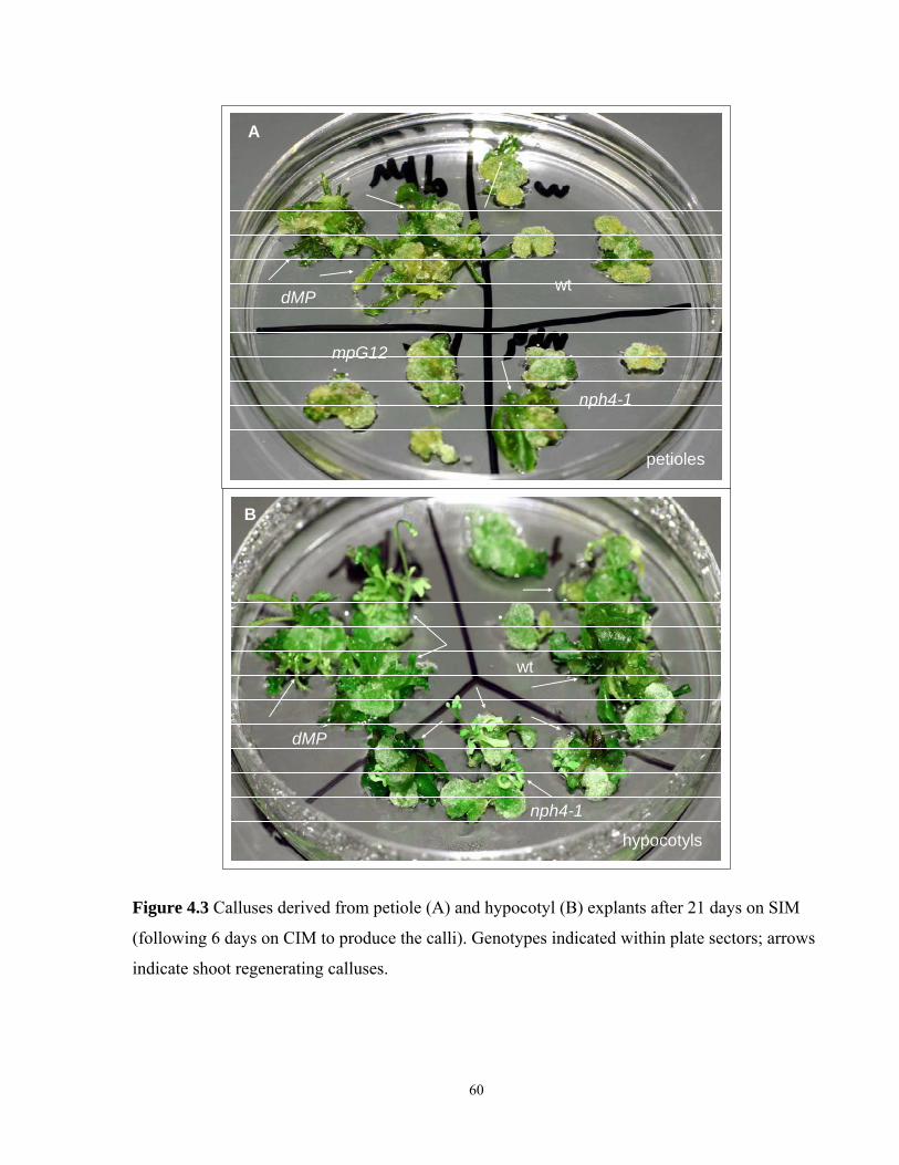
petioles
A
dMP
mpG12
nph4-1
wt
B
dMP
nph4-1
wt
hypocotyls
Figure 4.3 Calluses derived from petiole (A) and hypocotyl (B) explants after 21 days on SIM
(following 6 days on CIM to produce the calli). Genotypes indicated within plate sectors; arrows
indicate shoot regenerating calluses.
60
calluses, regenerated shoots (Fig. 4.1 A). Shoot regeneration in mpG12 was much lower than that
in wild type, and only 8%, or 9 out of 118 calluses, developed shoots (Fig. 4.1 A). Once again,
shoot regeneration in dMP was very strong compared to that in wild type, and a majority of dMP
calluses (71%, or 105 out of 148) formed shoots (Fig. 4.1 A).
The mean number of shoots per shoot regenerating callus also showed differences in shoot
regeneration ability among the genotypes (Fig. 4.1 B). In wild type, the mean number of shoots
per shoot regenerating callus was 2.7±0.42, range 1 to 7 in 22 calluses. The mean number of
shoots was significantly lower in nph4-1 and mpG12 calluses compared to that in wild type. In
nph4-1, the mean number of shoots was 1.8±0.28, range 1 to 6 in 21 calluses (P<0.1) (Fig. 4.1
B). The mpG12 calluses had 1.4±0.43 shoots per callus, range 1 to 5 in 9 calluses (P<0.05) (Fig.
4.1 B). In this category of calluses, the mean number of shoots in nph4-1 and mpG12 did not
differ significantly. In dMP calluses the mean number of shoots per shoot regenerating callus
was 5.9±0.39, range 1 to 24 in 230 calluses (P<0.001) (Fig. 4.1 B).
Collectively, these data showed strongly elevated shoot regeneration ability in dMP calluses
derived from petiole explants. This was indicated by the higher frequency of shoot regenerating
calluses, and by the higher number of shoots formed in the shoot regenerating calluses. Shoot
regeneration in the arf mutant genotypes, nph4-1 and mpG12, was generally lower than that in
wild type. Shoot regeneration was slightly decreased in the nph4-1 calluses, and greatly
decreased in the mpG12 calluses. Thus, the presumably elevated MP function in the dMP
calluses resulted in greatly increased shoot regeneration. Further, the loss of the MP function in
the mpG12 mutants resulted in greatly decreased shoot regeneration. Meanwhile, the loss of the
NPH4 function in the nph4-1 mutants had a more moderate effect on shoot regeneration, when
the calluses derived from petiole explants were compared on SIM.
4.3.4 Shoot regeneration in calluses derived from hypocotyl explants
The genotype related difference in shoot regeneration ability was relatively moderate when
calluses derived from hypocotyl explants were compared on SIM (Fig. 4.1 and 4.3 B). Generally,
shoot regeneration in calluses derived from these explants was strongest among all the types of
explants (cotyledon, leaf, petiole, and adventitious roots) (Fig. 4.1). All the tested genotypes
(wild type, dMP, and nph4-1) had a relatively high rate of shoot regeneration on SIM (Fig. 4.1).
61
No mpG12 calluses could be tested in this category, because of the lack of hypocotyls in the
mpG12 seedlings. In wild type, 63%, or 97 out of 153 calluses, regenerated shoots (where n
represents the total number of explants in four independent replicas of the experiment). Shoot
regeneration in nph4-1 was slightly higher than that in wild type, and 74%, or 109 out of 147
nph4-1 calluses, formed shoots (Fig. 4.1 A). Once again, shoot regeneration in dMP calluses was
stronger compared to that in wild type, and a majority of dMP calluses (88%, or 127 out of 145)
produced shoots (Fig. 4.1 A).
The mean number of shoots per shoot regenerating callus indicated the difference in shoot
regeneration ability among the genotypes more clearly (Fig. 4.1 B). In wild type, the mean
number of shoots per shoot regenerating callus was 4.14±0.23, range 1 to 10 in 97 calluses. The
mean number of shoots was significantly less in the nph4-1 calluses compared to that in the wild
type ones (3.4±0.19 shoots per callus, range 1 to 10 in 109 calluses, P<0.05). In the dMP
calluses, the mean number of shoots was much higher than that in the wild type calluses
(7.68±0.39, range 1 to 24 in 127 calluses, P<0.001).
Collectively, these data showed the increase in shoot regeneration in dMP calluses derived from
hypocotyl explants. This was indicated by a greater frequency of shoot regenerating calluses, and
by a higher mean number of shoots formed by the shoot regenerating calluses. Shoot
regeneration in the arf mutant genotype nph4-1 gave mixed results. On the one hand, the
frequency of shoot regenerating calluses in nph4-1 was higher than that in wild type. On the
other hand, the average number of shoots per shoot regenerating callus was significantly lower
than that in wild type. Thus, when compared to wild type, dMP calluses again showed increased
shoot regeneration on SIM. nph4-1 calluses demonstrated a moderate increase in the frequency
of shoot regenerating calluses, which produced significantly fewer shoots per callus compared to
those in wild type.
4.3.5 Shoot regeneration in calluses derived from adventitious root explants
Shoot regeneration in calluses derived from adventitious root explants partly differed from shoot
regeneration profiles in other types of explants (cotyledon, leaves, petioles, and hypocotyls) (Fig.
4.1 and 4.4). In this category of calluses, the strongest shoot regeneration was observed in wild
type, where 49%, or 120 out of 247 calluses, regenerated shoots (where n represents the total
62
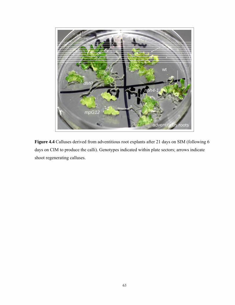
dMP
mpG12
wt
nph4-1
adventitious roots
Figure 4.4 Calluses derived from adventitious root explants after 21 days on SIM (following 6
days on CIM to produce the calli). Genotypes indicated within plate sectors; arrows indicate
shoot regenerating calluses.
63
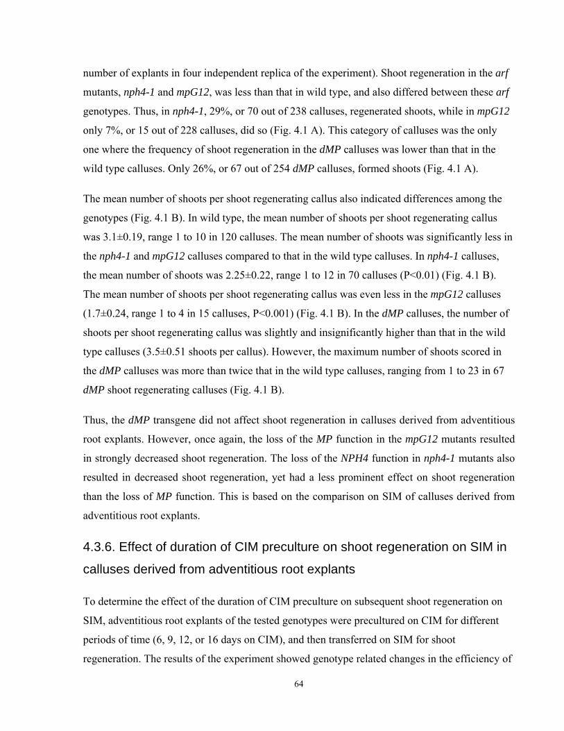
number of explants in four independent replica of the experiment). Shoot regeneration in the arf
mutants, nph4-1 and mpG12, was less than that in wild type, and also differed between these arf
genotypes. Thus, in nph4-1, 29%, or 70 out of 238 calluses, regenerated shoots, while in mpG12
only 7%, or 15 out of 228 calluses, did so (Fig. 4.1 A). This category of calluses was the only
one where the frequency of shoot regeneration in the dMP calluses was lower than that in the
wild type calluses. Only 26%, or 67 out of 254 dMP calluses, formed shoots (Fig. 4.1 A).
The mean number of shoots per shoot regenerating callus also indicated differences among the
genotypes (Fig. 4.1 B). In wild type, the mean number of shoots per shoot regenerating callus
was 3.1±0.19, range 1 to 10 in 120 calluses. The mean number of shoots was significantly less in
the nph4-1 and mpG12 calluses compared to that in the wild type calluses. In nph4-1 calluses,
the mean number of shoots was 2.25±0.22, range 1 to 12 in 70 calluses (P<0.01) (Fig. 4.1 B).
The mean number of shoots per shoot regenerating callus was even less in the mpG12 calluses
(1.7±0.24, range 1 to 4 in 15 calluses, P<0.001) (Fig. 4.1 B). In the dMP calluses, the number of
shoots per shoot regenerating callus was slightly and insignificantly higher than that in the wild
type calluses (3.5±0.51 shoots per callus). However, the maximum number of shoots scored in
the dMP calluses was more than twice that in the wild type calluses, ranging from 1 to 23 in 67
dMP shoot regenerating calluses (Fig. 4.1 B).
Thus, the dMP transgene did not affect shoot regeneration in calluses derived from adventitious
root explants. However, once again, the loss of the MP function in the mpG12 mutants resulted
in strongly decreased shoot regeneration. The loss of the NPH4 function in nph4-1 mutants also
resulted in decreased shoot regeneration, yet had a less prominent effect on shoot regeneration
than the loss of MP function. This is based on the comparison on SIM of calluses derived from
adventitious root explants.
4.3.6. Effect of duration of CIM preculture on shoot regeneration on SIM in
calluses derived from adventitious root explants
To determine the effect of the duration of CIM preculture on subsequent shoot regeneration on
SIM, adventitious root explants of the tested genotypes were precultured on CIM for different
periods of time (6, 9, 12, or 16 days on CIM), and then transferred on SIM for shoot
regeneration. The results of the experiment showed genotype related changes in the efficiency of
64
0
10
20
30
40
50
60
70
80
90
100
wt 6d n=
72
wt 9 d
n=35
wt 12d n
=22
wt 16 d
n=12
dMP 6d n=73
dMP 9d n
=34
dMP 12d
n=24
dMP 16d n=
14
nph4-1
6d n=67
nph4-1 9d
n=36
nph4
-1 12d n=25
nph4
-1 16d n=12
mpG12 6d n
=68
mpG12 9d n=32
mpG12 12d n
=24
mpG12 16 n=
12
Freq
uenc
y of
sho
ot re
gene
ratin
g ca
lluse
s, %
A
0
2
4
6
8
10
12
14
16
wt 6d n
=47
wt 9d n
=31
wt 12d
n=6
wt 16d
n=1
dMP 6d
n=18
dMP 9d
n=29
dMP 12
d n=13
dMP 16
d n=5
nph4
-1 6d n=
22
nph4
-1 9d n=
29
nph4
-1 12d n=
17
nph4
-1 16d n=
4
mpG12
6d n=11
mpG12
9d n=5
mpG12
12d n=2
mpG12
16d n=1
# sh
oots
per
sho
ot re
geer
atin
g ca
llus
B
Figure 4.5 Percentage of shoot regenerating calluses (A) and mean number of shoots per shoot regenerating callus (B) in calluses derived from adventitious root explants after 21 days on SIM depending on the time on CIM (6, 9, 12 and 16 days).
In (A) “n” represent the total number of explants used in the experiments; in (B) “n” is the number of shoot regenerating calluses. Bars indicate SE.
65
shoot regeneration depending on the time on CIM (Fig. 4.5). The results of a single experiment
are presented below.
In the experiment, when the adventitious root explants were exposed to CIM for 6 days (a
standard protocol, see Materials and Methods, Chapter 2.8), shoot regeneration was strongest in
the wild type calluses, and 64%, or 46 out of 72 calluses, formed shoots. Shoot regeneration
among all other genotypes was much lower compared to that in wild type. Specifically, shoot
regeneration in the arf mutants, nph4-1 and mpG12, was lower than that in wild type. Thus, 33%
(22 out of 67) nph4-1 calluses, and only 16% (11 out of 68) mpG12 calluses regenerated shoots
(Fig. 4.5 A). In dMP, the frequency of shoot regenerating calluses was also lower than that in
wild type, and only 25% (18 out of 73 calluses) formed shoots (Fig. 4.5 A).
The number of shoots per shoot regenerating callus differed slightly among the genotypes, where
the only significant difference was detected between wild type and mpG12. In wild type, the
mean number of shoots per shoot regenerating callus was 2.55±0.28, range 1 to 9 in 46 calluses.
In dMP, the mean number of shoots was 4.38±1.3, range 1 to 20 in 18 calluses. In nph4-1, the
mean number of shoots was 2.9±0.54, range 1 to 12 in 22 shoot regenerating calluses (Fig. 4.5
B). In mpG12, the mean number of shoots was significantly less than that in wild type (1.64±0.3,
range 1 to 4 in 11 calluses, P<0.05) (Fig. 4.5 B).
When the time on CIM was extended to 9 days, the shoot regeneration profile changed
significantly. Generally, shoot regeneration increased, and became relatively similar in all the
genotypes. An exception was mpG12, where shoot regeneration stayed consistently low
(Fig.4.5). At 9 days, 86%, or 30 out of 35 wild type calluses; 85%, or 29 out of 34 dMP calluses;
81%, or 29 out of 36 nph4-1 calluses; and only 16%, or 5 out of 32 mpG12 calluses, regenerated
shoots (Fig. 4.5 A). Also, in this experimental condition, the mean number of shoots per shoot
regenerating callus generally increased in all the genotypes, except for mpG12. In wild type, the
mean number of shoots per shoot regenerating callus was 4.87±0.5, range 1 to 12 in 31 calluses.
In dMP, the mean number of shoots was 13.31±1.23, range 1 to 26 in 29 calluses (P<0.001). In
nph4-1, the mean number of shoots was 7.1±0.78, range 1 to 16 in 29 calluses (P<0.05) (Fig.4.5
B). In mpG12, the mean number of shoots per shoot regenerating callus was significantly less
than that in wild type (1.2±0.19, range 1 to 2 in 5 calluses, P<0.001) (Fig. 4.5 B).
66
Extension of the time on CIM to 12 days affected shoot regeneration in the genotypes differently.
In wild type, shoot regeneration was strongly reduced compared to that at 9 days on CIM, and
only 27% (6 out of 22) wild type calluses regenerated shoots. Shoot regeneration was also
reduced in the dMP and nph4-1 calluses. However, these reductions were not as strong as in the
wild type calluses. Specifically, 54% (13 out of 24) dMP calluses, and 68% (17 out of 25) nph4-
1 calluses regenerated shoots (Fig. 4.5 A). Shoot regeneration in mpG12 calluses was also lower,
with only 8%, or 2 out of 24 calluses, regenerating shoots (Fig. 4.5 A).
The mean number of shoots per shoot regenerating callus was also reduced compared to that of
the previous experimental settings (9 days on CIM) in all the genotypes, except mpG12. In wild
type, the mean number of shoots per shoot regenerating callus was 3±1, range 1 to 8 in 6
calluses. The mean number of shoots per shoot regenerating callus was significantly higher in
dMP compared to that in wild type (9.85±2.27, range 1 to 30 in 13 calluses, P<0.05) (Fig. 4.5 B).
The mean number of shoots in nph4-1 was insignificantly higher than that in wild type
(4.88±0.7, range 1 to 10 in 17 calluses) (Fig. 4.5 B). The number of shoots scored in 2 out of 24
mpG12 shoot regenerating calluses was 1 and 2, which was consistent with all the data for shoot
regeneration in this genotype (Fig. 4.5 B).
A further drop in shoot regeneration was observed when the time on CIM was extended to 16
days (Fig. 4.5). Only 8%, or 1 out of 12 wild type calluses, regenerated shoots. A decrease in
shoot regeneration was also detected in dMP and nph4-1. However, shoot regeneration in these
genotypes was still higher than that in wild type. Only 36%, or 5 out of 14 dMP calluses, and
33%, or 4 out of 12 nph4-1 calluses, were still capable of shoot regeneration. Shoot regeneration
ability in mpG12 did not change, and 8%, or 1 out of 12 calluses, regenerated shoots.
Because of a generally low number of shoot regenerating calluses, a statistical analysis could not
be performed to discriminate the differences in the mean number of shoots per shoot
regenerating callus in this category. In wild type, the single shoot regenerating callus produced 2
shoots. In dMP, the mean number of shoots was 5.4±2.4, range 1 to 10 in 5 calluses (Fig. 4.5 B).
In nph4-1, the mean number was 2±0.4, range 1 to 3 in 4 calluses (Fig. 4.5 B). In mpG12, the
single shoot regenerating callus produced one shoot (Fig. 4.5 B).
67
Taken together, these data indicate that the increased time on CIM affected shoot regeneration in
wild type, dMP, and nph4-1, but not in mpG12. While the extension of CIM preculture from 6 to
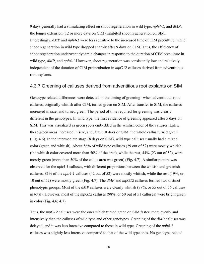
9 days generally had a stimulating effect on shoot regeneration in wild type, nph4-1, and dMP,
the longer extension (12 or more days on CIM) inhibited shoot regeneration on SIM.
Interestingly, dMP and nph4-1 were less sensitive to the increased time of CIM preculture, while
shoot regeneration in wild type dropped sharply after 9 days on CIM. Thus, the efficiency of
shoot regeneration underwent dynamic changes in response to the duration of CIM preculture in
wild type, dMP, and nph4-1.However, shoot regeneration was consistently low and relatively
independent of the duration of CIM preincubation in mpG12 calluses derived from adventitious
root explants.
4.3.7 Greening of calluses derived from adventitious root explants on SIM
Genotype related differences were detected in the timing of greening--when adventitious root
calluses, originally whitish after CIM, turned green on SIM. After transfer to SIM, the calluses
increased in size, and turned green. The period of time required for greening was clearly
different in the genotypes. In wild type, the first evidence of greening appeared after 5 days on
SIM. This was visualized as green spots embedded in the whitish color of the calluses. Later,
these green areas increased in size, and, after 10 days on SIM, the whole callus turned green
(Fig. 4.6). In the intermediate stage (8 days on SIM), wild type calluses usually had a mixed
color (green and whitish). About 56% of wild type calluses (29 out of 52) were mostly whitish
(the whitish color covered more than 50% of the area), while the rest, 44% (23 out of 52), were
mostly green (more than 50% of the callus area was green) (Fig. 4.7). A similar picture was
observed for the nph4-1 calluses, with different proportions between the whitish and greenish
calluses. 81% of the nph4-1 calluses (42 out of 52) were mostly whitish, while the rest (19%, or
10 out of 52) were mostly green (Fig. 4.7). The dMP and mpG12 calluses formed two distinct
phenotypic groups. Most of the dMP calluses were clearly whitish (98%, or 55 out of 56 calluses
in total). However, most of the mpG12 calluses (98%, or 50 out of 51 calluses) were bright green
in color (Fig. 4.6; 4.7).
Thus, the mpG12 calluses were the ones which turned green on SIM faster, more evenly and
intensively than the calluses of wild type and other genotypes. Greening of the dMP calluses was
delayed, and it was less intensive compared to those in wild type. Greening of the nph4-1
calluses was slightly less intensive compared to that of the wild type ones. No genotype related
68
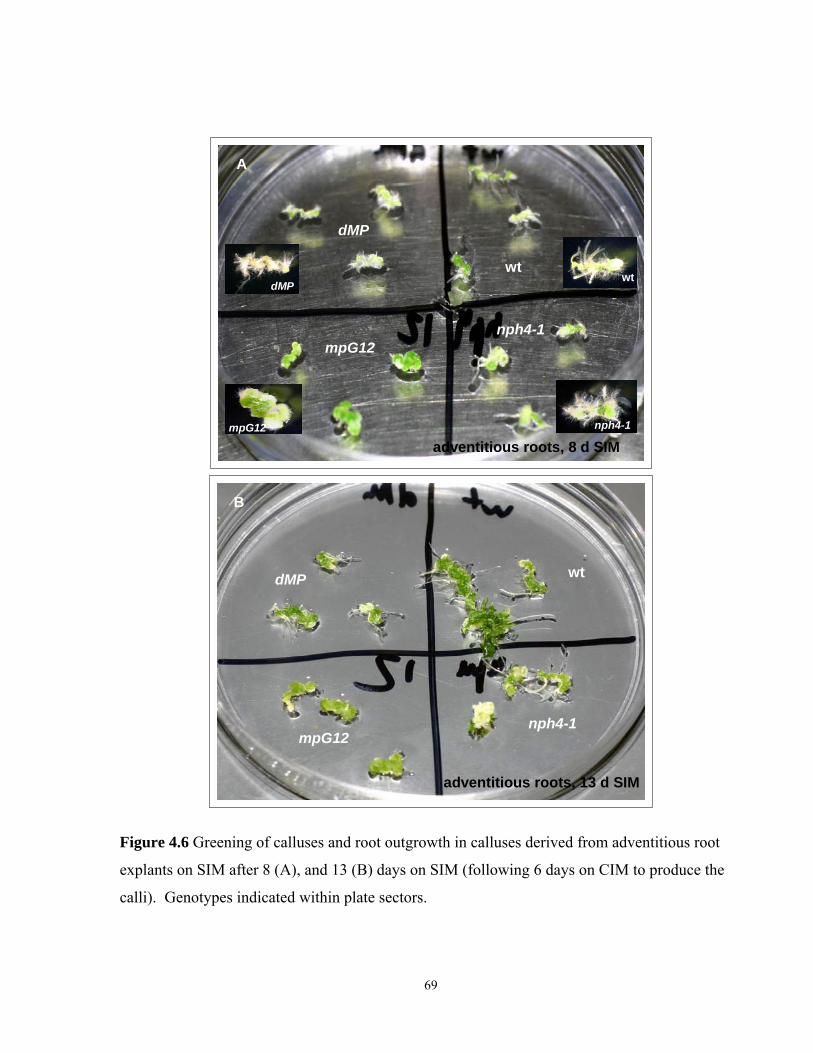
dMPwt
mpG12 nph4-1
A
adventitious roots, 8 d SIM
mpG12
dMP
nph4-1
wt
dMP
mpG12nph4-1
wt
adventitious roots, 13 d SIM
B
Figure 4.6 Greening of calluses and root outgrowth in calluses derived from adventitious root
explants on SIM after 8 (A), and 13 (B) days on SIM (following 6 days on CIM to produce the
calli). Genotypes indicated within plate sectors.
69
0102030405060708090
100
wt n=52 dMP n=56 nph4-1 n=52 mpG12 n=51
frequ
ency
, %
lightgreen
wtn=52
dMPn=56
nph4-1n=52
mpG12n=51
roots, 8 days on SIM
perc
enta
ge, %
Figure 4.7 Percentage of green versus whitish calluses after 8 days on SIM in adventitious root
derived calluses; n, number of explants.
70
difference in greening was established for calluses derived from other organs than roots, because
they were already greenish at the time of their transfer on SIM.
4.4. Discussion and Conclusions
The results in this chapter indicate that the efficiency of shoot regeneration depends on the
genotype, the type of organ used as the explant, and the duration of preculture on CIM. As
shown earlier (Akama et al., 1992; Chatfield and Raizada, 2008), the Columbia ecotype of
Arabidopsis thaliana is relatively recalcitrant to shoot regeneration, especially when cotyledons
were used to induce shoot regeneration in vitro. In the experiments here, shoot regeneration in
wild type varied dramatically depending on the type of organ used as the explant. In wild type,
the frequency of shoot regenerating calluses was negligible in cotyledon derived calluses,
moderate in leaf and petiole derived calluses, and relatively high in hypocotyl and adventitious
root derived calluses. A similar dependence of shoot regeneration efficiency on the type of
explant was clearly detected in nph4-1 calluses. However, shoot regeneration in dMP and
mpG12 calluses derived from these explants differed from those in wild type. Shoot regeneration
in mpG12 was very low regardless of the organ derivation of the callus. In contrast, shoot
regeneration in dMP was strong in calluses derived from aerial organs (cotyledons, leaves,
petioles, and hypocotyls), where it was significantly and consistently higher than that in wild
type. No elevation of shoot regeneration was recorded in the dMP calluses derived from
adventitious root explants.
These data indicate that the presumably elevated MP function in dMP promotes shoot
regeneration in calluses derived from aerial explants. Consistent with this, loss of MP function in
mpG12 mutants lead to greatly reduced shoot regeneration. This was indicated by the low
frequency of shoot regenerating calluses, and by the low number of shoots per callus in mpG12.
Loss of NPH4 function in nph4-1 mutants had a relatively modest effect on shoot regeneration.
The frequency of shoot regenerating calluses and the number of shoots per callus were only
slightly lower in nph4-1 compared to those in wild type.
MP/ARF5 and NPH4/ARF7 have a sequence similarity, interact with each other, and have
partially redundant functions (Liscum and Reed, 2002; Hardtke et al., 2004; discussed in General
Introduction, Chapter 1.2.5). As shown here, during de novo shoot initiation, the NPH4 gene
71
could not completely substitute for the loss of MP function. The MP gene is implicated in the
regulation of several key shoot meristem genes, such as CUC2 and STM (Aida et al., 2002), and
the ESR1/DRN gene (Cole et al., 2009). Moreover, MP is involved in the control of cytokinin
signaling through the regulation of activity A-type ARR15 and ARR7 (Zhao et al., 2010). The
meristem genes, such as CUC, STM, and ESR1/DRN, and some of the ARR genes are implicated
in de novo shoot regeneration (reviewed in Chapter 1.6.1; Hwang and Sheen, 2001; Osakabe et
al., 2002; To et al., 2004; Zhang and Lemaux, 2005). Thus, the observed strong impairment of
shoot regeneration in mpG12 mutants, as well as the consistently elevated shoot regeneration in
dMP calluses derived from aerial explants, may be explained by changes in the expression levels
of these genes in the mpG12 and dMP genotypes. The failure of dMP to promote shoot
regeneration from root derived calluses may be explained by the fact that MP, and hence dMP,
are not normally expressed in mature roots (Dr. W. Ckurshumova, personal communication).
However, low shoot regeneration in mpG12 may contradict the suggestion of the irrelevance of
MP to shoot regeneration in root derived calluses. Future studies are required to determine
whether dMP expression in root derived calluses is similar to that in calluses derived from other
organs.
No data demonstrating the involvement of NPH4 in the regulation of shoot meristem genes were
found in the literature. Instead, it has been shown that NPH4/ARF7 plays a central role in the
modulation of auxin-dependent differential growth in the aerial parts of Arabidopsis plants
(Stowe-Evans et al., 1998, Watahiki et al., 1999; reviewed in Chapter 1.2.5). Auxin insensitivity
has been shown for aerial organs, but not for the roots of nph4 mutants (Watahiki and
Yamamoto, 1997). However, here shoot regeneration in nph4-1 calluses derived from
adventitious root explants, with presumably normal auxin sensitivity and callusogenesis (Chapter
3.3.6), was significantly lower than that in the wild type calluses. This could reflect the partially
redundant function that NPH4 has with MP in forming the apical part of embryos, including the
shoot meristem (Hardtke et al., 2004). The contribution of NPH4 could be subtle, and completely
dispensable, in calluses cultured for more than 9 days.
In summary, decreased MP/ARF5 and NPH4/ARF7 levels in mpG12 and nph4-1 mutants had a
different impact on de novo shoot regeneration in these genotypes. While shoot regeneration in
nph4-1 was only slightly decreased compared to that in wild type, shoot regeneration in mpG12
was consistently, and markedly, reduced in calluses derived from any type of explant in all sets 72

of experiments. In contrast, elevated MP activity in dMP had a strong stimulating effect on shoot
regeneration when aerial organs were used as explants. Thus, MP played an important role in de
novo shoot meristem initiation.
73
Chapter 5
Root regeneration under conditions of altered auxin signal
transduction
5.1 Background and rationale
In tissue culture, regeneration of adventitious roots occurs in response to a high auxin to low
cytokinin ratio, or when auxin is present as a sole hormone in the root inducing medium (RIM)
(Miller and Skoog, 1957; Gaba, 2005). Rooting is often a critical step in the plant propagation
industry, because many species are extremely recalcitrant regarding adventitious root formation
(de Klerk, 2002; Schwarz et al., 2005). Although adventitious roots have been routinely induced
in response to auxin (Callis, 2005; Schwarz et al., 2005), the molecular mechanisms of this event
are unclear (Schwarz et al., 2005; Rose et al, 2006).
In tissue culture, the acquisition of competence by cells for either shoot or root formation occurs
during the preincubation stage on CIM (Christian and Warnick, 1985; Che et al., 2007) - the
stage when promeristemoid structures are formed (Schwarz et al., 2005). The specification of
organ development depends on subsequent hormonal stimuli on SIM or RIM. Ozawa et al.
(1998) - using the temperature-sensitive mutants, srd1, srd2 and srd3 -demonstrated that the
competence to form organs is acquired progressively, first roots and then shoots (Ozawa et al.,
1998).
In vitro experiments with leaf explants have shown that root primordia are formed from the
procambial-like cells of veins. These function as pluripotent stem cells, with the ability to form
either root primordia or vascular tissues in response to added auxin (Attfield and Evans, 1991;
Rose et al., 2006). Unlike the hormone auxin, cytokinin inhibits root formation in planta (Aloni
et al., 2006) and in vitro (Sugiyama 1999; Kubo and Kakimoto, 2000). Auxin insensitivity causes
impairment in root regeneration in culture. The auxin-tolerant rac mutants in tobacco are
defective in primary root formation and the regeneration of adventitious roots (Sugiyama, 1999,
and references therein). By contrast, cytokinin insensitivity promotes root and root primordia
formation even on cytokinin containing media (Kubo and Kakimoto, 2000; Kakani et al., 2009).
Che et al. (2006) demonstrated that molecular signatures for callus formation and root
74
regeneration are quite similar, since both are triggered by auxin-rich media. They also showed
that competence for root regeneration correlates with the increased expression of IAA genes on
CIM.
Besides their other features (for details see Chapter 1.2.5), ARF5/MP and ARF7/NPH4 are
implicated in root formation. However, mp seedlings lack primary roots (Berleth and Jurgens,
1993). Increased auxin responses and increased adventitious root formation have been shown for
cotyledon explants of the GOF MP line, dMP (Krogan et al., 2006). However, root formation
from cultured cotyledons of mp mutants is strongly decreased (Mattsson et al., 2003). Also, mp
mutants show auxin insensitivity and reduction of DR5 expression in leaf primordia (Mattsson et
al., 2003). The nph4 mutants exhibit auxin resistance in the aerial parts of plants. They have
severely impaired expression of many primary auxin-dependent genes and the auxin-responsive
reporter DR5 (Watahiki and Yamamoto, 1997; Stowe-Evans et al., 1998; Wang et al., 2005).
Also, NPH4, together with ARF19, regulates lateral and adventitious root formation (Tatematsu
et al., 2004; Wilmoth et al, 2005; Okushima et al., 2007). Wilmoth et al. (2005) showed that, in
the presence of exogenous IAA, the excised nph4 roots formed fewer lateral roots. The nph4
excised hypocotyls formed fewer adventitious roots than did wild type roots and hypocotyls. By
contrast, nph4 arf19 double mutants mainly lacked lateral roots.
In this chapter, the roles of the MP and NPH4 genes in de novo root regeneration in tissue culture
are explored. The extent to which root regeneration in each of the investigated genotypes is
further influenced by the origin of the respective tissue sample is also explored.
5.2 Specific research objectives
This project investigates the possible influence on de novo root regeneration of auxin signaling
through ARF5/MP and ARF7/NPH4. To this end, the loss-of-function mutants mpG12 (Hardtke
and Berleth, 1998), and nph4-1 (Harper et al., 2000), and the gain of function transgene line dMP
with truncated dimerization domains III and IV (reviewed in Chapter 1.2.5; Fig. 1.2; Krogan,
2006) were tested to determine the influence of these genotypes on adventitious root
regeneration in vitro. All the genotypes used in the experiments were in the Col-0 background
and were compared to the Col-0 wild type. Explants of different organs (cotyledons, leaves, and
adventitious roots) were first cultured on CIM for 6 days and then transferred to RIM (reviewed
75
in Chapter 2.8; Valvekens et al., 1988). Additionally, root formation in calluses derived from
adventitious root explants, and cultured on SIM after 6 days on CIM, was compared among the
genotypes. The frequency of root regeneration, a calculation based on the pooled data of all
replicas of the experiments, was compared among the tested genotypes after 21 days on RIM or
on SIM, as was the mean number of roots per root regenerating callus (mean +/- SE).
5.3 Results
5.3.1 Root regeneration in cotyledon derived calluses on RIM
There were some genotype related differences in root regeneration frequency, and in the mean
number of roots per root regenerating callus (Fig. 5.1; 5.2 A; 5.3). In wild type, only 67% of
calluses (63 out of 89) regenerated roots (Fig. 5.1 A). Among the rest of the genotypes, the
proportion of root regenerating calluses was higher than that in wild type, and relatively similar:
96% of dMP calluses (84 out of 88); 91% of nph4-1 calluses (76 out of 85); and, 93% of mpG12
calluses (71 out of 77) regenerated roots (Fig. 5.1 A).
The mean number of roots per root regenerating callus was quite similar among wild type, nph4-
1, and mpG12 calluses. It was significantly higher only in dMP (Fig. 5.1 B). In wild type, the
mean number of roots was 8.6±0.69, range 2 to 25 roots per calluses in 63 root regenerating
calluses. In nph4-1, the mean number of roots was 8.72±0.53, range 1 to 22 roots in 76 calluses.
In mpG12, this number was 8±0.5, range 1 to 20 in 71 calluses (Fig. 5.1 B). The mean number of
roots was significantly higher in dMP, equal to 17.3±0.82, range 3 to 35 in 84 root regenerating
calluses (P<0.001) (Fig. 5.1 B).
To summarize, in calluses derived from cotyledon explants, root regeneration in nph4-1 and
mpG12 was not impaired. It was either equal to that in wild type (in the mean number of roots
per root regenerating callus), or even exceeded that in wild type (in the frequency of root
regenerating calluses). Moreover, root regeneration in dMP calluses was significantly increased
compared to that in wild type.
76
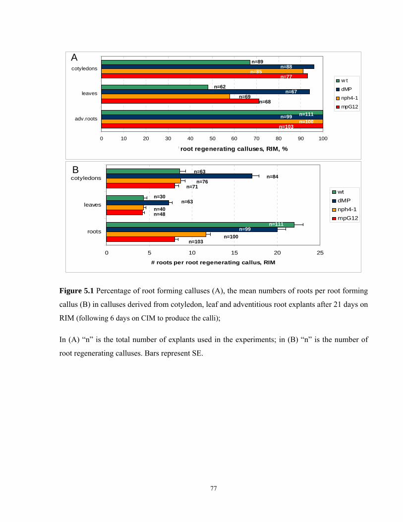
0 10 20 30 40 50 60 70 80 90 100
adv.roots
leaves
cotyledons
Frequency of root regenerating calluses, RIM, %
w t
dMP
nph4-1
mpG12
0 5 10 15 20 25
roots
leaves
cotyledons
# roots per root regenerating callus, RIM
wtdMPnph4-1mpG12
B
A n=89n=88
n=85n=77
n=62n=67
n=69n=68
n=111n=99n=100
n=103
n=63n=84
n=76n=71
n=30n=63
n=40n=48
n=111n=99
n=100n=103
Figure 5.1 Percentage of root forming calluses (A), the mean numbers of roots per root forming
callus (B) in calluses derived from cotyledon, leaf and adventitious root explants after 21 days on
RIM (following 6 days on CIM to produce the calli);
In (A) “n” is the total number of explants used in the experiments; in (B) “n” is the number of
root regenerating calluses. Bars represent SE.
77
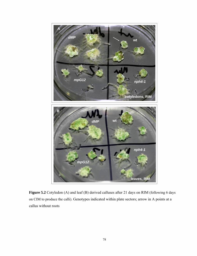
A
cotyledons, RIM
dMP
mpG12
wt
nph4-1
dMP
mpG12
wt
nph4-1
leaves, RIM
B
Figure 5.2 Cotyledon (A) and leaf (B) derived calluses after 21 days on RIM (following 6 days
on CIM to produce the calli). Genotypes indicated within plate sectors; arrow in A points at a
callus without roots
78
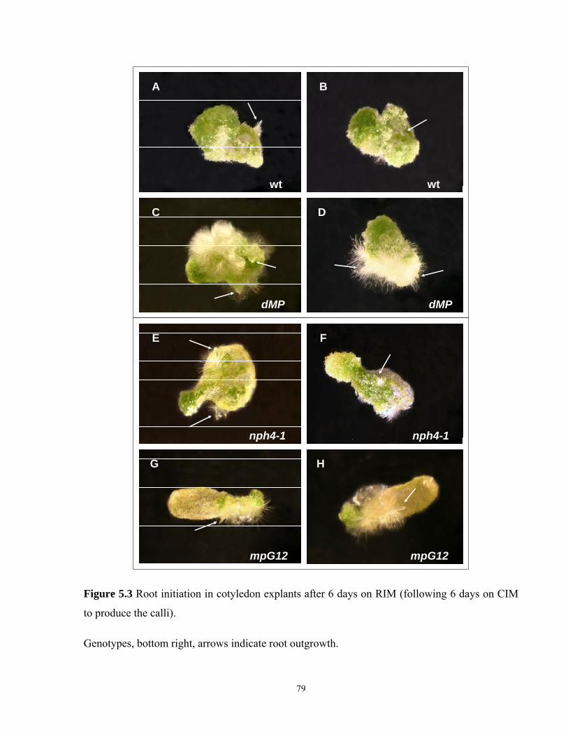
A B
C D
wt wt
dMP dMP
E F
G H
nph4-1 nph4-1
mpG12 mpG12
Figure 5.3 Root initiation in cotyledon explants after 6 days on RIM (following 6 days on CIM
to produce the calli).
Genotypes, bottom right, arrows indicate root outgrowth.
79
5.3.2 Root regeneration in leaf derived calluses on RIM
Root regeneration in calluses derived from leaf explants was generally lower than that in
cotyledon derived calluses (Fig. 5.1; 5.2 B). In wild type, only 48% (30 out of 62) calluses
regenerated roots. Once again, this frequency was the lowest one among the genotypes (Fig. 5.1
A). In the arf mutants, nph4-1 and mpG12, the frequency of root regenerating calluses was
higher than that in wild type: 58% of nph4-1 calluses (40 out of 69); and, 71% of mpG12
calluses (48 out of 68) regenerated roots (Fig. 5.1 A). The highest frequency of root regenerating
calluses was scored again in dMP, where 94% of calluses (63 out of 67) formed roots (Fig.
5.1A).
The mean number of roots per root regenerating callus was similar among wild type, nph4-1, and
mpG12 calluses (Fig. 5.1B). However, the mean number of roots was significantly higher in
dMP (Fig. 5.1 B). In wild type, the mean number of roots per root regenerating callus was
4.35±0.35, range 2 to 10 per callus in 30 calluses. In nph4-1, the mean number of roots was
4.3±0.31, range 2 to 8 in 40 calluses. In mpG12 the mean number was 4.2±0.26, range 2 to 9 in
48 calluses (Fig. 5.1 B). The mean number of roots was significantly higher in dMP, equal to
7.3±0.39, range 2 to 13 in 63 calluses (P<0.001) (Fig. 5.1 B).
To summarize, in leaf derived calluses, root regeneration in the arf mutants, nph4-1 and mpG12,
was not lower than that in wild type. However, in dMP, root regeneration was significantly
greater than that in wild type.
5.3.3 Root regeneration in calluses derived from adventitious roots on RIM
Since root explants are able to produce roots on RIM without CIM preincubation (Ozawa et. al,
1988), all explants in this experiment formed roots. The genotype related differences were
manifested only in the number of roots per root regenerating callus (Fig. 5.1; 5.4; 5.5). In wild
type, the mean number of roots was 22.12±1, range 2 to 50 in 111 calluses. The mean number of
roots in dMP calluses was comparable to that in wild type (19.86±0.95, range 1 to 45 in 99
calluses) (Fig. 5.1 B). By contrast, the mean number of roots per callus was significantly lower in
nph4-1 (11.57±0.59, range 2 to 30 in 100 calluses, P<0.001); and especially in mpG12 calluses
(7.99±0.42, range 1 to 18 in 103 calluses, P<0.001). The difference in the mean number of roots
per callus between nph4-1 and mpG12 was also statistically significant (P<0.001) (Fig. 5.1 B). 80
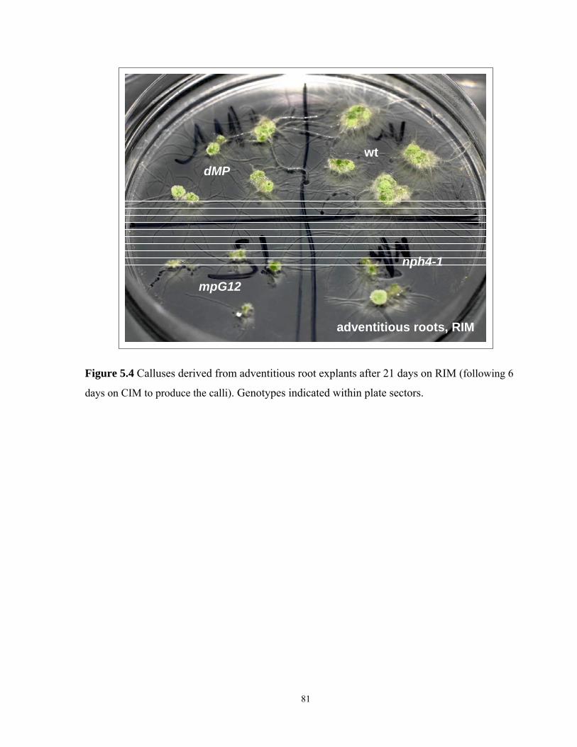
dMP
mpG12
nph4-1
wt
adventitious roots, RIM
Figure 5.4 Calluses derived from adventitious root explants after 21 days on RIM (following 6
days on CIM to produce the calli). Genotypes indicated within plate sectors.
81
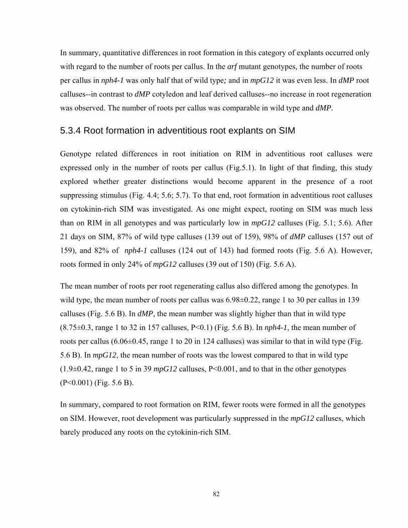
In summary, quantitative differences in root formation in this category of explants occurred only
with regard to the number of roots per callus. In the arf mutant genotypes, the number of roots
per callus in nph4-1 was only half that of wild type; and in mpG12 it was even less. In dMP root
calluses--in contrast to dMP cotyledon and leaf derived calluses--no increase in root regeneration
was observed. The number of roots per callus was comparable in wild type and dMP.
5.3.4 Root formation in adventitious root explants on SIM
Genotype related differences in root initiation on RIM in adventitious root calluses were
expressed only in the number of roots per callus (Fig.5.1). In light of that finding, this study
explored whether greater distinctions would become apparent in the presence of a root
suppressing stimulus (Fig. 4.4; 5.6; 5.7). To that end, root formation in adventitious root calluses
on cytokinin-rich SIM was investigated. As one might expect, rooting on SIM was much less
than on RIM in all genotypes and was particularly low in mpG12 calluses (Fig. 5.1; 5.6). After
21 days on SIM, 87% of wild type calluses (139 out of 159), 98% of dMP calluses (157 out of
159), and 82% of nph4-1 calluses (124 out of 143) had formed roots (Fig. 5.6 A). However,
roots formed in only 24% of mpG12 calluses (39 out of 150) (Fig. 5.6 A).
The mean number of roots per root regenerating callus also differed among the genotypes. In
wild type, the mean number of roots per callus was 6.98±0.22, range 1 to 30 per callus in 139
calluses (Fig. 5.6 B). In dMP, the mean number was slightly higher than that in wild type
(8.75±0.3, range 1 to 32 in 157 calluses, P<0.1) (Fig. 5.6 B). In nph4-1, the mean number of
roots per callus (6.06±0.45, range 1 to 20 in 124 calluses) was similar to that in wild type (Fig.
5.6 B). In mpG12, the mean number of roots was the lowest compared to that in wild type
(1.9±0.42, range 1 to 5 in 39 mpG12 calluses, P<0.001, and to that in the other genotypes
(P<0.001) (Fig. 5.6 B).
In summary, compared to root formation on RIM, fewer roots were formed in all the genotypes
on SIM. However, root development was particularly suppressed in the mpG12 calluses, which
barely produced any roots on the cytokinin-rich SIM.
82
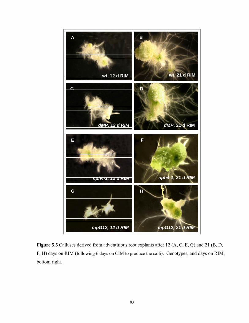
A
wt, 12 d RIM
C
dMP, 12 d RIM
B
D
wt, 21 d RIM
dMP, 21 d RIM
E
nph4-1, 12 d RIM
G
mpG12, 12 d RIM
F
H
nph4-1, 21 d RIM
mpG12, 21 d RIM
Figure 5.5 Calluses derived from adventitious root explants after 12 (A, C, E, G) and 21 (B, D,
F, H) days on RIM (following 6 days on CIM to produce the calli). Genotypes, and days on RIM,
bottom right.
83
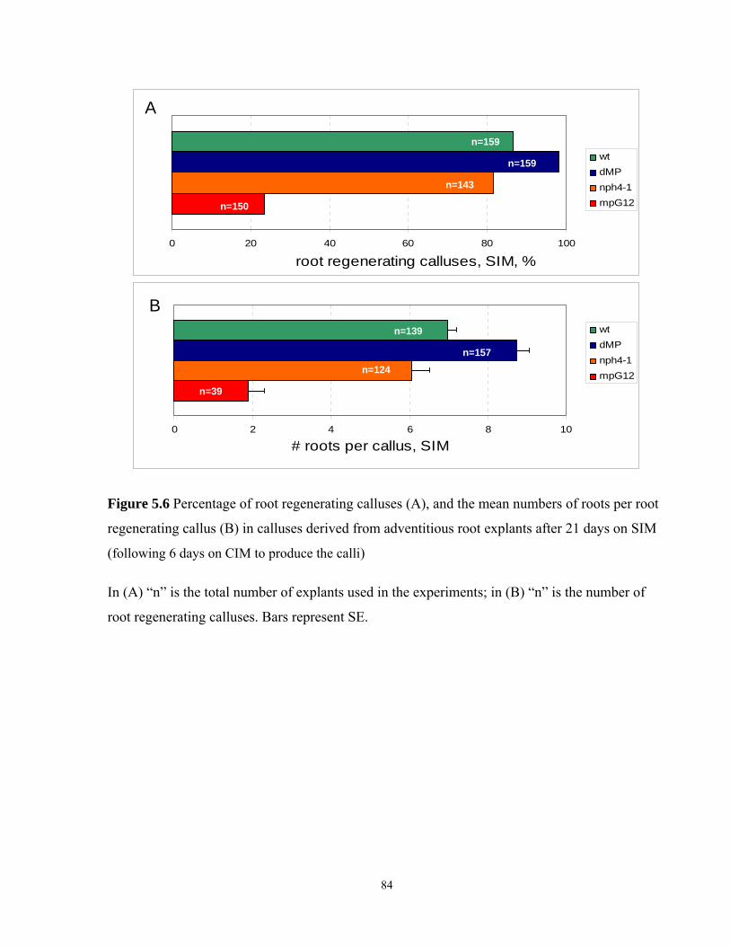
0 2 4 6 8 10
# roots per callus, SIM
wtdMPnph4-1mpG12
0 20 40 60 80 100
Frequency of root regenerating calluses, SIM, %
wtdMPnph4-1mpG12
A
B
n=159
n=159
n=143
n=150
n=139
n=157n=124
n=39
Figure 5.6 Percentage of root regenerating calluses (A), and the mean numbers of roots per root
regenerating callus (B) in calluses derived from adventitious root explants after 21 days on SIM
(following 6 days on CIM to produce the calli)
In (A) “n” is the total number of explants used in the experiments; in (B) “n” is the number of
root regenerating calluses. Bars represent SE.
84
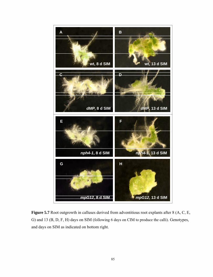
A B
C D
wt, 8 d SIM wt, 13 d SIM
dMP, 8 d SIM dMP, 13 d SIM
E F
G H
nph4-1, 8 d SIM nph4-1, 13 d SIM
mpG12, 8 d SIM mpG12, 13 d SIM
Figure 5.7 Root outgrowth in calluses derived from adventitious root explants after 8 (A, C, E,
G) and 13 (B, D, F, H) days on SIM (following 6 days on CIM to produce the calli). Genotypes,
and days on SIM as indicated on bottom right.
85
5.4 Discussion and Conclusions
The results in this chapter show that root regeneration differed among the auxin signal
transduction genotypes, and was also influenced by the origin of the explant. Quite unexpectedly,
root regeneration responses were only partially correlated with the assumed level of auxin
sensitivity in the explants.
Root regeneration on RIM in cotyledon and leaf derived calluses was elevated only in the dMP
explants, yet undiminished in the arf mutant genotypes, nph4-1 and mpG12. Earlier Krogan
(2006) showed that dissected dMP cotyledons were highly competent for adventitious root
formation when they were cultured in liquid RIM containing IBA. By contrast, rooting of mp
cotyledons in similar conditions was greatly decreased compared to that in wild type (Mattsson
et al., 2003). The discrepancy between the results of this study and the previous data on root
regeneration in mpG12 cotyledon explants can possibly be explained by a difference in the
experimental methods of rooting. Culture on liquid RIM may trigger different developmental
processes compared to those after pre-incubation on CIM, during which competence for
organogenesis stimuli is acquired (Christian and Warnick, 1985; Che et al., 2007; see Chapter
2.8 for details).
Another possible explanation for the normal root regeneration here in the aerial explants of the
arf mutant genotypes, particularly mpG12, may be related to the different molecular properties of
the two auxins, 2,4-D in CIM, and IBA in RIM. IBA is one of the natural forms of auxin with
physiological properties similar to IAA (Rashotte et al., 2003; Gaba, 2005). 2,4-D is a powerful
synthetic auxin, used for callus initiation (Gaba, 2005), which is known to be a good influx and a
bad efflux substrate - the qualities which allow 2,4-D to accumulate intracellularly in high
concentrations (Delbarre et al., 1996; Estelle, 1998). Possibly, the high intracellular auxin (2,4-
D) concentration during CIM incubation diminished differences in the relative auxin responses
among the tested genotypes. Also, the genotype related sensitivity of the explants to these two
auxins can differ (Estelle, 1998).
All the calluses derived from the adventitious root explants produced roots on RIM. The calluses
of the arf mutant genotypes (nph4-1 and mpG12) had a lower mean number of roots per callus.
Differences in the mean number of roots per callus were the only discriminative phenotypical
86
trait found in this category of explants. Also, lower root numbers in the arf mutants were only in
partial agreement with the original levels of auxin sensitivity in root explants, where low auxin
sensitivity is known for mpG12 (Mattsson et al., 2003), but not for nph4-1 roots (Watahiki and
Yamamoto, 1997; Okushima et all., 2005).
Another explanation for the lower root formation on RIM in the arf root derived calluses may be
their genotype related ability for lateral root formation. Root development in root derived
calluses is initiated from the pericycle of the root explants, i.e., it is similar to lateral root
formation (Casimiro et al., 2003; Atta et al., 2009; Sugimoto et al., 2010 ). Auxin stimulates the
development of lateral roots (Casimiro et al., 2003; De Smet et al., 2010). Because of that, the
lower root numbers on RIM found in nph4-1 and mpG12 could reflect their ability for lateral root
formation in response to auxin. As shown recently, ARF7/NPH4 and ARF5/MP are components
of a bimodular organogenesis system, and both ARFs are required for proper organogenesis and
lateral root formation in response to auxin (De Smet et al., 2010). LOF of these ARFs can lead to
reduced lateral root formation; and possibly, to lower number of roots in the mpG12 and nph4-1
adventitious root explants on RIM.
In this study, calluses derived from adventitious roots on SIM, had abundant root formation in
most of the wild type, nph4-1, and dMP calluses--although in lesser numbers compared to that
on RIM. However, root formation was dramatically reduced in mpG12. This was in broad
correlation with known root auxin sensitivities, where mpG12, the only genotype with reduced
auxin sensitivity in roots, also had greatly reduced root formation (Watahiki and Yamamoto,
1997; Mattsson et al., 2003; Krogan, 2006). Cytokinin inhibits root formation (Aloni et al.,
2006). When roots appear on cytokinin-rich SIM after CIM, this occurs against the external
hormonal signal. The latter is overcome by the inherent ability of roots to produce lateral roots,
which is normally an auxin-dependent process (Casimiro et al., 2003; Hardtke, 2006). However,
in the results here, the difference in genotype related auxin sensitivity comes out more clearly on
SIM, rather than on RIM. The reasons for this are as yet unclear. To attempt to clarify them,
further study with a wide range of mutants with altered auxin sensitivity would be required.
Also in this study, cytokinin counter activity on SIM was inversely correlated to auxin
sensitivity in the genotypes. In these results, low auxin sensitivity in mpG12 can also be paired
with elevated cytokinin sensitivity. The latter was also indicated by early and intensive greening
of mpG12 roots on SIM (discussed in Chapter 4.3.7). The lack of roots, or root primordia, and 87
the intensive greening of calluses on SIM are known as cytokinin responses (Sugiyama 1999;
Kubo and Kakimoto, 2000; Gaba, 2005). As shown recently, ARF5/MP is implicated in crosstalk
between auxin and cytokinin transduction pathways (Zhao et al., 2010; see for details Chapter
1.2.5). Thus, auxin/cytokinin balance and cytokinin sensitivity may be changed in the mpG12
explants due to the implication of MP in auxin/cytokinin crosstalk, and due to the interrelated
and antagonistic activity of these hormones in organogenesis (Coenen and Lomax, 1997;
Nordstrom et al., 2003; Zhao et al., 2010). These changes can result in visually noticeable
elevated cytokinin responses, and a lack of roots in the mpG12 root derived calluses on SIM.
88
Chapter 6
General Discussion and Conclusions
6.1 Effects of altered MP/ARF5 and NPH4/ARF7 activities on
callusogenesis and de novo generation of shoot and root
Plant cells have the ability to generate a whole plant from a wide variety of tissues and organs,
or even from a single cell (Skoog and Miller, 1957; Vogel, 2005; Sugimoto et al., 2010).
Several researchers have shown that auxin signaling genes may be involved in de novo shoot
and root formation (Coenen and Lomax, 1998; Gordon et al., 2007; Chatfield and Raizada,
2008; Zhao et al., 2008; Kakani et al., 2009). Auxin signaling operates through auxin response
factors (ARFs), a family of transcription factors that regulate the expression of auxin
responsive genes (reviewed in Teale, 2006; Guilfoyle and Hagen, 2007). In this research, the
LOF arf mutants, mpG12 and nph4-1, and the GOF transgene line dMP with truncated
regulatory domains III and IV (Krogan, 2006; Fig. 1.2) were tested to determine the roles of
altered MP and NPH4 activity on callus formation and de novo organogenesis in vitro. The
results of the study indicated genotype, and organ related differences during developmental
processes in plant regeneration.
Detailed discussion of the results related to each type of organogenesis is presented at the end
of each result chapter (3.4; 4.4; 5.4). The main focus of this section is the discussion and
general conclusions of the main results.
6.1.1 NPH4/ARF7 in developmental processes in tissue culture
The results of this study indicated that callus proliferation, root formation, and shoot
regeneration were diminished in some of the nph4-1 explants, though relatively normal in others
(Fig. 3.1; 4.1; 5.1). The observed variations in the organogenesis responses correlated with the
level of auxin sensitivity in the tested genotypes only during callus proliferation, but not always
during root and shoot formation.
Callusogenesis and root formation - developmental processes induced by auxin--have been
considered to share similar molecular pathways (Gaba, 2005; Che et al., 2006; Atta et al., 2009; 89
Sugimoto et al., 2010). However, in this study, callusogenesis and root regeneration in nph4-1
mutants were affected differently. For example, only nph4-1 aerial organs were defective in
callusogenesis, and only calluses derived from adventitious roots had fewer roots compared to
those in wild type. Interestingly, only aerial parts of nph4-1 plants have been known to be auxin
insensitive (Watahiki and Yamamoto, 1997; Stowe-Evans et al., 1998; Okushima et al., 2005).
The diminished level of auxin sensitivity in the nph4-1 aerial explants correlated with the
diminished callus proliferation, but it did not correlate with the ability for root regeneration (Fig.
3.1).
Callus proliferation depends on the activation of cell cycle genes in response to auxin and
cytokinin signals (Hemerly et al., 1993; Soni et al., 1995; Shaul et al., 1996; Riou-Khamlichi et
al., 1999; Sugiyama, 1999; Richard et al., 2002; Che et al., 2007). AuxREs elements have been
found in the promoters of various cyclin genes and some CDKs (Richard et al., 2002; Perrot-
Rechenmann, 2010). In that light, NPH4/ARF7 function in aerial organs might be related to the
auxin mediated activation of cell cycle genes.
The most straightforward interpretation of the apparently normal callusogenesis in nph4-1
adventitious roots is that auxin signal transduction is not defective in nph4-1 roots – because of
the presence of other ARFs, particularly ARF19, which can compensate for NPH4 function
specifically in this organ (Okushima et al., 2005). This interpretation is plausible, because it is
consistent with the observed absence of root defects, and normal root auxin sensitivity in nph4
single mutants (Watahiki and Yamamoto, 1997; Stowe-Evans et al., 1998; Okushima et al.,
2005). This is contrasted by the appearance of root specific defects in the nph4 arf19 double
mutants (Tatematsu et al., 2004; Okushima et al., 2005; Wilmoth et al, 2005; De Smet et al.,
2010).
Similarly, apparently normal de novo root development in nph4-1 aerial explants (cotyledons
and leaves) (Fig. 5.1) reflects the lack of an NPH4 requirement for de novo rooting in aerial
organs. This may be due to ARF redundancy in these organs (Hardtke et al., 2004; Tatematsu
et al., 2004; Wilmoth et al., 2005). By contrast, the results suggest that the molecular
mechanisms of root initiation in adventitious root explants are different from those in aerial
explants, as the latter seem to selectively require NPH4.
90
Shoot regeneration in nph4-1 calluses also reflected the origin of the explants. Shoot
regeneration was relatively mildly, or not at all, affected in explants from aerial organs. By
contrast, shoot regeneration in nph4-1 adventitious root explants was significantly less than that
in wild type (Fig.4.1). Thus, regarding the NPH4 requirement in organogenesis, the organ origin
of the nph4-1 explants determines the efficiency of de novo shoot, and root formation.
These organ related similarities in the efficiency of root and shoot regeneration may indicate that
these processes share a common developmental pathway. This statement accords with the model
in which shoot or root organogenesis is preceded by a state of tissue competence for either type
of organogenesis (Christianson and Warnick, 1985; Schwarz et al., 2005).
For example, tissue structures similar to lateral root meristems (LRMs) develop during callus
proliferation on CIM (Che et al., 2007; Atta et al., 2009; Sugimoto et al., 2010). Interestingly,
similar structures were also found in aerial explants when they were cultured on CIM (Sugimoto
et al., 2010). When transferred on RIM or SIM, LRM-like structures convert to either root or
shoot meristems (Che et al., 2007; Atta et al., 2009). Taking into consideration that lateral root
formation is slightly diminished in nph4-1 roots (Okushima et al., 2005; Wilmoth et al., 2005),
nph4-1 adventitious root explants may have fewer organogenesis-competent cells that are the
precursors of lateral root meristems. This may explain reduced shoot and root regeneration in
nph4-1 adventitious root explants. The organogenesis aspect of the NPH4 function was almost
irrelevant to the auxin sensitivity of nph4-1 organs, as measured by auxin sensitivity assays
(Watahiki and Yamamoto, 1997; Stowe-Evans et al., 1998; Okushima et al., 2005).
The variation in organogenesis productivity among the different organs and types of explants of
the same genotype is a well known fact in tissue culture research (Akama et al., 1992; Zhao et
al., 2002; Gaba, 2005). The question remains, however, of how calluses are able to maintain their
organ related identity, which causes the variations in organogenesis activity among calluses from
different explants. One explanation is the variation in endogenous hormone levels among plant
organs, which can affect their organogenesis responses in vitro (Davies, 2004; Gordon et al.,
2007). Also, the difference in epigenetic regulation may be a factor contributing to the variation
in organogenesis activity among plant organs (Zhao et al., 2008). Calluses generally comprise
heterogeneous mixtures of mostly undefined cell types (Schwarz et al., 2005). A further analysis
91
of calluses derived from various organs is necessary to understand the causes of organ related
differences in organogenesis responses.
6.1.2 MP/ARF5 in auxin and cytokinin mediated developmental processes
The MP gene together with NPH4 plays a key role in embryonic, vascular, and phyllotactic
responses (Hardtke et al., 2004). However, the overexpression of NPH4 can not rescue the
phenotype of mp mutants, which indicates only unilateral redundancy of their developmental
roles (Hardtke et al., 2004; Wilmoth et al., 2005). The results in this study showed that
callusogenesis and root regeneration had relatively similar features in mpG12 and nph4-1
mutants. The quantitative differences that distinguish callusogenesis and rooting responses in
mpG12 from those in nph4-1 possibly reflect the levels of contribution of NPH4 and MP in a
particular process (Fig. 3.1; 5.1). However, the ability for shoot regeneration strongly
distinguished mpG12 from nph4-1 mutants (Fig. 4.1).
In particular, the impairment of callus proliferation in mpG12 aerial explants was less
pronounced than that in nph4-1. However, dissimilar to nph4-1, the fresh weight of the mpG12
adventitious root derived calluses was below that of the wild type ones. This may indicate that
some level of auxin insensitivity exists in all mpG12 organs. The generally milder phenotype of
mpG12 aerial explants on CIM can possibly be explained by the presence of the functional
NPH4 gene. NPH4 contribution to callusogenesis seems stronger than that of the MP gene.
Based on the callusogenesis phenotype of mpG12 mutants, the possible contribution of the MP
gene to the regulation of cell cycle genes may also be a consideration (Richard et al., 2002;
Perrot-Rechenmann, 2010).
Root regeneration in mpG12 and nph4-1 explants had rather similar features. Based on normal
root regeneration in the mpG12 aerial explants, two suggestions can be made. Either MP is not
involved in de novo root formation in aerial explants; or, other ARFs substitute for the lack of
MP function (Hardtke et al., 2004; Wilmoth et al., 2005). This might be surprising, given MP
requirement for embryonic root formation, but it has previously been noted that this does not
imply a similar role in other cell types (Berleth and Jurgens, 1993). Similarly to NPH4, MP can
facilitate root formation in calluses derived from adventitious root explants, as indicated by the
result that fewer roots were formed in the mpG12 adventitious root calluses. The low root
92
formation in mpG12 root explants may reflect the regulation of lateral root formation by the
MP gene, shown recently by De Smet et al. (2010).
Normally, the MP gene is not expressed in mature root tissue (Dr. W. Ckurshumova, personal
communication), although MP expression is reactivated during callus proliferation (Dr. W.
Ckurshumova, personal communication), and during lateral root initiation (De Smet et al.,
2010). This concords with the data presented in this thesis, which indicate that the MP gene
might be involved in callusogenesis and root formation in adventitious root explants based on
the phenotype of mpG12 mutants.
The major difference between mpG12 and nph4-1 mutants was in their ability for shoot
regeneration (Fig. 4.1). Unlike in nph4-1, shoot regeneration was consistently very low in all the
types of mpG12 explants. Hardtke et al. (2004) demonstrated that the functions of NPH4 and MP
during SAM formation in embryos are only partially redundant, and the NPH4 gene could not
rescue the loss of MP functions in mp mutants. Likewise, the low rate of de novo shoot
regeneration in mpG12 may indicate one of the major differences between MP and NPH4
functions in de novo SAM formation.
As previously reported (Przemeck et al., 1996), the overall morphology of the developing
mpG12 shoots was relatively normal during vegetative stages. Similarly, in this study, no major
abnormalities were detected in regenerated shoots on SIM. Taking into consideration that the
competence to form organs is acquired sequentially, first roots and then shoots (Ozawa et al.,
1998), and that de novo root regeneration in mpG12 explants was not impaired, it is possible to
suggest that MP function is somehow related to the conversion of LRM-like structures to shoot
meristems in response to cytokinin (Che et al., 2007; Atta et al., 2009; Sugimoto et al., 2010).
A growing body of research shows that MP activity is linked to regulation of shoot meristem size
and formation (Vidaurre et al., 2007; Schuetz et al., 2008; Zhao et al., 2010), and to the activity
of several key meristem genes (Aida et al., 2002; Cole et al., 2009; Chapter 1.2.5). Moreover, the
MP gene facilitates auxin-cytokinin crosstalk via the auxin mediated regulation of two A-type
ARRs (Zhao et al., 2010). The strong phenotype of mpG12 mutants in shoot regeneration also
demonstrates the key role of the MP/ARF5 gene in de novo SAM initiation.
93
6.1.3 Enhancement of de novo organogenesis in the dMP genotype
MP function is elevated in the dMP genotype due to a constitutive MP activity in the truncated
dMP transgene (Fig. 1.2; Krogan, 2006). Krogan (2006) demonstrated that auxin responses are
strongly elevated in the dMP genotype. His results also showed a high capacity of cotyledon
explants for rooting in liquid RIM (Krogan, 2006). The data of this study further indicated an
enhancement of organogenesis responses in dMP, such as callusogenesis, root regeneration,
and particularly strongly, shoot regeneration. This enhancement, however, was detected only in
the aerial, but not in the adventitious root explants, indicating the organ dependent character of
these responses in the dMP genotype.
The dependence of organogenesis responses on MP activity in the GOF and LOF genotypes
(dMP and mpG12) turned out to be complex. Elevated organogenesis responses in dMP were
not always paired with a reduction of the same responses in LOF mpG12, as was observed
during shoot regeneration in aerial explants (Fig. 4.1). However, that was not always the case
in other organogenesis responses and explant types. Moreover - though to a different degree -
shoot organogenesis was diminished in the adventitious root explants of dMP and mpG12 (Fig.
4.1). Thus, the data of this study indicated that the effect of MP on organogenesis processes
depended on the level of gene activity only in particular processes, such as shoot regeneration
in aerial explants. In other cases, the increase in organogenesis responses by dMP was not
significant, or it may have been prevented by some unknown limiting factors. For example,
low shoot regeneration in dMP adventitious roots was greatly elevated when the time of CIM
preculture was increased, although the latter had no effect on mpG12 explants (Fig. 4.5). This
may indicate that some factors suppressing shoot regeneration in dMP were eliminated by the
prolonged time on CIM. More experiments would need to be done to be able to characterize
the factors and their molecular bases that possibly counteract MP activity in organogenesis
responses.
6.2 General Conclusion
According to the results of this study, callusogenesis, and root and shoot regeneration showed
genotype and organ related differences. Loss of MP and NPH4 functions affected callus and root
regeneration in mpG12 and nph4-1 mutants in a relatively similar manner. However, a major
94
difference in organogenesis responses between these arf mutants was indicated in their shoot
regeneration. Putatively elevated MP activity levels in the dMP genotype seem to have resulted
in the promotion of root regeneration, callus growth, and shoot regeneration in aerial, but not in
adventitious root explants. The different levels of gene redundancy, and the joint functions of
MP and NPH4 (Hardtke et al., 2004) during de novo organogenesis can be suggested based on
the results of this study. Overall, screening arf mutant genotypes in tissue culture assays can be a
powerful tool to elucidate unknown functions of ARF genes, as demonstrated in this thesis. The
possibly dominant effects of other ARF gene transgenes on organogenesis processes in vitro can
be of significant interest for plant biotechnology, if they can be reproduced in suitable plant
species - ones with normally poor regeneration properties.
95
References Aida, M., Ishida, T., Fukaki, H., Fujisawa, H. and Tasaka, M. (1997) Genes involved in organ
separation in Arabidopsis: an analysis of the cup-shaped cotyledon mutant. Plant Cell, 9,
841-857.
Aida, M., Ishida, T. and Tasaka, M. (1999). Shoot apical meristem and cotyledon formation
during Arabidopsis embryogenesis: interaction among the CUP-SHAPED COTYLEDON
and SHOOT MERISTEMLESS genes. Development, 126, 1563-1570.
Aida, M., Vernoux, T., Furutani, M., Traas, J. and Tasaka, M. (2002). Roles of RIN-FORMED1
and MONOPTEROUS in pattern formation of the apical region of the Arabidopsis
embryo. Development, 129, 3965-3974.
Akama, K., Shiraishi, H., Ohta, S., Nakamura, K., Okada, K. and Shimura, Y. (1992). Efficient
transformation of Arabidopsis thaliana: comparison of the efficiencies with various
organs, plant ecotypes and Agrobacterium strains. Plant Cell Reports, 12, 7-11.
Aloni, R., Aloni E., Langhans, M. and Ullrich, C. (2006). Role of cytokinin and auxin in shaping
root architecture: regulating vascular differentiation, lateral root initiation, root apical
dominance and root gravitropism. Annals of Botany (Lond), 97, 883-893.
Atta, R., Laurens, L., Boucheron-Dubuisson, E., Guivarc, A., Carnero, E., Giraudat-Pautot, V.,
Rech, P. and Chriqui, D. (2009). Pluripotency of Arabidopsis xylem pericycle underlies
shoot regeneration from root and hypocotyl explants grown in vitro. Plant Journal, 57,
626–644.
Attfield, E. and Evans, P. (1991). Developmental pattern of root and shoot organogenesis in
cultured leaf explants of Nicotiana tabacum cv. Xanthi nc. Journal of Experimental
Botany, 42, 51-57
Banno, H., Ikeda, Y., Niu, Q. and Chua, N. (2001). Overexpression of Arabidopsis ESR1 induces
initiation of shoot regeneration. Plant Cell, 13, 2609-261.
96
Barton, M. K. and Poethig, R. S. (1993). Formation of the shoot apical meristem in Arabidopsis
thaliana: an analysis of development in the wild type and in the shoot meristemless
mutant. Development, 119, 823-831.
Benkova, E., Michniewiz, M., Sauer, M., Teichmann, T., Seifertova, D., Jurgens, G. and Friml,
J. (2003) Local efflux dependent auxin gradients as a common module for plant organ
formation. Cell, 115, 591–602.
Berleth, T. and Jurgens, G. (1993). The role of the MONOPTEROUS gene in organizing the
basal body region of the Arabidopsis embryo. Development, 118, 575-587.
Berleth, T., Krogan, N. and Scarpella, E. (2004) Auxin signals - turning genes on and turning
cells around. Current Opinion in Plant Biology, 7, 553-563.
Boerjan, W., Cervera, M. T., Delarue, M., Beeckman, T., Dewitte, T., Bellini, C., Caboche, M.,
Van Onckelen, H., Van Montagu, M. and lnzé, D. (1995). superroot, a recessive mutation
in Arabidopsis, confers auxin overproduction. Plant Cell, 7, 1405-1419.
Callis, J. (2005). Auxin action. Nature, 435, 436-437.
Caponetti, J. D., Gray, J. D. and Trigiano, R. N. (2005). History of plant tissue culture and cell
culture. In Trigiano R.N and Gray D.J., Plant Development and Biotechnology. CRC
Press, 9-15.
Cary, A., Che, P. and Howell, S. (2002). Developmental events and shoot apical meristem gene
expression patterns during shoot development in Arabidopsis thaliana. Plant Journal, 32,
867-877.
Cary, A., Uttamchandani, S.J., Smets, R., Van Onckelen, H.A. and Howell, S.H. (2001).
Arabidopsis mutants with increased organ regeneration in tissue culture are more
competent to respond to hormonal signals. Planta, 213, 700-707.
Casimiro, I., Beeckman, T., Graham, N., Bhalerao, R., Zhang, H., Casero, P., Sandberg, G. and
Bennett, M. (2003). Dissecting Arabidopsis lateral root development. Trends Plant
Science, 8, 165–171
97
Catterou, M., Dubois, F., Smets, R., Vaniet, S., Kichey, T., van Onckelen, H., Sangwan-
Norreel1, B. S. and Sangwan R. S. (2002). hoc: an Arabidopsis mutant overproducing
cytokinins and expressing high in vitro organogenic capacity. Plant Journal, 30, 273-287.
Chaudhury, A. M., Letham, S., Craig, S. and Dennis, E. S. (1993). amp1 - a mutant with high
cytokinin levels and altered embryonic pattern, faster vegetative growth, constitutive
photomorphogenesis and precocious flowering. Plant Journal, 4, 907-916.
Chatfield, S. and Raizada, M. (2008). Ethylene and shoot regeneration: hookless1 modulates de
novo shoot organogenesis in Arabidopsis thaliana. Plant Cell Report, 27, 655-666.
Che, A., Gingerich, D., Lall, S. and Howell, S. (2002). Global and cytokinin-related gene
expression changes during shoot development in Arabidopsis. Plant Cell, 14, 2771-2785.
Che, P., Lall, S., Nettleton, D. and Howell, S. (2006). Gene expression programs during shoot,
root, and callus development in Arabidopsis tissue culture. Plant Physiology, 141, 620-
637.
Che, P., Lall, S. and Howell, S. (2007). Developmental steps in acquiring competence for shoot
development in Arabidopsis tissue culture. Planta, 226, 1183-1194.
Christianson, M. and Warnick, D. (1983). Competence and determination in the process of in
vitro shoot organogenesis. Developmental Biology, 95, 288-293.
Christianson, M. and Warnick, D. (1984). Phenocritical times in the process of in vitro shoot
organogenesis. Developmental Biology, 101, 382-390.
Christianson, M. and Warnick, D. (1985). Temporal requirement for phytohormone balance in
the control of organogenesis in vitro. Developmental Biology, 12, 494-497.
Christianson, M. and Warnick, D. (1988). Organogenesis in vitro as a developmental process.
Hortscience, 23, 515-519.
Coenen, C. and Lomax, T. (1997). Auxin-cytokinin interactions in higher plants: old problems
and new tools. Trends in Plant Science, 2, 325-363.
98
Cole, M., Chandler, J., Weijers, D., Jacobs, B., Comelli, P. and Werr, W. (2009).
DORNRÖSCHEN is a direct target of the auxin response factor MONOPTEROS in the
Arabidopsis embryo. Development, 136, 1643-1651.
Daimon, Y., Takabe, K. and Tasaka, M. (2003). The CUP-SHAPED COTYLEDON genes
promote adventitious shoot formation on calli. Pant Cell Physiology, 44, 113-121.
Davies, P. (2004) The plant hormones: Their nature, occurrence and function. In Davies, P.J.
(ed.), Plant Hormones: Biosyntheis, Signal Tranduction, Action! Kluwer Academic
Publishers, Dordrecht, The Netherlands, 1-15.
De Klerk, G. (2002). Rooting of microcuttings: theory and practice. In Vitro Cellular and
Developmental Biology – Plant. 38, 415-422.
De Klerk, G., Keppel, M., Brugge J. and Meekes H. (1995). Timing of the phases in adventitious
root formation in apple microcuttings. Journal of Experimental Botany, 46, 965-972.
De Smet, I., Lau, S., Vob, U., Vanneste, S., Benjamins, R., Rademacherg, E., Schlereth, A., De
Rybel, B., Vassileva, V., Grunewald, W., Naudts, M., Levesque, M., Ehrismann, J., Inzé
D., Luschnig, C., Benfey, P, Weijers, D., Montagu, V., Bennett, M., Jürgens, G. and
Beeckman, T. (2010). Bimodular auxin response controls organogenesis in Arabidopsis.
PNAS, 107, | 2705–2710.
Del Pozo, J., Lopez-Matas, A., Ramirez-Parra, E. and Gutierrez, C. (2005). Hormonal control of
the plant cell cycle. Physiologia Plantarum, 123, 173-183
Delarue, M., Prinsen, E., Van Onckelen, H., Caboche, M. and Bellini, C. (1998). Sur2 mutations
of Arabidopsis thaliana define a new locus involved in the control of auxin homeostasis.
Plant Journal, 14, 603-611.
Delbarre, A., Muller, P., Imhoff, V. and Guern J. (1996). Comparison of mechanisms controlling
uptake and accumulation of 2,4-dichlorophenoxy acetic acid, naphthalene-l-acetic acid,
and indole-3-acetic acid in suspension-cultured tobacco cells. Planta, 198, 532-541.
Estelle, M. (1998). Polar Auxin Transport: New Support for an Old Model. The Plant Cell, 10,
1775-1778. 99
Fletcher, J., Brand, U., Running, M., Simon, R. and Meyerowitz E. (1999). Signaling of cell fate
decisions by CLAVATA3 in Arabidopsis shoot meristems. Science, 283, 1911-1914.
Frank, M., Rupp, H., Prinsen, E., Motyka, V., Onckelen, H. and Schmulling, T. (2000). Hormone
autotrophic growth and differentiation identifies mutant lines of Arabidopsis with altered
cytokinin and auxin content or signaling. Plant Physiology, 122, 721-729.
Friml J., Vieten, A., Sauer, M., Weijers, D., Schwarz, H., Hamann, T., Offringa, R. and Jurgens,
G. (2003). Efflux-dependent auxin gradients establish the apical–basal axis of
Arabidopsis. Nature, 426, 147-153.
Gaba, V. (2005). Plant growth regulators in plant tissue culture and development. In Trigiano
R.N and Gray D.J., Plant Development and Biotechnology. CRC Press, 87-99.
Gallois, J., Nora, F., Mizukami, Y. and Sablowski, R. (2004). WUSCHEL induces shoot stem
cell activity and developmental plasticity in the root meristem. Genes and Development,
18, 375–380.
Gordon, S., Heisler, M., Venugopala, G., Ohno, C., Das, P. and Meyerowitz, E. (2007). Pattern
formation during de novo assembly of the Arabidopsis shoot meristem. Development,
134, 3539-3548.
Gray, D. (2005). Propagation from nonmeristematic tissues: nonzygotic embryogenesis. In
Trigiano R.N and Gray D.J., Plant Development and Biotechnology. CRC Press, 187-
199.
Gray, W. (2004). Hormonal regulation of plant growth and development. PLoS Biology, 2: e311,
1270-1273.
Gray, W. and Estelle, M. (2000). Function of the ubiquitin-proteasome pathway in auxin
response. Trends in Biochemical Science, 25, 133-138.
Gray, W., Kepinski, S., Rouse, D., Leyser, O. and Estelle, M. (2001). Auxin regulates SCFTIR1-
dependent degradation of AUX/IAA proteins. Nature, 414, 271-276.
100
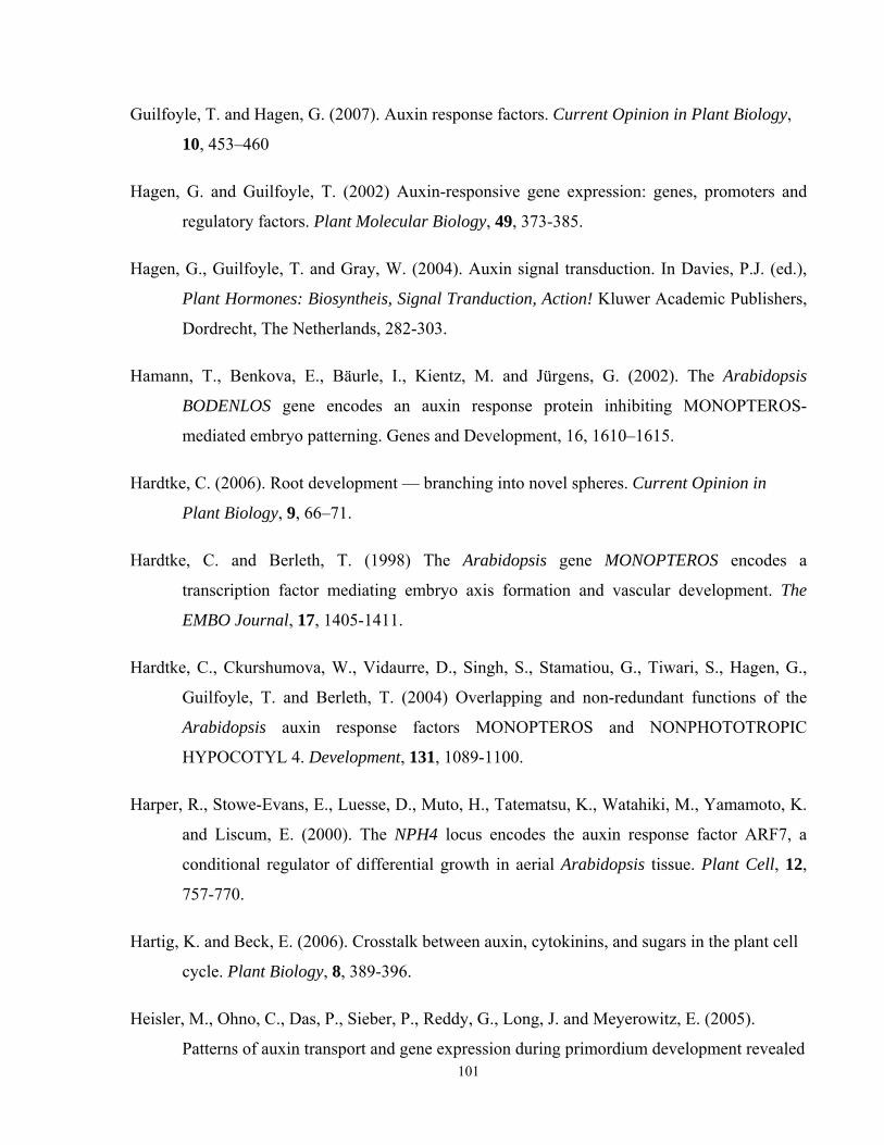
Guilfoyle, T. and Hagen, G. (2007). Auxin response factors. Current Opinion in Plant Biology,
10, 453–460
Hagen, G. and Guilfoyle, T. (2002) Auxin-responsive gene expression: genes, promoters and
regulatory factors. Plant Molecular Biology, 49, 373-385.
Hagen, G., Guilfoyle, T. and Gray, W. (2004). Auxin signal transduction. In Davies, P.J. (ed.),
Plant Hormones: Biosyntheis, Signal Tranduction, Action! Kluwer Academic Publishers,
Dordrecht, The Netherlands, 282-303.
Hamann, T., Benkova, E., Bäurle, I., Kientz, M. and Jürgens, G. (2002). The Arabidopsis
BODENLOS gene encodes an auxin response protein inhibiting MONOPTEROS-
mediated embryo patterning. Genes and Development, 16, 1610–1615.
Hardtke, C. (2006). Root development — branching into novel spheres. Current Opinion in
Plant Biology, 9, 66–71.
Hardtke, C. and Berleth, T. (1998) The Arabidopsis gene MONOPTEROS encodes a
transcription factor mediating embryo axis formation and vascular development. The
EMBO Journal, 17, 1405-1411.
Hardtke, C., Ckurshumova, W., Vidaurre, D., Singh, S., Stamatiou, G., Tiwari, S., Hagen, G.,
Guilfoyle, T. and Berleth, T. (2004) Overlapping and non-redundant functions of the
Arabidopsis auxin response factors MONOPTEROS and NONPHOTOTROPIC
HYPOCOTYL 4. Development, 131, 1089-1100.
Harper, R., Stowe-Evans, E., Luesse, D., Muto, H., Tatematsu, K., Watahiki, M., Yamamoto, K.
and Liscum, E. (2000). The NPH4 locus encodes the auxin response factor ARF7, a
conditional regulator of differential growth in aerial Arabidopsis tissue. Plant Cell, 12,
757-770.
Hartig, K. and Beck, E. (2006). Crosstalk between auxin, cytokinins, and sugars in the plant cell
cycle. Plant Biology, 8, 389-396.
Heisler, M., Ohno, C., Das, P., Sieber, P., Reddy, G., Long, J. and Meyerowitz, E. (2005).
Patterns of auxin transport and gene expression during primordium development revealed 101
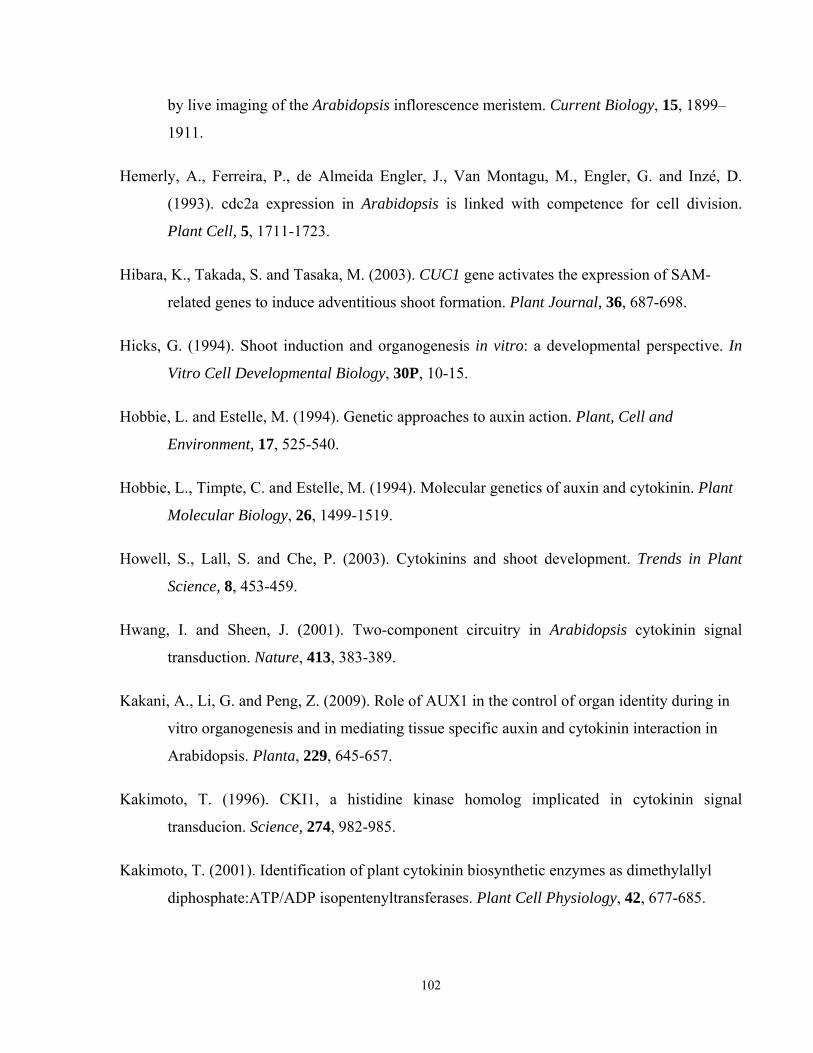
by live imaging of the Arabidopsis inflorescence meristem. Current Biology, 15, 1899–
1911.
Hemerly, A., Ferreira, P., de Almeida Engler, J., Van Montagu, M., Engler, G. and Inzé, D.
(1993). cdc2a expression in Arabidopsis is linked with competence for cell division.
Plant Cell, 5, 1711-1723.
Hibara, K., Takada, S. and Tasaka, M. (2003). CUC1 gene activates the expression of SAM-
related genes to induce adventitious shoot formation. Plant Journal, 36, 687-698.
Hicks, G. (1994). Shoot induction and organogenesis in vitro: a developmental perspective. In
Vitro Cell Developmental Biology, 30P, 10-15.
Hobbie, L. and Estelle, M. (1994). Genetic approaches to auxin action. Plant, Cell and
Environment, 17, 525-540.
Hobbie, L., Timpte, C. and Estelle, M. (1994). Molecular genetics of auxin and cytokinin. Plant
Molecular Biology, 26, 1499-1519.
Howell, S., Lall, S. and Che, P. (2003). Cytokinins and shoot development. Trends in Plant
Science, 8, 453-459.
Hwang, I. and Sheen, J. (2001). Two-component circuitry in Arabidopsis cytokinin signal
transduction. Nature, 413, 383-389.
Kakani, A., Li, G. and Peng, Z. (2009). Role of AUX1 in the control of organ identity during in
vitro organogenesis and in mediating tissue specific auxin and cytokinin interaction in
Arabidopsis. Planta, 229, 645-657.
Kakimoto, T. (1996). CKI1, a histidine kinase homolog implicated in cytokinin signal
transducion. Science, 274, 982-985.
Kakimoto, T. (2001). Identification of plant cytokinin biosynthetic enzymes as dimethylallyl
diphosphate:ATP/ADP isopentenyltransferases. Plant Cell Physiology, 42, 677-685.
102
Klee, H. and Estelle, M. (1991). Molecular genetic approaches to plant hormone biology. Annu.
Rev. Plant Physiol. Plant Mol. BioI., 42, 529-551.
Konishi, M. and Sugiyama, M. (2003). Genetic analysis of adventitious root formation with a
novel series of temperature-sensitive mutants of Arabidopsis thaliana. Development, 130,
5637-5647.
Krogan, N. (2006). Transcriptional regulation mediated by the Arabidopsis auxin response factor
MONOPTEROS. In Graduate Department of Botany, vol. PhD (ed., pp. 257). Toronto:
University of Toronto, 170-228.
Kubo, M. and Kakimoto, T. (2000). The CYTOKININ-HYPERSENCITIVE genes of Arabidopsis
negatively regulate the cytokinin-signaling pathway for cell division and chloroplast
development. Plant Journal, 23, 385-394.
Kunkel1, T., Niu, T., Chan, Y. and Chua N. (1999). Inducible isopentenyl transferase as a high
efficiency marker for plant transformation. Nature Biotechnology, 17, 916-919.
Kyozuka, J. (2007). Control of shoot and root meristem function by cytokinin. Current Opinion
in Plant Biology, 10, 442–446.
Lakshmanan, P., Ng, S., Loh, C. and Goh, C. (1997). Auxin, cytokinin and ethylene
differentially regulate specific developmental states associated with shoot bud
morphogenesis in leaf tissues of mangosteen (Garcinia mangostana L.) cultured in vitro.
Plant Cell Physiology, 38, 59-64.
Laxmi, A., Paul, L., Raychaudhuri, A., Peters, J. and Khurana, J. (2006). Arabidopsis cytokinin-
resistant mutant, cnr1, displays altered auxin responses and sugar sensitivity. Plant
Molecular Biology, 62, 409–425.
Lenhard, M., Juergens, G. and Laux, T. (2002) The WUSCHEL and HOOTMERISTEMLESS
genes fulfill complimentary roles in Arabidopsis shoot meristem regulation.
Development, 129, 3195-3206
Leyser, O. (2006). Dynamic integration of auxin transport and signaling. Current Biology, 16,
R424-R433. 103
Ljung, K., Bhalerao, R. and Sandberg, G. (2001). Sites and homeostatic control of auxin
biosynthesis in Arabidopsis during vegetative growth. Plant Journal, 28, 465-474.
Li, Z. and Gray, D. (2005) Genetic engineering technologies. In Trigiano R.N and Gray D.J.,
Plant Development and Biotechnology. CRC Press, p. 241-250.
Li, J., Dai, X. and Zhao, Y. (2006). A role for auxin response factor 19 in auxin and ethylene
signaling in Arabidopsis. Plant Physiology, 140, 899-908.
Lincoln, C., Britton, J. and Estelle, M. (1990). Growth and development of the axr1 mutants of
Arabidopsis. Plant Cell, 2, 1071-1080.
Liscum, E. and Reed, J. (2002). Genetics of Aux/IAA and ARF action in plant growth and
development. Plant Molecular Biology, 49, 387–400.
Long, J., Moan, E., Medford, J. and Barton, M. (1996). A member of the KNOTTED class of
homeodomain proteins encoded by the STM gene of Arabidopsis. Nature, 379, 66-69.
Mattsson, J., Ckurshumova, W. and Berleth, T. (2003) Auxin signaling in Arabidopsis leaf
vascular development. Plant Physiology, 131, 1327-1339.
Mayer, U., Ruiz, R., Berleth, T., Misera, S. and Jurgens, G. (1991). Mutations affecting body
organization in the Arabidopsis embryo. Nature, 353, 402-407.
Mayer, K., Schoof, H., Haecker, A., Lenhard, M., Jurgens, G. and Laux, T. (1998). Role of
WUSCHEL in regulating stem cell fate in the Arabidopsis shoot meristem. Cell, 95, 805-
815.
Maxwell, B. and Kieber, J. (2004). Cytokinin signal transduction. In Davies, P.J. (ed.), Plant
Hormones: Biosynthesis, Signal Transduction, Action! Kluwer Academic Publishers,
Dordrecht, The Netherlands, 321-349.
Mordhorst, A., Charbit, E. and de Vries, S. (2005) Some developmental and molecular aspects of
somatic embryogenesis. In Trigiano R.N and Gray D.J., Plant Development and
Biotechnology. CRC Press, 201-209.
104
Moubayidin, L., Mambro, R., and Sabatini, S. (2009). Cytokinin–auxin crosstalk. Trends in
Plant Science. 14, 557-562.
Muller, B. and Sheen, L. (2007a) Cytokinin signaling pathway. Science STKE, cm4. Available at
http://www.stke.org/cgi/content/full/2007/407/cm4
Muller, B. and Sheen, L. (2007b). Advances in cytokinin signaling. Science, 318, 68-69.
Nagpal, P., Ellis, C., Weber, H., Ploense, S., Barkawi, L., Guilfoyle, T., Hagen, G., Alonso, J.,
Cohen, J., Farmer, E., Ecker, J. and Reed, J. (2005) Auxin response factors ARF6 and
ARF8 promote jasmonic acid production and flower maturation. Development, 132,
4107-4118.
Nordstrom, A., Tarkowski, P., Tarkowska, D., Norbaek, R., Åstot, C., Dolezal, K. and Sandberg,
G. (2004). Auxin regulation of cytokinin biosynthesis in Arabidopsis thaliana: a factor of
potential importance for auxin–cytokinin-regulated development. PNAS, 101, 8039-8044.
Okushima, Y., Overvoorde, P., Arima, K., Alonso, J., Chan, A., Chang, C., Ecker, J., Hughes,
B., Lui, A., Nguyen, D., Onodera, C., Quach, H., Smith, A., Yu, G., and Theologisa, A.
(2005). Functional genomic analysis of the AUXIN RESPONSE FACTOR gene family
members in Arabidopsis thaliana: unique and overlapping functions of ARF7 and ARF19.
The Plant Cell, 17, 444–463.
Osakabe, Y., Miyata, S., Urao, T., Seki, M., Shinozaki, K. and Yamaguchi-Shinozaki, K. (2002).
Overexpression of Arabidopsis response regulators, ARR4/ATRR1/IBC7 and
ARR8/ATRR3, alters cytokinin responses differently in the shoot and in callus formation.
Biochemical and Biophysical Research Communications, 293, 806-815.
Ozawa, S., Yasutani, I., Fukuda, H., Komamine, A. and Sugiyama, M. (1998). Organogenic
responses in tissue culture of srd mutants of Arabidopsis thaliana. Development, 125,
135-142.
Paciorek, T. and Friml, J. (2006). Auxin signaling. Journal of Cell Science, 119, 1199-1202.
105
Peret, B., Rybel, B., Casimiro, I., Benkova, E., Swarup, R., Laplaze, L., Beeckman, T. and
Bennett, M. (2009). Arabidopsis lateral root development: an emerging story. Trends in
Plant Science, 14, 399-408.
Perrot-Rechenmann, C. (2010). Cellular responses to auxin: division versus expansion. Cold
Spring Harbor Perspectives in Biology, 2, a001446, 1-15
Petrášek, J., Mravec, J., Bouchard, R., Blakeslee, J., Abas, M., Seifertová, D., Wiśniewska, J.,
Tadele, Z., Kubeš, M., Čovanová, M., Dhonukshe, P., Skůpa, P., Benková, E., Perry, L.,
Křeček, P., Lee, O.R., Fink, G., Geisler, M., Murphy, A., Luschnig, C., Zažímalová, E.
and Friml, J. (2006). PIN proteins perform a rate-limiting function in cellular auxin
efflux. Science, 312, 914-918.
Przemeck, G., Mattsson, J., Hardtke, C., Sung, Z. and Berleth, T. (1996). Studies on the role of
the Arabidopsis gene MONOPTEROUS in vascular development and plant cell
axialization. Planta, 200, 229-237.
Rashotte, A., Poupart, J., Waddell, C. and Muday, G. (2003). Transport of the two natural
auxins, Indole-3-Butyric acid and indole-3-acetic acid, in Arabidopsis. Plant Physiology,
133, 761–772.
Reinhardt, D., Mandel, T. and Kuhlemeier, C. (2000). Auxin regulates the initiation and radial
position of plant lateral organs. The Plant Cell, 12, 507–518.
Remington, D., Vision, T., Guilfoyle, T. and Reed, J. (2004). Contrasting modes of
diversification in the Aux/IAA and ARF gene families. Plant Physiology, 135, 1738–
1752.
Riefler, M., Novak, O., Strnad, M. and Schmulling, T. (2006). Arabidopsis cytokinin receptor
mutants reveal functions in shoot growth, leaf senescence, seed size, germination, root
development, and cytokinin metabolism. Plant Cell, 18, 40-54.
Richard, C., Lescot, M., Inze, D. and De Veylder, L. (2002). Effect of auxin, cytokinin, and
sucrose on cell cycle gene expression in Arabidopsis thaliana cell suspension cultures.
Plant Cell, Tissue and Organ Culture, 69, 167–176.
106
Riou-Khamlichi, C., Huntley, R., Jacqmard, A. and Murray, J. (1999). Cytokinin activation of
Arabidopsis cell division through a D-type cyclin. Science, 283, 1541-1544.
Rogg, L., Lasswell, J. and Bartel, B. (2001). Gain-of-function mutation in IAA28 suppresses
lateral root development. The Plant Cell, 13, 465–480.
Rose, R., Wang, X., Nolan, K. and Rolfe, B. (2006). Root meristems in Medicago truncatula
tissue culture arise from vascular-derived procambial-like cells in a process regulated by
ethylene. Journal of Experimental Botany, 57, 2227-2235.
Rybel, B., Vassileva, V., Parizot, B., Demeulenaere, M., Grunewald, W., Audenaert, D.,
Campenhout, J., Overvoorde, P., Jansen, L., Vanneste, S., Möller, B., Wilson, M.,
Holman, T., Isterdael, G., Brunoud, G., Vuylsteke, M., Vernoux, T., Veylder, L., Inzé,
D., Weijers, D., Bennett, M. and Beeckman, T. (2010). A novel Aux/IAA28 signaling
cascade activates GATA23-dependent specification of lateral root founder cell identity.
Current Biology, 20, 1697-1706.
Scarpella, E., Marcos, D., Friml, J. and Berleth T. (2006). Control of leaf vascular patterning by
polar auxin transport. Genes and Development, 20, 1015-1027.
Schuetz, M., Berleth, T. and Mattsson, J. (2008). Multiple MONOPTEROS-dependent pathways
are involved in leaf initiation. Plant Physiology, 148, 870–880,
Schwarz, O., Sharma, A. and Beaty, R. (2005). Propagation from nonmeristematic tissues:
organogenesis. In Trigiano R.N and Gray D.J., Plant Development and Biotechnology.
CRC Press, 159-172.
Shaul, O., Van Montagu, M. and Inze, D. (1996). Cell cycle control in Arabidopsis. Annals of
Botany, 78, 283-288.
Shin, R., Burch, A., Huppert, K., Tiwari, S., Murphy, A., Guilfoyle, T. and Schachtman, D.
(2007). The Arabidopsis transcription factor MYB77 modulates auxin signal
transduction. Plant Cell, 19, 2440–2453.
107
Sieberer, T., Hauser, M., Seifert, G. and Luschnig, C. (2003). PROPORZ1, a putative
Arabidopsis transcriptional adaptor protein, mediates auxin and cytokinin signals in the
control of cell proliferation. Current Biology, 13, 837–842.
Skoog, F. and Miller, C. (1957). Chemical regulation of growth and organ formation in plant
tissue cultured in vitro. Symp. Soc. Exp. Biology, 11, 118-131.
Smalle, J., Kurepa, J., Yang, P., Babiychuk, E., Kushnir, S., Durski, A. and Vierstra, R. (2002).
Cytokinin growth responses in Arabidopsis involve the 26S proteasome subunit RPN12.
Plant Cell, 14, 17-32.
Soni, R., Carmichael, J., Shah, Z. and Murray, J. (1995). A family of cyclin D homologs from
plants differentially controlled by growth regulators and containing the conserved
retinoblastoma protein interaction motif. Plant Cell, 7, 85-103.
Stowe-Evans, E., Harper, R., Motchoulski, A. and Liscum, E. (1998) NPH4, a conditional
modulator of auxin-dependent differential growth responses in Arabidopsis. Plant
Physiology, 118, 1265-1275.
Sugiyama, M. (1999). Organogenesis in vitro. Current Opinion in Plant Biology, 2, 61-64.
Sugimoto, K., Jiao, J. and Meyerowitz, E. (2010). Arabidopsis regeneration from multiple tissues
occurs via a root development pathway. Developmental Cell, 18, 463–471.
Sun, J., Niu, Q., Tarkowski, P., Zheng, B., Tarkowska, D., Sandberg, G., Chua, N. and Zuo, J.
(2003). The Arabidopsis AtIPT8/PGA22 gene encodes an isopentenyl transferase that is
involved in de novo cytokinin biosynthesis. Plant Physiology, 131, 167–176,
Swarup, R., Parry, G., Graham, N., Allen, T., and Bennett, M. (2002). Auxin cross-talk:
integration of signaling pathways to control plant development. Plant Molecular Biology,
49, 411-426.
Swarup, R., Kargul J., Marchant, A, Zadik, D., Rahman, A., Mills, R., Yemm, A., May, S.,
Williams, L., Millnere, P., Tsurumi, S., Moore, J., Napier, R., Kerr, I. D. and Bennetta,
M. J. (2004). Structure-functional analysis of the presumptive Arabidopsis auxin
permease AUX1. Plant Cell, 16, 3069-3083. 108
Tajima, Y., Imamura, A., Kiba, T., Amano, Y., Yamashino, T. and Mizuno, T. (2004).
Comparative studies on the type-B response regulators revealing their distinctive
properties in thehis-to-asp phosphorelay signal transduction of Arabidopsis thaliana.
Plant Cell Physiology, 45, 28-39.
Takada, S., Hibara, K., Ishida, T. and Tasaka, M. (2001). The CUP-SHAPED COTYLEDON1
gene of Arabidopsis regulates shoot apical meristem formation. Development, 128, 1127-
1135.
Tamaki, H., Konishi, M., Daimon, Y., Aida, M., Tasaka, M. and Sugiyama, M. (2009).
Identification of novel meristem factors involved in shoot regeneration through the
analysis of temperature-sensitive mutants of Arabidopsis. Plant Journal, 57, 1027–1039.
Tatematsu, K., Kumagai, S., Muto, H., Sato, A., Watahiki, M., Harper, R., Liscum, E. and
Yamamoto, K. (2004). MASSUGU2 encodes Aux/IAA19, an auxin-regulated protein that
functions together with transcriptional activator NPH4/ARF7 to regulate differential
growth responses of hypocotyl and formation of lateral roots in Arabidopsis thaliana.
Plant Cell, 16, 379-393.
Teale, W., Paponov, I. and Palme, K. (2006). Auxin in action: signaling, transport and the
control of plant growth and development. Nature Reviews in Molecular Cell Biology, 7,
847-859.
To, J., Haberer, G., Ferreira, J., Deruere, J., Mason, M., Schaller, G., Alonso, J., Ecker, J. and
Kiebera, J. (2004). Type-A Arabidopsis response regulators are partially redundant
negative regulators of cytokinin signaling. Plant Cell, 16, 658–671.
Ulmasov T., Murfett J., Hagen G. and Guilfoyle T. (1997). Aux/lAA proteins repress expression
of reporter genes containing natural and highly active synthetic auxin
response elements. Plant Cell, 9, 1963-1971.
Ulmasov, T., Hagen, G. and Guilfoyle, T. (1999a) Activation and repression of transcription by
auxin-response factors. PNAS, 96, 5844-5849.
109
Ulmasov, T., Hagen, G. and Guilfoyle, T. (1999b) Dimerization and DNA binding of auxin
response factors. The Plant Journal, 19, 309-319.
Vanneste, S. and Friml, J. (2009). Auxin: a trigger for change in plant development. Cell, 136,
1005-1016.
Vieten, A., Sauer, M., Brewer, P. and Friml, J. (2007). Molecular and cellular aspects of auxin-
transport-mediated development. Trends in Plant Science, 12, 160-168.
Vidaurre, D., Ploense, S., Krogan, N. and Berleth, T. (2007). AMP1 and MP antagonistically
regulate embryo and meristem development in Arabidopsis. Development, 134, 2561-
2567.
Vogel, G. (2005). How does a single somatic cell become a whole plant? Science, 86, 309.
Vogler, H. and Kuhlemeier, C. (2003). Simple hormones but complex signaling. Current
Opinion in Plant Biology, 6, 51-56.
Wang, S., Tiwari, S., Hagen, G. and Guilfoyle, T. (2005). AUXIN RESPONSE FACTOR7
restores the expression of auxin-responsive genes in mutant Arabidopsis leaf mesophyll
protoplasts. Plant Cell, 17, 1979-1993.
Watahiki, M. and Yamamoto, K. (1997) The massugu1 mutation of Arabidopsis identified with
failure of auxin-induced growth curvature of hypocotyl confers auxin insensitivity to
hypocotyl and leaf. Plant Physiology, 115, 419-426.
Watahiki, M., Tatematsu, K., Fujihira, K., Yamamoto, M. and Yamamoto, K. (1999). The MSG1
and AXR1 genes of Arabidopsis are likely to act independently in growth-curvature
responses of hypocotyls. Planta, 207, 362-369.
Weijers, D., Benkova, E., Jager, K., Schlereth, A., Hamann, T., Kientz1, M., Wilmoth, J., Reed,
J. and Jurgens, G. (2005). Developmental specificity of auxin response by pairs of ARF
and Aux/IAA transcriptional regulators. The EMBO Journal, 24, 1874–1885
110
Wenzel, C., Schuetz, M., Yu, Q. and Mattsson, J. (2007). Dynamics of MONOPTEROS and PIN-
FORMED1 expression during leaf vein pattern formation in Arabidopsis thaliana. Plant
Journal, 49, 387-398.
Wilmoth, J., Wang S., Tiwari S., Joshi A., Hagen G., Guilfoyle T. and Alonso J. (2005).
NPH4/ARF7 and ARF19 promote leaf expansion and auxin-induced lateral root
formation. The Plant Journal, 43, 118-130.
Yasutani, I., Ozawa, S., Nishida, T., Sugiyama, M. and Komamine, A. (1994). Isolation of
temperature-sensitive mutants of Arabidopsis thaliana that are defective in the
redifferentiation of shoots. Plant Physiology, 105, 815-822.
Zhang, S. and Lemaux, P. (2004). Molecular analysis of in vitro shoot organogenesis. Critical
Reviews in Plant Sciences, 23, 325-335.
Zhang S. and Lemaux P. (2005). Molecular aspects of in vitro shoot organogenesis. In Trigiano
R.N and Gray D.J., Plant Development and Biotechnology. CRC Press, 173-185.
Zhang, K., Diederich, L. and John P. (2005). The cytokinin requirement for cell division in
cultured Nicotiana plumbaginifolia cells can be satisfied by yeast Cdc25 protein tyrosine
phosphatase. Implications for mechanisms of cytokinin response and plant development.
Plant Physiology, 137, 308-316.
Zhao Q., Fisher, R. and Auer, C. (2002). Developmental phases and STM expression during
Arabidopsis shoot organogenesis. Plant Growth Regulation, 37, 223–231.
Zhao X., Su Y., Cheng Z. and Zhang X. (2008) Cell fate switch during in vitro plant
organogenesis. Journal of Integrative Plant Biology, 50, 816-824.
Zhao, Z., Andersen, S., Ljung, K., Dolezal, K., Miotk, A., Schultheiss, S. and Lohmann,, J.
(2010). Hormonal control of the shoot stem-cell niche. Nature, 465, 1089- 1092.
Zubko, E., Adams, C., Machaekova, I., Malbeck, J., Scollan, C. and Meyer, P. (2002).
Activation tagging identifies a gene from Petunia hybrida responsible for the production
of active cytokinins in plants. Plant Journal, 29, 797-808.
111

112
Zuo, J., Niu, Q., Ikeda, Y. and Chua, N. H. (2002). Marker-free transformation: increasing
transformation frequency by the use of regeneration-promoting genes. Current Opinion
in Biotechnology, 13, 173–180.


![Unraveling the Evolution of Auxin Signaling - Plant Physiology · Unraveling the Evolution of Auxin Signaling1[C][W] ... PIN-FORMED orthologs calls for a deeper investigation of their](https://img.pdfslide.us/doc/110x75/5c79a60109d3f200208caa28/unraveling-the-evolution-of-auxin-signaling-plant-unraveling-the-evolution.jpg)
![LBD29-Involved Auxin Signaling Represses NAC Master · LBD29-Involved Auxin Signaling Represses NAC Master Regulators and Fiber Wall Biosynthesis1[OPEN] Kwang-Hee Lee,a Qian Du,a](https://img.pdfslide.us/doc/110x75/5ec8d6b920162707222fa0c8/lbd29-involved-auxin-signaling-represses-nac-lbd29-involved-auxin-signaling-represses.jpg)