Embed Size (px)
Citation preview

ORIGINAL PAPER
Organization of aliphatic chains grafted on nanofibrillatedcellulose and influence on final properties
Karim Missoum • Julien Bras •
Mohamed Naceur Belgacem
Received: 12 June 2012 / Accepted: 6 September 2012 / Published online: 19 September 2012
� Springer Science+Business Media B.V. 2012
Abstract Chemical surface modification of nanofibr-
illated cellulose (NFC) was performed using a long
aliphatic isocyanate chain. Different molar ratios of the
coupling agents were tested, i.e., 1, 10, 30 equivalents
with respect to hydroxyl groups of the NFC surface. FE-
SEM analyses revealed that there were no changes in
their morphology thus keeping nanofibril-like structure
with about 30 nm of diameter. All these samples were
characterized by different techniques (e.g., FTIR) to
check the efficiency of the grafting. Hydrophobic NFC
were achieved whatever the grafting agent ratio. The
Degree of Substitution was determined by Elemental
Analyses and the Degree of Substitution of the Surface
was calculated thanks to X-ray Photoelectron Spectros-
copy data. Combining these two techniques, the Internal
Degree of Substitution was proposed for the first time. It
indicates if the modification occurs also within NFC
internal layers. Surface (contact angle), rheological
(water suspension viscosity) and thermal properties
(ThermoGravimetric Analysis) of grafted NFC do not
follow the expected linear evolution of properties with
the increase of molar ratio. X-Ray Diffraction analyses
showed that the grafted aliphatic chains display crystal-
line waxy domains at some ratios. A model for aliphatic
chain organization at the surface is proposed and clearly
explained for the first time why a compromise in molar
ratio is necessary to achieve best properties.
Keywords Nanofibrillated cellulose (NFC) �Chemical surface modification � Aliphatic chain
organization � Degree of surface substitution (DSS) �Thermal properties
Introduction
The last decade has been focused on obtaining
efficient material from cellulose with a very strong
interest on nano-scaled cellulose-based elements.
There are two main families of nano-cellulose: the
cellulose nano-crystals (or whiskers) obtained by acid
hydrolysis of a cellulose-rich substrate and the cellu-
lose nanofibrils (or NFC) obtained by different
combinations of enzymatic, chemical and/or mechan-
ical treatments of these starting raw materials. Very
recent reviews give detailed information for each
material (Habibi et al. 2010; Siro and Plackett 2010)
and emphasize the out-standing impact on the
mechanical properties of the ensued bionanocompos-
ites. (Berglund and Peijs 2010; Eichhorn et al. 2010; Liu
et al. 2011; Siqueira et al. 2010a). In the present work,
experiments are focused on NFC. These cellulose
microfibrils (MFC, NFC) were first obtained by Herrick
et al. (1983) and Turbak et al. (1983) in 1983 by a
K. Missoum � J. Bras (&) � M. N. Belgacem
Laboratory of Pulp and Paper Science (LGP2), 461, rue de
la papeterie, BP65, 38402 St-Martin-d’Heres Cedex,
France
e-mail: [email protected];
123
Cellulose (2012) 19:1957–1973
DOI 10.1007/s10570-012-9780-7

mechanical disintegration of wood pulp (Zimmermann
et al. 2010). Such a mechanical treatment yields the
production of gelly-like aqueous suspension of nanofi-
brils at very low concentration. The diameter of
nanofibrils obtained with these processes is in the range
of 10–50 nm, whereas the typical length is several
micrometers (Chinga-Carrasco and Syverud 2010;
Walther et al. 2011). Different pretreatment such as
enzymatic (Paakko et al. 2007; Siqueira et al. 2010d;
Syverud et al. 2011) or TEMPO mediated process (Saito
and Isogai 2004; Saito et al. 2007; Isogai et al. 2011),
have nowadays been developed to obtain more homo-
geneous suspension and limit energy consumption.
All cellulose nanofibrils (NFC) tend to form an
aqueous gel at very low concentration (2 % wt.) due to
their important specific surface area and high number
of hydrogen bonds arising from hydroxyl groups
present at their surface. This feature handicaps their
use in several applications, such as coated products
(low solid content and high viscosity) or composites.
In fact, it is impossible to use them at dry state without
strong tendency to form aggregates or even film-like
material. In order to overcome these drawbacks,
different solutions are studied, but the most common
one is the surface chemical modification, aiming at
transforming hydroxyl groups into other functions
thus limiting (or even totally avoiding) the hydrogen
bonds establishment.
Over the last decade, many processes of cellulose
fibers surface modification have been investigated
(Gandini and Belgacem 2011). Some of the reported
approaches involved the grafting of polymers onto the
surface of the fibers either by ‘‘grafting from’’ [like
Ring Opening Polymerization—ROP (Lonnberg et al.
2006; Roy et al. 2005) and Atom Transfer Radical
Polymerization—ATRP (Carlmark and Malmstrom
2003; Coskun and Temuz 2005)] or by ‘‘grafting onto’’
[following the procedure with bifunctionnal molecule
bridge (Gaiolas et al. 2009; Ly et al. 2010; Paquet et al.
2010; Krouit et al. 2008)]. The other strategy consists
in grafting small molecules at the surface of fibers
using acid chloride, anhydrides, silanes or isocyanates.
Nevertheless, even if most of these strategies have
already been tested onto cellulose nanocrystals as
recently reviewed (Lin et al., 2012), only few works
have been reported on the grafting of nanofibrillated
cellulose. We can quote NFC modifications by
trimethylsilylation (Lu et al. 2008), ring opening
polymerization of poly(e-caprolactone) (Lonnberg
et al. 2011), cerium induced grafting (Stenstad et al.
2008), surface acetylation (Jonoobi et al. 2010;
Tingaut et al. 2010), carboxymethylation (Eyholzer
et al. 2010) or carbanilation (Siqueira et al. 2009,
2010b).
To the best of our knowledge, none of these papers
studied the superficial and the internal degrees of
substitution and they did not show the influence of
molar ratio on the organization of the grafted agent at
NFC surface. Indeed the target of our work is to
determine and understand the effect of the molar ratio
on the final properties of grafted moieties on NFC.
Only Berlioz et al. (2009) dealt with similar surface
versus internal organization but this work is different
in terms of grafting conditions (gas esterification),
characterization techniques (bulk analyses : XRD and
CP-MAS NMR) and the investigated raw materials
(nanocrystals and bacterial cellulose aggregated by
freeze-drying). Moreover, in our study, using XPS and
FE-SEM gives rise to a ‘‘real’’ surface scrutiny (XPS)
with high resolution (FE-SEM). NFC final properties
like thermal properties (TGA) or surface and rheolog-
ical properties (contact angle and rheology) have also
been studied, in this work.
So, in comparison to the previous study in our
group (Siqueira et al. 2010b), in which only one ratio
have been tested, different stoichiometric ratios
([coupling agent]/[superficial OH functions]) have
been investigated in the present work and the influence
of degree of surface substitution has been discussed in
detail in order to explain final properties of resulting
grafted NFC. A special focus on aliphatic chain
organization at the surface is proposed thanks to
deeper X-Ray diffraction analyses.
Materials and methods
Materials
Raw materials
Native eucalyptus fibers used in this work were
obtained from FIBRIA (Sao Paolo, Brazil). The
coupling agent (n-octadecyl isocyanate), as well as
the solvents (ethanol, acetone, toluene and dichloro-
methane) and the catalyst (IUPAC name: dibutyl(do-
decanoyloxy)stannyl dodecanoate, common name:
dibutyltin dilaurate), were purchased from Aldrich
1958 Cellulose (2012) 19:1957–1973
123

Co (FRANCE). All chemicals were reagent grade and
used as received without further purification. Deion-
ized water was used in all experiments.
Methods
Nanofibrillated cellulose (NFC)
Nanofibrillated cellulose suspension was produced
from eucalyptus sulphite wood pulp after enzymatic
pre-treatment (Endoglucanase Novozym� 476 sup-
plied by Novozymes, Denmark, 0.1 M, 2 h, 50 �C).
Endoglucanase was chosen regarding their ability to
cut macromolecular cellulose chains at their extremity
and not in the middle of the chain. A suspension of
bleached eucalyptus fibers (2.0 % w/v) was disinte-
grated using a microfluidizer apparatus, Model M-110
EH-30. The slurry was injected through the Z-shape
chamber of the apparatus under a high pressure. The
Interaction Chamber (IXC) hosted cells of different
sizes (400, 200 and 100 lm). The fibers suspension
was passed 3, 4 and 5 times in the Chamber fibrillation
containing the three mentioned above different cells,
respectively. Solid content of the treated suspensions
was around 2 % (w/w).
Chemical surface modification of NFC
Carbanilation reactions were performed following the
reaction conditions developed by Siqueira et al.
(2010b). The temperature was changed in our case.
The aqueous suspension (150 g of suspension at
2 %wt. which correspond to 3 g of dried NFC), was
first solvent exchanged from water to acetone by
several successive centrifugations and re-dispersion
operations. Centrifugation operations were conducted
at 10,000 rpm for 10 min and re-dispersion steps,
performed with high shear rate (Ultra-Turrax GT18) at
9,500–13,500 rpm for 15 s. Exchange solvent was
performed in 4 successive steps.
The resulting acetone-based suspension was added
in a three-necked round-bottomed flask of 250 mL,
equipped with a reflux condenser. The system was
kept under dynamic flow of N2 during the whole
reaction time. The reaction mixture was heated to
65 �C, in order to remove acetone. At the same time,
186 mL of toluene is added dropwise to perform the in
situ solvent exchange by removing slowly acetone and
introducing toluene. At the end of toluene addition,
1 mL of n-butyltindilaurate, as a catalyst (1 mL) was
added to the reaction medium. The temperature of the
reaction mixture was then increased to 105 �C and
thermo-stated using a contact thermometer. The
temperature of system was kept at 105 �C, for 2 h
after the isocyanate addition.
The quantity of octadecyl isocyanate has been
calculated as equivalents with respect to the fraction of
hydroxyl groups available at the surface of cellulosic
nanofibers. For this study, it has been considered that
only 4 % of hydroxyl groups were available at the
surface due to some calculations established by
Siqueira et al. (2010b) with similar dimensions of
NFC. Such assumptions have been proposed to
determine the surface hydroxyl groups content
because modeling of flexible heterogeneous nanofi-
brils is still under investigation. Some recent work,
(Majoinen et al. 2011), have proposed an estimation of
the amount of hydroxyl group present at the surface on
cellulose nanocrystals which are more homogeneous
and calibrated system.
After cooling at room temperature, the toluene
suspension of modified NFC was then filtered and
washed with dichloromethane (3 9 100 mL) and with
ethanol (3 9 100 mL) under vacuum, in order to
remove the formed by-products during the reaction
(amines/urethanes), the unreacted physically adsorbed
molecules and the excess of isocyanate (when
needed). Moreover, a soxhlet extraction was per-
formed for 24 h using a mixture ethanol/dichloro-
methane with a ratio 1/1 (v/v) to complete the
purification of modified NFC. Each reaction with
different molar ratio has been triplicated.
Scanning Electron Microscopy (FE-SEM)
A scanning electron microscope equipped with a field
emission gun (FE-SEM), model Zeiss Ultra column 55
Gemini, was used to observe NFC. The accelerating
voltage (EHT) was 3 kV for a working distance of
6.4 mm. A droplet of diluted suspension was then
deposited onto a substrate covered with carbon tape.
After drying, samples were coated with a 2 nm layer of
Au/Pd (Gold/Palladium) to ensure their conductivity.
Sample preparations were at least duplicated and a
minimum of 10 images by samples were observed
with digital image analysis (Image J) for calculating
dimensions. FE-SEM images selected in figures are
representative to the sample.
Cellulose (2012) 19:1957–1973 1959
123

X-Ray Diffraction (XRD)
The (wide angle) X-Ray Diffraction analysis was
performed on powder obtained with air-dried neat
NFC suspensions kept at ambient temperature (23 �C)
and relative humidity (28.8 %). The grafted samples
are obtained by casting and the ensuing films flakes
were milled to produce powder. The samples were
placed in a 2.5 mm deep cell and measurements were
performed with a PANanalytical, X’Pert PRO MPD
diffractometer equipped with an X’celerator detector.
The operating conditions of the refractometer were:
Copper Ka radiation (1.5418 A), 2h (Bragg angle)
between 5 and 60�, step size 0.067�, counting time
90 s. The degree of crystallinity was evaluated using
the Buschle-Diller and Zeronian Equation (Buschle-
Diller and Zeronian 1992):
Ic ¼ 1� I1
I2
ð1Þ
where: I1 is the intensity at the minimum (2h = 18�)
and I2 is the intensity associated with the crystalline
region of cellulose (2h = 22.5�). All measurements
were made at least in duplicates and averaged.
Infrared spectroscopy (FTIR-ATR)
Infrared spectra were recorded, on film for unmodified
NFC and powder form for modified NFC, using a
Mattson 5000 spectrometer. The sample under inves-
tigation was deposited and pressed against the ZnSe
crystal of an attenuated total reflectance (ATR)
spectrophotometer. The torque applied was kept
constant to ensure a same pressure on each sample.
All spectra were recorded between 4,000 and
700 cm-1, with a resolution of 4 cm-1 and 16 scans.
For each sample, a minimum of 2 spectra were
obtained on different area of the film or the powder.
Elemental analysis (E.A)
Elemental analysis was carried out by the ‘‘Service
Centrale d’Analyse (Vernaison, France)’’ of the
‘‘Centre National de la Recherche Scientifique’’
(CNRS). Carbon, Hydrogen, Nitrogen and Oxygen
contents were measured for unmodified NFC and
modified NFC. The data collected has allowed deter-
mining the degree of substitution (DS) which is the
number of grafted hydroxyl groups per anhydroglu-
cose unit according to the following equation:
DS ¼ 72:07�%C � 162:14
295:51�%C � 228:19ð2Þ
where C is the relative carbon content in the sample and
72.07, 162.14, 295.51 and 228.19 correspond to the
carbon mass of anhydroglucose unit, mass of anhydro-
glucose unit, mass of n-octadecyl isocyanate and carbon
mass of n-octadecyl isocyanate respectively. The anal-
yses were performed twice and average was used.
X-ray Photoelectron Spectroscopy (XPS)
X-ray photoelectron spectroscopy (XPS) experiments
were carried out using an XR3E2 apparatus (Vacuum
Generators, UK) equipped with monochromated Mg KaX-ray source (1,253.6 eV) and operating at 15 kV under
a current of 20 mA. Samples were placed in an ultra-
high-vacuum chamber (10-8 mbar) with electron collec-
tion by a hemispherical analyzer at a 90� angle. Signal
decomposition was determined using Spectrum NT, and
the overall spectrum was shifted to ensure that the C–C/
C–H contribution to the C1s signal occurred at 285.0 keV.
Comparison of the elementary surface composition was
performed using the following equation:
O=C ¼ ðI1=I2Þ � ðS2=S1Þ ð3Þ
where Ii is the intensity of signal i (carbon, oxygen, or
nitrogen) and Si (SC = 0.00170, SO = 0.00477 and
SN = 0.00299) denotes the atomic sensitivity factor
whose values were calculated from:
Si ¼Tikiri
4pð4Þ
with Ti, ki and ri being the transmission energy, the
electron inelastic mean free path, and the photoioni-
zation cross section for the X-ray source, respectively.
Ti depends on the atomic kinetic energy Eikin (eV)
according to:
Ti ¼1
ðEkini Þ
0:7ð5Þ
with ECkin = 966.6 eV, EO
kin = 722.6 eV, and ENkin =
851.6 eV. The Penn algorithm was used to calculate
the electron inelastic mean free path k (kC = 2.63 nm,
kO = 2.11 nm, and kN = 2.39 nm) and the values
were taken from Scofield (1976) (rC = 1, rO = 2.85,
and rN = 1.77).
1960 Cellulose (2012) 19:1957–1973
123

XPS was performed on the dried powder of
modified eucalyptus nanofibers. The XPS analysis
for unmodified NFC (reference sample) was per-
formed on a dried film treated in the same condition,
but in the absence of the grafting agent and submitted
to the same extraction procedure.
Contact angle measurement
Contact angle measurements were carried out by depos-
iting different water droplets at the surface of the studied
substrates and recording the angles formed using an OCA
dataphysics system equipped with a CCD camera. The
contact angle and drop volume acquisition was realized
during the first 60 s after deposition taking 4images/s. For
unmodified NFC, the measurement was performed on
dried film and on pellets for modified NFC. All measure-
ments were performed 7 times for each sample.
Thermo gravimetric analyses measurements
A Setaram 92-12 TGA was used. About 50 mg of the
sample were placed in the sample pan and tested with a
heating rate of 10 �C/min from ambient temperature to
700 �C under nitrogen flow. Experiments were at least
duplicated and averaged.
Rheology measurements Rheological measure-
ments of the neat and modified NFC suspension, re-
dispersed in water using 3 % of sodium dodecylsulfate
(SDS), w/w with respect to the dried NFC, were
carried out using a controlled stress rheometer (MCR
301, Anton Paar Physica, Austria), with a parallel plate
fixture (diameter 25 mm with gap of 1 mm) at 25.0 �C
controlled via a Peltier system. A solvent trap was
used to prevent solvent (water) evaporation. Flow
curves were plotted from the corresponding transient
tests (apparent viscosity, g (Pa.s), versus time at
constant shear rate, �c (s-1) at different shear rates) in a
wide range from 0.001 to 1 s-1. Flow curves were
made in duplicate at each tested storage time (600 s).
Results and discussions
Morphology and structure of neat NFC and grafted
NFC
As already mentioned, different pretreatments have
been developed with enzymes (Paakko et al. 2007;
Siqueira et al. 2010c, d; Syverud et al. 2011), or
involving chemical reactions (Saito and Isogai 2004;
Saito et al. 2006, 2007), in order to decrease the energy
consumption of cellulose fiber disintegration process.
This leads to the production of totally different kinds
of NFC with different final properties, as described
recently (Siqueira et al. 2010d). Therefore, it is very
important to specify the NFC under the conditions
used to isolate them, whenever one should deal with
them. The results presented on this work have been
obtained with an enzyme (cellulase) pretreated
bleached eucalyptus fibers disintegrated in a micro-
fludizer meaning that mainly OH groups are present at
NFC surface. In fact, such treatment conditions do not
induce any chemical change (such as oxidation) on the
substrate surface. The XPS results detailed latter
confirmed this assumption.
The diameter of nanofibrillated cellulose was
determined by digital image analysis (ImageJ) of
FE-SEM pictures, as presented in the Fig. 1. The
average diameter of neat NFC was about 22 ± 5 nm
(a minimum of 50 measurements was performed). The
micrograph shows that nanofibrils are strongly entan-
gled. After grafting, FE-SEM micrographs of NFC
show similar average diameter 30 ± 8, 34 ± 9 and
32 ± 7 nm for the sample grafted with 1 molar equiv.,
10 equiv and 30 equiv respectively. These figures
have been confirmed by AFM (not shown) which gives
30, 32, 35 nm for the samples grafted with 1, 10 and 30
molar equivalent respectively. It is worth to note that
no morphology modifications are observed after
grafting. According to XRD analyses (presented
latter) the crystallinity index is similar for each
samples. These two features confirm the relevance of
non-swelling solvent used in our procedure. Moreover
the ‘‘peeling effect’’ reported by Berlioz et al. (2009)
and Cetin et al. (2009) on cellulose nanocrystals, is
negligible in the case of nanofibrillated cellulose
grafted with fatty chains. It is due to the length of the
material (higher DP) which still contains appreciable
amounts of hemicellulose and amorphous cellulose
contrary to cellulose nanocrystals. Moreover the
reaction by-products formed in our case (octadecan-
amine or dioctadecylurea), are less aggressive than
HCl present in Berlioz’s study, which prevents the
NFC from this swelling and peeling effect. Only
surface grafting could occur and the size of the fatty
chain (2 nm) on the surface could explain the slight
diameter increases. Moreover, the increasing of the
Cellulose (2012) 19:1957–1973 1961
123

diameter could also be induced by the increase of the
distance between two cellulosic chains at the first
surface layers as represented in the Scheme 1. In the
native material, there is a well superposed and
organized cellulosic chain packing. After the grafting,
lower quantity of hydrogen bonds and some steric
repulsion may occur between two cellulosic chains at
the first surface layers increasing slightly the diameter
of the NFC.
Moreover, we can notice in Fig. 1 that the grafted
samples seem to yield less entangled NFC than that of
neat counterpart due to limitation of hydrogen inter-
action, proving by the way the NFC grafting with
obtention of hydrophobic NFC. This is also simply
proved by checking the NFC water suspensions
homogeneity or the NFC films after drying, as shown
in Fig. 2.
Efficiency of NFC grafting
FTIR spectroscopy was used to follow the efficiency
of each grafting for the different reaction conditions.
Fig. 3 shows FTIR spectra obtained from: (a) neat
NFC and NFC grafted using: (b) 1 molar equivalence;
(c) 10 molar equivalences and (d) 30 molar equiva-
lences of octadecyl isocyanate. Before the chemical
treatment the cellulosic nano-fibers display several
bands characteristic to cellulose macromolecules at
3,350 cm-1 (O–H), 1,110 cm-1 (C–O of secondary
alcohol) (used for the normalization of all spectra) and
2,868 and 2,970 cm-1 (C–H from –CH2–).
After reaction with the isocyanates, a characteristic
band assigned to urethane bonding at 1,735 cm-1 has
appeared. A substantial increase of the bands at 2,868
and 2,970 cm-1 corresponding to asymmetric and
symmetric –CH2 – stretches from fatty chain was also
observed. The peak associated with the vibration of
adsorbed water at 1,650 cm-1 strongly decreased after
modification, probably because of the hydrophobic
behavior of the modified material.
Elemental Analyses (E.A) and X-ray Photoelectron
Spectroscopy (XPS) were performed in order to
quantify the grafting efficiency and to establish the
degrees of substitution. Thus, E.A gives rise to the
determination of the average degree of substitution
(DS), whereas from XPS data the degree of
100nm – EHT 3kV Grenoble INP CMTC 200nm – EHT 3kV Grenoble INP CMTC
200nm – EHT 3kV Grenoble INP CMTC 200nm – EHT 3kV Grenoble INP CMTC
Neat NFC NFC 1equiv
NFC 30equivNFC 10equiv
100nm – EHT 3kV Grenoble INP CMTC 200nm – EHT 3kV Grenoble INP CMTC
200nm – EHT 3kV Grenoble INP CMTC 200nm – EHT 3kV Grenoble INP CMTC
Neat NFC NFC 1equiv
NFC 30equivNFC 10equiv
Fig. 1 Pictures for Neat NFC and grafted with 1, 10 and 30 molar equivalence as indicated in the figure obtained by Scanning Electron
Microscopy equipped with Field Emission Gun
1962 Cellulose (2012) 19:1957–1973
123
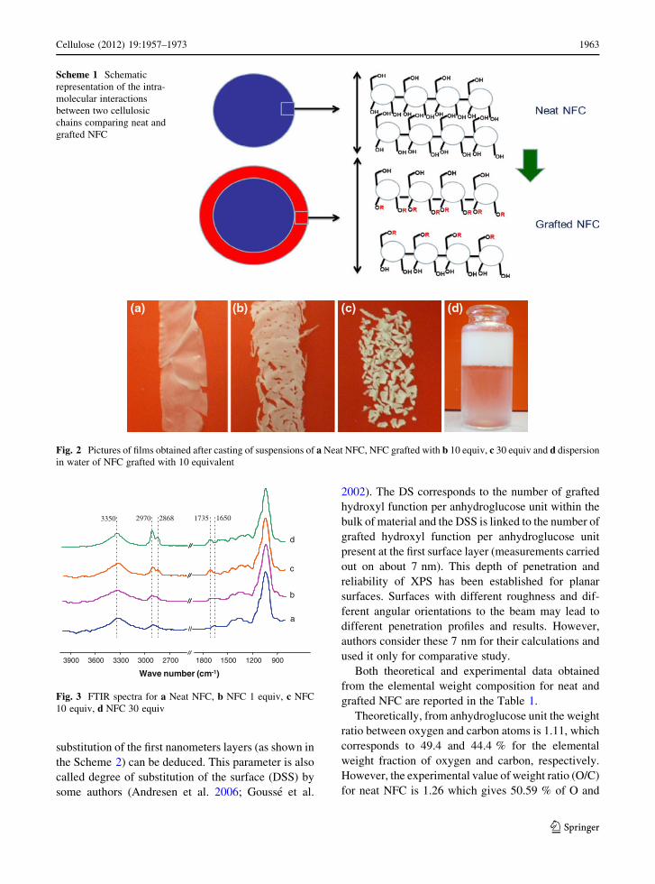
substitution of the first nanometers layers (as shown in
the Scheme 2) can be deduced. This parameter is also
called degree of substitution of the surface (DSS) by
some authors (Andresen et al. 2006; Gousse et al.
2002). The DS corresponds to the number of grafted
hydroxyl function per anhydroglucose unit within the
bulk of material and the DSS is linked to the number of
grafted hydroxyl function per anhydroglucose unit
present at the first surface layer (measurements carried
out on about 7 nm). This depth of penetration and
reliability of XPS has been established for planar
surfaces. Surfaces with different roughness and dif-
ferent angular orientations to the beam may lead to
different penetration profiles and results. However,
authors consider these 7 nm for their calculations and
used it only for comparative study.
Both theoretical and experimental data obtained
from the elemental weight composition for neat and
grafted NFC are reported in the Table 1.
Theoretically, from anhydroglucose unit the weight
ratio between oxygen and carbon atoms is 1.11, which
corresponds to 49.4 and 44.4 % for the elemental
weight fraction of oxygen and carbon, respectively.
However, the experimental value of weight ratio (O/C)
for neat NFC is 1.26 which gives 50.59 % of O and
Scheme 1 Schematic
representation of the intra-
molecular interactions
between two cellulosic
chains comparing neat and
grafted NFC
Fig. 2 Pictures of films obtained after casting of suspensions of a Neat NFC, NFC grafted with b 10 equiv, c 30 equiv and d dispersion
in water of NFC grafted with 10 equivalent
1500 9001200180027003000330036003900
Wave number (cm-1)
b
c
d
3350 2970 2868 16501735
a
Fig. 3 FTIR spectra for a Neat NFC, b NFC 1 equiv, c NFC
10 equiv, d NFC 30 equiv
Cellulose (2012) 19:1957–1973 1963
123

40.38 % of C atoms. The difference could be
explained by the presence of some O-rich impurities
and by experimental errors (Labet et al. 2007). The
presence of hemicelluloses (generally slightly richer
in O atoms) in NFC suspension can also explain this
difference.
Surface layer composition of neat NFC and grafted
samples at different molar ratio 1, 10 and 30 equiva-
lences have been compared by XPS. The use of XPS to
ascertain the efficiency of grafting was practiced very
extensively the last decade and showed to be a very
powerful technique to detect various changes at the
surface. The XPS wide spectra (not shown) of the four
samples show that in all cases the main peaks are
detected at 285 and 532 eV, corresponding to C and O
atoms, respectively. These spectra show also the
appearance of a new peak at 398 eV, attributed to N
atoms, whose concentration at the surface of grafted
samples increases with increasing the stoichiometric
ratio, as summarized in Table 2. Qualitatively, two
striking differences after grafting are noteworthy. On
one hand, the characteristic signal at 285 eV of C1S
increases with the grafting due to the evolution of the
molar ratio with the presence of long aliphatic chain
Scheme 2 Schematic representation of cellulosic chain contained in one NFC with different parameters used for the calculation of the
degree of substitution interne (DSI)
Table 1 Experimental and
corrected elemental weight
composition for neat and
grafted NFC obtained by
elemental analysis
Samples Experimental values Normalized values
%C %H %N %O %C %O
Neat NFC 40.38 6.19 \0.10 50.59 44.44 49.38
NFC
1 equiv
44.64 6.85 0.57 46.20 49.46 45.09
NFC
10 equiv
50.43 7.52 1.11 38.28 55.50 37.36
NFC
30 equiv
54.16 8.47 1.77 33.65 59.61 32.85
Table 2 Mass concentration of each element for neat and grafted sample correlated to deconvolution C1S obtained by XPS
Sample Experimental Values Decomposition of C1S
%C %O %N O/C C1(%) C2(%) C3(%) C4(%) C1/C3 C4/C3
Neat NFC 60.6 39.4 \0.1 0.65 15.1 67.8 16.8 0.4 0.90 0.02
NFC
1 equiv
63.8 34.8 1.1 0.55 20.6 52.1 22.9 5.8 0.82 0.28
NFC
10 equiv
67.9 29.9 1.8 0.44 35.8 43.0 15.1 6.1 2.4 0.40
NFC
30 equiv
82.0 14.9 3.1 0.18 63.4 24.7 7.7 3.3 8.6 0.32
1964 Cellulose (2012) 19:1957–1973
123

from the octadecyl carbamate. The ratio (O/C),
reported in the Table 2, for all tested materials,
decreased with the augmentation of the molar ratio.
On the other hand, the signal of nitrogen appears at
405 eV, which is also due to isocyanate moieties.
Moreover based on these XPS results, an approxima-
tion of the grafted molecule density can be calculated
(e.g., 0.4 OH/nm2 for 1 equivalent). So the compar-
ison to the OH density at the one surface layer (e.g., 0.2
OH/nm2 for 30 nm of width for one NFC) clearly
proves that the grafting occurs also at some internal
macromolecules.
The first relevant works dealing with the use of
X-ray Photoelectron spectroscopy to characterize
cellulose substrates were reported by Gray’s group
(Dorris and Gray 1978a, b; Katz and Gray 1980). The
deconvolution of C1s peak was reported by Ahmed
et al. (1987) showing that three entities are associated
with carbon signal and centered at 285.0, 286.7 and
288.3 eV. These moieties were attributed to C1 (C–H),
C2 (C–O) and C3 (O–C–O and/or C=O), respectively.
Figure 4 shows similar peaks in the XPS spectra
deconvolution of C1S signal, whatever the sample.
In theory (Belgacem and Gandini 2009), pure
cellulose exhibits two peaks in its deconvoluted C1s
XPS spectra, namely (1) C–O at 286.7 eV and
associated to alcohols and ethers groups. This peak
is noted as C2 and corresponds to 5 carbon atoms, and
(2) O–C–O at 288.3 eV attributed to acetal moieties.
This signal is noted C3 and corresponds to one carbon
atom.
In Fig. 4, two additional peaks are observed for
cellulose reference, namely: C1 and C4. As previously
mentioned C1 signal corresponds to non-oxidized
alkane-type carbon atoms associated with the presence
of residual lignin, extractive substances and fatty
acids. C4 peak was assigned to carboxylic functions
originating from glucuronic acids borne by hemicel-
luloses (Johansson et al. 2004, 2005) and present at the
surface of lignocellulosic fibers and pulps destined to
papermaking and used in our NFC production. In fact,
such a raw material is generally known to contain up to
30 % of this amorphous family.
In these works, it was also established that the
surface O/C ratio for pure cellulose (theoretical
formula) is 0.83. For the majority of virgin cellulose
(avicel, wood pulps, annual plants, etc.), this ratio is
systematically lower, because of the presence C-rich
molecular segments at the surface of the solids under
study. Table 2 confirms this assumption, in fact neat
Reference
0
200
400
600
800
1000
1200
282283284285286287288289290291292293294
Co
un
ts
C1
C2
C3
C4
NFC 1 equiv
0
200
400
600
800
1000
1200
1400
282283284285286287288289290291292293294
Co
un
ts
NFC 10 equiv
0
200
400
600
800
1000
1200
1400
282283284285286287288289290291292293294
Binding Energy (eV)
Co
un
ts
NFC 30 equiv
0
400
800
1200
1600
2000
2400
282283284285286287288289290291292293294
Binding Energy (eV)
Co
un
ts
C1
C2
C3
C4
C1
C2
C3
C4
C1
C2
C3
C4
Fig. 4 Decomposition of the C1s signal into its constituent contribution for neat and grafted NFC as mentioned in the figure
Cellulose (2012) 19:1957–1973 1965
123

NFC presents a lower ratio O/C in comparison to
theoretical value, i.e., 0.65 and 0.83, respectively. This
difference could be attributed to the surface pollution
by hydrocarbons adsorbed at the surface of nanofibers.
Recently, Johansson et al. (2011) proved also a
possible adaptation of the NFC surface depending on
the solvent used. Indeed Johansson et al. proved that
depending on the solvent used with NFC, XPS
analysis give strong difference. In this publication,
DMA and toluene based NFC suspensions were dried
and then analyzed using XPS. NFC dried from DMA
present higher O/C ratio than those dried from toluene.
After deconvolution only C2 and C3 peaks appear for
DMA dried NFC contrary to toluene dried NFC where
C1 and C4 are also present. Thus, this could also
explain the difference obtained in our case.
A deconvolution of the signal C1S presented in
Fig. 4, is required to quantify the grafting and
corroborate the occurrence of surface grafting. This
deconvolution reveals four peaks, which are attributed
to C1 (C–H), C2 (C–O), C3 (O–C–O and/or C=O) and
C4 (O–C=O), with a binding energy of 285.0, 286.6,
287.8 and 289.2 eV, respectively, as summarized in
the Table 2. This table shows that the intensity of C1
(C–C/C–H) increases strongly, from around 15 to
65 %, for the virgin and highly grafted NFCs,
respectively. Each glucose moiety possess only one
C3-carbon, the ratio C1/C3 reflects the number of
aliphatic carbons per glucose unit. The C1/C3 ratio
shifted from 0.9 for neat NFC to 0.82, 2.40 and 8.60 for
the NFC grafted with 1, 10 and 30 molar equivalence,
respectively. This is the consequence of the strong
impact of the C18 aliphatic chain. It is worth to note
that the C1/C3 ratio for the lowest NFC grafting
conditions (with 1 equivalent molar ratio) does not fit
the increasing trend, probably because of low amounts
of the coupled molecules. Similar analysis can be
applied to C4/C3 ((O=C=O)/(O–C–O)) ratio which is
also increasing with increasing the stoichiometric
ratios between the grafting molecules and the con-
centration of NFC superficial OH. The absolute values
of C4 signals (link to the carbamate functions)
increased with increasing the [NCO]/[OH] ratios.
These results clearly evidence the occurrence of
covalent bonding between the coupling molecules
and cellulose surface.
Unfortunately, except technique like TOF–SIMS, it
is quite difficult to know the composition of one
surface layer. So XPS data could be used in order to
determinate the DS of the surface (DSS) but taking
into account the first surface layers. For the calculation
of the DSS, several methods can be considered, but the
most common is based on Gousse et al. work (Gousse
et al. 2002), who defined the DSS (calculation done on
the amount of nitrogen) as follow:
DSS ¼ MAGU � x
100�MNð Þ � Mgroup grafted � x� � ð6Þ
where MAGU is the molar weight of one anhydroglu-
cose unit (162.14 g mol-1), MN the molar weight of
one atom of nitrogen (14 g mol-1), Mgroup_grafted the
molecular mass of the grafted moieties
(295.51 g mol-1) and x the mass concentration of
nitrogen. Table 3 reports the DS values calculated
from elemental analyses and the DSS determined
using XPS. Another DS, called Degree of Substitution
of Internal NFC (DSI), can then be calculated based on
the idea that XPS correspond to around 7 nm of depth
of analysis. Combining elemental analyses and XPS
data, the DS and the DSS can be used for the
determination of this Internal Degree of Substitution
(DSI). As mentioned before, this value could be very
interesting to determine in order to assess the depth at
which the grafting reaction took place. To the best of
our knowledge, the following parameter is proposed
for the first time:
DSI ¼ NAGUtot � DS� NAGUsurf � DSS
NAGUtot � NAGUsurfð7Þ
where NAGUtot is the total number of cellulose chains
which contains the cross section of one nanofibril,
NAGUsurf corresponds to the number of cellulose chains
under scrutiny during the XPS measurements, as
represented in the Scheme 2. The DS and the DSS are
the degree of substitution calculated from elemental
analysis and XPS measurements, respectively. NAGUtot
number was calculated as follow:
Table 3 DS, DSS and DSI calculated from elemental analysis
and XPS data
Samples DS (E.A) DSS (XPS) DSI
(XPS & E.A)
NFC 1 equiv 0.10 0.14 0.07
NFC 10 equiv 0.29 0.34 0.26
NFC 30 equiv 0.47 0.97 0.08
1966 Cellulose (2012) 19:1957–1973
123
NAGUtot ¼D
Wð8Þ
where D is the mean diameter of NFC and W the width
of one anhydroglucose unit (Hon and Shiraishi 2001)
(0.5889 nm). NAGUsurf is determined from the ratio:
NAGUsurf ¼D:A� 2
Wð9Þ
where: D.A is the XPS depth of analysis, 2 is used to
take into account both edges of the nanofibers and W
the width of one anhydroglucose unit.
Comparing the DS, DSS and DSI values, it seems
that the grafting occurred mainly at the surface of the
NFC for [NCO]/[OH] molar ratios of 1 and 30 equiv-
alent samples, as summarized in Table 3. It was
expected that DS is lower than DSS for all samples. DS
values are obtained from elemental analysis as previ-
ously discussed. It corresponds to a bulk analysis of all
materials. DSS is obtained from XPS data and
correspond to a surface characterization. Moreover,
the condition for the grafting, as demonstrated before,
was studied to be occurred only at the surface of NFCs.
Also all grafts are located at the surface, which can
explain the higher value of DSS in comparison to DS.
Therefore, for the first molar ratio, since there is no
excess of the grafting agent, the reaction is limited to
the hydroxyl groups present at the surface of NFC
substrate. Concerning the highest molar ratio (30
equivalent), the important amount of reagent intro-
duced in the media may induce quick saturation of the
hydroxyl groups present at the surface. The aliphatic
chains grafted can also hinder the diffusion of other
isocyanate moieties into the bulk of the materials,
especially because the reaction is carried out in non-
swelling conditions of solvent, pH and ionic force.
That is why there is a higher DSS and a low DSI for
this sample. However, the NFC sample, grafted using
a molar ratio of 10, shows that deeper modification has
occurred in the bulk of the nanofibrillated cellulose.
This result is hard to rationalize but it does not
constitute an experimental artifact. Indeed, this unex-
pected result was confirmed by repeating the exper-
iment several times. An explanation is proposed in
coming section.
So the DSI value seems to be a good way to check if
the grafting is strictly performed at the surface. In
some cases, the internal substitution is too small to
induce a significant size change of the NFCs as
observed previously. Even if this internal substitution
could be also attributed to highly substituted hemicel-
luloses and amorphous region of cellulose, the DSI
seems very helpful to understand surface versus
internal grafting.
Organization of grafted aliphatic chain
onto cellulose nanofibers and ensued properties
of NFC
The crystalline structure of grafted NFC and neat NFC
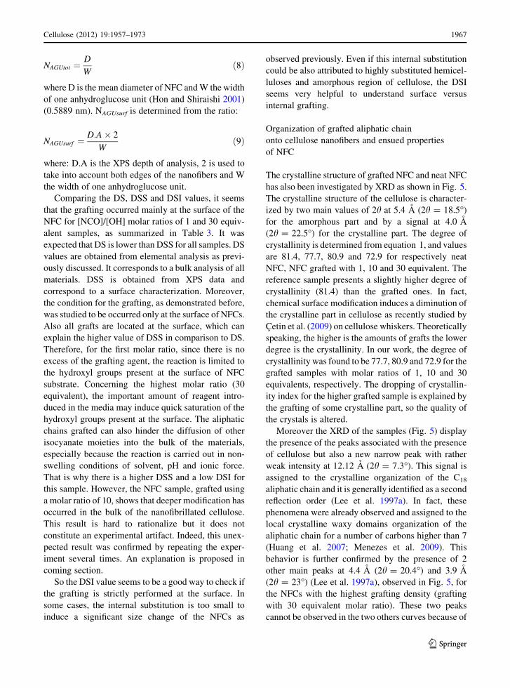
has also been investigated by XRD as shown in Fig. 5.
The crystalline structure of the cellulose is character-
ized by two main values of 2h at 5.4 A (2h = 18.5�)
for the amorphous part and by a signal at 4.0 A
(2h = 22.5�) for the crystalline part. The degree of
crystallinity is determined from equation 1, and values
are 81.4, 77.7, 80.9 and 72.9 for respectively neat
NFC, NFC grafted with 1, 10 and 30 equivalent. The
reference sample presents a slightly higher degree of
crystallinity (81.4) than the grafted ones. In fact,
chemical surface modification induces a diminution of
the crystalline part in cellulose as recently studied by
Cetin et al. (2009) on cellulose whiskers. Theoretically
speaking, the higher is the amounts of grafts the lower
degree is the crystallinity. In our work, the degree of
crystallinity was found to be 77.7, 80.9 and 72.9 for the
grafted samples with molar ratios of 1, 10 and 30
equivalents, respectively. The dropping of crystallin-
ity index for the higher grafted sample is explained by
the grafting of some crystalline part, so the quality of
the crystals is altered.
Moreover the XRD of the samples (Fig. 5) display
the presence of the peaks associated with the presence
of cellulose but also a new narrow peak with rather
weak intensity at 12.12 A (2h = 7.3�). This signal is
assigned to the crystalline organization of the C18
aliphatic chain and it is generally identified as a second
reflection order (Lee et al. 1997a). In fact, these
phenomena were already observed and assigned to the
local crystalline waxy domains organization of the
aliphatic chain for a number of carbons higher than 7
(Huang et al. 2007; Menezes et al. 2009). This
behavior is further confirmed by the presence of 2
other main peaks at 4.4 A (2h = 20.4�) and 3.9 A
(2h = 23�) (Lee et al. 1997a), observed in Fig. 5, for
the NFCs with the highest grafting density (grafting
with 30 equivalent molar ratio). These two peaks
cannot be observed in the two others curves because of
Cellulose (2012) 19:1957–1973 1967
123

the lower grafting surface density of the corresponding
samples. Even if, the last two peaks overlapped with
those corresponding to cellulose, their shapes (a
shoulder and a very sharp peak) can nevertheless be
clearly noticed.
In order to confirm the proposed mechanism, an
analysis at low angle was carried out. In fact, the first
order reflection of the C18 aliphatic chain could be
observed at 36.8 A (2h = 2.4�) (Lee et al. 1997b) and
can confirm local crystalline waxy domains structure
at the surface of grafted cellulose nanofibrils. The
results presented in the Fig. 6 show a well-defined
peak at 36.8 A (2h = 2.4�). The presence of this
shoulder is not due to a measurement artifact, since a
substrate of platinum was also characterized and
presented. Therefore, the obtained results strongly
suggest that the grafted aliphatic chain at the surface of
the NFC tend to form local crystalline waxy-like
domains and helps to propose a surface organization of
grafting in Scheme 3.
Indeed based on previous results (DSS and XRD)
and NFC properties (described latter), the organization
of the structure of the grafted layers could be
represented as sketched in Scheme 3. This hypothesis
allows explaining the crystallinity index evolution, the
local crystalline domain structure and the different
value of DSS. It is also a clear explanation for the
optimum of properties which will be presented in next
chapter. Indeed some characterizations (e.g., contact
angle and rheological measurement) were then per-
formed in order to highlight this organization.
In addition, contact angle measurements were
performed in order to point out the hydrophobic
5 10 15 20 25 30 35 40 45 50 55
Inte
nsity
XRD_Reference
5 10 15 20 25 30 35 40 45 50 55
2θ
Inte
nsity
XRD_1equiv
5 10 15 20 25 30 35 40 45 50 55
Inte
nsity
XRD_10equiv
5 10 15 20 25 30 35 40 45 50 55
Inte
nsity
XRD_30equiv
2θ
2θ 2θ
Fig. 5 X-Ray Diffraction patterns of neat NFC and the grafted samples as indicated in the figure
0 5 10
2θ
Inte
nsity
(co
unts
)
low angle analyses (a)
NFC_30equiv at high angle (b)
Platinium substrate (c)
(a)(b)(c)
Fig. 6 X-Ray Diffraction patterns for NFC_30 equiv at low
angle (a), NFC_30 equiv (b) and the substrate in platinum used
for the analyses (c)
1968 Cellulose (2012) 19:1957–1973
123

behavior of the grafted nanofibers comparing to neat
NFC. The results are presented in the Fig. 7. As
expected the contact angle values of grafted NFC are
higher than the neat NFC. Theoretically, the highest
molar ratio corresponds to the highest contact angle
value. However, the lowest contact angle value,
around 80�, is observed for NFC grafted using 10
molar equivalences and the higher for NFC grafted in
the condition 1 and 30 molar equivalence, around 90�respectively (±2�). Even after several measurements,
the same behavior is always observed. This might be
due to the organization of fatty chain at the surface as
previously detailed with Scheme 3. Indeed the high
amount of fatty chain can be organized as crystalline
phase due to important Van der Waals interactions. In
our case, organization at the surface can also be
assessed by keeping in mind that the degree of
substitution of surface (DSS) is increasing. In this
case, linear increase of contact angle should be
observed but it is not the case. This confirms our
assumption and can be explained by the higher
quantity of accessible zones at 10 eq. comparing to
1 eq. or 30 eq. grafted NFC, as proposed in Scheme 3.
Thermograms obtained by TGA measurements and
presented in Fig. 8 show clearly similar impact of the
grafting onto NFC surface. The grafted samples
display an enhanced thermal resistance and so a lower
sensitivity towards the degradation of the material in
comparison to the neat NFC. The Table 4 summarizes
the temperature to reach a certain relative weight loss.
For instance at 241 �C, the neat NFC lost 20 % of
weight. The same value is reached for higher temper-
ature for the grafted sample, i.e., 317, 286, 331 �C for
1 equiv, 10 equiv and 30 equiv respectively. In this
case the resistance to temperature is clearly high-
lighted by the ability of the grafts to be organized at the
surface to protect NFCs. The sample grafted using a 10
times molar ratio displayed a lower value than other
Accessible zone
Scheme 3 Schematic representation of the grafted NFC for the different ratio used for the chemical reaction as mentioned in the
scheme
0
20
40
60
80
100
120
0 100 200 300 400 500 600
Time (s)
Co
nta
ct a
ng
le (
deg
ree)
Reference Contact angle 1 equiv = 89.5° + - 1°
Contact angle 10 equiv = 79,5° + - 1° Contact angle 30 equiv = 90,1° + - 1°
Fig. 7 Contact angle versus
time performed with water
for (times) Reference,
(diamond) NFC 10 equiv,
(triangle) NFC 1 equiv and
(square) NFC 30 equiv
Cellulose (2012) 19:1957–1973 1969
123
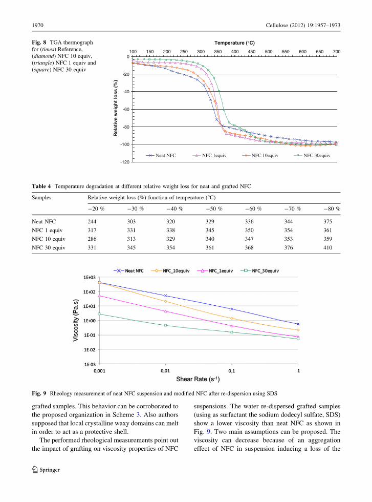
grafted samples. This behavior can be corroborated to
the proposed organization in Scheme 3. Also authors
supposed that local crystalline waxy domains can melt
in order to act as a protective shell.
The performed rheological measurements point out
the impact of grafting on viscosity properties of NFC
suspensions. The water re-dispersed grafted samples
(using as surfactant the sodium dodecyl sulfate, SDS)
show a lower viscosity than neat NFC as shown in
Fig. 9. Two main assumptions can be proposed. The
viscosity can decrease because of an aggregation
effect of NFC in suspension inducing a loss of the
-120
-100
-80
-60
-40
-20
0100 150 200 250 300 350 400 450 500 550 600 650 700
Temperature (°C)
Rel
ativ
e w
eig
ht
loss
(%
)
Neat NFC NFC 1equiv NFC 10equiv NFC 30equiv
Fig. 8 TGA thermograph
for (times) Reference,
(diamond) NFC 10 equiv,
(triangle) NFC 1 equiv and
(square) NFC 30 equiv
Table 4 Temperature degradation at different relative weight loss for neat and grafted NFC
Samples Relative weight loss (%) function of temperature (�C)
-20 % -30 % -40 % -50 % -60 % -70 % -80 %
Neat NFC 244 303 320 329 336 344 375
NFC 1 equiv 317 331 338 345 350 354 361
NFC 10 equiv 286 313 329 340 347 353 359
NFC 30 equiv 331 345 354 361 368 376 410
Fig. 9 Rheology measurement of neat NFC suspension and modified NFC after re-dispersion using SDS
1970 Cellulose (2012) 19:1957–1973
123

nanoscale dimension. It seems that this is not the case,
as confirmed by FE-SEM characterization. The second
explanation is the lower number of hydrogen bonds
between NFC as a result of the grafts which impedes
such interactions. Figure 9 reveals the diminution of
the viscosity for grafted samples. Moreover, the
sample grafted with 10 equiv has once again a different
behavior with a higher viscosity than other modified
substrates. It can be attributed to the ‘‘accessible zone’’
(presented in Scheme 3) which is not modified and still
able to form hydrogen bond interactions.
Thanks to all characterizations, the proposed sur-
face organization appears to be the correct explana-
tion. It proves that a compromise in molar ratio is then
necessary to achieve the best properties. Either very
low or high grafting should be targeted in such NFC
chemical modification process. That is the first time
such compromise is proposed and proved.
Conclusions
This work shows that NFC substrate can be efficiently
grafted by different molar ratio of fatty isocyanate and
that the grafting density increases with increasing the
molar ratio of the grafting agent. Moreover, thanks to
XPS, an approach dealing with surface versus bulk NFC
chemical modification is proposed with definition of a
new quantitative parameter (DSI). It helps discussing the
grafted molecule organization at the NFC surface.
Indeed, depending on the molar ratio, the grafted
methylene groups tend to form local crystalline waxy-
like domains resulting from lateral interaction between
the aliphatic chains. Depending on the molar ratio,
different surface organizations are assessed and pro-
posed for the first time. Results of NFC physico-
chemical properties confirmed the suggested organiza-
tion. They proved that such surface organization monitor
final NFC properties and that a compromise in molar
ratio is then necessary to achieve the best properties.
Acknowledgments This research was supported by the ‘‘Scale-
Up of Nanoparticles in modern PAPermaking’’ (SUNPAP)
project of the seven framework program of European research.
References
Ahmed A, Adnot A, Grandmaison JL, Kaliaguine S, Doucet J
(1987) ESCA analysis of cellulosic materials. Cellulose
Chem Technol 21(5):483–492
Andresen M, Johansson LS, Tanem B, Stenius P (2006) Prop-
erties and characterization of hydrophobized microfibril-
lated cellulose. Cellulose 13:665–677
Belgacem MN, Gandini A (2009) Natural fibre-surface modifi-
cation and characterisation. Natural fibre reinforced poly-
mer composites: from macro to nanoscale chapter 2. In:
Sabu T, Pothan L (eds) Cellulose fibre reinforced polymer
composites. Old City Publishing, Philadelphie, pp 14–46
Berglund LA, Peijs T (2010) Cellulose biocomposites—from
bulk moldings to nanostructured systems. MRS Bull
35(03):201–207
Berlioz S, Molina-Boisseau S, Nishiyama Y, Heux L (2009)
Gas-phase surface esterification of cellulose microfibrils
and whiskers. Biomacromolecules 10(8):2144–2151
Buschle-Diller G, Zeronian SH (1992) Enhancing the reactivity
and strength of cotton fibers. J Appl Polym Sci 45(6):
967–979
Carlmark A, Malmstrom E (2003) ATRP grafting from cellulose
fibers to create block-copolymer grafts. Biomacromole-
cules 4(6):1740–1745
Cetin NS, Tingaut P, Ozmen N, Henry N, Harper D, Dadmun M,
Sebe G (2009) Acetylation of cellulose nanowhiskers with
vinyl acetate under moderate conditions. Macromol Biosci
9(10):997–1003
Chinga-Carrasco G, Syverud K (2010) Computer-assisted
quantification of the multi-scale structure of films made of
nanofibrillated cellulose. J Nanopart Res 12(3):841–851
Coskun M, Temuz MM (2005) Grafting studies onto cellulose
by atom-transfer radical polymerization. Polym Int
54(2):342–347
Dorris GM, Gray D (1978a) The surface analysis of paper and
wood fibres by ESCA (electron spectroscopy for chemical
analysis). I. Application to cellulose and lignin. Cellulose
Chem Technol 12:9–23
Dorris GM, Gray D (1978b) The surface analysis of paper and
wood fibres by ESCA. II. Surface composition of
mechanical pulps. Cellulose Chem Technol 12:721–734
Eichhorn SJ, Dufresne A, Aranguren M, Marcovich NE, Ca-
padona JR, Rowan SJ, Weder C, Thielemans W, Roman M,
Renneckar S, Gindl W, Veigel S, Keckes J, Yano H, Abe K,
Nogi M, Nakagaito AN, Mangalam A, Simonsen J, Benight
AS, Bismarck A, Berglund LA, Peijs T (2010) Review:
current international research into cellulose nanofibres and
nanocomposites. J Mater Sci 45:1–33
Eyholzer C, Bordeanu N, Lopez-Suevos F, Rentsch D, Zim-
mermann T, Oksman K (2010) Preparation and charac-
terization of water-redispersible nanofibrillated cellulose
in powder form. Cellulose 17:19–30
Gaiolas C, Belgacem MN, Silva L, Thielemans W, Costa AP,
Nunes M, Silva MJS (2009) Green chemicals and process
to graft cellulose fibers. J Colloid Interface Sci
330(2):298–302
Gandini A, Belgacem MN (2011) Physical & chemical methods
of fiber surface modification. In: Zafeiropoulos E (ed)
Interface engineering in natural fibre composites for max-
imum performance. Woodhead Publishing, Cambridge,
UK, pp 3–42
Gousse C, Chanzy H, Excoffier G, Soubeyrand L, Fleury E
(2002) Stable suspensions of partially silylated cellulose
whiskers dispersed in organic solvents. Polymer
43(9):2645–2651
Cellulose (2012) 19:1957–1973 1971
123

Habibi Y, Lucia LA, Rojas OJ (2010) Cellulose nanocrystals:
chemistry, self-assembly, and applications. Chem Rev
110(6):3479–3500
Herrick FW, Casebier RL, Hamilton JK, Sandberg KR (1983)
Microfibrillated cellulose: morphology and accessibility.
J Appl Polym Sci 28(1):797–813
Hon DN-S, Shiraishi N (2001) Wood and cellulosic chemistry,
vol chapter 3: structure of cellulose. Marcel Dekker, New
York, USA
Huang B, Ge JJ, Li Y, Hou H (2007) Aliphatic acid esters of (2-
hydroxypropyl) cellulose—effect of side chain length on
properties of cholesteric liquid crystals. Polymer 48(1):
264–269
Isogai A, Saito T, Fukuzumi H (2011) TEMPO-oxidized cel-
lulose nanofibers. Nanoscale 3(1):71
Johansson L, Campbell J, Koljonen K, Kleen M, Buchert J
(2004) On surface distributions in natural cellulosic fibres.
Surf Interface Anal 36(8):706–710
Johansson L, Campbell J, Fardim P, Hulten A, Boisvert J,
Ernstsson M (2005) An XPS round robin investigation on
analysis of wood pulp fibres and filter paper. Surf Sci
584(1):126–132
Johansson L, Tammelin T, Campbell J, Setala H, Osterberg M
(2011) Experimental evidence on medium driven cellulose
surface adaptation demonstrated using nanofibrillated cel-
lulose. Soft Matter 22(7):10917–10924
Jonoobi M, Harun J, Mathew A, Hussein M, Oksman K (2010)
Preparation of cellulose nanofibers with hydrophobic sur-
face characteristics. Cellulose 17(2):299–307
Katz S, Gray DG (1980) Solvent extraction for the ESCA
analysis of paper. Svensk Papperstidning 8:226–228
Krouit M, Bras J, Belgacem MN (2008) Cellulose surface
grafting with polycaprolactone by heterogeneous click-
chemistry. Eur Polymer J 44(12):4074–4081
Labet M, Thielemans W, Dufresne A (2007) Polymer grafting
onto starch nanocrystals. Biomacromolecules 8(9):2916–
2927
Lee JL, Pearce EM, Kwei TK (1997a) Morphological devel-
opment in alkyl-substituted semiflexible polymers. Mac-
romolecules 30(26):8233–8244
Lee JL, Pearce EM, Kwei TK (1997b) Side-chain crystallization
in alkyl-substituted semiflexible polymers. Macromole-
cules 30(22):6877–6883
Lin N, Huang J, Dufresne A (2012) Preparation, properties and
applications of polysaccharide nanocrystals in advanced
functional nanomaterials: a review. Nanoscale 11(4):3274–
3294
Liu A, Walther A, Ikkala O, Belova L, Berglund LA (2011) Clay
nanopaper with tough cellulose nanofiber matrix for fire
retardancy and gas barrier functions. Biomacromolecules
12(3):633–641
Lonnberg H, Zhou Q, Brumer H, Teeri TT, Malmstrom E, Hult
A (2006) Grafting of cellulose fibers with poly(e-capro-
lactone) and poly(l-lactic acid) via ring-opening polymer-
ization. Biomacromolecules 7(7):2178–2185
Lonnberg H, Larsson K, Lindstrom T, Hult A, Malmstrom E
(2011) Synthesis of polycaprolactone-grafted microfibril-
lated cellulose for use in novel bionanocomposites—
influence of the graft length on the mechanical properties.
ACS Appl Mater Interfaces 3(5):1426–1433
Lu J, Askel P, Drzal LT (2008) Surface modification of mi-
crofibrillated cellulose for epoxy composite applications.
Polymer 49(5):1285–1296
Ly B, Bras J, Sadocco P, Belgacem MN, Dufresne A, Thiele-
mans W (2010) Surface functionalization of cellulose by
grafting oligoether chains. Mater Chem Phys 120(2–3):
438–445
Majoinen J, Walther A, McKee JR, Kontturi E, Aseyev V,
Malho JM, Ruokolainen J, Ikkala O (2011) Polyelectrolyte
brushes grafted from cellulose nanocrystals using Cu-
mediated surface-initiated controlled radical polymeriza-
tion. Biomacromolecules 12(8):2997–3006
Menezes AJd, Siqueira G, Curvelo AAS, Dufresne A (2009)
Extrusion and characterization of functionalized cellulose
whiskers reinforced polyethylene nanocomposites. Poly-
mer 50(19):4552–4563
Paakko M, Ankerfors M, Kosonen H, Nykanen A, Ahola S,
Osterberg M, Ruokolainen J, Laine J, Larsson PT, Ikkala
O, Lindstrom T (2007) Enzymatic hydrolysis combined
with mechanical shearing and high-pressure homogeniza-
tion for nanoscale cellulose fibrils and strong gels. Bio-
macromolecules 8(6):1934–1941. doi:10.1021/bm061
215p
Paquet O, Krouit M, Bras J, Thielemans W, Belgacem MN
(2010) Surface modification of cellulose by PCL grafts.
Acta Mater 58(3):792–801
Roy D, Guthrie JT, Perrier S (2005) Graft polymerization:
grafting poly(styrene) from cellulose via reversible addi-
tion-fragmentation chain transfer (RAFT) polymerization.
Macromolecules 38(25):10363–10372
Saito T, Isogai A (2004) TEMPO-mediated oxidation of native
cellulose. The effect of oxidation conditions on chemical
and crystal structures of the water-insoluble fractions.
Biomacromolecules 5(5):1983–1989. doi:10.1021/bm04
97769
Saito T, Nishiyama Y, Putaux J-L, Vignon M, Isogai A (2006)
Homogeneous suspensions of individualized microfibrils
from TEMPO-catalyzed oxidation of native cellulose.
Biomacromolecules 7(6):1687–1691
Saito T, Kimura S, Nishiyama Y, Isogai A (2007) Cellulose
nanofibers prepared by TEMPO-mediated oxidation of
native cellulose. Biomacromolecules 8(8):2485–2491
Scofield JH (1976) Hartree-Slater subshell photoionization
cross-sections at 1254 and 1487 eV. J Electron Spectrosc
Relat Phenom 8(2):129–137
Siqueira G, Bras J, Dufresne A (2009) Cellulose whiskers versus
microfibrils: influence of the nature of the nanoparticle and
its surface functionalization on the thermal and mechanical
properties of nanocomposites. Biomacromolecules 10(2):
425–432
Siqueira G, Bras J, Dufresne A (2010a) Cellulosic bionano-
composites: a review of preparation, properties and appli-
cations. Polymers 2(4):728–765
Siqueira G, Bras J, Dufresne A (2010b) New process of chem-
ical grafting of cellulose nanoparticles with a long chain
isocyanate. Langmuir 26(1):402–411
Siqueira G, Tapin-Lingua S, Bras J, Perez DdS, Dufresne A
(2010c) Mechanical properties of natural rubber nano-
composites reinforced with cellulosic nanoparticles
obtained from combined mechanical shearing, and
1972 Cellulose (2012) 19:1957–1973
123

enzymatic and acid hydrolysis of sisal fibers. Cellulose
18(1):57–65
Siqueira G, Tapin-Lingua S, Bras J, Perez DdS, Dufresne A
(2010d) Morphological investigation of nanoparticles
obtained from combined mechanical shearing, and enzy-
matic and acid hydrolysis of sisal fibers. Cellulose
17(6):1147–1158
Siro I, Plackett D (2010) Microfibrillated cellulose and new
nanocomposite materials: a review. Cellulose 17(3):
459–494
Stenstad P, Andresen M, Tanem B, Stenius P (2008) Chemical
surface modifications of microfibrillated cellulose. Cellu-
lose 15(1):35–45
Syverud K, Chinga-Carrasco G, Toledo J, Toledo PG (2011) A
comparative study of eucalyptus and Pinus radiata pulp
fibres as raw materials for production of cellulose nanofi-
brils. Carbohydr Polym 84(3):1033–1038
Tingaut P, Zimmermann T, Lopez-Suevos F (2010) Synthesis
and characterization of bionanocomposites with tunable
properties from poly(lactic acid) and acetylated micro-
fibrillated cellulose. Biomacromolecules 11:454–464
Turbak AF, Snyder FW, Sandberg KR (1983) Microfibrillated
cellulose, a new cellulose product: properties, uses, and
commercial potential. J Appl Polym Sci 28(1):815–827
Walther A, Timonen JVI, Dıez I, Laukkanen A, Ikkala O (2011)
Multifunctional high-performance biofibers based on wet-
extrusion of renewable native cellulose nanofibrils. Adv
Mater 23(26):2924–2928
Zimmermann T, Bordeanu N, Strub E (2010) Properties of
nanofibrillated cellulose from different raw materials and
its reinforcement potential. Carbohydr Polym 79(4):1086–
1093
Cellulose (2012) 19:1957–1973 1973
123


![GRAFTED TOMATO - Iserv1].pdf · GRAFTED TOMATO Grafted onto ... Grafting joins the top part of one plant (the scion) to the root ... (TPIE) - January 18-20, 2012 Spring Trials in](https://img.pdfslide.us/doc/110x75/5aa1ea047f8b9a436d8c452d/grafted-tomato-1pdfgrafted-tomato-grafted-onto-grafting-joins-the-top-part.jpg)