Embed Size (px)
Citation preview

University of WollongongResearch Online
Faculty of Engineering and Information Sciences -Papers: Part A Faculty of Engineering and Information Sciences
2013
Organ point dose measurements in clinical multislice computed tomography (MSCT)examinations with the MOSkin™ radiationdosimeterC P. L LianUniversity of Wollongong, [email protected]
A YoungSt Vincent's Hospital Sydney
D CutajarUniversity of Wollongong, [email protected]
N FreemanSt. Vincent's Hospital, Sydney
Anatoly B. RosenfeldUniversity of Wollongong, [email protected]
Research Online is the open access institutional repository for the University of Wollongong. For further information contact the UOW Library:[email protected]
Publication DetailsLian, C. P. L., Young, A., Cutajar, D., Freeman, N. & Rozenfeld, A. B. (2013). Organ point dose measurements in clinical multi slicecomputed tomography (MSCT) examinations with the MOSkin™ radiation dosimeter. Radiation Measurements, 55 56-59.

Organ point dose measurements in clinical multi slice computedtomography (MSCT) examinations with the MOSkin™ radiationdosimeter
AbstractThis study reports on the application of the MOSkin™ dosimeter in MSCT imaging for the real-timemeasurement of absorbed organ point doses in a tissue-equivalent female anthropomorphic phantom.MOSkin™ dosimeters were placed within the phantom to measure absorbed point organ doses for 2commonly applied clinical scan protocols, namely the renal calculus scan and the pulmonary embolus scan.Measured organ doses in the imaged field of view were found to be in the dose range 4.7-9.5 mGy and16.2-27.4 mGy for the renal calculus scan and pulmonary scan protocols respectively. For the derivation ofeffective dose, using the more recent ICRP 103 tissue weighting factors (w T) compared to that of the ICRP60 wT resulted in a difference in the derived effective dose by up to 0.8 mSv (-20%) in the renal calculusprotocol and up to 1.8 mSv (18%) in the pulmonary embolus protocol. This difference is attributed to thereduced radiosensitivity of the gonads and the increased radiosensitivity of breast tissue in the latest ICRP 103assigned wT. The results of this study show that the MOSkin™ dosimeter is a useful real-time tool for the directassessment of organ doses in clinical MSCT examinations. © 2012 Elsevier Ltd. All rights reserved.
Keywordsdose, point, organ, measurements, clinical, dosimeter, multi, slice, computed, tomography, msct,examinations, moskin, radiation
DisciplinesEngineering | Science and Technology Studies
Publication DetailsLian, C. P. L., Young, A., Cutajar, D., Freeman, N. & Rozenfeld, A. B. (2013). Organ point dose measurementsin clinical multi slice computed tomography (MSCT) examinations with the MOSkin™ radiation dosimeter.Radiation Measurements, 55 56-59.
This journal article is available at Research Online: http://ro.uow.edu.au/eispapers/1221

Organ Point Dose Measurements in Clinical Multi Slice Computed Tomography (MSCT) examinations with the MOSkinTM Radiation Dosimeter C.P.L. Liana,*, A. Youngb, D. Cutajara, N. Freemanc, A.B. Rosenfelda
aCentre for Medical Radiation Physics, Dept of Engineering Physics, Faculty of Engineering, University of Wollongong, Wollongong NSW 2522, Australia *Corresponding author Tel: + 612 4221 3507; Fax: +612 4221 5474 Email: [email protected] b St Vincent’s Hospital, Department of Nuclear Medicine and PET, Sydney NSW 2010, Australia c St Vincent’s Hospital, Department of Radiation Oncology, Sydney NSW 2010, Australia
Abstract
This study reports on the application of the MOSkinTM
dosimeter in MSCT imaging for the real-time
measurement of absorbed organ point doses in a tissue- equivalent female anthropomorphic phantom.
MOSkinTM dosimeters were placed within the phantom to measure absorbed point organ doses for 2 commonly
applied clinical scan protocols, namely the renal calculus scan and the pulmonary embolus scan. Measured
organ doses in the imaged field of view were found to be in the dose range 4.7 to 9.5 mGy and 16.2 to 27.4 mGy
for the renal calculus scan and pulmonary scan protocols respectively. For the derivation of effective dose,
using the more recent ICRP 103 tissue weighting factors (wT) compared to that of the ICRP 60 wT resulted in a
difference in the derived effective dose by up to 0.8 mSv (-20%) in the renal calculus protocol and up to 1.8
mSv (18%) in the pulmonary embolus protocol. This difference is attributed to the reduced radiosensitivity of
the gonads and the increased radiosensitivity of breast tissue in the latest ICRP 103 assigned wT. The results of
this study show that the MOSkinTM
dosimeter is a useful real-time tool for the direct assessment of organ doses
in clinical MSCT examinations.
Keywords
radiation dosimetry, CT, MOSFET, organ dose, tissue weighting factor, ICRP
Organ Point Dose Measurements in Clinical Multi Slice Computed Tomography (MSCT) examinations with the MOSkinTM Radiation Dosimeter
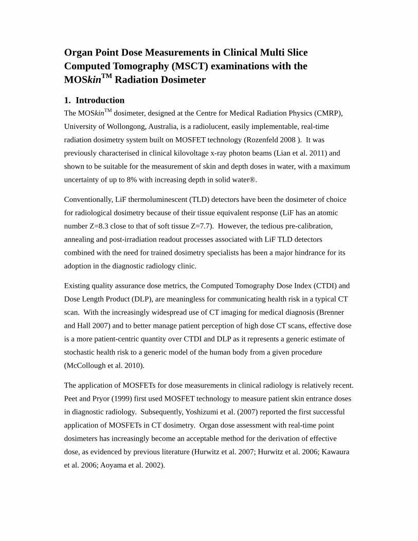
1. Introduction
The MOSkinTM dosimeter, designed at the Centre for Medical Radiation Physics (CMRP),
University of Wollongong, Australia, is a radiolucent, easily implementable, real-time
radiation dosimetry system built on MOSFET technology (Rozenfeld 2008 ). It was
previously characterised in clinical kilovoltage x-ray photon beams (Lian et al. 2011) and
shown to be suitable for the measurement of skin and depth doses in water, with a maximum
uncertainty of up to 8% with increasing depth in solid water®.
Conventionally, LiF thermoluminescent (TLD) detectors have been the dosimeter of choice
for radiological dosimetry because of their tissue equivalent response (LiF has an atomic
number Z=8.3 close to that of soft tissue Z=7.7). However, the tedious pre-calibration,
annealing and post-irradiation readout processes associated with LiF TLD detectors
combined with the need for trained dosimetry specialists has been a major hindrance for its
adoption in the diagnostic radiology clinic.
Existing quality assurance dose metrics, the Computed Tomography Dose Index (CTDI) and
Dose Length Product (DLP), are meaningless for communicating health risk in a typical CT
scan. With the increasingly widespread use of CT imaging for medical diagnosis (Brenner
and Hall 2007) and to better manage patient perception of high dose CT scans, effective dose
is a more patient-centric quantity over CTDI and DLP as it represents a generic estimate of
stochastic health risk to a generic model of the human body from a given procedure
(McCollough et al. 2010).
The application of MOSFETs for dose measurements in clinical radiology is relatively recent.
Peet and Pryor (1999) first used MOSFET technology to measure patient skin entrance doses
in diagnostic radiology. Subsequently, Yoshizumi et al. (2007) reported the first successful
application of MOSFETs in CT dosimetry. Organ dose assessment with real-time point
dosimeters has increasingly become an acceptable method for the derivation of effective
dose, as evidenced by previous literature (Hurwitz et al. 2007; Hurwitz et al. 2006; Kawaura
et al. 2006; Aoyama et al. 2002).

The main objectives of this study were firstly; to apply the MOSkinTM dosimeter to the
measurement of organ point doses in a tissue-equivalent female anthropomorphic phantom
for 2 clinical MSCT imaging examinations, namely, the CT renal calculus and the CT
pulmonary embolus scans. Secondly, to compare the derived effective doses as a result of the
recently introduced ICRP 103 over the ICRP 60 tissue weighting factors (wT) to our
measurements.
2. Materials and Methods
2.1 MSCT Scanner Protocols
A 16-slice General Electric (GE) Discovery 670 NM/CT SPECT/CT scanner was used at 120
kVp tube potential with a measured HVL of 0.8 mm Cu.
Table 1 shows the two clinical imaging protocols which were studied in this work, the CT
renal calculus protocol for the detection of renal stones in the pelvic region and the
pulmonary embolus protocol for the detection of blood vessel blockage in the chest region.
The scan range for the renal calculus CT examination extended from the upper region of the
diaphragm to the pubic symphasis of the anthropomorphic phantom corresponding to a scan
length of 250 mm. For the pulmonary embolus CT examination, the scan range extended
from the upper end of the lung apex to the lower region of the diaphragm of the
anthropomorphic phantom corresponding to a scan length of 300 mm.
Table 1: Imaging parameters for 16-slice MSCT single phasic scan of the abdominal-pelvic region for a renal calculus protocol; and of the chest for a pulmonary embolus (PE) protocol
Imaging Parameters
Protocol name Renal Calculus PE
Tube potential (kVp) 120 120 Fixed mAs 150 360 Effective mAsa 54.5 130.9 Table movement (mm rot-1) 27.5 27.5 Pitch 1.375 1.375 Detector configuration 16 x 1.25 16 x 1.25 Beam width (mm) 20.0 20.0 Planned scan length (mm) 250 300 Exposed scan length (mm) 278 328 Coverage time (s) 5.05 5.97

2.2 Dosimeter Calibration
The two dosimeters applied in this work were the CMRP MOSkinTM dosimeter and
Gafchromic XR-QA2 film (International Specialty Products ).
To match as closely as possible the applied CT beam quality and dose levels used in this
study, the dosimeters were calibrated in a 150 kVp, HVL=0.63 mm Cu orthovoltage beam on
a Gulmay D3300 unit against the Markus parallel plate ionisation chamber (Model N23343,
PTW-Frieburg, Germany) with 100 mGy dose delivered to the surface of a solid water®
phantom (RMI 457 Gammex,Wisconsin, USA).
2.3 Methodology
The use of a cross-sectional anatomical atlas (Kieffer and Heitzman 1979) enabled us to
identify the location of various organs of dosimetric interest throughout the anthropomorphic
phantom (The Alderson Radiation Therapy Phantom, Radiology Support Devices, NJ, USA).
Within each numbered phantom slab, the detectors were placed within the boundaries of the
demarcated organ region to determine average point organ dose as illustrated in Figure 1.
In the scan field-of-view, with the exception of the remainder organs, each recorded organ
point dose measurement was the average of 3 individual dosimeter readings. Each point dose
measurement was repeated twice to avoid random errors associated with the measurement.
For measurement of dose to the remaining organs, each point dose measurement was
obtained by a single dosimeter placed in the centroid of the organ.
Figure 1: Application of the MOSkinTM () and film () dosimeters for CT organ point dose measurement in the anthropomorphic phantom. The dotted line demarcates the anatomical position of the heart.
Our preliminary measurements indicated that out-of-field doses, particularly for dose
measurements beyond the boundaries of the scan field of view (more than a distance of 5 cm
equivalent to 2 slab lengths), lie beyond the low dose detection threshold of both dosimeters

and cannot be reliably measured. As such, the use of the headless phantom in this work was
justified as the distances from the primary beam to brain tissue was more than 8 cm.
To overcome the low dose detection threshold of the dosimeters used, we scaled up the
technical parameters of the imaging protocols. In the low-dose renal calculus protocol, we
doubled the tube current and scanned 3 times before taking a dose measurement. The
cumulative dose measured was then scaled down by a factor of 6, to determine organ dose in
a single phase MSCT scan. In the case of the pulmonary embolus protocol, we scanned 4
times before taking a dose measurement. The cumulative dose was then scaled down by a
factor of 4 to determine organ dose in a single phase MSCT scan.
3. Results
3.1 Depth dose response of dosimeters
The depth dose response of the MOSkinTM and film dosimeters were characterized from the
surface of the solid water® phantom at an applied surface dose of 100 mGy, to a depth of 100
mm. Figure 2 shows the obtained depth dose characterisation plot. It was found that both the
MOSkinTM dosimeter and the film dosimeter matched the gold-standard Markus ionisation
chamber tissue-equivalent response with a maximum uncertainty of ±10% at 100 mm depth.
This uncertainty is acceptable in diagnostic radiology measurements according to the IAEA
report (International Atomic Energy Agency 2007) which states that a 20% measurement
accuracy is considered acceptable in diagnostic radiology dosimetry where organ doses are
low and where the uncertainty for an absolute risk for a stochastic effect is high .

Figure 2: Characterization of the depth dose response of the MOSkinTM dosimeter and film with 100 mGy delivered dose to the surface of the phantom
3.2 Measured Organ doses
Figures 3 and 4 show the results of our organ point dose assessments with the MOSkinTM
dosimeter and the Gafchromic XR-QA2 film for the CT renal calculus protocol and the CT
pulmonary embolus protocol respectively. Organs in the imaged field of view were found to
be in the dose range 4.7 to 9.5 mGy and 16.2 to 27.4 mGy for the renal calculus (RC) and
pulmonary embolus (PE) CT scan protocols respectively.
Within the imaged field of view, point doses obtained with the MOSkinTM dosimeter and film
compared favourably to within 20% of each other, with the exception of measured average
point breast dose (up to 40% difference for the PE protocol) and measured bone surface dose
(up to 37% difference for the RC protocol). This difference may be explained by the
electronic disequilibrium created by the presence of the air-tissue interface and bone-tissue
interface for the assessment of breast dose and bone surface dose respectively.
For doses outside the imaged field of view, particularly at the boundaries of the scan field of
view (within 5 mm of the specified scan field), doses recorded by both dosimeters differed by
up to 33% for both imaging protocols.
0%
10%
20%
30%
40%
50%
60%
70%
80%
90%
100%
0.0
10.0
20.0
30.0
40.0
50.0
60.0
70.0
80.0
90.0
100.0
0 20 40 60 80 100 120
Percentage Depth dose (PDD)
Dose (mGy)
Depth in solid water (mm)
XR‐QA2 film
Markus Chamber
MOSkin

Figure 3: Renal calculus CT imaging scan. Point organ dose measurements obtained with the MOSkinTM dosimeter and Gafchromic XR-QA2 film (a) 18 organ locations of interest as specified in ICRP 60 (b) 20 organ locations of interest as specified in ICRP 103. Error bars refer to the range of dose measurements obtained with 3 individual point dosimeter readouts.
Imaged field-of-view
Imaged field-of-view

Figure 4: Pulmonary embolus CT imaging scan. Point organ dose measurements obtained with the MOSkinTM dosimeter and Gafchromic XQ-QA2 film (a) 19 organs of interest, ICRP 60 (b) 21 organs of interest, ICRP 103. Error bars refer to the range of dose measurements obtained with 3 individual point dosimeter readouts.
Imaged field-of-view
Imaged field-of-view
3.3 Evaluation of Effective Dose
Effective dose E is a summation of organ doses multiplied by individual tissue weighting
factors. For this study, we used the following equation to derive effective dose E.
where wT is the tissue weighting factor and HT is organ dose measurement in mSv.
For the renal calculus protocol, E was found to be 4.0 mSv and 3.2 mSv when the ICRP 60
and ICRP 103 wT were respectively applied to measured organ doses. Derived E from film
and MOSkinTM point dose measurements were in agreement. The decrease in derived E from
4.0 to 3.2 mSv using E103 wT is primarily due to the recent decrease in the assigned wT for
gonadal tissue (ovaries in the primary scan field of view) from 0.20 in ICRP 60 to 0.08 in
ICRP 103.
For the pulmonary embolus protocol, E was found to be 9.2 mSv and 10.6 mSv with the
applied ICRP 60 and ICRP 103 wT respectively. Derived E from film and MOSkinTM point
dose measurements agreed to within 10% of each other. The increase in derived E using E103
wT may be explained by the recent increase in the assigned wT of the radiosensitive breasts in
the scan field of view from 0.05 (ICRP 60) to 0.12 (ICRP 103).
4. Discussion and Conclusion
Experimental depth dose characterisation studies done with the MOSkinTM and XR-QA2 film
dosimeter showed both dosimeters to be suitable for the assessment of depth doses due to
their tissue equivalent response at depth in a clinical kilovoltage beam in the dose range 20
mGy to 100 mGy at a clinical tube potential of 150 kVp (0.63 mm Cu HVL). The MOSkinTM
dosimeter has an added advantage over film because of its ability to measure doses in real-
time.
Organ doses measured by the MOSkinTM and film dosimeters in this study generally agreed
to within 20%. The difference in measured doses is attributed to the changing energy
spectrum and dosimeter uncertainties with increasing depth. Larger differences in point dose
measurements between the two dosimeters may be attributed to differences in the positioning
of the x-ray tube start and end angle due to the CT helical beam not falling on the same organ
position on the same point of the arc (Yoshizumi et al. 2007).

We found that the application of ICRP 60 wT and ICRP 103 wT to our measured organ doses
resulted in a difference in the derived effective dose by up to 0.8 mSv (-20%) in the renal
calculus protocol and up to 1.8 mSv (18%) in the pulmonary embolus protocol. This
difference is attributed to the reduced radiosensitivity of the gonads and the increased
radiosensitivity of breast tissue with the updated ICRP 103 wT.
Based on the results of this study, we have shown that the MOSkinTM dosimeter is feasible for
implementation in the clinical diagnostic x-ray CT setting, with the added advantage of its
real-time dose readout capability. Derived effective doses by applying the ICRP wT to the
dosimeter- measured organ doses in an anthropomorphic phantom may lead to improved
communication of stochastic health risks to CT patients after taking into account patient
specific age-, gender and body habitus.
Acknowledgements The authors wish to thank Dr Martin Carolan for access to the Gulmay D3300 superficial/ orthovoltage therapy unit for the calibration of dosimeters used in this work.
References
Aoyama, T., Koyama, S., Kawaura, C., 2002. An in-phantom dosimetry system using pin silicon photodiode radiation sensors for measuring organ doses in x-ray CT and other diagnostic radiology. Med Phys 29 (7), 1504-1510. Brenner, D., Hall, E., 2007. Computed Tomography — An Increasing Source of Radiation Exposure. N Engl J Med 357 (22), 2277-2284. Hurwitz, L., Reiman, R., Yoshizumi, T., Goodman, P., Toncheva, G., Nguyen, G., et al., 2007. Radiation dose from contemporary cardiothoracic multidetector CT protocols with an anthropomorphic female phantom: implications for cancer induction. Radiol 245 (3), 742-750. Hurwitz, L.M., Yoshizumi, T.T., Reiman, R.E., Paulson, E.K., Frush, D.P., Nguyen, G.T., et al., 2006. Radiation Dose to the Female Breast from 16-MDCT Body Protocols. AJR 186, 1718-1722. International Atomic Energy Agency, 2007. Technical Reports Series no. 457 Dosimetry in Diagnostic Radiology: An International Code of Practice. Vienna. International Specialty Products, Product Specification Sheet- Gafchromic XR Series, Wayne, NJ, USA. Kawaura, C., Aoyama, T., Koyama, S., Achiwa, M., Mori, M., 2006. Organ and effective dose evaluation in diagnostic radiology based on in-phantom dose measurements with novel photodiode-dosemeters. Radiat Prot Dosimetry 118 (4), 421-430.

Kieffer, S.A., Heitzman, E.R., 1979. An Atlas of Cross-Sectional Anatomy: Computed Tomography, Ultrasound, Radiography, Gross Anatomy. Lian, C.P.L., Othman, M.A.R., Cutajar, D., Butson, M., Guatelli, S., Rosenfeld, A.B., 2011. Monte Carlo study of the energy response and depth dose water equivalence of the MOSkin radiation dosimeter at clinical kilovoltage photon energies. Austral Phys & Eng Sci Med 34 (2), 273-279. McCollough, C.H., Christner, J.A., Kofler, J.M., 2010. How Effective is Effective Dose as a Predictor of Radiation Risk? AJR 194, 890-896. Peet, D.J., Pryor, M.D., 1999. Evaluation of a MOSFET radiation sensor for the measurement of entrance surface dose in diagnostic radiology. Br J Radiol 72, 562-568. Rozenfeld, A.B., 2008. Radiation Sensor and Dosimeter (MOSkin). PCT/AU2008/000788 Australia. Yoshizumi, T., Goodman, P., Frush, D., Nguyen, G., Toncheva, G., Sarder, M., et al., 2007. Validation of Metal Oxide Semiconductor Field Effect Transistor Technology for Organ Dose Assessment During CT: Comparison with Thermoluminescent Dosimetry. Am J Roentgenol 188 (5), 1332-1336.