Embed Size (px)
Citation preview
O’Reilly et al. Hypoxia and Heart Development
1
Gene-environment interaction demonstrates the vulnerability of the embryonic heart
Victoria C. O’Reillya, Kylie Lopes Floroa,†, Hongjun Shia,†, Bogdan E. Chapmanb, Jost I. Preisa,
Alexander C. Jamesa, Gavin Chapmana,b, Richard P. Harveya,c,d, Randall S. Johnsone, Stuart M.
Grieveb,f,g, Duncan B. Sparrowa,c and Sally L. Dunwoodiea,c,d,*.
a: Developmental and Stem Cell Biology Division, Victor Chang Cardiac Research Institute, 405
Liverpool Street, Darlinghurst, Sydney, NSW 2010, Australia
b. School of Molecular Bioscience, Molecular Bioscience Building G08, University of Sydney,
NSW 2006, Australia
c: St Vincent’s Clinical School, Faculty of Medicine, University of New South Wales, de Lacy
Building, St Vincent's Hospital, Darlinghurst, Sydney, NSW 2010, Australia
d. School of Biotechnology and Biomolecular Sciences, University of New South Wales, Sydney,
NSW 2052, Australia
e. Department of Physiology, Development & Neuroscience, University of Cambridge, Downing
Street, Cambridge CB2 3EG, United Kingdom
f. Department of Radiology, Royal Prince Alfred Hospital, Missenden Road, Camperdown, Sydney,
NSW 2050, Australia
g. Sydney Translational Imaging Laboratory, Sydney Medical School, Edward Ford Building A27,
University of Sydney, Sydney, NSW 2006, Australia
* Corresponding author: Professor Sally L. Dunwoodie, Developmental and Stem Cell Biology
Division, Victor Chang Cardiac Research Institute, 405 Liverpool Street, Darlinghurst, Sydney,
NSW 2010, Australia. email: [email protected]; Tel: +612 9295 8613; Fax: +612
9295 8668
† These authors contributed equally to this work
O’Reilly et al. Hypoxia and Heart Development
2
Author email addresses:
VCO: [email protected]
KLF: [email protected]
BEC: [email protected]
JIP: [email protected]
ACJ: [email protected]
RPH: [email protected]
RSJ: [email protected]
SMG: [email protected]
DBS: [email protected]
SLD: [email protected]
O’Reilly et al. Hypoxia and Heart Development
3
ABSTRACT
Mammalian embryos develop in a low oxygen environment. The transcription factor hypoxia
inducible factor 1a (HIF1α) is a key element in the cellular response to hypoxia. Complete deletion
of Hif1α from the mouse conceptus causes extensive placental, vascular and heart defects, resulting
in embryonic lethality. However the precise role of Hif1α in each of these organ systems remains
unknown. To further investigate, we conditionally-deleted Hif1α from mesoderm, vasculature and
heart individually. Surprisingly, deletion from these tissues did not recapitulate the same severe
heart phenotype or embryonic lethality. Placental insufficiency, such as occurs in the complete
Hif1α null, results in elevated cellular hypoxia in mouse embryos. We hypothesized that subjecting
the Hif1α conditional null embryos to increased hypoxic stress might exacerbate the effects of
tissue-specific Hif1α deletion. We tested this hypothesis using a model system mimicking placental
insufficiency. We found that the majority of embryos lacking Hif1α in the heart died when exposed
to non-physiological hypoxia. This was a heart-specific phenomenon, as HIF1α protein
accumulated predominantly in the myocardium of hypoxia-stressed embryos. Our study
demonstrates the vulnerability of the heart to lowered oxygen levels, and that under such conditions
of non-physiological hypoxia the embryo absolutely requires Hif1α to continue normal
development. Importantly, these findings extend our understanding of the roles of Hif1α in
cardiovascular development.
KEYWORDS: HIF1a; heart development; mouse; hypoxia; gene-environment interaction
O’Reilly et al. Hypoxia and Heart Development
4
INTRODUCTION
The hypoxia inducible factor 1 (HIF1) is a key element in the cell's response to hypoxia. It is a
heterodimeric transcription factor consisting of HIF1α and aryl hydrocarbon receptor nuclear
translocator (ARNT). HIF1 affects processes as broad as angiogenesis, inflammation, epithelial to
mesenchymal transition, metabolism, differentiation, proliferation and apoptosis (Eisinger-
Mathason et al., 2013; Eltzschig and Carmeliet, 2011; Lendahl et al., 2009; Prabhakar and
Semenza, 2012; Schlaeger et al., 1997). In cell culture, HIF1α has a half-life of <5 minutes at a PO2
of 140 mmHg (20% oxygen) (Wang et al., 1995). HIF1α has enhanced stability and activity when
cellular PO2 falls below 42 mmHg, with a half maximal response at 10.5 mmHg and a maximal
response at 3.5 mmHg (Jiang et al., 1996; Lando et al., 2002a; Lando et al., 2002b).
Compared with adults, mammalian embryos develop in a low oxygen environment (physiological
hypoxia). At sea level the partial pressure of oxygen (PO2) is ~155 mmHg with oxygen comprising
21% of air, but adult tissue PO2 levels are reported to range widely, with direct measurement
techniques detecting the highest levels in lung tissue and the lowest levels in the superficial skin
(reviewed in Carreau et al (2011) and Land (2004)). During gestation, human maternal arterial and
venous PO2 are around half that of air, and low oxygen tensions are a feature of the gestational
environment from conception (~55 mmHg) through embryo implantation and early placentation (0-
13 mmHg). Even in late gestation, where placental gas exchange facilitates oxygen delivery to the
fetus, the PO2 values for the umbilical artery and vein are ~30% that of the respective maternal
values. Cellular PO2 can also be measured indirectly using nitroimidazole derivatives such as
pimonidazole hydrochloride (hypoxyprobe™, Raleigh et al., 1999). Such techniques have revealed
that tissues in different regions of the mouse embryo have a PO2 of <10 mmHg at a variety of stages
of development (Dunwoodie, 2009; Lee et al., 2001; Ream et al., 2008).
O’Reilly et al. Hypoxia and Heart Development
5
A number of studies report the effects of loss of Hif1α in mice, but evidence suggests that the story
may not be so simple. Firstly, it is accepted that Hif1α is essential for normal mouse embryo
development and survival (Compernolle et al., 2003; Cowden Dahl et al., 2005; Iyer et al., 1998;
Kotch et al., 1999; Ryan et al., 1998). However, complete deletion of Hif1α from the mouse
conceptus causes extensive placental and cardiovascular defects, resulting in elevated cellular
hypoxia, and lethality by embryonic day (E) 10.5. Here, the lack of placental formation creates a
hypoxic environment for embryogenesis. Therefore, we do not know the specific roles of Hif1α in
cardiovascular development during normal embryogenesis; that is embryogenesis under conditions
of physiological hypoxia with a functioning placenta. It is possible that Hif1α is required equally
for the development of the placenta, vasculature and heart, and that these three interdependent
systems develop in parallel. Alternatively, Hif1α might have a primary function in only one of these
systems, which then impacts on the development of the others. Secondly, conditional deletion of
Hif1α indicates roles in bone development, and in adult mice roles in angiogenesis and heart
function (Huang et al., 2004; Kisanuki et al., 2001; Tang et al., 2004). Moreover, during the course
of our study, additional roles for Hif1α have been indicated in development of the limb, heart and
hematopoietic system (Amarilio et al., 2007; Imanirad et al., 2013; Krishnan et al., 2008; Provot et
al., 2007). However, incomplete deletion of Hif1α and other factors (see Discussion) also suggests
that we still do not have a clear understanding of its role during cardiovascular development.
Here we address the specific role(s) of Hif1α in development of the heart and vasculature of the
mouse by deleting Hif1α from these systems individually, using a floxed Hif1α allele and Cre
recombinase technology. Surprisingly, deletion of Hif1α from these tissues individually did not
recapitulate the same severe cardiovascular phenotype or embryonic lethality as seen in complete
Hif1α null conceptuses. This indicates that Hif1a is not required for development of the heart or
vasculature under normal conditions. We further demonstrate that there is a strong gene-
O’Reilly et al. Hypoxia and Heart Development
6
environment interaction occurring in this system, whereby elevated cellular hypoxia, such as occurs
in the complete Hif1α null due to placental insufficiency, exacerbated the effects of Hif1α deletion.
In these experiments, the majority of embryos lacking Hif1α in the heart died when exposed to non-
physiological hypoxia. This appeared to be a heart-specific phenomenon, as nuclear HIF1α protein
accumulated predominantly in the myocardium of hypoxia-stressed embryos. We conclude that of
all the tissues in the developing mouse embryo, the heart is the most susceptible to gestational
hypoxia, and that Hif1α expression in the developing heart is crucial for embryo survival under
such hypoxic conditions.
O’Reilly et al. Hypoxia and Heart Development
7
METHODS
Mouse lines and genotyping
This research was performed following the guidelines, and with the approval, of the Garvan
Institute of Medical Research/St Vincent’s Animal Experimentation Ethics Committee, research
approvals 09/33 and 12/33. Mouse lines carrying targeted alleles and randomly inserted transgenes
used in these studies are as follows:
Tek-Cre [Tg(Tek-cre)12Flv] (Koni et al., 2001);
MesP1Cre [MesP1tm2(cre)Ysa ] (Saga et al., 1999);
Hif1αflox [Hif1αtm3Rsjo] (Ryan et al., 2000);
and Hif1α∆ [Hif1αtm3.1Rsjo]. These mouse line were all backcrossed for a minimum of ten
generations into the C57BL/6J genetic background, and were maintained on this background.
Nkx2-5IRESCre [Nkx2-5tm2(cre)Rph] (Stanley et al., 2002). For this line, mice were maintained on a
129/SvJ;SJL;C57BL/6J hybrid genetic background.
Mice and embryos were genotyped by PCR using the following primers:
Cre 5’-CATTTGGGCCAGCTAAACAT-3’ and
5#-ATTCTCCCACCGTCAGTACG-3’
Hif1α∆ 5’- TGGGGATGAAAACATCTGCT-3’ and 5’-GCAGTTAAGAGCACTAGTTG-3’
Hif1αflox 5’-GCTAAGGAAGTAAGCACCTGGA-3’ and 5’-TCGACGTTCAGAACTCATCCT-3’
Mice heterozygous for the Hif1α∆ allele (Hif1α∆/+) were crossed with mice carrying various Cre
alleles. For the transgenic Cre allele (Tek-Cre), Hif1α∆/+ Cre+ males were mated with Hif1αflox/flox
females, generating three control genotypes: Hif1α+/flox Cre-; Hif1α+/flox Cre+; Hif1α∆/flox Cre-; and
the conditionally deleted genotype Hif1α∆/flox Cre+. For knock-in Cre alleles (MesP1Cre and Nkx2-
5IRESCre), male double heterozygotes (Hif1α∆/+ Cre+/-) were mated with Hif1αflox/flox females,
generating three control genotypes: Hif1α+/flox Cre-/-; Hif1α+/flox Cre+/-; Hif1α∆/flox Cre-/-; and the
O’Reilly et al. Hypoxia and Heart Development
8
conditionally deleted genotype Hif1α∆/flox Cre+/-. Embryos and placentas were collected at various
stages of development, and were weighed and genotyped.
Heart morphology analysis
Heart morphology of all conditionally deleted embryos (Hif1α∆/flox Cre+/-) was assessed using wax
sectioning. To increase throughput, about half of control embryos were initially screened by
magnetic resonance imaging (MRI). All defects visualized by MRI were subsequently wax
sectioned for confirmation. The remaining control embryos were wax sectioned without prior MRI
analysis. For wax sectioning, whole E17.5 embryos were fixed in Bouin’s solution overnight, the
heart-lung complex manually dissected, and 7 µm frontal sections (with respect to the heart) were
cut and stained with haematoxylin and eosin by standard methods. For MRI, whole E17.5 embryos
were fixed in either Bouin’s solution or 4% paraformaldehyde in PBS, then two embryos set in
tubes in 4% agarose gel containing a Gadolinium-based contrast agent (2 mM Gadopentetate
dimeglumine, Bayer). The embryos were stored at 4 °C prior to MRI and were imaged within 7
days of preparation. High resolution ex vivo MRI images were acquired using a vertical bore 9.4 T
magnet (Oxford Instruments) interfaced to a Biospec Bruker spectrometer running ParaVision 5.1
(Bruker). A shielded gradient system with a maximum gradient strength of 548 mTesla/m and a rise
time of 160 µs was used (Magnex Scientific). The resonator for all experiments was a 15 mm
birdcage coil (m2m Imaging Corp). 3D gradient echo images (3DGE) T1-weighted images were
acquired using a protocol similar to that previously described at an isotropic voxel resolution of 35
µm (Schneider et al., 2003). The following parameters were used: NA = 2; TE = 11 ms; TR = 40
ms; 90 degree excitation pulse; matrix = 768 x 512 x 512; FOV: 18 mm x 18 mm x 27 mm.
Frequency encoding was performed along the long axis of the magnet (bandwidth = 105 Hz/pixel).
Total acquisition time per embryo was approximately 6 hours. Data were reconstructed off-line
using custom code written in MATLAB (The MathWorks Co). Data were initially zero-filled to a
nominal isotropic pixel resolution of 17.5 µm, then reformatted for ease of interpretation as axial
O’Reilly et al. Hypoxia and Heart Development
9
images with a cropped field of view containing a single embryo only. Image analysis was
performed on a Macintosh personal computer using Osirix (http://www.osirix-viewer.com, Rosset
et al., 2004). Data were initially viewed in the axial plane, then were reformatted using oblique cuts
in the 3D module to resolve any ambiguities. Heart morphology for all samples was initially
assessed by the same operator, with classification of heart defects confirmed by an independent
observer. Samples with ventricular septal defects (VSD) were further classified as double-outlet
right ventricle (DORV) if on a frontal section the aorta was positioned wholly to the right of the
ventricular septum (VS), and as overriding aorta if the aorta was positioned equally over both
ventricles.
Quantitative PCR
Genomic DNA was isolated from E9.5 heart tissue, and qPCR performed using TaqMan® probes
for Hif1α (Custom Plus Assay designed to detect both Hif1α wild type and Hif1αflox alleles) and β-
actin (Mm02619580_g1). Assays were performed in triplicate and a standard curve was generated
using embryonic hearts from Cre-negative embryos.
For transcript analysis, E9.5 embryo hearts were isolated, mRNA extracted (PureLink™ kit with
DNaseI treatment, Invitrogen), and reverse transcribed (SuperScript® VILO™ cDNA synthesis kit,
Invitrogen). Transcript levels were determined using the following TaqMan® gene expression
assays (Applied Biosystems): Hif1α (Mm01283757_m1), β-actin (Mm01205647_g1), Tbx5
(Mm00803518_m1) and Nkx2-5 (Mm00657783_m1). Assays were performed in triplicate with
FAM-labelled test probe and VIC-labelled β-actin probe as an internal control. Reactions were
done using Lightcycler® 480 Probes Master (Roche Applied Science), and were run on a Roche
Lightcycler® 480 (Roche Applied Science).
Maternal low oxygen exposure
O’Reilly et al. Hypoxia and Heart Development
10
Dams were exposed to varying levels of reduced oxygen at normal atmospheric pressure for 8 hours
at E9.5 as previously described (Sparrow et al., 2012).
Quantification of Embryonic Levels of Hypoxia
Wild-type C57BL/6J dams were injected peritoneally with hypoxyprobe™ (pimonidazole
hydrochloride, Hypoxyprobe Inc) at 60 mg/kg prior to three hours of maternal low oxygen
exposure. Embryos (E9.5) and embryonic tissues (E12.5) were collected and frozen prior to whole
cell protein extraction and western blotting using FITC-conjugated IgG1 monoclonal antibody
(Hypoxyprobe Inc) and Rabbit anti-FITC HRP antibody (Hypoxyprobe Inc) as previously described
(Sparrow et al., 2009). The β-tubulin loading control was quantified using a goat anti-β-tubulin
antibody (T5201, Sigma-Aldrich) and a donkey anti-goat AlexaFluor™ 680 secondary antibody
(Jackson ImmunoResearch).
Immunostaining and mitotic index determination
HIF1α expression was analyzed by immunofluorescence in either wholemount or on wax sections
using an anti-HIF1α antibody (NB100-449, Novus Biologicals) and a donkey anti-rabbit Cy3
secondary antibody (Jackson ImmunoResearch) as previously described (Geffers et al., 2007).
Nuclei were counterstained with TO-PRO3 (Life Technologies). Images were captured on a LSM
710 confocal microscope (Carl Zeiss Microscopy). Myocardial proliferation was assessed by
wholemount immunofluorescence using an anti-phospho-Histone H3 (Ser10) antibody (06-570,
Merck Millipore) and a donkey anti-rabbit Cy3 antibody (Jackson ImmunoResearch). Nuclei were
counterstained with DAPI. A z series of optical sections 1.5 µm apart encompassing the entire heart
were captured. Every 20th optical section, a total of 4-7 sections per heart, was analyzed using
ImageJ software (NIH) and the mean mitotic index calculated. This was expressed as the percentage
of phospho-Histone H3 positive area per total myocardial area.
O’Reilly et al. Hypoxia and Heart Development
11
Statistical analyses
Statistical analyses were performed using Microsoft Excel and Prism 6 (GraphPad Software Inc).
Statistical significance of embryo survival across genotype groups was tested by Chi-squared test.
For statistical analysis of weights, embryo and placenta weights from Hif1α deleted samples
(Hif1α∆/flox Cre+/-) were normalized against the averaged weights of control littermates, and the
normalized weights for each genotype group were analyzed using Student’s t-test. Significance in
the heart defect prevalence between genotype groups was tested using one-tailed Fisher’s exact test.
Significance of qPCR, western blots and mitotic index were tested using Student’s t-test. Where the
data were bounded by zero (RT qPCR), the data were transformed using natural logarithms to
correct for right-skew prior to analysis by Student’s t-test.
O’Reilly et al. Hypoxia and Heart Development
12
RESULTS
Deletion of Hif1αα from mesoderm leads to heart defects and embryo lethality
Complete deletion of Hif1α from the mouse conceptus results in extensive placental, vascular and
heart defects, resulting in embryonic lethality by embryonic day (E) 10.5 (Compernolle et al., 2003;
Cowden Dahl et al., 2005; Iyer et al., 1998; Kotch et al., 1999; Ryan et al., 1998). Therefore it is
difficult to identify where Hif1α specifically functions during development. We used conditional
deletion to directly address this issue. We first conditionally deleted Hif1α from mesoderm using
the MesP1Cre allele. This allele is expressed in embryonic and extra-embryonic mesoderm from
E6.5 (Saga et al., 1999) and therefore deletes DNA in a broad range of mesoderm in both the
embryo, including the heart and vasculature, as well as in the placenta. We crossed Hif1α∆/+
MesP1Cre+/- males with Hif1αflox/flox females and collected embryos and placentas at E17.5 (when
heart development is essentially complete). As a comparison, we intercrossed Hif1α∆/+ mice, and
collected embryos and placentas at E17.5. As expected from previous studies, complete deletion of
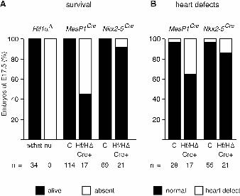
Hif1α results in 100% embryo lethality by E17.5 (Fig. 1A), By contrast, deletion of Hif1α from
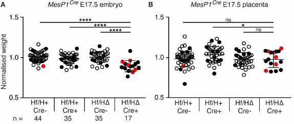
mesoderm led to ~55% embryo lethality (Fig. 1A; Table S1). Surviving conditionally-deleted
embryos were significantly lighter than their control littermates (Fig. S1). Analysis of heart
morphology at E17.5 identified a significantly increased prevalence of heart defects in
conditionally-deleted embryos (6/17 compared with 1/28 of controls analyzed; Fig. 1B; Table S1;
Fig. S1A, defective embryos indicated in red). The heart defects in the conditionally deleted
embryos included ventricular septal defect (VSD), overriding aorta (OA) and ectopia cordis (EC)
(Fig. 2; Table S1). In summary, Hif1α is required in mesoderm for survival and for normal
development of the heart. Its absence from mesoderm leads to ~55% embryo lethality, and 35% of
surviving conditionally-deleted embryos develop heart defects (Fig. 1A,B).
O’Reilly et al. Hypoxia and Heart Development
13
Deletion of Hif1αα from vascular endothelial cells impacts on development of the placenta and
embryo
Deletion of Hif1α from mesoderm resulted in significant heart defects and embryonic lethality. The
mesoderm gives rise to both the vasculature (embryonic and placental) and the heart, and we
considered that expression of Hif1α in either or both of these tissues might be important for causing
heart defects and/or lethality. We first sought to determine if the observed defects were caused by
loss of Hif1α in the vasculature alone. We therefore conditionally deleted Hif1α from vascular
endothelial cells (VECs) and hematopoietic cells using the Tek-Cre transgene (Constien et al.,
2001; Tang et al., 2004). This transgene drives expression of Cre recombinase in all VECs from
E7.5 (Koni et al., 2001) in both embryonic and extra-embryonic vessels, including those of the yolk
sac and placenta. In the heart, Tek-Cre is expressed in the endocardium, the myocardial vasculature,
the mesenchymal cells of the proximal outflow tract, the atrioventricular canal and in the
developing leaflets of the pulmonary and aortic valves (Kisanuki et al., 2001; Schlaeger et al.,
1997). We crossed Hif1α∆/+ Tek-Cre+ males with Hif1αflox/flox females and collected embryos and
placentas at E17.5. Deletion of Hif1α from VECs had no affect on embryo survival (Table S2),
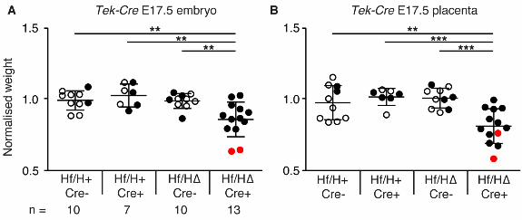
although both embryos and placentas were significantly lighter than controls (Fig. S2). Vascular
defects such as hemorrhage or edema were not apparent in embryos. Analysis of heart morphology
at E17.5 identified heart defects in 15% of conditionally-deleted embryos (2/13 compared with 0/10
of controls analyzed; Table S2; Fig. S2, defective embryos indicated in red). However the
prevalence of heart defects in these embryos was not significantly increased over controls. The
heart defects were VSD with OA or double-outlet right ventricle (DORV) (Table S2; Fig. 2).
Notably, the embryos with heart defects were significantly lighter than the other embryos of the
same genotype (Fig. S2A). In summary, Hif1α is required in the vasculature of the yolk sac,
placenta and/or embryo for normal embryo and placental growth, and its loss in VECs may
contribute to a low prevalence of heart defects.
O’Reilly et al. Hypoxia and Heart Development
14
Deletion of Hif1αα from cardiac precursor cells results in reduced placental and embryo
weight, and may impact heart development
Since the majority of heart defects and embryonic lethality caused by the loss of Hif1α in
mesoderm did not appear to be due to a requirement for Hif1α in VECs, we next sought to
conditionally delete Hif1α from cardiac precursors and derivatives using the Nkx2-5IRESCre allele.
This allele is expressed in the first and second heart fields from E7.75, with recombinase activity
evident throughout the myocardium as development proceeds (Stanley et al., 2002). Later in
development, this allele is also expressed in a number of extra-cardiac regions, including
derivatives of the pharyngeal floor and foregut endoderm including the lung epithelium, and parts
of the liver, thyroid, spleen, pancreas and stomach (Stanley et al., 2002). We crossed Hif1α∆/+
Nkx2-5IRESCre+/- males with Hif1αflox/flox females and collected embryos and placentas at E17.5.
Deletion of Hif1α from the heart did not affect embryo survival (Fig. 1A; Table S3), but these
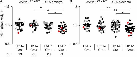
embryos and their placentas were significantly lighter than controls (Fig. S3). Analysis of heart
morphology revealed that 14% of these embryos had heart defects (3/21 embryos with VSD, OA,
DORV, straddling and overriding tricuspid valve (SOTV); Fig 1B; Fig. S3, defective embryos
indicated in red). However, 2/55 controls also had heart defects (VSD with OA), therefore this was
not a significantly increased prevalence. In summary, Hif1α expression in the heart is required for
normal embryo and placenta growth, and its absence from the heart during development may
contribute to a low prevalence of heart defects.
Hif1αα is deleted from the heart using MesP1Cre or Nkx2-5IRESCre
Although deletion of Hif1α from all embryonic and extra-embryonic tissues causes completely
penetrant heart defects and embryo lethality (Compernolle et al., 2003; Cowden Dahl et al., 2005;
Iyer et al., 1998; Kotch et al., 1999; Ryan et al., 1998), in our study where Hif1α is essentially
deleted from the mesoderm (MesP1Cre) or the heart (Nkx2-5IRESCre), we observed a much reduced
O’Reilly et al. Hypoxia and Heart Development
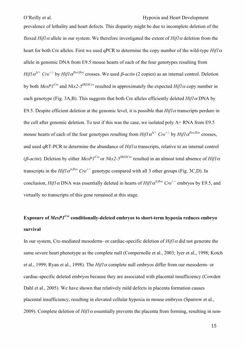
15
prevalence of lethality and heart defects. This disparity might be due to incomplete deletion of the
floxed Hif1α allele in our system. We therefore investigated the extent of Hif1α deletion from the
heart for both Cre alleles. First we used qPCR to determine the copy number of the wild-type Hif1α
allele in genomic DNA from E9.5 mouse hearts of each of the four genotypes resulting from
Hif1α∆/+ Cre+/- by Hif1αflox/flox crosses. We used β-actin (2 copies) as an internal control. Deletion
by both MesP1Cre and Nkx2-5IRESCre resulted in approximately the expected Hif1α copy number in
each genotype (Fig. 3A,B). This suggests that both Cre alleles efficiently deleted Hif1α DNA by
E9.5. Despite efficient deletion at the genomic level, it is possible that Hif1α transcripts perdure in
the cell after genomic deletion. To test if this was the case, we isolated poly A+ RNA from E9.5
mouse hearts of each of the four genotypes resulting from Hif1α∆/+ Cre+/- by Hif1αflox/flox crosses,
and used qRT-PCR to determine the abundance of Hif1α transcripts, relative to an internal control
(β-actin). Deletion by either MesP1Cre or Nkx2-5IRESCre resulted in an almost total absence of Hif1α
transcripts in the Hif1α∆/flox Cre+/- genotype compared with all 3 other groups (Fig. 3C,D). In
conclusion, Hif1α DNA was essentially deleted in hearts of Hif1α∆/flox Cre+/- embryos by E9.5, and
virtually no transcripts of this gene remained at this stage.
Exposure of MesP1Cre conditionally-deleted embryos to short-term hypoxia reduces embryo
survival
In our system, Cre-mediated mesoderm- or cardiac-specific deletion of Hif1α did not generate the
same severe heart phenotype as the complete null (Compernolle et al., 2003; Iyer et al., 1998; Kotch
et al., 1999; Ryan et al., 1998). The Hif1α complete null embryos differ from our mesoderm- or
cardiac-specific deleted embryos because they are associated with placental insufficiency (Cowden
Dahl et al., 2005). We have shown that relatively mild defects in placenta formation causes
placental insufficiency, resulting in elevated cellular hypoxia in mouse embryos (Sparrow et al.,
2009). Complete deletion of Hif1α essentially prevents the placenta from forming, resulting in non-
O’Reilly et al. Hypoxia and Heart Development
16
physiological hypoxia in embryos (Ryan et al., 1998). In such embryos, the expected increase in
requirement of HIF1α may lead to defects in tissues lacking this gene. We hypothesized that
subjecting the Hif1α conditional null embryos to increased hypoxic stress might exacerbate the
prevalence or severity of heart defects. To test this hypothesis, we used our method of exposing
pregnant mice to an atmosphere with reduced oxygen concentration at normal pressure for 8 hours
at E9.5 (Sparrow et al., 2012). Whilst this induces short-term embryonic hypoxia, it is unlikely to
match the placental insufficiency that likely occurs over a number of days in Hif1α nulls;
nevertheless it is sufficient to increase the severity and penetrance of vertebral defects in
genetically-susceptible embryos (Sparrow et al., 2012). We investigated the effects of maternal low
oxygen exposure on embryos with Hif1α deleted from mesoderm using the MesP1Cre allele. Crosses
were performed as above, then pregnant mice were exposed to an atmosphere with reduced oxygen
for 8 hours at E9.5. After exposure, pregnant mice were returned to normoxia and the embryos
allowed to develop until harvest at E17.5. Embryo and placenta weights were recorded, and heart
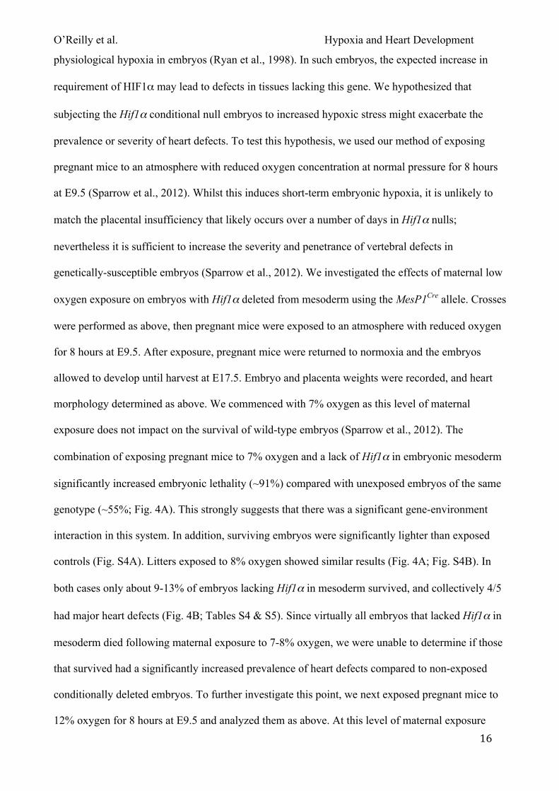
morphology determined as above. We commenced with 7% oxygen as this level of maternal
exposure does not impact on the survival of wild-type embryos (Sparrow et al., 2012). The
combination of exposing pregnant mice to 7% oxygen and a lack of Hif1α in embryonic mesoderm
significantly increased embryonic lethality (~91%) compared with unexposed embryos of the same
genotype (~55%; Fig. 4A). This strongly suggests that there was a significant gene-environment
interaction in this system. In addition, surviving embryos were significantly lighter than exposed
controls (Fig. S4A). Litters exposed to 8% oxygen showed similar results (Fig. 4A; Fig. S4B). In
both cases only about 9-13% of embryos lacking Hif1α in mesoderm survived, and collectively 4/5
had major heart defects (Fig. 4B; Tables S4 & S5). Since virtually all embryos that lacked Hif1α in
mesoderm died following maternal exposure to 7-8% oxygen, we were unable to determine if those
that survived had a significantly increased prevalence of heart defects compared to non-exposed
conditionally deleted embryos. To further investigate this point, we next exposed pregnant mice to
12% oxygen for 8 hours at E9.5 and analyzed them as above. At this level of maternal exposure
O’Reilly et al. Hypoxia and Heart Development
17
embryo lethality was not increased over that observed under normoxia (Fig. 4A). Embryos were
significantly lighter than exposed controls (Fig. S4C). Analysis of heart morphology revealed a
slight but non-significant increase in the prevalence of heart defects in embryos lacking Hif1α in
mesoderm comparing maternal exposure to 12% oxygen or normoxia (9/17 compared with 6/17;
Fig. 4B; Tables S1 & S6). Taken together, these data demonstrate that maternal exposure to
lowered oxygen levels significantly increases the prevalence of lethality in embryos lacking Hif1α
in mesoderm. Interestingly, maternal exposure at 7-8% oxygen resulted in an almost completely
penetrant embryonic lethality (~89%; Fig. 4A), similar to the fully penetrant lethality reported for
Hif1α complete null embryos. This suggests that maternal exposure to short-term hypoxia can in
part replicate the non-physiological hypoxia generated by placental insufficiency occurring in
complete Hif1α null embryos.
Exposure of Nkx2-5IRESCre conditionally-deleted embryos to short-term hypoxia reduces
embryo survival
Having demonstrated an increased requirement for Hif1α in embryonic mesoderm under hypoxic
conditions, we next sought to determine if a lack of Hif1α in the heart alone could be sufficient to
impair embryo survival and/or heart development under such conditions. We investigated the
effects of maternal low oxygen exposure on embryos with Hif1α deleted from the heart using the
Nkx2-5IRESCre allele. Crosses were performed as above, pregnant mice were exposed to an
atmosphere with reduced oxygen for 8 hours at E9.5, returned to normoxia, and the embryos
allowed to develop until E17.5 before harvest. Embryo and placenta weights were recorded, and
heart morphology determined as above. In the MesP1Cre experiments described above, there was no
observed difference between the phenotypes of embryos whose mothers were exposed to 7% or 8%
oxygen. Therefore we used the higher oxygen concentration for the Nkx2-5IRESCre experiments. The
combination of exposing pregnant mice to 8% oxygen and a lack of Hif1α in the embryonic heart
significantly reduced embryo survival (~64%) compared with unexposed embryos (~9%; Fig. 4C).
O’Reilly et al. Hypoxia and Heart Development
18
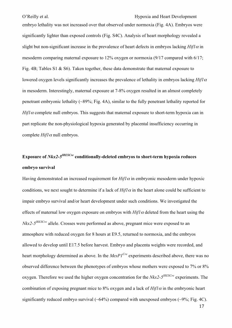
In addition, surviving embryos were significantly lighter than exposed controls (Fig. S5A). Analysis
of heart morphology showed a slight but non-significant increase in the prevalence of heart defects
in embryos lacking Hif1α in the heart between 8% oxygen and normoxia (2/9 compared with 3/21;
Fig. 4D; Tables S3 & S7). Similarly, maternal exposure to 9% oxygen also significantly reduced the
survival of embryos lacking Hif1α in the heart (~77%) compared with embryos of the same
genotype exposed to normoxia (~9% lethality; Fig. 4C). In addition, surviving embryos were
significantly lighter than exposed controls (Fig. S5B). Analysis of heart morphology showed a
small but non-significant increase in the prevalence of heart defects in embryos lacking Hif1α in the
heart at 9% oxygen and normoxia (2/6 compared with 3/21; Fig. 4D; Tables S3 & S8). Given that
exposing embryos lacking Hif1α in the heart to 8-9% oxygen profoundly affected lethality, we
exposed pregnant mice carrying such embryos to 12% oxygen for 8 hours at E9.5 and analyzed
them as above. In embryos lacking Hif1α in the heart, maternal exposure to 12% oxygen did not
affect survival (Fig. 4C), nor did it affect the prevalence of heart defects compared with normoxia
(5/32 compared with 3/21; Fig. 4D; Tables S3 & S9). Surviving embryos were significantly lighter
than exposed controls (Fig. S5C). Taken together, these data demonstrate that maternal exposure to
lowered oxygen levels significantly increases the prevalence of embryonic lethality, and may also
increase the prevalence of heart defects in embryos lacking Hif1α in the heart.
Maternal exposure at 8-9% oxygen, a level of oxygen that does not adversely impact the
survival of control embryos, resulted in ~64-77% lethality of embryos lacking Hif1α in the heart. In
addition, heart defects were observed in 22-33% of surviving conditionally deleted embryos (Fig.
4C,D). To further investigate the timing of lethality, we repeated the crosses described above.
Pregnant mice were exposed to 9% oxygen for 8 hours, then returned to normoxia prior to embryo
collection at E10.5. Control mice were maintained in normoxic conditions until embryo harvest at
E10.5. At this stage, embryos of all genotypes were present in equal numbers, but there was a
significantly increased prevalence of abnormality in conditionally deleted embryos exposed to 9%
oxygen compared with normoxia (11/18 compared with 1/17; Tables S10 & S11). At 9% oxygen,
O’Reilly et al. Hypoxia and Heart Development
19
2/18 of these embryos were very small and were necrotic; 9/18 were smaller than littermates and 6
of these also had an enlarged pericardial sac (data not shown). However, despite these defects all of
the non-necrotic embryos (9/18) had normal heart morphology, with correct looping and normal
chamber morphology (data not shown). This suggests that embryos lacking Hif1α in the heart are
very vulnerable to in utero exposure to lowered oxygen levels, and such exposure rapidly triggers
embryonic lethality.
Short-term exposure of pregnant mice to low oxygen increases embryonic hypoxia and
stabilisation of HIF1αα in the heart.
To investigate the molecular consequences of short-term low oxygen exposure in embryos, we
examined cellular hypoxia and the response of HIF1α protein in embryos immediately following
such exposure. Firstly, we used hypoxyprobe™ (pimonidazole hydrochloride) to measure cellular
hypoxia in embryos. Hypoxyprobe™ is reductively activated, forming adducts with thiol groups in
cells experiencing < 2% oxygen (Mahy et al., 2003; Raleigh et al., 1999). These adducts can be
detected using a specific antibody (Kennedy et al., 1997). To assess the relative levels of cellular
hypoxia in exposed and unexposed embryos, we injected pregnant mice with hypoxyprobe™ at
E9.5, exposed them to 8% oxygen for 3 hours (whilst control mice remained at normoxia), then
harvested the embryos and quantified the hypoxia using western blotting as previously described
(Sparrow et al., 2009). Cellular hypoxia was dramatically increased in exposed embryos (Fig.
5A,B). Secondly, we compared HIF1α protein levels in exposed and control embryos in vivo using
wholemount immufluorescence. Hif1α is constitutively transcribed and translated in cells, but under
normoxic conditions the HIF1α protein has an extremely short half-life of less than 5 min (Huang et
al., 1996; Yu et al., 1998). Under hypoxic conditions HIF1α is stabilized and rapidly accumulates in
the nucleus, activating its target genes (Kallio et al., 1998). We exposed pregnant mice to 9%
oxygen for 3 or 8 hours at E9.5 (whilst control mice remained at normoxia), then harvested the
embryos and performed immunofluorescence in wholemount. Nuclear HIF1α was not detected in
O’Reilly et al. Hypoxia and Heart Development
20
any region of control embryos (Fig. 5D-F) or those exposed for 3 hours (not shown), whereas
embryos exposed for 8 hours had readily detectable levels of nuclear HIF1α specifically localized
in the myocardium (Fig. 5G-I). To determine if this result reflected an intrinsic susceptibility of
myocardium to hypoxia, we next examined the localization of HIF1α under increased hypoxic
stress. We exposed wild type embryos to the lowest possible oxygen level (5.5% oxygen for 8 hours
(Sparrow et al., 2012)), and used immunofluorescence to detect HIF1α protein (Fig. S6). In these
embryos, nuclear HIF1α was once again detected in the myocardium (Fig. S6F) and also in gut
epithelium (data not shown). Low level nuclear accumulation was also detectable in forebrain
epithelium (Fig. S6H). Taken together, these data demonstrate that short-term maternal exposure to
mildly lowered oxygen levels (9%) at E9.5 is sufficient to cause cellular hypoxia in the embryos by
3 hours, and localisation of HIF1α protein to the nucleus of embryonic myocardial cells by 8 hours.
Furthermore, the vast majority of HIF1α protein accumulation was in myocardial cells, suggesting
that this tissue is likely to be much more susceptible to non-physiological hypoxia than the other
tissues of the developing embryo.
Myocardial proliferation is reduced in hearts lacking Hif1αα
Short-term exposure of embryos in utero to reduced oxygen levels caused accumulation of HIF1α
protein in the nucleus of the myocardium. Additionally, it caused rapid demise of embryos that
were unable to respond to this hypoxic insult due to the lack of Hif1α in the heart. As embryo death
did not appear to be due to a disruption of gross heart morphology, we next examined the cellular
response of myocardium lacking Hif1α to cellular hypoxia. In particular, we investigated cell
proliferation and cell death. We exposed pregnant mice to 9% oxygen for 8 hours at E9.5 (whilst
control mice remained at normoxia), and then harvested the embryos immediately after maternal
exposure. To examine proliferation, these embryos were co-stained with an antibody for
phosphorylated Histone H3 and the nuclear marker DAPI, and the myocardial mitotic index
O’Reilly et al. Hypoxia and Heart Development
21
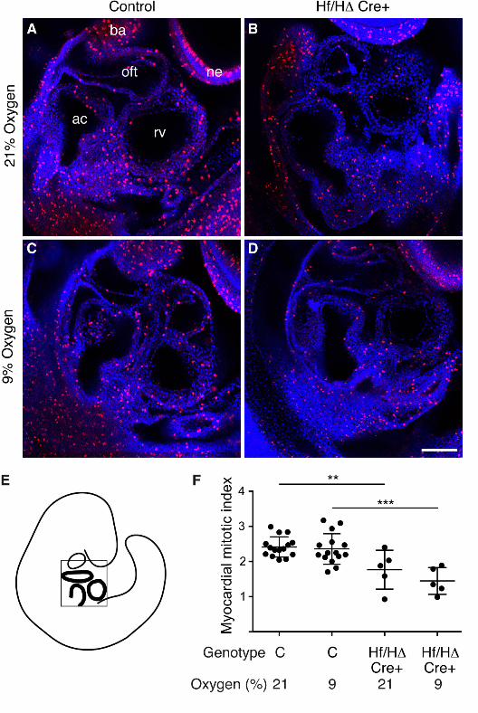
calculated (Fig. 6). Embryos lacking Hif1α in the heart showed a significant drop in the myocardial
mitotic index compared with control genotypes. This drop occurred regardless of whether or not
their mothers had been exposed to reduced oxygen levels. By contrast, for both wild type embryos
and embryos lacking Hif1α in the heart, maternal exposure to 9% oxygen did not cause
significantly reduced myocardial proliferation compared with unexposed embryos (Fig. 6F). To
examine cell death, embryos generated as described above were stained with LysoTracker Red
(Life Technologies). Surprisingly, staining was completely absent from the heart, despite being
present elsewhere in the embryo, regardless of genotype or oxygen concentration (data not shown).
These data demonstrate that myocardial proliferation is significantly reduced in hearts lacking
Hif1α compared with controls, but that maternal exposure to reduced oxygen levels does not alter
embryonic myocardial cell proliferation or cell death. Therefore the hypoxia-induced death of
embryos lacking cardiac Hif1α is not due to a lack of cellular proliferation in the heart, or to an
increase in cell death.
O’Reilly et al. Hypoxia and Heart Development
22
DISCUSSION
Hif1α deletion from the mouse conceptus leads to completely penetrant embryonic lethality by E10
due to a collapse in the development of the cardiovascular system of the embryo, yolksac and
placenta (Compernolle et al., 2003; Cowden Dahl et al., 2005; Iyer et al., 1998; Kotch et al., 1999;
Ryan et al., 1998). These studies have shown that Hif1α is required for normal development, but
they do not specifically identify the tissues in which Hif1α functions, or if Hif1α is actually
required in the embryo when the placenta is functioning (which is not the case in the complete
Hif1α null). We have addressed these issues by conditionally deleting Hif1α from specific cell
types and tissues, resulting in a number of key findings. Most significantly, we demonstrate that
Hif1α is not required for development of the cardiovascular system when the embryo develops in
the context of a functioning placenta. Additionally, we find that Hif1α is primarily required in the
heart when the embryo is exposed to lowered oxygen conditions.
Firstly, we showed that Hif1α is not required in VECs of the embryo, yolksac or placenta for either
normal morphological development or for embryo survival, using conditional deletion with Tek-
Cre. However there was an impact on embryo and placental growth, and a slight but non-significant
increase in the prevalence of heart defects. This lack of lethality might have been predicted as
progeny of a Hif1αflox/flox x Tek-Cre intercross survive to adulthood (Tang et al., 2004). However in
that study, an embryonic phenotype may have been averted due to incomplete conditional deletion
of Hif1α throughout the conceptus during development. Others have reported incomplete Hif1α
deletion when using homozygous Hif1αflox alleles (Krishnan et al., 2008; Mason et al., 2004). By
contrast, our study design maximized Hif1α deletion in VECs by using Hif1α∆/flox embryos that
only require Tek-Cre to act on a single Hif1αflox allele. Our finding that Hif1α is not overtly
required in VECs during development is intriguing, as Hif1α is required for angiogenesis during
wound healing and tumorigenesis in adult mice (Tang et al., 2004). Our results suggest that
O’Reilly et al. Hypoxia and Heart Development
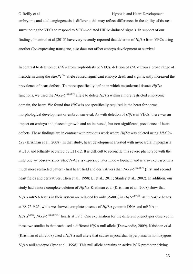
23
embryonic and adult angiogenesis is different; this may reflect differences in the ability of tissues
surrounding the VECs to respond to VEC-mediated HIF1α-induced signals. In support of our
findings, Imanirad et al (2013) have very recently reported that deletion of Hif1α from VECs using
another Cre-expressing transgene, also does not affect embryo development or survival.
In contrast to deletion of Hif1α from trophoblasts or VECs, deletion of Hif1α from a broad range of
mesoderm using the MesP1Cre allele caused significant embryo death and significantly increased the
prevalence of heart defects. To more specifically define in which mesodermal tissues Hif1α
functions, we used the Nkx2-5IRESCre allele to delete Hif1α within a more restricted embryonic
domain, the heart. We found that Hif1α is not specifically required in the heart for normal
morphological development or embryo survival. As with deletion of Hif1α in VECs, there was an
impact on embryo and placenta growth and an increased, but non-significant, prevalence of heart
defects. These findings are in contrast with previous work where Hif1α was deleted using MLC2v-
Cre (Krishnan et al., 2008). In that study, heart development arrested with myocardial hyperplasia
at E10, and lethality occurred by E11-12. It is difficult to reconcile this severe phenotype with the
mild one we observe since MLC2v-Cre is expressed later in development and is also expressed in a
much more restricted pattern (first heart field and derivatives) than Nkx2-5IRESCre (first and second
heart fields and derivatives, Chen et al., 1998; Li et al., 2011; Stanley et al., 2002). In addition, our
study had a more complete deletion of Hif1α: Krishnan et al (Krishnan et al., 2008) show that
Hif1α mRNA levels in their system are reduced by only 35-80% in Hif1α∆/flox; MLC2v-Cre hearts
at E8.75-9.25, while we showed complete absence of Hif1α genomic DNA and mRNA in
Hif1α∆/flox; Nkx2-5IRESCre+/- hearts at E9.5. One explanation for the different phenotypes observed in
these two studies is that each used a different Hif1α null allele (Dunwoodie, 2009). Krishnan et al
(Krishnan et al., 2008) used a Hif1α null allele that causes myocardial hyperplasia in homozygous
Hif1α null embryos (Iyer et al., 1998). This null allele contains an active PGK promoter driving
O’Reilly et al. Hypoxia and Heart Development
24
expression of the neo gene. The Hif1α null allele used in our study does not contain PGK-neo, and
does not cause myocardial hyperplasia (Ryan et al., 1998). A third Hif1α allele exists that also lacks
myocardial hyperplasia (Compernolle et al., 2003). It is therefore possible that the myocardial
hyperplasia and embryonic lethality reported by Krishnan et al (Krishnan et al., 2008) stems from
the particular Hif1α null allele used, rather than the loss of Hif1α in the myocardium per se
(Dunwoodie, 2009). It is also possible that the genetic background of the mice used in the different
studies has bearing on the phenotype. Our study used C57BL/6J mice (>10 backcrosses) whilst
Krishnan et al (Krishnan et al., 2008) used mice of mixed background (C57BL/6; 129SvJ; 129S1).
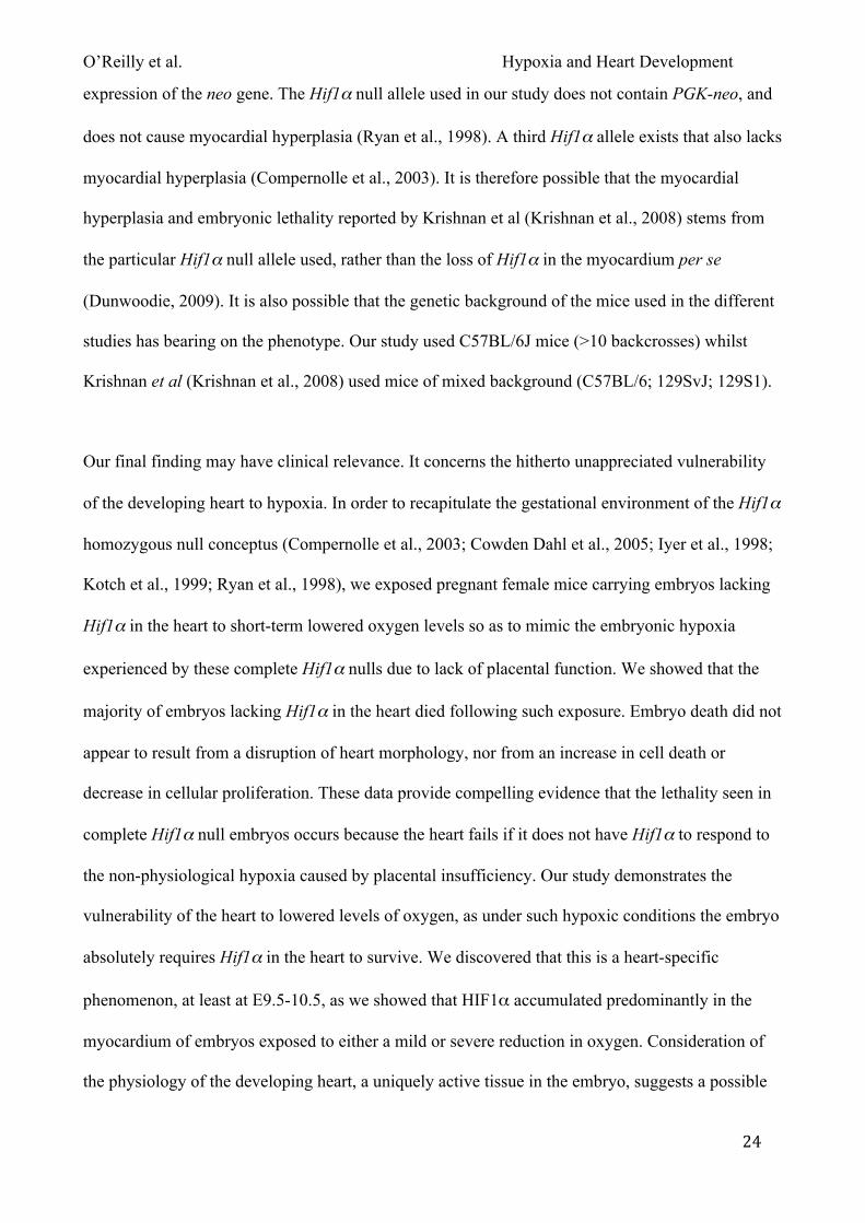
Our final finding may have clinical relevance. It concerns the hitherto unappreciated vulnerability
of the developing heart to hypoxia. In order to recapitulate the gestational environment of the Hif1α
homozygous null conceptus (Compernolle et al., 2003; Cowden Dahl et al., 2005; Iyer et al., 1998;
Kotch et al., 1999; Ryan et al., 1998), we exposed pregnant female mice carrying embryos lacking
Hif1α in the heart to short-term lowered oxygen levels so as to mimic the embryonic hypoxia
experienced by these complete Hif1α nulls due to lack of placental function. We showed that the
majority of embryos lacking Hif1α in the heart died following such exposure. Embryo death did not
appear to result from a disruption of heart morphology, nor from an increase in cell death or
decrease in cellular proliferation. These data provide compelling evidence that the lethality seen in
complete Hif1α null embryos occurs because the heart fails if it does not have Hif1α to respond to
the non-physiological hypoxia caused by placental insufficiency. Our study demonstrates the
vulnerability of the heart to lowered levels of oxygen, as under such hypoxic conditions the embryo
absolutely requires Hif1α in the heart to survive. We discovered that this is a heart-specific
phenomenon, at least at E9.5-10.5, as we showed that HIF1α accumulated predominantly in the
myocardium of embryos exposed to either a mild or severe reduction in oxygen. Consideration of
the physiology of the developing heart, a uniquely active tissue in the embryo, suggests a possible
O’Reilly et al. Hypoxia and Heart Development
25
mechanism for the embryonic lethality in the absence of structural heart defects. The energetic
needs of the post implantation mouse embryo (E5.6-E6.5) are almost entirely met in this low
oxygen environment by glycolysis (Wales et al., 1995). As development proceeds, the proportion of
energy derived from glucose oxidation increases with the increasing vascularisation of the embryo.
However at E9.5 oxidative phosphorylation is still a minor contributor (Folmes et al., 2012a;
Folmes et al., 2012b; Wales et al., 1995). At this stage cardiomyocytes have only a few fragmented
mitochondria with poorly defined and organized cristae, and so are primarily dependent on
anaerobic glycolysis for ATP generation, as is the rest of the embryo (Hom et al., 2011; Wales et
al., 1995). Extensive growth is a feature of subsequent embryonic development (E9.5-13.5). Such
growth can only be sustained with elevated cardiac output, and the increasing bioenergetic demands
of the cardiomyocytes require efficient oxidative ATP generation. Networks of elongated and
branched mitochondria with abundant organized cristae are a feature of cardiomyocytes at E13.5
(Hom et al., 2011). Thus between E9.5 and E13.5 is a critical time for the embryo as
cardiomyocytes transition between anaerobic and aerobic glycolysis. This transition needs to be
balanced against the increasing cardiovascular requirements of the rapidly growing embryo. Clearly
an adequate oxygen supply is crucial for this transition and for sustaining embryonic growth and
viability. Thus it might be predicted that when we expose embryos to lowered oxygen levels at E9.5
for eight hours, we disrupt this critical balance between embryonic growth demands, contractility of
the myocardium, ATP requirement and oxygen. Such a disruption is clearly seen in the
accumulation of nuclear HIF1α in the myocardium, indicating that there is insufficient oxygen to
sustain the metabolic activity of the cardiomyocytes. However in the absence of Hif1α, the effects
of hypoxia cannot be redressed by the normal compensatory mechanisms co-ordinated by HIF1α.
Failure of these compensatory mechanisms then rapidly results in significant embryonic lethality.
Our discovery that the heart is extremely vulnerable to reduced levels of oxygen is potentially
clinically relevant for two reasons. Firstly, during embryonic development, short-term exposure to
O’Reilly et al. Hypoxia and Heart Development
26
low oxygen causes cellular hypoxia of the myocardium. This, coupled with a genetic predisposition
that impairs any of the systems that deliver, monitor and/or respond to oxygen, or are required for
cardiomyocyte contractile function, or mitochondria and energy metabolism, may lead to death in
utero. Secondly, in cases of congenital heart disease where chronic underperformance of the
cardiovascular system occurs even post-surgery, such as in hyperplastic left heart syndrome, an
impaired response to cardiomyocyte hypoxia (Gaber et al., 2013) may explain the variable
outcomes experienced by these patients. In conclusion, our Hif1α-centered studies shed light on
these clinical situations and may in time lead to identification of individuals at increased risk of
death in utero or after cardiac surgery, and to the development of preventative therapies.
O’Reilly et al. Hypoxia and Heart Development
27
ACKNOWLEDGEMENTS
We thank Yumiko Saga for providing mouse lines; BioCORE staff; and Herbert Smith for
generously donating the confocal microscope used in this study.
SOURCES OF FUNDING
The work was supported by the Australian National Health and Medical Research Council: Project
Grants 303705, 404805 and 1019776; and Senior Research Fellowships 514900 and 1042002.
DISCLOSURES
None.
O’Reilly et al. Hypoxia and Heart Development
28
REFERENCES
Amarilio, R., Viukov, S. V., Sharir, A., Eshkar-Oren, I., Johnson, R. S., Zelzer, E., 2007. HIF1alpha
regulation of Sox9 is necessary to maintain differentiation of hypoxic prechondrogenic cells during
early skeletogenesis. Development. 134, 3917-28.
Carreau, A., El Hafny-Rahbi, B., Matejuk, A., Grillon, C., Kieda, C., 2011. Why is the partial
oxygen pressure of human tissues a crucial parameter? Small molecules and hypoxia. J Cell Mol
Med. 15, 1239-53.
Chen, J., Kubalak, S. W., Chien, K. R., 1998. Ventricular muscle-restricted targeting of the
RXRalpha gene reveals a non-cell-autonomous requirement in cardiac chamber morphogenesis.
Development. 125, 1943-9.
Compernolle, V., Brusselmans, K., Franco, D., Moorman, A., Dewerchin, M., Collen, D.,
Carmeliet, P., 2003. Cardia bifida, defective heart development and abnormal neural crest migration
in embryos lacking hypoxia-inducible factor-1alpha. Cardiovasc Res. 60, 569-79.
Constien, R., Forde, A., Liliensiek, B., Grone, H. J., Nawroth, P., Hammerling, G., Arnold, B.,
2001. Characterization of a novel EGFP reporter mouse to monitor Cre recombination as
demonstrated by a Tie2 Cre mouse line. Genesis. 30, 36-44.
Cowden Dahl, K. D., Fryer, B. H., Mack, F. A., Compernolle, V., Maltepe, E., Adelman, D. M.,
Carmeliet, P., Simon, M. C., 2005. Hypoxia-inducible factors 1alpha and 2alpha regulate
trophoblast differentiation. Mol Cell Biol. 25, 10479-91.
Dunwoodie, S. L., 2009. The role of hypoxia in development of the Mammalian embryo. Dev Cell.
17, 755-73.
Eisinger-Mathason, T. S., Zhang, M., Qiu, Q., Skuli, N., Nakazawa, M. S., Karakasheva, T., Mucaj,
V., Shay, J. E., Stangenberg, L., Sadri, N., Pure, E., Yoon, S. S., Kirsch, D. G., Simon, M. C., 2013.
Hypoxia-Dependent Modification of Collagen Networks Promotes Sarcoma Metastasis. Cancer
Discov.
Eltzschig, H. K., Carmeliet, P., 2011. Hypoxia and inflammation. N Engl J Med. 364, 656-65.
O’Reilly et al. Hypoxia and Heart Development
29
Folmes, C. D., Dzeja, P. P., Nelson, T. J., Terzic, A., 2012a. Metabolic plasticity in stem cell
homeostasis and differentiation. Cell Stem Cell. 11, 596-606.
Folmes, C. D., Dzeja, P. P., Nelson, T. J., Terzic, A., 2012b. Mitochondria in control of cell fate.
Circ Res. 110, 526-9.
Gaber, N., Gagliardi, M., Patel, P., Kinnear, C., Zhang, C., Chitayat, D., Shannon, P., Jaeggi, E.,
Tabori, U., Keller, G., Mital, S., 2013. Fetal Reprogramming and Senescence in Hypoplastic Left
Heart Syndrome and in Human Pluripotent Stem Cells during Cardiac Differentiation. Am J Pathol.
183, 720-34.
Geffers, I., Serth, K., Chapman, G., Jaekel, R., Schuster-Gossler, K., Cordes, R., Sparrow, D. B.,
Kremmer, E., Dunwoodie, S. L., Klein, T., Gossler, A., 2007. Divergent functions and distinct
localization of the Notch ligands DLL1 and DLL3 in vivo. J Cell Biol. 178, 465-76.
Hom, J. R., Quintanilla, R. A., Hoffman, D. L., de Mesy Bentley, K. L., Molkentin, J. D., Sheu, S.
S., Porter, G. A., Jr., 2011. The permeability transition pore controls cardiac mitochondrial
maturation and myocyte differentiation. Dev Cell. 21, 469-78.
Huang, L. E., Arany, Z., Livingston, D. M., Bunn, H. F., 1996. Activation of hypoxia-inducible
transcription factor depends primarily upon redox-sensitive stabilization of its alpha subunit. J Biol
Chem. 271, 32253-9.
Huang, Y., Hickey, R. P., Yeh, J. L., Liu, D., Dadak, A., Young, L. H., Johnson, R. S., Giordano, F.
J., 2004. Cardiac myocyte-specific HIF-1alpha deletion alters vascularization, energy availability,
calcium flux, and contractility in the normoxic heart. FASEB J. 18, 1138-40.
Imanirad, P., Solaimani Kartalaei, P., Crisan, M., Vink, C., Yamada-Inagawa, T., de Pater, E.,
Kurek, D., Kaimakis, P., van der Linden, R., Speck, N., Dzierzak, E., 2013. HIF1alpha is a
regulator of hematopoietic progenitor and stem cell development in hypoxic sites of the mouse
embryo. Stem Cell Res. 12, 24-35.
O’Reilly et al. Hypoxia and Heart Development
30
Iyer, N. V., Kotch, L. E., Agani, F., Leung, S. W., Laughner, E., Wenger, R. H., Gassmann, M.,
Gearhart, J. D., Lawler, A. M., Yu, A. Y., Semenza, G. L., 1998. Cellular and developmental
control of O2 homeostasis by hypoxia-inducible factor 1 alpha. Genes Dev. 12, 149-62.
Jiang, B. H., Semenza, G. L., Bauer, C., Marti, H. H., 1996. Hypoxia-inducible factor 1 levels vary
exponentially over a physiologically relevant range of O2 tension. Am J Physiol. 271, C1172-80.
Kallio, P. J., Okamoto, K., O'Brien, S., Carrero, P., Makino, Y., Tanaka, H., Poellinger, L., 1998.
Signal transduction in hypoxic cells: inducible nuclear translocation and recruitment of the
CBP/p300 coactivator by the hypoxia-inducible factor-1alpha. EMBO J. 17, 6573-86.
Kennedy, A. S., Raleigh, J. A., Perez, G. M., Calkins, D. P., Thrall, D. E., Novotny, D. B., Varia,
M. A., 1997. Proliferation and hypoxia in human squamous cell carcinoma of the cervix: first report
of combined immunohistochemical assays. Int J Radiat Oncol Biol Phys. 37, 897-905.
Kisanuki, Y. Y., Hammer, R. E., Miyazaki, J., Williams, S. C., Richardson, J. A., Yanagisawa, M.,
2001. Tie2-Cre transgenic mice: a new model for endothelial cell-lineage analysis in vivo. Dev
Biol. 230, 230-42.
Koni, P. A., Joshi, S. K., Temann, U. A., Olson, D., Burkly, L., Flavell, R. A., 2001. Conditional
vascular cell adhesion molecule 1 deletion in mice: impaired lymphocyte migration to bone
marrow. J Exp Med. 193, 741-54.
Kotch, L. E., Iyer, N. V., Laughner, E., Semenza, G. L., 1999. Defective vascularization of HIF-
1alpha-null embryos is not associated with VEGF deficiency but with mesenchymal cell death. Dev
Biol. 209, 254-67.
Krishnan, J., Ahuja, P., Bodenmann, S., Knapik, D., Perriard, E., Krek, W., Perriard, J. C., 2008.
Essential role of developmentally activated hypoxia-inducible factor 1alpha for cardiac
morphogenesis and function. Circ Res. 103, 1139-46.
Land, S. C., 2004. Hochachka's "Hypoxia Defense Strategies" and the development of the pathway
for oxygen. Comp Biochem Physiol B Biochem Mol Biol. 139, 415-33.
O’Reilly et al. Hypoxia and Heart Development
31
Lando, D., Peet, D. J., Gorman, J. J., Whelan, D. A., Whitelaw, M. L., Bruick, R. K., 2002a. FIH-1
is an asparaginyl hydroxylase enzyme that regulates the transcriptional activity of hypoxia-
inducible factor. Genes Dev. 16, 1466-71.
Lando, D., Peet, D. J., Whelan, D. A., Gorman, J. J., Whitelaw, M. L., 2002b. Asparagine
hydroxylation of the HIF transactivation domain a hypoxic switch. Science. 295, 858-61.
Lee, Y. M., Jeong, C. H., Koo, S. Y., Son, M. J., Song, H. S., Bae, S. K., Raleigh, J. A., Chung, H.
Y., Yoo, M. A., Kim, K. W., 2001. Determination of hypoxic region by hypoxia marker in
developing mouse embryos in vivo: a possible signal for vessel development. Dev Dyn. 220, 175-
86.
Lendahl, U., Lee, K. L., Yang, H., Poellinger, L., 2009. Generating specificity and diversity in the
transcriptional response to hypoxia. Nat Rev Genet. 10, 821-32.
Li, P., Cavallero, S., Gu, Y., Chen, T. H., Hughes, J., Hassan, A. B., Bruning, J. C., Pashmforoush,
M., Sucov, H. M., 2011. IGF signaling directs ventricular cardiomyocyte proliferation during
embryonic heart development. Development. 138, 1795-805.
Mahy, P., De Bast, M., Gallez, B., Gueulette, J., Koch, C. J., Scalliet, P., Gregoire, V., 2003. In
vivo colocalization of 2-nitroimidazole EF5 fluorescence intensity and electron paramagnetic
resonance oximetry in mouse tumors. Radiother Oncol. 67, 53-61.
Mason, S. D., Howlett, R. A., Kim, M. J., Olfert, I. M., Hogan, M. C., McNulty, W., Hickey, R. P.,
Wagner, P. D., Kahn, C. R., Giordano, F. J., Johnson, R. S., 2004. Loss of skeletal muscle HIF-
1alpha results in altered exercise endurance. PLoS Biol. 2, e288.
Prabhakar, N. R., Semenza, G. L., 2012. Adaptive and maladaptive cardiorespiratory responses to
continuous and intermittent hypoxia mediated by hypoxia-inducible factors 1 and 2. Physiol Rev.
92, 967-1003.
Provot, S., Zinyk, D., Gunes, Y., Kathri, R., Le, Q., Kronenberg, H. M., Johnson, R. S., Longaker,
M. T., Giaccia, A. J., Schipani, E., 2007. Hif-1alpha regulates differentiation of limb bud
mesenchyme and joint development. J Cell Biol. 177, 451-64.
O’Reilly et al. Hypoxia and Heart Development
32
Raleigh, J. A., Chou, S. C., Arteel, G. E., Horsman, M. R., 1999. Comparisons among pimonidazole
binding, oxygen electrode measurements, and radiation response in C3H mouse tumors. Radiat Res.
151, 580-9.
Ream, M., Ray, A. M., Chandra, R., Chikaraishi, D. M., 2008. Early fetal hypoxia leads to growth
restriction and myocardial thinning. Am J Physiol Regul Integr Comp Physiol. 295, R583-95.
Rosset, A., Spadola, L., Ratib, O., 2004. OsiriX: an open-source software for navigating in
multidimensional DICOM images. J Digit Imaging. 17, 205-16.
Ryan, H. E., Lo, J., Johnson, R. S., 1998. HIF-1 alpha is required for solid tumor formation and
embryonic vascularization. EMBO J. 17, 3005-15.
Ryan, H. E., Poloni, M., McNulty, W., Elson, D., Gassmann, M., Arbeit, J. M., Johnson, R. S.,
2000. Hypoxia-inducible factor-1alpha is a positive factor in solid tumor growth. Cancer Res. 60,
4010-5.
Saga, Y., Miyagawa-Tomita, S., Takagi, A., Kitajima, S., Miyazaki, J., Inoue, T., 1999. MesP1 is
expressed in the heart precursor cells and required for the formation of a single heart tube.
Development. 126, 3437-47.
Schlaeger, T. M., Bartunkova, S., Lawitts, J. A., Teichmann, G., Risau, W., Deutsch, U., Sato, T.
N., 1997. Uniform vascular-endothelial-cell-specific gene expression in both embryonic and adult
transgenic mice. Proc Natl Acad Sci U S A. 94, 3058-63.
Schneider, J. E., Cassidy, P. J., Lygate, C., Tyler, D. J., Wiesmann, F., Grieve, S. M., Hulbert, K.,
Clarke, K., Neubauer, S., 2003. Fast, high-resolution in vivo cine magnetic resonance imaging in
normal and failing mouse hearts on a vertical 11.7 T system. J Magn Reson Imaging. 18, 691-701.
Sparrow, D. B., Boyle, S. C., Sams, R. S., Mazuruk, B., Zhang, L., Moeckel, G. W., Dunwoodie, S.
L., de Caestecker, M. P., 2009. Placental insufficiency associated with loss of Cited1 causes renal
medullary dysplasia. J Am Soc Nephrol. 20, 777-86.
Sparrow, D. B., Chapman, G., Smith, A. J., Mattar, M. Z., Major, J. A., O'Reilly, V. C., Saga, Y.,
Zackai, E. H., Dormans, J. P., Alman, B. A., McGregor, L., Kageyama, R., Kusumi, K.,
O’Reilly et al. Hypoxia and Heart Development
33
Dunwoodie, S. L., 2012. A mechanism for gene-environment interaction in the etiology of
congenital scoliosis. Cell. 149, 295-306.
Stanley, E. G., Biben, C., Elefanty, A., Barnett, L., Koentgen, F., Robb, L., Harvey, R. P., 2002.
Efficient Cre-mediated deletion in cardiac progenitor cells conferred by a 3'UTR-ires-Cre allele of
the homeobox gene Nkx2-5. Int J Dev Biol. 46, 431-9.
Tang, N., Wang, L., Esko, J., Giordano, F. J., Huang, Y., Gerber, H. P., Ferrara, N., Johnson, R. S.,
2004. Loss of HIF-1alpha in endothelial cells disrupts a hypoxia-driven VEGF autocrine loop
necessary for tumorigenesis. Cancer Cell. 6, 485-95.
Wales, R. G., Martin, K. L., Leese, H. J., 1995. Glucose utilization by components of the mouse
conceptus during early embryogenesis. J Reprod Fertil. 104, 125-32.
Wang, G. L., Jiang, B. H., Rue, E. A., Semenza, G. L., 1995. Hypoxia-inducible factor 1 is a basic-
helix-loop-helix-PAS heterodimer regulated by cellular O2 tension. Proc Natl Acad Sci U S A. 92,
5510-4.
Yu, A. Y., Frid, M. G., Shimoda, L. A., Wiener, C. M., Stenmark, K., Semenza, G. L., 1998.
Temporal, spatial, and oxygen-regulated expression of hypoxia-inducible factor-1 in the lung. Am J
Physiol. 275, L818-26.
O’Reilly et al. Hypoxia and Heart Development
34
FIGURE CAPTIONS
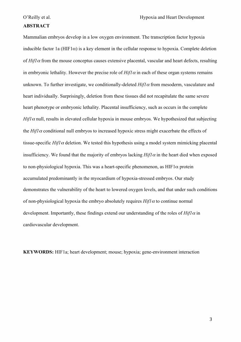
Figure 1: Embryonic survival and prevalence of heart defects following embryonic Hif1αα
deletion
(A) Histograms showing the estimated percentage of embryos that survived until E17.5 following
total embryonic Hif1α deletion (Hif1α∆), or conditional deletion in mesoderm (MesP1Cre) or cardiac
progenitor cells (Nkx2-5Cre). (B) Histograms showing the percentage of heart defects present in
surviving E17.5 embryos that were analyzed for heart morphology following embryonic Hif1α
deletion in mesoderm (MesP1Cre) or cardiac progenitor cells (Nkx2-5Cre). wt/het represents pooled
homozygous wild type and heterozygous Hif1α∆ embryos, null represents homozygous Hif1α∆
embryos, C represents pooled embryos from all three control genotypes from Cre crosses, and
Hf/H∆ Cre+ represents conditionally deleted embryos.
Suggested image size = 1 column
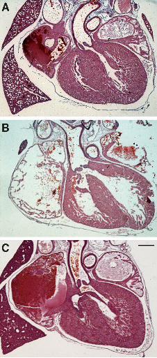
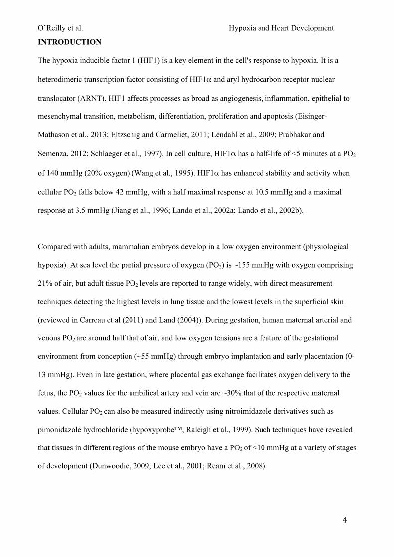
Figure 2: Examples of heart defects identified following Hif1αα deletion
Histological sections of E17.5 mouse hearts after conditional deletion of Hif1α showing examples
of the most common types of defects identified. (A) normal heart, (B) ventricular septal defect
(VSD) with overriding aorta (OA) from a Hif1α∆/flox Tek-Cre embryo, (C) straddling and overriding
tricuspid valve (SOTV) from a Hif1α∆/flox Nkx2-5IRESCre embryo. Scale bar represents 350 µm
Suggested image size = 60 mm
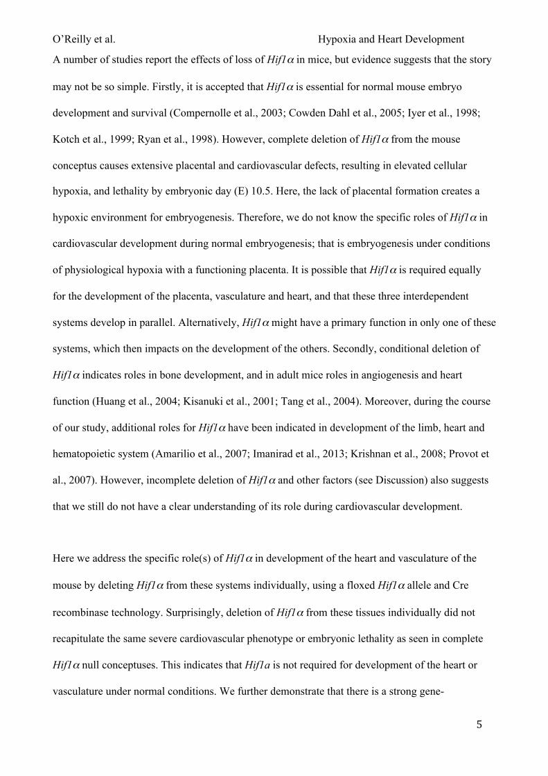
Figure 3: Hif1αα genomic and transcript levels in Hif1αα∆/flox Mesp1Cre and Nkx2-5IRESCre mouse
lines
(A,B) Genomic DNA was isolated from E9.5 embryonic hearts from the Hif1α∆/flox Mesp1Cre (A) or
Nkx2-5IRESCre (B) mouse lines. Hif1α copy number was determined using qPCR with β-actin as an
internal control. (C,D) poly A+ RNA was extracted from E9.5 embryonic hearts from the Hif1α∆/flox
O’Reilly et al. Hypoxia and Heart Development
35
Mesp1Cre (C) or Nkx2-5IRESCre (D) mouse lines. Relative levels of Hif1α transcripts were determined
by qRT-PCR using β-actin as an internal control. Error bars indicate standard deviations. For
transcript levels, statistical significance was determined using a one-tailed Student’s t-test on data
transformed using natural logarithms to correct for zero-bounded data. **** p<0.0001. Hf/H+ Cre-,
Hf/H+ Cre+ and Hf/HΔ Cre- represent the three control genotypes; and Hf/H∆ Cre+ represents
conditionally deleted embryos.
Suggested image size = 1.5 column
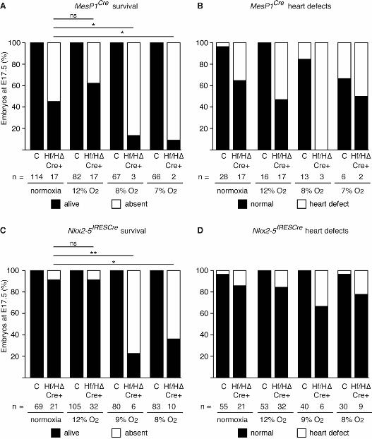
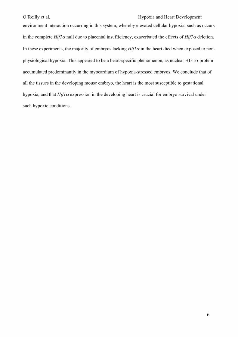
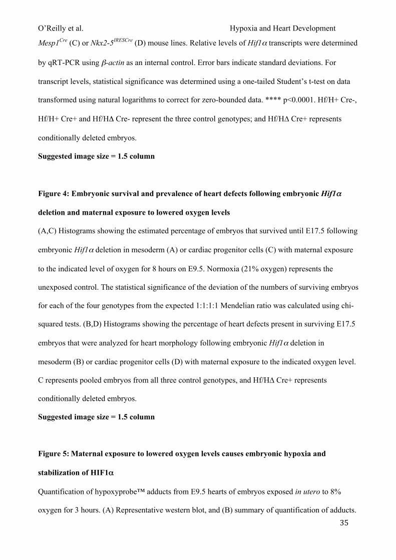
Figure 4: Embryonic survival and prevalence of heart defects following embryonic Hif1αα
deletion and maternal exposure to lowered oxygen levels
(A,C) Histograms showing the estimated percentage of embryos that survived until E17.5 following
embryonic Hif1α deletion in mesoderm (A) or cardiac progenitor cells (C) with maternal exposure
to the indicated level of oxygen for 8 hours on E9.5. Normoxia (21% oxygen) represents the
unexposed control. The statistical significance of the deviation of the numbers of surviving embryos
for each of the four genotypes from the expected 1:1:1:1 Mendelian ratio was calculated using chi-
squared tests. (B,D) Histograms showing the percentage of heart defects present in surviving E17.5
embryos that were analyzed for heart morphology following embryonic Hif1α deletion in
mesoderm (B) or cardiac progenitor cells (D) with maternal exposure to the indicated oxygen level.
C represents pooled embryos from all three control genotypes, and Hf/H∆ Cre+ represents
conditionally deleted embryos.
Suggested image size = 1.5 column
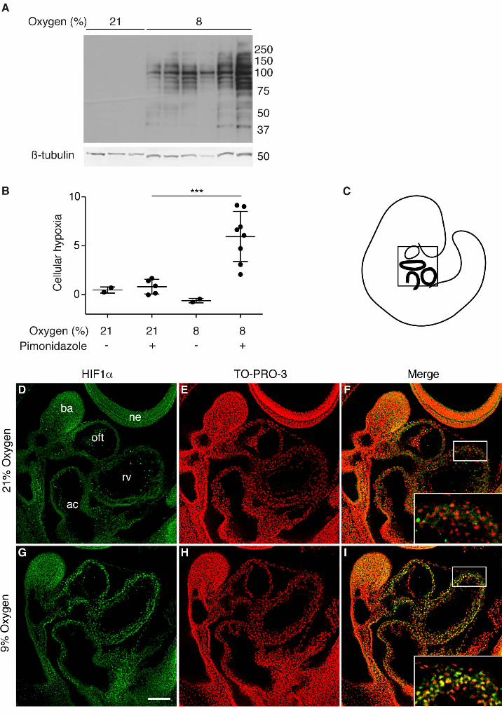
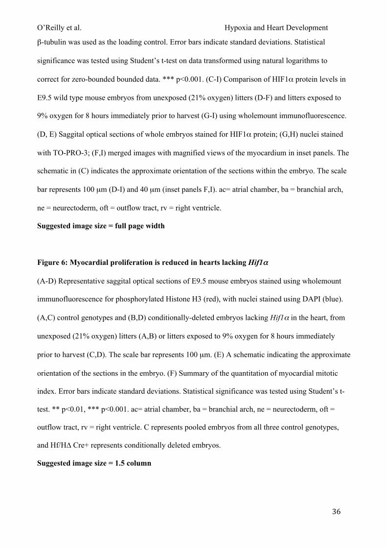
Figure 5: Maternal exposure to lowered oxygen levels causes embryonic hypoxia and
stabilization of HIF1αα
Quantification of hypoxyprobe™ adducts from E9.5 hearts of embryos exposed in utero to 8%
oxygen for 3 hours. (A) Representative western blot, and (B) summary of quantification of adducts.
O’Reilly et al. Hypoxia and Heart Development
36
β-tubulin was used as the loading control. Error bars indicate standard deviations. Statistical
significance was tested using Student’s t-test on data transformed using natural logarithms to
correct for zero-bounded bounded data. *** p<0.001. (C-I) Comparison of HIF1α protein levels in
E9.5 wild type mouse embryos from unexposed (21% oxygen) litters (D-F) and litters exposed to
9% oxygen for 8 hours immediately prior to harvest (G-I) using wholemount immunofluorescence.
(D, E) Saggital optical sections of whole embryos stained for HIF1α protein; (G,H) nuclei stained
with TO-PRO-3; (F,I) merged images with magnified views of the myocardium in inset panels. The
schematic in (C) indicates the approximate orientation of the sections within the embryo. The scale
bar represents 100 µm (D-I) and 40 µm (inset panels F,I). ac= atrial chamber, ba = branchial arch,
ne = neurectoderm, oft = outflow tract, rv = right ventricle.
Suggested image size = full page width
Figure 6: Myocardial proliferation is reduced in hearts lacking Hif1αα
(A-D) Representative saggital optical sections of E9.5 mouse embryos stained using wholemount
immunofluorescence for phosphorylated Histone H3 (red), with nuclei stained using DAPI (blue).
(A,C) control genotypes and (B,D) conditionally-deleted embryos lacking Hif1α in the heart, from
unexposed (21% oxygen) litters (A,B) or litters exposed to 9% oxygen for 8 hours immediately
prior to harvest (C,D). The scale bar represents 100 µm. (E) A schematic indicating the approximate
orientation of the sections in the embryo. (F) Summary of the quantitation of myocardial mitotic
index. Error bars indicate standard deviations. Statistical significance was tested using Student’s t-
test. ** p<0.01, *** p<0.001. ac= atrial chamber, ba = branchial arch, ne = neurectoderm, oft =
outflow tract, rv = right ventricle. C represents pooled embryos from all three control genotypes,
and Hf/H∆ Cre+ represents conditionally deleted embryos.
Suggested image size = 1.5 column
O’Reilly et al. Hypoxia and Heart Development
1
SUPPLEMENTAL FIGURE CAPTIONS
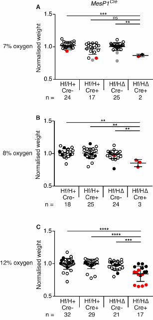
Supplemental Figure 1: Deletion of Hif1αα from mesoderm using MesP1Cre results in reduced
embryo and placental weight at E17.5
Graphs showing normalized weights of (A) embryos and (B) placentas at E17.5. Error bars indicate
standard deviations. Student’s t-test was used to compare the weights of conditionally-deleted
embryos and placentas to those of the three control genotypes. Embryos with heart defects are
shown in red, those with normal heart morphology are shown in black, and those that were not
analyzed for heart morphology are shown by empty icons. ns = not significant, * = p<0.05, **** =
p<0.0001. Hf/H+ Cre-, Hf/H+ Cre+ and Hf/HΔ Cre- represent the three control genotypes; and
Hf/H∆ Cre+ represents conditionally deleted embryos.
Supplemental Figure 2: Hif1αα deletion from vascular endothelial cells using Tek-Cre results
in reduced embryo and placental weight at E17.5
Graphs showing normalized weights of (A) embryos and (B) placentas at E17.5. Error bars indicate
standard deviations. Student’s t-test was used to compare the weight of conditionally-deleted
embryos and placentas to the three control genotypes. Embryos with heart defects are shown in red,
those with normal heart morphology are shown in black, and those that were not analyzed for heart
morphology are shown by empty icons. ** = p<0.01, *** = p<0.001. Hf/H+ Cre-, Hf/H+ Cre+ and
Hf/HΔ Cre- represent the three control genotypes; and Hf/H∆ Cre+ represents conditionally deleted
embryos.
Supplemental Figure 3: Hif1αα deletion from the cardiac progenitor cells using Nkx2-5IRESCre
results in reduced embryo and placental weight at E17.5
Graphs showing normalized weights of (A) embryos and (B) placentas at E17.5. Error bars indicate
standard deviations. Student’s t-test was used to compare the weight of conditionally-deleted
embryos and placentas to the three control genotypes. Embryos with heart defects are shown in red,
O’Reilly et al. Hypoxia and Heart Development
2
those with normal heart morphology are shown in black, those with normal heart morphology that
were analyzed by MRI alone are shown in grey, and those that were not analyzed for heart
morphology are shown by empty icons. ns = not significant, * = p<0.05, ** = p<0.01. Hf/H+ Cre-,
Hf/H+ Cre+ and Hf/HΔ Cre- represent the three control genotypes; and Hf/H∆ Cre+ represents
conditionally deleted embryos.
Supplemental Figure 4: Affects of maternal low oxygen exposure at E9.5 in the Hif1αα∆/flox
Mesp1Cre model
Graphs showing normalized weights of embryos at E17.5 after an eight hour E9.5 exposure to low
oxygen: (A) 7%, (B) 8% and (C) 12%. Error bars indicate standard deviations. Student’s t-test was
used to compare the weight of conditionally-deleted embryos and placentas to the three control
genotypes. Embryos with severe heart defects are shown in red, those with normal heart
morphology are shown in black, those with normal heart morphology that were analyzed by MRI
alone are shown in grey, and those that were not analyzed for heart morphology are shown by
empty icons. ns = not significant, ** = p<0.01, *** = p<0.001, **** = p<0.0001. Hf/H+ Cre-,
Hf/H+ Cre+ and Hf/HΔ Cre- represent the three control genotypes; and Hf/H∆ Cre+ represents
conditionally deleted embryos.
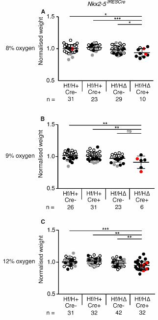
Supplemental Figure 5: Affects of maternal low oxygen exposure at E9.5 in the Hif1αα∆/flox
Nkx2-5IRESCre model
Graphs showing normalized weights of embryos at E17.5 after an eight hour maternal exposure to
low oxygen on E9.5: 8% (A), 9% (B) and 12% (C). Error bars indicate standard deviations.
Student’s t-test was used to compare the weight of conditionally-deleted embryos and placentas to
the three control genotypes. Embryos with severe heart defects are shown in red, those with normal
heart morphology are shown in black, those with normal heart morphology that were analyzed by
MRI alone are shown in grey, and those that were not analyzed for heart morphology are shown by
O’Reilly et al. Hypoxia and Heart Development
3
empty icons. ns = not significant, * = p<0.05, ** = p<0.01, *** = p<0.001. Hf/H+ Cre-, Hf/H+
Cre+ and Hf/HΔ Cre- represent the three control genotypes; and Hf/H∆ Cre+ represents
conditionally deleted embryos.
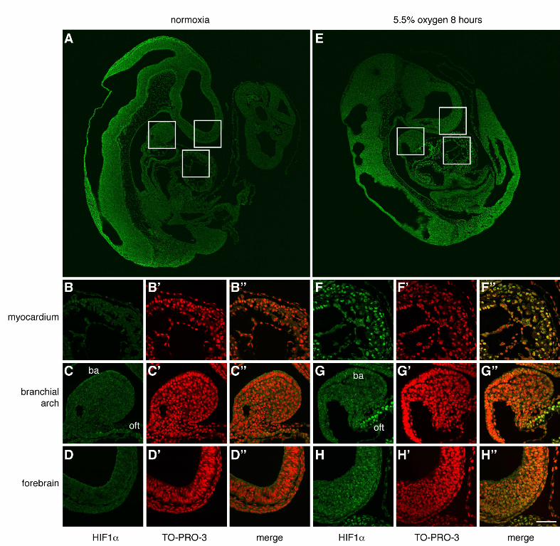
Supplemental Figure 6: Maternal exposure to extremely low oxygen levels causes nuclear
accumulation of HIF1αα predominantly in myocardium
Comparison of HIF1α protein levels in E9.5 wild type mouse embryos from unexposed (normoxia,
21% oxygen) litters (A-D) and litters exposed to 5.5% oxygen for 8 hours immediately prior to
harvest (E-H) using immunofluorescence on wax sections. (A,E) Saggital sections of whole
embryos stained for HIF1α protein, with white boxes indicating the locations of the magnified
views. (B-D and F-H) magnified views of myocardium, branchial arch and forebrain epithelium
stained for HIF1α protein; (B’-D’ and F’-H’) nuclei stained with TO-PRO-3; (B”-D” and F”-H”)
merged images. The scale bar represents 170 µm (A,E) and 65 µm (B-D and F-H). ba = branchial
arch, oft = outflow tract.
O’Reilly et al. Hypoxia and Heart Development
4
SUPPLEMENTAL TABLES
Supplemental Table 1
MesP1Cre Hif1αflox/+
Cre-
Hif1αflox/+
Cre+
Hif1α∆/flox
Cre-
Hif1α∆/flox
Cre+
p value
Embryos 44 35 35 17 0.008a
Hearts analyzed 8 11 9 17
Heart defects 1
CAVC
0 0 6
VSD, OA,
EC
0.008b
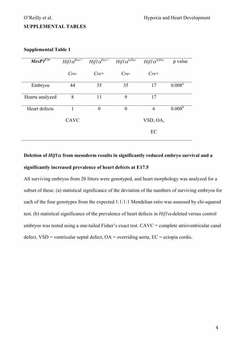
Deletion of Hif1αα from mesoderm results in significantly reduced embryo survival and a
significantly increased prevalence of heart defects at E17.5
All surviving embryos from 20 litters were genotyped, and heart morphology was analyzed for a
subset of these. (a) statistical significance of the deviation of the numbers of surviving embryos for
each of the four genotypes from the expected 1:1:1:1 Mendelian ratio was assessed by chi-squared
test. (b) statistical significance of the prevalence of heart defects in Hif1α-deleted versus control
embryos was tested using a one-tailed Fisher’s exact test. CAVC = complete atrioventricular canal
defect, VSD = ventricular septal defect, OA = overriding aorta, EC = ectopia cordis.
O’Reilly et al. Hypoxia and Heart Development
5
Supplemental Table 2
Tek-Cre Hif1αflox/+
Cre-
Hif1αflox/+
Cre+
Hif1α∆/flox
Cre-
Hif1α∆/flox
Cre+
p value
Embryos 10 7 10 13 0.62a
Hearts analyzed 2 4 4 13
Heart defects 0 0 0 2
VSD, OA,
DORV
0.31b
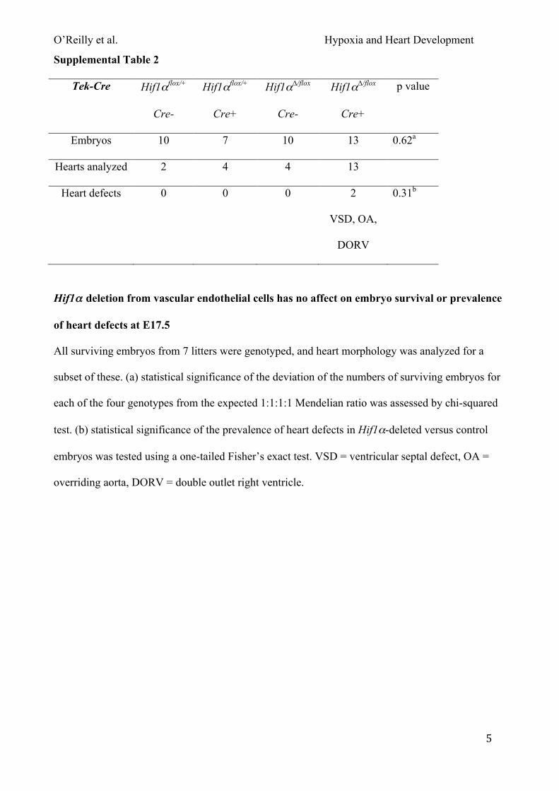
Hif1αα deletion from vascular endothelial cells has no affect on embryo survival or prevalence
of heart defects at E17.5
All surviving embryos from 7 litters were genotyped, and heart morphology was analyzed for a
subset of these. (a) statistical significance of the deviation of the numbers of surviving embryos for
each of the four genotypes from the expected 1:1:1:1 Mendelian ratio was assessed by chi-squared
test. (b) statistical significance of the prevalence of heart defects in Hif1α-deleted versus control
embryos was tested using a one-tailed Fisher’s exact test. VSD = ventricular septal defect, OA =
overriding aorta, DORV = double outlet right ventricle.
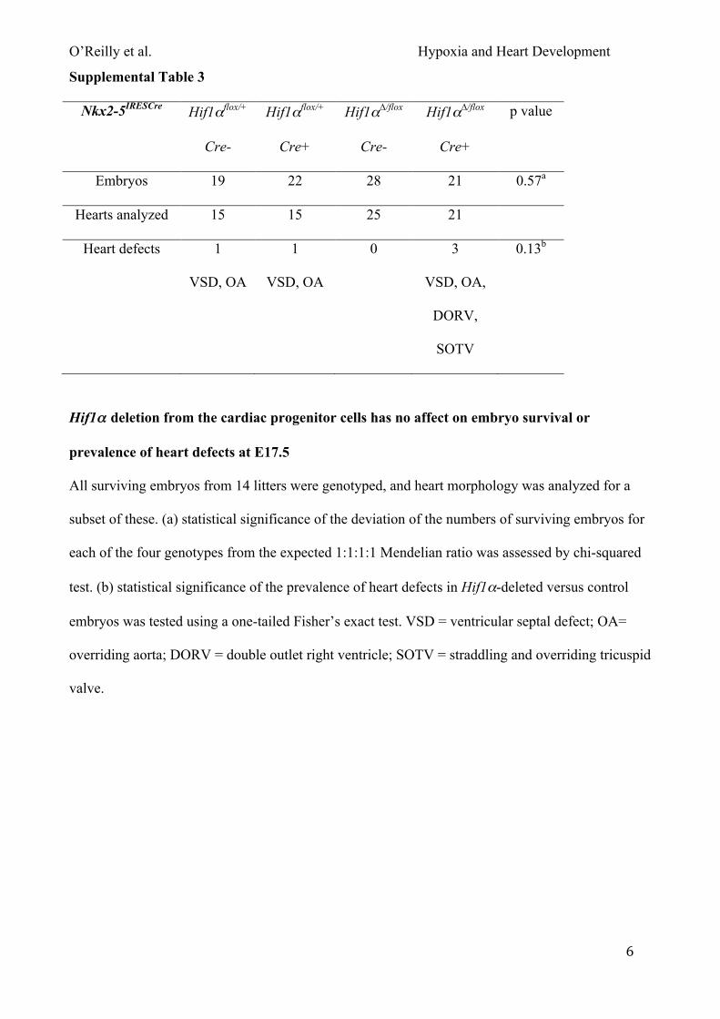
O’Reilly et al. Hypoxia and Heart Development
6
Supplemental Table 3
Nkx2-5IRESCre Hif1αflox/+
Cre-
Hif1αflox/+
Cre+
Hif1α∆/flox
Cre-
Hif1α∆/flox
Cre+
p value
Embryos 19 22 28 21 0.57a
Hearts analyzed 15 15 25 21
Heart defects 1
VSD, OA
1
VSD, OA
0 3
VSD, OA,
DORV,
SOTV
0.13b
Hif1αα deletion from the cardiac progenitor cells has no affect on embryo survival or
prevalence of heart defects at E17.5
All surviving embryos from 14 litters were genotyped, and heart morphology was analyzed for a
subset of these. (a) statistical significance of the deviation of the numbers of surviving embryos for
each of the four genotypes from the expected 1:1:1:1 Mendelian ratio was assessed by chi-squared
test. (b) statistical significance of the prevalence of heart defects in Hif1α-deleted versus control
embryos was tested using a one-tailed Fisher’s exact test. VSD = ventricular septal defect; OA=
overriding aorta; DORV = double outlet right ventricle; SOTV = straddling and overriding tricuspid
valve.
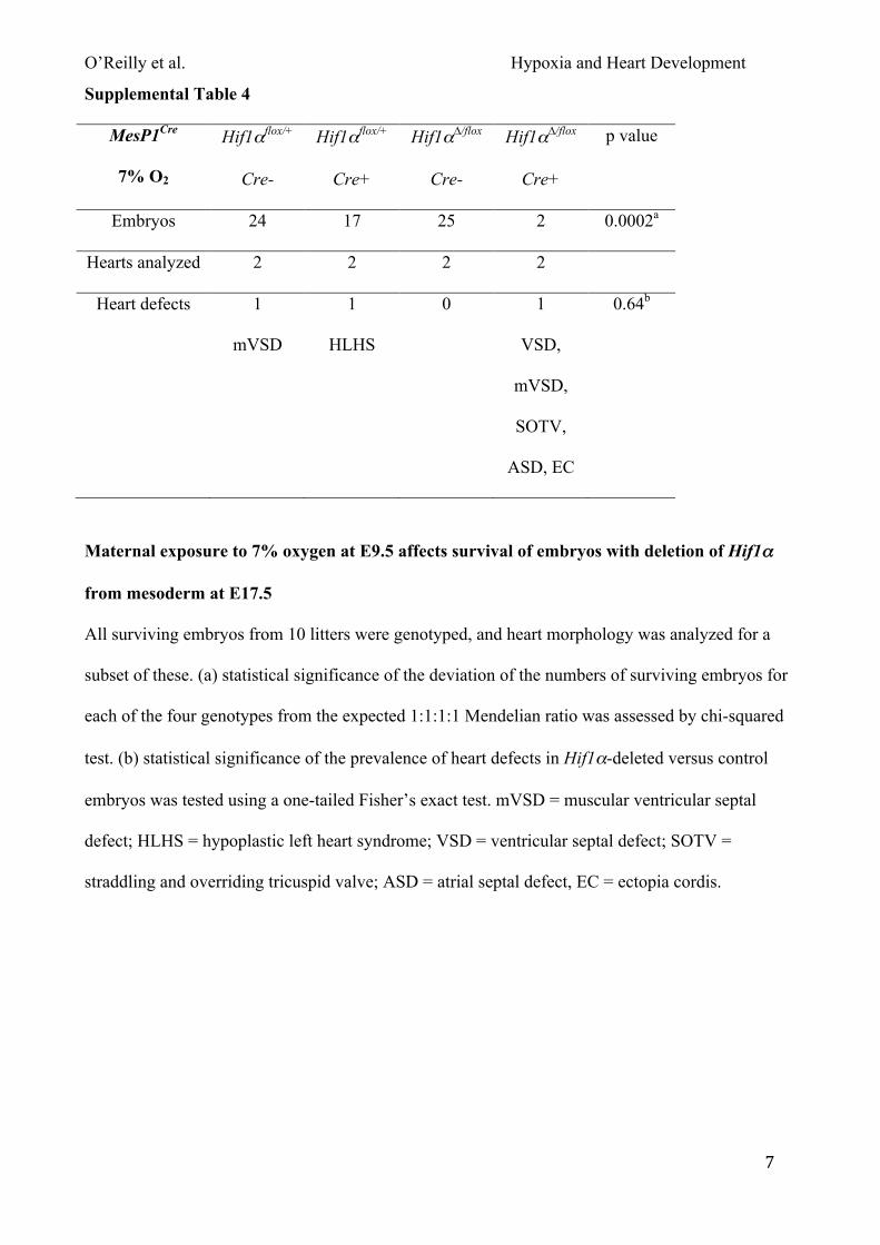
O’Reilly et al. Hypoxia and Heart Development
7
Supplemental Table 4
MesP1Cre
7% O2
Hif1αflox/+
Cre-
Hif1αflox/+
Cre+
Hif1α∆/flox
Cre-
Hif1α∆/flox
Cre+
p value
Embryos 24 17 25 2 0.0002a
Hearts analyzed 2 2 2 2
Heart defects 1
mVSD
1
HLHS
0 1
VSD,
mVSD,
SOTV,
ASD, EC
0.64b
Maternal exposure to 7% oxygen at E9.5 affects survival of embryos with deletion of Hif1αα
from mesoderm at E17.5
All surviving embryos from 10 litters were genotyped, and heart morphology was analyzed for a
subset of these. (a) statistical significance of the deviation of the numbers of surviving embryos for
each of the four genotypes from the expected 1:1:1:1 Mendelian ratio was assessed by chi-squared
test. (b) statistical significance of the prevalence of heart defects in Hif1α-deleted versus control
embryos was tested using a one-tailed Fisher’s exact test. mVSD = muscular ventricular septal
defect; HLHS = hypoplastic left heart syndrome; VSD = ventricular septal defect; SOTV =
straddling and overriding tricuspid valve; ASD = atrial septal defect, EC = ectopia cordis.
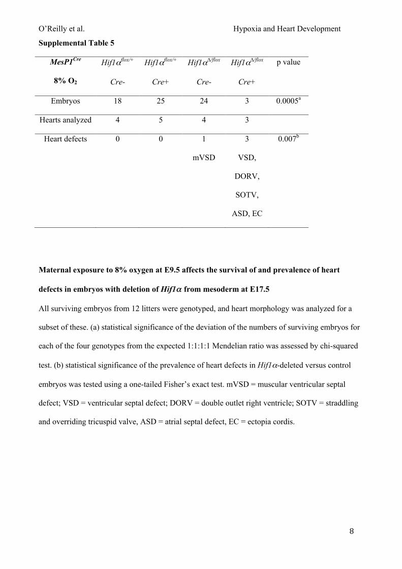
O’Reilly et al. Hypoxia and Heart Development
8
Supplemental Table 5
MesP1Cre
8% O2
Hif1αflox/+
Cre-
Hif1αflox/+
Cre+
Hif1α∆/flox
Cre-
Hif1α∆/flox
Cre+
p value
Embryos 18 25 24 3 0.0005a
Hearts analyzed 4 5 4 3
Heart defects 0 0 1
mVSD
3
VSD,
DORV,
SOTV,
ASD, EC
0.007b
Maternal exposure to 8% oxygen at E9.5 affects the survival of and prevalence of heart
defects in embryos with deletion of Hif1αα from mesoderm at E17.5
All surviving embryos from 12 litters were genotyped, and heart morphology was analyzed for a
subset of these. (a) statistical significance of the deviation of the numbers of surviving embryos for
each of the four genotypes from the expected 1:1:1:1 Mendelian ratio was assessed by chi-squared
test. (b) statistical significance of the prevalence of heart defects in Hif1α-deleted versus control
embryos was tested using a one-tailed Fisher’s exact test. mVSD = muscular ventricular septal
defect; VSD = ventricular septal defect; DORV = double outlet right ventricle; SOTV = straddling
and overriding tricuspid valve, ASD = atrial septal defect, EC = ectopia cordis.
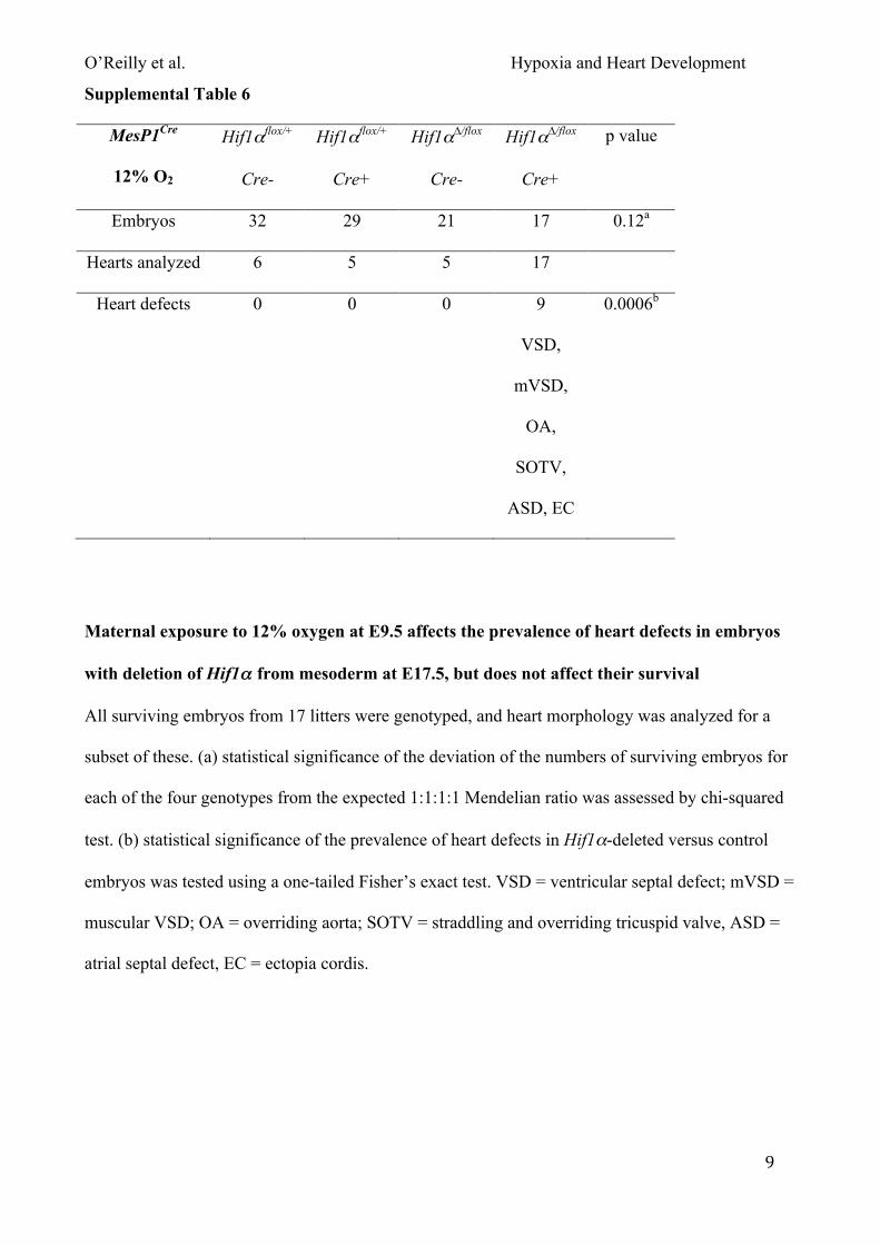
O’Reilly et al. Hypoxia and Heart Development
9
Supplemental Table 6
MesP1Cre
12% O2
Hif1αflox/+
Cre-
Hif1αflox/+
Cre+
Hif1α∆/flox
Cre-
Hif1α∆/flox
Cre+
p value
Embryos 32 29 21 17 0.12a
Hearts analyzed 6 5 5 17
Heart defects 0 0 0 9
VSD,
mVSD,
OA,
SOTV,
ASD, EC
0.0006b
Maternal exposure to 12% oxygen at E9.5 affects the prevalence of heart defects in embryos
with deletion of Hif1αα from mesoderm at E17.5, but does not affect their survival
All surviving embryos from 17 litters were genotyped, and heart morphology was analyzed for a
subset of these. (a) statistical significance of the deviation of the numbers of surviving embryos for
each of the four genotypes from the expected 1:1:1:1 Mendelian ratio was assessed by chi-squared
test. (b) statistical significance of the prevalence of heart defects in Hif1α-deleted versus control
embryos was tested using a one-tailed Fisher’s exact test. VSD = ventricular septal defect; mVSD =
muscular VSD; OA = overriding aorta; SOTV = straddling and overriding tricuspid valve, ASD =
atrial septal defect, EC = ectopia cordis.
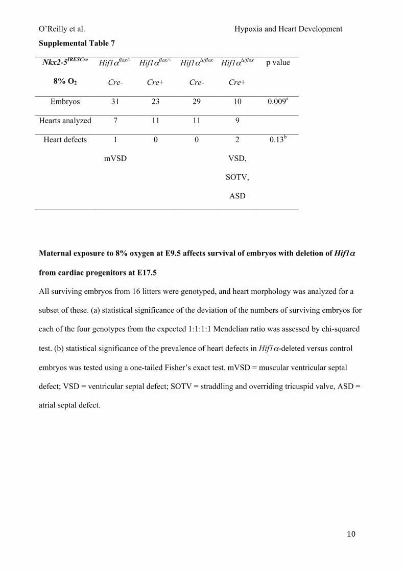
O’Reilly et al. Hypoxia and Heart Development
10
Supplemental Table 7
Nkx2-5IRESCre
8% O2
Hif1αflox/+
Cre-
Hif1αflox/+
Cre+
Hif1α∆/flox
Cre-
Hif1α∆/flox
Cre+
p value
Embryos 31 23 29 10 0.009a
Hearts analyzed 7 11 11 9
Heart defects 1
mVSD
0 0 2
VSD,
SOTV,
ASD
0.13b
Maternal exposure to 8% oxygen at E9.5 affects survival of embryos with deletion of Hif1αα
from cardiac progenitors at E17.5
All surviving embryos from 16 litters were genotyped, and heart morphology was analyzed for a
subset of these. (a) statistical significance of the deviation of the numbers of surviving embryos for
each of the four genotypes from the expected 1:1:1:1 Mendelian ratio was assessed by chi-squared
test. (b) statistical significance of the prevalence of heart defects in Hif1α-deleted versus control
embryos was tested using a one-tailed Fisher’s exact test. mVSD = muscular ventricular septal
defect; VSD = ventricular septal defect; SOTV = straddling and overriding tricuspid valve, ASD =
atrial septal defect.
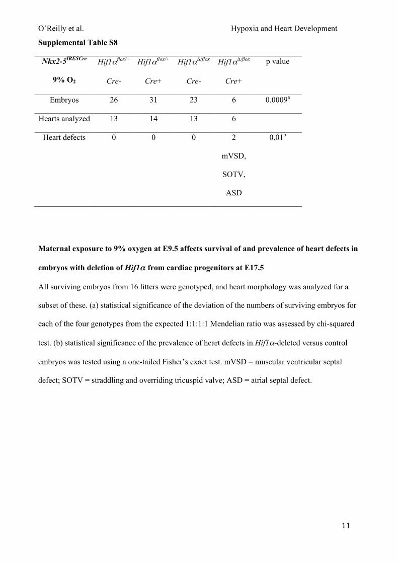
O’Reilly et al. Hypoxia and Heart Development
11
Supplemental Table S8
Nkx2-5IRESCre
9% O2
Hif1αflox/+
Cre-
Hif1αflox/+
Cre+
Hif1α∆/flox
Cre-
Hif1α∆/flox
Cre+
p value
Embryos 26 31 23 6 0.0009a
Hearts analyzed 13 14 13 6
Heart defects 0 0 0 2
mVSD,
SOTV,
ASD
0.01b
Maternal exposure to 9% oxygen at E9.5 affects survival of and prevalence of heart defects in
embryos with deletion of Hif1αα from cardiac progenitors at E17.5
All surviving embryos from 16 litters were genotyped, and heart morphology was analyzed for a
subset of these. (a) statistical significance of the deviation of the numbers of surviving embryos for
each of the four genotypes from the expected 1:1:1:1 Mendelian ratio was assessed by chi-squared
test. (b) statistical significance of the prevalence of heart defects in Hif1α-deleted versus control
embryos was tested using a one-tailed Fisher’s exact test. mVSD = muscular ventricular septal
defect; SOTV = straddling and overriding tricuspid valve; ASD = atrial septal defect.
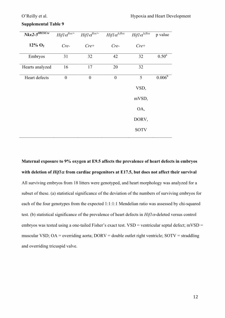
O’Reilly et al. Hypoxia and Heart Development
12
Supplemental Table 9
Nkx2-5IRESCre
12% O2
Hif1αflox/+
Cre-
Hif1αflox/+
Cre+
Hif1α∆/flox
Cre-
Hif1α∆/flox
Cre+
p value
Embryos 31 32 42 32 0.50a
Hearts analyzed 16 17 20 32
Heart defects 0 0 0 5
VSD,
mVSD,
OA,
DORV,
SOTV
0.006b
Maternal exposure to 9% oxygen at E9.5 affects the prevalence of heart defects in embryos
with deletion of Hif1αα from cardiac progenitors at E17.5, but does not affect their survival
All surviving embryos from 18 litters were genotyped, and heart morphology was analyzed for a
subset of these. (a) statistical significance of the deviation of the numbers of surviving embryos for
each of the four genotypes from the expected 1:1:1:1 Mendelian ratio was assessed by chi-squared
test. (b) statistical significance of the prevalence of heart defects in Hif1α-deleted versus control
embryos was tested using a one-tailed Fisher’s exact test. VSD = ventricular septal defect; mVSD =
muscular VSD; OA = overriding aorta; DORV = double outlet right ventricle; SOTV = straddling
and overriding tricuspid valve.
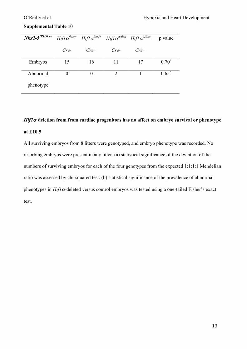
O’Reilly et al. Hypoxia and Heart Development
13
Supplemental Table 10
Nkx2-5IRESCre Hif1αflox/+
Cre-
Hif1αflox/+
Cre+
Hif1α∆/flox
Cre-
Hif1α∆/flox
Cre+
p value
Embryos 15 16 11 17 0.70a
Abnormal
phenotype
0 0 2 1 0.65b
Hif1αα deletion from from cardiac progenitors has no affect on embryo survival or phenotype
at E10.5
All surviving embryos from 8 litters were genotyped, and embryo phenotype was recorded. No
resorbing embryos were present in any litter. (a) statistical significance of the deviation of the
numbers of surviving embryos for each of the four genotypes from the expected 1:1:1:1 Mendelian
ratio was assessed by chi-squared test. (b) statistical significance of the prevalence of abnormal
phenotypes in Hif1α-deleted versus control embryos was tested using a one-tailed Fisher’s exact
test.
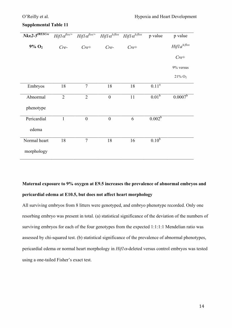
O’Reilly et al. Hypoxia and Heart Development
14
Supplemental Table 11
Nkx2-5IRESCre
9% O2
Hif1αflox/+
Cre-
Hif1αflox/+
Cre+
Hif1α∆/flox
Cre-
Hif1α∆/flox
Cre+
p value p value
Hif1a∆/flox
Cre+
9% versus
21% O2
Embryos 18 7 18 18 0.11a
Abnormal
phenotype
2 2 0 11 0.01b 0.0007b
Pericardial
edema
1 0 0 6 0.002b
Normal heart
morphology
18 7 18 16 0.10b
Maternal exposure to 9% oxygen at E9.5 increases the prevalence of abnormal embryos and
pericardial edema at E10.5, but does not affect heart morphology
All surviving embryos from 8 litters were genotyped, and embryo phenotype recorded. Only one
resorbing embryo was present in total. (a) statistical significance of the deviation of the numbers of
surviving embryos for each of the four genotypes from the expected 1:1:1:1 Mendelian ratio was
assessed by chi-squared test. (b) statistical significance of the prevalence of abnormal phenotypes,
pericardial edema or normal heart morphology in Hif1α-deleted versus control embryos was tested
using a one-tailed Fisher’s exact test.