Embed Size (px)
Citation preview
University of Groningen
FDG-PET/CT in staging and treatment of esophageal cancerSchreurs, Liesbeth Maria Antonia
IMPORTANT NOTE: You are advised to consult the publisher's version (publisher's PDF) if you wish to cite fromit. Please check the document version below.
Document VersionPublisher's PDF, also known as Version of record
Publication date:2014
Link to publication in University of Groningen/UMCG research database
Citation for published version (APA):Schreurs, L. M. A. (2014). FDG-PET/CT in staging and treatment of esophageal cancer Groningen: s.n.
CopyrightOther than for strictly personal use, it is not permitted to download or to forward/distribute the text or part of it without the consent of theauthor(s) and/or copyright holder(s), unless the work is under an open content license (like Creative Commons).
Take-down policyIf you believe that this document breaches copyright please contact us providing details, and we will remove access to the work immediatelyand investigate your claim.
Downloaded from the University of Groningen/UMCG research database (Pure): http://www.rug.nl/research/portal. For technical reasons thenumber of authors shown on this cover page is limited to 10 maximum.
Download date: 18-03-2018
46
Chapter 2
Better Assessment of Nodal Metastases by PET/CT
Fusion compared to side-by-side PET /CT in
Oesophageal Cancer
Liesbeth M.A. Schreurs
Bareld B. Pultrum
Klaas P. Koopmans
Christian C. Verhoef
Pieter L. Jager
Gooitzen M. van Dam
Henk Groen
Erik J. van der Jagt
John Th.M. Plukker
PET/CT fusion in staging esophageal cancer
47
Abstract
Background
Recently, positron emission tomography/computed tomography (PET/CT) has
been introduced in the staging of oesophageal cancer. The impact of PET/CT
fusion in comparison to side-by-side PET/CT in these tumours, was analyzed.
Patients and Methods
In 61 patients, 18-F-fluorodeoxyglucose (FDG)-PET and multidetector (md)-CT
were performed within a two week interval. Software-fusion of md-CT and FDG-
PET was correlated with side-by-side FDG-PET/CT reading by two independent
investigators. The gold standard was the pathological outcome or clinical
evidence of progression during the first year of follow-up.
Results
In 18 patients (18/61; 30%), nodal staging improved with software-fusion. The
number of nodal metastases increased in five patients and decreased in four
patients, leading to up-staging in one patient (2%) and down-staging in three
patients (5%). In nine cases (15%), certainty and localization of metastases
improved. However, the number of distant metastases did not change and
software-fusion did not have an influence on resectability.
Conclusion
PET/CT fusion substantially improves detection and localization of nodal
metastases and may have an impact on locoregional treatment options.
PET/CT fusion in staging esophageal cancer
48
Introduction
In cancer of the oesophagus and gastro-oesophageal junction (GOJ) curatively
intended resection, eventually with neoadjuvant chemoradiation, is the most
effective treatment option.1 As cure is only possible in the absence of distant
metastases or local invasion into vital surrounding structures, optimal staging
is indispensable for adequate preoperative patient selection preventing
unnecessary surgical exploration.
Endoscopic ultrasonography (EUS) in combination with fine needle aspiration
(FNA), multidetector computed tomography (md-CT), and ultrasound (US) of
the cervical region are commonly used staging methods.2 EUS-FNA is the most
accurate in detecting nodal involvement and gross mediastinal invasion,
whereas CT is the best imaging technique in detecting distant metastases.3 In
the last decade, positron emission tomography with 18-F-fluorodeoxyglucose
(FDG-PET) has become a frequently used staging technique.4-7 FDG-PET is
especially applied for the detection of regional lymph node and distant
metastases not visible on CT or EUS. Hence, pre-therapeutic FDG-PET alters
assessment of the tumour stage in 15-22% of the patients.8-12
Despite the improvements in preoperative staging, it is still difficult to determine
resectability accurately. Depending on the pre-operative work-up, local invasion and
distant metastases are found in 10-60% of patients during exploration.13-20 A
combination of PET/CT images is thought to increase significantly the accuracy of
staging because it correlates functional PET information with high resolution
anatomical CT results.21-26 Previously, it was common practice to correlate FDG-PET
with CT visually, but recently hybrid PET/CT or PET/CT fusion have gained more
support. In this study the accuracy of PET/CT fusion in staging patients with cancer
of the oesophagus or GOJ was evaluated and compared with visual correlation of
PET and md-CT.
PET/CT fusion in staging esophageal cancer
49
Patients and Methods
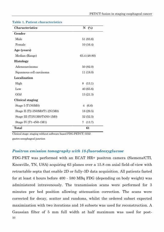
Patient characteristics
Retrospectively the medical records of 85 patients, who were staged by FDG-PET
and md-CT and treated for cancer of the oesophagus or GOJ between January 2001
and November 2004 were analyzed. Exclusion criteria were, treatment with
neoadjuvant chemoradiation or a history of other malignancies. Twenty-four
patients were excluded, either because the CT data from rural hospitals were
missing (n=10) or the CT-slices were too thick (n=14). All the other patients were
staged with EUS, 16-64 md-CT with slices of at least three mm and FDG-PET
within two weeks of the time of presentation. In these 61 patients, it was feasible to
perform a software-based PET/CT fusion. The mean age was 63.4 (SD±8.0; range 48-
80) years (Table 1). Fifty patients (82%) presented with an adenocarcinoma of the
oesophagus. Most of the tumours (87%) were localized in the distal part of the
oesophagus (n=40) or at the GOJ (n=13). Depending on tumour invasion and lymph
node involvement, the tumours were divided into stage I-IV according to the
tumour-node-metastasis (TMN) staging system of the Union Internationale Contre
Le Cancer (UICC).27
Computed tomography
Multidetector row CT imaging was performed with a 16-64 md-CT scanner
(Somatom Sensation, Siemens Medical Systems, Erlangen, Germany). The CT scans
(collimation 16 x 1.5 mm) were performed from the neck to the upper abdomen with
both intravenous and oral contrast. The reconstructed slices had a thickness of 3
mm with a 1.5 mm effective section thickness.
PET/CT fusion in staging esophageal cancer
50
Table 1. Patient characteristics
Characteristics N (%)
Gender
Male 51 (83.6)
Female 10 (16.4)
Age (years)
Median (Range) 63.4 (48-80)
Histology
Adenocarcinoma 50 (82.0)
Squamous cell carcinoma 11 (18.0)
Localization
High 8 (13.1)
Low 40 (65.6)
GOJ 13 (21.3)
Clinical staging
Stage I (T1N0M0) 4 (6.6)
Stage II (T2-3N0M0/T1-2N1M0) 18 (29.5)
Stage III (T3N1M0/T4N0-1M0) 32 (52.5)
Stage IV (T1-4N0-1M1) 7 (13.7)
Total 61
Clinical stage: staging without software based FDG-PET/CT, GOJ:
gastro-oesophageal junction
Positron emission tomography with 18-fluorodeoxyglucose
FDG-PET was performed with an ECAT HR+ positron camera (Siemens/CTI,
Knoxville, TN, USA) acquiring 63 planes over a 15.8 cm axial field-of-view with
retractable septa that enable 2D or fully-3D data acquisition. All patients fasted
for at least 4 hours before 400 - 580 MBq FDG (depending on body weight) was
administered intravenously. The transmission scans were performed for 3
minutes per bed position allowing attenuation correction. The scans were
corrected for decay, scatter and randoms, whilst the ordered subset expected
maximization with two iterations and 16 subsets was used for reconstruction. A
Gaussian filter of 5 mm full width at half maximum was used for post-
PET/CT fusion in staging esophageal cancer
51
smoothing of the reconstructed images.28 Data acquisition started in whole body
mode 90 minutes after injection, for 5 minutes per bed position from the crown
to the mid-femur.
Diagnostic evaluation of CT and PET findings
The images of the md-CT, EUS and FDG-PET techniques were reviewed
independently by two experienced nuclear physicians. Round hypo-dense lymph
nodes larger than 5 mm and lymph nodes measuring 10 mm or more on CT were
determined to be pathological. Pathological lymph nodes at the celiac axis were
classified as M1a metastases in the case of distal oesophageal cancer or as M1b
metastases in the mid or proximal tumours. Cervical metastases were classified as
M1a in the case of proximal cancer and as M1b for mid or distal tumours. The FDG-
uptake was scored on a four-point intensity scale: ‘normal’ (physiological), ‘slightly
increased’, ‘moderately increased’ and ‘intensely increased’. These lesions were
interpreted as: ‘absolutely benign’, ‘probably benign’, ‘indeterminate’, ‘probably
malignant’ and ‘definitely malignant’. All the ‘indeterminate’, ‘probably malignant’
and ‘definitely malignant’ lesions were identified as hotspots. Suspect lesions were
occasionally confirmed by FNA cytology, by pathological examination during or after
surgery, or by radiological and clinical follow-up of at least one year. The lymph
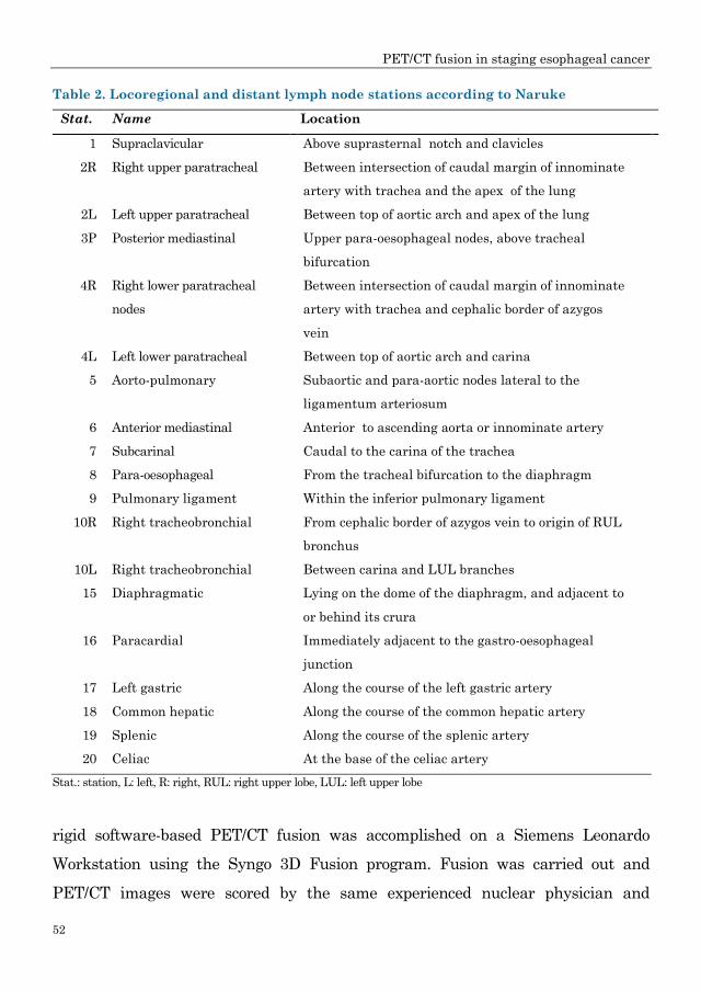
nodes were defined according to the Naruke lymph node stations (Table 2).
PET/CT fusion compared with side-by-side PET/CT reading
Together with an experienced radiologist all the reviewed FDG-PET and CT results
were visually correlated (side-by-side) and scored by the same nuclear medicine
physicians. Lymph nodes >1 cm on CT imaging without FDG-uptake on PET
imaging, were scored as negative on visually correlated FDG-PET/CT staging. The
PET/CT fusion in staging esophageal cancer
52
Table 2. Locoregional and distant lymph node stations according to Naruke
Stat. Name Location
1 Supraclavicular Above suprasternal notch and clavicles
2R Right upper paratracheal Between intersection of caudal margin of innominate
artery with trachea and the apex of the lung
2L Left upper paratracheal Between top of aortic arch and apex of the lung
3P Posterior mediastinal Upper para-oesophageal nodes, above tracheal
bifurcation
4R Right lower paratracheal
nodes
Between intersection of caudal margin of innominate
artery with trachea and cephalic border of azygos
vein
4L Left lower paratracheal Between top of aortic arch and carina
5 Aorto-pulmonary Subaortic and para-aortic nodes lateral to the
ligamentum arteriosum
6 Anterior mediastinal Anterior to ascending aorta or innominate artery
7 Subcarinal Caudal to the carina of the trachea
8 Para-oesophageal From the tracheal bifurcation to the diaphragm
9 Pulmonary ligament Within the inferior pulmonary ligament
10R Right tracheobronchial From cephalic border of azygos vein to origin of RUL
bronchus
10L Right tracheobronchial Between carina and LUL branches
15 Diaphragmatic Lying on the dome of the diaphragm, and adjacent to
or behind its crura
16 Paracardial Immediately adjacent to the gastro-oesophageal
junction
17 Left gastric Along the course of the left gastric artery
18 Common hepatic Along the course of the common hepatic artery
19 Splenic Along the course of the splenic artery
20 Celiac At the base of the celiac artery
Stat.: station, L: left, R: right, RUL: right upper lobe, LUL: left upper lobe
rigid software-based PET/CT fusion was accomplished on a Siemens Leonardo
Workstation using the Syngo 3D Fusion program. Fusion was carried out and
PET/CT images were scored by the same experienced nuclear physician and
PET/CT fusion in staging esophageal cancer
53
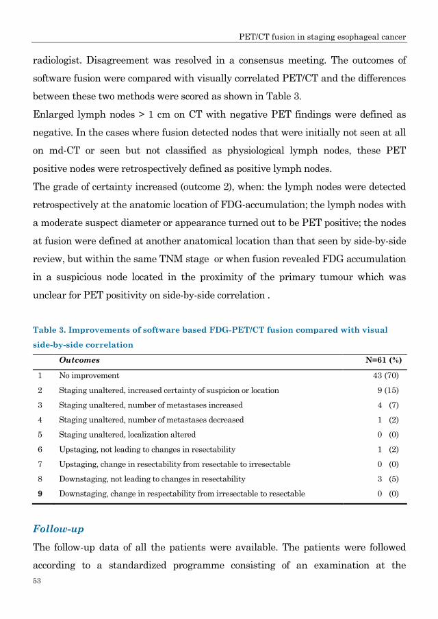
radiologist. Disagreement was resolved in a consensus meeting. The outcomes of
software fusion were compared with visually correlated PET/CT and the differences
between these two methods were scored as shown in Table 3.
Enlarged lymph nodes > 1 cm on CT with negative PET findings were defined as
negative. In the cases where fusion detected nodes that were initially not seen at all
on md-CT or seen but not classified as physiological lymph nodes, these PET
positive nodes were retrospectively defined as positive lymph nodes.
The grade of certainty increased (outcome 2), when: the lymph nodes were detected
retrospectively at the anatomic location of FDG-accumulation; the lymph nodes with
a moderate suspect diameter or appearance turned out to be PET positive; the nodes
at fusion were defined at another anatomical location than that seen by side-by-side
review, but within the same TNM stage or when fusion revealed FDG accumulation
in a suspicious node located in the proximity of the primary tumour which was
unclear for PET positivity on side-by-side correlation .
Table 3. Improvements of software based FDG-PET/CT fusion compared with visual
side-by-side correlation
Outcomes N=61 (%)
1 No improvement 43 (70)
2 Staging unaltered, increased certainty of suspicion or location 9 (15)
3 Staging unaltered, number of metastases increased 4 (7)
4 Staging unaltered, number of metastases decreased 1 (2)
5 Staging unaltered, localization altered 0 (0)
6 Upstaging, not leading to changes in resectability 1 (2)
7 Upstaging, change in resectability from resectable to irresectable 0 (0)
8 Downstaging, not leading to changes in resectability 3 (5)
9 Downstaging, change in respectability from irresectable to resectable 0 (0)
Follow-up
The follow-up data of all the patients were available. The patients were followed
according to a standardized programme consisting of an examination at the
PET/CT fusion in staging esophageal cancer
54
outpatient department every three months for the first two years and every six
months thereafter for a total period of five years.
Statistical analysis
Sensitivity, specificity and accuracy were calculated for both visual correlation and
PET/CT fusion. Both the techniques were compared in nonparametric paired
analysis using the McNemar test and p-values < 0.05 were considered statistically
significant.
PET/CT fusion in staging esophageal cancer
55
Results
In 18 patients (30%), an improvement in the detection of locoregional and/or distant
lymph node metastases was observed on fused PET/CT compared to visually
correlated CT and PET (Table 3). Details of these 18 patients are summarized in
Table 4.
Increased certainty of localization and number of metastases
In patients 1 to 9, the certainty of suspicious lymph nodes increased on PET/CT
fusion without altering the clinical nodal staging. In patients 1 and 2, enlarged
nodes were seen on md-CT in the paracardial region (patient 1) and near the left
gastric artery (patient 2). On side-by-side correlation it was not possible to
distinguish whether these nodes were PET positive or not, but fusion revealed FDG-
uptake in these nodes. In one patient (3), the lymph nodes were visible on CT at
Naruke 4/5 and 17 without FDG accumulation on primary PET review. However,
FDG-uptake was noted in two nodes after correction for the difference in height of
the diaphragm vault. Only two nodes (Naruke 4/5 and 20) were eventually
submitted for pathological examination as resection was abandoned because of
tumour invasion in the pericardium (T4 stage). In patient 4, two nodes both > 1 cm
(mean 1.6 x 1.1 ) at Naruke 16, did not show any FDG uptake, indicating a benign
enlargement. Fusion revealed that there was indeed nodal FDG-uptake but that it
was assimilated by FDG accumulation from the primary tumour. In patient 5, local
and distant lymph nodes were detected by CT, as well as skeletal and lung
metastases by PET. With precise anatomical correlation, PET/CT fusion could
identify exactly the Naruke stations that were involved. Cytological proof was taken
only from Naruke 18 and the six month clinical/radiological follow-up was taken as
validation. In patients 7 and 8 a small paracardial (patient 6) and para-oesophageal
node (patients 7) was missed on CT, although these lesions were suspected on PET
and EUS. In patients 8 and 9, PET, CT and EUS did not agree on the localization of
PET/CT fusion in staging esophageal cancer
56
suspected lymph nodes. PET/CT fusion enabled accurate identification and
localization in the four nodes.
Number of metastases altered, stage unaltered
In four patients (10 to 13), the total number of nodal metastases increased as more
pathological lymph nodes were observed on PET/CT fusion, though the TNM stage
remained unaltered. In patient 10, PET/CT fusion revealed lymph node metastases
close to the tumour (Naruke 8 and 16) which were not suspect on CT and were
thought to be primary tumour tissue at first PET diagnosis. In patient 12, an
initially missed node metastasis at Naruke 16 was detected on PET/CT fusion. Two
other enlarged lymph nodes >1 cm at Naruke 8 and 17 did not show any FDG-
uptake on visual correlation. In patient 13, no nodal involvement was observed on
PET scanning, but on PET/CT fusion there was indeed FDG- uptake in the para-
oesophageal lymph nodes.
In one patient (patient 14), the number of metastases decreased. CT detected three
lymph nodes > 1 cm, but on side-by-side fusion it was impossible to determine
whether these nodes were involved due to their close proximity. PET/CT fusion
clearly showed that one of these nodes did not absorb any FDG. Unfortunately, at
exploration, pathologically proven cervical metastasis was found and resection was
abandoned. Hence, PET/CT findings of enlarged lymph nodes at Naruke 8 were
verified by the 12-months follow-up.
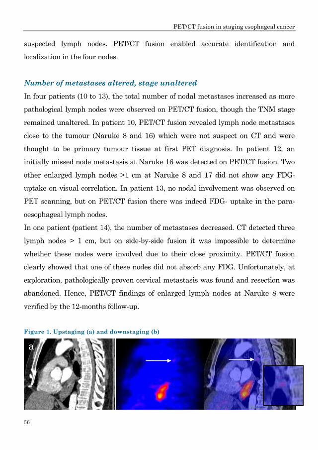
Figure 1. Upstaging (a) and downstaging (b)
a
PET/CT fusion in staging esophageal cancer
57
(a) Patient 15: FDG-PET, low suspicion for both nodal and skeletal metastases, but without suspicion on md-CT or
EUS. Software fusion clearly showed metastatic nodal metastasis in the para-oesophageal region. Magnification
shows the involved lymph node.
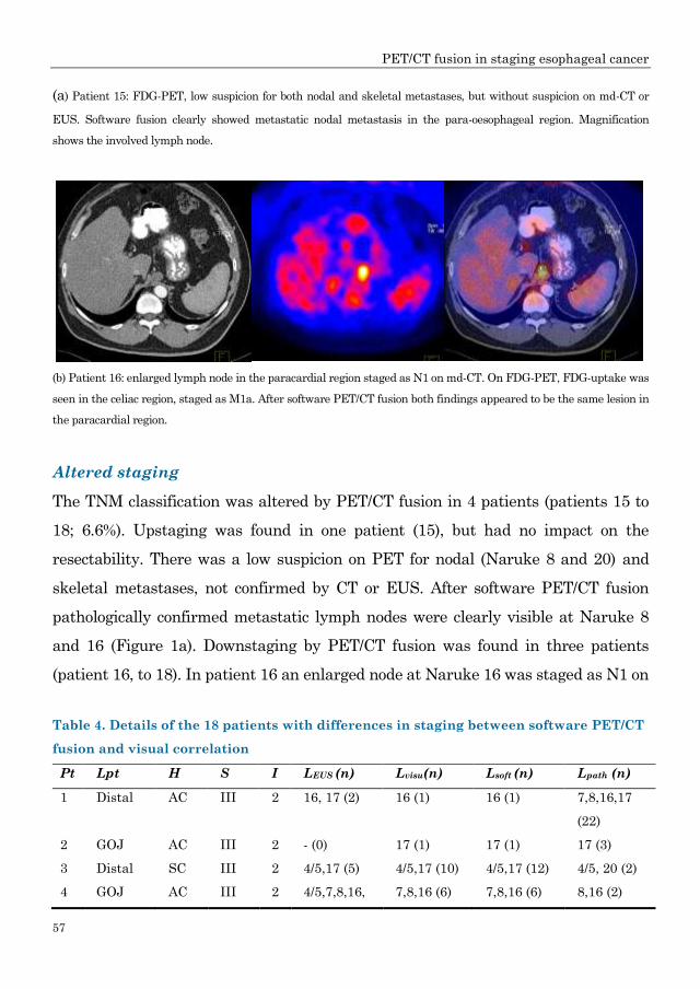
(b) Patient 16: enlarged lymph node in the paracardial region staged as N1 on md-CT. On FDG-PET, FDG-uptake was
seen in the celiac region, staged as M1a. After software PET/CT fusion both findings appeared to be the same lesion in
the paracardial region.
Altered staging
The TNM classification was altered by PET/CT fusion in 4 patients (patients 15 to
18; 6.6%). Upstaging was found in one patient (15), but had no impact on the
resectability. There was a low suspicion on PET for nodal (Naruke 8 and 20) and
skeletal metastases, not confirmed by CT or EUS. After software PET/CT fusion
pathologically confirmed metastatic lymph nodes were clearly visible at Naruke 8
and 16 (Figure 1a). Downstaging by PET/CT fusion was found in three patients
(patient 16, to 18). In patient 16 an enlarged node at Naruke 16 was staged as N1 on
Table 4. Details of the 18 patients with differences in staging between software PET/CT
fusion and visual correlation
Pt Lpt H S I LEUS (n) Lvisu(n) Lsoft (n) Lpath (n)
1 Distal AC III 2 16, 17 (2) 16 (1) 16 (1) 7,8,16,17
(22)
2 GOJ AC III 2 - (0) 17 (1) 17 (1) 17 (3)
3 Distal SC III 2 4/5,17 (5) 4/5,17 (10) 4/5,17 (12) 4/5, 20 (2)
4 GOJ AC III 2 4/5,7,8,16, 7,8,16 (6) 7,8,16 (6) 8,16 (2)
b
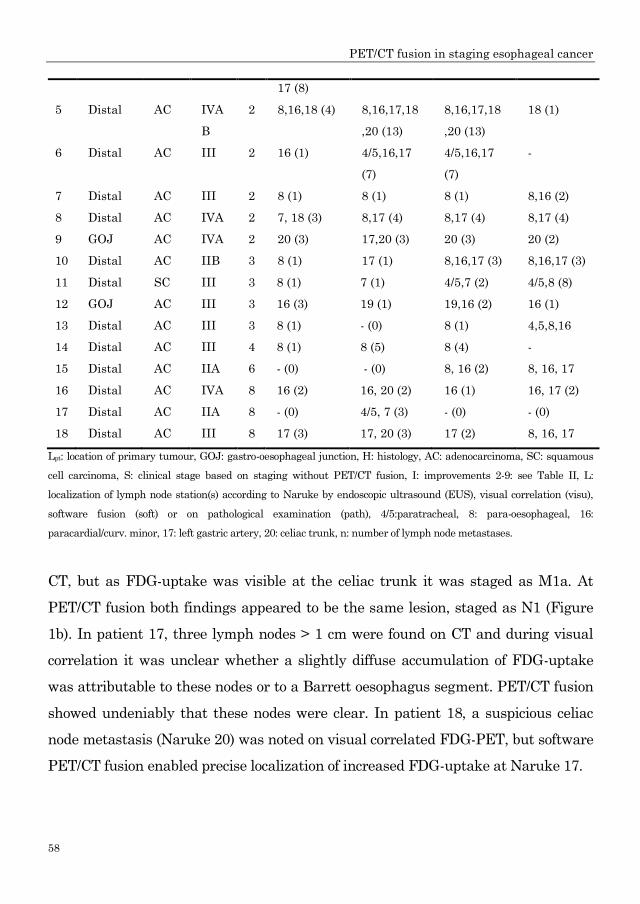
PET/CT fusion in staging esophageal cancer
58
17 (8)
5 Distal AC IVA
B
2 8,16,18 (4) 8,16,17,18
,20 (13)
8,16,17,18
,20 (13)
18 (1)
6 Distal AC III 2 16 (1) 4/5,16,17
(7)
4/5,16,17
(7)
-
7 Distal AC III 2 8 (1) 8 (1) 8 (1) 8,16 (2)
8 Distal AC IVA 2 7, 18 (3) 8,17 (4) 8,17 (4) 8,17 (4)
9 GOJ AC IVA 2 20 (3) 17,20 (3) 20 (3) 20 (2)
10 Distal AC IIB 3 8 (1) 17 (1) 8,16,17 (3) 8,16,17 (3)
11 Distal SC III 3 8 (1) 7 (1) 4/5,7 (2) 4/5,8 (8)
12 GOJ AC III 3 16 (3) 19 (1) 19,16 (2) 16 (1)
13 Distal AC III 3 8 (1) - (0) 8 (1) 4,5,8,16
14 Distal AC III 4 8 (1) 8 (5) 8 (4) -
15 Distal AC IIA 6 - (0) - (0) 8, 16 (2) 8, 16, 17
16 Distal AC IVA 8 16 (2) 16, 20 (2) 16 (1) 16, 17 (2)
17 Distal AC IIA 8 - (0) 4/5, 7 (3) - (0) - (0)
18 Distal AC III 8 17 (3) 17, 20 (3) 17 (2) 8, 16, 17
Lpt: location of primary tumour, GOJ: gastro-oesophageal junction, H: histology, AC: adenocarcinoma, SC: squamous
cell carcinoma, S: clinical stage based on staging without PET/CT fusion, I: improvements 2-9: see Table II, L:
localization of lymph node station(s) according to Naruke by endoscopic ultrasound (EUS), visual correlation (visu),
software fusion (soft) or on pathological examination (path), 4/5:paratracheal, 8: para-oesophageal, 16:
paracardial/curv. minor, 17: left gastric artery, 20: celiac trunk, n: number of lymph node metastases.
CT, but as FDG-uptake was visible at the celiac trunk it was staged as M1a. At
PET/CT fusion both findings appeared to be the same lesion, staged as N1 (Figure
1b). In patient 17, three lymph nodes > 1 cm were found on CT and during visual
correlation it was unclear whether a slightly diffuse accumulation of FDG-uptake
was attributable to these nodes or to a Barrett oesophagus segment. PET/CT fusion
showed undeniably that these nodes were clear. In patient 18, a suspicious celiac
node metastasis (Naruke 20) was noted on visual correlated FDG-PET, but software
PET/CT fusion enabled precise localization of increased FDG-uptake at Naruke 17.
PET/CT fusion in staging esophageal cancer
59
Sensitivity, specificity and accuracy
Although not statistically significant (p=0.250), the diagnostic accuracy of
PET/CT fusion (87%; 53/61) was slightly better than side-by-side visualization
(82%; 50/61) in the assessment of locoregional metastases. Sensitivity and
specificity of side-by-side visualization were 80% (12/15) and 83% (38/46),
respectively. The sensitivity and specificity of PET/CT fusion in the assessment
of nodal metastases were both 87%; 13/15 and 40/46, respectively.
PET/CT fusion in staging esophageal cancer
60
Discussion
This study showed that software-fused PET/CT had a supplementary value over
visually correlated FDG-PET and md-CT in the assessment of nodal metastases in
30% of the patients with cancer of the oesophagus. Improved assessment of
locoregional tumour foci is necessary for appropriate surgical treatment, but also for
more accurately planned target volumes, particularly in the neoadjuvant
chemoradiation treatment of these tumours.29,30 Refinement of the nodal assessment
was found by PET/CT fusion compared to the side-by-side visualisation method,
even though the N-stage itself was not significantly affected. It is this refinement
that is of major importance in radiotherapy planning.
However, there are some potential pitfalls in the interpretation of PET/CT fusion
images. Although md-CT has a high accuracy in detecting enlarged lymph nodes, its
specificity for metastases is low. Previous studies showed that lymph nodes of > 1
cm on CT without FDG-uptake on PET are usually benign.23,31,32 Visual correlation
between PET and CT is usually sufficient to determine this difference. Therefore,
improvements in staging were not taken into account when summarizing
improvements of software fusion compared to visual fusion. This statement should
be interpreted with caution as it was difficult to visualize the regional lymph nodes
near the primary tumour. In many cases, FDG-uptake in the primary tumour may
mask nearby lesions, due to the assimilation of FDG-uptake in both. The para-
oesophageal and paracardial areas are particularly difficult to interpret in this way.
The lymph nodes can be categorized only as benign on the aforementioned criteria
when they are not in the proximity of the primary tumour. Software fusion can be
helpful in identifying whether these lymph nodes are located near the primary
tumour. Another pitfall in the determination of nodal metastases on PET/CT fusion
is the difficulty in exact pathological localization. Only meticulous marking during
surgery with mapping of all visible or palpable nodes region by region in the
resected specimen according to the Japanese method makes correct identification
possible.
PET/CT fusion in staging esophageal cancer
61
There are also some inherent difficulties in software PET/CT fusion. Firstly, the
outlining in software fusion depends, to a certain extent, on human expertise and
appraisal, as does its evaluation. Therefore, small inter-observer variation is
inherent to this kind of science.31 To overcome this problem in the present study, all
the fusion images were studied and scored separately by an experienced nuclear
physician and a radiologist. Disagreements were solved in a consensus meeting.
Secondly, the software-fused images consist of two images from two different
techniques at different times. Consequently body posture and position differ
between the md-CT and PET. Fortunately, structures close to the spine, like the
oesophagus, show only minimal movements and are therefore very suitable for
fusion, though the position of the diaphragm often does not match as md-CT is
conducted at maximum inspiration and PET during moderate respiration.
Therefore, it is difficult to fuse PET and CT images below the vault of the
diaphragm. Some authors have reported a failure of 30-39% in software PET/CT
fusion of the evaluated cases.32,33 However, these studies also described an increased
success rate if the PET transmission data were incorporated in the fusion process for
attenuation correction.
Recently hybrid PET/CT scanners have become available. The use of hybrid
scanners partly overcomes these above mentioned limitations because PET and md-
CT are performed simultaneously, in the same body posture and nearly at the same
diaphragm position. Several studies comparing hybrid PET/CT with visually
correlated FDG-PET/CT have reported an improvement of 22-49% in the detection,
localization and characterization of malignant lesions with an accuracy of 90-96%.21-
23,34-36 Nevertheless, hybrid FDG-PET/CT scanners still consist of two separate
scanners in one combined device, and difficulties may occur in the application of
these scanners. The quality of the md-CT scan as part of a hybrid scan is frequently
inferior to the quality of a separate md-CT scan, because md-CT is primarily based
on anatomical mapping for precise localization of structures for FDG-PET.
Additionally, oral contrast fluid is not administered, accurate imaging of pulmonary
PET/CT fusion in staging esophageal cancer
62
and hepatic lesions might be problematic due to respiratory movements and timing
for arterial/venous imaging are not optimal. Furthermore, as earlier research has
revealed no benefit of FDG-PET in stage I and II disease, hybrid scanning seems to
be of limited value compared to md-CT in this group of patients.37 It is a relatively
expensive investigation used in a whole population, while PET scanning will have
no additional value in some subgroups.
In conclusion, fusion of FDG-PET and md-CT images improves the detection and/or
localization of locoregional metastases in oesophageal cancer cases with a more
accurate differentiation between physiological and pathological lesions.
Acknowledgements
This study was partially supported by a ZonMw program for Health Care Efficiency
Research.
PET/CT fusion in staging esophageal cancer
63
Reference List
(1) Parkin DM: Global cancer statistics in the year 2000. Lancet Oncol 2 (9): 533-543, 2001.
(2) Patel AN and Buenaventura PO: Current staging of esophageal carcinoma. Surg Clin
North Am 85 (3): 555-567, 2005.
(3) Mariette C, Balon JM, Maunoury V, Taillier G, Van Seuningen I and Triboulet JP:
Value of endoscopic ultrasonography as a predictor of long-term survival in oesophageal
carcinoma. Br J Surg 90 (11): 1367-1372, 2003.
(4) Flamen P, Lerut T, Haustermans K, Van Cutsem E, Mortelmans L. Position of positron
emission tomography and other imaging diagnostic modalities in esophageal cancer. Q J
Nucl Med Mol Imaging 48 (2): 96-108, 2004.
(5) Kato H, Miyazaki T, Nakajima M et al : The incremental effect of positron emission
tomography on diagnostic accuracy in the initial staging of esophageal carcinoma.
Cancer 103 (1): 148-156, 2005.
(6) Meltzer CC, Luketich JD, Friedman D et al: Whole-body FDG positron emission
tomographic imaging for staging esophageal cancer - comparison with computed
tomography. Clin Nucl Med 25 (11): 882-887, 2000.
(7) van Westreenen HL, Westerterp M, Bossuyt PMM et al: Systematic review of the
staging performance of F-18-fluorodeoxyglucose positron emission tomography in
esophageal cancer. J Clin Oncol 22 (18): 3805-3812, 2004.
(8) Block MI, Patterson GA, Sundaresan RS et al: Improvement in staging of esophageal
cancer with the addition of positron emission tomography. Ann Thorac Surg 64 (3): 770-
776, 1997.
(9) Flamen P, Lerut A, van Custem E et al: The utility of positron emission tomography for
the diagnosis and staging of recurrent esophageal cancer. J Thorac Cardiovasc Surg 120
(6): 1085-1092, 2000.
(10) Heeren PAM, Jager PL, Bongaerts F, van Dullemen H, Sluiter W and Plukker JTM:
Detection of distant metastases in esophageal cancer with F-18-FDG PET. J Nucl Med
45 (6): 980-987, 2004.
(11) Kole AC, Plukker JT, Nieweg OE and Vaalburg W: Positron emission tomography for
staging of oesophageal and gastroesophageal malignancy. Br J Cancer 78 (4): 521-527,
1998.
PET/CT fusion in staging esophageal cancer
64
(12) Luketich JD, Friedman DM, Weigel TL et al: Evaluation of distant metastases in
esophageal cancer: 100 consecutive positron emission tomography scans. Ann Thorac
Surg 68 (4): 1133-1136, 1999.
(13) Clements DM, Bowrey DJ and Havard TJ: The role of staging investigations for
oesophago-gastric carcinoma. Eur J Surg Oncol 30 (3): 309-312, 2004.
(14) Ellis FH, Heatley GJ, Krasna MJ, Williamson WA and Balogh K: Esophagogastrectomy
for carcinoma of the esophagus and cardia: A comparison of findings and results after
standard resection in three consecutive eight-year intervals with improved staging
criteria. J Thorac and Cardiovasc Surg 113 (5): 836-846, 1997.
(15) Hulscher JBF, van Dijkum EJMN, de Wit LT et al: Laparoscopy and laparoscopic
ultrasonography in staging carcinoma of the gastric cardia. Eur J Surg 166 (11): 862-
865, 2000.
(16) Menon KV and Dehn TCB: Multiport staging laparoscopy in esophageal and cardiac
carcinoma. Dis Esophagus 16 (4): 295-300, 2003.
(17) Sagar PM, Gauperaa T, Sueling H, Mcmahon MJ and Johnston D: An audit of the
treatment of cancer of the esophagus. Gut 35 (7): 941-945, 1994.
(18) Sariego J, Mosher S, Byrd M, Matsumoto T and Kerstein M: Prediction of outcome in
resectable esophageal-carcinoma. J Surg Oncol 54 (4): 223-225, 1993.
(19) Sondenaa K, Skaane P, Nygaard K and Skjennald A: Value of computed-tomography in
preoperative evaluation of resectability and staging in esophageal-carcinoma. Eur J
Surg 158 (10): 537-540, 1992.
(20) van Westreenen HL, Heeren PAM, van Dullemen HM et al: Positron emission
tomography with F-18-fluorodeoxyglucose in a combined staging strategy of esophageal
cancer prevents unnecessary surgical explorations. J Gastrointest Surg 9 (1): 54-61,
2005.
(21) Bar-Shalom R, Yefremov N, Guralnik L et al: Clinical performance of PET/CT in
evaluation of cancer: additional value for diagnostic Imaging and patient management.
J Nucl Med 44 (8): 1200-1209, 2003.
(22) Bar-Shalom R, Guralnik L, Tsalic M et al: The additional value of PET/CT over PET in
FDG imaging of oesophageal cancer. Eur J Nucl Med Mol Imaging 32 (8): 918-924, 2005.
PET/CT fusion in staging esophageal cancer
65
(23) Jadvar H, Henderson RW and Conti PS: 2-deoxy-2-[F-18]fluoro-D-glucose-positron
emission tomography/computed tomography imaging evaluation of esophageal cancer.
Mol Imaging Biol 8 (3): 193-200, 2006.
(24) Munden RF, Macapinlac HA and Erasmus JJ: Esophageal cancer: the role of integrated
CT-PET in initial staging and response assessment 333after preoperative therapy. J
Thorac Imaging 21 (2): 137-145, 2006.
(25) Rampin L, Rubello D, Nanni C and Fanti S: Value of PET-CT fusion imaging in avoiding
potential pitfalls in the interpretation of F-18-FDG accumulation in the distal
oesophagus. Eur J Nucl Med Mol Imaging 32 (8): 990-992, 2005.
(26) Antoch G, Saoudi N, Kuehl H et al: Accuracy of whole-body dual-modality fluorine-18-2-
fluoro-2-deoxy-D-glucose positron emission tomography and computed tomography
(FDG-PET/CT) for tumor staging in solid tumors: comparison with CT and PET. J Clin
Oncol 22 (21): 4357-4368, 2004.
(27) Sobin LH and Fleming ID: TNM classification of malignant tumors, fifth edition (1997).
Cancer 80 (9): 1803-1804, 1997.
(28) Lonneux M, Borbath I, Bol A et al: Attenuation correction in whole-body FDG
oncological studies: the role of statistical reconstruction. Eur J Nucl Med 26 (6): 591-598,
1999.
(29) Leong T, Everitt C, Yuen K et al: A prospective study to evaluate the impact of FDG-
PET on CT-based radiotherapy treatment planning for oesophageal cancer. Radiother
Oncol 78 (3): 254-61, 2006.
(30) Moureau-Zabotto L, Touboul E, Lerouge D et al: Impact of CT and 18F-deoxyglucose
positron emission tomography image fusion for conformal radiotherapy in esophageal
carcinoma. Int J Radiat Oncol Biol Phys 63 (2): 340-345, 2005.
(31) Delbeke D, Coleman RE, Guiberteau MJ et al: Procedure guideline for tumor imaging
with F-18-FDG PET/CT 1.0. J Nucl Med 47 (5): 885-895, 2006.
(32) Allen-Auerbach M, Quon A, Weber WA et al: Comparison between 2-deoxy-2-[F-
18]fluoro-D-Glucose positron emission tomography and positron emission
tomography/computed tomography hardware fusion for staging of patients with
lymphoma. Mol Imaging Biol 6 (6): 411-416, 2004.
PET/CT fusion in staging esophageal cancer
66
(33) Slomka PJ, Dey D, Przetak C, Aladl UE and Baum RP: Automated 3-dimensional
registration of stand-alone F-18-FDG whole-body PET with CT. J Nucl Med 44 (7): 1156-
1167, 2003.
(34) Cerfolio RJ, Ojha B, Bryant AS, Raghuveer V, Mountz JM and Bartolucci AA: The
accuracy of integrated PET-CT compared with dedicated PET alone for the staging of
patients with nonsmall cell lung cancer. Ann Thorac Surg 78 (3): 1017-1023, 2004.
(35) Charron M, Beyer T, Bohnen NN et al: Image analysis in patients with cancer studied
with a combined PET and CT scanner. Clin Nucl Med 25 (11): 905-910, 2000.
(36) Townsend DW, Beyer T, Blodgett TM: PET/CT scanners: a hardware approach to image
fusion. Semin Nucl Med 33 (3): 193-204, 2003.
(37) Westreenen HL: Positron emission tomography in staging of oesophageal cancer [thesis]
Groningen; University of Groningen 2005.