Embed Size (px)
DESCRIPTION
manual lens meter, trial lens set,trial frames,cross cylinder, flippers,visual charts,pupil distance meter check us at www.intercomoptic.com contact us at [email protected]
Citation preview
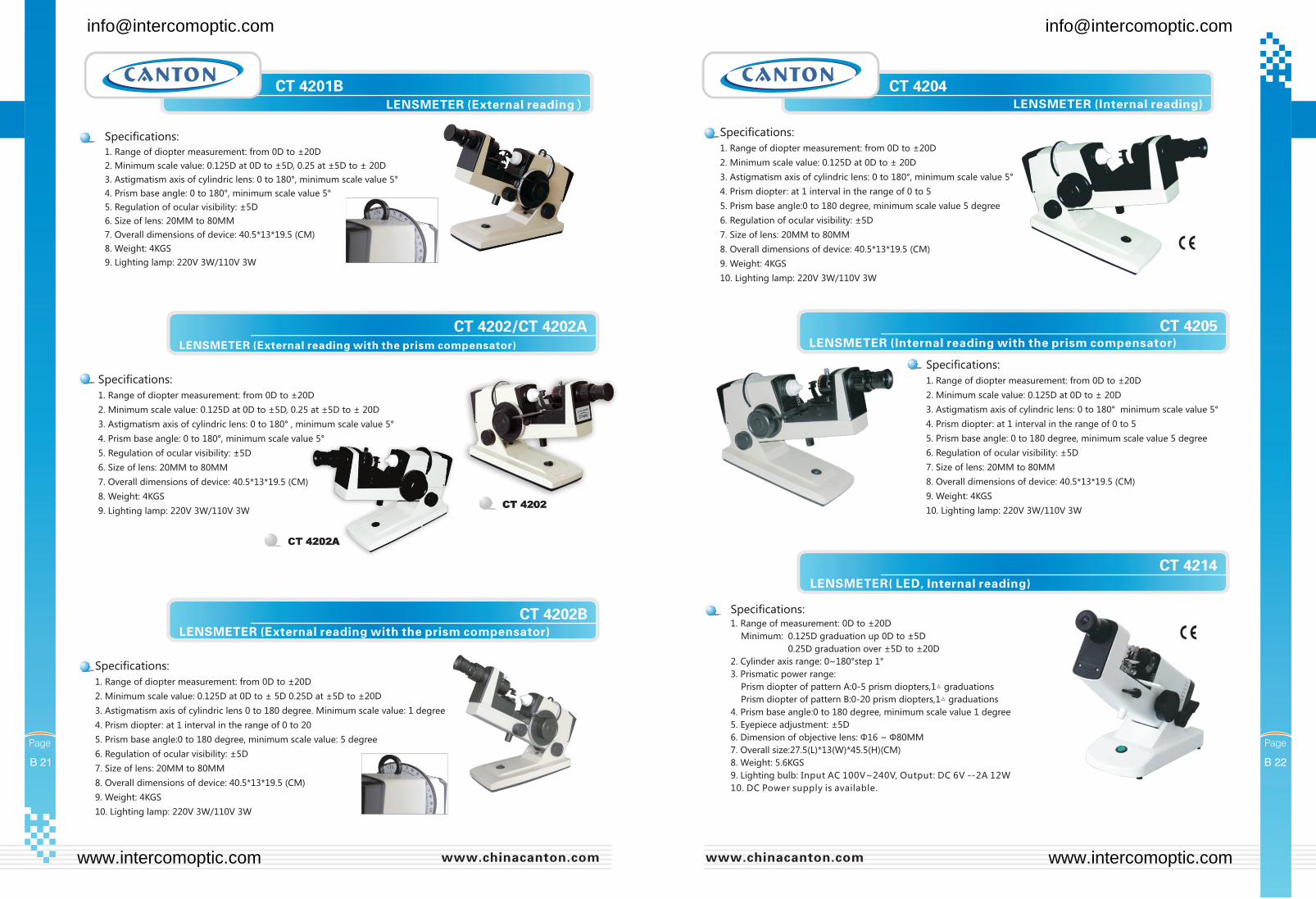
LENSMETER( LED, Internal reading)
[email protected] [email protected]
www.intercomoptic.com www.intercomoptic.com
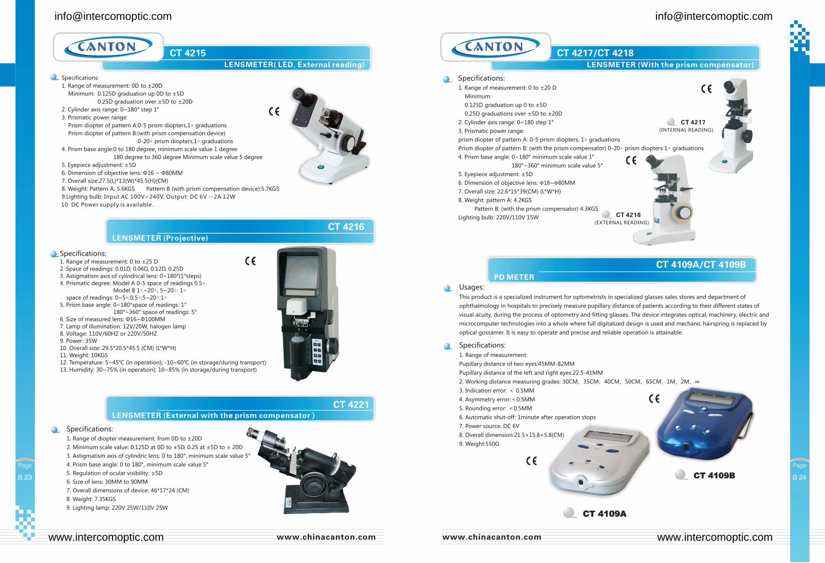
LENSMETER( LED, External reading)
[email protected] [email protected]
www.intercomoptic.com www.intercomoptic.com
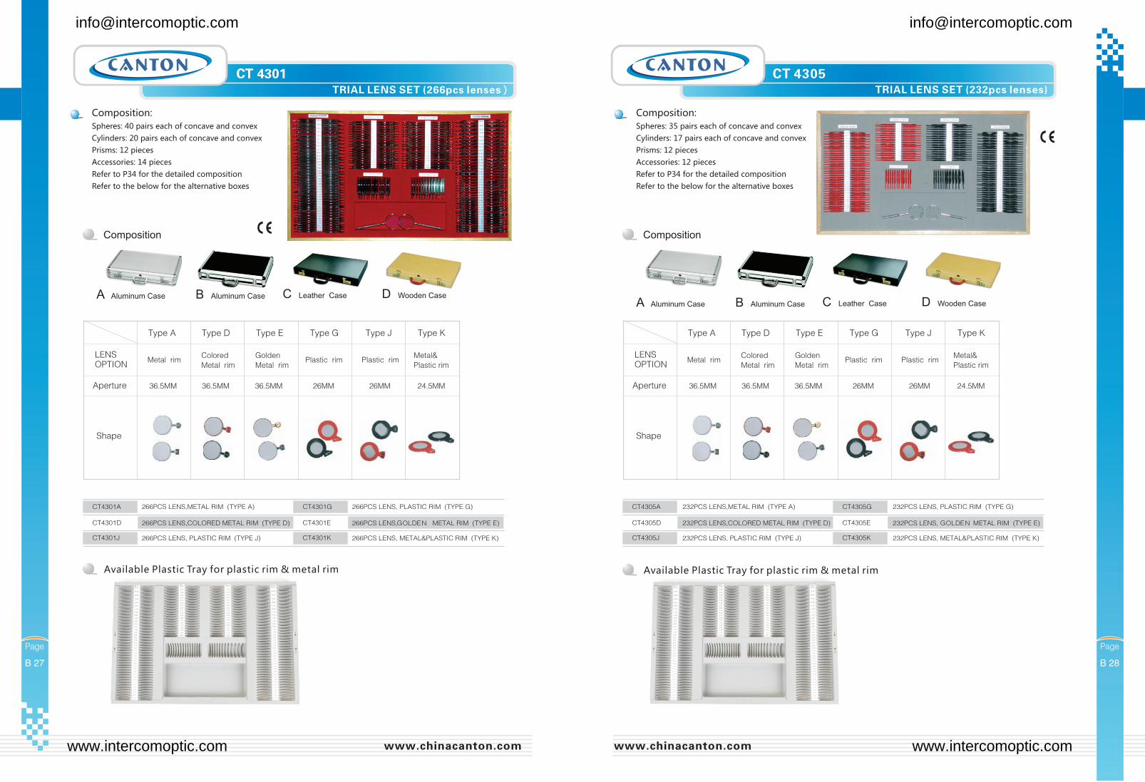
Available Plastic Tray for plastic rim & metal rim Available Plastic Tray for plastic rim & metal rim
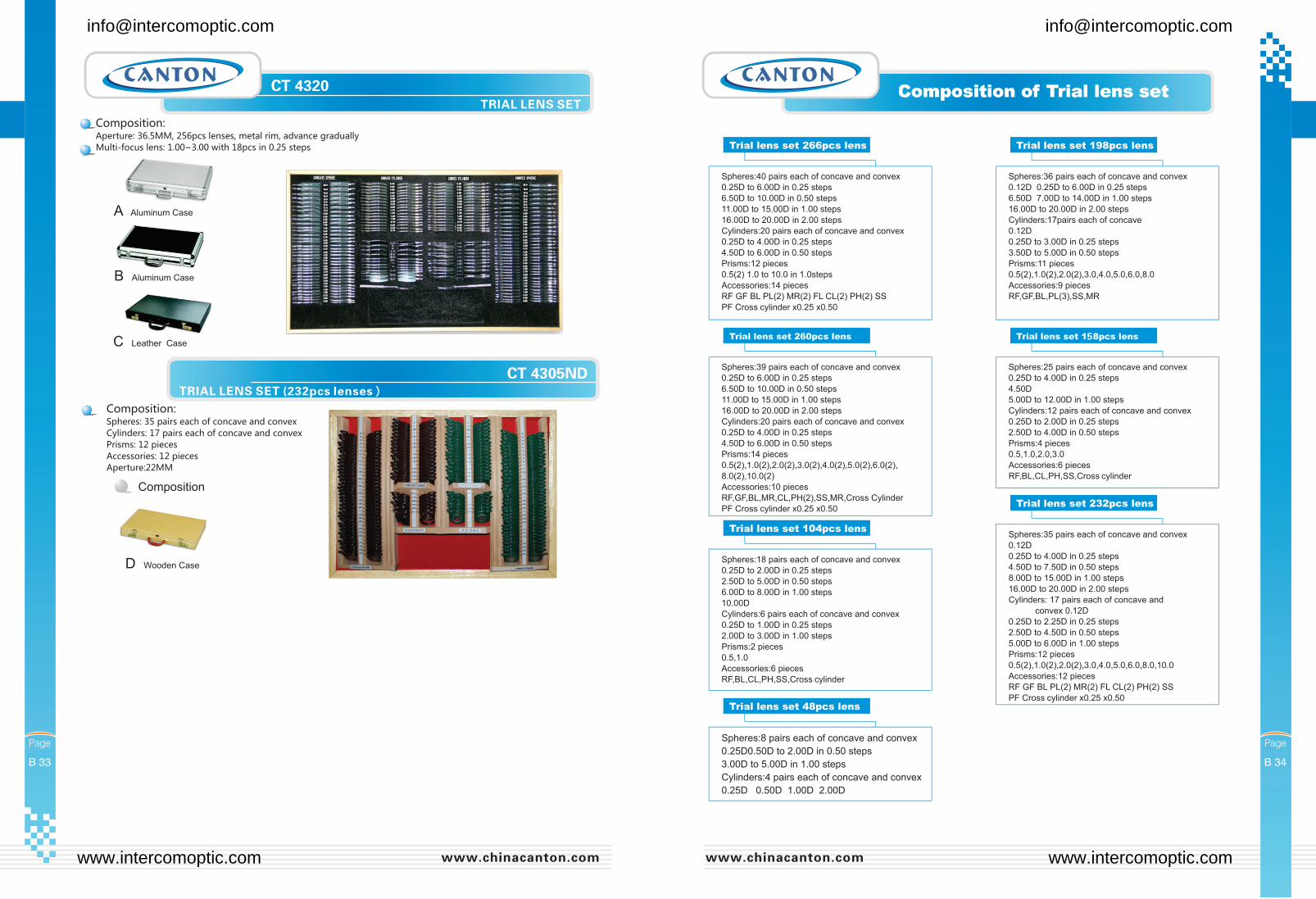
CT 4305
[email protected] [email protected]
www.intercomoptic.com www.intercomoptic.com
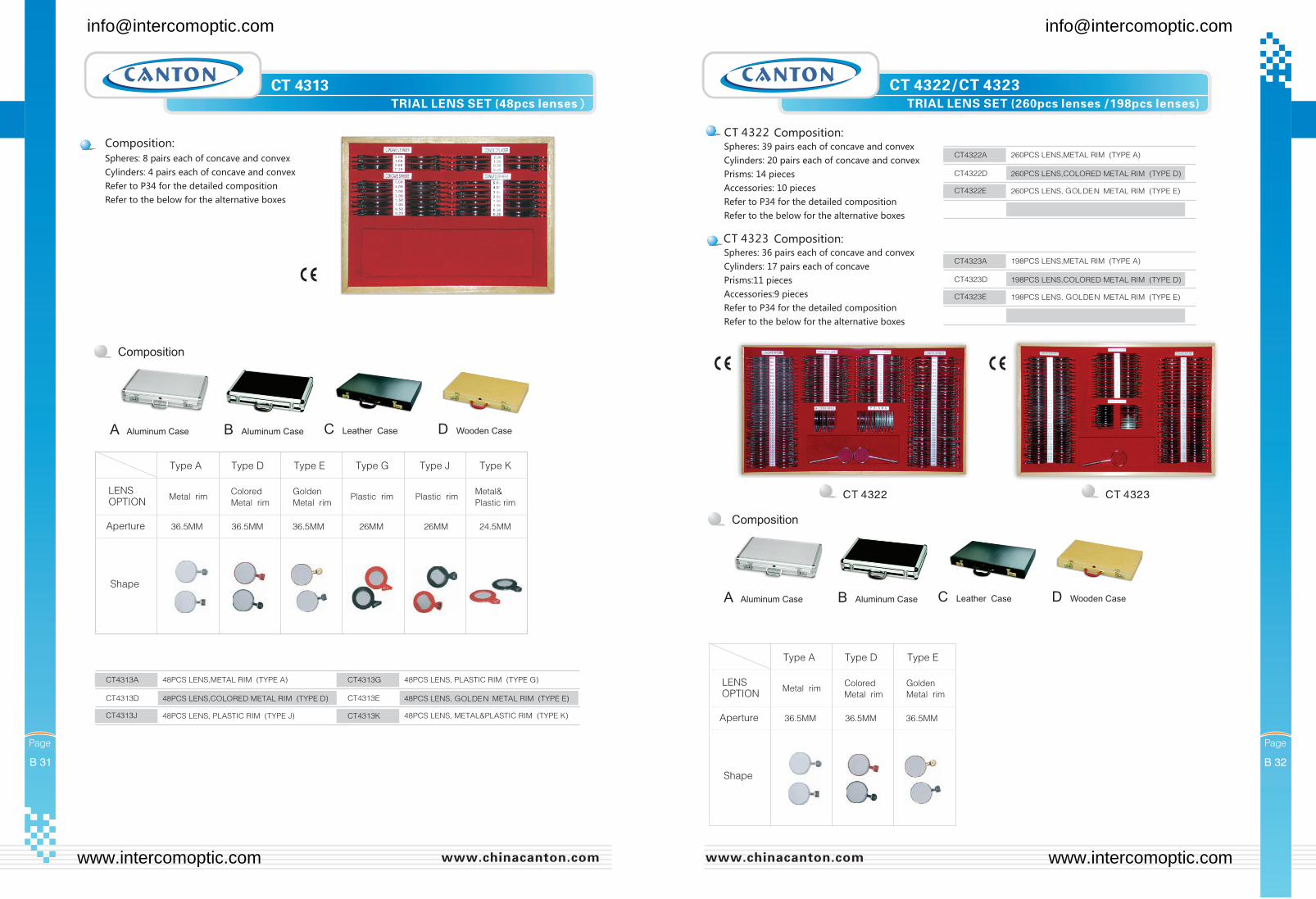
CT 4322 CT 4323
CT 4322/CT 4323TRIAL LENS SET (260pcs lenses /198pcs lenses)
[email protected] [email protected]
www.intercomoptic.com www.intercomoptic.com
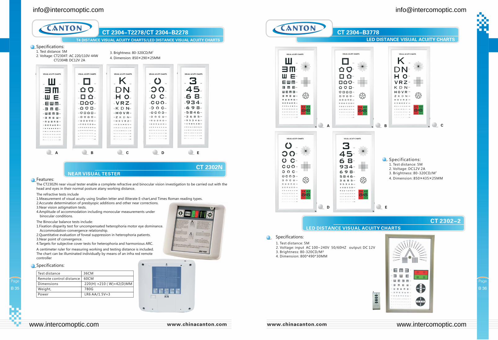
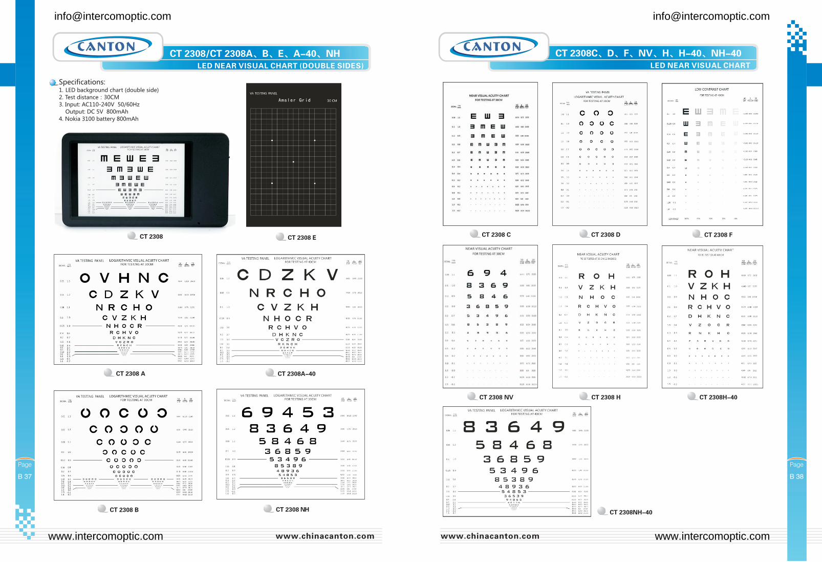
CT 2302-2LED DISTANCE VISUAL ACUITY CHARTS
1. Test distance: 5M
2. Voltage: input AC 100~240V 50/60HZ output: DC 12V
3. Brightness: 80-320CD/M²
4. Dimension: 800*490*30MM
[email protected] [email protected]
www.intercomoptic.com www.intercomoptic.com
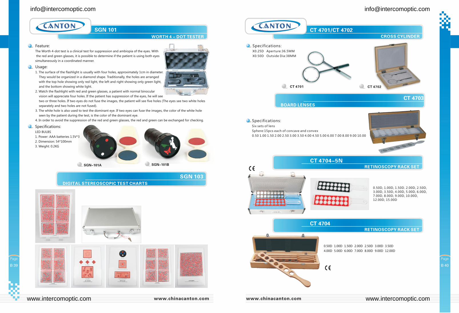
Feature:
The Worth 4-dot test is a clinical test for suppression and ambiopia of the eyes. With
the red and green glasses, it is possible to determine if the patient is using both eyes
simultaneously in a coordinated manner.
Usage:
1. The surface of the flashlight is usually with four holes, approximately 1cm in diameter.
They would be organized in a diamond shape. Traditionally, the holes are arranged
with the top hole showing only red light, the left and right showing only green light,
and the bottom showing white light.
2. Watch the flashlight with red and green glasses, a patient with normal binocular
vision will appreciate four holes. If the patient has suppression of the eyes, he will see
two or three holes. If two eyes do not fuse the images, the patient will see five holes (The eyes see two white holes
separately and two holes are not fused).
3. The white hole is also used to test the dominant eye. If two eyes can fuse the images, the color of the white hole
seen by the patient during the test, is the color of the dominant eye.
4. In order to avoid the suppression of the red and green glasses, the red and green can be exchanged for checking.
SGN 103DIGITAL STEREOSCOPIC TEST CHARTS
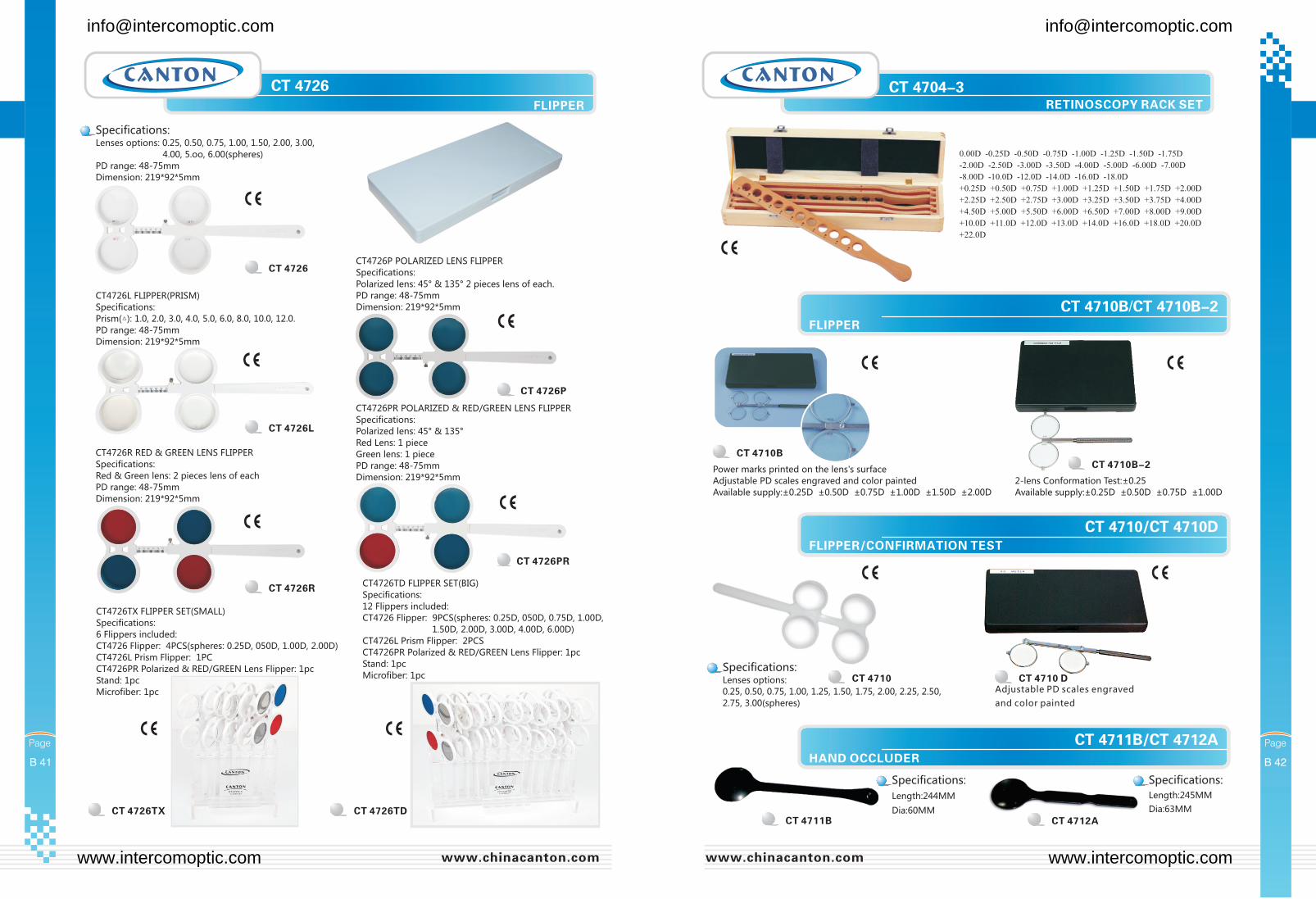
CT 4704-5N
0.50D, 1.00D, 1.50D. 2.00D, 2.50D,
3.00D, 3.50D, 4.00D, 5.00D, 6.00D,
7.00D, 8.00D, 9.00D, 10.00D,
12.00D, 15.00D
[email protected] [email protected]
www.intercomoptic.com www.intercomoptic.com
CT 4726TX
CT4726TX FLIPPER SET(SMALL)
Specifications:
6 Flippers included:
CT4726 Flipper: 4PCS(spheres: 0.25D, 050D, 1.00D, 2.00D)
CT4726L Prism Flipper: 1PC
CT4726PR Polarized & RED/GREEN Lens Flipper: 1pc
Stand: 1pc
Microfiber: 1pc
CT 4726TD
CT4726TD FLIPPER SET(BIG)
Specifications:
12 Flippers included:
CT4726 Flipper: 9PCS(spheres: 0.25D, 050D, 0.75D, 1.00D,
1.50D, 2.00D, 3.00D, 4.00D, 6.00D)
CT4726L Prism Flipper: 2PCS
CT4726PR Polarized & RED/GREEN Lens Flipper: 1pc
Stand: 1pc
Microfiber: 1pc
[email protected] [email protected]
www.intercomoptic.com www.intercomoptic.com
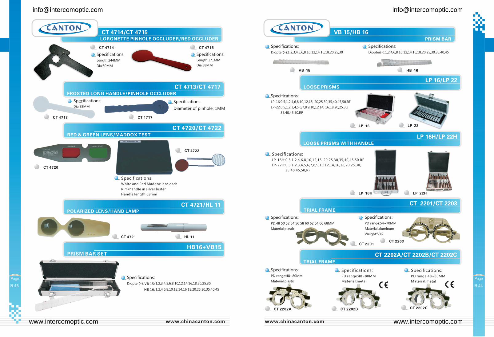
CT 4720/CT 4722RED & GREEN LENS/MADDOX TEST
CT 4722
CT 4720
PRISM BAR SET
HB16+VB15
[email protected] [email protected]
www.intercomoptic.com www.intercomoptic.com
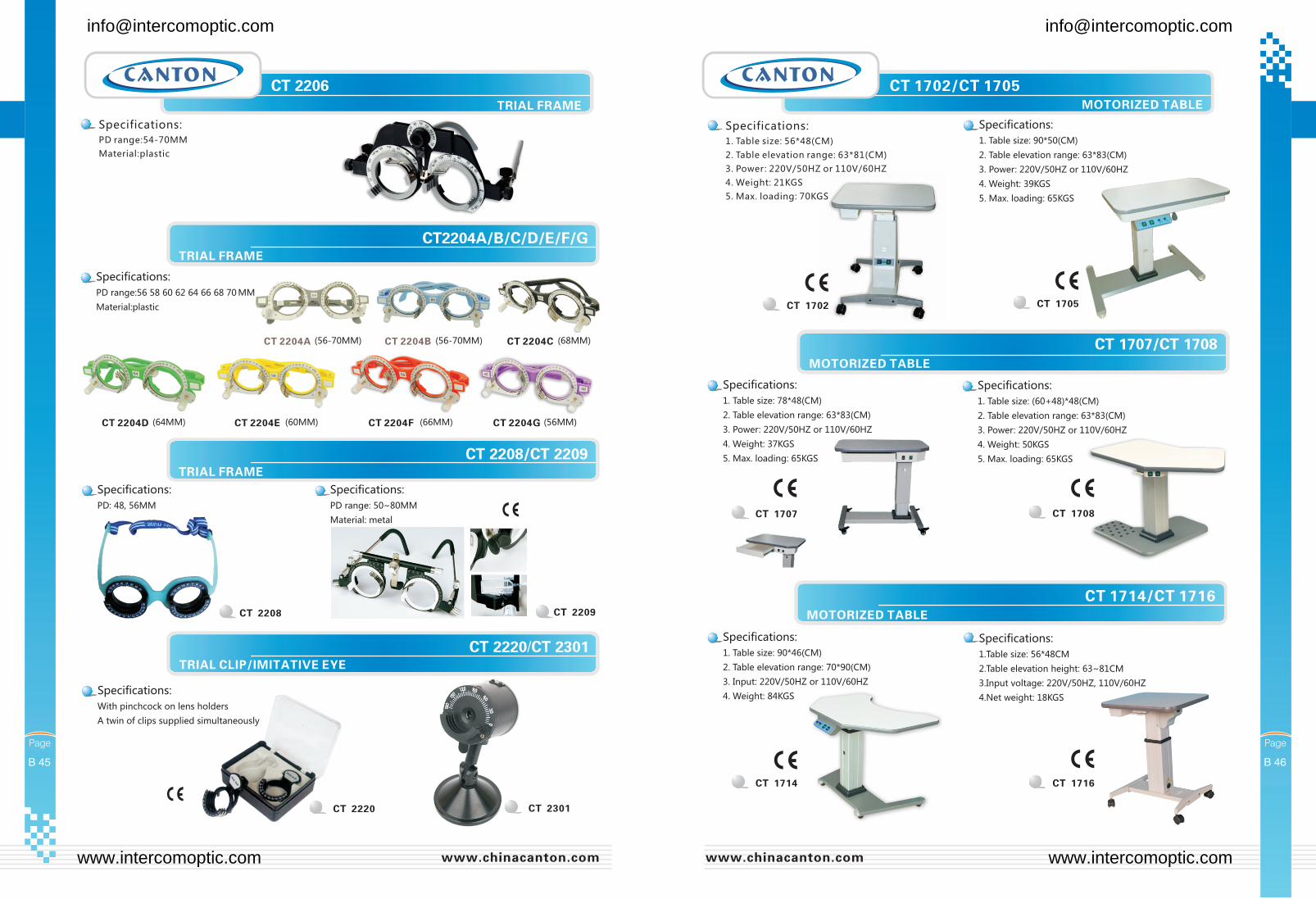
CT 1714/CT 1716
CT 1702/CT 1705
[email protected] [email protected]
www.intercomoptic.com www.intercomoptic.com
CT 1723/CT 1730
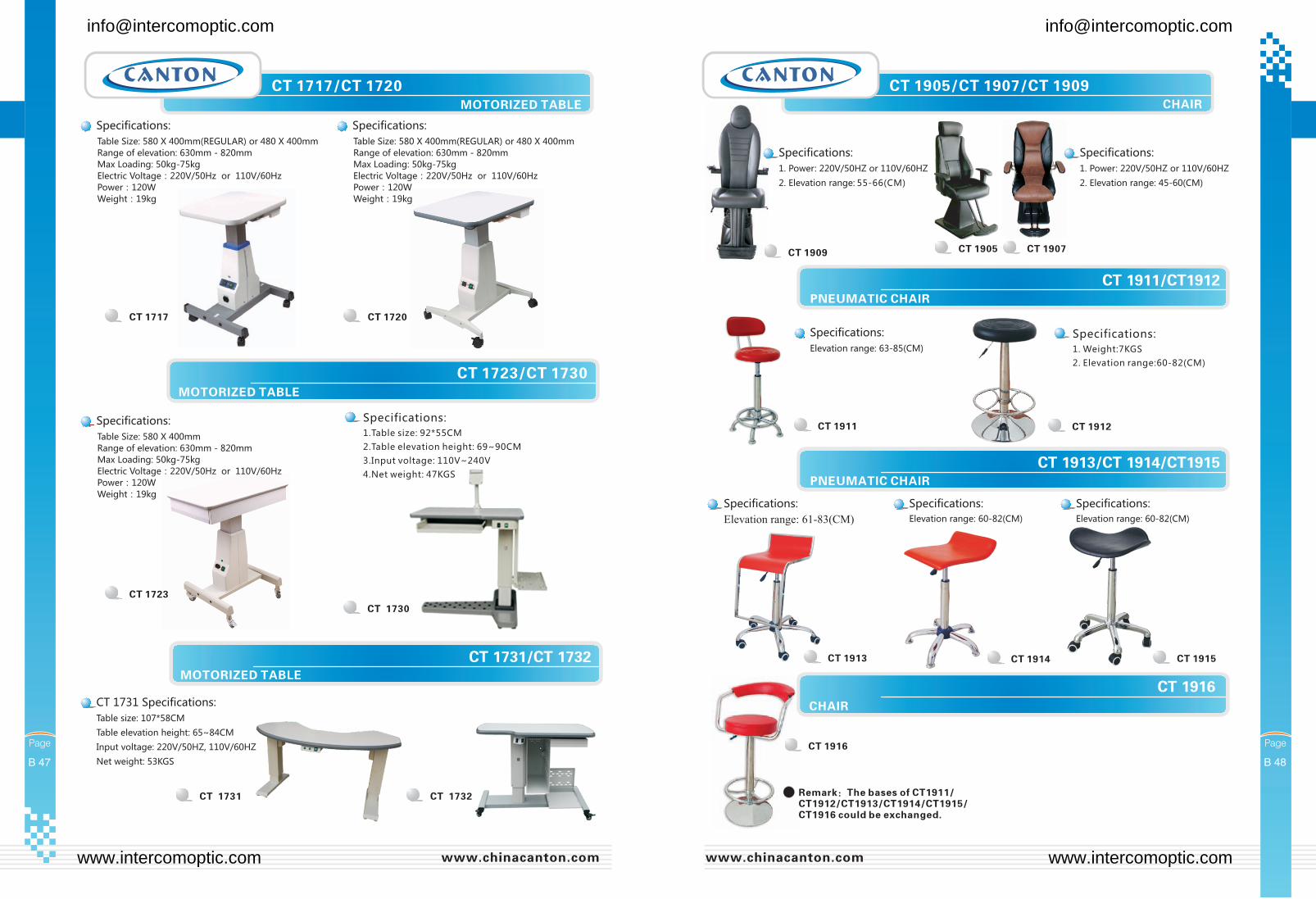
CT 1717/CT 1720
CT 1720CT 1717
CT 1723
CT 1905/CT 1907/CT 1909
CT 1909
Table Size: 580 X 400mm(REGULAR) or 480 X 400mm
Range of elevation: 630mm - 820mm
Max Loading: 50kg-75kg
Electric Voltage:220V/50Hz or 110V/60Hz
Power:120W
Weight:19kg
Table Size: 580 X 400mm(REGULAR) or 480 X 400mm
Range of elevation: 630mm - 820mm
Max Loading: 50kg-75kg
Electric Voltage:220V/50Hz or 110V/60Hz
Power:120W
Weight:19kg
Table Size: 580 X 400mm
Range of elevation: 630mm - 820mm
Max Loading: 50kg-75kg
Electric Voltage:220V/50Hz or 110V/60Hz
Power:120W
Weight:19kg
[email protected] [email protected]
www.intercomoptic.com www.intercomoptic.com
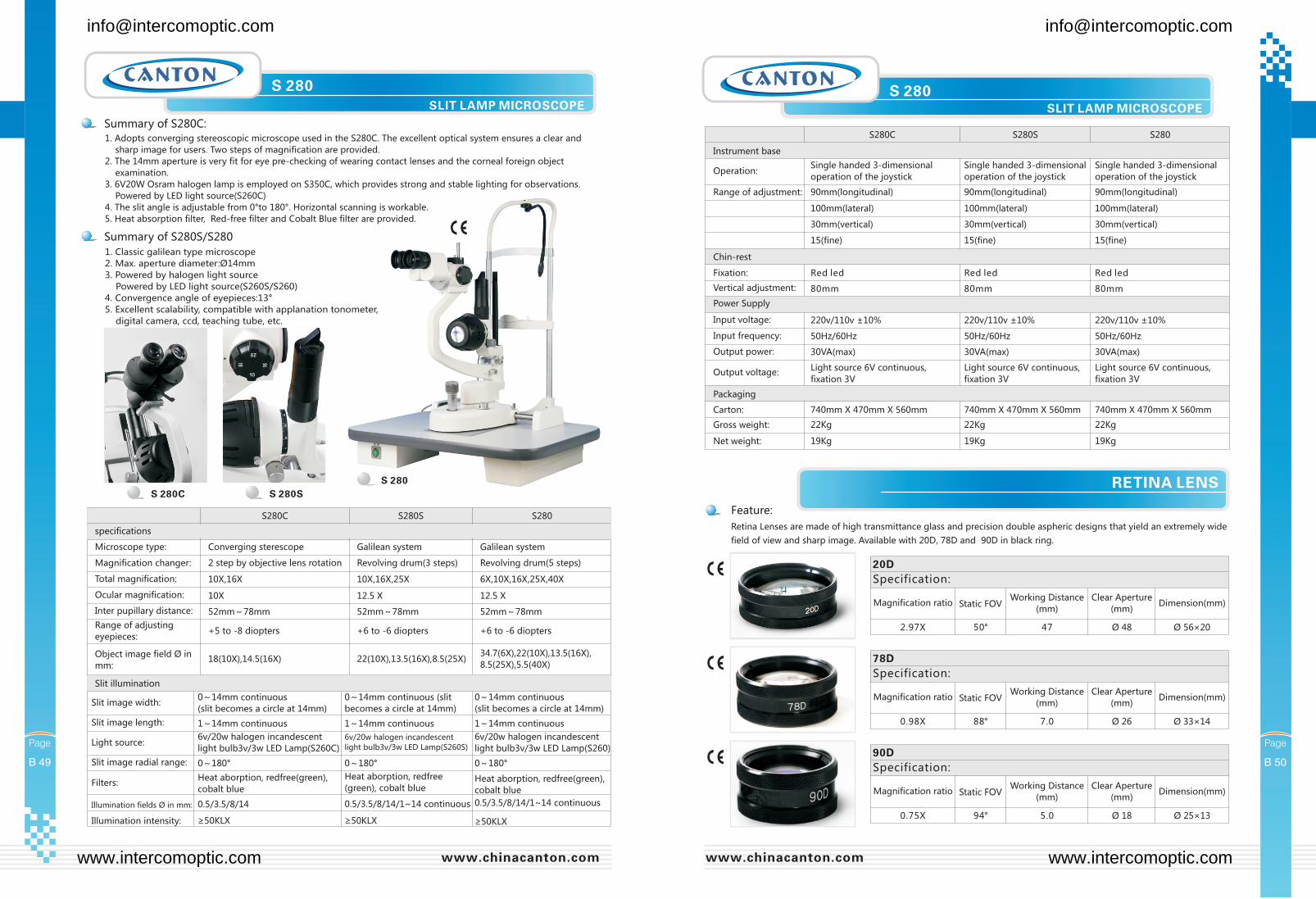
Feature:
Retina Lenses are made of high transmittance glass and precision double aspheric designs that yield an extremely wide
field of view and sharp image. Available with 20D, 78D and 90D in black ring.
20D
Specification:
Magnification ratio Static FOVWorking Distance
(mm)
Clear Aperture
(mm)Dimension(mm)
2.97X 50° 47 Ø 48 Ø 56×20
78D
Specification:
Magnification ratio Static FOVWorking Distance
(mm)
Clear Aperture
(mm)Dimension(mm)
0.98X 88° 7.0 Ø 26 Ø 33 14×
90D
Specification:
Magnification ratio Static FOVWorking Distance
(mm)
Clear Aperture
(mm)Dimension(mm)
0.75X 94° 5.0 Ø 18 Ø 25×13
1. Adopts converging stereoscopic microscope used in the S280C. The excellent optical system ensures a clear and
sharp image for users. Two steps of magnification are provided.
2. The 14mm aperture is very fit for eye pre-checking of wearing contact lenses and the corneal foreign object
examination.
3. 6V20W Osram halogen lamp is employed on S350C, which provides strong and stable lighting for observations.
Powered by LED light source(S260C)
4. The slit angle is adjustable from 0°to 180°. Horizontal scanning is workable.
5. Heat absorption filter, Red-free filter and Cobalt Blue filter are provided.
1. Classic galilean type microscope
2. Max. aperture diameter:Ø14mm
3. Powered by halogen light source
Powered by LED light source(S260S/S260)
4. Convergence angle of eyepieces:13°
5. Excellent scalability, compatible with applanation tonometer,
digital camera, ccd, teaching tube, etc.
S 280SLIT LAMP MICROSCOPE
S 280SLIT LAMP MICROSCOPE
S280C S280S S280
specifications
Microscope type: Converging sterescope Galilean system Galilean system
Magnification changer: 2 step by objective lens rotation Revolving drum(3 steps) Revolving drum(5 steps)
Total magnification: 10X,16X 10X,16X,25X 6X,10X,16X,25X,40X
Ocular magnification: 10X 12.5 X 12.5 X
Inter pupillary distance: 52mm~78mm 52mm~78mm 52mm~78mm
Range of adjusting
eyepieces:+5 to -8 diopters +6 to -6 diopters +6 to -6 diopters
Object image field Ø in
mm:18(10X),14.5(16X) 22(10X),13.5(16X),8.5(25X)
34.7(6X),22(10X),13.5(16X),
8.5(25X),5.5(40X)
Slit illumination
Slit image width:0~14mm continuous
(slit becomes a circle at 14mm)
0~14mm continuous (slit
becomes a circle at 14mm)
0~14mm continuous
(slit becomes a circle at 14mm)
Slit image length: 1~14mm continuous 1~14mm continuous 1~14mm continuous
Light source:6v/20w halogen incandescent
light bulb3v/3w LED Lamp(S260C)
6v/20w halogen incandescent
light bulb3v/3w LED Lamp(S260S)
6v/20w halogen incandescent
light bulb3v/3w LED Lamp(S260)
Slit image radial range: 0~180° 0~180° 0~180°
Filters:Heat aborption, redfree(green),
cobalt blue
Heat aborption, redfree
(green), cobalt blueHeat aborption, redfree(green),
cobalt blue
Illumination fields Ø in mm:
Illumination intensity:
0.5/3.5/8/14
≥50KLX
0.5/3.5/8/14/1~14 continuous
≥50KLX ≥50KLX
0.5/3.5/8/14/1~14 continuous
Instrument base
Operation:
Range of adjustment:
Single handed 3-dimensional
operation of the joystick
90mm(longitudinal) 90mm(longitudinal) 90mm(longitudinal)
100mm(lateral) 100mm(lateral) 100mm(lateral)
30mm(vertical) 30mm(vertical) 30mm(vertical)
15(fine) 15(fine) 15(fine)
Single handed 3-dimensional
operation of the joystick
Single handed 3-dimensional
operation of the joystick
Chin-rest
Fixation:
Vertical adjustment:
Power Supply
Input voltage:
Input frequency:
Output power:
Output voltage:
Red led Red led Red led
80mm 80mm 80mm
220v/110v ±10% 220v/110v ±10% 220v/110v ±10%
50Hz/60Hz 50Hz/60Hz 50Hz/60Hz
30VA(max) 30VA(max) 30VA(max)
Light source 6V continuous,
fixation 3V
Light source 6V continuous,
fixation 3V
Light source 6V continuous,
fixation 3V
Packaging
Carton:
Gross weight:
Net weight:
740mm X 470mm X 560mm 740mm X 470mm X 560mm 740mm X 470mm X 560mm
22Kg 22Kg 22Kg
19Kg 19Kg 19Kg
S280C S280S S280
RETINA LENS
[email protected] [email protected]
www.intercomoptic.com www.intercomoptic.com
Chin-rest
Fixation:
Vertical adjustment:
Power Supply
Input voltage:
Input frequency:
Output power:
Output voltage:
Red led Red led Red led
80mm 80mm 80mm
220v/110v ±10% 220v/110v ±10% 220v/110v ±10%
50Hz/60Hz 50Hz/60Hz 50Hz/60Hz
30VA(max) 30VA(max) 30VA(max)
Light source 6V continuous,
fixation 3V
Light source 6V continuous,
fixation 3V
Light source 6V continuous,
fixation 3V
Packaging
Carton:
Gross weight:
Net weight:
740mm X 470mm X 560mm 740mm X 470mm X 560mm 740mm X 470mm X 560mm
22Kg 24Kg 24Kg
19Kg 21Kg 21Kg
S350C S350S S350
S 350
S 350C
S 350S
S 350 S 350SLIT LAMP MICROSCOPE SLIT LAMP MICROSCOPE
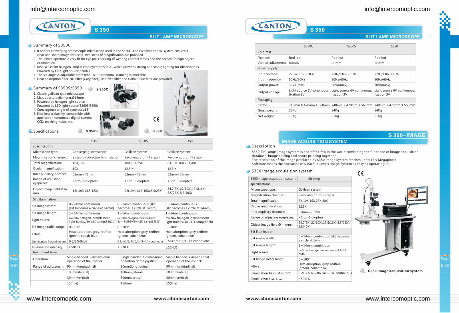
1. It adopts converging stereoscopic microscope used in the S350C. The excellent optical system ensures a
clear and sharp image for users. Two steps of magnification are provided.
2. The 14mm aperture is very fit for eye pre-checking of wearing contact lenses and the corneal foreign object
examination.
3. 6V20W Osram halogen lamp is employed on S350C, which provides strong and stable lighting for observations.
Powered by LED light source(S360C)
4. The slit angle is adjustable from 0°to 180°. Horizontal scanning is workable.
5. Heat absorption filter, ND filter (Grey filter), Red-free filter and Cobalt Blue filter are provided.
1. Classic galilean type microscope
2. Max. aperture diameter:Ø14mm
3. Powered by halogen light source
Powered by LED light source(S360S/S360)
4. Convergence angle of eyepieces:13°
5. Excellent scalability, compatible with
application tonometer, digital camera,
CCD, teaching tube, etc.
S350C S350S S350
specifications
Microscope type: Converging sterescope Galilean system Galilean system
Magnification changer: 2 step by objective lens rotation Revolving drum(3 steps) Revolving drum(5 steps)
Total magnification: 10X,16X 10X,16X,25X 6X,10X,16X,25X,40X
Ocular magnification: 10X 12.5 X 12.5 X
Inter pupillary distance: 52mm~78mm 52mm~78mm 52mm~78mm
Range of adjusting
eyepieces:+5 to -8 diopters +6 to -6 diopters +6 to -6 diopters
Object image field Ø in
mm:18(10X),14.5(16X) 22(10X),13.5(16X),8.5(25X)
34.7(6X),22(10X),13.5(16X),
8.5(25X),5.5(40X)
Slit illumination
Slit image width:0~14mm continuous
(slit becomes a circle at 14mm)
0~14mm continuous (slit
becomes a circle at 14mm)
0~14mm continuous
(slit becomes a circle at 14mm)
Slit image length: 1~14mm continuous 1~14mm continuous 1~14mm continuous
Light source:6v/20w halogen incandescent
light bulb3v/3w LED Lamp(S360C)
6v/20w halogen incandescent
light bulb3v/3w LED Lamp(S360S)
6v/20w halogen incandescent
light bulb3v/3w LED Lamp(S360)
Slit image radial range: 0~180° 0~180° 0~180°
Filters:Heat aborption, grey, redfree
(green), cobalt blue
Heat aborption, grey, redfree
(green), cobalt blue
Heat aborption, grey, redfree
(green), cobalt blue
Illumination fields Ø in mm:
Illumination intensity:
0.5/3.5/8/14
≥50KLX
0.2/1/2/3/5/10/14/1~14 continuous
≥50KLX ≥50KLX
0.5/3.5/8/14/1~14 continuous
Instrument base
Operation:
Range of adjustment:
Single handed 3-dimensional
operation of the joystick
90mm(longitudinal) 90mm(longitudinal) 90mm(longitudinal)
100mm(lateral) 100mm(lateral) 100mm(lateral)
30mm(vertical) 30mm(vertical) 30mm(vertical)
15(fine) 15(fine) 15(fine)
Single handed 3-dimensional
operation of the joystick
Single handed 3-dimensional
operation of the joystick
S350 Slit Lamps Image System is one of the few in the world combining the functions of image acquisition,
database, image editing and photo printing together.
The resolution of the image produced by S350 Image System reaches up to 17.9 Megapixels.
Software makes the operation of S350 Slit Lamps Image System as easy as operating PC.
S350 image acquisition system
IMAGE ACQUISITION SYSTEM
S 350-IMAGE
Magnification changer: Revolving drum(5 steps)
Total magnification: 6X,10X,16X,25X,40X
Ocular magnification: 12.5X
Inter pupillary distance: 52mm~78mm
Range of adjusting eyepieces: +6 to -6 diopters
Object image field Ø in mm:34.7(6X),22(10X),13.5(16X),8.5(25X),
5.5(40X)
Slit illumination
Slit image width:0~14mm continuous (slit becomes
a circle at 14mm)
Slit image length: 1~14mm continuous
Light source:6v/20w halogen incandescent light
bulb
Slit image radial range: 0~180°
Filters:Heat aborption, grey, redfree
(green), cobalt blue
Illumination fields Ø in mm:
Illumination intensity:
0.2/1/2/3/5/10/14/1~14 continuous
≥50KLX
Microscope type: Galilean system
slit lamp
specifications
S350 image acquisition system:
[email protected] [email protected]
www.intercomoptic.com www.intercomoptic.com
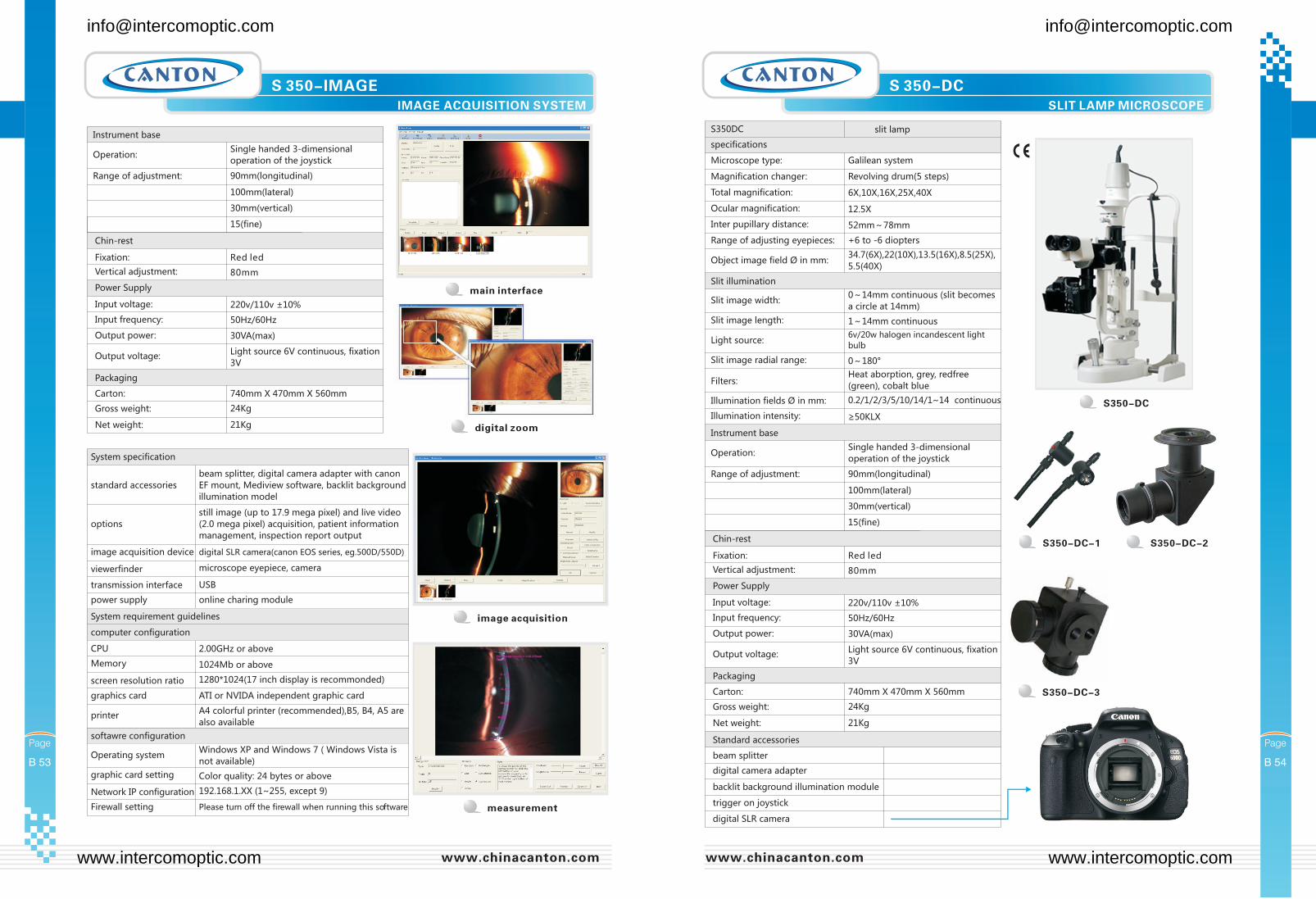
image acquisition
measurement
S 350-IMAGEIMAGE ACQUISITION SYSTEM
Chin-rest
Fixation:
Vertical adjustment:
Power Supply
Input voltage:
Input frequency:
Output power:
Output voltage:
Red led
80mm
220v/110v ±10%
50Hz/60Hz
30VA(max)
Light source 6V continuous, fixation
3V
Packaging
Carton:
Gross weight:
Net weight:
740mm X 470mm X 560mm
24Kg
21Kg
Instrument base
Operation:
Range of adjustment:
Single handed 3-dimensional
operation of the joystick
90mm(longitudinal)
100mm(lateral)
30mm(vertical)
15(fine)
main interface
digital zoom
options
still image (up to 17.9 mega pixel) and live video
(2.0 mega pixel) acquisition, patient information
management, inspection report output
image acquisition device digital SLR camera(canon EOS series, eg.500D/550D)
viewerfinder microscope eyepiece, camera
transmission interface USB
standard accessories
beam splitter, digital camera adapter with canon
EF mount, Mediview software, backlit background
illumination model
System specification
power supply online charing module
CPU
Operating system
2.00GHz or above
Windows XP and Windows 7 ( Windows Vista is
not available)
Memory
graphic card setting
1024Mb or above
Color quality: 24 bytes or above
screen resolution ratio
Network IP configuration
1280*1024(17 inch display is recommonded)
192.168.1.XX (1~255, except 9)
computer configuration
softawre configuration
System requirement guidelines
graphics card
Firewall setting
printer
ATI or NVIDA independent graphic card
Please turn off the firewall when running this software
A4 colorful printer (recommended),B5, B4, A5 are
also available
S 350-DCSLIT LAMP MICROSCOPE
Chin-rest
Fixation:
Vertical adjustment:
Power Supply
Input voltage:
Input frequency:
Output power:
Output voltage:
Red led
80mm
220v/110v ±10%
50Hz/60Hz
30VA(max)
Light source 6V continuous, fixation
3V
Packaging
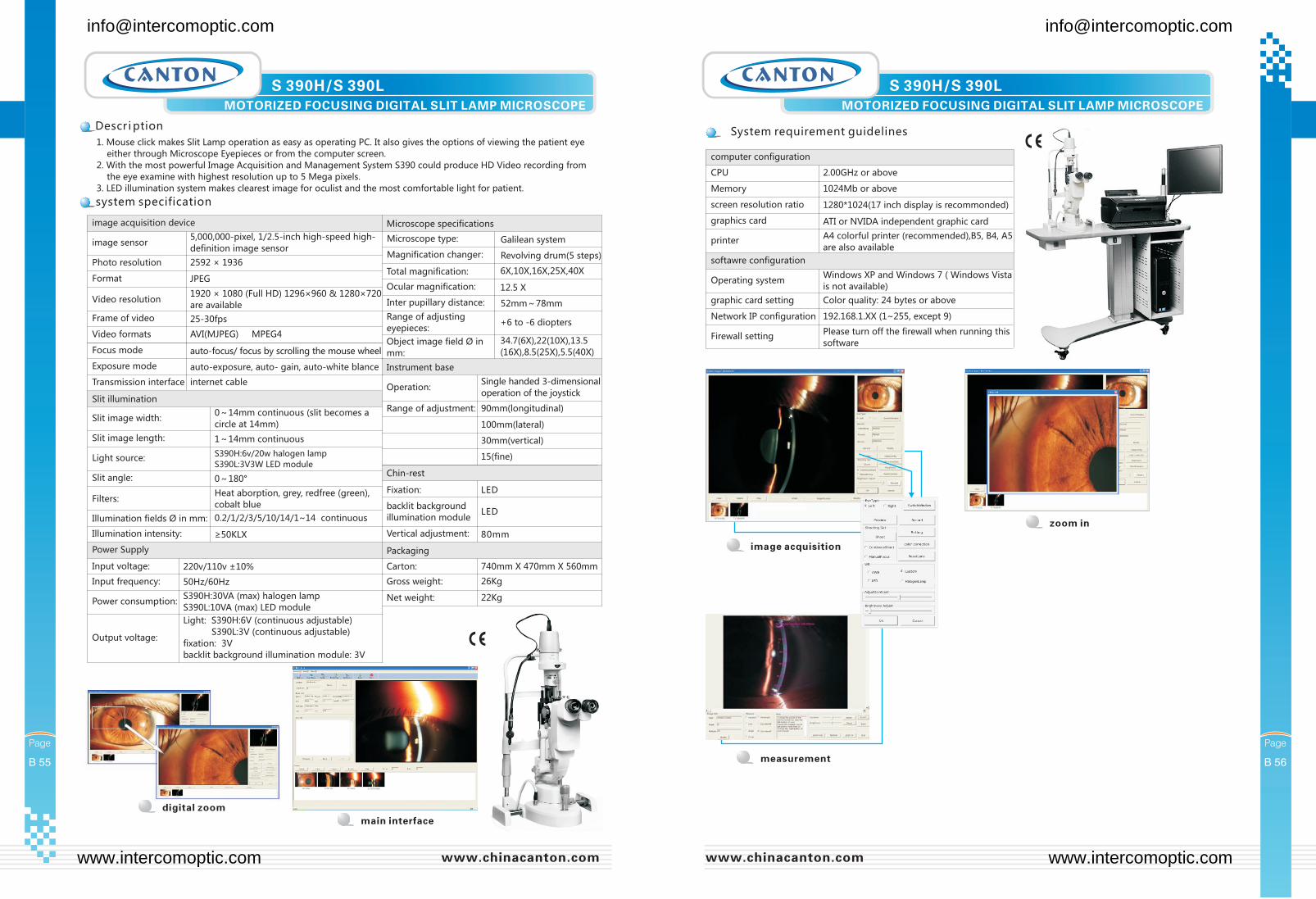
Standard accessories
Carton:
beam splitter
Gross weight:
digital camera adapter
Net weight:
backlit background illumination module
trigger on joystick
digital SLR camera
740mm X 470mm X 560mm
24Kg
21Kg
Magnification changer: Revolving drum(5 steps)
Total magnification: 6X,10X,16X,25X,40X
Ocular magnification: 12.5X
Inter pupillary distance: 52mm~78mm
Range of adjusting eyepieces: +6 to -6 diopters
Object image field Ø in mm:34.7(6X),22(10X),13.5(16X),8.5(25X),
5.5(40X)
Slit illumination
Slit image width:0~14mm continuous (slit becomes
a circle at 14mm)
Slit image length: 1~14mm continuous
Light source:6v/20w halogen incandescent light
bulb
Slit image radial range: 0~180°
Filters:Heat aborption, grey, redfree
(green), cobalt blue
Illumination fields Ø in mm:
Illumination intensity:
0.2/1/2/3/5/10/14/1~14 continuous
≥50KLX
Instrument base
Operation:
Range of adjustment:
Single handed 3-dimensional
operation of the joystick
90mm(longitudinal)
100mm(lateral)
30mm(vertical)
15(fine)
Microscope type: Galilean system
slit lamp
specifications
S350DC
S350-DC
S350-DC-1 S350-DC-2
S350-DC-3
[email protected] [email protected]
www.intercomoptic.com www.intercomoptic.com
1. Mouse click makes Slit Lamp operation as easy as operating PC. It also gives the options of viewing the patient eye
either through Microscope Eyepieces or from the computer screen.
2. With the most powerful Image Acquisition and Management System S390 could produce HD Video recording from
the eye examine with highest resolution up to 5 Mega pixels.
3. LED illumination system makes clearest image for oculist and the most comfortable light for patient.
S 390H/S 390L S 390H/S 390LMOTORIZED FOCUSING DIGITAL SLIT LAMP MICROSCOPE MOTORIZED FOCUSING DIGITAL SLIT LAMP MICROSCOPE
Chin-rest
Fixation:
backlit background
illumination module
Vertical adjustment:
Power Supply
Input voltage:
Input frequency:
Power consumption:
Output voltage:
LED
LED
80mm
220v/110v ±10%
50Hz/60Hz
S390H:30VA (max) halogen lamp
S390L:10VA (max) LED module
Light: S390H:6V (continuous adjustable)
S390L:3V (continuous adjustable)
fixation: 3V
backlit background illumination module: 3V
Packaging
Carton:
Gross weight:
Net weight:
740mm X 470mm X 560mm
26Kg
22Kg
Photo resolution 2592 × 1936
Format JPEG
Video resolution
Focus mode
1920 × 1080 (Full HD) 1296×960 & 1280×720
are available
auto-focus/ focus by scrolling the mouse wheel
Frame of video
Exposure mode
25-30fps
auto-exposure, auto- gain, auto-white blance
Video formats
Transmission interface
AVI(MJPEG),MPEG4
internet cable
Microscope type: Galilean system
Magnification changer:
Inter pupillary distance:
Revolving drum(5 steps)
52mm~78mm
Total magnification:
Range of adjusting
eyepieces:
Ocular magnification:
Object image field Ø in
mm:
6X,10X,16X,25X,40X
+6 to -6 diopters
12.5 X
34.7(6X),22(10X),13.5
(16X),8.5(25X),5.5(40X)
Instrument base
Operation:
Range of adjustment:
Single handed 3-dimensional
operation of the joystick
90mm(longitudinal)
100mm(lateral)
30mm(vertical)
15(fine)
image sensor
image acquisition device
5,000,000-pixel, 1/2.5-inch high-speed high-
definition image sensor
Microscope specifications
Slit illumination
Slit image width:0~14mm continuous (slit becomes a
circle at 14mm)
Slit image length: 1~14mm continuous
Light source:S390H:6v/20w halogen lamp
S390L:3V3W LED module
Slit angle: 0~180°
Filters:Heat aborption, grey, redfree (green),
cobalt blue
Illumination fields Ø in mm:
Illumination intensity:
0.2/1/2/3/5/10/14/1~14 continuous
≥50KLX
main interface
zoom in
digital zoom
Memory
Network IP configuration
1024Mb or above
192.168.1.XX (1~255, except 9)
screen resolution ratio
Firewall setting
1280*1024(17 inch display is recommonded)
Please turn off the firewall when running this
software
graphics card ATI or NVIDA independent graphic card
Operating systemWindows XP and Windows 7 ( Windows Vista
is not available)
CPU
graphic card setting
2.00GHz or above
Color quality: 24 bytes or above
computer configuration
printerA4 colorful printer (recommended),B5, B4, A5
are also available
softawre configuration
image acquisition
measurement
[email protected] [email protected]
www.intercomoptic.com www.intercomoptic.com