Embed Size (px)
Citation preview

Dr Simon Barnard Optometric examination of children aged 0 – 5 years
1
Optometric examination of children aged 0 – 5 years
Dr Simon Barnard PhD BSc FCOptom FAAO DCLP DipClinOptom
Tutor in Ocular Disease, Department of Optometry &
Visual Science City University
&
Director of Ocular Medicine Institute of Optometry
London
Ronald Brown Group Evening Lecture 8th January 2004
Ronald Brown 8th January 2004

Dr Simon Barnard Optometric examination of children aged 0 – 5 years
2
Ronald Brown 8th January 2004

Dr Simon Barnard Optometric examination of children aged 0 – 5 years
3
Table of Contents
Table of Contents..................................................................................................3 Introduction ...........................................................................................................4 History & Symptoms .............................................................................................5 How clearly does the child see? ...........................................................................5
Angular vs. Morphoscopic .................................................................................5 Alternate Occlusion ...........................................................................................6 Preferential Looking ..........................................................................................6 Picture Tests .....................................................................................................7 Letter tests ........................................................................................................8 Tests for near vision ..........................................................................................9
What is the child’s refractive status?.....................................................................9 Assessment of Refraction .................................................................................9
Colour vision .......................................................................................................11 Pupillary reflexes.................................................................................................11 Visual fields.........................................................................................................11 Eye position, ocular motor balance and binocularity ...........................................13
Is the child straight ? .......................................................................................13 Vergence.........................................................................................................14 Stereopsis .......................................................................................................16 Stereopsis .......................................................................................................17
Are the eyes healthy ? ........................................................................................17 Tonometry .......................................................................................................17 Slit lamp ..........................................................................................................18 The fundus ......................................................................................................18 Sedation and anaesthesia...............................................................................19 Methods of retinal examination........................................................................19 Further reading................................................................................................22
Ronald Brown 8th January 2004
Dr Simon Barnard Optometric examination of children aged 0 – 5 years
4
Introduction
It is often stated that children must not be treated as miniature adults. Whilst this is true in many regards, children aged around 3 years and older must be treated with the respect given to adults. Certain tests and procedures need to be explained to them, even at 3 years, and permission gently sought for certain procedures such as instillation of eye drops. The practice should present a friendly environment with books, toys, miniature table and chairs, blackboard, and a video all helping to make a child more comfortable. Needless to say, the toilet can be the most important room in the practice. It is preferable for the optometrist to go and out to greet the child and parents rather than having them brought to the consulting room, especially at the first visit. The consulting room may be intimidating to a young child and this should be borne in mind. One should try and make a verbal connection early on and try and strike up conversation with child to “break the ice”. Observe the child on the way to and in the consulting room. Clumsiness, fidgetiness and “hyperactivity” may be apparent and my direct the optometrist to searching questions about possible developmental difficulties or dyspraxic tendencies. Who should be with the child? The author believes it imperative to have a parent (or both if present) at this age and older. However, siblings can be very distracting and should, if possible, be accommodated in the waiting room. For this age group and younger one should direct questions relating to history and symptoms to the parent or guardian and determine if the visit is purely routine or whether there are any signs or reported symptoms. Always take special note of an observation made by a grandmother as she will be even more experienced. Careful symptom taking is most important and a tentative diagnosis is often in your mind even before you commence the examination, e.g., frequent blinking in a 5 year-old is most usually behavioral in origin, sometimes due to allergy and rarely due to other causes. The optometrist must work efficiently and it may be that you will return to tidy up some history up after the examination when you have a child with a short span of attention.
Ronald Brown 8th January 2004

Dr Simon Barnard Optometric examination of children aged 0 – 5 years
5
A six-metre room, whilst being a luxury, does remove the need for a mirror with the confusion that sometimes results. During the routine one may keep up a constant babble of talk (or attention seeking noises) especially during some tests such as retinoscopy and ophthalmoscopy. Screening and routine checks v. patients with presenting signs or symptoms In a screening situation where one is examining an unselected group most children will be normal. The child may be considered “normal” unless proven otherwise. Given present signs or symptoms, a positive history, most children will be found to have an anomaly and should be considered “abnormal” until proven otherwise.
History & Symptoms The parent should be asked if the visit is for a routine eye examination of whether there are any “problems”. The word “problems” will invite the parent to describe any signs noticed or symptoms reported by the child. As with all symptoms and signs, one should determine when they first started, and, when applicable, their duration and frequency. Having determined whether the visit is routine or because of concerns, the optometrist needs to enquire of the child’s general health and history and of any medications being taken. Enquiries should be made as to whether the child was born normally at full term and of “normal weight”. If the baby was small, did he require oxygen ? Further questioning should be aimed at family history with a specific mention of “squint” or “lazy eye”.
How clearly does the child see? Angular vs. Morphoscopic When using symbols or letters one must be aware that measurement of vision with single symbols or letters (angular) will produce an estimate of vision that is better than if measured with a row (or grouped) symbols or letters
Ronald Brown 8th January 2004

Dr Simon Barnard Optometric examination of children aged 0 – 5 years
6
(morphoscopic). This difference, due to the crowding phenomenon is enhanced in strabismic amblyopia and nystagmus. Various paediatric vision tests are designed to introduce crowding. Table 1 lists a variety of vision tests with suggestions as to what ages they may be employed with. Infants and children vary in their stage of development and “intelligence”. Accordingly, the optometrist may sometimes find that tests designed for an older age group may be employed in a child who is younger but very responsive. Infants < 18 months 18 months to 3 years 3 to 5 years Vision
Preferential looking (forced choice or Visual Acuity Cards); Cardiff Test; response to occlusion
Visual Acuity Cards; Kay Pictures; Cardiff ; Sheridan-Gardiner; Sonksen-Silver, Lea symbols
Kay Pictures; Cardiff ; Sheridan- Gardiner; Cambridge Crowding; Sonksen-Silver; Glasgow Acuity Cards, Lea symbols
Table 1. Vision tests that may be employed at different ages With the age range being discussed in these lectures, virtually all tests of vision can be utilized as 5 year olds can often recognize capital letters employed by, for example, a conventional Snellen Chart. Various tests will be discussed and the reader is invited to view Table 1 to determine the approximate age range that might be used for the test. Visual acuity increases with age on all tests and the measured acuity at any age depends upon the technique used. Generally one should use most difficult test child can cope with and taking in to account the child's age and intellect. Alternate Occlusion Whilst suitable from few weeks, this totally objective test relies on the practitioner assessing any differential behavioral response of the infant to covering each eye separately. Essentially it attempts to give a clue as to the likelihood of equality of vision. Preferential Looking This requires practise and more experience than other procedures but can be surprisingly rapid. Originally a laboratory based procedure requiring an observer, the mother to hold the infant and a third person to change the grating cards, it has been adapted into more clinically based tests.
Ronald Brown 8th January 2004

Dr Simon Barnard Optometric examination of children aged 0 – 5 years
7
Clinical Preferential Looking
Available as Teller cards and Keeler Acuity Cards these series of cards allow a single practitioner to assess acuity in the practice without the need for a third assistant. Typically the cards assess vision from 6/960 - 6/3.
Cardiff Acuity Cards
The ingenious Cardiff Cards use a combination of preferential viewing and vanishing optotypes. Although pictures are displayed, the infant or young child is not expected to name the symbol but rather, the optometrist, looking around the side or over the top of the card determines whether the patient is preferentially viewing the top or bottom of the card. Measures from 6/60 - 6/6 can be obtained. As with any test on infants, it can be a challenge to occlude one eye for any period of time and Opticlude may employed. Picture Tests
Kay Pictures Kay picture (singles) employ readily recogniseable and readily named pictures. This is a useful test for some children. It is available in a morphoscopic version which introduces crowding.
LH (Lea) Symbols Lea These symbols(circle, square, house, apple), which are based upon a logMAR design, are widely used and recommended by professional eye organizations and pediatric groups because they blur equally and are equally difficult to distinguish, helping to eliminate guessing. The symbols are spaced so the distance between the symbols on each line equals the size of the symbols on that line.
Ronald Brown 8th January 2004

Dr Simon Barnard Optometric examination of children aged 0 – 5 years
8
Since the symbols blur equally at threshold, this discourages a sense of failure on the part of the child. When no longer recognized, the symbols transform into circles (rings or balls). The child will still perceive that he or she is answering "correctly" while the examiner can easily detect the acuity threshold. This can be especially beneficial when testing visually impaired children who are accustomed to experiencing failures in all vision testing situations. Letter tests
Sheridan Gardiner Long a mainstay of paediatric vision screening, this test, employs a book of single letters which the practitioner presents to the child at a distance of 3 or 6 metres. The child has a card showing the various letters and is encouraged to match them by pointing. It conforms closely to Snellen principles.
Sonksen-Silver This is essentially a morphoscopic version of the Sheridan Gardiner singles
Cambridge Crowding cards Again, similar to Sheridan Gardiner and Sonksen-Silver but produces crowding of the target letter by displaying a letter above, below as well as on either side.
Glasgow Acuity Cards Glasgow Acuity Cards have been designed to include features that should allow change in letter acuity to be detected in pre-school children, especially those undergoing vision therapy training. The test is performed at 3 m and incorporates several design features which have been used previously in adult charts but are new to children’s' test charts. These include: linear progression of letter sizes using a log scale; letters of approximately equal legibility; equal number of letters per line; control of contour interaction; screening cards to determine initial level of acuity. The test is reportedly quick and easy to perform .
Ronald Brown 8th January 2004

Dr Simon Barnard Optometric examination of children aged 0 – 5 years
9
Tests for near vision
Maclure Near Chart
This test employs lower case reduced Snellen design with text graded to cater for different age groups.
Lea Symbols The near test is based upon the same principles as the distance design.
What is the child’s refractive status?
Assessment of Refraction An objective refraction will be the method employed in all but the brightest of 5 year-old. Even then, subjective refraction will invariably be somewhat crude. Photorefractive techniques are rarely employed in optometric practice.
Autorefractors The use of autorefractors is rarely applicable to paediatric practice. Cycloplegia is necessary and co-operation from the child is required in terms of fixation.
Retinoscopy
Retinoscopy is the mainstay of the refractive routine in the 0 – 5 year age groups. One needs to be adaptable with both working distance and where the infant or child is positioned. The use of a pushchair or even the floor for an infant can be very useful. There are paediatric trial frames and lenses available but with infants, lenses can be held by the optometrist in front of the infant or baby. Lens racks are also useful.
Ronald Brown 8th January 2004

Dr Simon Barnard Optometric examination of children aged 0 – 5 years
10
The practitioner must decide upon when to employ cycloplegia. Cyclopentolate hydrochloride 0.5% or 1% is most commonly used. Atropine may be occasionally indicated in the presence of dark irides and an already constant esotropia. It is imperative to request permission to use eye drops from the parent. In children aged over 3 years it is usually worth taking time to explain the procedure, show how small the drop is and explain that it will feel only like splashing the eye when in the bath. This does not always work and when really necessary, a drop can be placed on the closed lid margin/lashes. Whilst this may need to be repeated, enough drug usually leaches in to the tears to provide adequate cycloplegia. A spray type formulation may also be employed. The Mohindra technique is an adaptation of near fixation retinoscopy and is most useful for refracting infants and babies without using cycloplegia. With the infant sitting on the mother’s lap, one eye is occluded (if possible) and the room is made completely dark. Retinoscopy is carried out at 50 cm with the baby fixating the retinoscopy light. Using a lens rack, reversal is achieved. Rather than adding –2.00 DS for the working distance, Mohindra suggests adding –1.25 DS. Whilst some literature shows good correlation with cycloplegic refraction other research suggest that it is not so accurate on hypermetropic patients. Dynamic retinoscopy should be employed to assess accommodative status and is particularly pertinent to Down’s syndrome children whom, research suggests frequently under accommodate at near.
Ronald Brown 8th January 2004

Dr Simon Barnard Optometric examination of children aged 0 – 5 years
11
Colour vision Whilst colour vision can be tested on 4 and 5 year olds, care must be taken not to diagnose definitively any anomalies, as children can be unreliable at this age. The Ishihara unlettered and City University paediatric tests are useful for younger children. All children (including girls) should be screened at the first reasonable opportunity.
Pupillary reflexes These are tested in the same way as for older patients. Direct, consensual, near and “swinging light” for RAPD should all be tested.
Visual fields The only reasonably easy and clinically applicable test that can be employed on children aged 3 to around 5 years is Kinetic Outline Perimetry, a technique adapted by the author (Barnard, 1989) from the outline perimetry first described by Kestenbaum. With one eye of the child’s eye occluded and with the child fixating the optometrist’s nose, the practitioner brings a small toy on a wand around from behind the child’s head using a working distance of 10 cm. The optometrist observes when the child’s eye moves to take up fixation of the target and compares the position of the toy to the “outline” of the child’s face.
Ronald Brown 8th January 2004

Dr Simon Barnard Optometric examination of children aged 0 – 5 years
12
kinetic outline perimetry
Ronald Brown 8th January 2004

Dr Simon Barnard Optometric examination of children aged 0 – 5 years
13
Eye position, ocular motor balance and binocularity Is the child straight ? To assess the eye position of an infant a two-step procedure may be used. First, the angle lambda can be measured (which under clinical circumstances, is sometimes incorrectly named angle kappa), and second the Hirschberg test may be performed.
Angle Lambda Angle lambda is defined as the angle between the line of sight and the papillary axis. First occlude one eye. Direct the light from a pen torch, positioned immediately below the practitioner’s sighting eye, into the patient’s eye from a distance of 50 cm. The practitioner then estimates, or measures, the distance in millimetres between the corneal reflex and the centre of the patient’s pupil. If the corneal reflex is situated exactly in the centre of the pupil then angle lambda is zero. If it is nasally displaced it is a positive angle lambda. A 1 mm nasal displacement is recorded as +1.0 angle lambda.
Hirschberg test
Having estimate angle lambda for each eye, the Hirschberg technique may be used by encouraging the infant to fixate binocularly the pen torch as before. The position of each corneal reflex is noted. If one of the reflexes is displaced with respect to the other and to its previous position, this suggests the presence of a squint. This is rather insensitive as a 1mm displacement is equivalent to approximately 20 prism dioptres Hirschberg may be useful to differentiate strabismus from epicanthus.
Ronald Brown 8th January 2004

Dr Simon Barnard Optometric examination of children aged 0 – 5 years
14
Prisms can be used to realign the reflex of the deviating eye with the amount of prism giving some indication of the amplitude of the deviation. This is known as the Krimsky test.
Bruckner Test
A bright light from an ophthalmoscope is directed towards the patient from 1 m. The brightness of each red reflex compared. If they appear same then the patient is construed to have binocular fixation. If different then the ‘lighter’ reflex is from abnormal eye and may indicate strabismus or pathology
Cover Test The cover test is the most commonly employed test for eye position. A suitable target should be chosen to induce accommodation. The thumb can be used to occlude infants. For the cover-uncover test a period of 4 to 5 seconds of occlusion should be employed.
Alternate cover test This is particularly useful to detect small vertical deviations that can be apparent by carefully watching for movements of the upper lids during the test.
Vergence This is assessed on binocular patients with an appropriate target. Whilst a fine letter target may be used on a 5-year old, younger children will co-operate better if a small picture is used. A small light source may be needed for babies.
Near point of convergence (NPC) (reflex) NPC is assessed objectively for this age group with the practitioner first assessing if the convergence reflex can be elicited, then determining the near point and how well maintained.
Jump convergence (voluntary) This can be assessed from around 4 years upwards.
Ronald Brown 8th January 2004

Dr Simon Barnard Optometric examination of children aged 0 – 5 years
15
Fusion
Whilst fusional reserves can be measured on 5-year olds and older using conventional methods, a simpler test for the presence of the fusion reflex is very useful for infants from 4 to 6 months of age and older. This is done by interposing a 5∆ or 10∆ base out prism in front of one eye and observing for a re-fixation movement
Amplitudes of accommodation
Dynamic retinoscopy is invaluable for this age group. Subjective amplitudes can be measured on 5 year olds using the RAF rule. For younger children a “jump” response can be tested. Interpose a Using a tiny (N4 or N5) size picture in front of the young child at a distance of 10 cm. If the child can recognise the picture this is an indication that accommodation is substantially normal.
Eye movement One needs to be adaptable for infants and young children. Whereas a pen torch might be optimum, a toy might give better results. Remember that your face \will be a strong stimulus and picking up and rotating a baby may be usefully employed. Look for furrowing of the eyebrows on upward gaze.
Ronald Brown 8th January 2004

Dr Simon Barnard Optometric examination of children aged 0 – 5 years
Ronald Brown 8th January 2004
16

Dr Simon Barnard Optometric examination of children aged 0 – 5 years
17
Stereopsis The presence of stereopsis indicates that optical, neural and motor functions of both eyes are adequate. The presence of stereopsis can be demonstrated from 3 months of age and it further improves rapidly at around 2.5 years reaching adult levels at 3 to 4 years of age. As with visual acuity, practitioners must have available a variety of tests as certain tests are appropriate at different ages.
Lang Test The Lang Test uses cylindrical gratings and is used without spectacles. It can be used from six months of age if the practitioner observes which part of the card the child looks or points at. However the Lang has a poor range (version I, 1200-550'' and version II, 600 to 200'') and in one study 63% of subjects with mild anisometropia passed the Lang I. Both have monocular clues and produce over estimations of stereoacuity but are a very useful test in clinical practice.
Frisby Test The Frisby Test consists of perspex plates and is also used without spectacles. The disparity is produced by the thickness of the plate and the test requires the child to identify which of four squares has a circle within it.
TNO Test The TNO Test can be used from 18 months and requires red and green glasses or goggles. It is very accurate, has a good range (480” to 15 “) and there are no monocular clues, however some children find the concept difficult to understand.
Titmus and/or Randot Test Titmus and/or Randot. These tests use polaroid goggles and are simple to understand. They have a good range of stereo-acuity but often over-estimate the level of stereo-acuity due to monocular clues.
Are the eyes healthy ?
Tonometry The Keeler Pulsair enables ocular tensions to be relatively easily assessed. Applanation tonometry can be most difficult to perform satisfactorily on all but the most co-operate 5 year old.
Ronald Brown 8th January 2004
Dr Simon Barnard Optometric examination of children aged 0 – 5 years
18
Slit lamp Slit lamp can be used on all ages. The child can sit or kneel on the chair, or be raised further by a cushion. Alternatively the child may sit on his mother’s lap. Babies can be wrapped in a blanket and held horizontally. The fundus It could be argued that whoever carries out an eye examination should take responsibility for examining the retina. This poses a dilemma for optometrists in different parts of the world. Optometrists in some countries are not licensed to use mydriatics (e.g., Portugal, Spain, France). In those countries where mydriatics are used (e.g., UK, Holland, USA) not every optometrist dilates the pupil of every patient on every occasion. In some countries, optometrists are forbidden to examine the retina (e.g., Greece). If mydriasis is not used, then a full view of the fundus cannot be expected and a thorough examination of the ocular fundi in children poses additional difficulties. Mydriatics commonly used are the anti-muscarinics Tropicamide 0.5% or 1% and the sympathomimetic, Phenylephrine 2.5%. Sympathomimetics are contraindicated in premature babies and phenylephrine 10% is contraindicated in children and must never be used in infants as it may cause a cardiovascular hypertensive crisis.
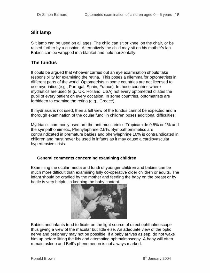
General comments concerning examining children Examining the ocular media and fundi of younger children and babies can be much more difficult than examining fully co-operative older children or adults. The infant should be cradled by the mother and feeding the baby on the breast or by bottle is very helpful in keeping the baby content. Babies and infants tend to fixate on the light source of direct ophthalmoscope thus giving a view of the macular but little else. An adequate view of the optic nerve and periphery may not be possible. If a baby arrives asleep, do not wake him up before lifting the lids and attempting ophthalmoscopy. A baby will often remain asleep and Bell’s phenomenon is not always marked.
Ronald Brown 8th January 2004

Dr Simon Barnard Optometric examination of children aged 0 – 5 years
19
Many optometrists make fundoscopy a game. One technique would be to tell the child that that the examiner is going to look into the eye to see what the child has eaten for breakfast. Another would be to ask the child to look for an imaginary animal or spider in different parts of the room. Do not make the room completely dark as this can frighten some children. Sedation and anaesthesia If a full examination of the ocular fundus of a baby or young child is critically important e.g., suspected pathology then the ophthalmologist may use a sedative e.g., chloral hydrate, or a general anaesthetic (Taylor, 1990). Methods of retinal examination
Non-Photographic methods Direct ophthalmoscopy The direct ophthalmoscope has the advantage of being relatively easy to use on co-operative patients. It provides a reasonable magnification of x15 for the emmetropic eye. Another advantage is that a view of the fundus can be obtained without the use of a mydriatic drug making it an important technique for “routine” examinations and in countries where optometrists cannot use mydriatics. However, because it is a monocular technique, stereopsis is not afforded and also the field of view is poor, only 10° in the emmetropic eye (two disc diameters). Another disadvantage is the relatively poor illumination as compared to indirect techniques but with the clear media of most children, this is less of a disadvantage. A useful fundus examination with the direct ophthalmoscope can be obtained on co-operative young children (3 years and over) but younger children and infants more difficult. It is important to seek anatomical details, particularly the disc and macular. Indirect ophthalmoscopy Indirect methods of ophthalmoscopy employ a condensing lens to form an inverted and laterally reversed image which the optometrist then views.
Ronald Brown 8th January 2004

Dr Simon Barnard Optometric examination of children aged 0 – 5 years
20
The main advantages of indirect systems are stereopsis and large field of view making changes in colour and elevation easier to detect. The illumination is also brighter than direct making viewing through media opacities easier. The head set indirect is a very important technique for babies and children and, depending on the power of the condensing lens used may give between 45° to 60° field of view. This technique should be used prior to direct to obtain an overall view or impression of the retina with a + 20 D lens giving a compromise with regard to magnification and field of view. A +30 D may be used to obtain a larger field and a +14 D used to obtain greater detail. It is necessary to dilate the pupil with an anti-muscarinic. The combination of slit lamp microscope with a fundus lens (e.g., Volk) is also a very useful technique but the ability to carry out this technique is affected more by the co-operation of the patient. The potential field of view with, for example, a +90 D Volk Superfield is 120°, although the field of illumination employed is much less. The magnification of the image is x 0.7 and, when this is viewed through the slit lamp with a x 20 magnification, then a similar magnification to that of direct is obtained but with the added advantages of good illumination, stereopsis and wide field of view. The use of a mydriatic is preferable although with a naturally large pupil, a 3-D view of the disc can be sometimes obtained without dilating.
Image courtesy of Andrew Field www.academy.org.uk
The AO or Recichert monocular indirect ophthalmoscope is also very useful for examining children providing an erect image and a field of view of 20°. Contact fundus lenses e.g., Goldman 3-mirror This technique is rarely indicated for children as great co-operation is required. A central lens provides 30O view of posterior pole with mirrors used peripheral retinal examination .
Ronald Brown 8th January 2004

Dr Simon Barnard Optometric examination of children aged 0 – 5 years
21
Imaging systems Direct ophthalmoscopy and headset indirect are most commonly used in paediatric optometric practice but there are ways in which retinal examination can be improved. Modern digital imaging systems enable the optometrists to obtain excellent images of the ocular fundi of children, usually without the need to dilate. An infra- red system allows the practitioner to focus on the retina without any other ambient illumination. The camera flash is faster than the pupil response thus enabling image capture. Digital fundus cameras Digital fundus cameras are superb for photographing children aged 3 or 4 years and older. Routine use helps in terms of serial comparison and is especially helpful in comparing optic nerve appearances over time. Can digital imaging help us with babies and children ? OptoMap Laser Scanning Ophthalmoscope This instrument offers a breakthrough in paediatric eye care with a number of advantages. No mydriatic is required for pupils larger than 1 ¾ mm; a field of view of up to 200° is photographed and a view of retina and choroid may be viewed separately or combined. The confocal system means that everything is always in focus. This means that lining up the baby exactly is less critical, and that a view, even if not the full field, is obtained.
Optomap Laser Scanning Camera and Image
Ronald Brown 8th January 2004

Dr Simon Barnard Optometric examination of children aged 0 – 5 years
22
Infants < 18 months 18 months to 3 years
3 to 5 years
External exam (inc S/L)
√ √ √
Pupillary reflexes √ √ √ Ophthalmoscopy Attempt Attempt √ Ocular motility Binocular : Fixation on face,
squeaky toy, pen torch Binocular : pen torch , toy
Binocular (& monocular): pen torch
Eye position/ ocular motor balance
Hirschberg/Krimsky/ Bruckner; near cover test
Hirschberg/Krimsky/ Bruckner, near cover test, distance cover test sometimes possible
Distance and near cover test
Fusion Gross convergence (light or toy); 5-10∆ base out by six months
Convergence ( fine picture on fixation stick or toy); 10∆ base out
Convergence ( fine picture on fixation stick or toy); > 10∆ base out
Stereopsis Lang Lang , Titmus fly /animals, TNO butterfly
Several stereo tests
Visual fields - Kinetic outline perimetry at as young as 2½ years
Kinetic outline perimetry
Colour vision - - Ishihara (Unlettered), Ishihara specific numbers, Fletcher-Hamblin
Accommodation Objective (retinoscopy) Objective; Picture recognition
Objective; Picture recognition; "push up subjective"; accommodative facility; AC/A ratio
Table 2. A guide to examination procedures for different age groups of children in primary care optometric practice Further reading
Ronald Brown 8th January 2004

Dr Simon Barnard Optometric examination of children aged 0 – 5 years
23
Barnard S & Edgar D (1996) Pediatric Eye Care, Blackwell Science, Oxford Evans B. (1999) Pickwell’s Anomalies of Binocular Vision, Butterworth-
Heinnemann, Oxford Taylor D (1990) Pediatric Ophthalmology, Blackwell Science, Oxford e-mail: [email protected]
Ronald Brown 8th January 2004