Embed Size (px)
Citation preview

OpportunisticInfections Associated with HumanImmunodeficiency Virus Infection
Definition
• Acquired immunodeficiency syndrome (AIDS)-related opportunistic infections are defined as those infections that occur with increased frequency or severity in patients with human immunodeficiency virus (HIV) infection or AIDS.
Epidemiology
• The incidence of HIV-related opportunistic infections depends on the degree of immunosuppression and environmental exposure.
• The occurrence of specific infections in some cases is due to primary infection; in other cases, disease is the result of reactivation of latent infection.
PROSPECTIVE MONITORING
• The CD4+ T-cell count
• HIV viral load
• Clinical findings
Microbiology• The constellation of infections that characterize AIDS is
unique: Pneumocystis pneumonia, Toxoplasma encephalitis, cytomegaloviral retinitis, pneumococcal pneumonia, disseminated Mycobacterium aviumcomplex, cryptosporidiosis, cryptococcal meningitis, and Mycobacterium tuberculosis infection. The occurrence of these infections individually or in a cluster should prompt consideration of underlying HIV infection/AIDS in any patient without a clear predisposing immunodeficiency.
• The organisms that cause HIV-related opportunistic infections include bacteria, fungi, viruses, and protozoa. Some are transmitted person to person, whereas others are present in certain environmental niches.
Diagnosis• Given the broad range of pathogens that can cause
infectious syndromes in patients with HIV infection/AIDS, and the potential toxicities of therapeutic agents, specific microbiologic diagnoses should be established when possible. AIDS-related opportunistic infections are diagnosed by a wide variety of techniques, including bacterial and fungal and viral culture, serum or body fluid antigen assays or polymerase chain reaction assays, colorimetric and immunofluorescent stain of secretions or tissue, and histology.

MANAGEMENT OF ANTIRETROVIRAL THERAPY FOR
PATIENTS WITH ACUTE OPPORTUNISTIC INFECTION

GENERAL PRINCIPLESOF MANAGEMENT:
• Primary Px
• Prompt Dx
• effective ART
• Reevaluation
• Secondary Px
• Drug interactions
• IRIS

PCP
• PCP was the clinical manifestation that originally suggested to clinicians that a new syndrome, AIDS, was occurring in patients who appeared to be previously healthy.
• Pneumocystis causes disease almost exclusively in the lungs
• Chest tightness or exercise intolerance
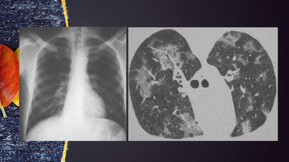
• Infiltrates in chest radiographs
• Hypoxemia in ABG
Diagnosis:
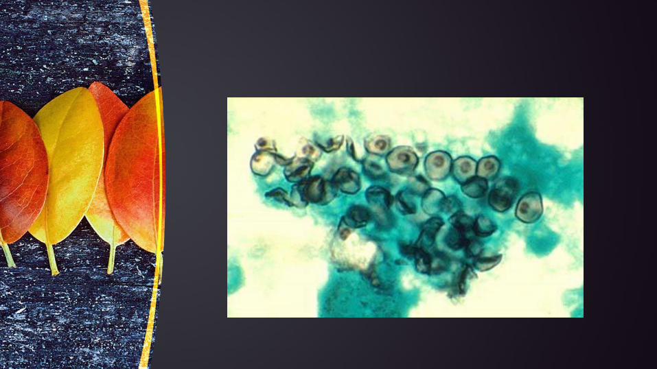
• visualization of Pneumocystis by colorimetric or immunofluorescent stain in sputum, bronchoalveolar lavage, or tissue is definitive for diagnosis of PCP
• Nucleic acid detection systems for PCP that use oral washes, gargles, sputum, or bronchoalveolar lavage
• β1-glucan detection in serum or bronchoalveolar lavage is not sufficiently sensitive or specific
Poor prognosis:
• an alveolar-arterial gradient greater than 30 mm Hg
• a severely abnormal chest radiograph
• a large number of organisms detected on lavage or biopsy
• comorbid conditions
• Delayed treatment

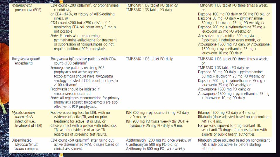
Primary prophylaxis

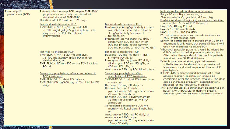
treatment
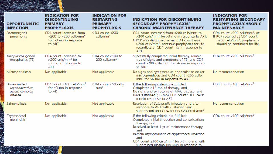
Indications for Discontinuing and Restarting Opportunistic Infection Prophylaxis
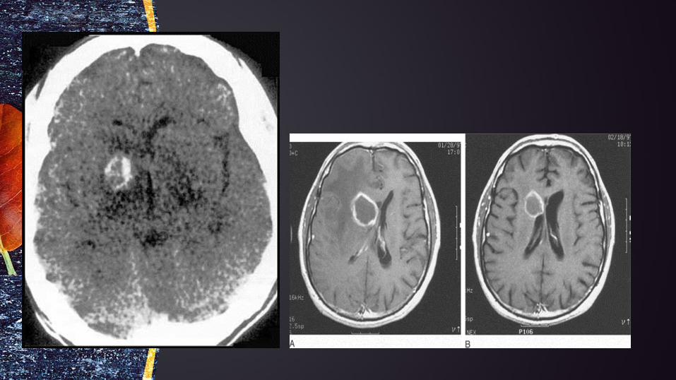
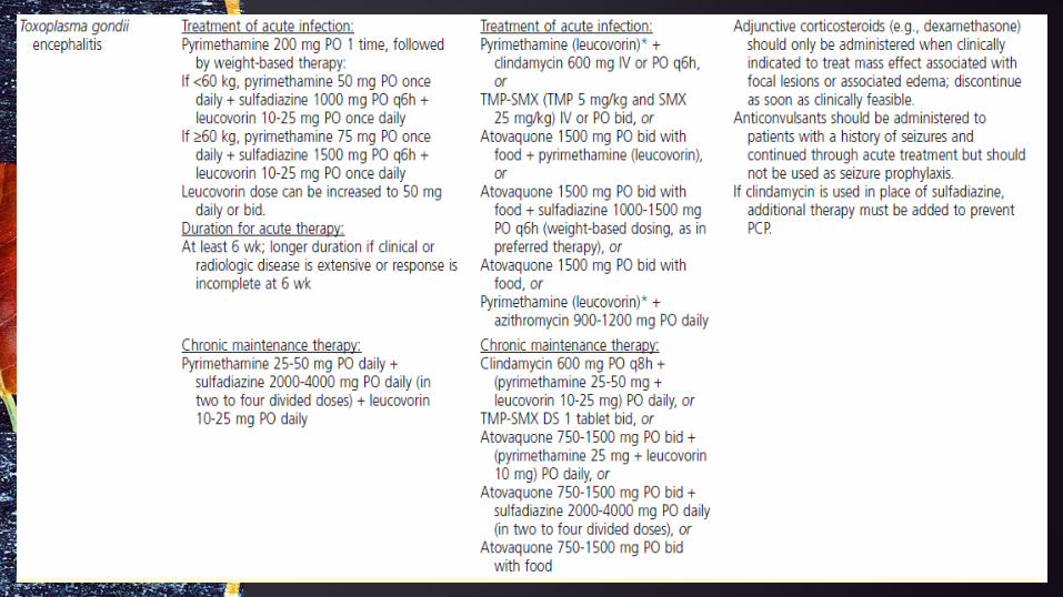
Toxoplasma gondii
• primarily by reactivation of latent disease rather than by primary infection
• manifests most often as cerebral disease presenting as fever, headache, confusion, motor defects, and seizures
• Retinochoroiditis, pneumonitis, disseminated disease, and a sepsis-like syndrome, less frequent.
• If an HIV-infected patient with a CD4+ T-cell count of less than 100 cells/mm3 presents with a space-occupying cerebral lesion that involves gray matter, the differential diagnosis should focus on two entities: toxoplasmosis and lymphoma.

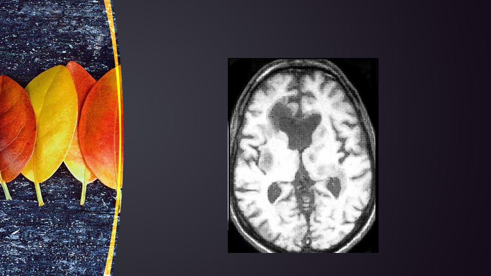
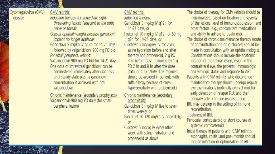
Cytomegalovirus
CMV retinitis
• CD4+ T-cell counts less than 50 cells/mm3
• Rapidly damage the macula and optic disk , ultimately blindness
• Diagnosis :clinical

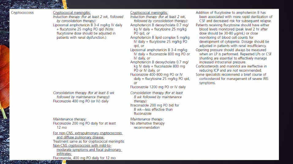
Cryptococcus neoformans
• Meningitis is the most frequent manifestation of cryptococcosis in HIV-infected patients
• fever, headache, neck stiffness, or photophobia
• In CD4+ T-cell counts less than 50 cells/mm
• Pulmonary or cutaneous manifestations
• Diagnosis: elevated protein levels and numbers of mononuclear cells and decreased glucose concentration in CSF
• CSF and serum cryptococcal antigen tests

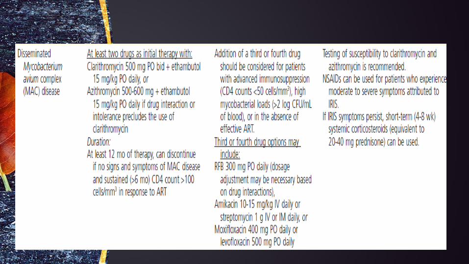
Mycobacterium avium Complex
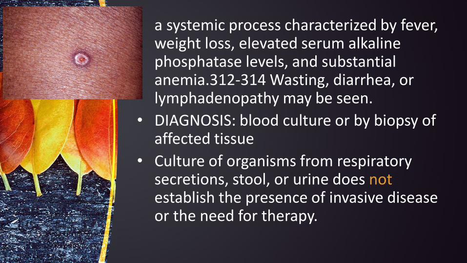
• a systemic process characterized by fever, weight loss, elevated serum alkaline phosphatase levels, and substantial anemia.312-314 Wasting, diarrhea, or lymphadenopathy may be seen.
• DIAGNOSIS: blood culture or by biopsy of affected tissue
• Culture of organisms from respiratory secretions, stool, or urine does not establish the presence of invasive disease or the need for therapy.

Thank you