Embed Size (px)
Citation preview

Chapter 5
Corticosteroids affect the
testicular androgen production
in male common carp,
Cyprinus carpio L.
Co-authors: Jan G.D. Lambert, Henk J.Th. Goos
submitted
1
2
3
4
5
6
7
8
9
10
11
12
13
14
15
16
17
18
19
20
21
22
23
24
25
26
27
28
29
30
31
32
33
34
35
36
37
38
39
40
41
42
C o r t i s o l i n h i b i t s t h e t e s t i c u l a r a n d r o g e n p r o d u c t i o n • 7 1
Opmaak • 005 14-03-2001 12:07 Page 71

1
2
3
4
5
6
7
8
9
10
11
12
13
14
15
16
17
18
19
20
21
22
23
24
25
26
27
28
29
30
31
32
33
34
35
36
37
38
39
40
41
42
7 2 • C h a p t e r 5
Opmaak • 005 14-03-2001 12:07 Page 72

A b s t r a c t
Our previous experiments to study the effect of cortisol on pubertal devel-opment in carp showed that repeated temperature stress, but especially pro-longed feeding with cortisol containing food pellets caused a retardation of thefirst waves of spermatogenesis, and a decrease in 11-ketotestosterone (11KT) andLH plasma levels.
The objective of the present study was to investigate whether the decreasein plasma 11KT is caused by a direct effect of cortisol on the steroid producingcapacity of the testis or by an indirect effect such as a decrease in plasma LH.Adolescent and pubertal isogenic male common carp were fed with either cor-tisol containing food pellets or control food pellets over a prolonged period. Ourresults indicate that cortisol has a direct inhibitory effect on the testicular andro-gen secretion, independent of the LH secretion. Furthermore, the pubertal peri-od is critical to the influence of cortisol regarding testicular androgen secretion.The effect is no longer observed at adolescence.
I n t r o d u c t i o n
In all teleost species, including the common carp, cortisol is the major cor-ticosteroid produced by the interrenals under influence of stress (Barton &Iwama, 1991). Cortisol plays a key role in the restoration of homeostasis duringor after stress and has frequently been indicated as the major factor mediatingthe suppressive effect of stress on reproduction. The developmental period du-ring which the animal acquires the capacity to reproduce is defined as puberty.The basis of pubertal maturation is the development of the gonads and theendocrine system that regulates reproductive processes, the brain-pituitary-gonad (BPG) axis.
Our previous studies demonstrated that in common carp, repeated tempe-rature-induced stress caused a retardation of the first waves of spermatogenesis(chapter 2). Furthermore, long-term cortisol treatment resulted in a similar
1
2
3
4
5
6
7
8
9
10
11
12
13
14
15
16
17
18
19
20
21
22
23
24
25
26
27
28
29
30
31
32
33
34
35
36
37
38
39
40
41
42
C o r t i s o l i n h i b i t s t h e t e s t i c u l a r a n d r o g e n p r o d u c t i o n • 7 3
Opmaak • 005 14-03-2001 12:07 Page 73

effect on spermatogenesis, accompanied by a decrease in plasma LH and plasma11-ketotestosterone (11KT) (Consten et al., 2001a). Several studies indicate that11KT has an important function of 11KT during sexual maturation. 11KT hasbeen shown to stimulate spermatogenesis in African catfish (Clar ias gar iepinus)(Cavaco et al., 1998b), the common carp (Cypr inus carpio) (Komen, personalcommunication) and in Japanese eel (Anguilla japonica) (Miura et al., 1991).
The reduction of plasma sex steroids due to stress or cortisol has beendemonstrated in a variety of vertebrate species (mammals: Norman and Smith,1992, Charpenet et al., 1981; reptiles: Moore et al., 1991, Mahmoud & Licht,1997; amphibians: Coddington & Cree, 1995 and fish: Pickering et al., 1987a,Carragher et al., 1989, Foo & Lam., 1993a). In mammals, the steroid producingcells of the testis, the Leydig cells, have been shown to contain glucocorticoidreceptors (Schultz et al., 1993) and therefore corticosteroids may exert a directeffect on the steroidogenesis. In vitro experiments suggest that stress or corti-costeroids decrease the Leydig cell sensitivity to gonadotropins (Charpenet etal., 1981, Orr & Mann, 1992) either by reducing the LH receptor content(Bambino and Hsueh, 1981) or by inhibiting the 17α-hydroxylase and/or C17,20-lyase activity (Fenske, 1997). In fish, the data on the direct effect of cortisol onsteroidogenesis are less consistent compared to mammals. Carragher andSumpter (1990) and Pankhurst et al. (1995a) found a reduction of 17β-estradi-ol and testosterone secretion by cultured ovarian follicles. In other species (gold-fish (Carassius auratus), common carp and the sparid Pagrus auratus), how-ever, Pankhurst et al. (1995b) found no evidence that the inhibitory effects ofstress on reproduction are mediated by the action of cortisol on ovariansteroidogenesis directly.
The aim of this study was to investigate if the observed decrease in plasma11KT levels is caused by a direct effect of cortisol on the steroid producingcapacity of the testis or via a decreased LH secretion. Furthermore, we wereinterested if the negative effects of cortisol are correlated with age and develop-ment of the fish.
M a t e r i a l a n d M e t h o d s
A n i m a l s
Isogenic male common carp (designated as strain E4xR3R8) were pro-duced by crossing a homozygous gynogenetic E4 female (Komen et al., 1991)with a YY-male of an unrelated homozygous androgenetic male R3R8 (Bongerset al., 1997). Fry were produced and raised in the facilities of the Departmentfor Fish Culture and Fisheries (Agricultural University, Wageningen, TheNetherlands) and transported at 21 days post hatching (dph) to the fish facilitiesat the Utrecht University.
1
2
3
4
5
6
7
8
9
10
11
12
13
14
15
16
17
18
19
20
21
22
23
24
25
26
27
28
29
30
31
32
33
34
35
36
37
38
39
40
41
42
7 4 • C h a p t e r 5
Opmaak • 005 14-03-2001 12:07 Page 74

During the experiment, the fish were kept at 25°C in a flow-through sys-tem, exposed to a 12:12 hours light-dark regime and fed pelleted dry food(Trouw, Putten,The Netherlands) at a daily ration of 20 g/kg-0.8. Immature fishwere allowed to acclimatize till 63 dph after which the experiment started orwere kept until adolescence.
E x p e r i m e n t 1 : m a t u r i n g f i s h
Cortisol (Steraloids Inc. Wilton, USA) treated food (100 mg/kg food) wasprepared as described by Pickering et al. (1987). One hundred and twenty ani-mals were equally divided over two groups. One group received control food,the other group the cortisol-treated food from 63 dph onwards as described pre-viously (Consten et al., 2001).
Fish from both groups (n=20) were sampled at several time-intervals du-ring the pubertal development, at 94 dph (early puberty), 100 dph (late puberty)and 120 dph (first wave of spermatogenesis completed). The fish were anaes-thetized in TMS (Tricaine Methane Sulfonate, Crescent Research Chemicals,Phoenix AZ, USA). Body weight was determined and blood was collected bypuncturing the caudal vasculature. After blood sampling fish were immediatelydecapitated and testes were removed for determining the gonadosomatic index(GSI) and in vitro incubation for determination of the steroid synthesizingcapacity.
E x p e r i m e n t 2 : a d o l e s c e n t f i s h
Forty-eight adolescent fish were equally divided over two groups and wereeither fed, starting at 138 dph, control food or cortisol containing food, similarto the maturing fish. At 165, 183 and 197 dph fish were sampled (n=8) accor-ding to the same procedure as in experiment 1.
A n d r o g e n s e c r e t i o n i n v i t r o
The in vitro determination of the steroid secretory capacity of testicular tis-sue of maturing fish in experiment 1 was performed as described by Cavaco etal. (1998b). In short, the left and right testis of each male were divided in equalhalves. Each half testis was weighed separately and transferred to a separate wellof a 24-wells Costar plate, containing 0.5 ml HEPES buffered L-15 medium (15 mM HEPES, 100,000 U/l penicillin/streptomycin, pH 7.4).The testis halveswere then cut into fragments of approximately 2 mm3. The culture medium ofthe four halve testis was taken off and replaced by 0.5 ml medium, with or with-out dexamethasone (Sigma, St. Louis, USA) (150 ng/ml medium) and contain-ing increasing amounts of LH (0, 10, 30 and 100 ng LH/ml medium). Carp pitu-itary extract, in which LH content determined by radioimmunoassay (RIA), wasused as LH source. After incubation for 20 hours at 25°C, the medium wasremoved, heated for 1 hour at 80°C and centrifuged at 10,000 g for 30 min at
1
2
3
4
5
6
7
8
9
10
11
12
13
14
15
16
17
18
19
20
21
22
23
24
25
26
27
28
29
30
31
32
33
34
35
36
37
38
39
40
41
42
C o r t i s o l i n h i b i t s t h e t e s t i c u l a r a n d r o g e n p r o d u c t i o n • 7 5
Opmaak • 005 14-03-2001 12:07 Page 75

room temperature. The supernatant was stored at -20°C until steroid hormonemeasurement by RIAs.
Testicular tissue from each adolescent fish separately was prepared asdescribed by Schulz et al. (1994). Then, for each fish five wells of a 24-wellsCostar plate, containing 0.5 ml L-15 medium, were filled with 100 mg testicu-lar tissue. The medium was taken off and replaced by 1 ml medium containingincreasing amounts of LH (carp pituitary extract), respectively 0, 10, 30, 100 and300 ng LH/ml medium, in the absence or presence of dexamethasone (Sigma,St. Louis, USA) (150 ng/ml medium). After an incubation of 20 hours at 25°C,the medium was treated as in experiment 1.
P i t u i t a r y e x t r a c t a n d p l a s m a L H
Luteinizing Hormone (LH) was quantified in pituitary extract and in plas-ma using a homologous RIA (slightly modified from Goos et al., 1986). Purifiedcarp LHβ subunit (a gift from Dr. E. Burzawa-Gérard) was used for the prepara-tion of standards and for 125I-labeling. Anti-LHβ (internal code # 6.3) was usedas first antibody.
Plasma LH levels were measured in all animals. In common carp, as in manyspecies, the presence of a follicle-stimulating hormone (FSH) has also beendemonstrated (Van Der Kraak et al., 1992). However, a FSH specific assay is notavailable.
S t e r o i d R a d i o i m m u n o a s s a y s ( R I A )
The steroid levels in both plasma (11KT) and medium (11KT and 11-ketoandrostenedione, OA) were determined by RIA as described previously(Schulz, 1985). In most male teleosts 11KT is considered to be the most domi-nant androgen in the plasma (Borg, 1994).Also in the male common carp 11KThas been found to be the major androgen produced by the testes (Barry et al.,1990, Koldras et al., 1990). However, in immature common carp OA is the mainandrogen produced by the testes (Komen, personal communication).
S t a t i s t i c s
All results are expressed as mean ± SEM. Plasma levels of 11KT and LH aregiven as ng per ml plasma. In vitro data are given as ng of steroid secreted, cor-rected for total testis weight. All results on the treatment effect of cortisol wereprocessed for statistical analysis by Student's T-test (p<0.05). In vitro data wereprocessed by one-way ANOVA, followed by Fisher's least significant differencetest (p<0.05).
1
2
3
4
5
6
7
8
9
10
11
12
13
14
15
16
17
18
19
20
21
22
23
24
25
26
27
28
29
30
31
32
33
34
35
36
37
38
39
40
41
42
7 6 • C h a p t e r 5
Opmaak • 005 14-03-2001 12:07 Page 76
R e s u l t s
G o n a d o s o m a t i c i n d e x ( G S I )
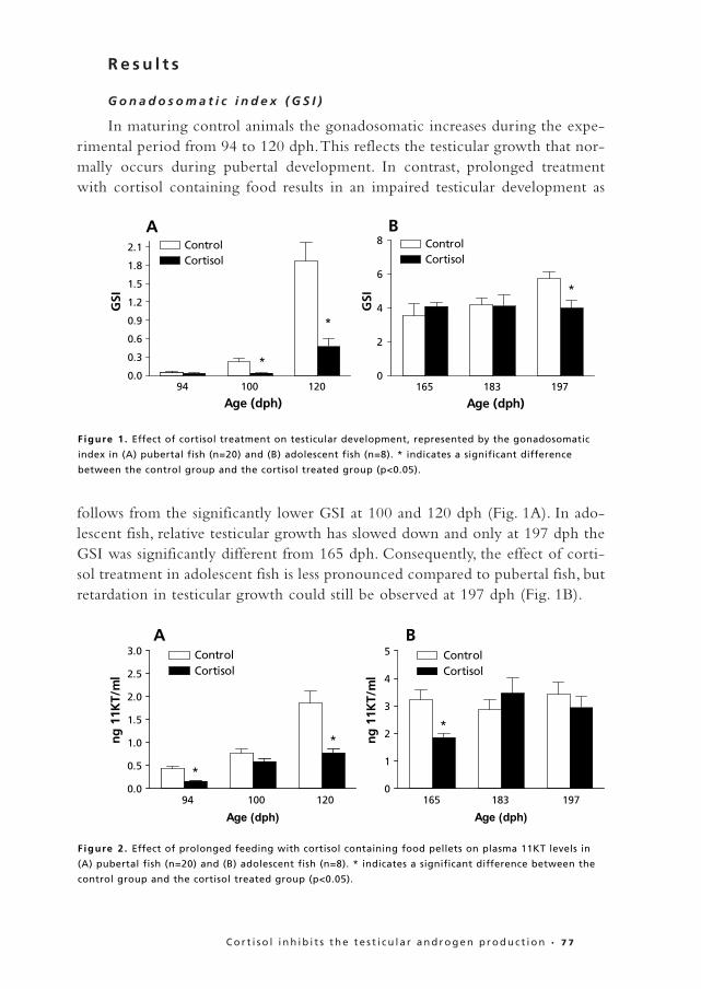
In maturing control animals the gonadosomatic increases during the expe-rimental period from 94 to 120 dph.This reflects the testicular growth that nor-mally occurs during pubertal development. In contrast, prolonged treatmentwith cortisol containing food results in an impaired testicular development as
follows from the significantly lower GSI at 100 and 120 dph (Fig. 1A). In ado-lescent fish, relative testicular growth has slowed down and only at 197 dph theGSI was significantly different from 165 dph. Consequently, the effect of corti-sol treatment in adolescent fish is less pronounced compared to pubertal fish, butretardation in testicular growth could still be observed at 197 dph (Fig. 1B).
1
2
3
4
5
6
7
8
9
10
11
12
13
14
15
16
17
18
19
20
21
22
23
24
25
26
27
28
29
30
31
32
33
34
35
36
37
38
39
40
41
42
C o r t i s o l i n h i b i t s t h e t e s t i c u l a r a n d r o g e n p r o d u c t i o n • 7 7
94 100 1200.0
0.3
0.6
0.9
1.2
1.5
1.8
2.1 ControlCortisol
*
*
Age (dph)
GSI
165 183 1970
2
4
6
8 ControlCortisol
*
Age (dph)
GSI
A B
Figure 1 . Effect of cortisol treatment on testicular development, represented by the gonadosomatic
index in (A) pubertal fish (n=20) and (B) adolescent fish (n=8). * indicates a significant difference
between the control group and the cortisol treated group (p<0.05).
94 100 1200.0
0.5
1.0
1.5
2.0
2.5
3.0 ControlCortisol
*
*
Age (dph)
ng
11K
T/m
l
165 183 1970
1
2
3
4
5 ControlCortisol
*
Age (dph)
ng
11K
T/m
l
A B
Figure 2 . Effect of prolonged feeding with cortisol containing food pellets on plasma 11KT levels in
(A) pubertal fish (n=20) and (B) adolescent fish (n=8). * indicates a significant difference between the
control group and the cortisol treated group (p<0.05).
Opmaak • 005 14-03-2001 12:07 Page 77
P l a s m a h o r m o n e l e v e l s
Similar to the GSI in maturing control fish, plasma 11KT levels increasedduring pubertal development. Plasma 11KT levels of cortisol treated animals aresignificantly lower compared to control animals (at 100 dph, the difference is notsignificant) (Fig. 2A). In adolescent control fish, plasma 11KT levels have furtherincreased and remain at the same level during the experimental period. In cor-tisol treated adolescent fish the 11KT levels at 165 dph are still behind the con-trol values. However, during the experimental period the 11KT levels in corti-sol treated fish become equal to the control values (Fig. 2B).
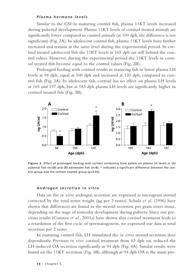
Prolonged feeding with cortisol results in maturing fish in lower plasma LHlevels at 94 dph, equal at 100 dph and increased at 120 dph, compared to con-trol fish (Fig. 3A). In adolescent fish, cortisol has no effect on plasma LH levelsat 165 and 197 dph, but at 183 dph plasma LH levels are significantly higher incortisol treated fish (Fig. 3B).
A n d r o g e n s e c r e t i o n i n v i t r o
Data on the in vitro androgen secretion are expressed as microgram steroidcorrected by the total testes weight (µg per 2 testes). Schulz et al. (1996) haveshown that differences are found in the steroid secretion per gram testes tissue,depending on the stage of testicular development during puberty. Since our pre-vious results (Consten et al., 2001a) have shown that cortisol treatment leads toa retardation of the first cycle of spermatogenesis, we expressed our data as totalsecretion per 2 testes.
In maturing control fish, LH stimulated the in vitro steroid secretion dosedependently. Previous in vivo cortisol treatment from 63 dph on, reduced theLH-induced OA secretion significantly at 94 dph (Fig. 4A). Similar results werefound on the 11KT secretion (Fig. 4B), although at 94 dph OA is the main pro-
1
2
3
4
5
6
7
8
9
10
11
12
13
14
15
16
17
18
19
20
21
22
23
24
25
26
27
28
29
30
31
32
33
34
35
36
37
38
39
40
41
42
7 8 • C h a p t e r 5
94 100 1200.0
0.5
1.0
1.5
2.0 ControlCortisol
*
*
Age (dph)
ng
LH
/ml
165 183 1970.0
0.5
1.0
1.5
2.0
2.5
3.0 ControlCortisol
*
Age (dph)
ng
LH
/ml
A B
Figure 3 . Effect of prolonged feeding with cortisol containing food pellets on plasma LH levels in (A)
pubertal fish (n=20) and (B) adolescent fish (n=8). * indicates a significant difference between the con-
trol group and the cortisol treated group (p<0.05).
Opmaak • 005 14-03-2001 12:07 Page 78

duct produced by the testes. In vitro treatment with dexamethasone resulted ina reduction of the LH-induced androgen secretion as well (Fig. 4A&B).
At 100 dph, OA is still the main androgen produced by the testes. However,both OA and 11KT production have increased, compared to 94 dph (Fig.4C&D). Cortisol treatment in vivo resulted in significantly lower LH-inducedOA and 11KT secretion in vitro. The in vitro treatment with dexamethasonecaused a reduction in the secretion of OA and 11KT, but this reduction is notsignificant due to the somewhat larger variation.
1
2
3
4
5
6
7
8
9
10
11
12
13
14
15
16
17
18
19
20
21
22
23
24
25
26
27
28
29
30
31
32
33
34
35
36
37
38
39
40
41
42
C o r t i s o l i n h i b i t s t h e t e s t i c u l a r a n d r o g e n p r o d u c t i o n • 7 9
0.0 3 10 30 1000.0
0.5
1.0
1.5
2.0 ControlCortisolControl+DexCortisol+Dex **
**
ng LH/ml medium
µg O
A p
er 2
tes
tis
0.0 3 10 30 1000.00
0.05
0.10
0.15
0.20
0.25
0.30 ControlCortisolControl+DexCortisol+Dex
***
**
**
ng LH/ml medium
µg 1
1KT
per
2 t
esti
s
0.0 3 10 30 1000
2
4
6
8 ControlCortisolControl+DexCortisol+Dex
ng LH/ml medium
µg O
A p
er 2
tes
tis
*
0.0 3 10 30 1000.0
0.5
1.0
1.5
2.0 ControlCortisolControl+DexCortisol+Dex
*
ng LH/ml medium
µg 1
1KT
per
2 t
esti
s
0.0 3 10 30 1000.0
0.5
1.0
1.5
2.0
2.5
3.0 ControlCortisolControl+DexCortisol+Dex
ng LH/ml medium
µg 1
1KT
per
2 t
esti
s
*
**
*
**
94 100 1200
1
2
3
4
5
6
7
8 ControlCortisol
*
Age (dph)
Rat
io O
A/1
1KT
A B
C D
E F
F igure 4 . Effect of in vivo cortisol treatment and in vitro dexamethasone (Dex) treatment on the in
vitro steroid secretion of pubertal fish, expressed as µg steroid per 2 testes (n=10). (A) OA secretion at
94 dph, (B) 11KT secretion at 94 dph, (C) OA secretion at 100 dph, (D) 11KT secretion at 100 dph, (E) 11KT
secretion at 120 dph, (F) ratio between OA and 11KT secreted in vitro. * indicates a significant difference
between the control group and the cortisol treated group (p<0.05). ** indicates a significant difference
of the control group with all other groups(p<0.05). *** indicates a difference between the control group
and the cortisol and dexamethasone treated group(p<0.05).
Opmaak • 005 14-03-2001 12:07 Page 79

At 120 dph, when in control animals 11KT is becoming the main steroidproduced by the testes, both basal and LH-induced 11KT secretion are signifi-cantly reduced after prolonged in vivo cortisol treatment (Fig. 4E).The in vitrodexamethasone treatment has a comparable effect as both basal and LH-stimu-lated 11KT secretion are affected. Similar results were observed for the OAsecretion (data not shown). The ratio OA/11KT shows that cortisol treatment
not only affects the androgen production quantitatively, but also its pattern. Incontrol animals there is a shift towards 11KT secretion at 120 dph, while corti-sol treatment caused the relative high production of OA to be maintained at thisage (Fig. 4F).
In contrast, in adolescent fish, there is no effect of either in vivo cortisoltreatment or in vitro dexamethasone treatment on the in vitro basal and LH-induced androgen secretion throughout the experiment (Fig. 5A-C).
1
2
3
4
5
6
7
8
9
10
11
12
13
14
15
16
17
18
19
20
21
22
23
24
25
26
27
28
29
30
31
32
33
34
35
36
37
38
39
40
41
42
8 0 • C h a p t e r 5
0.0 3 10 30 100 3000
3
6
9
12
15 ControlCortisolControl+DexCortisol+Dex
ng LH/ml medium
µg 1
1KT
per
2 t
esti
s
0.0 3 10 30 100 3000
3
6
9
12
15 ControlCortisolControl+DexCortisol+Dex
ng LH.ml mediumµg
11K
T p
er 2
tes
tis
0 3 10 30 100 3000
6
12
18
24
30 ControlCortisolControl+DexCortisol+Dex
ng LH/ml medium
µg 1
1KT
per
2 t
esti
s
A B
C Figure 5 . Effect of in vivo cortisol treatment
and in vitro dexamethasone (Dex) treatment
on the in vitro 11KT secretion of adolescent
fish, expressed as µg 11KT per 2 testes (n=8)
at (A) 165 dph, (B) 183 dph and (C) 197 dph.
Opmaak • 005 14-03-2001 12:07 Page 80

D i s c u s s i o n
Previous work has shown that prolonged treatment with cortisol caused aretardation of the first waves of spermatogenesis, which are associated with theonset of puberty. This was accompanied by a decrease in plasma 11-ketotestos-terone (11KT) (Consten et al., 2001a). In the present study we show that theobserved decrease in plasma 11KT levels is caused by a direct effect of cortisolon the steroid producing capacity of the testis and is probably independent ofthe LH secretion.
As previously observed, cortisol treatment of maturing fish caused a retar-dation of pubertal testicular development as reflected by the lower GSI, thelower plasma 11KT levels, and the lower plasma LH levels at the onset of puber-tal development, 94 dph. However, at 100 dph we observe plasma LH levelsequal to the control group and at 120 dph the plasma levels in cortisol treatedfish are even significantly elevated, but plasma 11KT levels are still lower thancontrol fish. These results suggest that the decrease in plasma androgen levels isnot caused by an effect of cortisol on LH levels. Pankhurst & Van Der Kraak(2000) also found evidence that the inhibitory effect of stress on plasma sexsteroids is independent of the plasma LH levels.
In contrast, in adolescent fish we observe no effect of cortisol treatment onthe testicular development at 165 and 183 dph. Only at 197 dph an inhibitoryeffect of cortisol treatment becomes apparent.At 165 dph, plasma 11KT levels isstill lower in cortisol treated adolescent fish compared to controls, but during theexperimental period they increase to same values as the controls. From theseobservations we conclude that fish become less sensitive to cortisol during sex-ual maturation. Apparently, cortisol sensitivity depends on the maturational sta-tus of the animal. Indeed, Pankhurst & Van Der Kraak (2000), demonstrated thatin female rainbow trout the effect of cortisol on ovarian steroidogenesis dependson the stage of the reproductive cycle.
In mammals, cortisol may have a direct effect on the Leydig cells, since theyhave been shown to possess glucocorticoid receptors (Schultz et al., 1993).Studies by Charpenet et al. (1981) and by Orr & Mann (1992) demonstrate thatstress decreases the sensitivity of the Leydig cell to gonadotropins. This may becaused by reducing the LH receptor content (Bambino and Hsueh, 1981) or byinhibiting the 17α-hydroxylase and/or C17,20-lyase activity (Fenske, 1997).
Our results demonstrate that prolonged exposure to cortisol reduced theandrogen secreting capacity of the testis. Both OA and 11KT secretion in vitroare significantly reduced and also the difference in the ratio OA/11KT showsonce more that the testicular development in cortisol treated animals is retard-ed since the ratio still reflects a more immature pattern. Our results are notappropriate to reveal the precise mechanism via which cortisol affects the tes-ticular androgen production. It is, however, unlikely that prolonged exposure to
1
2
3
4
5
6
7
8
9
10
11
12
13
14
15
16
17
18
19
20
21
22
23
24
25
26
27
28
29
30
31
32
33
34
35
36
37
38
39
40
41
42
C o r t i s o l i n h i b i t s t h e t e s t i c u l a r a n d r o g e n p r o d u c t i o n • 8 1
Opmaak • 005 14-03-2001 12:07 Page 81

cortisol causes a decrease in LH receptor content, since the sensitivity to LH isunchanged. At 100 dph and 120 dph the stimulation factor (data not shown) ofLH is similar for control and cortisol treated animals.We therefore hypothesizethat cortisol affects the enzyme activity involved in the androgen production. Ina successive study we will investigate this hypothesis, as well as the possibilitythat cortisol competitively inhibits the conversion of 11β-hydroxyandrostene-dione (OHA) into OA.
In contrast, in vitro treatment with dexamethasone does appear to affect LHsensitivity. At 94 and 120 dph, testes taken from control animals do not show anincrease in the androgen production upon LH stimulation in the presence ofdexamethasone. In several studies corticosteroids have been suggested (e.g.Pankhurst & Van Der Kraak, 2000; Valli et al., 2000) and shown (reviewed byBorski, 2000) to mediate their inhibiting effect by interfering with signal trans-duction. In rat Leydig cells, chronic treatment with corticosterone diminishedthe production of testosterone, as well as the basal and LH-stimulated cyclicAMP production (Sankar et al., 2000). Based on these results we hypothesizethat the in vitro effect of dexamethasone in our experiments may be caused byan interference of corticosteroids with the LH signal transduction, therebyblocking the LH response and thus the LH-induced secretion of 11KT and OA.
In summary, we showed that cortisol has a direct inhibitory effect on thetesticular androgen secretion, and not via plasma LH levels. The underlyingmechanism may involve an inhibitory effect on expression of the steroid pro-ducing enzymes, substrate inhibition of enzymes that have a function in the con-version of cortisol as well as androgen precursors. Moreover, a direct interferencewith the LH signal transduction can not be excluded. Furthermore, our resultsdemonstrate that cortisol sensitivity depends on the maturational status of theanimal.
1
2
3
4
5
6
7
8
9
10
11
12
13
14
15
16
17
18
19
20
21
22
23
24
25
26
27
28
29
30
31
32
33
34
35
36
37
38
39
40
41
42
8 2 • C h a p t e r 5
Opmaak • 005 14-03-2001 12:07 Page 82