Embed Size (px)
Citation preview
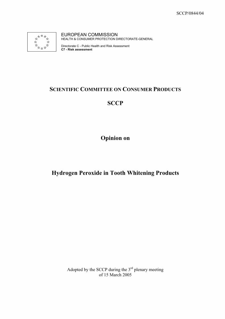
SCCP/0844/04
EUROPEAN COMMISSION HEALTH & CONSUMER PROTECTION DIRECTORATE-GENERAL Directorate C - Public Health and Risk Assessment C7 - Risk assessment
SCIENTIFIC COMMITTEE ON CONSUMER PRODUCTS
SCCP
Opinion on
Hydrogen Peroxide in Tooth Whitening Products
Adopted by the SCCP during the 3rd plenary meeting of 15 March 2005
SCCP/0844/04 Opinion on hydrogen peroxide in tooth whitening products
TABLE OF CONTENTS 1. BACKGROUND 3 2. TERMS OF REFERENCE 3 3. OPINION 4 4. CONCLUSION 40 5. MINORITY OPINION 6. REFERENCES 41 7. ACKNOWLEDGEMENTS 49
2
SCCP/0844/04 Opinion on hydrogen peroxide in tooth whitening products
1. BACKGROUND The Scientific Committee on Cosmetics and Non Food Products intended for Consumers (SCCNFP) has been consulted and expressed its views on the safety of hydrogen peroxide tooth whitening systems in several occasions, most recently on 17 September 2002 (SCCNFP/0602/02, final). In October 2003 the SCCNFP was asked to clarify its opinion. On the basis of the dossiers already submitted, the SCCNFP adopted opinion SCCNFP/0752/03 of 20 October 2003 on “The Use of Hydrogen Peroxide in Tooth Whitening Products, Clarification concerning its opinion of the Scientific Committee on Cosmetic Products and Non-Food Products intended for Consumers”, concluding following: “It is known that the use of tobacco, and alcohol abuse, cause an increased risk of oral cancer. Hydrogen peroxide may enhance this risk. This effect cannot be quantified. It is not anticipated that the tooth whitening products of the type being discussed will represent a risk of oral cancer in people neither using tobacco nor abusing alcohol. The tooth whitening products of the type being discussed should only be used under the surveillance of a dentist. These tooth whitening product should not be freely available to consumers.” In September 2003, a Member State provided the European Commission with the publication of a review “Tooth bleaching - A Critical Review of the Biological Aspects” Crit.Rev.Oral.Biol.Med 14(4):292-304 (2003), and in November the European Commission received from COLIPA1 a third submission on “Hydrogen Peroxide and Hydrogen Peroxide releasing Substances used in Tooth Whitening Products” with new human clinical safety data, data on pharmacokinetics, on market experience and on the carcinogenic risk. In February the European Commission received an evaluation by the French Committee on Cosmetology on hydrogen peroxide and hydrogen peroxide releasing substances in tooth whitening products. In addition the European Commission has received documents from COLIPA and PHD Pharmaceuticals NV on hydrogen peroxide and hydrogen peroxide releasing substances in tooth whitening products. The SCCP was requested to clarify whether the terms “tooth whitening products” and “tooth bleaching products” used in the opinion of the Scientific Committee defines the same kind of cosmetic products. 2. TERMS OF REFERENCE The SCCP is requested to answer the following questions: 1. Does the SCCP agree that the new additional data provide the necessary reassurance to
support the safety of up to 6% hydrogen peroxide in tooth whitening products freely and directly available to consumer in various application forms (strips, trays, etc.)?
1 COLIPA - European Cosmetics Toiletry and Perfumery Association
3
SCCP/0844/04 Opinion on hydrogen peroxide in tooth whitening products
2. Considering the new additional data provided, does the SCCP recommend that any specific
information should be provided to consumers related to the safe use of these tooth whitening products?
3. If the answer to the question on free and direct availability to consumer is negative, would
the SCCP identify and quantify any remaining risks that need to be addressed taking into account in particular the overall data on pharmacokinetics and exposure?
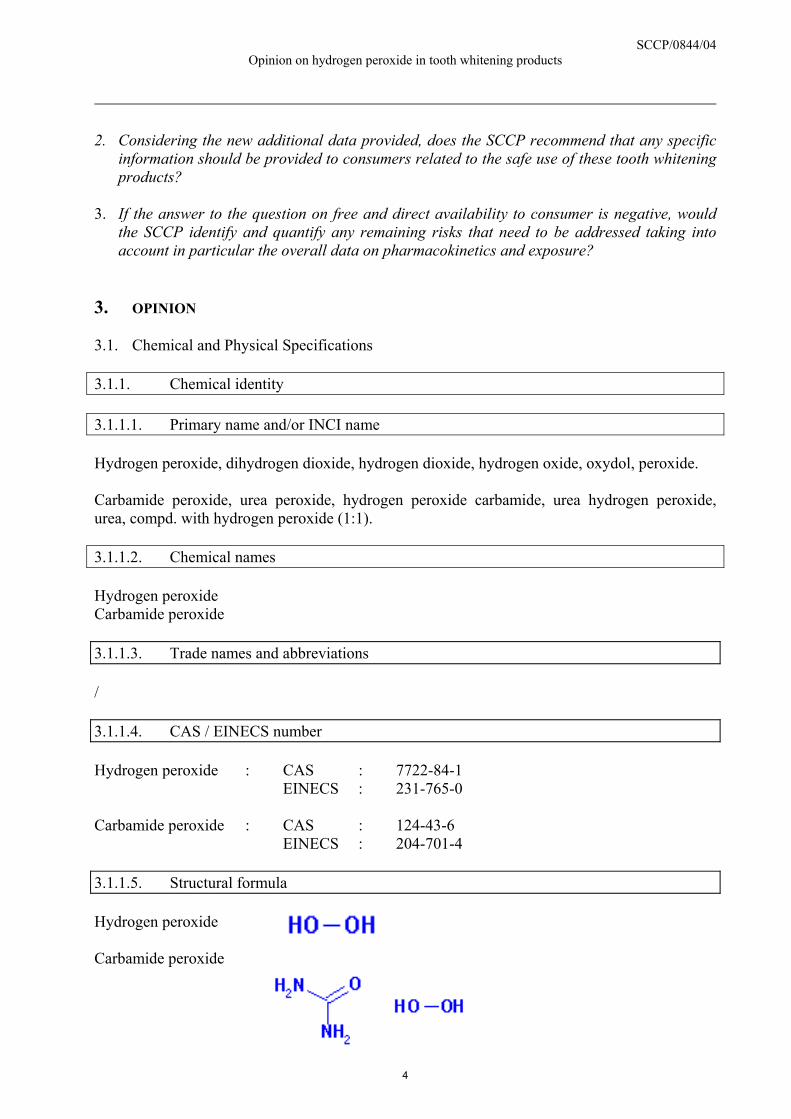
3. OPINION 3.1. Chemical and Physical Specifications 3.1.1. Chemical identity 3.1.1.1. Primary name and/or INCI name Hydrogen peroxide, dihydrogen dioxide, hydrogen dioxide, hydrogen oxide, oxydol, peroxide. Carbamide peroxide, urea peroxide, hydrogen peroxide carbamide, urea hydrogen peroxide, urea, compd. with hydrogen peroxide (1:1). 3.1.1.2. Chemical names Hydrogen peroxide Carbamide peroxide 3.1.1.3. Trade names and abbreviations / 3.1.1.4. CAS / EINECS number Hydrogen peroxide : CAS : 7722-84-1
EINECS : 231-765-0 Carbamide peroxide : CAS : 124-43-6
EINECS : 204-701-4 3.1.1.5. Structural formula Hydrogen peroxide Carbamide peroxide
4
SCCP/0844/04 Opinion on hydrogen peroxide in tooth whitening products
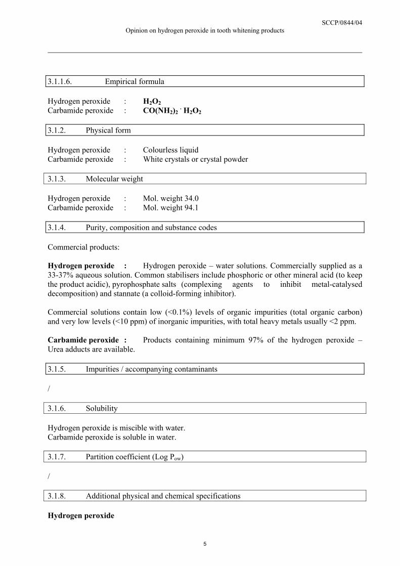
3.1.1.6. Empirical formula Hydrogen peroxide : H2O2 Carbamide peroxide : CO(NH2)2 . H2O2 3.1.2. Physical form Hydrogen peroxide : Colourless liquid Carbamide peroxide : White crystals or crystal powder 3.1.3. Molecular weight Hydrogen peroxide : Mol. weight 34.0 Carbamide peroxide : Mol. weight 94.1 3.1.4. Purity, composition and substance codes Commercial products: Hydrogen peroxide : Hydrogen peroxide – water solutions. Commercially supplied as a 33-37% aqueous solution. Common stabilisers include phosphoric or other mineral acid (to keep the product acidic), pyrophosphate salts (complexing agents to inhibit metal-catalysed decomposition) and stannate (a colloid-forming inhibitor). Commercial solutions contain low (<0.1%) levels of organic impurities (total organic carbon) and very low levels (<10 ppm) of inorganic impurities, with total heavy metals usually <2 ppm. Carbamide peroxide : Products containing minimum 97% of the hydrogen peroxide – Urea adducts are available. 3.1.5. Impurities / accompanying contaminants / 3.1.6. Solubility Hydrogen peroxide is miscible with water. Carbamide peroxide is soluble in water. 3.1.7. Partition coefficient (Log Pow) / 3.1.8. Additional physical and chemical specifications Hydrogen peroxide
5
SCCP/0844/04 Opinion on hydrogen peroxide in tooth whitening products
Pure H2O2 (not commercially available in EU)
Melting point : -0.4oC Boiling point : 150-152oC Density : 1.4425 g/cm3 Vapour pressure : 3 hPa
Carbamide peroxide Melting point : 75-85oC Boiling point : not available Density : 1.4 g/cm3 Vapour pressure : not available
3.2. Function and uses Hydrogen peroxide is capable of undergoing numerous reactions (e.g., molecular additions, substitutions, oxidations and reductions). It is a strong oxidant and can form free radical by homolytic cleavage. Carbamide peroxide contains approximately 35% hydrogen peroxide and forms hydrogen peroxide and urea in liquid solution. 750,000 tonnes hydrogen peroxide (calculated as 100% H2O2) were produced in Europe in 1995. The main usage of hydrogen peroxide is in production of chemicals and bleaching of cellulose pulp and textiles. Small quantities are used for such purposes as disinfection of eye contact lenses, disinfections of wounds and mouth washing. Both hydrogen peroxide and carbamide peroxide are used for hair bleaching, oral antiseptics, dentifrices, oxidation of permanent waves, hair relaxer, ear drops, sores and tooth bleaching. Peroxide compounds including hydrogen peroxide and carbamide peroxide have been used in various dental procedures for many years. Reports of using peroxides to bleach or whiten teeth can be traced back to more than a century ago. Current peroxide containing whiteners used in USA can be classified into 3 categories: 1) Those containing high concentration of hydrogen peroxide (30-35%) or carbamide peroxide (35%) for professional use only; 2) materials that are dispensed by dentists and used by patients at home (up to 10% hydrogen peroxide or 16% carbamide peroxide); and 3) over-the-counter products with hydrogen peroxide content up to 6% and available to consumers for home use (Li, 1996). The first articles on bleaching teeth using night guard whitening bleaching were published in 1989 (Christensen, 1989a, b; Haywood and Heymann, 1989). The whitening effect is due to degradation of high molecular weight, complex organic molecules that reflect a specific wavelength of light and is responsible for the colour of the stain. The resulting degradation products are of lower molecular weights and are less complex molecules that reflect less light and result in a reduction or elimination of the discoloration (Flaitz and Hicks, 1996). Both the dentin and the enamel change colour as a result of the easy passage of the peroxide and urea through the tooth. Extended treatment times have been developed for difficult situations. Heavy tobacco stains may require as much as three months of treatment. Tetracycline-stained teeth have responded in two to six months of nightly treatment, although not to the extent of normal teeth. Single dark teeth can also be bleached successfully. It is reported in one study that after 18 months 74% and after 3 years 62% of patients whose teeth were bleached, still exhibited stable
6
SCCP/0844/04 Opinion on hydrogen peroxide in tooth whitening products
colour, and “touch-up” generally requires only one to two days of retreatment for each week taken for initial treatment (see Marshall et al., 1995, Haywood, 1997). In another study (Leonard, 1998) 17% (4 persons) responded that there were no obvious change in the colour 13-25 months after the treatment, while 57% (13 persons) stated that there was a slight darkening, but it was not noticeable by other people. 75-89 months after the treatment, 10% (3 persons) responded there were no obvious change in colour and 25% (7 persons) that there was a slight darkening, but it was not noticeable by other persons. 48% (14 persons) responded that there had been some darkening, but they had retreated the teeth back to acceptable colour. Several different techniques are used for bleaching teeth at home. The peroxide may be placed in dentist produced trays or commercial available trays. Later strips containing from 6% and up to 14% hydrogen peroxide have become commercially available. The textured strip is made of polyethylene and the backing is made of polyester. The strips are designed to adhere directly to the surface of the teeth. The strips should be worn twice a day for 30 minutes over a period of 14 days. After using all of the upper teeth strips, the process is repeated on the lower teeth. After premarketing period, the strips have been extensively marketed in USA from May 2001. Recently, peroxide containing paint-on gels for tooth whitening have become available. The gel stays on the teeth overnight for a certain time period. For the purpose of this Opinion the terms “tooth whitening products” and “tooth bleaching products” define the same kind of products. 3.3. Toxicological Evaluation 3.3.1. Acute toxicity 3.3.1.1. Acute oral toxicity Hydrogen peroxide:
* Oral LD50 - values for rats vary between 600-1617 mg/kg bw (Y.Li, unpublished; Ito et al., 1976).
* A 16-month-old boy was found playing with an empty bottle that had contained about 230
g of 3% hydrogen peroxide solution. The container had a cracked lid that allowed the contents to be sucked. White foam emerged from the child’s mouth and nose. He then walked to bed and was found dead 10 hours later. In a post-mortem examination there was frothy blood in the right ventricle of the heart and the portal venous system. The gastric mucosa was red and the brain oedematous. Histopathological examination showed oedema in the lungs, and diffuse interstitial emphysema was evident. Gas emboli are found within the pulmonary vasculature and gastric and intestinal lymphatics. Clear vacuoles were also found within the walls of the gastrointestinal tract, in the spleen, kidney and myocardium (Cina et al., 1994). The estimated dose of hydrogen peroxide ingested was 7 g, about 600 mg/kg bw for a boy of 11.6 kg.
* An uncommon route of absorption from a cavity presumably lined by well-vascularized
granulomatous tissue involved an obese 54-year-old male who underwent irrigation of an infected and fistulous herniorrhaphy wound with 5 x 20 ml volume of 3 % hydrogen peroxide. Not all irrigating volume seemed to have drained from the wound. On the fifth
7
SCCP/0844/04 Opinion on hydrogen peroxide in tooth whitening products
irrigation, the patient suddenly lost consciousness, showed cardiac shock and fell to coma which lasted for 15 min. There was no indication of red cell damage. ECG showed signs of transient myocardial ischaemia. The patient made a full recovery within 3 days. The authors attributed this occurrence to widespread embolization of oxygen microbubbles, especially to the cerebral and coronary arteries (Bassan et al., 1982). If it is presumed that as much as one half of the volume of the irrigating solution was absorbed, the hydrogen peroxide dose would have been 1.5 g implying for an obese person (assumed weight of 100 kg) about 15 mg/kg bw.
* Oxygen embolism has been reported in several infants following intestinal irrigation with
hydrogen peroxide to remove meconium (Danis et al., 1967; Shaw et al., 1967). In one case a 36-hour old infant died following use of 1% hydrogen peroxide to remove inspissated meconium from the bowel due to meconium ileus (Shaw et al., 1967).
Tooth whiteners containing 10-22% carbamide peroxide:
* Oral LD50 - values for rats reported >5.000 mg/kg bw (Rope [Report], 1993; Huang [Report], 1996; Adam-Rodwell et al., 1994; Cherry et al., 1993). (It appears that LD50 studies with rats for doses less than 5.000 mg/kg bw has not been performed)
* Oral LD50 - values for mice vary between 87.2-143.8 mg/kg bw (Woolverton et al., 1993).
3.3.1.2. Acute dermal toxicity Hydrogen peroxide
* Dermal LD50 -values for rats vary between 700->7500 mg/kg bw (FDA, 1983).
* Dermal LD50 –values in rabbits about 630 mg/kg bw (FDA, 1983). 3.3.1.3. Acute inhalation toxicity / 3.3.2. Irritation and corrosivity 3.3.2.1. Skin irritation Hydrogen peroxide:
* Skin irritation tests in rabbits with concentration of hydrogen peroxide of 3-8% were non-
irritating to intact and abraded skin following exposure for 24 hours under occlusive
8
SCCP/0844/04 Opinion on hydrogen peroxide in tooth whitening products
dressing (cited in ECETOC, 1996). Irritation was slight following 4 hour exposure to 10% hydrogen peroxide and mild with 35% hydrogen peroxide. Desquamation occurred in 2 of 6 animals at day 14 with the latter concentration (Aguinaldo et al. [Abstract], 1992).
Tooth whiteners containing 10-22% carbamide peroxide:
* Primary irritation of the skin of rabbits was not found with tooth whitener (Rope [Report], 1993).
3.3.2.2. Eye irritation
Hydrogen peroxide:
* Eye irritation studies with rabbits indicate that a 5% hydrogen peroxide solution is non-irritant to mild-irritant (Weiner et al. [Abstract], 1990).
* Several drops of a 2-5% solution induced much clouding of the cornea and inflammation
of the conjunctiva of rabbit eyes. A 1% solution applied repeatedly caused conjunctival hyperaemia and slight corneal haze, followed by recovery (Koster, 1921 as quoted by Grant, 1986).
* Testing of eye irritancy for hydrogen peroxide with the Draize method indicated that 5%
solution was slightly irritating (FMC, 1987a), 8% solution was moderately irritating (EU classification irritating) (FMC, 1987b), and 10% solution was highly irritating (EU classification risk of serious damage to eyes) (FMC, 1985).
* A woman who had inadvertently stored a contact lens in a 3% hydrogen peroxide
disinfectant solution experienced hyperaemia, tearing, and eyelid spasm (Knoph, 1984).
* In 10 human volunteers, the threshold of detection for irritation was about 0.1% when hydrogen peroxide was administered as drops directly to the eye (McNally, 1990).
* When a hydrogen peroxide solution was administered to the eye of human volunteers via
soaking contact lenses, the threshold of detection for hydrogen peroxide irritation was less than 0.03% (McNally, 1990).
3.3.2.3. Mucous membrane irritation
Hydrogen peroxide:
* 1 or 1.2% hydrogen peroxide applied to the gingivae or tongues of anaesthetised dogs by continuous drip caused oedema, followed by destruction and sloughing of the cornified epithelial layer of the gingivae (Martin et al., 1968, Dorman and Bishop, 1970).
9
SCCP/0844/04 Opinion on hydrogen peroxide in tooth whitening products
Tooth whiteners containing 10-22% carbamide peroxide: * No evidence of oral mucosal irritation after applying tooth whiteners containing 10% or
22% carbamide peroxide for up to 6 week in experiments with rats, hamsters and rabbits has been reported (Rope [Report], 1993; Huang [Report], 1996; Adam-Rodwell et al., 1994; Li et al. [Abstract], 1996; Webb [Report], 1996).
* Stomach gavage of 15 and 50 mg/kg bw carbamide peroxide or 150 and 500 mg/kg bw
whitener containing 10% carbamide peroxide produced ulceration of gastric mucosa in the 1-hour rats; the lesions appeared to be healing after 24 hours (Dahl and Becher, 1995).
* Stomach gavage of doses up to 2,000 mg/kg bw of tooth whiteners containing 10%
carbamide peroxide or 70 mg hydrogen peroxide was given weekdays for 15 weeks or 6 months to Chinese hamsters. Cyclophosphamide and water served as control substances. (Concentration and results with cyclophosphamide not stated). Histopathological findings of the gastroduodenal tissue were comparable among the groups (Li et al. [Abstract], 1993).
3.3.3. Skin sensitisation Hydrogen peroxide:
* Ten guinea pigs were exposed to 3 or 6% hydrogen peroxide on intact or abraded skin and
by intradermal injections of 0.1 ml of test solution in saline. Test solutions were re-applied 9 times over a 2 week period prior to a challenge to evaluate sensitisation. The final reactions did not indicate induction of skin sensitization with either solution (DuPont [Report], 1953).
* A case report observed skin sensitisation reaction from two women who had been exposed
to hydrogen peroxide as an ingredient in commercial hair dyes. Both women tested positively to 3% hydrogen peroxide and numerous other ingredients in the hair dyes. The author reported that 156 hairdressers patch tested negatively to 3% hydrogen peroxide. In general, the data do not provide evidence that hydrogen peroxide is a skin sensitiser in man (Aguire et al., 1994).
3.3.4. Dermal / percutaneous absorption Biological membranes are highly permeable to hydrogen peroxide and it is expected that hydrogen peroxide is readily taken up by the cells constituting the absorption surfaces, but at the same time it is effectively metabolised, and it is uncertain to what extent the unchanged substance may enter the blood circulation. 3.3.5. Repeated dose toxicity 3.3.5.1. Repeated Dose (28 days) oral / dermal / inhalation toxicity /
10
SCCP/0844/04 Opinion on hydrogen peroxide in tooth whitening products
3.3.5.2. Sub-chronic (90 days) oral / dermal / inhalation toxicity Hydrogen peroxide Mice Mice drinking 0.15% hydrogen peroxide (about 150 mg/kg/day) ad libitum grew normally and developed no visible abnormalities during a 35-week test period (FDA, 1983). Necropsy results show changes in the liver, kidney and stomach and small intestine. Hydrogen peroxide solutions at >1% (> 1 g/kg/day) caused pronounced weight loss and death of mice within 2 weeks (FDA, 1983). Mice (C57BL/6N, catalase deficient) (groups of 15/sex) received solutions of 0, 100, 300, 1000 or 3000 ppm hydrogen peroxide in distilled water for 13 weeks. Mild-minimal duodenal mucosal hyperplasia was noted in animals receiving 1000 and 3000 ppm hydrogen peroxide and in one male receiving 300 ppm hydrogen peroxide for 13 weeks. All effects noted during treatment period, including the duodenal hyperplasia, were reversible during the 6 weeks recovery period. The NOAEL was 100 ppm or 26 and 37 mg/kg/day hydrogen peroxide for males and females respectively (Weiner et al., 1998). Rats When rats were administered hydrogen peroxide by oral gastric tube 6 days weekly for 90 days, the dose of 506 mg/kg suppressed bodyweight gain, decreased food consumption, and caused changes in haematology, blood chemistry, and organ weights. Principal organ affected was gastric mucosa, and the effect was local. The no-observed-effect-level (NOEL) of hydrogen peroxide was 56.2 mg/kg/day (Ito et al., 1976). In another rat study (Kawasaki et al., 1969) the NOAEL of hydrogen peroxide was 30 mg/kg/day, when the animals were treated by oral gastric tubing daily for 100 days. The same study showed no adverse effect in rats receiving a diet containing 6 mg hydrogen peroxide in 20 g of food (about 12 mg/kg/day). Humans Several studies have reported the use of 0.75 or 1.5% hydrogen peroxides as a mouthwash or mouth rinse for periods of up to three months. Tombes et al. (1993) reported discoloration of the mucosal surfaces and the tongue following 5 weeks of rinsing 4 times daily with 0.75% or 1.5% hydrogen peroxide-containing solutions. Shibly et al. (1997) reported no adverse effects from a 1.5% hydrogen peroxide mouthwash, used for 60 days. Winer et al. (1991) reported that 4 times a day use of a 1.5% hydrogen peroxide mouthwash for 90 days resulted in no intraoral soft tissue adverse effects. Use of 3% hydrogen peroxide 3 to 5 times per day as a mouth rinse resulted in mucosal irritations in 2 individuals with prior tissue injury. The pre-existing lesions worsened after exposure to hydrogen peroxide (Rees and Orth, 1986). Herrin et al. (1987) have shown that use of 3% hydrogen peroxide with sodium bicarbonate did not cause lesions in healthy individuals. Gingival lesions were seen in patients who used home care solutions employing 5 M sodium chloride in addition to 3% hydrogen peroxide and sodium bicarbonate.
11
SCCP/0844/04 Opinion on hydrogen peroxide in tooth whitening products
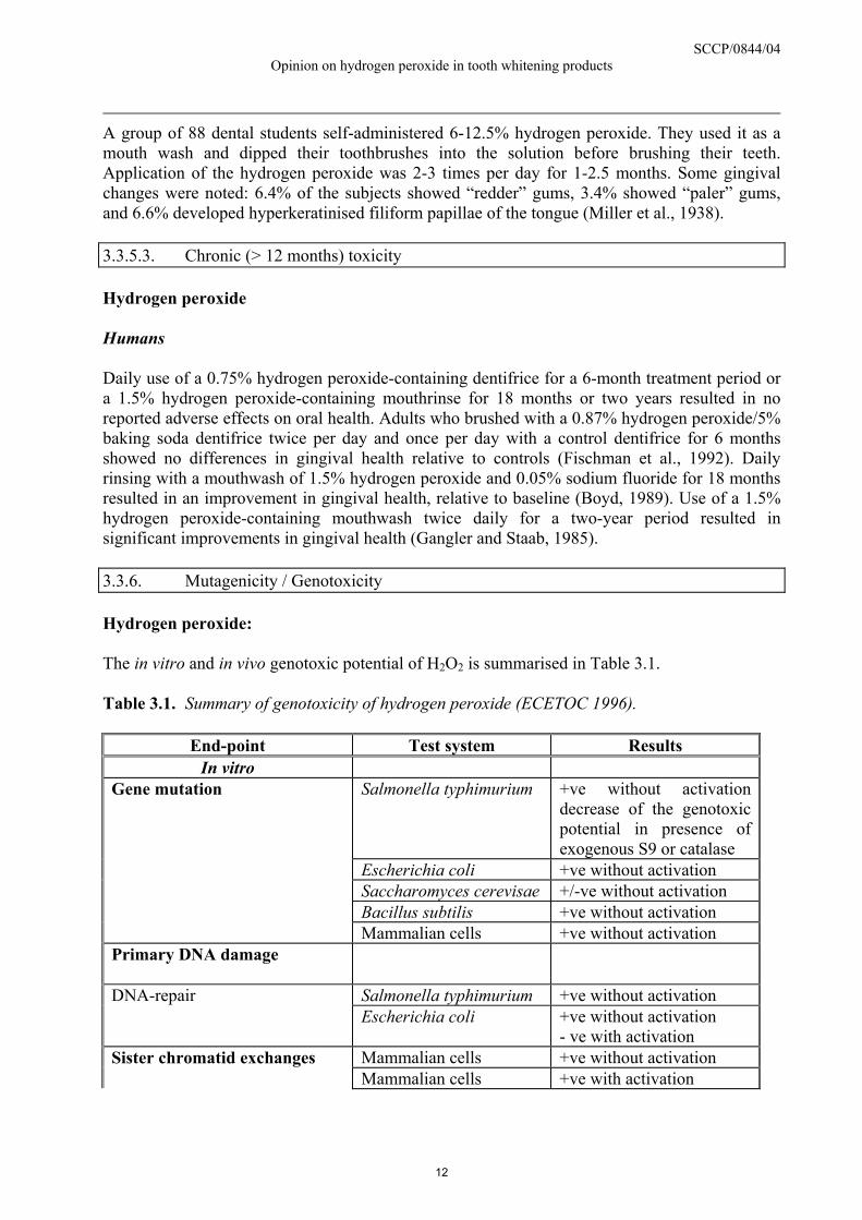
A group of 88 dental students self-administered 6-12.5% hydrogen peroxide. They used it as a mouth wash and dipped their toothbrushes into the solution before brushing their teeth. Application of the hydrogen peroxide was 2-3 times per day for 1-2.5 months. Some gingival changes were noted: 6.4% of the subjects showed “redder” gums, 3.4% showed “paler” gums, and 6.6% developed hyperkeratinised filiform papillae of the tongue (Miller et al., 1938). 3.3.5.3. Chronic (> 12 months) toxicity Hydrogen peroxide Humans Daily use of a 0.75% hydrogen peroxide-containing dentifrice for a 6-month treatment period or a 1.5% hydrogen peroxide-containing mouthrinse for 18 months or two years resulted in no reported adverse effects on oral health. Adults who brushed with a 0.87% hydrogen peroxide/5% baking soda dentifrice twice per day and once per day with a control dentifrice for 6 months showed no differences in gingival health relative to controls (Fischman et al., 1992). Daily rinsing with a mouthwash of 1.5% hydrogen peroxide and 0.05% sodium fluoride for 18 months resulted in an improvement in gingival health, relative to baseline (Boyd, 1989). Use of a 1.5% hydrogen peroxide-containing mouthwash twice daily for a two-year period resulted in significant improvements in gingival health (Gangler and Staab, 1985). 3.3.6. Mutagenicity / Genotoxicity Hydrogen peroxide: The in vitro and in vivo genotoxic potential of H2O2 is summarised in Table 3.1. Table 3.1. Summary of genotoxicity of hydrogen peroxide (ECETOC 1996).
End-point Test system Results In vitro
Gene mutation Salmonella typhimurium +ve without activation decrease of the genotoxic potential in presence of exogenous S9 or catalase
Escherichia coli +ve without activation Saccharomyces cerevisae +/-ve without activation Bacillus subtilis +ve without activation Mammalian cells +ve without activation Primary DNA damage
DNA-repair Salmonella typhimurium +ve without activation Escherichia coli +ve without activation - ve with activation Sister chromatid exchanges Mammalian cells +ve without activation Mammalian cells +ve with activation
12
SCCP/0844/04 Opinion on hydrogen peroxide in tooth whitening products
End-point Test system Results Mammalian cells +ve without activation
decrease of the SCE induction in presence of exogenous S9 or catalase
Chromosomal aberration Mammalian cells +ve without activation In vivo
Gene mutation Drosophila melanogaster -ve
Salmonella typhimurium (host mediated assay in mice)
+ve
Chromosomal aberration Micronucleus or metaphase analysis
Mice and rats -ve
In addition to the studies reported by ECETOC (1996) it has been found that hydrogen peroxide in concentrations of 0.2 µg/ml induces cell transformation in the Syrian hamster embryo assay (Mikalsen et al., 1990). Hydrogen peroxide enhanced N-methyl-N-nitrosourea (MNU)-initiated transformation of MYP3 cells, an anchorage-dependent non-tumorgenic rat bladder epithelial cell line. Moreover, hydrogen peroxide treatment alone also caused transformation. The transformants induced by MNU plus hydrogen peroxide or hydrogen peroxide alone formed high-grade transitional cell carcinomas when injected into nude mice (Okamato et al., 1996). Hydrogen peroxide inhibited gap junction intercellular communication (GJIC) in WB-F-344 rat liver epithelial cells with an I50 of 6.8 µg/ml. The results indicated that the effects were not caused by free radical damage (Upham et al., 1997). In other systems it has been found that hydrogen peroxide enhances GJIC (Mikalsen and Sanner, 1994). Conclusion on mutagenicity (EU, 2003) Hydrogen peroxide is a mutagen and genotoxicant in a variety of in vitro test systems. The responses observed were modified by the presence of degrading enzymes (catalase), the extent of formation of hydroxyl radicals by Fenton reaction, and the cells repair abilities. Regarding in vivo genotoxicity, studies employing modern methodologies have explored DNA repair in liver cells of rats administered hydrogen peroxide by intravenous infusion for 30 minutes (CEFIC, 1997), as well as micronucleus formation in mice in the context of a 2-week drinking water exposure (Du Pont, 1995), or after a single intraperitoneal injection (CEFIC, 1995), all with a negative outcome. Intravenous administration of hydrogen peroxide in the in vivo-in vitro unscheduled DNA synthesis study ensured that the substance had a fair chance to reach the target (liver) cells, although the duration of exposure was limited (CEFIC, 1997). In the micronucleus study by oral drinking water exposure (Du Pont, 1995), the systemic fate of hydrogen peroxide was uncertain, and there was no decrease in the ratio of polychromatic/normochromatic erythrocytes in the bone marrow. In the other micronucleus study (CEFIC, 1995), a single intraperitoneal injection of a large dose of hydrogen peroxide somehow affected the bone marrow (because the PE/NE decreased), but the absence of micronucleus formation must be viewed with caution because of the presumably very short lifetime of hydrogen peroxide. With a view to exploring target tissue in vivo genotoxicity and mutagenicity as a pre-screen for carcinogenicity, hydrogen peroxide 0.2-3.2% solutions in ethanol were applied to the skin of Sencar mice twice weekly for 4 weeks (Society for Plastic Industry, 1997). There was no
13
SCCP/0844/04 Opinion on hydrogen peroxide in tooth whitening products
indication of induced DNA damage (increased 8-OH-dG), c-Ha-ras mutations, epidermal hyperplasia and dermal cellularity changes. Thus at low concentrations, and with a low application frequency, hydrogen peroxide did not induce local mutagenicity in this tissue model. In conclusion, the available studies are not in support of a significant genotoxicity/mutagenicity for hydrogen peroxide under in vivo conditions. A wider database of genotoxicity and mutagenicity observations on other relevant target tissues in direct contact with hydrogen peroxide is, however, desirable. Mechanistic studies suggest that cells are adapted to repair DNA damage caused by oxidants; on the other hand there is some evidence that hydrogen peroxide may inhibit the repair of DNA lesions inflicted by other types of reactive chemicals (Churg et al., 1995, Pero et al., 1990, Hu et al., 1995). According to the principles followed in the EU, hydrogen peroxide is not classified as a mutagen. Tooth whiteners The genotoxicity of tooth whiteners has been investigated in a number of studies. Two studies (Adam-Rodwell et al., 1994; Lee [Report], 1996) found that tooth whiteners containing 10% carbamide peroxide were not mutagenic in the Salmonella test. Other studies showed a dose response effect of tooth whiteners containing 10% carbamide peroxide in TA102 when tested without S9 (Li et al. [Abstract], 1992; Li [Report], 1997). In the test with S9, the tooth whiteners were not mutagenic. When comparing data obtained from hydrogen peroxide and carbamide peroxide examined in the same test, the observed effect of tooth whiteners appears to be associated with their peroxide contents (Li et al. [Abstract], 1992; Li [Report], 1997). Several in vivo studies on peroxide containing tooth whiteners detected no genotoxicity. No increased frequency of micronuclei was observed in bone marrow cells of mice that were gavage-fed with two solutions containing 10% carbamide peroxide (Woolveton et al., 1993). Three tooth whiteners containing 10% carbamide peroxide did not increase the SCE frequency in bone marrow cells of Chinese hamsters and mice after the animals were incubated with up to a dose of 10 g/kg (Li et al. [Abstract], 1992, 1993; Lee [Report], 1996). Also using the SCE assay, a tooth whitener paste containing 10% carbamide peroxide was found to be non-genotoxic when administered to rats at doses ranging from 0.1 to 1.0 g/kg for 5 days (Adam-Rodwell et al., 1994). A long term study showed that oral administration of tooth whiteners of 10% carbamide peroxide up to 2 g/kg daily on week days for 3 or 6 months did not affect the SCE frequency of bone marrow cells of Chinese hamsters (Li et al.[Abstract], 1993). The effect of four bleaching agents containing hydrogen peroxide or carbamide peroxide was studied in different E. coli strains with various capabilities to repair damages to DNA. The bleaching agents tested decreased the survival fractions of all strains studied and the effect was greatest on the strains with the lowest ability to repair DNA damage. The authors conclude that the results on dental bleaching agents generate biological effect like the ionising radiations and that their use must be strictly controlled by a dentist in order to prevent any contact with gingival and mucous tissues (Zouain-Ferreira et al., 2002). Whitening gel containing hydrocarbon-oxo-borate complex were compared with commercial hydrogen peroxide and carbamide peroxide products. The effects of human epithelial cell line for induction of DNA damage and subsequent induction of apoptosis and necrosis have been studied. The study was used in MCF-7 (human breast cancer cells). The result show that the two hydrogen peroxide and the one carbamide peroxide based products induce significant DNA
14
SCCP/0844/04 Opinion on hydrogen peroxide in tooth whitening products
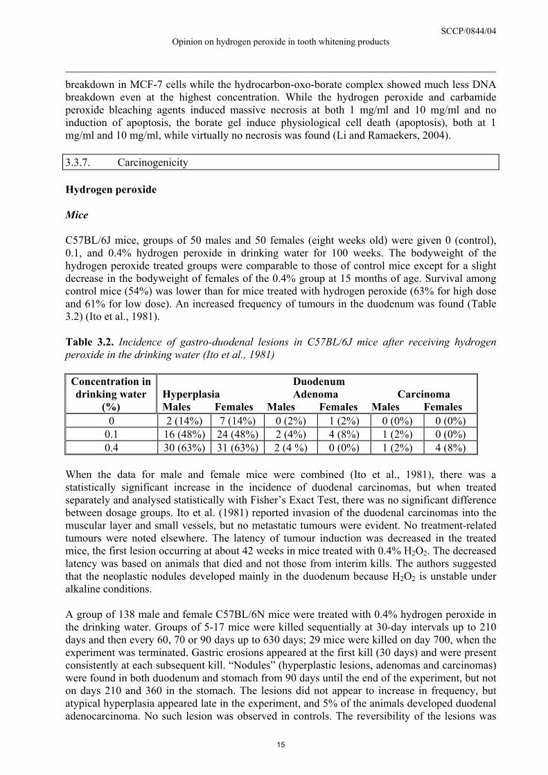
breakdown in MCF-7 cells while the hydrocarbon-oxo-borate complex showed much less DNA breakdown even at the highest concentration. While the hydrogen peroxide and carbamide peroxide bleaching agents induced massive necrosis at both 1 mg/ml and 10 mg/ml and no induction of apoptosis, the borate gel induce physiological cell death (apoptosis), both at 1 mg/ml and 10 mg/ml, while virtually no necrosis was found (Li and Ramaekers, 2004). 3.3.7. Carcinogenicity Hydrogen peroxide Mice C57BL/6J mice, groups of 50 males and 50 females (eight weeks old) were given 0 (control), 0.1, and 0.4% hydrogen peroxide in drinking water for 100 weeks. The bodyweight of the hydrogen peroxide treated groups were comparable to those of control mice except for a slight decrease in the bodyweight of females of the 0.4% group at 15 months of age. Survival among control mice (54%) was lower than for mice treated with hydrogen peroxide (63% for high dose and 61% for low dose). An increased frequency of tumours in the duodenum was found (Table 3.2) (Ito et al., 1981). Table 3.2. Incidence of gastro-duodenal lesions in C57BL/6J mice after receiving hydrogen peroxide in the drinking water (Ito et al., 1981)
Concentration in drinking water
(%)
Duodenum Hyperplasia Adenoma Carcinoma Males Females Males Females Males Females
0 2 (14%) 7 (14%) 0 (2%) 1 (2%) 0 (0%) 0 (0%) 0.1 16 (48%) 24 (48%) 2 (4%) 4 (8%) 1 (2%) 0 (0%) 0.4 30 (63%) 31 (63%) 2 (4 %) 0 (0%) 1 (2%) 4 (8%)
When the data for male and female mice were combined (Ito et al., 1981), there was a statistically significant increase in the incidence of duodenal carcinomas, but when treated separately and analysed statistically with Fisher’s Exact Test, there was no significant difference between dosage groups. Ito et al. (1981) reported invasion of the duodenal carcinomas into the muscular layer and small vessels, but no metastatic tumours were evident. No treatment-related tumours were noted elsewhere. The latency of tumour induction was decreased in the treated mice, the first lesion occurring at about 42 weeks in mice treated with 0.4% H2O2. The decreased latency was based on animals that died and not those from interim kills. The authors suggested that the neoplastic nodules developed mainly in the duodenum because H2O2 is unstable under alkaline conditions. A group of 138 male and female C57BL/6N mice were treated with 0.4% hydrogen peroxide in the drinking water. Groups of 5-17 mice were killed sequentially at 30-day intervals up to 210 days and then every 60, 70 or 90 days up to 630 days; 29 mice were killed on day 700, when the experiment was terminated. Gastric erosions appeared at the first kill (30 days) and were present consistently at each subsequent kill. “Nodules” (hyperplastic lesions, adenomas and carcinomas) were found in both duodenum and stomach from 90 days until the end of the experiment, but not on days 210 and 360 in the stomach. The lesions did not appear to increase in frequency, but atypical hyperplasia appeared late in the experiment, and 5% of the animals developed duodenal adenocarcinoma. No such lesion was observed in controls. The reversibility of the lesions was
15
SCCP/0844/04 Opinion on hydrogen peroxide in tooth whitening products
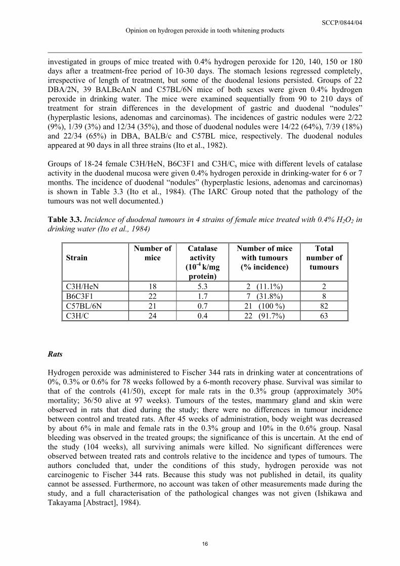
investigated in groups of mice treated with 0.4% hydrogen peroxide for 120, 140, 150 or 180 days after a treatment-free period of 10-30 days. The stomach lesions regressed completely, irrespective of length of treatment, but some of the duodenal lesions persisted. Groups of 22 DBA/2N, 39 BALBcAnN and C57BL/6N mice of both sexes were given 0.4% hydrogen peroxide in drinking water. The mice were examined sequentially from 90 to 210 days of treatment for strain differences in the development of gastric and duodenal “nodules” (hyperplastic lesions, adenomas and carcinomas). The incidences of gastric nodules were 2/22 (9%), 1/39 (3%) and 12/34 (35%), and those of duodenal nodules were 14/22 (64%), 7/39 (18%) and 22/34 (65%) in DBA, BALB/c and C57BL mice, respectively. The duodenal nodules appeared at 90 days in all three strains (Ito et al., 1982). Groups of 18-24 female C3H/HeN, B6C3F1 and C3H/Cs mice with different levels of catalase activity in the duodenal mucosa were given 0.4% hydrogen peroxide in drinking-water for 6 or 7 months. The incidence of duodenal “nodules” (hyperplastic lesions, adenomas and carcinomas) is shown in Table 3.3 (Ito et al., 1984). (The IARC Group noted that the pathology of the tumours was not well documented.) Table 3.3. Incidence of duodenal tumours in 4 strains of female mice treated with 0.4% H2O2 in drinking water (Ito et al., 1984)
Strain
Number of mice
Catalase activity
(10-4 k/mg protein)
Number of mice with tumours (% incidence)
Total number of tumours
C3H/HeN 18 5.3 2 (11.1%) 2 B6C3F1 22 1.7 7 (31.8%) 8 C57BL/6N 21 0.7 21 (100 %) 82 C3H/C 24 0.4 22 (91.7%) 63
Rats Hydrogen peroxide was administered to Fischer 344 rats in drinking water at concentrations of 0%, 0.3% or 0.6% for 78 weeks followed by a 6-month recovery phase. Survival was similar to that of the controls (41/50), except for male rats in the 0.3% group (approximately 30% mortality; 36/50 alive at 97 weeks). Tumours of the testes, mammary gland and skin were observed in rats that died during the study; there were no differences in tumour incidence between control and treated rats. After 45 weeks of administration, body weight was decreased by about 6% in male and female rats in the 0.3% group and 10% in the 0.6% group. Nasal bleeding was observed in the treated groups; the significance of this is uncertain. At the end of the study (104 weeks), all surviving animals were killed. No significant differences were observed between treated rats and controls relative to the incidence and types of tumours. The authors concluded that, under the conditions of this study, hydrogen peroxide was not carcinogenic to Fischer 344 rats. Because this study was not published in detail, its quality cannot be assessed. Furthermore, no account was taken of other measurements made during the study, and a full characterisation of the pathological changes was not given (Ishikawa and Takayama [Abstract], 1984).
16
SCCP/0844/04 Opinion on hydrogen peroxide in tooth whitening products
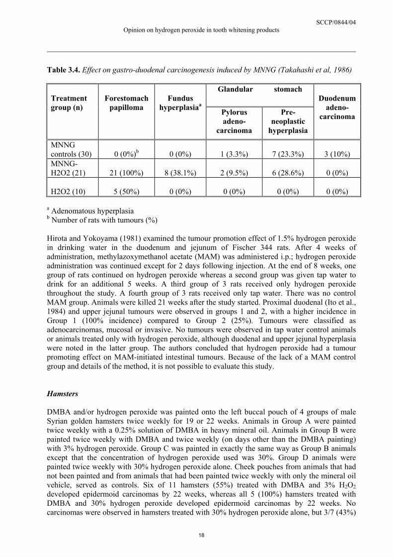
In other studies, forestomach papillomas were observed in rats exposed to hydrogen peroxide in drinking water (1%) (see Takahashi et al., 1986 [below]). Hydrogen peroxide in initiation – promotion experiments Mice Groups of 60 female Sencar mice, aged 7 to 9 weeks, were used to test the tumour-promoting (A), tumour-initiating (B) and complete carcinogenic (C) activity of hydrogen peroxide on the skin. Mice in experiment (A) received a single topical application of 10 nmol DMBA in 0.2 ml acetone, followed one week later by applications of a 30% solution of hydrogen peroxide diluted 1:1 (once and twice weekly), 1:2 or 1:5 in 0.2 ml acetone twice weekly for 25 weeks. Controls received acetone alone. The proportions of mice with papillomas at 25 weeks were 0/60 (0%) (controls), 3/58 (5%), 5/59 (8%), 6/59 (10%) and 6/60 (10%), respectively. Mice in experiment (B) received a single topical application of hydrogen peroxide diluted 1:1 in 0.2 ml acetone, or acetone alone (controls), followed one week later by twice-weekly applications of 2 µg 12-O-tetradecanoylphorbol 13-acetate (TPA) in acetone for 25 weeks. Papillomas were found after 25 weeks in 3/56 (5%) and 6/58 (10%) control and hydrogen peroxide-treated animals, respectively. Mice in experiment (C) received twice-weekly topical applications of hydrogen peroxide diluted 1:1 in 0.2 ml acetone for 25 week; 3/57 (5%) had papillomas at that time. No squamous-cell carcinoma was found when these animals were observed up to 50 weeks (Klein-Szanto and Slaga, 1982) (The IARC Working Group noted the absence of a DMBA-treated control group for the promotion experiment and the short duration of the experiment for complete carcinogenicity evaluation). In similar studies, mice were treated dermally for up to 58 weeks with 3% or 5% hydrogen peroxide following initiation with DMBA (Shamberger, 1972; Bock et al., 1975; Kurokawa et al., 1984). In these studies there were no significant increases in the incidence of skin tumours, although epidermal hyperplasia was evident in most of the mice treated. Rats Takahashi et al. (1986) examined the potential of hydrogen peroxide to promote N-methyl-N’-nitro-N-nitrosoguanidine (MNNG) initiated gastric tumours in rats. Two groups of rats (n=30 and 21) received MNNG-treated drinking water and food supplemented with 10% sodium chloride, the water of one group being supplemented with 1% hydrogen peroxide for 7 weeks ad libitum after which the animals were maintained on normal food and tap water. A third group (n=10) was not given MNNG or a sodium chloride supplemented diet, but was administered 1% hydrogen peroxide in the drinking water. Adenocarcinomas were observed in the pyloric stomach and duodenum of the MNNG-treated rats, and “preneoplastic hyperplasia” was observed in the pylorus (Table 3.4). In rats treated with MNNG and hydrogen peroxide, there was no enhancement in the number of gastrointestinal tumours, although all treated animals exhibited forestomach papillomas; these also occurred in rats treated only with hydrogen peroxide in the drinking water. No carcinoma development was noted in the stomach or duodenum. Erosions and ulcerations also occurred in the fundic mucosa of the stomach of the hydrogen peroxide treated rats. The authors concluded that, in contrast to the study of Hirota and Yokoyama (1981, see below), no enhancement of duodenal tumours occurred, although characteristic diffuse lesions, showing fusion of the villi, were observed throughout the duodenum.
17
SCCP/0844/04 Opinion on hydrogen peroxide in tooth whitening products
Table 3.4. Effect on gastro-duodenal carcinogenesis induced by MNNG (Takahashi et al, 1986)
Glandular stomach Treatment group (n)
Forestomach
papilloma
Fundus
hyperplasiaa Pylorus adeno-
carcinoma
Pre-neoplastic
hyperplasia
Duodenum
adeno-carcinoma
MNNG controls (30)
0 (0%)b
0 (0%)
1 (3.3%)
7 (23.3%)
3 (10%)
MNNG-H2O2 (21)
21 (100%)
8 (38.1%)
2 (9.5%)
6 (28.6%)
0 (0%)
H2O2 (10)
5 (50%)
0 (0%)
0 (0%)
0 (0%)
0 (0%)
a Adenomatous hyperplasia b Number of rats with tumours (%) Hirota and Yokoyama (1981) examined the tumour promotion effect of 1.5% hydrogen peroxide in drinking water in the duodenum and jejunum of Fischer 344 rats. After 4 weeks of administration, methylazoxymethanol acetate (MAM) was administered i.p.; hydrogen peroxide administration was continued except for 2 days following injection. At the end of 8 weeks, one group of rats continued on hydrogen peroxide whereas a second group was given tap water to drink for an additional 5 weeks. A third group of 3 rats received only hydrogen peroxide throughout the study. A fourth group of 3 rats received only tap water. There was no control MAM group. Animals were killed 21 weeks after the study started. Proximal duodenal (Ito et al., 1984) and upper jejunal tumours were observed in groups 1 and 2, with a higher incidence in Group 1 (100% incidence) compared to Group 2 (25%). Tumours were classified as adenocarcinomas, mucosal or invasive. No tumours were observed in tap water control animals or animals treated only with hydrogen peroxide, although duodenal and upper jejunal hyperplasia were noted in the latter group. The authors concluded that hydrogen peroxide had a tumour promoting effect on MAM-initiated intestinal tumours. Because of the lack of a MAM control group and details of the method, it is not possible to evaluate this study. Hamsters DMBA and/or hydrogen peroxide was painted onto the left buccal pouch of 4 groups of male Syrian golden hamsters twice weekly for 19 or 22 weeks. Animals in Group A were painted twice weekly with a 0.25% solution of DMBA in heavy mineral oil. Animals in Group B were painted twice weekly with DMBA and twice weekly (on days other than the DMBA painting) with 3% hydrogen peroxide. Group C was painted in exactly the same way as Group B animals except that the concentration of hydrogen peroxide used was 30%. Group D animals were painted twice weekly with 30% hydrogen peroxide alone. Cheek pouches from animals that had not been painted and from animals that had been painted twice weekly with only the mineral oil vehicle, served as controls. Six of 11 hamsters (55%) treated with DMBA and 3% H2O2 developed epidermoid carcinomas by 22 weeks, whereas all 5 (100%) hamsters treated with DMBA and 30% hydrogen peroxide developed epidermoid carcinomas by 22 weeks. No carcinomas were observed in hamsters treated with 30% hydrogen peroxide alone, but 3/7 (43%)
18
SCCP/0844/04 Opinion on hydrogen peroxide in tooth whitening products
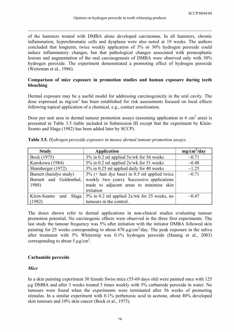
of the hamsters treated with DMBA alone developed carcinomas. In all hamsters, chronic inflammation, hyperchromatic cells and dysplasia were also noted at 19 weeks. The authors concluded that longterm, twice weekly application of 3% or 30% hydrogen peroxide could induce inflammatory changes, but that pathological changes associated with preneoplastic lesions and augmentation of the oral carcinogenesis of DMBA were observed only with 30% hydrogen peroxide. The experiment demonstrated a promoting effect of hydrogen peroxide (Weitzman et al., 1986). Comparison of mice exposure in promotion studies and human exposure during teeth bleaching Dermal exposure may be a useful model for addressing carcinogenicity in the oral cavity. The dose expressed as mg/cm2 has been established for risk assessments focused on local effects following topical application of a chemical, e.g., contact sensitisation. Dose per unit area in dermal tumour promotion assays (assuming application to 6 cm2 area) is presented in Table 3.5 (table included in Submission III except that the experiment by Klein-Szanto and Slaga (1982) has been added later by SCCP). Table 3.5. Hydrogen peroxide exposure in mouse dermal tumour promotion assays.
Study Application mg/cm2/day Bock (1975) 3% in 0.2 ml applied 5x/wk for 56 weeks ~0.71 Kurokowa (1984) 5% in 0.2 ml applied 2x/wk for 51 weeks ~0.48 Shamberger (1972) 3% in 0.25 ml applied daily for 40 weeks ~1.25 Burnett (hairdye study) Burnett and Goldenthal, 1988)
3% (+ hair dye base) in 0.5 ml applied twice weekly /two years). Successive applications made to adjacent areas to minimise skin irritation
~0.71
Klein-Szanto and Slaga (1982)
5% in 0.2 ml applied 2x/wk for 25 weeks, no tumours in the control.
~0.47
The doses shown refer to dermal applications in non-clinical studies evaluating tumour promotion potential. No carcinogenic effects were observed in the three first experiments. The last study the tumour frequency was 5% after initiation with the initiator DMBA followed skin painting for 25 weeks corresponding to about 470 µg/cm2/day. The peak exposure in the saliva after treatment with 5% Whitestrip was 0.1% hydrogen peroxide (Hannig et al., 2003) corresponding to about 5 µg/cm2. Carbamide peroxide Mice In a skin painting experiment 30 female Swiss mice (55-69 days old) were painted once with 125 µg DMBA and after 3 weeks treated 5 times weekly with 5% carbamide peroxide in water. No tumours were found when the experiments were terminated after 56 weeks of promoting stimulus. In a similar experiment with 0.1% perbenzoic acid in acetone, about 40% developed skin tumours and 10% skin cancer (Bock et al., 1975).
19
SCCP/0844/04 Opinion on hydrogen peroxide in tooth whitening products
Tooth whiteners Human case report In a press release from American Head and Neck Society a study presented at the 6th International Conference on Head and Neck Cancer is reported. Patients with primary oral cancer diagnosed at Georgetown University Medical Center between 1997 and 2003 were identified. Nineteen patients agreed to participate in the study. Three (16 percent) of patients reported a history of tooth whitener use in the past. There was no significant difference in age at diagnosis between the patients who used and did not use tooth whiteners, however the tooth whitener users tended to be younger (mean age 34.3 vs. 52.4, p = 0.11). Alcohol use and smoking history were similar in the two groups. The patients who used tooth whiteners were more likely to present with regional lymph node disease, than those who did not use tooth whiteners. All three patients presented with node positive disease as opposed to 3 of 16 (19 percent) patients without a history of tooth whitener use. The authors point out that the data do not necessarily suggest a causative relationship between the use of these products and the development of oral cancer. However, free radicals generated in the whitening process have carcinogenic potential, and therefore the use of these products in this patient population should be studied further (Burningham et al., 2004). Conclusions on carcinogenicity A drinking water study in mice showed that hydrogen peroxide caused duodenal hyperplasia at a high frequency and localised duodenal carcinomas at a low frequency. A subsequent study with different strains of mice showed a strong negative correlation between incidence of duodenal tumours and catalase activity in duodenal mucosa. In one study with rats a high incidence of forestomach papillomas were found after receiving 1% hydrogen peroxide in the drinking water. While humans do not have a forestomach, they do have comparable squamous epithelium tissues in the oral cavity and the upper 2-3 of the oesophagus. Thus, in principle, carcinogens targeting the forestomach squamous epithelium rodents are relevant for humans. Also, the target tissues for carcinogens may differ between experimental animals and humans and a forestomach carcinogen in rodent may target a different tissue in humans (IARC, 2003). Some tumour promotion studies indicate that hydrogen peroxide may act as a weak promoter. Hydrogen peroxide has a weak potential to induce local carcinogenic effects. The mechanism is unclear, but a genotoxic mechanism cannot be excluded. As regard to tumour promotion, several mechanisms might be operative; direct genotoxicity, impairment of DNA repair, and chronic inflammation. 3.3.8. Reproductive toxicity 3.3.8.1. Two generation reproduction toxicity
20
SCCP/0844/04 Opinion on hydrogen peroxide in tooth whitening products
Mice Male albino mice were given 0.33%, 1.0% or 3.0% hydrogen peroxide in drinking water. There were no controls. The mice at the highest dose would not drink the solution and were taken off the study. Mice were mated after 7 and 21 days on hydrogen peroxide. All females became pregnant within a few days and delivered litters of normal size. The concentration, morphology and mobility of the spermatozoa of the male mice receiving hydrogen peroxide in the drinking water over 3 and 6 weeks remained normal. In vivo, hydrogen peroxide had not significant spermicidal action in mice at concentration up to 1% in solution (Wales et al., 1959). Rats Male and female rats were administered hydrogen peroxide daily by gavage at doses of 1/10-1/5 LD50 (which was not specified) for 45 days. At the high dose, females showed modifications of the oestrus cycle and males reduced mobility of spermatozoa, without effects on the weight of the testicles. In a second experiment male and female rats received daily doses of 0.005, 0.05, 0.5, 5 and 50 mg hydrogen peroxide/kg bw by gavage for 6 months and were mated. Variations of the oestrous cycle in females were observed at 50 and 0.5 mg hydrogen peroxide/kg bw, but not at 5 mg/kg bw. Reduced mobility of spermatozoa in males was observed at 50 mg hydrogen peroxide/kg bw. No changes were observed in the morphology and weight of the testes. Among the high dose females, only 3/9 produced litters, compared to 7/9 in the control group. In addition, litter size and body weight gain of the offspring of the high dose females were reduced relative to those of control females (Antonova, 1974). The results of the study should be considered with caution because the information on the experiment is incomplete. In order to test the effect of ingested tooth whitener on early embryo development and growth, rats were intubated with 500 mg/kg whitener on day 2 of pregnancy. It was concluded that a) ingestion of tooth whitener containing 35% carbamide peroxide causes a loss of embryos sometimes between day 2 (treatment) and day 5 (collection), but that b) day 5 embryos have the same cell number both prior to and after 24 hour culture, and c) have the same ability to implant in vitro (Redmond et al. [Abstract], 1998). 3.3.8.2. Teratogenicity One study which addresses developmental toxicity has been conducted with Wistar rats Moriyama et al., 1982). Aqueous solutions of hydrogen peroxide were mixed with powdered feed to 10, 2, 0.1, or 0.02% and administered to groups of 5-8 pregnant rats for one week during “the critical period of pregnancy”. The foetuses were removed on day 20 for examinations (Study A). Separate dose groups of 2-3 rats were similarly treated, but the rats were allowed to go through normal delivery, and the offspring were followed-up for about four weeks (Study B). In Study A, at the high dose level the dam body weight did not increase markedly. Food consumption was reduced to about one third as compared to the other dose groups, for which there was no difference from controls. Foetal resorptions were increased and the foetal body weight was decreased; most of the foetuses were close to death. No external malformations were found in any of the dose groups. Haemorrhaging of internal organs (eye, parietal region of the brain, cardiopulmonary region, torso) was dose dependently increased in the dose range 0.1-10% H2O2. Skeletal hypoplasias occurred dose dependently at the two highest levels. In Study B, all the neonates of the 10% treatment group died within 1 week post partum, the body weights were low and the number of live births was decreased. In the other dose groups there were no major effects on the development of neonates. There are major uncertainties about the exposure and effect mechanism which cast doubt on the relevance of the study. H2O2 concentration in feed was reported to decrease to 1/10 after 24 hours and to virtually nil by 72 hours. The authors state
21
SCCP/0844/04 Opinion on hydrogen peroxide in tooth whitening products
that “the amount of residue was determined and consumption was estimated”; however, it is not stated how frequently fresh feed was prepared. Nevertheless, it seems likely that the dams indeed ingested hydrogen peroxide, and there was not much of an increase in dam body weight at the top dose level. There was no marked difference between the groups in placental weight. The authors proposed that the observed effects on foetal development were due to the breakdown of essential nutrients in food by hydrogen peroxide. Conclusions on reproductive toxicity No appropriate animal studies were available for a complete evaluation of reproductive and developmental toxicity. Limited studies with mice and rats exposed to hydrogen peroxide in drinking water suggested no grave disturbances on the male or female reproductive functions. The only available developmental toxicity study in Wistar rats which were fed on powdered feed mixed with hydrogen peroxide did show foetotoxic effects (Moriyama et al., 1982), but the study contains major uncertainties about the exposure and effect mechanisms. Although raising some further questions, the study cannot be used for an evaluation. 3.3.9. Toxicokinetics Hydrogen peroxide is a normal metabolite in the aerobic cells. It is produced from superoxide anion spontaneously or as a result of the activity of superoxide dismutase (SOD) (EC1.15.1.1). Superoxide radical undergoes dismutation quickly and spontaneously, but the enzymatic process occurs at a rate that is 1010-fold faster. Eukaryotic cells contain two kinds of SOD that are highly specific for superoxide (O2) as a substrate. Hydrogen peroxide occurs under most conditions at submicromolar concentrations. Hydrogen peroxide passes readily across biological membranes. Because it reacts slowly with organic substrates, it can diffuse considerable distances in biological systems. There are two main hydrogen peroxide metabolising enzymes, catalase and glutathione peroxidase which control the hydrogen peroxide concentration. Significant amounts of topically applied hydrogen peroxide can penetrate the epidermis or mucous membranes followed by rapid spontaneous or enzyme-catalysed decomposition to oxygen and water in the underlying tissue. The formation of gaseous oxygen causes capillary microembolism and prevents irrigation of tissues by blood resulting in a visible, reversible bleaching of the exposed tissue area. The local spontaneous or enzymatic-catalysed breakdown prevents it to enter the general circulation and thus its systemic distribution. The overall decomposition reaction of hydrogen peroxide in the present of catalase is as followed:
H2O2 + H2O2 → 2H2O + O2 Catalases are present at a wide range of concentrations in nearly all mammalian cells. Catalases are located in the subcellular compartments, mainly in peroxisomes. Soluble catalases were found in erythrocytes. The highest catalase activity is observed in cells of the duodenum, liver, spleen, kidney, blood, mucous membranes and other highly vascularised tissues. Peroxidases decompose hydrogen peroxide through the reaction:
H2O2 + 2RH → 2H2O+ R- R
22
SCCP/0844/04 Opinion on hydrogen peroxide in tooth whitening products
Relatively high peroxidase activities occur in human adrenal medulla, liver, kidney, leukocytes and saliva. In the oral cavity, salivary peroxidase and myeloperoxidase are the primary defences against bacterially derived peroxide. Salivary peroxidase activity, the conversion of hydrogen peroxide to water, is coupled with the conversion of thiocyanate to hypothiocyanate, which has bacteriostatic activity and reduces the formation of peroxide and dental plaque acid by bacteria. In the absence of salivary peroxidase and thiocyanate, the rate of production of hydrogen peroxide by bacteria in saliva is approximately 100 nmol/ml/hr and would lead to a steady-state level of 0.1 mM hydrogen peroxide in one hour (Thomas et al, 1994). In the presence of salivary peroxidase and thiocyanate, the steady-state level of peroxide was predicted to be maintained below 0.01 mM. Glutathione peroxidase can react with both hydrogen peroxide and organic hydroperoxides. Glutathione peroxidase is more efficient at low concentrations of hydrogen peroxide compared to catalase. Glutathione reduces hydrogen peroxide to water with formation of oxidised glutathione which is regenerated by glutathione reductase by consuming NADPH. The oxidative reactivity of hydrogen peroxide with biological molecules such as carbohydrates, proteins, fatty acids or nucleic acids is not pronounced in the absence of transition metals, except for a few nucleophilic reactions. In the presence of transition metals, particularly ferrous ions (Fe2), hydrogen peroxide can be reduced to hydroxyl radicals:
H2O2 + Fe2+ → OH. + OH- + Fe3+ The hydroxyl radical is highly reactive and will attack most molecules in living cells. Groups at extra risk Genetically determined traits (acatalasaemia, glucose-6-phosphate dehydrogenase (G6PD) deficiency) render humans more susceptible to peroxide toxicity. Acatalasemic individuals are more susceptible to hydrogen peroxide exposure because of a hereditary disorder in their hydrogen peroxide metabolising enzymes, i.e. the blood catalase activity level is below normal (hypocatalasemia). Acatalesemia is a rare (frequency 0.2-0.4%) genetic defect occurring particularly in the Orient (Ogata, 1991). It has been found that approximately half of the Japanese acatalasemic patients developed progressive gangrene of the mouth called Takahara’s disease. This condition is characterised by small, painful ulcers in the gingival crevices and tonsillar lacunae, attributed to excess levels of hydrogen peroxide generated by various microorganisms in the mouth without normal destruction by catalase. The total number of reported patients of acatalasemia worldwide in 1989 was 107 belonging to 52 families. Later two Hungarian acatalasaemic subjects have been reported (Góth, 1992). There appears to be two types of acatalasaemia. The Japanese type is the result of a splice mutation resulting in defective catalase synthesis (Góth and Páy, 1996). The Swiss type of acatalasaemia type is caused by point mutation resulting in catalase that is rapidly degraded. Swiss type acatalasaemic patients show no signs of oxidative damage (Góth and Páy, 1996). Another group of individuals more sensitive to hydrogen peroxide exposure is persons with G6PD deficiency. G6PD deficiency is a genetic disorder of erythrocytes (over 300 variants have been identified) in which the inability of affected cells to maintain NAD(P)H levels sufficient for
23
SCCP/0844/04 Opinion on hydrogen peroxide in tooth whitening products
the reduction of oxidised glutathione results in inadequate detoxification of hydrogen peroxide through glutathione peroxidase. It is estimated that about 400 million people throughout the world are deficient in G6PD. The frequency in G6PD deficiency in Europe is about 0.1%. A third group of individuals that might be more sensitive to hydrogen peroxide exposure is persons with xerostomia, or dry mouth, which occurs when the salivary glands are hypoactive. This may affect the degradation of hydrogen peroxide. However, two studies (Aguire et al., in press) indicate that the degradation of hydrogen peroxide in the oral cavity is not affected by xerostomia. Marshall et al. (2001) found no difference in the clearance of peroxide from the oral cavity when comparing adults with normal salivary flow and adult with diminished salivary flow (Sjorgren’s syndrome). In a Procter & Gamble sponsored clinical study (2000159), subjects with artificially induced xerostomia (via use of a rubber dental dam) experienced no adverse events after 10 days use of 6% hydrogen peroxide gel strips. Procter & Gamble claimed that due to the low levels of hydrogen peroxide in saliva during use of tooth whitening products and conversion of exogenous hydrogen peroxide to water and oxygen, hydrogen peroxide would not be expected to persist long enough in the body to reach G6PD deficient erythrocytes to precipitate an oxidative response. 3.3.10. Photo-induced toxicity 3.3.10.1. Phototoxicity / photoirritation and photosensitisation No data submitted 3.3.10.2. Phototoxicity / photomutagenicity / photoclastogenicity No data submitted 3.3.11. Human data 3.3.11.1 Exposure From submission II A study (study no. 2000045 and 2000143) was carried out by Procter & Gamble. Adult subjects (N=12) used either a 5.3% hydrogen peroxide gel strip that delivers 10.6 mg hydrogen peroxide/strip (200 mg of a 5.3% hydrogen peroxide gel; 5, 10, 30 or 60 minute treatments), a 10% carbamide peroxide (CP) tray that delivers ≈ 22-48 mg hydrogen peroxide (600-900 mg of a 3.6% hydrogen peroxide gel; 10, 30, 60 or 120 minute treatments) or a 20% CP tray that delivers ≈ 40-60 mg hydrogen peroxide (600-900 mg of a 6.7% hydrogen peroxide gel; 10, 30, 60 or 120 minute treatments). Treatment was on maxillary teeth only. It is concluded that hydrogen peroxide delivered at 6% in tooth whitening products (films, gels or varnished) intended for direct application to the teeth, degrades rapidly during wear time. This is indicative of the rapid degradation of hydrogen peroxide that would occur where direct contact with the gingival tissues immediately surrounding the teeth may result during wear time of such tooth whitening products. Salivary hydrogen peroxide levels are low during wear time (<0.02%), thereby demonstrating the minimal oral and systemic exposure that occurs with such tooth whitening formulations. The available peroxide resulting from the cosmetic use 4 strips with 6%
24
SCCP/0844/04 Opinion on hydrogen peroxide in tooth whitening products
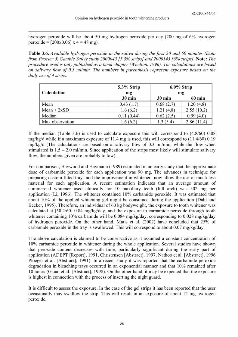
hydrogen peroxide will be about 50 mg hydrogen peroxide per day (200 mg of 6% hydrogen peroxide = [200x0.06] x 4 = 48 mg). Table 3.6. Available hydrogen peroxide in the saliva during the first 30 and 60 minutes (Data from Procter & Gamble Safety study 2000045 [5.3% strips] and 2000143 [6% strips]. Note: The procedure used is only published as a book chapter (Whelton, 1996). The calculations are based on salivary flow of 0.3 ml/min. The numbers in parenthesis represent exposure based on the daily use of 4 strips.
Calculation
5.3% Strip mg
30 min
6.0% Strip mg
30 min 60 min Mean 0.43 (1.7) 0.68 (2.7) 1.20 (4.8) Mean + 2xSD 1.6 (6.2) 1.21 (4.8) 2.55 (10.2) Median 0.11 (0.44) 0.62 (2.5) 0.99 (4.0) Max observation 1.6 (6.2) 1.3 (5.4) 2.86 (11.4)
If the median (Table 3.6) is used to calculate exposure this will correspond to (4.8/60) 0.08 mg/kg/d while if a maximum exposure of 11.4 mg is used, this will correspond to (11.4/60) 0.19 mg/kg/d (The calculations are based on a salivary flow of 0.3 ml/min, while the flow when stimulated is 1.5 – 2.0 ml/min. Since application of the strips most likely will stimulate salivary flow, the numbers given are probably to low). For comparison, Haywood and Haymann (1989) estimated in an early study that the approximate dose of carbamide peroxide for each application was 90 mg. The advances in technique for preparing custom fitted trays and the improvement in whiteners now allow the use of much less material for each application. A recent estimation indicates that an average amount of commercial whitener used clinically for 10 maxillary teeth (full arch) was 502 mg per application (Li, 1996). The whitener contained 10% carbamide peroxide. It was estimated that about 10% of the applied whitening gel might be consumed during the application (Dahl and Becker, 1995). Therefore, an individual of 60 kg bodyweight, the exposure to tooth whitener was calculated at [50.2/60] 0.84 mg/kg/day, and the exposure to carbamide peroxide through tooth whitener containing 10% carbamide will be 0.084 mg/kg/day, corresponding to 0.028 mg/kg/day of hydrogen peroxide. On the other hand, Matis et al. (2002) have concluded that 25% of carbamide peroxide in the tray is swallowed. This will correspond to about 0.07 mg/kg/day. The above calculation is claimed to be conservative as it assumed a constant concentration of 10% carbamide peroxide in whitener during the whole application. Several studies have shown that peroxide content decreases with time, particularly significant during the early part of application (ADEPT [Report], 1991, Christensen [Abstract], 1997, Nathoo et al. [Abstract], 1996 Ploeger et al. [Abstract], 1991). In a recent study it was reported that the carbamide peroxide degradation in bleaching trays occurred in an exponential manner and that 10% remained after 10 hours (Gaiao et al. [Abstract], 1998). On the other hand, it may be expected that the exposure is highest in connection with the process of inserting the night guard. It is difficult to assess the exposure. In the case of the gel strips it has been reported that the user occasionally may swallow the strip. This will result in an exposure of about 12 mg hydrogen peroxide.
25
SCCP/0844/04 Opinion on hydrogen peroxide in tooth whitening products
Comparisons of the use of the new strips with the custom fitted trays suggest that the total exposure to hydrogen peroxide is of the same order of magnitude. Marshall et al. (2001) determined the clearance of peroxide from the oral cavity after 1 minute brushing with a 3% hydrogen peroxide dentifrice. Seventy percent of the hydrogen peroxide decomposed during the minute of brushing for infants (3-4 years), juveniles (7-12 years), adults with normal salivary flow and adults with diminished salivary flow (Sjorgren’s syndrome). The degradation of 10% carbamide peroxide (≈3.6% hydrogen peroxide), worn in a custom-fitted tray, was determined over 10 hours (N=15). The degradation rate in the tray and in the gel on the teeth was rapid for the first hour, and then slowed, with more than 50% loss of active ingredient seen at 4 hours, and more than 85% loss following 10 hours of exposure. The degradation of “grab” sample from the reservoir of tooth no. 8 was slower. On average 56% remained after 4 hours and 23% after 10 hours (Matis et al., 1999). From submission III In an article by Mahony et al. (2003), both maxillary and mandibular teeth were treated with a 5.3% hydrogen peroxide paint-on gel. Peroxide concentrations in the tooth scraping sample after 10, 30 minutes, 1, 2 and 4 hours of daytime wear were 4.56, 3.28, 1.57, 0.51 and 0.14%, respectively. The median peroxide concentration in the saliva at 5, 10 and 20 minutes of daytime wear were 0.001, 0.0001 and 0.0001% respectively. The median peroxide concentrations in the saliva at 30 and 60 minutes were below the limit of detection (0.00007%). Overall, salivary hydrogen peroxide concentration was less than 0.033% measured at any time. Bleaching strips containing 10% hydrogen peroxide gel were used in a clinical trail involving 16 persons. The median hydrogen peroxide concentration after 5 minutes was 7.3%, 6.4% and 0.7% for strips, teeth and gingival, respectively, declining to 4.6%, 2.9% and 0.1% at 30 minutes. Salivary samples never exceeded a median concentration of 0.014% at any point. Median hydrogen peroxide concentration on strips and teeth remain about 2% over 60 minutes. (In the received summary report, it is written that 14% hydrogen peroxide strips was used) (Walden et al., 2004, report). Maxillary teeth were treated with 14% hydrogen peroxide strips (0.1 gm gel load) in a clinical trial involving 15 persons. The concentration was 13.4% on average after sampling 3 strips. The median concentration for the teeth after 10 minutes was 6.9% after 30 minutes 4.2% and 60 minutes wear time 2.9%. Using colorimetric analysis with glycerine normalized analysis, the results after 10 minutes was 11.1%, 30 minutes 8.7% and 60 minutes 8.5%. Thus, there is considerable difference which increase with time. The highest concentration in saliva was 0.073%, which occurred at 10 minutes time point. Strip hydrogen peroxide level decreased with wear time. After 30 minutes wear time, the median peroxide level on the strip ranged from 3.4 to 11% depending on the analytical method. Tooth hydrogen peroxide level also decreased with wear time. Depending on the analytical method the median peroxide concentration was 10.0 to 12.7% after 5 minutes of wear time and the levels ranged from 4.2 to 8.7% after 30 min. Median salivary peroxide level did not exceed 0.0725% for any time point in the study (Report 2003 009). Slezak et al. (2002) determined the concentration of hydrogen peroxide in saliva after application of a 6.5% hydrogen peroxide paint-on gel. The concentrations were 0.03%, 0.0042% and 0.0001% at 1, 5 and 15 minutes, respectively.
26
SCCP/0844/04 Opinion on hydrogen peroxide in tooth whitening products
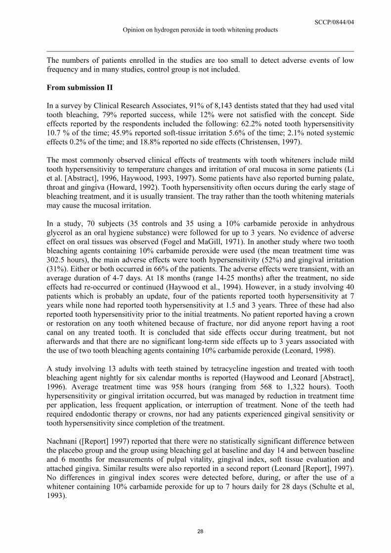
A study involving 17 persons was carried out with 6.5% hydrogen peroxide strips (0.2 g gel load, 13 mg hydrogen peroxide/strip) or 14% hydrogen peroxide strips (0.1 g gel load, 14 mg hydrogen peroxide/strip). The median peroxide concentrations are shown on Table 3.7. It is pointed out that for pair wise comparisons between treatment, there were no significant differences between the strips hydrogen peroxide level at 60 min and for gingival and saliva data for any time point and for area under the curve (Report 2003 046). Table 3.7. Concentration of hydrogen peroxide in the strip, at the teeth, at the gingival, and in the saliva during the first 60 minutes (Data from study 2003 046[6.5% and 14% strips]).
Measurement 0 min (%)
5 min (%)
10 min (%)
30 min (%)
60 min (%)
6.5% 7.0 5.5 5.6 4.4 3.0 Strip 14% 14.5 10 9.2 6.2 3.0
6.5% 4.4 3.8 2.6 1.7 Teeth 14% 7.4 6.7 4.4 2.4
6.5 0.60 0.16 0.17 0.08 Gingiva 14% 0.36 0.18 0.12 0.06
6.5% 0.007 0.006 0.005 0.001 Salivary 14% 0.011 0.013 0.009 0.002
The use of 5.3% or 6.5% hydrogen peroxide paint-on gel is presented in a clinical study involving 17 persons. When 6.5% paint-on gel is used, median concentrations after 2 and 5 minutes when retractor is used was 8.6% and 9.5%, respectively. In another study with no retractors, the concentration 0.5 minute after application was 10.6%. This increase after application is probably due to the rapid loss of alcohol in the gel (Report 2003 043). In a study where 6.5% hydrogen peroxide paint-on gel and 6% hydrogen peroxide strip were compared, it was concluded that on the average the hydrogen peroxide concentration on teeth and in saliva was statistically higher for strips compared to paint-on gel both at 5 and 30 minutes time point, while the concentration of paint-on appear to peak at 30 seconds application, the hydrogen peroxide concentration rapidly declined to less than 0.6% at 2% as the paint-on products exposed to saliva, salivary hydrogen peroxide peaked at 0.5 minutes (0.034%) and declined rapidly (Report 2002 126). Other studies The amount of peroxides released into saliva was related to the bleaching system and only partially influenced by the individual salivary flow rate. Bleaching with Vivastyle (10% carbamide peroxide, tray charged with 225 mg) led to lower release of peroxides into saliva compared to Whitestrips (5% H2O2) (Vivastyle: 0.8± 0.17 mg; Whitestrips: 1.5±0.84 mg). The peak exposures of hydrogen peroxide in the saliva were 0.06% with Vivastyle and 0.1% with Whitestrips. Salivary flow rate was not correlated to release of peroxides from the bleaching products (Hanning et al., 2003).
3.3.11.2 Clinical safety data
27
SCCP/0844/04 Opinion on hydrogen peroxide in tooth whitening products
The numbers of patients enrolled in the studies are too small to detect adverse events of low frequency and in many studies, control group is not included. From submission II In a survey by Clinical Research Associates, 91% of 8,143 dentists stated that they had used vital tooth bleaching, 79% reported success, while 12% were not satisfied with the concept. Side effects reported by the respondents included the following: 62.2% noted tooth hypersensitivity 10.7 % of the time; 45.9% reported soft-tissue irritation 5.6% of the time; 2.1% noted systemic effects 0.2% of the time; and 18.8% reported no side effects (Christensen, 1997). The most commonly observed clinical effects of treatments with tooth whiteners include mild tooth hypersensitivity to temperature changes and irritation of oral mucosa in some patients (Li et al. [Abstract], 1996, Haywood, 1993, 1997). Some patients have also reported burning palate, throat and gingiva (Howard, 1992). Tooth hypersensitivity often occurs during the early stage of bleaching treatment, and it is usually transient. The tray rather than the tooth whitening materials may cause the mucosal irritation. In a study, 70 subjects (35 controls and 35 using a 10% carbamide peroxide in anhydrous glycerol as an oral hygiene substance) were followed for up to 3 years. No evidence of adverse effect on oral tissues was observed (Fogel and MaGill, 1971). In another study where two tooth bleaching agents containing 10% carbamide peroxide were used (the mean treatment time was 302.5 hours), the main adverse effects were tooth hypersensitivity (52%) and gingival irritation (31%). Either or both occurred in 66% of the patients. The adverse effects were transient, with an average duration of 4-7 days. At 18 months (range 14-25 months) after the treatment, no side effects had re-occurred or continued (Haywood et al., 1994). However, in a study involving 40 patients which is probably an update, four of the patients reported tooth hypersensitivity at 7 years while none had reported tooth hypersensitivity at 1.5 and 3 years. Three of these had also reported tooth hypersensitivity prior to the initial treatments. No patient reported having a crown or restoration on any tooth whitened because of fracture, nor did anyone report having a root canal on any treated tooth. It is concluded that side effects occur during treatment, but not afterwards and that there are no significant long-term side effects up to 3 years associated with the use of two tooth bleaching agents containing 10% carbamide peroxide (Leonard, 1998). A study involving 13 adults with teeth stained by tetracycline ingestion and treated with tooth bleaching agent nightly for six calendar months is reported (Haywood and Leonard [Abstract], 1996). Average treatment time was 958 hours (ranging from 568 to 1,322 hours). Tooth hypersensitivity or gingival irritation occurred, but was managed by reduction in treatment time per application, less frequent application, or interruption of treatment. None of the teeth had required endodontic therapy or crowns, nor had any patients experienced gingival sensitivity or tooth hypersensitivity since completion of the treatment. Nachnani ([Report] 1997) reported that there were no statistically significant difference between the placebo group and the group using bleaching gel at baseline and day 14 and between baseline and 6 months for measurements of pulpal vitality, gingival index, soft tissue evaluation and attached gingiva. Similar results were also reported in a second report (Leonard [Report], 1997). No differences in gingival index scores were detected before, during, or after the use of a whitener containing 10% carbamide peroxide for up to 7 hours daily for 28 days (Schulte et al, 1993).
28
SCCP/0844/04 Opinion on hydrogen peroxide in tooth whitening products
It is stated in the dossier that a number of investigators reported that the use of 10% carbamide peroxide in anhydrous glycerol was effective in reducing risk of gingivitis (Zinner et al, 1978) and dental caries (Fogel and MaGill, 1971) and improving oral hygiene (Tartakow et al, 1978). Its use for four times daily up to 3 years did not have any adverse effects on gingival tissues or any evidence of other side effects. Procter & Gamble have reported several studies concerning the use of peroxide (2.7-7% hydrogen peroxide) containing tooth whitening products for less than 6 months, resulting in the same adverse events (oral soft tissue irritation and tooth sensitivity) observed in two week studies. The majority of the adverse events were mild and all had resolved within 3 days after the products use was discontinued. No adverse events resolution related to treatment was required. There was a trend toward a slight increase in adverse event incidence with increasing hydrogen peroxide concentration. Oral soft tissue irritation or oral hard tissue adverse incidence in groups using hydrogen peroxide products was not significantly different compared to the concurrent placebo in any study. In only 2 of 14 studies, the total adverse events incidence was statistically significantly greater in subjects using hydrogen peroxide compared to the concurrent placebo groups. Even 6 months continuous use of either a strip or a custom tray peroxide product caused the same mild, transient adverse events (tooth sensitivity and oral soft tissue irritation) as those observed after 14 or 28 days of product use. A company producing teeth bleaching strips has reported that some of the users of the gel strips have swallowed a strip. In several of these cases, consumers reported minor gastrointestinal symptoms. Tooth whiteners containing 10% carbamide peroxide The majority of the published peroxide based teeth whitening studies with carbamide peroxide are done with a type of product only available via the dental office. With this system, a carbamide peroxide gel is delivered in a custom-fitted mouthguard, designed to cover either the upper or lower dentition. The filled bleaching tray is worn at home from 2-3 hours for a daytime exposure to 8-10 hours for an overnight exposure. Treatment is generally daily, and ranges in duration from one week to six months, or until the patient is satisfied with the results achieved. Ten percent carbamide peroxide is a commonly used gel concentration and is equivalent to 3.6% hydrogen peroxide. Up to two weeks: Matis et al. (1998) reported 79% incidence of gingival “sensitivity” and 55% incidence of tooth sensitivity during a 2-weeks exposure to 10% carbamide peroxide. Kowitz et al. (1994) reported 1% of patients discontinuing use of 10% carbamide peroxide due to tooth sensitivity. No other adverse events were reported during this 2-weeks exposure. In both studies, adverse effects returned to normal following the bleaching period. Nathoo et al. (1994) reported no adverse effects of 2-weeks bleaching with 10% carbamide peroxide. Up to one month: In a 3-weeks exposure to 10% carbamide peroxide, 95 of the subjects reported tooth sensitivity and 32% reported minor oral discomfort (Reinhardt et al., 1993). Treatment with 10% carbamide peroxide for 4 weeks resulted in no changes in pulp sensitivity or pulpal response, as measured by electric pulp testing, although 14% of the subjects dropped from the study because of tooth sensitivity (Schulte et al., 1994). No changes in pulp sensitivity during 4 weeks exposure to 10% carbamide peroxide film-forming gel. None of the subjects reported oral soft tissue irritation (Kozlovsky et al., 1996).
29
SCCP/0844/04 Opinion on hydrogen peroxide in tooth whitening products
Beyond one month: A 38% incidence of adverse events (tooth sensitivity) in a 2 months study with 10% carbamide peroxide gel; symptoms resolved during treatment or immediately following treatment (Migliore et al., 1991). In a review article by Haywood et al. (1997) several longer-term studies with patients using 10% carbamide peroxide for 6 weeks up to 6 months were reported. Adverse events (tooth sensitivity and gingival irritation) were experienced by 67% of the clinical subjects; symptoms were gone 24 hours post-treatment. Leonard et al (1999) reported an 80% incidence of adverse events in a 6 month study with a 10% carbamide peroxide gel; resolution of symptoms was not reported. From Submission III The industry states that a large number (100) of clinical studies with around 4000 subjects have been conducted with a variety of tooth bleaching products delivering 5.3-16% hydrogen peroxide. Studies have evaluated effects of intended use (1-2 weeks), extended use (up to 3 months), exaggerated use (4 times application per day) and use by juveniles. Adverse events reported or observed in these studies were oral soft tissue irritation and tooth sensitivity. They were generally of mild severity, all resolved fully without dentist intervention and were without long term effect. All the studies submitted have been sponsored by the industry. The studies presented have been carried out by strips or paint-on gels. Hydrogen peroxide gels in custom tray were used only in a few cases. Two Week Use: Forty-four subjects were divided into two groups and used either 6% strips or a 3.3% hydrogen peroxide gel in a custom tray. Only the maxillary arch was treated twice daily for 30 minutes over a 14-day treatment period. Thirty-three percent of subjects using the 6% strips had oral soft tissue adverse events and 19% had tooth sensitivity. The numbers in the groups using 3.3% hydrogen peroxide tray system were 23% and 5%, respectively. All of the adverse effect in this study resolved during the study or within several days after product use has been discontinued (Report 2001 111, not received). Three months use: The safety of three months use of strips was evaluated. This product is designed as a one-week use and the present conditions represent a twelve times overuse. Forty subjects were divided into two groups and were assigned to either 6% hydrogen peroxide strips or 9.5% hydrogen peroxide strips. Subjects used their product on the maxillary teeth for 30 minutes twice a day for 3 months. For the 6% hydrogen peroxide strips, 6% of subjects had oral soft tissue adverse effects and 44% reported tooth sensitivity. For the 9.5% hydrogen peroxide strips, 6% of subjects had oral soft tissue adverse effects and 59% reported tooth sensitivity. One severe tooth sensitivity adverse effect was reported with the 9.5% hydrogen peroxide strips. All of the adverse effects resolved quickly when product use was discontinued (Report 2002 063).
Juvenile study: In twenty-eight day study, 9.5% hydrogen peroxide strips were used by adolescence (12-18 years). The product is designed as one-week kit, but the study represents a two-times overuse. Subjects either used 9.5% hydrogen peroxide strips (30 min, twice a day) or a 3.3% hydrogen peroxide gel in a custom tray (8 hours at night). Subject used their assigned product on the maxillary arch only for two weeks followed by their mandibular arch only for two weeks. In the 9.5% hydrogen peroxide strip group, 13% had oral soft tissue adverse effects and 18% reported tooth sensitivity. There were no oral soft tissue adverse effects in the 3.3% hydrogen peroxide tray group and 42% of the subjects reported tooth sensitivity. All of the adverse effects resolved quickly when product use was discontinued (Report 2003 016).
30
SCCP/0844/04 Opinion on hydrogen peroxide in tooth whitening products
Other studies A whitening product with 10% carbamide peroxide was used for 5 weeks on 5 women smokers and 6 women who were not smokers. The authors found with the use of biopsies an increase in the thickness of the epithelium producing an increase in cellular proliferation in the basement and parabasal membranes of the gingival epithelium. The authors pointed out that it is not possible to conclude that 10% carbamide peroxide is carcinogenic in clinical situations, but in the present study, it was possible to observe that it alters cellular proliferation and consequently, it could act as a tumour promoter (da Costa Filho et al., 2002). An in vivo study on the effect of carbamide peroxide on enamel was carried out. The action on the morphology of the enamel surface of two whitening products with 10% carbamide peroxide that are on the market was studied (Colgate Platinium and Starbrite). 24 subjects divided into two groups used the products for two weeks. Immediately after the treatment, porosity was increased in the Colgate group whilst erosive alterations were observed in the Starbrite group. After three months, the situation was as it was before treatment (Turkum et al., 2002). 3.3.12. Special investigations
Submission III states that the reactivity of peroxides is limited to endogenous and exogenous sources of colour – including dietary stains and possibly non-functional matrix components of the teeth. It is claimed that bleaching per se, even with concentration of up 16% hydrogen peroxide under exaggerated use conditions (up to 6 weeks in vitro), does not damage either enamel, coronal dentin (subsurface to bleaching) or root dentin and that bleaching did not disperse or dissolve smear layers of exposed root dentin. It is claimed that current bleaching systems do not adversely affect tooth vitality, since pulp concentrations of peroxide do not reach levels needed to produce damage. Bleaches do not significantly damage restorations, although restoring teeth should be avoided immediately after bleaching due to a transient reduction in bond strength, which quickly returns to normal. In vitro studies, under exaggerated conditions of use, have demonstrated release of small amounts of mercury from amalgams at levels, which are well within the limits for mercury exposure in the guidelines set out by the WHO. It is claimed that clinical studies have now demonstrated that increases in the concentration of peroxide within the gel on the strips, accompanied by decreases in gel amount, permit more efficient (faster/more) tooth bleaching with soft tissue/hypersensitivity side effects remaining more or less constant with comparison to tray applications of higher concentrations peroxides. 3.3.12.1. Market experience Humans are exposed to hydrogen peroxide (0.75%) in dentifrice products, as commercially available toothpastes. Monitoring of adverse reactions by the U.S Cosmetic, Toiletry and Fragrance Association (CTFA, 1994) indicated one adverse reaction for every 100,000 units sold. Reported reactions were primarily burning mouth and oral irritation. Symptoms generally subsided with cessation of the product used. The incidence and types of reactions were typical also of other dentifrices on the market without hydrogen peroxides. In summary, more than 35 million tooth whitening kits have now been sold via dentists and direct to consumers since May 2001 both in Europe and outside of Europe. Market experience continues to indicate that #6% hydrogen peroxide tooth whitening gels are well tolerated by consumers, with an adverse event incidence rate of 0.1%. The vast majority of adverse events are
31
SCCP/0844/04 Opinion on hydrogen peroxide in tooth whitening products
oral soft tissue irritation and tooth sensitivity, which are generally mild. All have resolved fully, typically within 24-48 hours, without the involvement of a dentist. Tooth bleaching products containing #6% hydrogen peroxide (present or released) have been available direct to consumers since May 2000. The top five complaints received by consumers have been mouth irritation, oral miscellaneous, tooth hypersensitivity, gastrointestinal, and stained teeth. Oral cavity related effects represent the majority of health effects reported, with 58% of symptoms reported being tooth sensitivity and 56% of symptoms reported being oral soft tissue irritation. Whitening products that contain peroxide are known to have the potential to produce oral irritation and tooth hypersensitivity. These effects are known to be transient in nature and resolve shortly after cessation of product use. 3.3.12.2. Bleaching effects on teeth and dental restoration materials The testing revealed no increase in bleach aggression to hard tissues associated with increased hydrogen peroxide concentrations in gels. This included assessments of bleach effects on coronal enamel and dentin, root dentin and common restoratives. Thus, bleaching at concentrations up to 16% hydrogen peroxide produced no effects on ultrastructure, surface morphology or physical properties. As with prior tests and literature precedent, amalgam restorations showed some surface reactivity with bleaching which can be associated with “corrosion processes”. The high microhardness and strength of these materials suggests that these changes are surface oriented and not of clinical relevance, though consideration of mercury release from treated amalgams warrants avoidance of treatments by patients where possible. Effects on enamel and dentin There are numerous published in vitro reports in the literature detailing the detrimental effects or lack of effects of peroxide-containing tooth whitening products on enamel microhardness (Seghi and Denry, 1992; Murchison et al., 1992), enamel resistance to abrasion (Seghi and Denry, 1992), dentin microhardness (Nathoo et al., 1994; Pecora et al., 1994), dentin roughening (Zalkind et al., 1996; Atrushkevich and Vasiukova, 1996), and restoration microhardness (Bailey and Swift-Ej, 1992; Nathoo et al., 1994). Results are dependent on the methodology used and the materials or products tested. Some authors have reported alterations of enamel surfaces, including shallow depression, increased porosity and slight erosion, associated with whitening treatments (Bitter, 1992; Bitter and Sander, 1993; Josey et al., 1996). In one study with two bleaching gels containing 16 and 35% carbamide peroxide, the authors concluded that the results indicated a need to warn patients of the potential for enamel alteration and its detrimental effect on tooth structure even if the long-term consequences have yet to be conclusively determined (Bitter 1998). It should be noted that studies have demonstrated that also soft drinks (e.g., Coca-Cola, Pepsi Cola) and fruit juices cause demineralisation and alteration of enamel (Grobler et al., 1990; Grando et al., 1996) which are comparable to those reported for whitening agents (McCracken and Haywood, 1996). A number of studies also reported minimal or no effects of whitening agents containing 10% carbamide peroxide on microhardness and mineral content of human enamel surfaces (Shannon et al., 1993; McCracken and Haywood, 1995, 1996; Nathoo et al., 1994; Murchison et al., 1992). Shannon et al. (1993) subjected enamel slabs to different bleaching agents containing 10% carbamide peroxide for 15 hours a day for 2- and 4-week periods and evaluated by scanning electron microscopy. During the remaining 9 hours, the slabs were exposed to human saliva in
32
SCCP/0844/04 Opinion on hydrogen peroxide in tooth whitening products
vivo. Significant surface alterations in enamel topography were observed for slabs treated with the bleaching solutions for 4 weeks. Cubbon and Ore (1991) and Hammel (1998) have reported two clinical cases of serious adverse effects on enamel associated with whitening agents, both of which involved the use of “over-the-counter” products. Crest Whitestrips gel containing up to 6.5% hydrogen peroxide was applied for up to 70 hours bleaching (five kits). Human tooth enamel specimens were cycled through a daily regime including salivary immersions and treatment with commercial tooth whitening gels containing hydrogen peroxide or carbamide peroxide. Following in vitro laboratory cycling, the teeth were cross-sectioned and remounted for observation of microhardness and ultrastructural characteristics in subsurface regions. It was concluded that the peroxide bleaching gels produced no changes in subsurface enamel and dentin ultrastructure or architecture. Tooth preparation - Human teeth were collected by dentists and periodontists in the course of their typical practice in the Cincinnati region. These teeth were collected as part of a longstanding Procter and Gamble program of tooth collection and preservation for their laboratory requirements. The results provided support for the clinical experience that vital tooth bleaching produces no effects on the structure or function of teeth (White et al., 2004a). Another technique for the assessment of bleaching safety to enamel and dentin is scanning electron microscopy. Numerous studies have indicated neglible changes in enamel surface texture associated with peroxide bleaching (McGuckin et al., 1992). When changes are observed, they are for the most part minor, involving the formation of shallow depressions or increased porosities. These are likely to be a side effect of the bleaching matrices. These changes are expected to be normalized through later prophylaxis or through salivary remineralization. The majority of studies confirming the safety of bleaching systems are contrasted with a few investigations that have shown surface degradative changes associated with bleaching processes. Rotstein et al. (1996) reported that application of 35% carbamide peroxide produced etching and demineralisation on dental enamel surfaces and in subsurface areas. Attin et al. (1997) assessed effects of bleaching on enamel concurrent with fluoride remineralization. While bleaching produced a slight surface softening in their protocol, the group found that topical fluoride reversed this effect, promoting surface hardening through remineralization. A 10% carbamide peroxide bleaching agent was evaluated against a placebo agent. Two hundred and forty dental fragments were randomly fixed on the vestibular surface of the first superior molars and second superior premolars of 30 volunteers. The results suggest that treatment with 10% carbamide peroxide bleaching materials for three weeks alters the enamel microhardness, although it does not seem to alter the dentin microhardness (Basting et al. 2001). This in vitro study aimed to evaluate the effect of bleaching agents on dentin microhardness during and after bleaching. Specimens were randomly assigned to seven groups using different bleaching agents as well as a placebo agent. The 42-day whitening treatment consisted of daily application of the agents to the dentin surface for 8 hours, followed by immersion in artificial saliva for 16 hours. After the bleaching treatment, specimens were kept immersed in artificial saliva for 14 days. Microhardness was measured at baseline as well as different times during bleaching and during the post-treatment period. It is concluded that throughout the bleaching treatment, depending on the agent applied, dentin showed a transitory decrease in microhardness
33
SCCP/0844/04 Opinion on hydrogen peroxide in tooth whitening products
values. In the post-treatment period, artificial salvia presented a remineralizing effect on the bleached surfaces (de Freitas et al. 2004). Research has been carried out supporting the hard tissue safety of bleaching processes associated with strip bleaching gels (White et al., 2000). Studies included the assessment of strip gels containing different peroxide concentrations ranging from 5.3 to 16% hydrogen peroxide. Studies also included the application of gels for time periods up to 5x recommended consumer use. In a novel in vitro cycling protocol, bleach activity was first confirmed with image analysis colorimetry of enamel and dentin surfaces. Surface microhardness and texture assessments were complemented with analyses on cross sections samples. Hardness evaluations were then further complemented with ultrastructural observations realized through application of 3D confocal laser scanning microscopy image reconstructions, carried out on naturally wet specimens. Results illustrate the safety of the 6% hydrogen peroxide bleaching gels to both topically treated enamel and dentin and to surface regions of these specimens for all concentrations of hydrogen peroxide and all exposure regimens. Effects on restorative materials While dental enamel is the focus of peroxide whitening reactions, dental restorative materials can be visualized as a collateral substrate for bleaching effects. With respect to bleach effects on restoration colour, research supports the conclusion that restorative materials are generally unaffected by peroxide bleaching procedures (Swift, 1997, 1998). However, composite restorations may lighten a very small amount during bleaching, but this is detectable by colorimeter measurements only. The effects of peroxide bleaching on restoration surface texture and chemistry are strongly dependent upon restoration type (Swift, 1998). Thus, porcelain or other ceramic restoratives as well as dental gold appear generally unaffected by bleaching procedures. Composite restorations, on the whole would seem to be more reactive to bleach effects, but these still may include only minor etching or softening depending upon treatment conditions. In studies of strip bleaching gels, glass ionomers were largely unaffected (Schenk-Meuster et al., 2002, Nathoo et al., 1994). The most noteworthy chemical interactions that have been reported with bleaching procedures include cements and amalgam restorations. Zinc phosphate cement has been previously observed to be completely solubilized by a carbamide peroxide bleaching gel (Christensen et al., 1991). Dental amalgams show signs of oxidative reactivity with bleaching gels with minor localized spotting and colour changes observed on amalgam surfaces (Schenk-Meuster et al., 2002). In vitro studies, under exaggerated conditions of use, have demonstrated release of very small amounts of mercury from amalgams, which are at levels well within the limits for mercury exposure in the guidelines of WHO (Hummert et al., 1993, Rotstein et al., 1997). Uptake of bleach and transport to dental pulp It is well established that a common adverse effect of vital tooth bleaching is dentinal hypersensitivity. As discussed previously, uptake studies have confirmed that peroxide is taken up into dental pulp from 30 – 35% peroxide in-office treatments and 6% peroxide consumer bleaching systems. Several studies have examined the effects of vital bleaching on pulp histology. These studies involve the use of vital teeth scheduled for orthodontic extraction that are then exposed to bleach or control treatments prior extraction, fixation and assessments.
34
SCCP/0844/04 Opinion on hydrogen peroxide in tooth whitening products
Researchers have observed that vital tooth bleaching produces histological evidence of minor inflammation of superficial layers of pulp adjacent to the pulp-dentin junction (Robertson and Melfi, 1980). It is noteworthy that the minor inflammatory response of the pulp to the introduction of bleaching seems to be concurrent with the pain response expressed by consumers having increased hypersensitivity. In two studies, extracted human teeth were sectioned above the cemento-enamel junction and oriented so that an enamel surface was immersed in a solution of hydrogen peroxide (Bowles and Ugwuneri, 1987) or a gel of hydrogen peroxide or carbamide peroxide (Cooper et al., 1992). After exposure, acetate buffer that had been placed in the pulp cavity was subjected to a peroxidase-based assay to measure the extent of peroxide penetration through enamel and dentin. Peroxide was detected in the pulp cavity as early as 15 minutes following exposure of enamel to 1, 10 or 30% hydrogen peroxide, and the amounts detected showed a significant dose relationship (Bowles and Ugwuneri, 1987). Carbamide peroxide appears to result in less penetration than the equivalent amount of hydrogen peroxide. Exposure to a 15% carbamide peroxide gel (equivalent to 5.3% hydrogen peroxide) resulted in a mean pulp cavity concentration of peroxide that was less than half that caused by exposure to gelled 5% hydrogen peroxide (Cooper et al., 1992). Laboratory research has demonstrated that hydrogen peroxide is readily transported through tooth enamel into dentin and pulp. Despite this uptake, the development of pulpal damage associated with vital tooth bleaching is remarkably low. It is pointed out that peroxide concentration from 14% hydrogen gel was far below levels required for the initiation of significant enzyme inhibition (White et al., 2004b). In a study of Slezak et al. (2002) the pulp penetration was studied with 6.5% hydrogen peroxide and 9% hydrogen peroxide paint-on gel. It was claimed that pulpal penetration over two 30 minutes applications of peroxide under in vitro conditions produced a level of approximately 1000 times lower than the amount of peroxide required to inhibit pulpal enzymes. The levels were also well below concentrations shown to result in no damage to the pulp tissue. 3.3.13. Safety evaluation (including calculation of the MoS) 3.3.13.1. Effects of concern Bottenberg (2004) points out: “Tooth bleaching is not just a simple cosmetic operation. It is important to diagnose correctly the cause of the staining or discoloration, to establish a prognosis whether the stain can be removed or not, to give the patient a complete clinical examination in order to establish other oral health problems be they related or not to the discoloration, and to counsel the patient about the best way of treating or not treating this esthetical problem.” “Before bleaching is performed, defective restorations should be replaced. All tooth and restoration surfaces must be thoroughly cleaned and adequate oral hygiene instruction are to be given to the patient.” The risk of adverse effects has not been the main focus in the design of clinical studies of external tooth-bleaching. For example, for a case-reference study that detects a doubling of the risk for an adverse effect that occurs at a level of 1:1000 in the reference group, the study group must have at least 1000 people, and for detection of a 10% increase in the risk, more than 10.000 people must be enrolled in the study (Dahl and Pallesen, 2003). In the clinical studies published
35
SCCP/0844/04 Opinion on hydrogen peroxide in tooth whitening products
on tooth-bleaching that address adverse effects, the number of participants have been small compared with the above numbers, and many studies did not have control groups. Moreover, the first articles on bleaching teeth using night guard whitening bleaching with hydrogen or carbamide peroxide were published 15 years ago and no studies seem to have been published on possible long-term effects. Therefore, a number of potential adverse effects of external tooth-bleaching cannot be assessed as this time. Conditions such as pre-existing tissue injury or the concurrent use of alcohol and/or tobacco while using tooth whiteners may also exacerbate their toxic effects. Hydrogen peroxide even at concentrations as low as 3%, may be especially harmful to oral tissues if they have been previously injured (Rees and Orth, 1986). Therefore, particular care should be taken in administering bleaching agents to patients with gingivitis, periodontal disease, or pre-existing gingival lesions, and to those using alcohol and tobacco (Tipton et al., 1995). This mixed exposure may be of concern since smokers are likely candidates for tooth bleaching. Tooth sensitivity is a common side-effect of external tooth bleaching. Data from various studies of tooth bleaching revealed that up to 65% of the patients reported increased tooth sensitivity. Tooth sensitivity normally persists for up to 4 days after the cessation of bleaching treatment, but a longer duration of up to 39 days has been reported. In clinical trials with bleaching with hydrogen or carbamide peroxide in custom-made trays, 25 to 40% of the patients reported gingival irritation during treatment. All bleaching materials demonstrate diffusion of hydrogen peroxide through dentin. Few investigators have addressed the possible pathophysiological effects on oral and pulpal tissues from long-term treatment. Claims of safety have been based largely on previous use of these peroxides as a short-term mouth-rinse adjunctive to routine oral hygiene procedures. One concern of the present evaluation is the long-term effect on pulp. The dental pulp is vulnerable through exposed dentin in patients with gingival retraction, attrition, cervical abrasion, and leaking restorations, and the gingiva may be exposed directly to hydrogen peroxide gels during treatment. Significant amounts of hydrogen peroxide diffuse through dentin after application of carbamide peroxide and hydrogen peroxide-based bleaching agents (Hanks et al., 1993). Bleaching agents can also enter the pulp via leakage from tooth restorations, particularly at the cemento-enamel junction and following thermal stress (Crim, 1992). Histological evaluation of the pulp after vital bleaching with 10 % carbamide peroxide revealed mild inflammatory changes in 4 out of 12 teeth both after 4 days and 14 days treatment, and no changes after 14 days treatment followed by “recovery” phase of 14 days (González-Ochoa 2002). Catalase activity in the dental pulp is very low and there is virtually no glutathione peroxide activity (Bowles and Burns, 1992). Application of a 3% hydrogen peroxide solution to the dentin of rat incisors caused emphysema and capillary stasis, and slowed down the blood circulation in the underlying pulp. Direct application of hydrogen peroxide to the pulp itself caused permanent damage to the capillary net (Gaengler, 1976). This study describes, however, extreme conditions which would not be expected to be present when hydrogen peroxide is used by humans in oral hygiene products. Data from laboratory studies documented increased mercury release from dental amalgams exposed to carbamide peroxide solutions for periods ranging from 8 hrs to 14-28 days. The amount mercury released varied with type of amalgam and type of bleaching agent and ranged from 4 to 30 times higher in saline controls. However, the clinical significance of the loss of
36
SCCP/0844/04 Opinion on hydrogen peroxide in tooth whitening products
mercury from amalgam is unclear. It is concluded that bleaching of teeth containing amalgam restoration should be approached with caution (Swift and Perdigao, 1998). It has been suggested that bleaching may increase the solubility of glass-ionomer and other cements. Furthermore, the bond strength between enamel and resin-based fillings was reduced in the first 24 hrs after bleaching. After 24 hrs, there was no difference in the strengths of dental composite resin cement bonds to bleached and non-bleached enamel. In the case of gel strips, it has been reported that the user occasionally may swallow the strip. This will result in an exposure of about 12 mg hydrogen peroxide. In several of these cases, consumers reported minor gastrointestinal symptoms. Stomach gavage of 4 – 10 mg carbamide peroxide produced ulceration of gastric mucosa in the rats within 1 hour (Dahl and Becher, 1995). Hydrogen peroxide has a weak potential to induce local carcinogenic effects both as a complete carcinogen and as a tumour promoter. The mechanisms are unclear, but a genotoxic mechanism cannot be excluded. As regards to tumour promotion, several mechanisms might be operative; direct genotoxicity, impairment of DNA repair, and chronic inflammation. Tobacco use and alcohol abuse are the main risk factors for cancer in the oral cavity (IARC, 1988, 2004, and In press). Since hydrogen peroxide can act as a promoter, it may increase the risk of oral cancer in persons that already have an increased risk of oral cancer due to tobacco use, alcohol abuse, or genetic predisposition. The risk may increase with repeated treatments with tooth whiteners. 3.3.13.2. Regulations IARC has concluded that there is “limited” evidence of carcinogenicity of hydrogen peroxide in experimental animals (IARC 1999). No labelling is required in EU for hydrogen peroxide solutions of less than 5%. Solutions containing 5 – 20% hydrogen peroxide are labelled harmful due to eye and skin irritation. According to Annex III of the Cosmetic Products Directive, oral hygiene products must not contain more than 0.1% hydrogen peroxide. 3.3.13.3. Calculation of MoS Not relevant. 3.3.14. Discussion
37
SCCP/0844/04 Opinion on hydrogen peroxide in tooth whitening products
The most commonly observed clinical side effects of treatments with tooth whiteners include tooth hypersensitivity to temperature changes and irritation of oral mucosa. These effects are observed at a very high frequency. Some patients have also reported burning palate, throat, and gingiva. Tooth hypersensitivity often occurs during the early stage of bleaching treatment, and it is usually transient. Studies designed to detect adverse effects of low frequency (e.g doubling the risk of the reference group) are lacking. No studies seem to be available concerning people that have reused tooth bleaching agents several times. Moreover, long-term studies seem to be lacking, except for a smaller study where less than 40 patients have been followed for up to 7 years after treatment. All bleaching materials demonstrate diffusion of hydrogen peroxide through dentin. Few investigators have addressed the possible pathophysiological effects on oral and pulpal tissues from long-term treatment. Most scanning electron microscopy showed little or no morphological changes in enamel surfaces treated with carbamide peroxide tooth whitening agents. Some authors have, however, reported alterations of enamel surfaces, including shallow depression, and increased porosity and slight erosion, associated with whitening treatments. It should be noted that peroxides are not used in simple aqueous solutions, but in gel matrices designed to increase stability of peroxide or facilitate transport of peroxide into the teeth during bleaching. Two clinical cases of serious adverse effects on enamel associated with whitening agents, both of which involve the use of over-the-counter products, have been reported. Prolonged treatment with bleaching agents might cause microstructural changes in amalgam surfaces and possibly increasing exposure of patients to mercury. The bleaching procedure produces a transient reduction on the bond strengths of enamel and dentin immediately following bleaching. Because of the transient reduction in bond strength, dentists should delay restoration placement for periods of one to two weeks after bleaching to ensure sufficient bond strength and retention on teeth. The recommendation of postponing restoration placement for a period following bleaching is also supported on grounds that this provides the clinician with a better opportunity for colour matching. Conditions such as pre-existing tissue injury or the concurrent use of alcohol and/or tobacco while using tooth whiteners may also exacerbate their toxic effects. Acute toxicity data is available for hydrogen peroxide and carbamide peroxide. The LD50 of hydrogen peroxide after oral application amounts from 600 to 1617 mg per kg body weight in rats. A 16-month-old boy died after ingestion of a 3% hydrogen peroxide solution corresponding to about 600 mg/kg bw. Dermal LD50 in rats varied between 700 - >7500 mg/kg bw in rats and was reported to be 630 mg/kg bw in rabbits. The oral LD50 of tooth whiteners containing 10-22% carbamide peroxide was reported >5000 mg/kg bw in rats and between 87 and 144 mg/kg bw in mice. Stomach gavage of 15 mg/kg bw of carbamide peroxide produced ulceration of gastric mucosa in rats observed after 1 hour; the lesions appeared to be healing after 24 hours (Dahl and Becher, 1995). Solutions containing 5 – 20% hydrogen peroxide are labelled harmful in EU due to eye and skin irritation.
38
SCCP/0844/04 Opinion on hydrogen peroxide in tooth whitening products
Hydrogen peroxide has not been shown to be allergenic. Biological membranes are highly permeable to hydrogen peroxide, and it is expected that hydrogen peroxide is readily taken up by the cells constituting the absorption surfaces, but at the same time it is effectively metabolized. It is uncertain to what extent the unchanged substance may enter the blood circulation. Hydrogen peroxide and carbamide peroxide are mutagenic in vitro. No mutagenic effects have been found in in vivo studies. Hydrogen peroxide has a weak potential to induce local carcinogenic effects. The mechanism is unclear, but a genotoxic mechanism cannot be excluded. As regards to tumour promotion, several mechanisms might be operative; direct genotoxicity, impairment of DNA repair, and chronic inflammation. Due to degradation of hydrogen peroxide in the oral cavity, it is unlikely that the use of tooth whitener will represent a cancer risk in persons that do not have an increased risk of oral cancer due to tobacco use, alcohol abuse, or genetic predisposition. The risk of oral cancer for such groups may increase with repeated treatments with tooth whiteners. On the basis of a case report it has been suggested that users of tooth whiteners should be further studied with regard to oral cancer (Burningham et al., 2004). Data on reproductive toxicity are limited and do not allow a complete evaluation. As the amount of peroxide available for systemic distribution is limited, no toxic effect on reproduction is expected. 3.3.15. Review of comments received in the public consultation. During the 2nd plenary meeting on 7 December 2004, the SCCP decided to undertake a public consultation on the basis of the “Preliminary opinion on Hydrogen Peroxide in Tooth Whitening Products”. Interested parties were invited to submit comments or pertinent scientific information by 31 January 2005. 24 comments were received. The main issues in the comments concerned the availability of tooth whitening products and possible adverse health effects including carcinogenicity.
Approximately half of the comments supported the notion that tooth whitening products should only be used after consultation with a dentist. Consumer and Public health organisations, dentist organisations, as well as professionals in Scandinavia and Ireland supported this notion. The cosmetic industry and their organisations, as well as some individuals in UK and USA, held the view that the products were safe and should be freely available. No new arguments not discussed in the preliminary opinion were submitted.
Several of the comments received, especially from the cosmetic industry, refer to the possible carcinogenic effects of hydrogen peroxide in the oral cavity and enhancement by use of tobacco and/or alcohol. It should be stressed that this is only one aspect in the Opinion. None of the comments have argued against the SCCP conclusion with regard to carcinogenicity, namely that hydrogen peroxide has a weak potential to induce local carcinogenic effects and may also act as a weak promoter.
The cosmetic industry and their organisations noted that over 100 published and unpublished clinical studies, comprising approximately 4000 subjects in total were available. In addition, there exists a 7.5-year follow-up study on a small group of tooth whitening products users. SCCP
39
SCCP/0844/04 Opinion on hydrogen peroxide in tooth whitening products
has noted that only 9 of the 15 persons in the long-term study agreed to clinical examination. Six studies, all with less than 100 people, had up to 6 months follow-up. The majority of the studies seemed to be less than 1.5 month and involve less than 150 persons. As pointed out in the preliminary Opinion, for a case-reference study to detect a doubling of the risk for an adverse effect that occurs at a level of 1:1000 in the reference group, the study group must have at least 1000 people. Thus, there is a need of good clinical studies during the use of tooth whitening products as well as long-term clinical data and epidemiological studies that assess the possible adverse effects of tooth whitening products within the oral cavity.
List of interested parties • Swedish Dental Association • Dublin Dental School & Hospital, Ireland • The National Board of Health and Welfare, Sweden • Department of Health, UK • Danish Consumer Council • Prof. U. Örtenger, The National Board of Health and Welfare, Gothenburg University,
Sweden • Scandinavian Institute of Dental Materials • Prof. F.L. Martin, Lancaster University, UK • Dr. D. Kinane, School of Dentistry, University of Louisville, USA • Prof. C. Wallmann, Dept. of Cariology, Gothenburg University, Sweden • Prof. J. Caldwell, University of Liverpool, UK • Dr. H. Maier, Ulm University, Germany (authorisation to publish not received) • Prof. E. Lynch, School of Dentistry, Northern Ireland • Mr. S. Freeman, GlaxoSmithKline • Prof. I.C. Munro, Cantox Health Services, Canada • Dr. J.-P. Dechesne, Colgate-Palmolive (partially confidential) • Mr. M. Barach, Ultradent products, USA • COLIPA, Belgium • CTFA, USA • CTPA, UK • Fullerton Regulatory & Clinical, USA • Eucomed European Federation of Medical Technology Manufacturers • BEUC, Belgium • Foundation for the Promotion of Oral Health, Poland 4. CONCLUSION In response to the questions asked, the SCCP is of the opinion that: tooth whitening products containing up to 0.1% hydrogen peroxide • The use of tooth whitening products up to 0.1% hydrogen peroxide is safe.
40
SCCP/0844/04 Opinion on hydrogen peroxide in tooth whitening products
tooth whitening products containing > 0.1% to 6.0 % hydrogen peroxide • The proper use of tooth whitening products containing > 0.1 to 6.0 % hydrogen peroxide (or
equivalent for hydrogen peroxide releasing substances) is considered safe after consultation with and approval of the consumer's dentist.
o The use of tooth whitening products is not recommended prior to or immediately after
dental restoration.
o Particular care should be taken in using tooth whitening products by persons with gingivitis and other periodontal diseases or defective restorations. Conditions such as pre-existing oral tissue injury or concurrent use of tobacco and/or alcohol may exacerbate the toxic effects of hydrogen peroxide (see e.g. section 3.3.13.1).
• There is an absence of good clinical data and long-term epidemiological studies that assess
the possible adverse effects within the oral cavity. • The new additional data supplied does not provide the necessary reassurance in terms of risk
assessment to support the safety of hydrogen peroxide up to 6 % in tooth whitening products freely and directly available to the consumer in various application forms (strips, trays, etc…). SCCP cannot quantify the risk of potential serious adverse effects in relation to the use of tooth whitening products.
5. MINORITY OPINION Not applicable 6. REFERENCES Adam-Rodwell G, Kong BM, Bagley DM, Tonucci D, Christina LM (1994). Safety profile of
Colgate Platinum Professional Tooth Whitening System. The Compendium of Continuing Education in Dentistry Suppl 17: S622-S626.
ADEPT (1991). Lightening natural teeth. ADEPT Report 2: 1-24. Aguinaldo ER, Weiner ML, Freeman C, McCarty JD, Fletcher MJ, McConnell F [Abstract]
(1992). Skin irritation studies with hydrogen peroxide, abstract 354. Toxicologist 12: 110. Aguire A, Zabala R, Sanz C, Landa N, Diaz-Peres JL (1994). Positive patch tests to hydrogen
peroxide in 2 cases. Contact Dermatitis 30: 113. Aguire A, Colon LA, Chen R (In press). Comparison of the rate of H2O2 degradation in the oral
cavity of normal and xerostomic adults subjects. Antonova A (1974). Argumentation hygiénique de la concentration limité admissible en
peroxyde d’hydrogéne dans les réservoirs d’eau. O Gigiena i sanit 10: 20-22. Atrushkevich VG, Vasiukova OM. (1996) [The effect of a whitening gel containing carbamide
peroxide on enamel and dentin ultrastructure]. Vliianie otbelivaiushchego gelia, soderzhashchego perekis’ karbamida, na ul’trastrukturu emali i dentina. Stomatologiia 75: 15-18.
Attin T, Kielbassa AM, Schwanenberg H, Hellwig E. Effect of fluoride treatment on the remineralization of bleached enamel. J Oral Rehabil 24: 282-286, 1997.
41
SCCP/0844/04 Opinion on hydrogen peroxide in tooth whitening products
Bailey SJ, Swift-Ej J. (1992). Effects of home bleaching products on composite resins. Quintessence Int 23: 489-494.
Bassan MM, Dudai M, Shalev O (1982). Near-fatal systemic oxygen embolism due to wound irritation with hydrogen peroxide. Postgrad Med J 58: 448-450.
Basting RT, Rodrigues AL, Serra MC (2001). The effect of 10% carbamide peroxide bleaching material on microhardness of sound and demineralised enamel and dentin in situ. Oper Dent 26: 531-539.
Bitter NC (1998). A scanning electron microscopic study of the long-term effect of bleaching agents on the enamel surface in vivo. General Dentistry January/February: 84-88.
Bitter NC (1992). A scanning electron microscopy study of the effect of bleaching agents on enamel: a preliminary report. J Prosthetic Dent 67: 82-855.
Bitter NC, Sander JL (1993). The effect of four bleaching agents on the enamel surface: a scanning electron microscopic study. Quintessence Int 24: 817-824.
Bock FG, Myers HK, Fox HW (1975). Cocarcinogenic activity of peroxy compounds. J Natl Cancer Inst 55: 1359-1361.
Bottenberg P (2004). Teeth whitening in dental practice. In Proceedings of Thirtieth Symposium of the Belgium Association of Dermato-Cosmetic Sciences. Pp A1-A8. Vrije Universiteit Brussel.
Bowles WH, Burns H (1992). Catalase/Peroxidase activity in dental pulp. J Endotontics 18: 527-529
Bowles WH, Ugwuneri Z (1987). Pulp chamber penetration by hydrogen peroxide following vital bleaching procedures. J Endodontics 13: 375-377.
Boyd RL (1989). Effects on gingivitis of daily rinsing with 1.5% H2O2. J Clin Periodont 16: 557-562.
Burningham AR, Davidson BJ, Malekzadeh S, Dasgupta R, Yoder BE, Newkirk KA (2004). Tooth Whiteners as a Risk Factor for Oral Cavity Squamous Cell Carcinoma: A Report of Cases. 6th International Conference on Head and Neck Cancer (http://www.sic2004.org) August 7-11, 2004, Washington, D.C.
Burnett CM, Goldenthal EI (1988). Multigeneration reproduction and carcinogenicity studies in Sprague-Dawley rats exposed topically to oxidative hair-colouring formulations containing p-phenylenediamine and other aromatic amines. Food Chem Toxicol. 26: 467-474.
CEFIC Peroxygen Sector Group (1995). Micronucleus test by intraperitoneal route in mice. Hydrogen peroxide. CIT/Study No. 12240 MAS/HYDROGEN PEROXIDE/CEFIC. Centre International de Toxicologie (C.I.T.), Miserey.
CEFIC Peroxygen Sector Group (1997). Hydrogen peroxide: measurement of unscheduled DNA synthesis in rat liver using in vivo and in vivo/in vitro procedures. Final report No. 514/24-1052. Covance Laboratories Limited, Harrogate.
Cherry DV, Bowers DE, Thomas L, Redmond AF (1993). Acute toxicological effects of ingested tooth whiteners in female rats. J Dental Research 72: 1298-1303.
Christensen GJ (1989a). Tooth bleaching, home-use products. Clinical Research Associates Newsletter 13: 1-3.
Christensen GJ (1989b). Tooth bleaching, home-use products - in vivo assays for presence of gel in trays. Clinical Research Associates Newsletter 13: 2.
Christensen GJ [Abstract] (1997). CRA clinical lab tests on 13 popular brands of home use bleaching products. Clinical Res Assoc Newsletter 21(4): 3.
Christensen GJ (1997). Bleaching teeth: Practitioner trends. JADA 128: 16S-18S. Christensen WG, Zena RB, Khan Z. The effect of vital bleaching solutions on dental luting
agents. J Dent Res 70: 475, 1991.
42
SCCP/0844/04 Opinion on hydrogen peroxide in tooth whitening products
Churg A, Keeling B, Gilks B, Porter S, Olive P (1995). Rat mesothelial and tracheal epithelial cells show equal DNA sensitivity to hydrogen peroxide-induced oxidant injury.Am J Physiol. (Lung Mol. Physiol 268: L832-L838.
Cina SJ, Downs JCU, Conradi SE (1994). Hydrogen peroxide: a source of lethal oxygen embolism. Am J Forensic Med Pathol 15: 44-50.
Cooper JS, Bokmeyer TJ, Bowles WH (1992). Penetration of the pulp chamber by carbamide peroxide bleaching agents. J Endodont 18: 315-317.
Crim GA (1992). Post-operative bleaching: Effct of microleakage. Am J Dent 5: 109-112. CTFA (US Cosmetic, Toiletry and Fragrance Association) (1994). Letter to ECETOC on 26
September 1994 by McEwen GN. CFTA, Washington DC. Cubbon T, Ore D (1991). Hard tissue and home tooth whiteners. CDS Review June: 32-35. da Costa Filho LC, da Costa CC, Sória ML, Taga R (2002). Effect of home bleaching and
smoking on marginal gingival epithelium proliferation : a histologic study on woman. J Oral Pathol Med 31: 473-480.
Dahl JE, Becher R (1995). Acute toxicity of carbamide peroxide and a commercially available tooth-bleaching agent in rat. J Dental Research 74: 710-714.
Dahl JE, Pallesen U (2003). Tooth bleaching – a critical review of the biological effects. Crit Rev Oral Biol Med 14: 292-304.
Danis RK, Brodeur AE, Shields J (1967). The danger of hydrogen peroxide as a colonic irrigating solution. J Pediatric Surg 2: 131.
de Freitas PM, Turssi CP, Hara AT, Serra MC (2004). Dentin microhardness during and after whitening treatments. Quintessence Int 35: 411-417.
Dorman HL, Bishop JG (1970). Production of experimental edema in dog tongue with dilute hydrogen peroxide. Oral Surgery, Oral Medicine, and Oral Pathology 29: 38-43.
Du Pont [Report] (1953). Primary irritancy and skin sensitization tests, Memphis hydrogen peroxide 3%, Medical research project. Du Pont, Wilmington, DE. Cited from ECETOC: Joint assessment of commodity chemicals No. 22 - hydrogen peroxide. European Center for Toxicology of Chemicals, Brussels, Belgium.
Du Pont. [Report] (1995). An evaluation of the stability and palatability of hydrogen peroxide in water and its potential genotoxicity in bone marrow when administered to mice in drinking water. Haskell Laboratory Report No. 723-94 (Sponsor: CEFIC Peroxygen Toxicity Group). E. I. Du Pont de Nemours and Company, Newark, DE.
ECETOC (1996) Hydrogen peroxide OEL Criteria Document CAS No. 7722-84-1. Special report No 10.
EU (2003) Risk assessment Report. Hydrogen peroxide. FDA (1983). Hydrogen peroxide: proposed affirmation of GRAS status as a direct human food
ingredient with specific limitations. Federal Register 48: 52323-53333. Fischman SL, Truelove RB, Hart R, Cancro LP (1992). The laboratory and cinical safety
evaluation of a dentifrice containing hydrogen peroxide and baking soda. J Clin Dent 3: 104-110.
Flaitz CM, Hicks MJ (1996). Effects of carbamide peroxide whitening agents on enamel surfaces and caries-like lesion formation: An SEM and polarized light microscopic in vitro study. J Dent Children July-August: 249-256.
FMC (1985). Preliminary eye irritation of 10% hydrogen peroxide in rabbits. FMC study number I84-851. FMC Toxicology Laboratory, Princeton, NJ.
FMC (1987a). 5% Hydrogen peroxide. Preliminary eye irritation study in rabbits. FMC study number I86-0949. FMC Toxicology Laboratory, Princeton, NJ.
FMC (1987b). Hydrogen peroxide 8% STD. Preliminary eye irritation study in rabbits. FMC study number I87-0950. FMC Toxicology Laboratory, Princeton, NJ.
43
SCCP/0844/04 Opinion on hydrogen peroxide in tooth whitening products
Fogel MS, MaGill JM (1971). Use of an antiseptic agent in orthodontic hygiene. Dental Survey 47: 50-54.
Gaengler P (1976). The response of the pulp-dentine system to drugs. Stomatol DDR 26: 327-330 (cited in ECETOC, 1996).
Gaengler P, Staab W (1985). Clinical controlled 2-year study of plaque control with chlorhexidine digluconate and hydrogen peroxide in patients with marginal periodontitis. ZAHN- MUND-KIEFERHEILKD 3: 261.
Gaiao U, Matis BA, Cochran MA, Blackman D, Schultz F [Abstract] (1998). Clinical study of 10% carbamide peroxide degradation in bleaching trays. J Dental Research 77 Special Issue A. Abstract No. 235
González-Ochoa JG (2002). Histological changes to dental pulp after vital bleaching with 10 % carbamide peroxide (dissertation). Indianapolis, IN: Indiana University School of Dentistry.
Góth L (1992). Characterisation of acatalasemia detected in two Hungarian sisters. Enzyme 46: 252-258.
Góth L, Páy A (1996). Genetic heterogeneity in acatalasemia. Electrophoresis 17: 1302-1303. Grando LJ, Tames DR, Cardoso AC, Gabilan NH (1996). In vitro study of enamel erosion
caused by soft drinks and lemon juice in deciduous teeth analysed by stereomicroscopy and scanning electron microscopy. Caries Res 30: 373-378.
Grant WM (1986). Toxicology of the eye, 3rd ed. Thomas, Springfield IL, 492-494. Grobler SR, Senekal PJC, Laubscher JA (1990). In vitro demineralization of enamel by orange
juice, apple juice, Pepsi Cola and Diet Pepsi Cola. Clinical Prevent Dent 12: 5-9. Hammel S (1998). Do-it-yourself tooth whitening is risky. US News and World Report p 66,
April 2. Hanks CT, Fat JC, Wataha JC, Corcoran JF (1993). Cytotoxicity and dentin permeability of
carbamide peroxide and hydrogen peroxide vital bleaching materials, in vitro. J Dental Research 72: 931-938.
Hannig C, Zech R, Henze E, Dorr-Tolui R, Attin T (2003). Determination of peroxides in saliva–kinetics of peroxide release into saliva during home-bleaching with Whitestrips and Vivastyle. Archives of Oral Biology 48: 559—566.
Haywood VB (1993). Commonly asked questions about nightguard vital bleaching. IDA-Journal Sept/Oct: 28-33.
Haywood VB (1997). Nightguard vital bleaching: Current concepts and research. JADA 128: 19S-25S.
Haywood VB, Heymann HO (1989). Nightguard vital bleaching. Quintessence International 20: 173-176.
Haywood VB, Leonard RH [Abstract] (1996). Six and 12-month Color Stability after 6-months Bleaching Tetracycline Teeth. J Dent Res 75: 379 (abstract #2891).
Haywood VB, Leonard RH, Dickinson GL (1997). Efficacy of six-months nightguard vital bleaching of tetracycline-stained teeth. J Esthet Dent 9: 13-19.
Haywood VB, Leonard RH, Nelson CF, Brunson WD (1994). Effectiveness, side effects and long-term status of nightguard vital bleaching. JADA 125: 1219-1226.
Herrin JR, Squier CA, Rubright WC (1987). Development of erosive gingival lesions after use of a home care technique. J Peridontol 58: 785-788.
Hirota N, Yokoyama T (1981). Enhancing effect of hydrogen peroxide upon duodenal and upper jejunal carcinogenesis in rats. Gann 72: 811-812.
Howard WR (1992). Patient-applied tooth whiteners. JADA 72: 57-60.
44
SCCP/0844/04 Opinion on hydrogen peroxide in tooth whitening products
Hu JJ, Dubin N, Kurland D, Ma BL, Roush GC (1995). The effects of hydrogen peroxide on DNA repair activities. Mutat Reseach 336: 193-201.
Huang J [Report] (1996). Analytical Report - Nite White Excel 22%: buccal mocosa irritation study and acute oral toxicity. BioScreen Testing Services, Project No. 291.
Hummert TW, Osborn JW, Norling BK, Cardenas HL. Mercury in solution following exposure of various amalgams to carbamide peroxides. Am J Dent 6: 305-309, 1993.
IARC (1988). Monograph on the Evaluation of Carcinogenic Risk Chemicals to Humans. Alcohol Drinking. 44: 35-321.
IARC (1999). Monograph on the Evaluation of Carcinogenic Risk Chemicals to Humans. Hydrogen peroxide. 71: 671-689.
IARC (2003). Predictive Value of Rodent Forestomach and Gastric Neuroendocrine Tumours in Evaluating Carcinogenic Risks to Humans. Views and expert opinions of an IARC Working Group, Lyon, 29. November – 1. December 1999. IARC Technical Publication No.39, Lyon, 2003
IARC (2004). Monograph on the Evaluation of Carcinogenic Risk Chemicals to Humans. Tobacco Smoke and Involuntary Smoking. 83: 49-1187.
IARC. Monograph on the Evaluation of Carcinogenic Risk Chemicals to Humans. Smokeless Tobacco. Vol 89. Lyon, France. In Press.
Ishikawa T, Takayama S (1984). Cited in: Information Bulletin on the Survey of Chemicals Being Tested for Carcinogenicity 11: 86.
Ito R, Kawamura H, Chang HS, Toda S, Matsuura S, Hidano T, Nakai S, Inayoshi Y, Matsuura M, Akuzawa K (1976). Safety study on hydrogen peroxide: acute and subacute toxicity. J Medical Society Toho Japan 23: 531-537.
Ito A, Naito M, Naito Y, Watanaee H (1982). Induction and characterization of gastroduodenal lesions in mice given continuous oral administration of hydrogen peroxide. Gann 73: 315-322.
Ito A, Watanaee H, Naito M, Naito Y (1981). Induction of duodenal tumors in mice by oral administration of hydrogen peroxide. Gann 72: 174-175.
Ito A, Watanaee H, Naito M, Naito Y, Kawashima K (1984). Correlation between induction of duodenal tumor by hydrogen peroxide and catalase activity in mice. Gann 75: 17-21.
Josey AL, Meyers IA, Komaniuk K, Symons AL (1996). The effect of a vital bleaching technique on enamel surface morphology and the bonding of composite resin to enamel. J Oral Rehab 23: 244-250.
Kawasaki C, Kondo M, Nagayama T, Takeuchi Y, Nagano H (1969). Effect of hydrogen peroxide on the growth of rats. J Food Hygienic Society Japan 10: 68-72.
Klein-Szanto AJP, Slaga T (1982). Effects of peroxides on rodent skin: epidermal hyperplasia and tumor promotion. J Investigative Dermatology 79: 30-34.
Knoph HL (1984). Reaction to hydrogen peroxide in a contact-lens wearer. Am J Ophthalmol 97: 796.
Kowitz GM, Nathoo SA, Rustogi KN, Chmielewski MB, Liang LJ, Wong R (1994). Clinical comparison of Colgate Platinum Toothwhitening System and Rembrandt Gel Pllus. Compendium Suppl 17: S646-S651.
Kozlovsky A, Sintov A, Artzi Z, Tal H (1996). Clinical efficacy of a degradable film-forming product containing carbamide peroxide to reduce tooth discolouration. Oral Health 86: 46-56.
Kurokawa Y, Takamura N, Matsushima Y, Imazawa T, Hayashi Y (1984). Studies on the promoting and complete carcinogenic activities of some oxidizing chemicals in skin carcinogenesis. Cancer Lett 24: 299-304.
Lee MA [Report] (1996). Final report: Salmonella typhimurium mutation assay - Ames test. Nelson Laboratory, No. 86914, January 1996.
45
SCCP/0844/04 Opinion on hydrogen peroxide in tooth whitening products
Leonard RH [Report] (1997). Final Report - Efficacy, side effects, and six-month status of a 10% carbamide peroxide whitening gel. University of North Carolina School of Dentistry, May 1997.
Leonard RH (1998). Efficacy, longevity, side effects, and patient perceptions of nightguard vital bleaching. Compend Contin Educ Dent 19: 766-781.
Leonard RH, Haywood VB, Eagle JC, Garland GE, Caplan DJ, Matthews KP, Taart ND (1999). Nightguard vital bleaching of tetracycline-stained teeth: 54 months post treatment. J Esthet Dent 11: 2265-277, 1999.
Li Y (1996) Biological properties of peroxide-containing tooth whiteners. Food and Chemical Toxicology 34: 887-904.
Li Y [Report] (1997). Final report - evaluation of mutagenic potential of Nite White Tooth Whitening Gel using the Ames Salmonella/Microsome Test. Biocompatibility and Toxicology Research Laboratory, Loma Linda University School of Dentistry, Study Number 97107, June 26.
Li Y, Ramaekers F (2004). Effect of various reactive oxygen donors on DNA damage. Submission by Pappas SA, PHD Pharmaceuticals NV to Carvalho A, DG Enterprise, 7 April.
Li Y, Noblitt T, Dunipace A, Stookey G [Abstract] (1992). Evaluation of genotoxicity of a tooth whitener. J Dental Research 71: 157.
Li Y, Noblitt T, Zhang A, Origel A, Kafrawy A, Stookey G [Abstract] (1993). Effect of long-term exposure to a tooth whitener. J Dental Research 72: 248.
Li Y, Noblitt T, Zhang W, Schymik M, Fang S, Kafrawy A, Xu Y, Klaunig JE, Stookey GK [Abstract](1996). Safety evaluation of Opalescence Sustained Release Whitening Gel. J Dental Research 75: 430.
Mahony C, Barker ML, Engel TM, Walden GL. Peroxide degradation kinetics of a direct application percarbonate bleaching film. Am J Dent 16: 9B-11B, 2003.
Marshall MV, Cancro LP, Fischman SL (1995). Hydrogen peroxide: A review of its use in dentistry. J Periodontol 66: 786-796.
Marshall MV, Gragg PP, Packman EW, Wright PM, Cancro LP (2001). Hydrogen peroxide decomposition in the oral cavity. Am J Dent 14: 39-45.
Martin JH, Bishop JG, Guentherman H, Dorman HL (1968). Cellular responses of gingiva to prolonged application of dilute hydrogen peroxide. Periodontics 39: 208-210.
Matis BA, Cochran MA, Eckert G, Carlson TJ (1998). The efficacy and safety of a 10% carbamide peroxide bleaching gel. Quintessence International 29: 555-563.
Matis BA, Gaiao U, Blackman D, Schultz FA, Eckert GJ (1999). In vivo degradation of bleaching gel used in whitening teeth. J Am Dental Assoc 130: 227-235.
Matis BA, Yousef M, Cochran MA, Eckert GJ (2002). Degradation of bleaching gels in vivo as a function of tray design and carbamide peroxide concentration. Oper Dent 27: 12-18.
McCracken MS, Haywood VB (1995). Effects of 10% carbamide peroxide on the subsurface hardness of enamel. Quintessence Int 26: 21-24.
McCracken MS, Haywood VB (1996). Demineralization effects of 10 percent carbamide peroxide. J Dent 24: 395-398.
McGuckin RS, Babin JF, Meyer BJ. Alterations in human enamel surface morphology following vital tooth bleaching. J Prosthetic Dentistry 68: 754-760, 1992.
McNally JJ (1990). Clinical aspects of topical application of dilute hydrogen peroxide solutions. CLAO J 16: S46-S51.
Migliore S, Damiani MG, Faucher A (1991). [Bleaching of vital teeth. Part one of a comparative study of two ambulatory methods]. Blanchiment des dents vitals Premi’ere partie d’une etude comparative de deux methods ambulatories. Information Dentaire 73: 1109-1114.
46
SCCP/0844/04 Opinion on hydrogen peroxide in tooth whitening products
Mikalsen SO, Kaalhus O, Reith A, Sanner T (1990). Role of catalase and oxidative stress in hepatic peroxisome proliferator-induced morphological transformation of Syrian hamster embryo cells. Int J Cancer 46: 950-957.
Mikalsen SO, Sanner T (1994). Increased gap junctional intercellular communication in Syrian hamster embryo cells treated with oxidative agents. Carcinogenesis 15: 381-387.
Miller SC, Sorrin SS, Greenhut WH, Pelzer RH (1938). Hydrogen peroxide and sodium perborate: their comparative oral irritant action. JADA 25: 1957-1973.
Moriyama I, Hiraoka K, Fujita M, Ichijo M and Ioka H (1982). Effects of food additive hydrogen peroxide studied in fetal development. [Japanese, English translation]. Acta Obst. Gynaec. Japan 34, 2149-2154.
Murchison DP, Charlton DG, Moore BK (1992). Carbamide peroxide bleaching: effects on enamel surface hardness and bonding. Operative Dent 17: 181-185.
Nachnani S [Report] (1997). Final Report - Efficacy of Nite White Whitening Gel: a clinical trial. University of California at Los Angeles School of Dentistry, March 1997.
Nathoo SA, Chmielewski MB, Kirkup RE (1994). Effects of Colgate Platinum Professional Tooth Whitening System on microhardness of enamel, dentin, and composite resins. Compend Contin Educ Dent, Suppl 17: 627-630.
Nathoo SA, Richter R, Smith S, Zhang YP [Abstract] (1996). Kinetics of carbamide peroxide degradation in bleaching trays. J Dental Res 75: 286.
Ogata M (1991). Acatalasemia. Hum Genet 86: 331-340. Okamoto M, Kawai K, Reznikoff CA, Oyasu R (1996). Transformation in vitro of a non-
tumorigenic rat urothelial cell line by hydrogen peroxide. Cancer Research 56: 4649-4653. Pero RW, Anderson MW, Doyle GA, Anna CH, Romagna F, Markowitz M, Bryngelsson C
(1990).Oxidative stress induces DNA damage and inhibits the repair of DNA lesions induced by Nacetoxy-2-acetylaminofluorene in human peripheral mononuclear leukocytes. Cancer Res 50: 4619-4625.
Pecora JD, Cruzfilho AM, Sousaneto MD, Silva RG (1994). In vitro action of various bleaching agents on the microhardness of human dentin. Brazilian Dent J 5: 129-134.
Ploeger BJ, Robison RA, Robinson DF, Christensen RP [Abstract] (1991). Quantitative in vivo comparison of five carbamide peroxide bleach gels. J Dental Res 70: 376.
Redmond AF, Leunda A, Wuu YD, Vanderheyden I, Pampfer S, Marchand C, De Hertogh R [Abstract] (1998). Tooth whitener ingestion and early embryo growth in rats. J Dental Res 77, Special issue B, Abstract no 1941.
Rees TD, Orth CF (1986). Oral ulcerations with use of hydrogen peroxide. J Peridontology 57: 689-692.
Reinhardt JW, Eivins SE, Swift EJ, Denehy GE (1993). A clinical study of nightguard vital bleaching. Quintessence International 24: 379-384.
Robertson WD, Melfi RC. Pulpal response to vital bleaching procedures. J Endodont 6: 645-649, 1980.
Rope BL [Report] (1993). Analytical Report - Nite white whitening gel: buccal mucosa irritation study, acute oral toxicity and primary skin irritation. BioScreen Testing Services, Project No. 2563-5.
Rotstein I, Dankner E, Goldman A, Heling I, Stabholz A, Zalkind M (1996). Histochemical analysis of dental hard tissues following bleaching. J Endodon 22: 23-25.
Rotstein I, Mor C, Arwaz JR (1997). Changes in surface levels of mercury, silver, tin, and copper of dental amalgam treated with carbamide peroxide and hydrogen peroxide in vitro. Oral Surg Oral Med Oral Path Oral Rad Endodon 83: 506-509.
Schenk-Meuster K, Duschner H, Kozak KM, Götz H, Zoladz JR, White DJ (2002). Dental restoration curing: Effects of bleaches on tomography and microhardness. J Dent Res 81 (Spec Iss A):
47
SCCP/0844/04 Opinion on hydrogen peroxide in tooth whitening products
Schulte JR, Morrissette DB, Gasior EF, Czajewski MV (1993). Clinical changes in the gingiva as a result of at-home bleaching. Compendium Continuing Education of Dentistry 14: 1362-1368.
Schulte JR, Morrissette DB, Gasior EJ, Czajewski MV (1994). The effects of blleaching applicatiion time on the dental pulp. J Am Dent Assoc 125: 1330-1335.
Seghi RR, Denry I (1992). Effects of external bleaching on indentation and abrasion characteristics of human enamel in vitro. J Dent Res 71: 1340-1344.
Shamberger RJ (1972). Increase of peroxidation in carcinogenesis. J Natl Cancer Inst 48: 1491-1496.
Shannon H, Spencer P, Cross K, Tira D (1993). Characterization of enamel exposed to 10% carbamide peroxide bleaching agents. Quintessence Int 24: 39-44.
Shaw A, Cooperman A, Fusco F (1967). Gas embolism produced by hydrogen peroxide. New England J Med 277: 238-241.
Shibly O, Cciancio SG, Kazmierczak M, Cohen RE, Mather ML, Ho A, Bessinger M (1997). Clinical evaluation of the effect of a hydrogen peroxide mouth rinse, sodium bicarbonate dentifrice, and mouth moisturizer on oral health. J Clin Dent 8: 145-149.
Slezak B, Santarpia P, Xu T, Monsul-Barnes V, Heu RT, Stranick M, Sullivan R, Petrou I, Bagley D, Li Y (2002). Safety profile of a new liquid whitening gel. [Clinical Trial. Journal Article. Randomized Controlled Trial] Compendium of Continuing Education in Dentistry.
Swift EJ. Restorative considerations with vital tooth bleaching. J Am Dent Ass 128: 60-64, 1997. Swift EJ. Effects of bleaching on teeth and restorations. Compendium of Continuing Education
in Dentistry 19: 815-820, 1998. Swift EJ, Perdigao J (1998). Effects of Bleaching of teeth and restorations. Compend Contin
Educ Dent 19: 815-820. Takahashi M, Hasegawa R, Furukawa F, Toyoda K, Sato H, Hayashi Y (1986). Effects of
ethanol, potassium metabisulfite, formaldehyde and hydrogen peroxide on gastric carcinogenesis in rats after initiation with N-metyl-N’-nitro-N-nitrosoguanidine. Gann 77: 118-124.
Tartakow DJ, Smith RS, Spinelli JA (1978). Urea peroxide solution in the treatment of gingivitis in orthodontics. Am J Orthodont 73: 560-567.
Thomas EL, Milligan TW, Joyner RE, Jefferson MM (1994). Antibacterial activity of hydrogen peroxide and the lactoperoxidase-hydrogen peroxide-thiocyanate system against oral streptococci. Infect Immun 62: 529-535.
Tipton DA, Braxton SD, Dabbous MK (1995). Effects of a bleaching agent on human gingival fibroblasts. J Periodontol 66: 7-13.
Tombes MB, Gallucci B (1993). The effects of hydrogen peroxide rinses on the normal oral mucosa. Nursing Res 42: 332-337.
Turkum M, Sevigan F, Pehlivan Y, Aktener BO (2002). Effects of 10% carbamide peroxide on the enamel surface morphology : a scanning electron microscopy study. J Esthet Restor Dent 14: 238-244.
Upham BL, Kang K-S, Cho H-Y, Trosko JE (1997). Hydrogen peroxide inhibits gap junctional intercellular communication in glutathione sufficient but not glutathione deficient cells. Carcinogenesis 18: 37-42.
Walden GL, McMillan DA, Sagel PA, Barker ML, Gerlach RW [Report] (2004). Kinetics of 10% hydrogen peroxide whitening strips. The Procto and Gamle Co., Mason, OH, USA.
Wales RG, White IG, Lamond DR (1959). The spermicidal activity of hydrogen peroxide in vitro and in vivo. J Endocrin 18: 236-244.
Webb M [Report] (1996). Subchronic (21-day) mucous membrane irritation study in the rat. NAMSA, Lab No. 95C 24392 00.
48
SCCP/0844/04 Opinion on hydrogen peroxide in tooth whitening products
Weiner M, Freeman C, McCarty JD, Aguinaldo ER, Fletcher MJ [Abstract] (1990). Eye irritation studies on three concentrations of hydrogen peroxide. J American College Toxicology part B, 49-50.
Weiner ML, Feeman C, Trochimowicz H, Brock W, De Gerlache J, Malinverno G, Mayr W, Regnier JF (1998). A 13-week drinking water study with 6-week recovery period in catalase-deficient mice with hydrogen peroxide. Toxicology Letters 95 (Suppl 1): 175.
Weitzman SA, Weitberg AB, Stossel TP, Schwartz J, Shklar G (1986). Effects of hydrogen peroxide on oral carcinogenesis in hamsters. J Periodontol 57: 685-688.
Whelton H (1996). Introduction: The anatomy and physiology of salivary glands. In: Edgar WM, O’Mullane DM, editors. Saliva and Oral Health. London: British Dental Assocation. P 1 – 41.
White DJ, Kozak KM, Zoladz JR (2004b). Bleaching uptake into pulp and models for transient bleach induced hypersensitivity. Accepted for AADR Meeting.
White DJ, Kozak KM, Zoladz JR, Duschner HJ, Gotz H (2000). Effects of tooth-whitening gels on enamel and dentin ultrastructure--a confocal laser scanning microscopy pilot study. Compendium of Continuing Education in Dentistry. Supplement. (29):S29-34.
White DJ, Kozak KM, Zoladz JR, Duschner HJ, Götz H (2004a). Effects of Crest Whitestrips bleaching on subsurface microhardness and ultrastructure of tooth enamel and coronal dentin. Am J Dent 17: 5-11.
Winer RA, Chauncey HH, Garcia R (1991). Effect of peroxyl mouthrinse on chlorhexidine staining of teeth. J Clin Dent 3: 15-18.
Woolveton CJ, Haywood VB, Heymann HO (1993). Toxicity of two carbamide peroxide products used in nightguard vital bleaching. Am J Dent 6: 310-314.
Zalkind M, Arwaz JR, Goldman A, Rotstein I (1996). Surface morphology changes in human enamel, dentin and cementum following bleaching – a scanning electron microscopy. Endod Dent Traumatol 12: 82-88.
Zinner DD, Duany LF, Llorente M (1978). Effects of urea peroxide in anhydrous glycerol on gingivitis and dental plaque. J Prevent Dentist 5: 38-40.
Zouain-Ferreira SL, Zouain-Ferreira TR, Da Silva CR, Cervautes Dras KR, Caldeira-de-Aranja A, Bernardo-Filho M (2002). Radiation induced-like effects of four home bleaching agents used for tooth whitening effect on bacterial cultures with different capabilities of repairing deoxyribonucleic : acid (DNA) damage. Cell Mol Biol 48 (5) 521-524.
7. ACKNOWLEDGEMENTS Members of the working group are acknowledged for their valuable contribution to this opinion. The members of the working group are: Dr. C. Chambers Prof. G. Degen Prof. C. Galli Prof. J. Krutmann Prof. J.-P. Marty Prof. T. Platzek Dr. S. Rastogi Prof. J. Revuz Prof. V. Rogiers Prof. T. Sanner (Chairman and rapporteur)
49
SCCP/0844/04 Opinion on hydrogen peroxide in tooth whitening products
50
Dr. J. van Engelen Dr. I.R. White