Embed Size (px)
Citation preview
© 2002 Nature Publishing Group
P E R S P E C T I V E S
49. Hilberg, F., Aguzzi, A., Howells, N. & Wagner, E. F. c-Jun is essential for normal mouse development andhepatogenesis. Nature 365, 179–181 (1993).
50. Hilberg, F. & Wagner, E. F. Embryonic stem (ES) cellslacking functional c-jun: consequences for growth anddifferentiation, AP-1 activity and tumorigenicity.Oncogene 7, 2371–2380 (1992).
51. Wang, Z. Q. et al. Bone and haematopoietic defects inmice lacking c-fos. Nature 360, 741–745 (1992).
52. Pulverer, B. J. et al. Co-purification of mitogen-activatedprotein kinases with phorbol ester-induced c-Jun kinaseactivity in U937 leukaemic cells. Oncogene 8, 407–415(1993).
53. Hibi, M., Lin, A., Smeal, T., Minden, A. & Karin, M.Identification of an oncoprotein- and UV-responsiveprotein kinase that binds and potentiates the c-Junactivation domain. Genes Dev. 7, 2135–2148 (1993).
54. Derijard, B. et al. JNK1: a protein kinase stimulated by UVlight and Ha-Ras that binds and phosphorylates the c-Jun activation domain. Cell 76, 1025–1037 (1994).
55. Bos, T. J. et al. Efficient transformation of chicken embryofibroblasts by c-Jun requires structural modification incoding and noncoding sequences. Genes Dev. 4,1677–1687 (1990).
56. Adler, V., Polotskaya, A., Wagner, F. & Kraft, A. S. Affinity-purified c-Jun amino-terminal protein kinaserequires serine/threonine phosphorylation for activity. J. Biol. Chem. 267, 17001–17005 (1992).
57. Adler, V., Unlap, T. & Kraft, A. S. A peptide encoding thec-Jun delta domain inhibits the activity of a c-jun amino-terminal protein kinase. J. Biol. Chem. 269,11186–11191 (1994).
58. Dai, T. et al. Stress-activated protein kinases bind directly tothe delta domain of c-Jun in resting cells: implications forrepression of c-Jun function. Oncogene 10, 849–855 (1995).
59. Black, E. J., Catling, A. D., Woodgett, J. R., Kilbey, A. &Gillespie, D. A. Transcriptional activation by the v-Junoncoprotein is independent of positive regulatoryphosphorylation. Oncogene 9, 2363–2368 (1994).
60. Hussain, S., Kilbey, A. & Gillespie, D. A. v-Jun repressesc-jun proto-oncogene expression in vivo through a 12-O-tetradecanoylphorbol-13-acetate-responsive element inthe proximal gene promoter. Cell Growth Differ. 9,677–686 (1998).
61. Kilbey, A., Black, E. J., Unlu, M. & Gillespie, D. A. The v-Jun oncoprotein replaces p39 c-Jun as thepredominant AP-1 constituent in ASV17-transformedfibroblasts: implications for SAPK/JNK-mediated signaltransduction. Oncogene 12, 2409–2418 (1996).
62. May, G. H., Allen, K. E., Clark, W., Funk, M. & Gillespie, D. A.Analysis of the interaction between c-Jun and c-Jun N-terminal kinase in vivo. J. Biol. Chem. 273,33429–33435 (1998).
63. van Dam, H. & Castellazzi, M. Distinct roles of Jun:Fosand Jun:ATF dimers in oncogenesis. Oncogene 20,2453–2464 (2001).
64. Vogt, P. K. Jun, the oncoprotein. Oncogene 20,2365–2377 (2001).
65. Stam, K. et al. Evidence of a new chimeric BCR/c-ABLmRNA in patients with chronic myelocytic leukemia andthe Philadelphia chromosome. N. Engl. J. Med. 313,1429–1433 (1985).
66. Kakizuka, A. et al. Chromosomal translocation t(15;17) inhuman acute promyelocytic leukemia fuses RAR α with a novelputative transcription factor, PML. Cell 66, 663–674 (1991).
67. Galili, N. et al. Fusion of a fork head domain gene toPAX3 in the solid tumour alveolar rhabdomyosarcoma.Nature Genet. 5, 230–235 (1993).
68. Hollstein, M., Sidransky, D., Vogelstein, B. & Harris, C. C. p53mutations in human cancers. Science 253, 49–53 (1991).
AcknowledgementsWork of the author is supported by grants from the NationalCancer Institute. This is manuscript number MEM 14965 of TheScripps Research Institute.
Online links
DATABASESThe following terms in this article are linked online to:GenBank: http://www.ncbi.nlm.nih.gov/Genbank/avian sarcoma virus 17 | SV40LocusLink: http://www.ncbi.nlm.gov/LocusLink/ABL | ATF family | BCR | C/EBP | DNase I | FKHR | Fos | FOS |FOS-B | Fra1 | Fra2 | Jun | JUN | Jun-B | Jun-D |metallothionein 2A | p53 | PAX3 | PML | RARαSaccharomyces Genome Database:http://genome-www.stanford.edu/Saccharomyces/Gcn4Access to this interactive links box is free online.
NATURE REVIEWS | CANCER VOLUME 2 | JUNE 2002 | 469
Histone methyltransferases, dietnutrients and tumour suppressors
Shi Huang
O P I N I O N
It is well known that an insufficiency ofdietary methyl-group donors can causecancer, and that a deficiency in methylationis characteristic of cancer, but howcarcinogenesis results from abnormalmethyl-donor metabolism has long remaineda matter of speculation. Recently, however, ithas been found that some histonemethyltransferases, which require methyldonors for activity, are tumour suppressors.
Abnormal diet accounts for more cancer-related deaths in the United States (~35%)than any other environmental or geneticfactor1. It has been known for severaldecades that many cancer cells have meta-bolic defects, and that carcinogenesis can becaused by a nutrient-deficient diet, but alink between these metabolic changes and arecognized cancer-causing event — such asmutation of a tumour-suppressor gene —has never been made. This lack of molecularunderstanding has hindered recognition ofthe role of nutrient metabolism in carcino-genesis, despite the great public-healthimplications of this area of investigation.
Dietary nutrients and their metabolicintermediates directly influence the activityof many enzymes. Methyltransferases, forexample, use S-adenosylmethionine (SAM)as the methyl-group donor, and the cellularlevels of SAM are determined by the dietaryintake of methyl-group donors, such asmethionine, folic acid and choline.Methyltransferases belong to a broad class ofenzymes, which includes: DNA methyltrans-ferases (DNMTs); RNA methyltrasferases;methyltransferases that function in themetabolism of amino acids, nucleotides andother small organic molecules; and proteinmethyltransferases that methylate aminoacids such as arginine, lysine and others.Until recently, interest in the role of methyl-transferases in tumorigenesis had focusedlargely on DNMTs. Genome-wide DNAhypomethylation and gene-specific DNAhypermethylation are both characteristics ofcancer2–4, which implies that DNMTs havedifferent effects on carcinogenesis. Theseapparently conflicting functions of DNMTs
in cancer remain poorly understood and willnot be a focus of this discussion.
My emphasis here will be on proteinmethylation. Until recently, the biological roleof protein methylation in mammalian cellshad not been studied, but cDNA clones forprotein methyltransferases have now becomeavailable. Five related arginine methyltrans-ferases have been found in mammalian cellsover the past few years. More recently, it wasrealized that a protein superfamily containsone of two similar catalytic core motifs —termed the SET (SU(VAR)3-9, E(Z), tritho-rax) and PR (PRDI-BF1 and RIZ1) domains— that are characteristic of lysine methyl-transferases. The activity of protein methyl-transferases that methylate arginine andlysine residues on histones is, at present, atopic of intense investigation5. These enzymesseem to be transcription cofactors that areinvolved in chromatin remodelling — assuch, they are important for long-term orpermanent gene activation and repressionthat is associated with cell memory.
The PR and SET domains were found inseveral human genes that are important forcancer development, before any knowledge oftheir function. Studies over the past decade onthese PR- and SET-containing cancer geneshave led to the idea that the PR and SETdomains are important in tumour suppressionand define a new class of tumour-suppressorgenes6. The recent discovery of methyltrans-ferase activity for the SET domain has unex-pectedly shed light on the long-recognized, butpoorly appreciated, relationship between car-cinogenesis and methylation deficiency.Whatevidence is there that protein methyltrans-ferases act as tumour suppressors, and how might this link to the dietary methyl defi-ciency that contributes to carcinogenesis?Independent discoveries are supportive of alink, and the convergence of these independentlines of investigation strongly indicates thatprotein methyltransferases have a broad andcrucial role in human cancer development, andare one of the most common targets of inacti-vation during carcinogenesis. For the first time,a bridge has been built to span the gap betweennutritional and genetic causes of cancer.
© 2002 Nature Publishing Group470 | JUNE 2002 | VOLUME 2 www.nature.com/reviews/cancer
P E R S P E C T I V E S
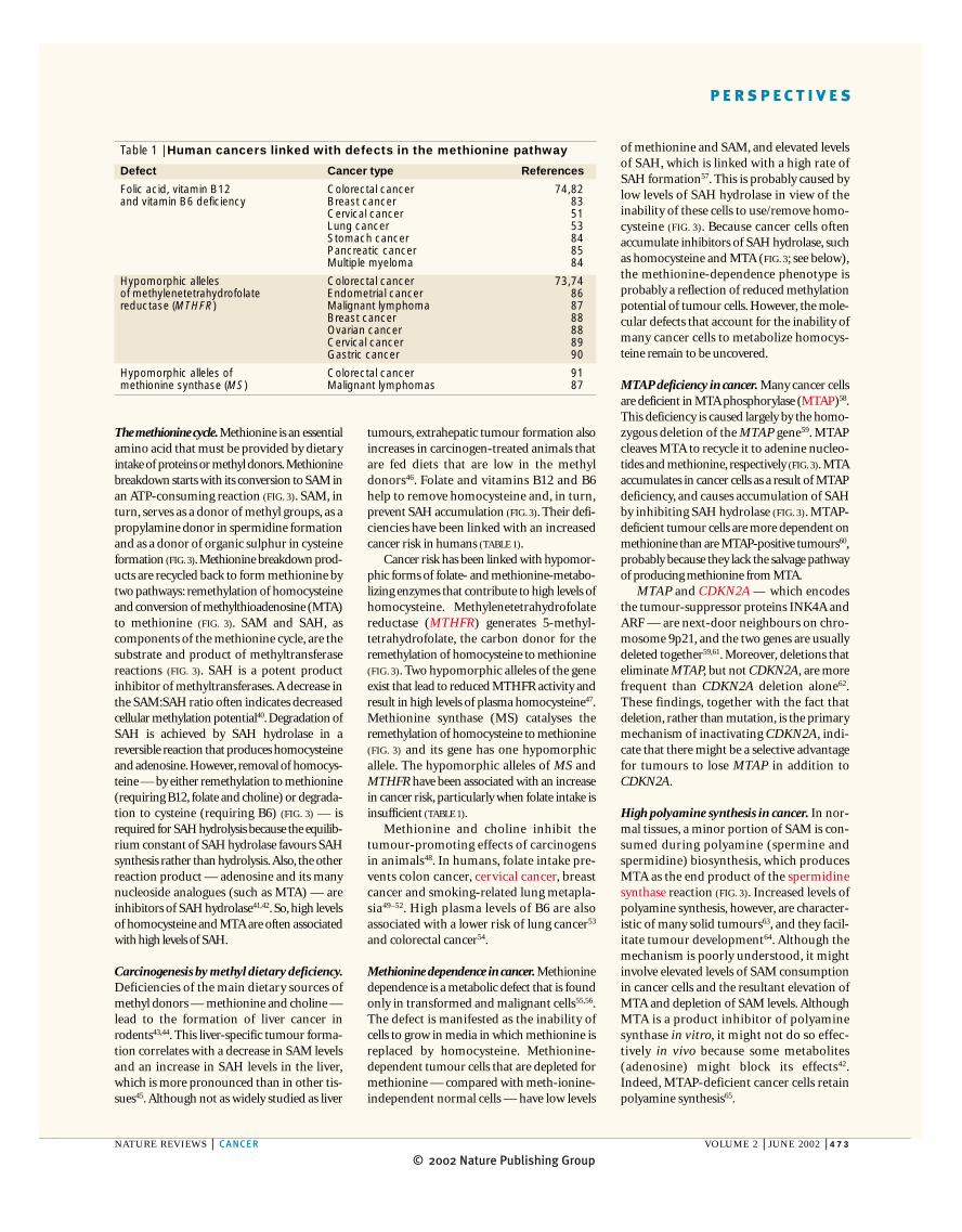
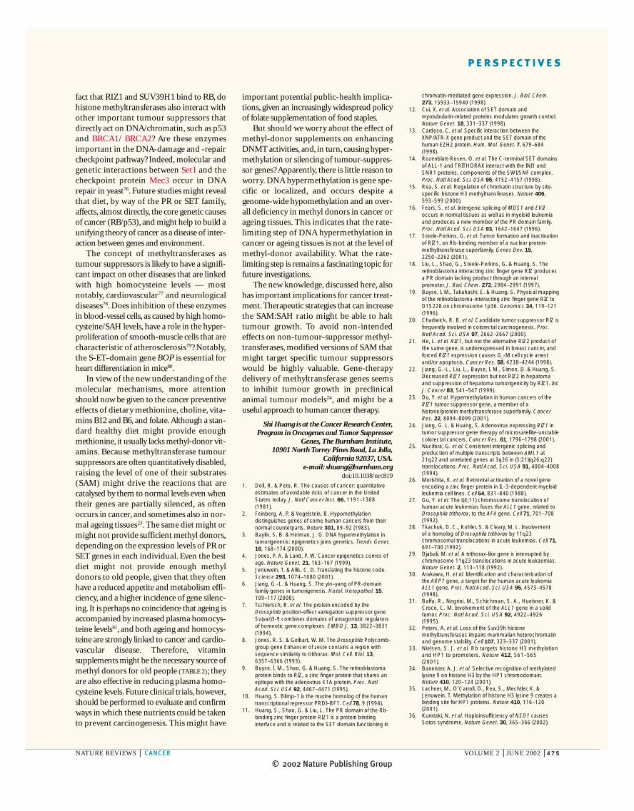
the PR and SET domains and between theplant enzymes and the PR/SET domains. Themain differences between the PR and SETdomains lie in the less conserved boxes — Aand B — in which the PR domain has severalconserved motifs that are not found in theSET domain (FIG. 1). Five related human genescontain a divergent A box that is separatedfrom their B and C boxes by a MYND zinc-finger motif; these genes are grouped underthe name S-ET domain or BOP family (seeONLINE TABLE 1). Finally, one human proteinhas ~20% overall identity to the plant lysinemethyltransferase Rubsico LSMT (large sub-unit methyltransferase) and is more related tothe plant enzyme than to either the PR- orSET- domain proteins (see ONLINE TABLE 1).
PR/SET genes in tumour suppressionAmong the >100 different human genes thatbelong to the broad class of SAM-dependentmethyltransferases, the first that wasunequivocally shown to be a tumour-sup-pressor was RIZ1 (REF. 17). RIZ1 also serves toillustrate a key role of the PR domain itself intumour suppression, which indicates thatother PR- and SET-containing genes mighthave a tumour-suppressor function. Apotential tumour-suppressor role has beenindicated for several other PR and SETgenes, but the data, briefly discussed below,remain circumstantial.
RIZ1. The RIZ1 gene was isolated originallyin a functional screen for proteins that bind tothe retinoblastoma (RB) tumour suppressor9.The gene produces two mRNA and proteinproducts, owing to alternative promoterusage18. It is only the full-length (1,719 aminoacids) product, RIZ1, that contains the PR
evolution and is more involved in metazoan-specific functions. Consistently, metazoanevolution is associated with a more dramaticincrease in the number of PR-domain genesthan in that of SET-domain genes. There are~7 SET genes in yeast, ~30 in Arabidopsisthaliana, ~20 in Drosophila, ~20 inCaenorhabditis elegans and ~27 in humans(see ONLINE TABLE 1). By contrast, there are noPR-domain proteins in yeast or A. thaliana,two in Drosophila, two in C. elegans and ~17in humans (see ONLINE TABLE 1). In spite ofthe differences between the two domains,protein-domain databases, such as Pfam (seeonline links box), and some recent literatureuse the name SET to represent both. Becausethe two domains are easily distinguishable— indeed, they were discovered indepen-dently — and it is not yet clear if they func-tion similarly, I will use the term PR/SET torefer to the superfamily that contains either aPR or a SET domain.
Repeated PSI-Blast (position-specific iterative basic local alignment search tool)analyses of sequence databases have revealedthat a plant protein lysine methyltransferasehas sequence similarity to the SET domain15.The homology (~7%) is borderline (FIG. 1),but as histone lysine methyltransferase activ-ity has been shown for the human proteinSUV39H1 and several other SET-domain pro-teins — which is mainly conferred by the SETdomain15 — similar activity is expected of thePR domain, because it is as homologous to theplant enzyme as the SET domain is (FIG. 1). Thehomology is restricted largely to the threeconserved boxes that were recognized initiallyfor the PR domain16, which is consistent witha functional similarity. Box C is the most conserved among the plant enzymes, between
The SET and PR domainThe SET domain was discovered as a120–150 amino-acid sequence homologypresent in several Drosophila genes that areinvolved in chromatin-mediated gene regu-lation during development7,8. The PRdomain was characterized as a ~130 amino-acid homology present in the humanretinoblastoma-protein-binding proteinRIZ1 and the human transcriptional repres-sor PRDI-BF1 (REFS 9,10). Using improvedbioinformatics tools, the PR domain waslater shown to share sequence homologywith the SET domain11. The commonresidues between the PR and SET domainsare also among the most conserved in eachdomain, indicating that they might sharesome functions. Indeed, a protein-bindinginterface activity has been described inde-pendently for both domains11–14.
Despite the sequence similarity betweenPR and SET domains, which is typically20–30% amino-acid identity, they are dis-tinctive, because identities among PRdomains or among SET domains are usuallyhigher — ~40%. Also, SET domains are pri-marily found at the carboxyl termini of pro-teins, whereas PR domains are mostlylocated at the amino termini. PR-domain-containing proteins commonly have zinc-finger DNA-binding domains, whereasSET- domain proteins lack obvious DNA-binding motifs. The PR-domain-containinggenes have the human-gene-mapping work-shop (HGMW)-approved gene symbolPRDM (PR-domain containing, with zincfingers). Finally, the SET domain, but notthe PR domain, is found in the yeastgenome, indicating that the PR domain isprobably a derivative of the SET domain in
253 GRGWGVRTLEKIRKNSFVMEYVGEIITSEE-(24)-VYTVDAAYY--GNISHFVNHSCDPNLQVYNVFID-(7)-RIAFF-ATRTIRAGEELTFDYNMQVDPVD
621 VAGWGIFIKDPVQKNEFISEYCGEIISQDE-(21)-DFVVDATRK--GNKIRFANHSVNPNCYAKVMMVNG--DHRIGIF-AKRAIQTGEELFFDYRYSQADAL
3838 IHGRGLFCKRNIDAGEMVIEYAGNVIRSIQ-(22)-EV-VDATMH--GNRARFINHSCEPNCYSRVINIDG--QKHIVIF-AMRKIYRGEELTYDYKFPIEDAS
60 EEVIGVMSKEYIPKGTRFGPLIGEIYTNDT-(19)-HHFIDGFNEEKSNWMRYVNPAHSPREQNLAACQN---GMNIY-FYTIKPIPANQELLVWYCRDFAERL
39 KTRIGVWATKPILKGKKFGPFVGDKKKRSQ-(16)-WMCIDATDPEKGNWLRYVNWACSGEEQNLFPLEI---NRAIY-YKTLKPIAPGEELLVWYNGEDNPEI
61 LQEEGVITAKTPVKASVVTEGLGLVALKDI-(136)-LRNENLVVVPMADL--INHSAGVTTEDHAY-(10)-WDYLFSLKSPLSVKAGEQVYIQYDLNKSNAE
56 LRDQGVVSGKSVAEPAVVPEGLGLVARRDI-(128)-LELNRESLTSMFEFEQINHNPAIKTEDYAY-(9)--RDLLFSLKSPVYVKAGEQVYIQYDLNKSNAE
SET containing
PR containing
Rubisco LSMT
SUV39HIEZH2MLL1
BLIMP1RIZ1
P. sativumA. thaliana
Box A Box B Box C
Figure 1 | Comparison of the SET and PR domains with a plant protein lysine methyltransferase. The aligned peptide sequences include the SET-domain regions of the human homologues of the three Drosophila founding members of the SET-domain family, the PR-domain regions of the two humanfounding members of the PR-domain family, and the PR/SET homology regions of the Rubisco LSMTs (Rubisco large subunit methyltransferase) from twodifferent plant species, Pisum sativum and Arabidopsis thaliana. Identical residues that are conserved among different plant species in the Rubisco ISMTs are ingreen; also in green are those residues in the SET or PR domain of human genes that are identical or similar to the conserved residues of plant enzymes. Themotifs of the PR domain that are boxed in red are present in most PR domains, but are absent in most SET domains, so can be used to distinguish the two.
© 2002 Nature Publishing Group
P E R S P E C T I V E S
expressed contain the carboxy-terminalSET domain of MLL1 (FIG. 2b). SET-minuschimeric proteins seem to exert a domi-nant-negative effect on the wild-type MLL1protein30. Also, 11q23 is deleted in somesolid tumours, and the MLL1 transcript isabsent in a gastric tumour-cell line withpartial gene duplication31. Disruption ofcertain SET-domain-mediated proteininteractions of MLL1 contributes to trans-formation12. These observations indicate,although are insufficient to conclude, thatMLL1 is involved in tumorigenesis by aloss-of-function mechanism.
SUV39H1. A potential tumour-suppressorrole of SUV39H1, and of its paralogueSUV39H2, is indicated by knockout micethat are prone to developing B-cell lymphomas, although whether a cell-autonomous defect is involved remainsunclear32. Loss of SUV39H1/SUV39H2function impairs heterochromatin andgenome stability, which might contribute totumour formation32. The B-cell tumours in
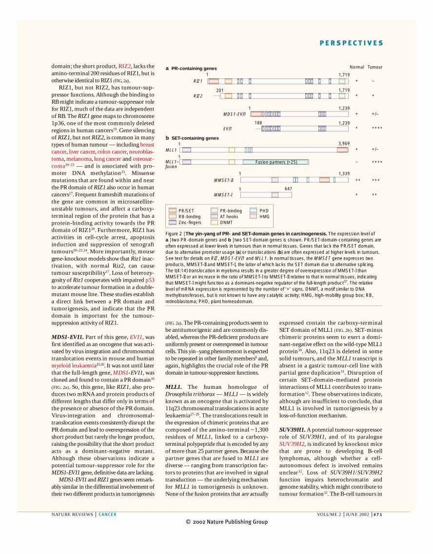
domain; the short product, RIZ2, lacks theamino-terminal 200 residues of RIZ1, but isotherwise identical to RIZ1 (FIG. 2a).
RIZ1, but not RIZ2, has tumour-sup-pressor functions. Although the binding toRB might indicate a tumour-suppressor rolefor RIZ1, much of the data are independentof RB. The RIZ1 gene maps to chromosome1p36, one of the most commonly deletedregions in human cancers19. Gene silencingof RIZ1, but not RIZ2, is common in manytypes of human tumour — including breastcancer, liver cancer, colon cancer, neuroblas-toma, melanoma, lung cancer and osteosar-coma20–22 — and is associated with pro-moter DNA methylation23. Missensemutations that are found within and nearthe PR domain of RIZ1 also occur in humancancers17. Frequent frameshift mutations ofthe gene are common in microsatellite-unstable tumours, and affect a carboxy-terminal region of the protein that has aprotein-binding activity towards the PRdomain of RIZ120. Furthermore, RIZ1 hasactivities in cell-cycle arrest, apoptosisinduction and suppression of xenografttumours20–22,24. More importantly, mousegene-knockout models show that Riz1 inac-tivation, with normal Riz2, can causetumour susceptibility17. Loss of heterozy-gosity of Riz1 cooperates with impaired p53to accelerate tumour formation in a double-mutant mouse line. These studies establish a direct link between a PR domain andtumorigenesis, and indicate that the PRdomain is important for the tumour-suppression activity of RIZ1.
MDS1-EVI1. Part of this gene, EVI1, wasfirst identified as an oncogene that was acti-vated by virus integration and chromosomaltranslocation events in mouse and humanmyeloid leukaemia25,26. It was not until laterthat the full-length gene, MDS1-EVI1, wascloned and found to contain a PR domain16
(FIG. 2a). So, this gene, like RIZ1, also pro-duces two mRNA and protein products ofdifferent lengths that differ only in terms ofthe presence or absence of the PR domain.Virus-integration and chromosomal-translocation events consistently disrupt thePR domain and lead to overexpression of theshort product but rarely the longer product,raising the possibility that the short productacts as a dominant-negative mutant.Although these observations indicate apotential tumour-suppressor role for theMDS1-EVI1 gene, definitive data are lacking.
MDS1-EVI1 and RIZ1 genes seem remark-ably similar in the differential involvement oftheir two different products in tumorigenesis
(FIG. 2a). The PR-containing products seem tobe antitumorigenic and are commonly dis-abled, whereas the PR-deficient products areuniformly present or overexpressed in tumourcells. This yin–yang phenomenon is expectedto be repeated in other family members6 and,again, highlights the crucial role of the PRdomain in tumour-suppression functions.
MLL1. The human homologue ofDrosophila trithorax — MLL1 — is widelyknown as an oncogene that is activated by11q23 chromosomal translocations in acuteleukaemia27–29. The translocations result inthe expression of chimeric proteins that arecomposed of the amino-terminal ~1,300residues of MLL1, linked to a carboxy-terminal polypeptide that is encoded by anyof more than 25 partner genes. Because thepartner genes that are fused to MLL1 arediverse — ranging from transcription fac-tors to proteins that are involved in signaltransduction — the underlying mechanismfor MLL1 in tumorigenesis is unknown.None of the fusion proteins that are actually
NATURE REVIEWS | CANCER VOLUME 2 | JUNE 2002 | 471
1,719
1,719
3,969
1,2391
1,3391
1
1
1
647
1,239188
1
201
PR/SETRB-bindingZinc-fingers
PHDHMG
PR-bindingAT hooksDNMT
Fusion partners (>25)
MLL1
MLL1–fusion
Normal Tumour
RIZ1
RIZ2
MDS1-EVI1
EVI1
MMSET-II
MMSET-I
a PR-containing genes
b SET-containing genes
+
+
+
+
+
–
++
+
–
+
+/–
++++
+/–
++++
+++
++
Figure 2 | The yin–yang of PR- and SET-domain genes in carcinogenesis. The expression level of a | two PR-domain genes and b | two SET-domain genes is shown. PR/SET-domain-containing genes areoften expressed at lower levels in tumours than in normal tissues. Genes that lack the PR/SET domain,due to alternative promoter usage (a) or translocations (b) are often expressed at higher levels in tumours.See text for details on RIZ, MDS1-EVI1 and MLL1. In normal tissues, the MMSET gene expresses twoproducts, MMSET-II and MMSET-I, the latter of which lacks the SET domain due to alternative splicing.The t(4:14) translocation in myeloma results in a greater degree of overexpression of MMSET-I thanMMSET-II or an increase in the ratio of MMSET-I to MMSET-II relative to that in normal tissues, indicatingthat MMSET-I might function as a dominant-negative regulator of the full-length product37. The relativelevel of mRNA expression is represented by the number of ‘+’ signs. DNMT, a motif similar to DNAmethyltransferases, but is not known to have any catalytic activity; HMG, high-mobility group box; RB,retinoblastoma; PHD, plant homeodomain.
© 2002 Nature Publishing Group472 | JUNE 2002 | VOLUME 2 www.nature.com/reviews/cancer
P E R S P E C T I V E S
In such a scenario, other functions of a PRor SET domain might be involved, such astheir protein-binding activity. Finally, it ispossible that certain PR or SET genes,regardless of their enzymatic activity, mightnot be involved in tumorigenesis. Methyl-transferase function per se, regardless ofsubstrates, does not necessarily predict arole in cancer, in view of the presence ofmany such enzymes that clearly do not haveany role in cancer. These possibilities not-withstanding, the yin–yang phenomenon,together with the link with methylationdeficiency, predicts that a PR or SET genewith active protein methyltransferase activ-ity is more likely to be a tumour-suppressorgene than an oncogene.
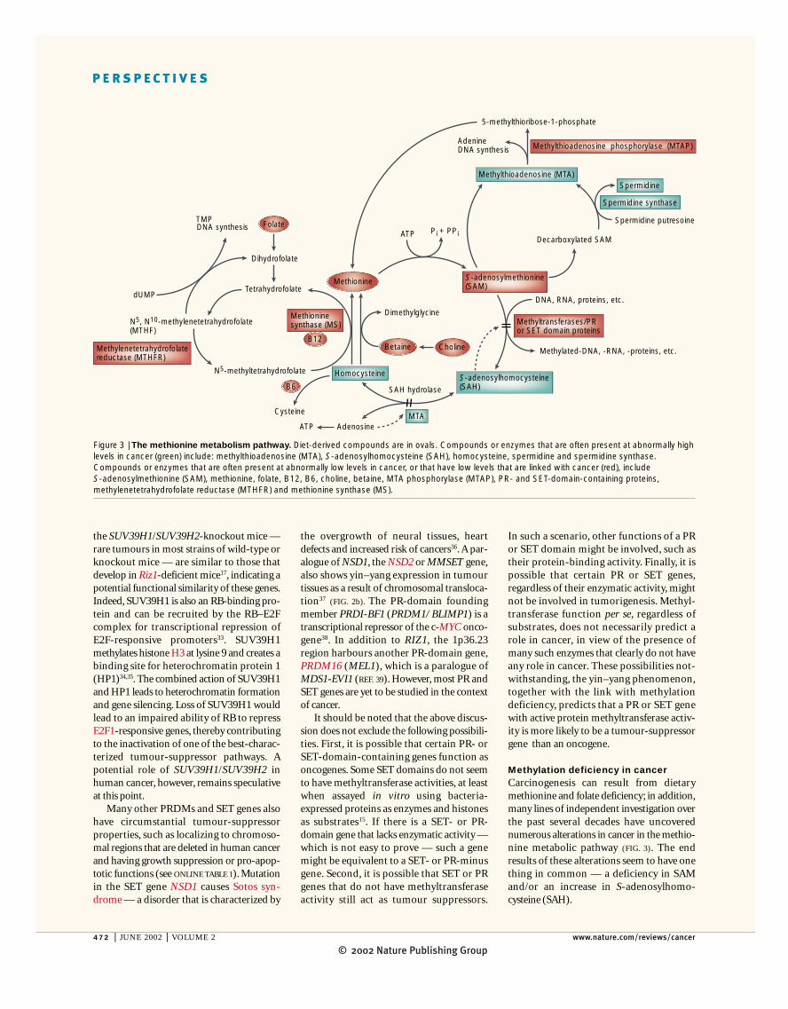
Methylation deficiency in cancerCarcinogenesis can result from dietarymethionine and folate deficiency; in addition,many lines of independent investigation overthe past several decades have uncoverednumerous alterations in cancer in the methio-nine metabolic pathway (FIG. 3). The endresults of these alterations seem to have onething in common — a deficiency in SAMand/or an increase in S-adenosylhomo-cysteine (SAH).
the overgrowth of neural tissues, heartdefects and increased risk of cancers36. A par-alogue of NSD1, the NSD2 or MMSET gene,also shows yin–yang expression in tumourtissues as a result of chromosomal transloca-tion37 (FIG. 2b). The PR-domain foundingmember PRDI-BF1 (PRDM1/ BLIMP1) is atranscriptional repressor of the c-MYC onco-gene38. In addition to RIZ1, the 1p36.23region harbours another PR-domain gene,PRDM16 (MEL1), which is a paralogue ofMDS1-EVI1 (REF. 39). However, most PR andSET genes are yet to be studied in the contextof cancer.
It should be noted that the above discus-sion does not exclude the following possibili-ties. First, it is possible that certain PR- orSET-domain-containing genes function asoncogenes. Some SET domains do not seemto have methyltransferase activities, at leastwhen assayed in vitro using bacteria-expressed proteins as enzymes and histonesas substrates15. If there is a SET- or PR-domain gene that lacks enzymatic activity —which is not easy to prove — such a genemight be equivalent to a SET- or PR-minusgene. Second, it is possible that SET or PRgenes that do not have methyltransferaseactivity still act as tumour suppressors.
the SUV39H1/SUV39H2-knockout mice —rare tumours in most strains of wild-type orknockout mice — are similar to those thatdevelop in Riz1-deficient mice17, indicating apotential functional similarity of these genes.Indeed, SUV39H1 is also an RB-binding pro-tein and can be recruited by the RB–E2Fcomplex for transcriptional repression ofE2F-responsive promoters33. SUV39H1methylates histone H3 at lysine 9 and creates abinding site for heterochromatin protein 1(HP1)34,35. The combined action of SUV39H1and HP1 leads to heterochromatin formationand gene silencing. Loss of SUV39H1 wouldlead to an impaired ability of RB to repressE2F1-responsive genes, thereby contributingto the inactivation of one of the best-charac-terized tumour-suppressor pathways. Apotential role of SUV39H1/SUV39H2 inhuman cancer, however, remains speculativeat this point.
Many other PRDMs and SET genes alsohave circumstantial tumour-suppressorproperties, such as localizing to chromoso-mal regions that are deleted in human cancerand having growth suppression or pro-apop-totic functions (see ONLINE TABLE 1). Mutationin the SET gene NSD1 causes Sotos syn-drome — a disorder that is characterized by
DNA synthesisTMP
dUMP
Dihydrofolate
Tetrahydrofolate
N5, N10-methylenetetrahydrofolate(MTHF)
N5-methyltetrahydrofolate
Dimethylglycine
DNA, RNA, proteins, etc.
Methylated-DNA, -RNA, -proteins, etc.
AdenosineATP
Cysteine
ATP Pi + PPiDecarboxylated SAM
Spermidine putresoine
AdenineDNA synthesis
5-methylthioribose-1-phosphate
Folate
B12
Methionine
Choline
B6
Betaine
SAH hydrolase
Homocysteine S-adenosylhomocysteine(SAH)
MTA
Spermidine synthase
SpermidineMethylthioadenosine (MTA)
Methioninesynthase (MS)
Methylenetetrahydrofolatereductase (MTHFR)
S-adenosylmethionine(SAM)
Methyltransferases/PR or SET domain proteins
Methylthioadenosine phosphorylase (MTAP)
Figure 3 | The methionine metabolism pathway. Diet-derived compounds are in ovals. Compounds or enzymes that are often present at abnormally highlevels in cancer (green) include: methylthioadenosine (MTA), S-adenosylhomocysteine (SAH), homocysteine, spermidine and spermidine synthase.Compounds or enzymes that are often present at abnormally low levels in cancer, or that have low levels that are linked with cancer (red), include S-adenosylmethionine (SAM), methionine, folate, B12, B6, choline, betaine, MTA phosphorylase (MTAP), PR- and SET-domain-containing proteins,methylenetetrahydrofolate reductase (MTHFR) and methionine synthase (MS).
© 2002 Nature Publishing Group
P E R S P E C T I V E S
of methionine and SAM, and elevated levelsof SAH, which is linked with a high rate ofSAH formation57. This is probably caused bylow levels of SAH hydrolase in view of theinability of these cells to use/remove homo-cysteine (FIG. 3). Because cancer cells oftenaccumulate inhibitors of SAH hydrolase, suchas homocysteine and MTA (FIG. 3; see below),the methionine-dependence phenotype isprobably a reflection of reduced methylationpotential of tumour cells. However, the mole-cular defects that account for the inability ofmany cancer cells to metabolize homocys-teine remain to be uncovered.
MTAP deficiency in cancer. Many cancer cellsare deficient in MTA phosphorylase (MTAP)58.This deficiency is caused largely by the homo-zygous deletion of the MTAP gene59. MTAPcleaves MTA to recycle it to adenine nucleo-tides and methionine, respectively (FIG. 3). MTAaccumulates in cancer cells as a result of MTAPdeficiency, and causes accumulation of SAH by inhibiting SAH hydrolase (FIG. 3). MTAP-deficient tumour cells are more dependent onmethionine than are MTAP-positive tumours60,probably because they lack the salvage pathwayof producing methionine from MTA.
MTAP and CDKN2A — which encodesthe tumour-suppressor proteins INK4A andARF — are next-door neighbours on chro-mosome 9p21, and the two genes are usuallydeleted together59,61. Moreover, deletions thateliminate MTAP, but not CDKN2A, are morefrequent than CDKN2A deletion alone62.These findings, together with the fact thatdeletion, rather than mutation, is the primarymechanism of inactivating CDKN2A, indi-cate that there might be a selective advantagefor tumours to lose MTAP in addition toCDKN2A.
High polyamine synthesis in cancer. In nor-mal tissues, a minor portion of SAM is con-sumed during polyamine (spermine andspermidine) biosynthesis, which producesMTA as the end product of the spermidinesynthase reaction (FIG. 3). Increased levels ofpolyamine synthesis, however, are character-istic of many solid tumours63, and they facil-itate tumour development64. Although themechanism is poorly understood, it mightinvolve elevated levels of SAM consumptionin cancer cells and the resultant elevation ofMTA and depletion of SAM levels. AlthoughMTA is a product inhibitor of polyaminesynthase in vitro, it might not do so effec-tively in vivo because some metabolites(adenosine) might block its effects42.Indeed, MTAP-deficient cancer cells retainpolyamine synthesis65.
The methionine cycle.Methionine is an essentialamino acid that must be provided by dietaryintake of proteins or methyl donors.Methioninebreakdown starts with its conversion to SAM inan ATP-consuming reaction (FIG. 3). SAM, inturn, serves as a donor of methyl groups, as apropylamine donor in spermidine formationand as a donor of organic sulphur in cysteineformation (FIG.3).Methionine breakdown prod-ucts are recycled back to form methionine bytwo pathways: remethylation of homocysteineand conversion of methylthioadenosine (MTA)to methionine (FIG. 3). SAM and SAH, as components of the methionine cycle, are thesubstrate and product of methyltransferasereactions (FIG. 3). SAH is a potent productinhibitor of methyltransferases.A decrease inthe SAM:SAH ratio often indicates decreasedcellular methylation potential40.Degradation ofSAH is achieved by SAH hydrolase in areversible reaction that produces homocysteineand adenosine.However, removal of homocys-teine — by either remethylation to methionine(requiring B12, folate and choline) or degrada-tion to cysteine (requiring B6) (FIG. 3) — isrequired for SAH hydrolysis because the equilib-rium constant of SAH hydrolase favours SAHsynthesis rather than hydrolysis.Also, the otherreaction product — adenosine and its manynucleoside analogues (such as MTA) — areinhibitors of SAH hydrolase41,42. So, high levelsof homocysteine and MTA are often associatedwith high levels of SAH.
Carcinogenesis by methyl dietary deficiency.Deficiencies of the main dietary sources ofmethyl donors — methionine and choline —lead to the formation of liver cancer inrodents43,44. This liver-specific tumour forma-tion correlates with a decrease in SAM levelsand an increase in SAH levels in the liver,which is more pronounced than in other tis-sues45. Although not as widely studied as liver
tumours, extrahepatic tumour formation alsoincreases in carcinogen-treated animals thatare fed diets that are low in the methyldonors46. Folate and vitamins B12 and B6help to remove homocysteine and, in turn,prevent SAH accumulation (FIG. 3). Their defi-ciencies have been linked with an increasedcancer risk in humans (TABLE 1).
Cancer risk has been linked with hypomor-phic forms of folate- and methionine-metabo-lizing enzymes that contribute to high levels ofhomocysteine. Methylenetetrahydrofolatereductase (MTHFR) generates 5-methyl-tetrahydrofolate, the carbon donor for theremethylation of homocysteine to methionine(FIG. 3). Two hypomorphic alleles of the geneexist that lead to reduced MTHFR activity andresult in high levels of plasma homocysteine47.Methionine synthase (MS) catalyses theremethylation of homocysteine to methionine(FIG. 3) and its gene has one hypomorphic allele. The hypomorphic alleles of MS andMTHFR have been associated with an increasein cancer risk, particularly when folate intake isinsufficient (TABLE 1).
Methionine and choline inhibit thetumour-promoting effects of carcinogensin animals48. In humans, folate intake pre-vents colon cancer, cervical cancer, breastcancer and smoking-related lung metapla-sia49–52. High plasma levels of B6 are alsoassociated with a lower risk of lung cancer53
and colorectal cancer54.
Methionine dependence in cancer. Methioninedependence is a metabolic defect that is foundonly in transformed and malignant cells55,56.The defect is manifested as the inability ofcells to grow in media in which methionine isreplaced by homocysteine. Methionine-dependent tumour cells that are depleted formethionine — compared with meth-ionine-independent normal cells — have low levels
NATURE REVIEWS | CANCER VOLUME 2 | JUNE 2002 | 473
Table 1 | Human cancers linked with defects in the methionine pathway
Defect Cancer type References
Folic acid, vitamin B12 Colorectal cancer 74,82and vitamin B6 deficiency Breast cancer 83
Cervical cancer 51Lung cancer 53Stomach cancer 84Pancreatic cancer 85Multiple myeloma 84
Hypomorphic alleles Colorectal cancer 73,74of methylenetetrahydrofolate Endometrial cancer 86reductase (MTHFR) Malignant lymphoma 87
Breast cancer 88Ovarian cancer 88Cervical cancer 89Gastric cancer 90
Hypomorphic alleles of Colorectal cancer 91methionine synthase (MS) Malignant lymphomas 87
© 2002 Nature Publishing Group474 | JUNE 2002 | VOLUME 2 www.nature.com/reviews/cancer
P E R S P E C T I V E S
humans, renders it difficult to extrapolateanimal results. So, the number of tumour-suppressor genes within the PR/SET super-family will probably be underestimated, butmight still prove to be more than the totalnumber (~25) of all presently knowntumour-suppressor genes.
Future directionsThe biological and biochemical functions oftumour-suppressor genes of the proteinmethyltransferase class are only now beginning to be studied. By methylating his-tones, these genes are crucial in chromatin-mediated long-term gene expression and inmaintaining cell memory or the differentiated phenotype. In view of the fact that cancer isnot only a disease of proliferation, but also ofde-differentiation, loss of protein methyltrans-ferase function might contribute directly tothe pseudo-stem-cell-like phenotypes of can-cer. The function of these enzymes on chro-matin might also contribute to the aneuploidythat is characteristic of cancer.
This general working model of proteinmethyltransferase activity in tumour sup-pression remains to be fine-tuned by futureinvestigations. Does a methyl-deficient dietcause inhibition of protein methyltrans-ferases and confer enhanced tumour forma-tion in animal models with hypomorphicalleles of these enzymes? Is global hypo-methylation — although not excludinglocalized (gene-specific) hypermethylation— of histones a feature of cancer? Can a cer-tain range of SAM or SAH levels linearly con-trol protein methyltransferase activities, andwhat is the range? Is the long-recognizedembryonic-stem-cell-like phenotype (reacti-vation of embryonic marker gene expres-sion) of cancer related to inactivation of thesegenes? Given that histone methylation is heri-table, as is apparent in cell memory, shouldthe term cancer epigenetics be redefined toalso include histone methylation? Given the
Therefore, although decreased DNMTsand several other previously suggested path-ways could result from methyl deficiency andmight be involved in carcinogenesis, they allinvoke indirect mechanisms and remain spec-ulative. By contrast, the discovery of tumoursuppressors among protein methyltrans-ferases provides a direct link from methylinsufficiency to genetic causes of cancer.
Are most members of the PR/SET super-family — ~44 in total (see ONLINE TABLE 1) —tumour-suppressor genes? Although this is apossibility, each member must individuallyqualify for the standard definition of atumour-suppressor gene — that is, loss ofgene activity by epigenetic silencing, geneticmutation or both must correlate withtumorigenesis. However, gene silencing ordosage reduction of a PR or SET gene mightalso be coupled with a methyl deficiency,which would make its partial or quantitativeloss more serious. The fact that methyl defi-ciency causes cancer indicates that quantita-tive inactivation of protein methyltrans-ferases is sufficient for carcinogenesis, whichmight explain why this class of tumour-sup-pressor genes is often silenced rather thanmutated17,23. In view of this, it is also proba-ble that certain PR or SET tumour-suppres-sor genes are primarily disabled at theenzyme function level, and would, therefore,rarely reveal themselves when examined atthe gene level using present methods of find-ing tumour-suppressor genes. Furthermore,mutation or gene silencing might notachieve complete inactivation of the gene’sfunction if the gene has paralogues. TheSET-domain family is particularly rich ingenes with paralogues; 16 members repre-sent only 6 orthologue genes. In the absenceof human genetic or DNA epigenetic evi-dence, animal knockout models would haveto be more heavily relied on. However, thepresence of genes that specifically act astumour suppressors in animals, but not
A common theme?Although the observations that link meta-bolic alterations of methionine with car-cinogenesis are overwhelming, the underly-ing molecular mechanism remains a matterof speculation. Several pathways have previ-ously been proposed to link methyl defi-ciency to carcinogenesis, which includeDNA methylation, polyamine synthesis,DNA damage and repair, the cell cycle andprotein kinase C63,66–69.
The different events that affect the methio-nine pathway all give rise to one common out-come: a methyl deficiency as defined by adecrease in the ratio of SAM:SAH and, in turn,inhibition of SAM-dependent transmethyla-tions. Because the human genome encodes>100 different methyltransferases that useSAM, the number of potential targets ofmethyl deficiency is vast. The most widelyinvestigated in the past has been the idea ofDNMT inhibition; a methyl-deficient diet isalso known to cause DNA hypomethylation66.DNA hypomethylation is thought to causeoncogene activation, DNA mutation/recombi-nation and genome instability. Few oncogenes,however, are activated by hypomethylation; itis also unclear whether hypomethylation iscausally linked with transcriptional activation.By contrast, DNA hypomethylation has beenshown to suppress tumour formation70.Hypermethylation seems more significantlyassociated with carcinogenesis3,4, so the impor-tance of DNMT inhibition in carcinogenesisremains uncertain.
The effect of folic-acid deficiency onDNA mutation and repair has also been con-sidered in the past; it causes the intracellular accumulation of dUMP (deoxyuridine 5′-monophosphate) and the subsequent incor-poration of uracil into DNA (FIG. 3). Theremoval of uracil during DNA repair byuracil DNA glycosylase might result in sin-gle- and double-stranded DNA breaks, theaccumulation of which is a putative risk fac-tor for cancer71. This mechanism, however, isunlikely to apply to the effects of methioninedeficiency, which are not known to causeDNA damage72, and is also inconsistent withthe observation that the hypomorphicMTHFR allele is a risk factor for cancer73,74
(TABLE 1). Reduced MTHFR activity increaseslevels of methyltetrahydrofolate (FIG. 3), and,in turn, leads to more efficient conversion ofdUMP to TMP (thymidine 5′-monophos-phate), thereby reducing the chance of mis-incorporation of uracil into DNA. Finally,dietary folate supplements seem to have nosignificant effect on Apc and Trp53 muta-tions in the dimethylhydrazine rat model ofcolorectal cancer75.
Table 2 | A potential cancer-prevention recipe of methyl donors
Nutrient Daily amount (%RDA) ‡ Food sources
Folid acid (B9) 800 µg (200%) Whole grains, asparagus, beans, broccoli
Vitamin B12* 250–500 µg (5,000–10,000%) Liver, fish, milk, eggs, meat
Vitamin B6 25–50 mg (1,000–2,000%) Whole grains, beans, liver, avocados
Choline* 0–500 mg (n/a) Beef, liver, eggs, cauliflower
TMG (betaine) * 0–1,000 mg (n/a) Sugar beets, asparagus, most foods
*These nutrients are more necessary for some vegetarians who might not consume enough proteins, andtherefore methionine and meat (the main source of vitamin B12), in their diets. Non-vegetarians usually getenough methionine and vitamin B12 from dietary meat. ‡RDA, the United States Food and DrugAdministration’s recommended daily allowance. Because the RDAs for many nutrients have little to do withoptimal body need, significantly higher than RDA amounts — but still safe and effective in reducing plasmahomocysteine levels as found in clinical trials — are recommended here for the methyl donors. n/a, notapplicable; TMG, trimethylglycine.
© 2002 Nature Publishing Group
P E R S P E C T I V E S
chromatin-mediated gene expression. J. Biol. Chem.273, 15933–15940 (1998).
12. Cui, X. et al. Association of SET domain andmyotubularin-related proteins modulates growth control.Nature Genet. 18, 331–337 (1998).
13. Cardoso, C. et al. Specific interaction between theXNP/ATR-X gene product and the SET domain of thehuman EZH2 protein. Hum. Mol. Genet. 7, 679–684(1998).
14. Rozenblatt-Rosen, O. et al. The C-terminal SET domainsof ALL-1 and TRITHORAX interact with the INI1 andSNR1 proteins, components of the SWI/SNF complex.Proc. Natl Acad. Sci. USA 95, 4152–4157 (1998).
15. Rea, S. et al. Regulation of chromatin structure by site-specific histone H3 methyltransferases. Nature 406,593–599 (2000).
16. Fears, S. et al. Intergenic splicing of MDS1 and EVIIoccurs in normal tissues as well as in myeloid leukemiaand produces a new member of the PR domain family.Proc. Natl Acad. Sci. USA 93, 1642–1647 (1996).
17. Steele-Perkins, G. et al. Tumor formation and inactivationof RIZ1, an Rb-binding member of a nuclear protein-methyltransferase superfamily. Genes Dev. 15,2250–2262 (2001).
18. Liu, L., Shao, G., Steele-Perkins, G. & Huang, S. Theretinoblastoma interacting zinc finger gene RIZ producesa PR domain lacking product through an internalpromoter. J. Biol. Chem. 272, 2984–2991 (1997).
19. Buyse, I. M., Takahashi, E. & Huang, S. Physical mappingof the retinoblastoma-interacting zinc finger gene RIZ toD1S228 on chromosome 1p36. Genomics 34, 119–121(1996).
20. Chadwick, R. B. et al. Candidate tumor suppressor RIZ isfrequently involved in colorectal carcinogenesis. Proc.Natl Acad. Sci. USA 97, 2662–2667 (2000).
21. He, L. et al. RIZ1, but not the alternative RIZ2 product ofthe same gene, is underexpressed in breast cancer, andforced RIZ1 expression causes G2-M cell cycle arrestand/or apoptosis. Cancer Res. 58, 4238–4244 (1998).
22. Jiang, G.-L., Liu, L., Buyse, I. M., Simon, D. & Huang, S.Decreased RIZ1 expression but not RIZ2 in hepatomaand suppression of hepatoma tumorigenicity by RIZ1. Int.J. Cancer 83, 541–547 (1999).
23. Du, Y. et al. Hypermethylation in human cancers of theRIZ1 tumor suppressor gene, a member of ahistone/protein methyltransferase superfamily. CancerRes. 22, 8094–8099 (2001).
24. Jiang, G. L. & Huang, S. Adenovirus expressing RIZ1 intumor suppressor gene therapy of microsatellite-unstablecolorectal cancers. Cancer Res. 61, 1796–1798 (2001).
25. Nucifora, G. et al. Consistent intergenic splicing andproduction of multiple transcripts between AML1 at21q22 and unrelated genes at 3q26 in (3;21)(q26;q22)translocations. Proc. Natl Acad. Sci. USA 91, 4004–4008(1994).
26. Morishita, K. et al. Retroviral activation of a novel geneencoding a zinc finger protein in IL-3-dependent myeloidleukemia cell lines. Cell 54, 831–840 (1988).
27. Gu, Y. et al. The t(4;11) chromosome translocation ofhuman acute leukemias fuses the ALL1 gene, related toDrosophila trithorax, to the AF4 gene. Cell 71, 701–708(1992).
28. Tkachuk, D. C., Kohler, S. & Cleary, M. L. Involvement of a homolog of Drosophila trithorax by 11q23chromosomal translocations in acute leukemias. Cell 71,691–700 (1992).
29. Djabali, M. et al. A trithorax-like gene is interrupted bychromosome 11q23 translocations in acute leukaemias.Nature Genet. 2, 113–118 (1992).
30. Arakawa, H. et al. Identification and characterization ofthe ARP1 gene, a target for the human acute leukemiaALL1 gene. Proc. Natl Acad. Sci. USA 95, 4573–4578(1998).
31. Baffa, R., Negrini, M., Schichman, S. A., Huebner, K. &Croce, C. M. Involvement of the ALL1 gene in a solidtumor. Proc. Natl Acad. Sci. USA 92, 4922–4926(1995).
32. Peters, A. et al. Loss of the Suv39h histonemethyltransferases impairs mammalian heterochromatinand genome stability. Cell 107, 323–337 (2001).
33. Nielsen, S. J. et al. Rb targets histone H3 methylationand HP1 to promoters. Nature 412, 561–565 (2001).
34. Bannister, A. J. et al. Selective recognition of methylatedlysine 9 on histone H3 by the HP1 chromodomain.Nature 410, 120–124 (2001).
35. Lachner, M., O’Carroll, D., Rea, S., Mechtler, K. &Jenuwein, T. Methylation of histone H3 lysine 9 creates abinding site for HP1 proteins. Nature 410, 116–120(2001).
36. Kurotaki, N. et al. Haploinsufficiency of NSD1 causesSotos syndrome. Nature Genet. 30, 365–366 (2002).
fact that RIZ1 and SUV39H1 bind to RB, dohistone methyltransferases also interact withother important tumour suppressors thatdirectly act on DNA/chromatin, such as p53and BRCA1/ BRCA2? Are these enzymesimportant in the DNA-damage and -repaircheckpoint pathway? Indeed, molecular andgenetic interactions between Set1 and thecheckpoint protein Mec3 occur in DNArepair in yeast76. Future studies might revealthat diet, by way of the PR or SET family,affects, almost directly, the core genetic causesof cancer (RB/p53), and might help to build aunifying theory of cancer as a disease of inter-action between genes and environment.
The concept of methyltransferases astumour suppressors is likely to have a signifi-cant impact on other diseases that are linkedwith high homocysteine levels — mostnotably, cardiovascular77 and neurologicaldiseases78. Does inhibition of these enzymesin blood-vessel cells, as caused by high homo-cysteine/SAH levels, have a role in the hyper-proliferation of smooth-muscle cells that arecharacteristic of atherosclerosis79? Notably,the S-ET-domain gene BOP is essential forheart differentiation in mice80.
In view of the new understanding of themolecular mechanisms, more attentionshould now be given to the cancer preventiveeffects of dietary methionine, choline, vita-mins B12 and B6, and folate. Although a stan-dard healthy diet might provide enoughmethionine, it usually lacks methyl-donor vit-amins. Because methyltransferase tumoursuppressors are often quantitatively disabled,raising the level of one of their substrates(SAM) might drive the reactions that arecatalysed by them to normal levels even whentheir genes are partially silenced, as oftenoccurs in cancer, and sometimes also in nor-mal ageing tissues23. The same diet might ormight not provide sufficient methyl donors,depending on the expression levels of PR orSET genes in each individual. Even the bestdiet might not provide enough methyldonors to old people, given that they oftenhave a reduced appetite and metabolism effi-ciency, and a higher incidence of gene silenc-ing. It is perhaps no coincidence that ageing isaccompanied by increased plasma homocys-teine levels81, and both ageing and homocys-teine are strongly linked to cancer and cardio-vascular disease. Therefore, vitaminsupplements might be the necessary source ofmethyl donors for old people (TABLE 2); theyare also effective in reducing plasma homo-cysteine levels. Future clinical trials, however,should be performed to evaluate and confirmways in which these nutrients could be takento prevent carcinogenesis. This might have
important potential public-health implica-tions, given an increasingly widespread policyof folate supplementation of food staples.
But should we worry about the effect ofmethyl-donor supplements on enhancingDNMT activities, and, in turn, causing hyper-methylation or silencing of tumour-suppres-sor genes? Apparently, there is little reason toworry. DNA hypermethylation is gene spe-cific or localized, and occurs despite agenome-wide hypomethylation and an over-all deficiency in methyl donors in cancer orageing tissues. This indicates that the rate-limiting step of DNA hypermethylation incancer or ageing tissues is not at the level ofmethyl-donor availability. What the rate-limiting step is remains a fascinating topic forfuture investigations.
The new knowledge, discussed here, alsohas important implications for cancer treat-ment. Therapeutic strategies that can increasethe SAM:SAH ratio might be able to halttumour growth. To avoid non-intendedeffects on non-tumour-suppressor methyl-transferases, modified versions of SAM thatmight target specific tumour suppressorswould be highly valuable. Gene-therapy delivery of methyltransferase genes seems to inhibit tumour growth in preclinical animal tumour models24, and might be a useful approach to human cancer therapy.
Shi Huang is at the Cancer Research Center,Program in Oncogenes and Tumor Suppressor
Genes, The Burnham Institute,10901 North Torrey Pines Road, La Jolla,
California 92037, USA.e-mail: [email protected]
doi:10.1038/nrc819
1. Doll, R. & Peto, R. The causes of cancer: quantitativeestimates of avoidable risks of cancer in the UnitedStates today. J. Natl Cancer Inst. 66, 1191–1308(1981).
2. Feinberg, A. P. & Vogelstein, B. Hypomethylationdistinguishes genes of some human cancers from theirnormal counterparts. Nature 301, 89–92 (1983).
3. Baylin, S. B. & Herman, J. G. DNA hypermethylation intumorigenesis: epigenetics joins genetics. Trends Genet.16, 168–174 (2000).
4. Jones, P. A. & Laird, P. W. Cancer epigenetics comes ofage. Nature Genet. 21, 163–167 (1999).
5. Jenuwein, T. & Allis, C. D. Translating the histone code.Science 293, 1074–1080 (2001).
6. Jiang, G.-L. & Huang, S. The yin–yang of PR-domainfamily genes in tumorigenesis. Histol. Histopathol. 15,109–117 (2000).
7. Tschiersch, B. et al. The protein encoded by theDrosophila position-effect variegation suppressor geneSu(var)3-9 combines domains of antagonistic regulatorsof homeotic gene complexes. EMBO J. 13, 3822–3831(1994).
8. Jones, R. S. & Gelbart, W. M. The Drosophila Polycomb-group gene Enhancer of zeste contains a region withsequence similarity to trithorax. Mol. Cell. Biol. 13,6357–6366 (1993).
9. Buyse, I. M., Shao, G. & Huang, S. The retinoblastomaprotein binds to RIZ, a zinc finger protein that shares anepitope with the adenovirus E1A protein. Proc. NatlAcad. Sci. USA 92, 4467–4471 (1995).
10. Huang, S. Blimp-1 is the murine homolog of the humantranscriptional repressor PRDI–BF1. Cell 78, 9 (1994).
11. Huang, S., Shao, G. & Liu, L. The PR domain of the Rb-binding zinc finger protein RIZ1 is a protein bindinginterface and is related to the SET domain functioning in
NATURE REVIEWS | CANCER VOLUME 2 | JUNE 2002 | 475
© 2002 Nature Publishing Group476 | JUNE 2002 | VOLUME 2 www.nature.com/reviews/cancer
P E R S P E C T I V E S
78. Seshadri, S. et al. Plasma homocysteine as a risk factorfor dementia and Alzheimer’s disease. N. Engl. J. Med.346, 476–483 (2002).
79. Tsai, J. C. Promotion of vascular smooth muscle cellgrowth by homocysteine: a link to atherosclerosis. Proc.Natl Acad. Sci. USA 91, 6369–6373 (1994).
80. Gottlieb, P. D. et al. BOP encodes a muscle-restrictedprotein containing MYND and SET domains and isessential for cardiac differentiation and morphogenesis.Nature Genet. 1 April 2002 (DOI:10.1038/ng866).
81. Brattstrom, L., Lindgren, A., Israelsson, B., Andersson, A.& Hultberg, B. Homocysteine and cysteine: determinantsof plasma levels in middle-aged and elderly subjects. J. Internal Med. 236, 633–641 (1994).
82. Giovannucci, E. et al. Alcohol, low-methionine–low-folatediets, and risk of colon cancer in men. J. Natl CancerInst. 87, 265–273 (1995).
83. Wu, K. et al. A prospective study on folate, B12, andpyridoxal 5′-phosphate (B6) and breast cancer. Cancer Epidemiol. Biomark. Prev. 8, 209–217 (1999).
84. Hsing, A. W. et al. Pernicious anemia and subsequentcancer. A population-based cohort study. Cancer 71,745–750 (1993).
85. Stolzenberg-Solomon, R. Z. et al. Pancreatic cancer riskand nutrition-related methyl-group availability indicators inmale smokers. J. Natl Cancer Inst. 91, 535–541 (1999).
86. Esteller, M., Garcia, A., Martinez-Palones, J. M.,Xercavins, J. & Reventos, J. Germ line polymorphisms incytochrome-P450 1A1 (C4887 CYP1A1) andmethylenetetrahydrofolate reductase (MTHFR) genes andendometrial cancer susceptibility. Carcinogenesis 18,2307–2311 (1997).
87. Matsuo, K. et al. Association between polymorphisms offolate- and methionine-metabolizing enzymes andsusceptibility to malignant lymphoma. Blood 97,3205–3209 (2001).
88. Gershoni-Baruch, R. et al. Association of the C677Tpolymorphism in the MTHFR gene with breast and/orovarian cancer risk in Jewish women. Eur. J. Cancer 36,2313–2316 (2000).
89. Piyathilake, C. J. et al. Methylenetetrahydrofolatereductase (MTHFR) polymorphism increases the risk ofcervical intraepithelial neoplasia. Anticancer Res. 20,1751–1757 (2000).
90. Shen, H. et al. Polymorphisms of 5,10-methylenetetrahydrofolate reductase and risk of gastriccancer in a Chinese population: a case–control study. Int.J. Cancer 95, 332–336 (2001).
91. Ma, J. et al. A polymorphism of the methionine synthasegene: association with plasma folate, vitamin B12,homocyst(e)ine, and colorectal cancer risk. CancerEpidemiol. Biomark. Prev. 8, 825–829 (1999).
Online links
DATABASESThe following terms in this article are linked online to:Cancer.gov: http://www.cancer.gov/cancer_information/breast cancer | cervical cancer | colon cancer | liver cancer |lung cancer | melanoma | myeloid leukaemia | neuroblastoma |osteosarcomaLocusLink: http://www.ncbi.nlm.nih.gov/LocusLink/Apc | BOP | BRCA1 | BRCA2 | CDKN2A | E2F1 | EVI1 | H3 |MDS1 | MTAP | MTHFR | MYC | NSD1 | NSD2 | p53 | PRDI-BF1 | PRDM16 | protein kinase C | RB | Riz1 | RIZ1 | RIZ2 |spermidine synthase | SUV39H1 | SUV39H2 | Trp53 | uracilDNA glycosylaseOMIM: http://www.ncbi.nlm.nih.gov/Omim/Sotos syndromeSaccharomyces Genome Database: http://genome-www.stanford.edu/Saccharomyces/Mec3 | Set1
FURTHER INFORMATIONPfam Database: www.sanger.ac.uk/cgi-bin/Pfam/getacc?PF00856Access to this interactive links box is free online.
57. Stern, P. H. & Hoffman, R. M. Elevated overall rates oftransmethylation in cell lines from diverse human tumors.In Vitro 20, 663–670 (1984).
58. Toohey, J. I. Methylthio group cleavage frommethylthioadenosine. Description of an enzyme and itsrelationship to the methylthio requirement of certain cellsin culture. Biochem. Biophys. Res. Commun. 78,1273–1280 (1977).
59. Nobori, T. et al. Genomic cloning of methylthioadenosinephosphorylase: a purine metabolic enzyme deficient inmultiple different cancers. Proc. Natl Acad. Sci. USA 93,6203–6208 (1996).
60. Tang, B. Defects in methylthioadenosine phosphorylaseare associated with but not responsible for methionine-dependent tumor cell growth. Cancer Res. 60,5543–5547 (2000).
61. Dreyling, M. H., Roulston, D., Bohlander, S. K., Vardiman, J. & Olopade, O. I. Codeletion of CDKN2 andMTAP genes in a subset of non-Hodgkin’s lymphomamay be associated with histologic transformation fromlow-grade to diffuse large-cell lymphoma. GenesChromosom. Cancer 22, 72–78 (1998).
62. Schmid, M. et al. Homozygous deletions ofmethylthioadenosine phosphorylase (MTAP) are morefrequent than p16INK4A (CDKN2) homozygous deletionsin primary non-small cell lung cancers (NSCLC).Oncogene 17, 2669–2675 (1998).
63. Pegg, A. E. Polyamine metabolism and its importance inneoplastic growth and a target for chemotherapy. CancerRes. 48, 759–774 (1988).
64. Megosh, L. et al. Increased frequency of spontaneous skintumors in transgenic mice which overexpress ornithinedecarboxylase. Cancer Res. 55, 4205–4209 (1995).
65. Redman, C. et al. Involvement of polyamines inselenomethionine induced apoptosis and mitoticalterations in human tumor cells. Carcinogenesis 18,1195–1202 (1997).
66. Wainfan, E. & Poirier, L. A. Methyl groups incarcinogenesis: effects on DNA methylation and geneexpression. Cancer Res. 52, S2071–S2077 (1992).
67. Rushmore, T. H. et al. A choline-devoid diet, carcinogenicin the rat, induces DNA damage and repair.Carcinogenesis 7, 1677–1680 (1986).
68. James, S. J., Miller, B. J., Cross, D. R., McGarrity, L. J. &Morris, S. M. The essentiality of folate for themaintenance of deoxynucleotide precursor pools, DNAsynthesis, and cell cycle progression in PHA-stimulatedlymphocytes. Environ. Health Perspect. 101 (Suppl. 5),173–178 (1993).
69. da Costa, K. A., Cochary, E. F., Blusztajn, J. K., Garner, S. C. & Zeisel, S. H. Accumulation of 1,2-sn-diradylglycerol with increased membrane-associatedprotein kinase C may be the mechanism for spontaneoushepatocarcinogenesis in choline-deficient rats. J. Biol.Chem. 268, 2100–2105 (1993).
70. Laird, P. W. et al. Suppression of intestinal neoplasia byDNA hypomethylation. Cell 81, 197–205 (1995).
71. Blount, B. C. et al. Folate deficiency causes uracilmisincorporation into human DNA and chromosomebreakage: implications for cancer and neuronal damage.Proc. Natl Acad. Sci. USA 94, 3290–3295 (1997).
72. Duthie, S. J., Grant, G. & Narayanan, S. Increased uracilmisincorporation in lymphocytes from folate-deficientrats. Br. J. Cancer 83, 1532–1537 (2000).
73. Houlston, R. S. & Tomlinson, I. P. Polymorphisms andcolorectal tumor risk. Gastroenterology 121, 282–301(2001).
74. Ma, J. et al. Methylenetetrahydrofolate reductasepolymorphism, dietary interactions, and risk of colorectalcancer. Cancer Res. 57, 1098–1102 (1997).
75. Sohn, K. J. et al. The effect of dietary folate on Apc andp53 mutations in the dimethylhydrazine rat model ofcolorectal cancer. Carcinogenesis 20, 2345–2350 (1999).
76. Corda, Y. et al. Interaction between Set1p andcheckpoint protein Mec3p in DNA repair and telomerefunctions. Nature Genet. 21, 204–208 (1999).
77. McCully, K. S. Homocystinuria, arteriosclerosis,methylmalonic aciduria, and methyltransferase deficiency:a key case revisited. Nutr. Rev. 50, 7–12 (1992).
37. Chesi, M. et al. The t(4;14) translocation in myelomadysregulates both FGFR3 and a novel gene, MMSET,resulting in IgH/MMSET hybrid transcripts. Blood 92,3025–3034 (1998).
38. Lin, Y., Wong, K.-K. & Calame, K. Repression of c-Myctranscription by Blimp-1, an inducer of terminal B celldifferentiation. Science 276, 596–598 (1997).
39. Mochizuki, N. et al. A novel gene, MEL1, mapped to1p36.3 is highly homologous to the MDS1/EVI1 geneand is transcriptionally activated in t(1;3)(p36;q21)-positive leukemia cells. Blood 96, 3209–3214 (2000).
40. Caudill, M. A. et al. Intracellular S-adenosylhomocysteineconcentrations predict global DNA hypomethylation intissues of methyl-deficient cystathionine β-synthaseheterozygous mice. J. Nutrit. 131, 2811–2818 (2001).
41. Hershfield, M. S. & Krodich, N. M. S-adenosyl-homocysteine hydrolase is an adenosine-binding protein:a target for adenosine toxicity. Science 202, 757–760(1978).
42. Williams-Ashman, H. G., Seidenfeld, J. & Galletti, P.Trends in the biochemical pharmacology of 5′-deoxy-5′-methylthioadenosine. Biochem. Pharmacol. 31, 277–288(1982).
43. Mikol, Y. B., Hoover, K. L., Creasia, D. & Poirier, L. A.Hepatocarcinogenesis in rats fed methyl-deficient,amino acid-defined diets. Carcinogenesis 4, 1619–1629(1983).
44. Ghoshal, A. K. & Farber, E. The induction of liver cancerby dietary deficiency of choline and methionine withoutadded carcinogens. Carcinogenesis 5, 1367–1370(1984).
45. Shivapurkar, N. & Poirier, L. A. Tissue levels of S-adenosyl-methionine and S-adenosylhomocysteine inrats fed methyl-deficient, amino acid-defined diets forone to five weeks. Carcinogenesis 4, 1051–1057 (1983).
46. Cravo, M. L. et al. Folate deficiency enhances thedevelopment of colonic neoplasia in dimethylhydrazine-treated rats. Cancer Res. 52, 5002–5006 (1992).
47. Christensen, B. et al. Correlation of a common mutationin the methylenetetrahydrofolate reductase gene withplasma homocysteine in patients with prematurecoronary artery disease. Arterioscler. Thromb. Vasc. Biol.17, 569–573 (1997).
48. Fullerton, F. R., Hoover, K., Mikol, Y. B., Creasia, D. A. &Poirier, L. A. The inhibition by methionine and choline ofliver carcinoma formation in male C3H mice dosed withdiethylnitrosamine and fed phenobarbital. Carcinogenesis11, 1301–1305 (1990).
49. Giovannucci, E. et al. Multivitamin use, folate, and coloncancer in women in the Nurses’ Health Study. Ann.Intern. Med. 129, 517–524 (1998).
50. Prinz-Langenohl, R., Fohr, I. & Pietrzik, K. Beneficial rolefor folate in the prevention of colorectal and breastcancer. Eur. J. Nutrit. 40, 98–105 (2001).
51. Butterworth, C. E. Jr, Hatch, K. D., Gore, H., Mueller, H. &Krumdieck, C. L. Improvement in cervical dysplasiaassociated with folic acid therapy in users of oralcontraceptives. Am. J. Clin. Nutr. 35, 73–82 (1982).
52. Heimburger, D. C. et al. Improvement in bronchialsquamous metaplasia in smokers treated with folate andvitamin B12. Report of a preliminary randomized,double-blind intervention trial. JAMA 259, 1525–1530(1988).
53. Hartman, T. J. et al. Association of the B-vitaminspyridoxal 5′-phosphate (B(6)), B(12), and folate with lungcancer risk in older men. Am. J. Epidemiol. 153, 688–694(2001).
54. Kato, I. et al. Serum folate, homocysteine and colorectalcancer risk in women: a nested case–control study. Br. J.Cancer 79, 1917–1922 (1999).
55. Chello, P. L. & Bertino, J. R. Dependence of 5-methyltetra-hydrofolate utilization by L5178Y murine leukemia cells invitro on the presence of hydroxycobalamin andtranscobalamin II. Cancer Res. 33, 1898–1904 (1973).
56. Hoffman, R. M. & Erbe, R. W. High in vivo rates ofmethionine biosynthesis in transformed human andmalignant rat cells auxotrophic for methionine. Proc. NatlAcad. Sci. USA 73, 1523–1527 (1976).

![The Sterol Methyltransferases SMT1, SMT2, and … Sterol Methyltransferases SMT1, SMT2, and SMT3 Influence Arabidopsis Development through Nonbrassinosteroid Products1[W][OA] Francine](https://img.pdfslide.us/doc/110x75/5ce0f3ae88c993700d8b6654/the-sterol-methyltransferases-smt1-smt2-and-sterol-methyltransferases-smt1-smt2.jpg)