Embed Size (px)
Citation preview

OPHTHALMIC SURGICAL MODELS
designs innovative surgical models, task trainers and teaching tools for theophthalmic industry. Our surgical models present the user with dexterity and coordination challenges of common surgical scenarios, allowing fundamental skills to be practiced using real surgical instruments. The models aim to replicate consistency, proportions and continuous interfaces between different ocular sub-structures, providing high fidelity tissue simulators that can be cut, dissected and sutured. Surgical models allow faculty to demonstrate surgical technique. Users gain proficiency and perfectsurgical skills in a safe, realistic, and non-stressful environment. Furthermore, synthetic models enable repeatable and standardized assessment of surgical technique. Our surgical models can be easily incorporated into any training program to help develop and perfect:Spatial Perception: Surgical approach, orientation, depth and scaleMotor Skills: Instrument control, muscle memory and hand-eye coordinationTechnical Understanding: Instrumentation, surgical sequence and best practices
BIONIKO
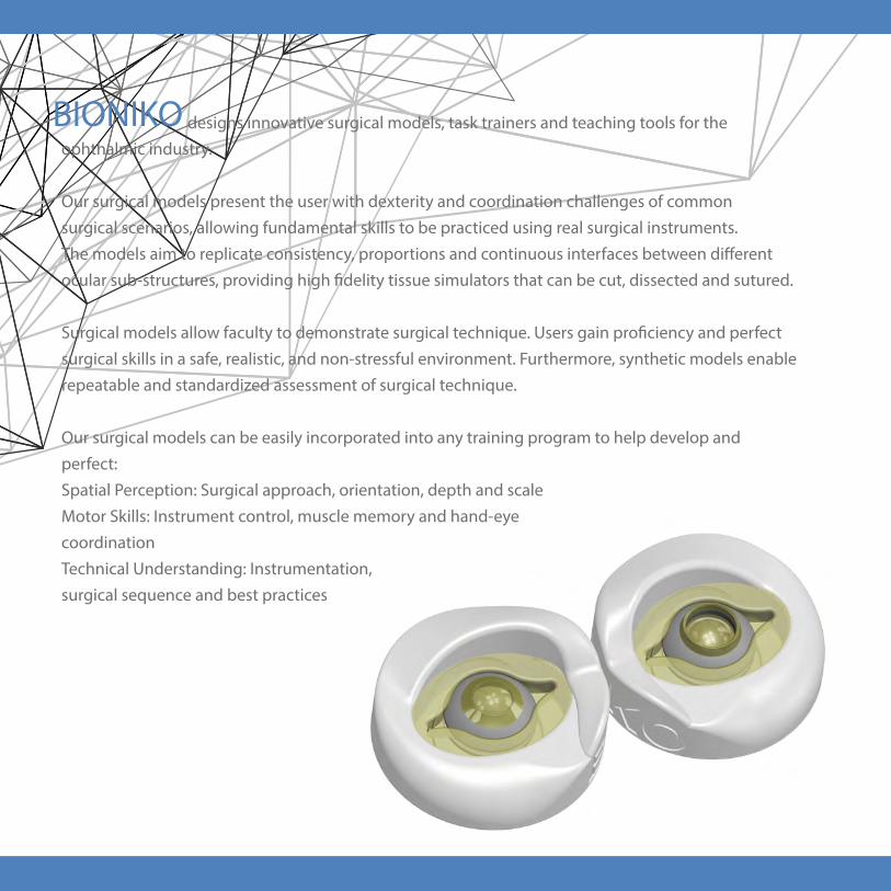
AVAILABLE
Training on demand for individual pace and needs
PORTABLE
Teach in the classroom, train in the OR and practice at home
REPEATABLE
Standardize training and assessment without model variability
AFFORDABLE
Surgical simulation tools at textbook cost
SIMPLE
Ready to use with minimum setup and assembly
SYNTHETIC
No refrigeration or special disposal required
FEATURES AND BENEFITS

www.bioniko.com | [email protected] | [email protected]
Suturing is a fundamental skill in ophthalmic surgery. However, it is a
common weakness among aspiring surgeons due to the lack of operative
experience and suitable training models. This skill is absent in virtual reality
tools, and animal models are not accurate or repeatable.
By presenting the main challenges of a
penetrating keratoplasty (PKP) scenario, the
KERATO task allows users to learn, train
and perfect the skills required to perform
precise suturing under a microscope.
The KERATO task consists in suturing
a corneal graft to a host limbus using
real surgical instruments. The user
will need to properly handle forceps,
needle holder, scissors and 10-0
suture to complete the task.
With practice the user will gain
confidence, reduce time to
completion, improve suture radiality
and spacing, maintain even and safe
distance to tissue edge and gain better
feel for suture tension during knot creation.
Pat. Pending
Suturing TaskKERATO

www.bioniko.com | [email protected]
www.bioniko.com | [email protected]
FEATURES AND BENEFITS
1 - CORNEA
2 - LIMBUS
3 - SCLERA
4 - GRAFT EDGE
5 - RECIPIENT EDGE
6 - SUTURE
KERATO
• Accurate tissue proportions
• Realistic tissue feel
• Repeatable and available
• Encourages awareness of tissue hydration
• Practice microsuturing techniques (continous/interrupted)
• Improve confidence and decrease fatigue
• Improve time to completion (Decrease OR time)
• Self asses execution by checking for:
- Suture radiality
- Safe and even distance to tissue edge
- Knot tension
- Even spacing between sutures

www.bioniko.com | [email protected]
Instrument control through ports is a fundamental skill in ophthalmic surgery.
However, it is a common weakness among aspiring surgeons due to the lack
of operative experience and suitable feedback from available models.
By presenting the main challenges of a capsulorhexis scenario, the RHEXIS
task allows users to learn, train and perfect the fine motor skills
required to properly use the wound as a fulcrum point
for instrument movement.
The task consists in performing a
capsulorhexis by manipulating
instruments through an incision on
a delicate limbus rim. Improper
instrument control will cause stress
and damage to the limbus, providing
feedback to the user. In addition to
a proper capsulorhexis, minimizing
damage to the limbus rim is key
to complete the task succesfully.
With practice the user will increase
confidence, reduce time to completion,
improve rhexis shape, size and centration,
and minimize stress on the wound.
Pat. Pending
RHEXISInstrument Control Task

RHEXIS
• Repeatable and available
• Limbus ridge designed to provide
• Improve surgical skills confidence
• Develop instrument control
• Decrease reliance on donor tissue for training
• Decrease wound size
• Improve time to completion
• Asses instrument control by checking wound integrity
• Assess execution by measuring rhexis size and centration
FEATURES AND BENEFITS
1 - LENS CAPSULE
2 - LIMBUS RIDGE
3 - SCLERA
4 - WOUND
5 - INSTRUMENT
www.bioniko.com | [email protected]
instrument handling feedback
• Encourages good practices:
- posterior incisions
- regrasping
- wound awareness

www.bioniko.com | [email protected]
The OJOS Extraocular model simulates the external anatomical
features of the eye, including conjunctiva, sclera, cornea and
rectus muscles. This model allows demonstration and practice of
numerous techniques requiring dissection and manipulation of
the conjunctiva and sclera. Procedures encountered
in glaucoma surgery, such as shunt implants and
trabeculectomies, can be demonstrated and
practiced on the model. The OJOS model
can be used in conjunction with the FLEX-ORBIT platform for added challenge and
realism.
Model EyesOJOS
Pat. Pending

www.bioniko.com | [email protected]
• “Loose” and continuous conjunctiva/tenon’s layer for realistic dissections
• Standardize your instruction and assessment around a consistent model
• Long shelf life
• No refrigeration needed
• No biohazard handling or disposal required
• Available on demand
• Decrease reliance on donor/animal tissue for training
FEATURES AND BENEFITS
1 - CORNEA
2 - CONJUNTIVA
3 - SCLERA
4 - MUSCLE INSERTION
5 - RECTUS MUSCLE
6 - OPTIC NERVE
7 - BASE
OJOS

www.bioniko.com | [email protected]
a
The PTERYGIUM model is based on a Pterygium excision with conjunctival
autograft scenario. Users will gain valuable hands-on training in delicate
ocular surface dissection and suturing techniques, which are essential skills in
ophthalmic surgery.
The PTERYGIUM model allows the user to demonstrate,
practice or evaluate dissection of the pterygium
head and body; autograft sizing, dissection
and harvesting; placement and suturing of
graft over scleral bed.
The FLEX-ORBIT (Sold separately)
serves as the holder for the PTERYGIUM
model and provides support, frame of
reference and the challenges posed by
facial features surrounding the eye.
Pat. Pending
PTERYGIUMModel

PTERYGIUM
• Practice excision techniques, complex conjunctival
dissection and conjunctival suturing
• Bilateral pterygia allows two simulations per model
• Layered conjunctiva (Conjunctiva- Tenon’s) allows
realistic autograft dissection
• Works with FLEX-ORBIT platform
FEATURES AND BENEFITS
1 - PTERYGIUM HEAD
2 - PTERYGIUM BODY
3 - CONJUNCTIVA
a. BULBAR CONJUNCTIVA
b. TENON’S CAPSULE
4 - LIMBUS
5 - SNAP RING
6 - SNAP RING GAP (TEMPORAL)
7 - SNAP RING APEX (NASAL)
8 - SCLERA
9 - CORNEA
www.bioniko.com | [email protected]

www.bioniko.com | [email protected]
FUNDUS is an innovative model for posterior segment training and simulation. Its
posterior segment includes a photo-realistic model of the central retina featuring
macula/fovea, optic disc/cup and retinal vasculature with accurate superior and
inferior arcades. Its anterior segment includes a central optical element and a
flexible pars plana that allows surgical intervention.
The model facilitates training in basic retinal examination technique and
instrumentation such as indirect ophthalmoscopy, slit-lamp, contact
and non-contact retinal lenses, and retinal cameras. It is
also an exceptional tool for demonstration, practice
and assessment of retinal instrument handling
and microscope skills such as use of surgical
contacts lenses, non-contact systems and
inverters.
Its modular design enables insertion of
foreign bodies or vitreous substitutes
to enhance training scenarios. It also
allows optical element removal and trans-
illumination to facilitate practice where
vitreo-retinal equipment is unavailable.
Pat. Pending
FUNDUSModel

FUNDUS
• Retinal model with macula/fovea, optic disc/cup and retinal
vasculature with accurate superior and inferior arcades
• Removable optical element simulates optical power
of the eye
• Removable anterior segment facilitates creation of surgical
scenarios
• Flexible pars plana allows surgical instrument insertion
• Translucent body enables transmitted/retro illumination
* Special orders: Iris 1mm-8mm, optical element AR coating and ILM,
FEATURES AND BENEFITS
1 - OPTICAL ELEMENT* (50D)
2 - CORNEAL RIM
3 - PARS PLANA
4 - SUPPORT RING
5 - MUSCLE INSERTION
6 - RECTUS MUSCLE
7 - GLOBE
8 - OPTIC NERVE
9 - BASE
10 - IRIS*
11 - FUNDUS*
A - MACULA / FOVEA
B- OPTIC DISC / CUP
C - VASCULATURE/ ARCADE
www.bioniko.com | [email protected]

www.bioniko.com | [email protected]
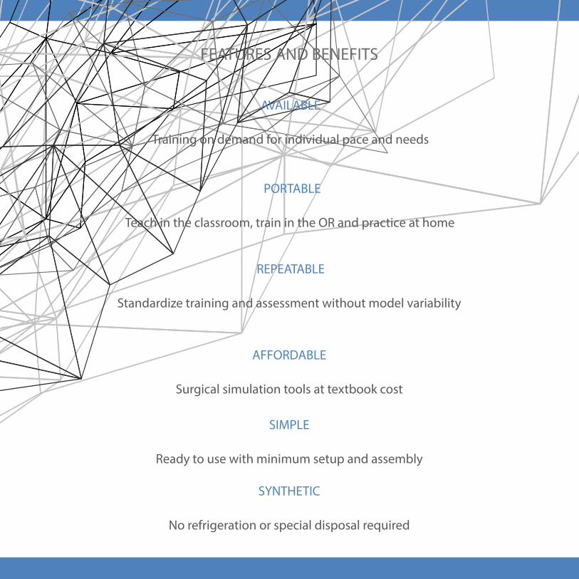
Strabismus and other eye-muscle related surgeries can be difficult to simulate with
animal or donor tissue, since enucleated globes lack muscles and their attachments
to the orbit. The STRABISMUS model features the four rectus muscles and works
with the FLEX-ORBIT to provide a natural attachment point at the back of the orbit.
The orbit also provides reference and realism by challenging the user to manipulate
instruments according to the orbit cavity and facial structures around the eye.
The model is ideal to demonstrate, practice and perfect the
surgical sequence and instrument handling of
a strabismus surgery scenario. Learning to
efficiently and precisely hook the muscles
on a first attempt is a fundamental skill
for novice surgeons. The model allows
the users to develop proper suture
placement and practice of adjustable
suturing techniques and permanent
knots.
The model is an excellent tool for
self-assessment and evaluation of final
muscle and eye position in the orbit and
intrascleral pass technique.
STRABISMUSModel
STRABISMUS
• Realistic muscle traction and feel
• Practice surgical procedure sequence and manipulation of instruments
• Available on demand
• Practice muscle suturing techniques that are not available with animal tissue
• Permits use of subcleral dye injections to obtain suture depth feedback
• Standardize your instruction and assessment around a consistent model
FEATURES AND BENEFITS
1 - CORNEA
2 - MUSCLE INSERTION
3 - RECTUS MUSCLE
4 - GLOBE
5 - OPTIC NERVE
6 - BASE
www.bioniko.com | [email protected]

www.bioniko.com | [email protected]
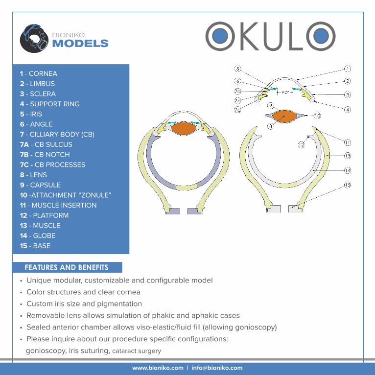
The OKULO Model is a modular and configurable platform for anterior segment
surgery simulation. It features a clear cornea, a flexible pigmented iris and a
removable/ crystalline lens.
This versatile model offers various iris pigmentation and pupil size options.
Choose between our standard blue or brown iris and 5 or 8 mm pupil size.
The iris can also be customized to create specific training scenarios.
The OKULO lens can be removed to re-create aphakic conditions or to replace
used lenses.
The OKULO Anterior Segment works with our
Posterior Segment Adapter and FLEX-ORBIT
to provide the most realistic surgical
simulation experience.
Pat. Pending
OKULOModel
OKULO
• Unique modular, customizable and configurable model
• Color structures and clear cornea
• Custom iris size and pigmentation
• Removable lens allows simulation of phakic and aphakic cases
• Sealed anterior chamber allows viso-elastic/fluid fill (allowing gonioscopy)
• Please inquire about our procedure specific configurations:
gonioscopy, iris suturing, cataract surgery
FEATURES AND BENEFITS
1 - CORNEA
2 - LIMBUS
3 - SCLERA
4 - SUPPORT RING
5 - IRIS
6 - ANGLE
7 - CILLIARY BODY (CB)
7A - CB SULCUS
7B - CB NOTCH
7C - CB PROCESSES
8 - LENS
9 - CAPSULE
10 -ATTACHMENT “ZONULE”
11 - MUSCLE INSERTION
12 - PLATFORM
13 - MUSCLE
14 - GLOBE
15 - BASE
www.bioniko.com | [email protected]

The ORBIT is the holder for all BIONIKO anterior segment models.
It provides an anatomical frame of reference and adds realism
to the surgical scenario by challenging the user to
manipulate instruments according to the facial
structures around the eye.
The ORBIT can be secured to any
smooth surface (horizontal or
vertical) with its integrated
suction cup and will still retain
a realistic degree of freedom
that simulates head and eye
movement.
There are right and left
ORBIT models to practice all
approaches: Right-Superior,
Right-Temporal, Left-Superior and
Left-Temporal
ORBIT
Pat. Pending
ModelORBIT
www.bioniko.com | [email protected]

ORBIT
www.bioniko.com | [email protected]
• Provides anatomical frame of reference to anterior segment models
• Practice superior (1) or temporal (4) approaches on both left (L) and right (R) eyes
• Suction-cup firmly attaches to any smooth surface while retaining realistic movement
• Compact and portable design
FEATURES AND BENEFITS
1 - BROW / SUPERIOR
2 - BRIDGE / NASAL
3 - ZYGOMATIC / INFERIOR
4 - TEMPORAL
5 - EYELID / SOCKET
6 - EYE MODEL
7 - SUCTION CUP

www.bioniko.com | [email protected]
Modular Training PlatformFLEX-ORBITSOCKET ADAPTER The FLEX-ORBIT modular training platform enhances and facilitates the
use of ex-vivo and synthetic eye models for training and R&D purposes.
It can position, secure and pressurize ex-vivo eyes of different sizes
(18-26mm Ø), while providing the user with an anatomical frame of
reference.
The FLEX-ORBIT is designed to receive and
complement the entire line of BIONIKO
synthetic models. Whole globe models
like OJOS and EXOS fit right in. With the
included socket adapter, the FLEX-ORBIT
can receive all anterior segment models
and task trainers, such as the RHEXIS and
KERATO. This makes the FLEX-ORBIT a versatile tool for any surgical training
program.
This compact platform is ideal for use in
the classroom, research lab, wet-lab and
even at the office to communicate with
colleagues and patients.
Pat. Pending

FLEX-ORBIT
www.bioniko.com | [email protected]
• Use with both synthetic and biological tissue models*
• Adjust eye position with posterior screws (6)
• Adjust intra-ocular pressure with anterior screws (5)
• Suction-cup firmly attaches to any smooth surface
• Socket adapter for anterior segment models
* For research use only
FEATURES AND BENEFITS
1 - SUPERIOR (BROW)
2 - NASAL (BRIDGE)
3 - INFERIOR
4 - TEMPORAL
5 - ANTERIOR SCREW 6 - POSTERIOR SCREW
7 - SUCTION CUP
8 - DRAIN/PORT
9 - SOCKET ADAPTER
10 - ADAPTER GROOVE
11 - SNAP RING
12 - SNAP RING GAP
13 - EYELID
14 - SOCKET BASE

www.bioniko.com | [email protected]
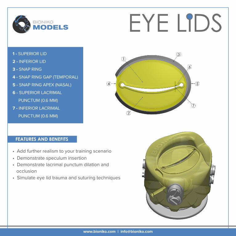
Add realism and difficulty to your simulation scenario by using the EYE LIDS
FLEX-ORBIT accessory. The flexible EYE LIDS are detachable and easily
inserted into the FLEX-ORBIT grove.
Use for demonstration, practice and assessment of
techniques such as speculum placement and
punctum plug placement. EYE LIDS features
superior and inferior puncta (0.6mm).
EYE LIDS can also be used as an
introductory ocular trauma model.
Eye lid laceration is not an everyday
encounter for many ophthalmologists
and EYE LIDS provides a basic platform
for training and communication with
peers and students.
Pat. Pending
EYE LIDSFLEX-ORBIT Accessory
EYE LIDS
• Add further realism to your training scenario
• Demonstrate speculum insertion
• Demonstrate lacrimal punctum dilation and
occlusion
• Simulate eye lid trauma and suturing techniques
FEATURES AND BENEFITS
1 - SUPERIOR LID
2 - INFERIOR LID
3 - SNAP RING
4 - SNAP RING GAP (TEMPORAL)
5 - SNAP RING APEX (NASAL)
6 - SUPERIOR LACRIMAL
PUNCTUM (0.6 MM)
7 - INFERIOR LACRIMAL
PUNCTUM (0.6 MM)
www.bioniko.com | [email protected]

www.bioniko.com | [email protected]
The CORDELIA recovery model was made possible by the valuable guidance and support of LIONS Vision Gift
Recovery Simulator
The CORDELIA recovery simulator is based on an in-situ excision
scenario. Eye bank technicians must master this technique to
successfully recover delicate corneal tissue from donors in the field.
CORDELIA is a task trainer that allows users
to learn and practice the tissue recovery
technique without using valuable
donor tissue, in a realistic yet
simple manner. Its repeatability
and availability makes it ideal for
developing standardized methods
of instruction and assessment.
The ORBIT (Sold separately) serves a
holder for the CORDELIA models and
provides support, frame of reference
and the challenges posed by facial features
surrounding the eye.

CORDELIA
• Practice corneal detachment technique
• Refine sequence and manipulation of surgical instruments
• Decrease reliance on donor tissue for training
• Available on demand
• Standardize your instruction and assessment around a consistent model
FEATURES AND BENEFITS
1 - SCLERAL LAYER2 - CHOROID LAYER3 - LIMBUS4 - SUPRA-CHOROIDAL
5 - SPUR6 - CORNEA7 - IRIS8 - STRUCTURAL RING9 - NOTCH
BB
DETAIL VIEW
SPACE
www.bioniko.com | [email protected]

www.bioniko.com | [email protected]
The EXOS model focuses on simulating the challenging steps of an enucleation
and corneal excision procedures. Learning to hook and transect the muscles and
optic nerve is a fundamental skill for eye-bank technicians. The EXOS is coupled
to our CORDELIA recovery simulator, allowing the practice
of corneal excision with greater realism.
Users can demonstrate and practice the
sequence and manipulation of surgical
instruments around anatomical
landmarks. The EXOS model serves
as a tool for introductory instruction
and competency reviews.
The FLEX-ORBIT (Sold separately)
serves as a holder for the EXOS
model and provides reference and
realism by challenging the user to
manipulate instruments according
to the orbit cavity and facial struc-
tures around the eye.
EXOSEnucleation Simulator
The EXOS Enucleation Simulator was made possible by the valuable guidance and support of Florida Lions Eye Bank

EXOS
• Realistic muscle traction and feel
• Practice enucleation and excision in one model
• Practice sequence and manipulation of surgical instruments
• Decrease reliance on donor tissue for training
• Available on demand
• Standardize your instruction and assessment around a consistent model
FEATURES AND BENEFITS
1 - CORDELIA MODEL
2 - MUSCLE INSERTION
3 - RECTUS MUSCLE
4 - GLOBE
5 - OPTIC NERVE
6 - BASE
www.bioniko.com | [email protected]

www.bioniko.comPhone: (507) BIONIKO
Email: [email protected]/user/BionikoDesign
© Copyright 2015 BIONIKO