Embed Size (px)
Citation preview
Objectives of ophthalmic epidemiology
• Establish the incidence and prevalence of eye disorders that cause vision impairment and/or blindness
• Determine the societal impact (social and economic) of vision loss
• Assess the potential and real impact of preventive and treatment efforts for eye problems
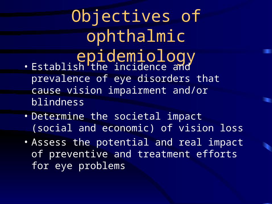
Causes of Worldwide Blindness• Cataract 17 million• Trachoma 6.0 million• Glaucoma 3.0 million• Xerophthalmia 0.5 million• Onchocerciasis 0.5 million• AMD 1.0 million• Diabetic retinopathy 0.25 million• Leprosy 0.25 million• Others 2.5 million
– 85% of blindness is in Africa and Asia– 85% of cases are potentially treatable or preventable
• Prevalence: – 0.125-0.25% in Western world– 0.2-1.5% (av 0.75%) in Asia– 0.3-3.1% (av 1.2%) in Africa
Allen Foster in Clinical Ophthalmology - Duane, ed. (1991)
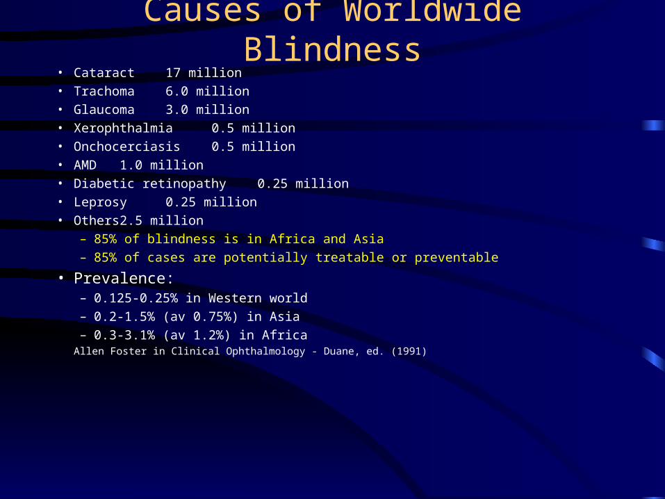
Aging and Blindness
• Prevalence (in Germany) :
– 15 % lose sight < 20 years old– 51% lose sight >50 and <80– 15 % lose sight > 80 years old
• Incidence:
– 50% of new cases are people over 80• “Imbalance” due to differences in life expectancy and duration of
blindness.– Blind < 10 years - 74%– Blind >10 years - 26%– Blind > 20 years - 10%
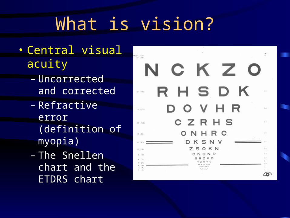
What is vision?
• Central visual acuity – Uncorrected and
corrected– Refractive error
(definition of myopia)
– The Snellen chart and the ETDRS chart
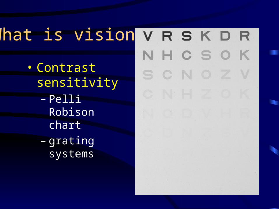
What is vision?
• Contrast sensitivity– Pelli Robison
chart– grating systems
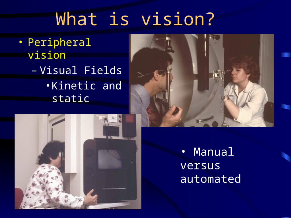
What is vision? • Peripheral vision
– Visual Fields • Kinetic and
static
• Manual versus automated
What is vision?
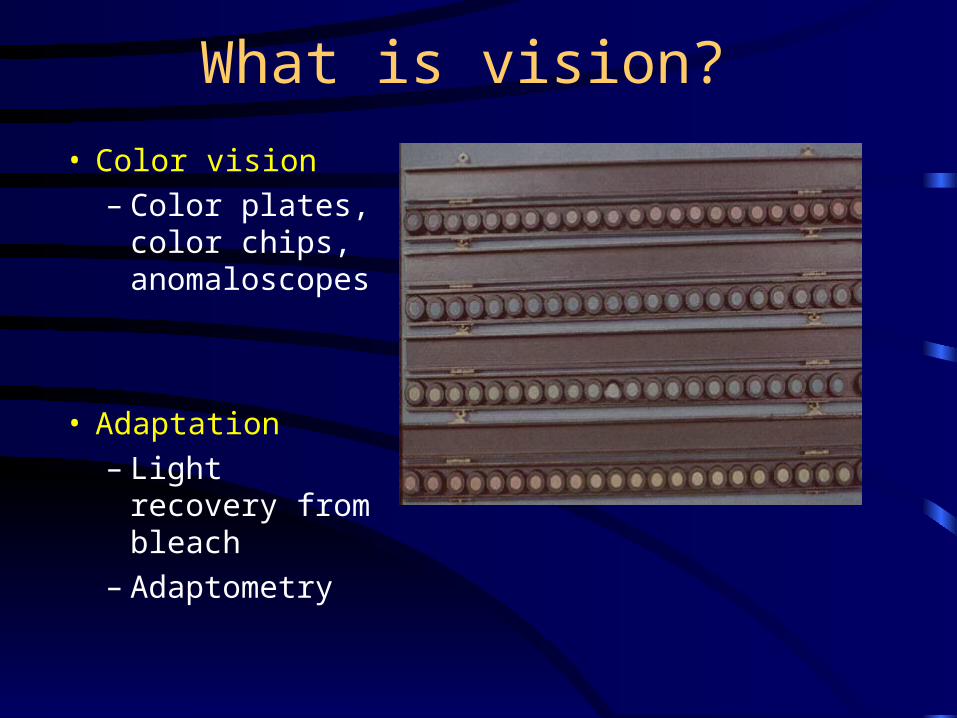
• Color vision– Color plates,
color chips, anomaloscopes
• Adaptation– Light recovery
from bleach– Adaptometry
What is vision?
• Other measures of visual function– Electrophysiology– Ocular movements– Visual function questionnaires - VF-14
• Initially validated for cataracts • more extensive use in all eye studies
What is the definition of blindness?
• 20/10 - 20/25: Normal• 20/30 - 20/60: Near-normal• 20/70 - 20/160 : Moderate vision impairment - eligible for education
assistance in US• 20/200 - 20/400: Severe vision impairment - legal blindness in US
(visual field < 20 degrees)• 20/500 - 20/1000: profound vision impairment - WHO and several
European countries definition of blindness (visual field < 10 degrees), CF < 3m
• < 20/1000: Near-total visual impairment: used by some developing countries as definition of blindness (visual field < 5 degrees), HM, LP
• NLP: Total visual impairment
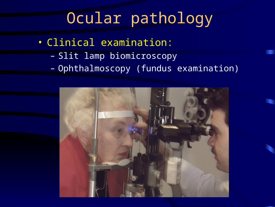
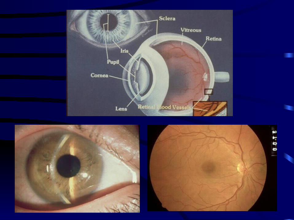
Ocular pathology
• Clinical examination:– Slit lamp biomicroscopy
– Ophthalmoscopy (fundus examination)
Ocular pathology
– Conjunctival scarring - CSP
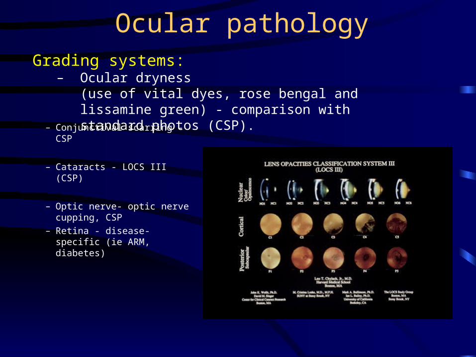
– Cataracts - LOCS III (CSP)
– Optic nerve- optic nerve cupping, CSP
– Retina - disease-specific (ie ARM, diabetes)
Grading systems:– Ocular dryness
(use of vital dyes, rose bengal and lissamine green) - comparison with standard photos (CSP).
Documentation of ocular pathology
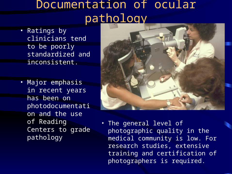
• Ratings by clinicians tend to be poorly standardized and inconsistent.
• Major emphasis in recent years has been on photodocumentation and the use of Reading Centers to grade pathology
• The general level of photographic quality in the medical community is low. For research studies, extensive training and certification of photographers is required.
Documentation of ocular pathology
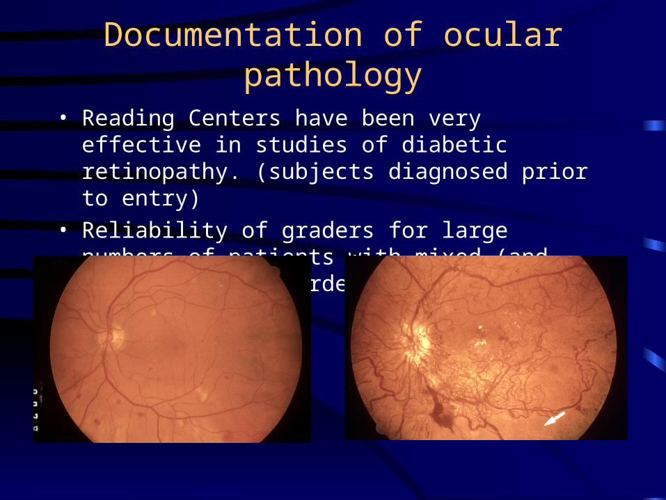
• Reading Centers have been very effective in studies of diabetic retinopathy. (subjects diagnosed prior to entry)
• Reliability of graders for large numbers of patients with mixed (and unspecified) disorders is unknown.
Specific issues in eye research• Is one assessing the subject or the eye?
• Relatedness between eyes of a single individual
• Research design with bilateral and monocular cases
• Use of the contralateral eye as a control
• Masking of the subject and observer
• Is one comparing the same definition of the disease among studies? (ie AMD, myopia, glaucoma)
• Diagnostic reliability, sample bias
• 10% of cases have vision loss from 2 different conditions, though studies often only cite the cause of the second eye.
Causes of vision loss• Trauma
– Recreational, work-related, military
• Systemic Disease – Diabetes, vascular disease, hypertension
• Aging/Eye Specific – Cataracts, age-related maculopathy, glaucoma
• Infectious – Trachoma, onchocerciasis, immunocompromised individuals
• Congenital/Hereditary - – Cataracts, malformations, glaucoma, retinal degenerations
• Nutritional and Toxic– Vitamin A deficiency, methanol
• Tumors– Metastatic, primary malignancies (children / adults)
Infectious causes of vision loss• Trachoma
– Affects 500 million
– Estimated 6 million are blind
• Onchocerciasis– Endemic across equatorial Africa (99%), some areas of South and Central
America
– 80 million exposed, 18 million infected, 2 million blind
– Transmitted by blackfly - filial nematode
– Treatment - vector control, ivermectin (annual dose for a minimum of 10 years)
• Other ID: leprosy, syphillis– Estimated 10-12 million cases of leprosy
– WHO estimated that 250,000 blind from disease
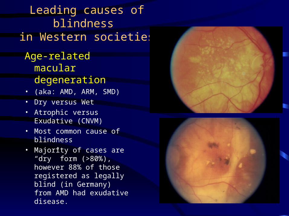
Leading causes of blindness in Western societies
Age-related macular degeneration
• (aka: AMD, ARM, SMD)
• Dry versus Wet
• Atrophic versus Exudative (CNVM)
• Most common cause of blindness
• Majority of cases are “dry” form (>80%), however 88% of those registered as legally blind (in Germany) from AMD had exudative disease.
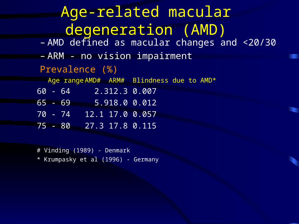
Age-related macular degeneration (AMD)– AMD defined as macular changes and <20/30– ARM - no vision impairment
Prevalence (%) Age range AMD# ARM# Blindness due to AMD*
60 - 64 2.3 12.3 0.007
65 - 69 5.9 18.0 0.012
70 - 74 12.1 17.0 0.057
75 - 80 27.3 17.8 0.115
# Vinding (1989) - Denmark
* Krumpasky et al (1996) - Germany
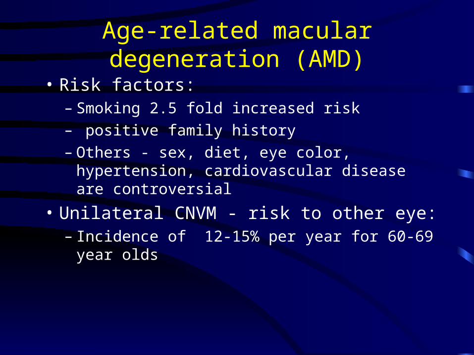
• Risk factors:– Smoking 2.5 fold increased risk– positive family history– Others - sex, diet, eye color, hypertension,
cardiovascular disease are controversial
• Unilateral CNVM - risk to other eye:– Incidence of 12-15% per year for 60-69 year
olds
Age-related macular degeneration (AMD)
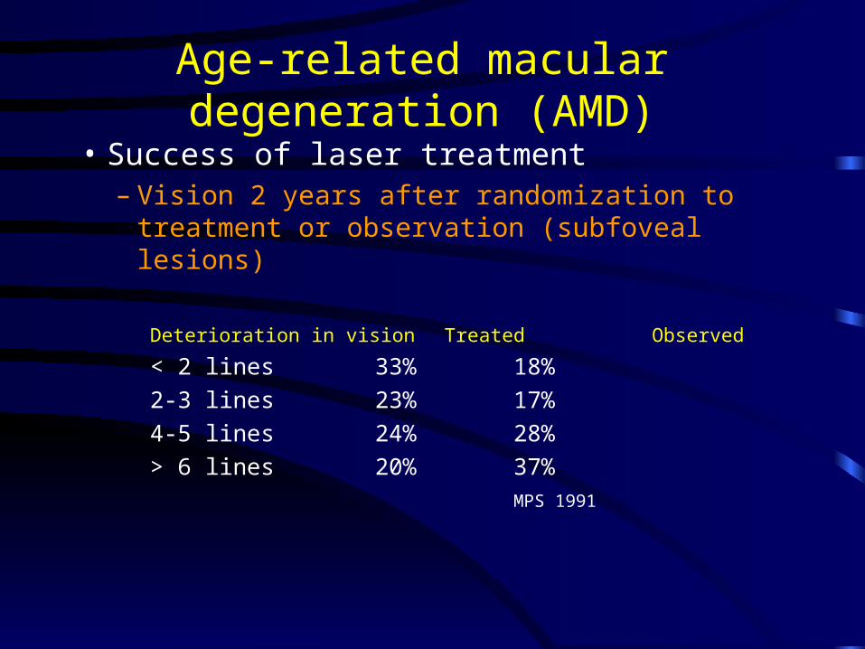
• Success of laser treatment– Vision 2 years after randomization to treatment or
observation (subfoveal lesions)
Deterioration in vision Treated Observed
< 2 lines 33% 18%
2-3 lines 23% 17%
4-5 lines 24% 28%
> 6 lines 20% 37%MPS 1991
Age-related macular degeneration (AMD)
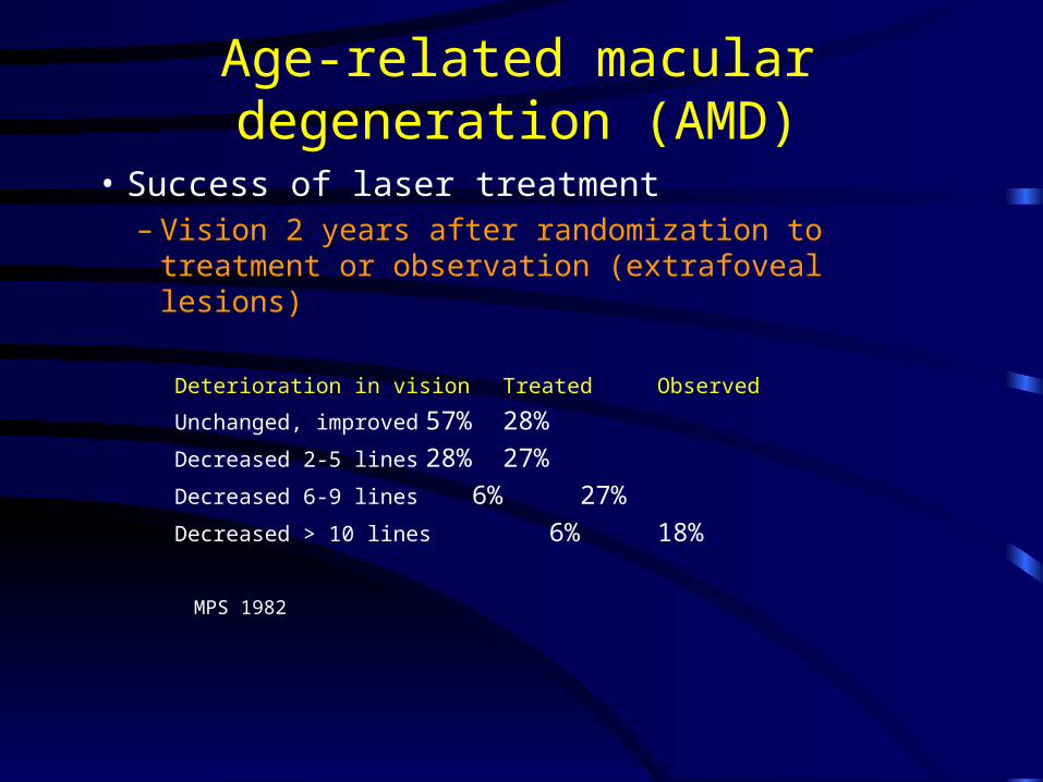
• Success of laser treatment– Vision 2 years after randomization to treatment or
observation (extrafoveal lesions)
Deterioration in vision Treated Observed
Unchanged, improved 57% 28%
Decreased 2-5 lines 28% 27%
Decreased 6-9 lines 6% 27%
Decreased > 10 lines 6% 18%
MPS 1982
Age-related macular degeneration (AMD)
Leading causes of blindness in Western societies
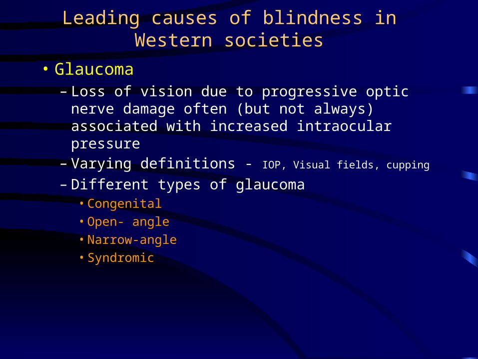
• Glaucoma – Loss of vision due to progressive optic nerve damage
often (but not always) associated with increased intraocular pressure
– Varying definitions - IOP, Visual fields, cupping
– Different types of glaucoma• Congenital
• Open- angle
• Narrow-angle
• Syndromic
• Glaucoma– Glaucoma affects 1.5-2.0% of population over the age of 40. Rises
with age up to 8% for those over 80
– Current prevalence is 15% of all cases of blindness (developed nations)
– Age of onset of blindness from glaucoma
• >60 years : 79%
– Those under 65 years old
• Glaucoma-related blindness associated with other conditions - 36%
– Those greater than 65 years old
• Glaucoma-related blindness associated with other conditions - 46%
Leading causes of blindness in Western societies
• Diabetic retinopathy– Women greater than men:
• 56% of the younger blind diabetic individuals
• 87% of the older blind diabetic individuals
– In those under the age of 65, diabetes is the most common cause of blindness
– However, 2/3 of diabetics do not become blind until after the age of 60
– Blindness from DR is a poor prognosis for survival
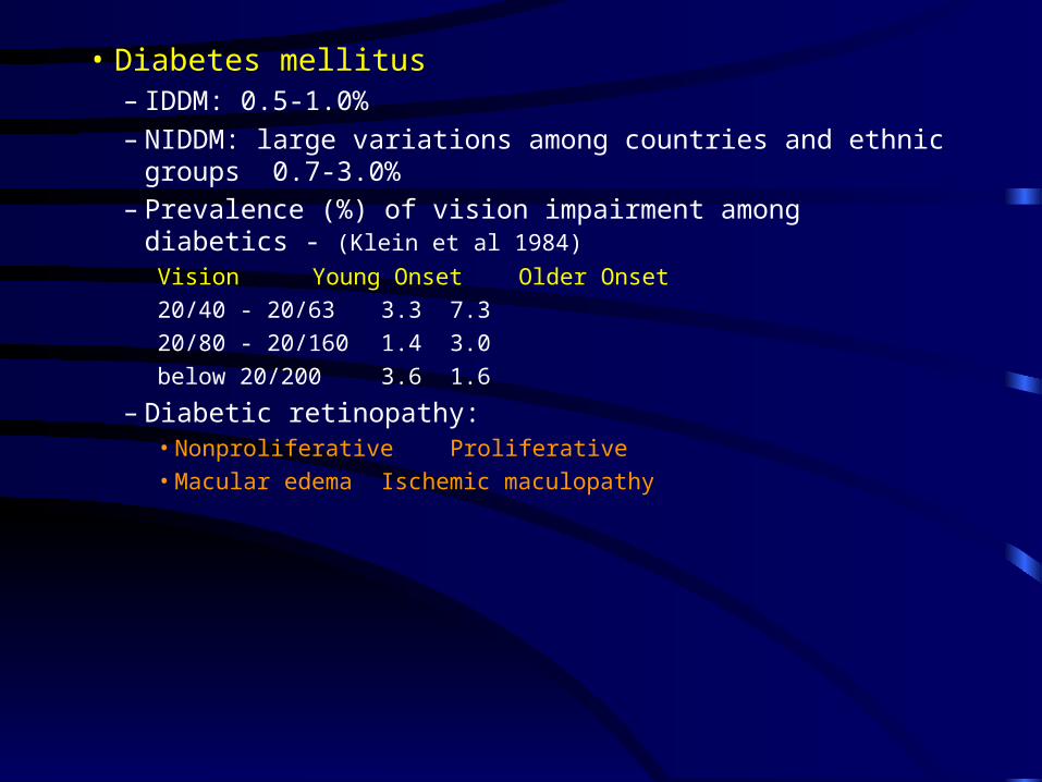
• Diabetes mellitus– IDDM: 0.5-1.0%
– NIDDM: large variations among countries and ethnic groups 0.7-3.0%– Prevalence (%) of vision impairment among diabetics - (Klein et al 1984)
Vision Young Onset Older Onset
20/40 - 20/63 3.3 7.3
20/80 - 20/160 1.4 3.0
below 20/200 3.6 1.6
– Diabetic retinopathy: • Nonproliferative Proliferative• Macular edema Ischemic maculopathy
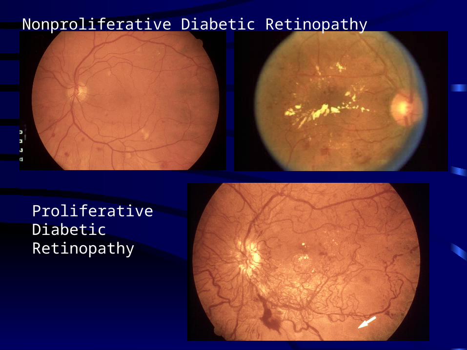
Proliferative Diabetic Retinopathy
Nonproliferative Diabetic Retinopathy
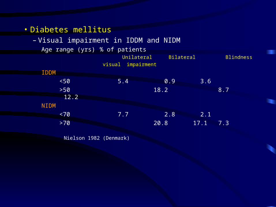
• Diabetes mellitus– Visual impairment in IDDM and NIDM
Age range (yrs) % of patients Unilateral Bilateral Blindness
visual impairment
IDDM
<50 5.4 0.9 3.6
>50 18.2 8.7 12.2
NIDM
<70 7.7 2.8 2.1
>70 20.8 17.1 7.3
Nielson 1982 (Denmark)
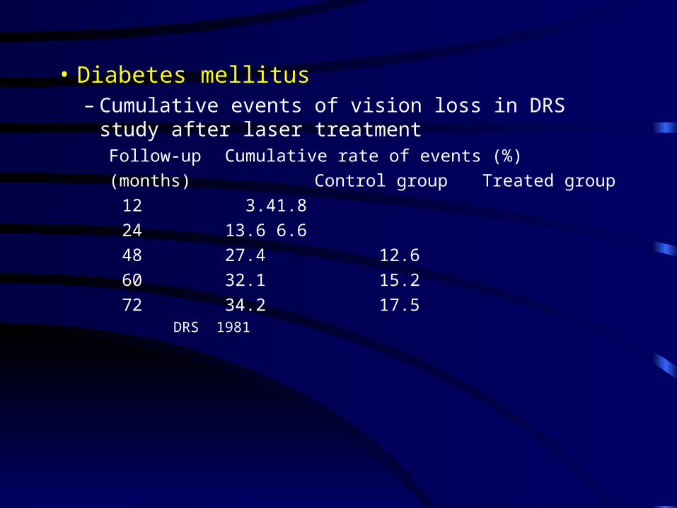
• Diabetes mellitus– Cumulative events of vision loss in DRS study after laser
treatmentFollow-up Cumulative rate of events (%)
(months) Control group Treated group
12 3.4 1.8
24 13.6 6.6
48 27.4 12.6
60 32.1 15.2
72 34.2 17.5DRS 1981
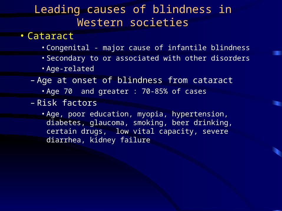
Leading causes of blindness in Western societies
• Cataract• Congenital - major cause of infantile blindness• Secondary to or associated with other disorders• Age-related
– Age at onset of blindness from cataract• Age 70 and greater : 70-85% of cases
– Risk factors• Age, poor education, myopia, hypertension, diabetes,
glaucoma, smoking, beer drinking, certain drugs, low vital capacity, severe diarrhea, kidney failure