Embed Size (px)
Citation preview
Operative Chest Wall Fixation with Osteosynthesis PlatesChristine Engel, MD, James C. Krieg, MD, Steven M. Madey, MD, William B. Long, MD, andMichael Bottlang, PhD
J Trauma. 2005;58:181–186.
Blunt chest wall trauma is a major cause of morbidity andmortality, especially in the presence of a flail chestwhere paradoxical inward movement of the flail seg-
ment in inspiration is found.1–3
Patients with a flail chest require aggressive pain con-trol, pulmonary toilet, and often intubation and mechanicalventilation to establish an internal pneumatic stabilizationof the flail segment. This may result in a prolonged ICUstay and pulmonary complications including pneumonia,septicemia and barotrauma.3–9 The high mortality rate ofup to 10 –36%3,10 –12 is partly due to the high prevalence ofassociated life-threatening extra-thoracic injuries. How-ever, one principle cause of death consists of pneumoniaand sepsis with prolonged intubation.3,13,14
Several potential advantages of operative chest wall sta-bilizations have been reported. These include reduced dura-tion of mechanical ventilation,4,7,8,13,15–17 shortened ICU stayand hospitalization,7,8,15 and decreased likelihood of clini-cally significant long-term respiratory dysfunction and skel-etal deformity.5,15
Despite the advantages of operative chest wall fixation,little consensus on the fixation technique exists.
This report describes three cases of flail chest injurymanaged by operative stabilization with plates and screws.The criteria for surgical intervention in this trauma center aretraumatic loss of 30% of pleural cavity volume, inability towean an awake patient from the ventilator, inability to controlchest wall pain despite epidural catheter, major air leak ormajor bleeding, or unstable sternal fracture with overlap.
In all three cases a standard posterolateral thoracotomywas performed. The serratus anterior was retracted anteriorlyand the latissimus dorsi was divided. To reach more craniallya small portion of the trapezius and the rhomboids weretransected in cases 2 and 3. The chest was always entered andhematoma was removed. Pelvic, mandibular and customized
reconstruction plates were used, with bending stiffness rang-ing from 1,936 over 414 to 56 kN mm2, respectively.
In addition to documentation of the technique, this reportdescribes the results obtained with three distinct osteosyn-thesis plates and provides a historic overview of alternativefixation means.
CASE 1A 34-year-old man was rolled over by a forklift in an
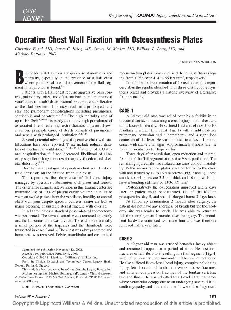
industrial accident, sustaining a crush injury to his chest andto his biceps bilaterally. He suffered fractures of ribs 3 to 10,resulting in a right flail chest (Fig. 1) with a mild posteriorpulmonary contusion and a hemothorax and a right lobecontusion of the liver. He was admitted to a Level I traumacenter with stable vital signs. Approximately 8 hours later herequired intubation for hypercarbia.
Three days after admission, open reduction and internalfixation of the flail segment of ribs 6 to 9 was performed. Theremaining injured ribs had isolated fractures without instabil-ity. Pelvic reconstruction plates were contoured to the chestwall and fixated by 12 to 16 mm screws (Fig. 2 and 3). Thesestainless steel plates are 3.5 mm thick and 10 mm wide andhave a bending stiffness of 1,936 kN mm2.
Postoperatively the oxygenation improved and 2 dayslater the patient could be extubated. He left the ICU onpostoperative day 5, and was discharged home 3 days later.
At follow-up examination 2 months after surgery, thepatient did not have any shortness of breath but the thoracot-omy site was tender to touch. He was able to return tofull-time employment 4 months after the injury. The promi-nent hardware continued to irritate him and was thereforeremoved half a year later.
CASE 2A 49-year-old man was crushed beneath a heavy object
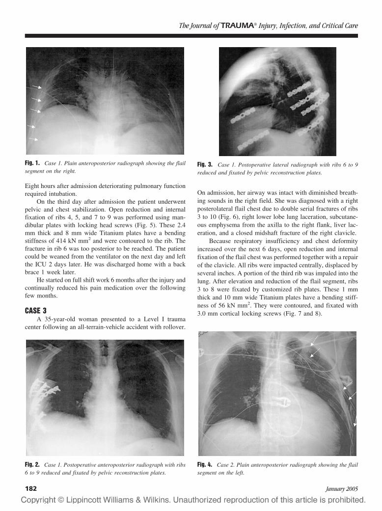
and remained trapped for a period of time. He sustainedfractures of left ribs 3 to 9 resulting in a flail segment (Fig. 4)with left pulmonary contusion and a left hemopneumothorax.He also suffered from closed head injury, complex pelvic ringinjury, left thoracic and lumbar transverse process fractures,and anterior compression fractures of the lumbar vertebraetwo and three. He was admitted to a Level I trauma centerwhere ventricular ectopy due to an underlying severe dilatedcardiomyopathy and traumatic anemia were also diagnosed.
Submitted for publication November 12, 2002.Accepted for publication February 5, 2003.Copyright © 2005 by Lippincott Williams & Wilkins, Inc.From the Clinical Research and Technology Center, Legacy Health
System, Portland, Oregon.This study has been supported by a Grant from the Legacy Foundation.Address for reprints: Michael Bottlang, PhD, Legacy Clinical Research
& Technology Center, 1225 NE 2nd Avenue, Portland, OR 97232; email:[email protected].
DOI: 10.1097/01.TA.0000063612.25756.60
CASEREPORT The Journal of TRAUMA! Injury, Infection, and Critical Care
Volume 58 • Number 1 181
Eight hours after admission deteriorating pulmonary functionrequired intubation.
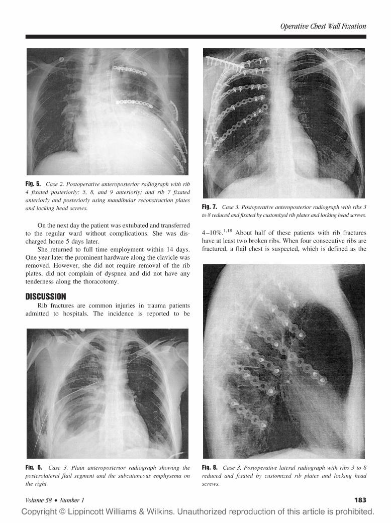
On the third day after admission the patient underwentpelvic and chest stabilization. Open reduction and internalfixation of ribs 4, 5, and 7 to 9 was performed using man-dibular plates with locking head screws (Fig. 5). These 2.4mm thick and 8 mm wide Titanium plates have a bendingstiffness of 414 kN mm2 and were contoured to the rib. Thefracture in rib 6 was too posterior to be reached. The patientcould be weaned from the ventilator on the next day and leftthe ICU 2 days later. He was discharged home with a backbrace 1 week later.
He started on full shift work 6 months after the injury andcontinually reduced his pain medication over the followingfew months.
CASE 3A 35-year-old woman presented to a Level I trauma
center following an all-terrain-vehicle accident with rollover.
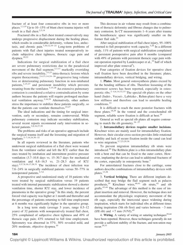
On admission, her airway was intact with diminished breath-ing sounds in the right field. She was diagnosed with a rightposterolateral flail chest due to double serial fractures of ribs3 to 10 (Fig. 6), right lower lobe lung laceration, subcutane-ous emphysema from the axilla to the right flank, liver lac-eration, and a closed midshaft fracture of the right clavicle.
Because respiratory insufficiency and chest deformityincreased over the next 6 days, open reduction and internalfixation of the flail chest was performed together with a repairof the clavicle. All ribs were impacted centrally, displaced byseveral inches. A portion of the third rib was impaled into thelung. After elevation and reduction of the flail segment, ribs3 to 8 were fixated by customized rib plates. These 1 mmthick and 10 mm wide Titanium plates have a bending stiff-ness of 56 kN mm2. They were contoured, and fixated with3.0 mm cortical locking screws (Fig. 7 and 8).
Fig. 1. Case 1. Plain anteroposterior radiograph showing the flailsegment on the right.
Fig. 2. Case 1. Postoperative anteroposterior radiograph with ribs6 to 9 reduced and fixated by pelvic reconstruction plates.
Fig. 3. Case 1. Postoperative lateral radiograph with ribs 6 to 9reduced and fixated by pelvic reconstruction plates.
Fig. 4. Case 2. Plain anteroposterior radiograph showing the flailsegment on the left.
The Journal of TRAUMA! Injury, Infection, and Critical Care
182 January 2005
On the next day the patient was extubated and transferredto the regular ward without complications. She was dis-charged home 5 days later.
She returned to full time employment within 14 days.One year later the prominent hardware along the clavicle wasremoved. However, she did not require removal of the ribplates, did not complain of dyspnea and did not have anytenderness along the thoracotomy.
DISCUSSIONRib fractures are common injuries in trauma patients
admitted to hospitals. The incidence is reported to be
4–10%.1,18 About half of these patients with rib fractureshave at least two broken ribs. When four consecutive ribs arefractured, a flail chest is suspected, which is defined as the
Fig. 5. Case 2. Postoperative anteroposterior radiograph with rib4 fixated posteriorly; 5, 8, and 9 anteriorly; and rib 7 fixatedanteriorly and posteriorly using mandibular reconstruction platesand locking head screws.
Fig. 6. Case 3. Plain anteroposterior radiograph showing theposterolateral flail segment and the subcutaneous emphysema onthe right.
Fig. 7. Case 3. Postoperative anteroposterior radiograph with ribs 3to 8 reduced and fixated by customized rib plates and locking head screws.
Fig. 8. Case 3. Postoperative lateral radiograph with ribs 3 to 8reduced and fixated by customized rib plates and locking headscrews.
Operative Chest Wall Fixation
Volume 58 • Number 1 183
fracture of at least four consecutive ribs in two or moreplaces.1,3,19 Up to 10–15% of blunt chest trauma injuries willresult in a flail chest.2,3,14
Fractured ribs in a flail chest treated conservatively mayundergo progressive displacement during the healing phase,which results in considerable deformity, volume loss, atelec-tasis, and chronic pain.7,10,12,19–21 Long-term problems ofpatients with flail chest injuries treated nonoperatively in-clude subjective chest tightness, thoracic cage pain anddyspnea.10,22
Indications for surgical stabilization of a flail chestare severe pulmonary restrictions due to the paradoxicalmovement of the flail segment,2,3,11 markedly overridingribs and severe instability,15,23 intra-thoracic lesions whichrequire thoracotomy,2,4,11,14,24 –26 progressive lung volumeloss or deteriorating pulmonary function in non-intubatedpatients,3,15,27 and persistent instability which preventsweaning from the ventilator.3,15,28 An extensive pulmonarycontusion is considered a relative contraindication by someauthors because the patients will require prolonged mechan-ical ventilation anyway.3,16,29 Alternatively, other authorsstress the importance to stabilize these patients surgically, sothat the patients can ventilate themselves.23,27
The ideal indications and the timing for surgical inter-vention, early or secondary, remains controversial. Whilepulmonary contusion may indicate secondary stabilization,several recent reports recommend early intervention in se-lected patients.3,8,16,27,30
The problems and risks of an operative approach includethe surgical trauma itself and the loosening and migration ofimplants.2–7,14,24,30–32
In all reports reviewed in the literature, patients whounderwent surgical stabilization of a flail chest were weanedfrom the ventilator earlier and left the ICU earlier than theones treated by intubation and intermittent positive pressureventilation (3.7–10.8 days vs. 15–30.7 days for mechanicalventilation and 6.8–16.5 vs. 21–28.3 days of ICUstay).3,5,7,8,16,17,28,31 The incidence of chest infection was7.6–24% in surgically stabilized patients versus 50–77% innonoperated patients.3,7,8
A prospective and randomized study of 18 patients whowere treated by surgical stabilization versus 19 patientstreated with internal pneumatic stabilization showed a shorterventilation time, shorter ICU stay, and lower incidence ofpneumonia in the operative group.8 In this study, the percentforced vital capacity was higher at 1 month and thereafter andthe percentage of patients returning to full time employmentat 6 months was significantly higher in the operative group.
In a long term study (average 5 years, range 0.5–12years) of 32 patients with nonoperatively treated flail chest,25% complained of subjective chest tightness and 49% ofthoracic cage pain; 43% returned to full time employment.Spirometry was abnormal in 57%. 50% revealed mild, and20% moderate, objective dyspnea.10
This decrease in air volume may result from a combina-tion of thoracic deformity and fibrous changes due to pulmo-nary contusion. In CT measurements 1–6 years after traumathe hemithoracic space was significantly smaller on theformer flail side.21
After surgical stabilization of flail chest injury, 95–100%returned to full preoperative work capacity.3,28 In a differentstudy, 11% of patients with surgical stabilization complainedof persistent postoperative pain after 6 months (comparedwith 49% of patients with persistent thoracic cage pain with-out operation reported by Landercasper et al.,10 half of whichimproved after plate removal3).
Four categories of fixation devices for operative chestwall fixation have been described in the literature; plates,intramedullary devices, vertical bridging, and wiring.
1. Plates. Most general osteosynthesis plates exceed byfar the bending stiffness of ribs. Therefore loosening of theoutermost screws has been reported, especially in osteo-porotic ribs.2– 4,6,14,32,33 The special rib plates on the otherhand (Judet-, Vecsei-, Labitzke-, Rehm-plate) have a verylow stiffness and therefore can lead to unstable healingconditions.33
It is difficult to reach the more posterior fractures withthese plates.18,29 In the ventral and the costochondral ribsegment, reliable screw fixation is difficult at best.29
General as well as special rib plates all require contour-ing to match the rib geometry.34–37
2. Intramedullary devices require smaller incisions.Kirschner wires are mainly used for intramedullary fixation.However, their circular cross-section provides little rotationalstability and lack of secure fixation inside the canal can leadto wire migration.5,7,23,29,36
To prevent migration intramedullary rib struts wereintroduced.20 The Rehbein plate is a thin intramedullary platewith a bent end that can be fixed to the outer cortex. How-ever, implanting the device can lead to additional fractures ofthe cortex, especially in osteoporotic bone.5
For anterolateral fractures close to the cartilage, someauthors describe combinations of intramedullary devices withplates.13,38
3. Vertical bridging. There are different implants de-scribed that may bridge the flail segment such as palacosprosthesis,39 Kirschner wires,40,41 rib struts,42 and ribgrafts.43,44 The advantage of this method is the ease of im-plant insertion and removal. However, the technique does notaccommodate the physiologic respiratory movement of therib cage, especially the intercostal space widening duringinspiration, which starts for individual ribs at different timesduring inspiration (5th rib first) and varies between !3 mm(I/II rib) and "7 mm (V/VI).45
4. Wiring. A variety of wiring or suturing techniques29,44
have been reported. However, these techniques generally do notprovide a sufficient stability of the fracture, and non-union mayoccur.29,33
The Journal of TRAUMA! Injury, Infection, and Critical Care
184 January 2005
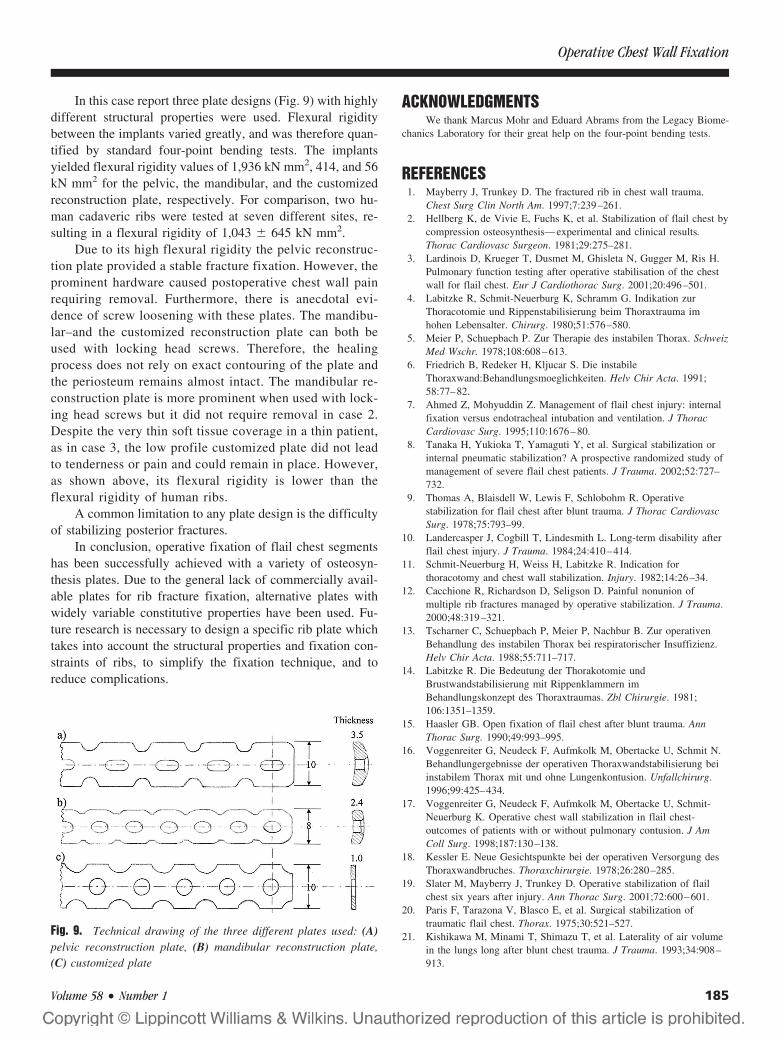
In this case report three plate designs (Fig. 9) with highlydifferent structural properties were used. Flexural rigiditybetween the implants varied greatly, and was therefore quan-tified by standard four-point bending tests. The implantsyielded flexural rigidity values of 1,936 kN mm2, 414, and 56kN mm2 for the pelvic, the mandibular, and the customizedreconstruction plate, respectively. For comparison, two hu-man cadaveric ribs were tested at seven different sites, re-sulting in a flexural rigidity of 1,043 # 645 kN mm2.
Due to its high flexural rigidity the pelvic reconstruc-tion plate provided a stable fracture fixation. However, theprominent hardware caused postoperative chest wall painrequiring removal. Furthermore, there is anecdotal evi-dence of screw loosening with these plates. The mandibu-lar–and the customized reconstruction plate can both beused with locking head screws. Therefore, the healingprocess does not rely on exact contouring of the plate andthe periosteum remains almost intact. The mandibular re-construction plate is more prominent when used with lock-ing head screws but it did not require removal in case 2.Despite the very thin soft tissue coverage in a thin patient,as in case 3, the low profile customized plate did not leadto tenderness or pain and could remain in place. However,as shown above, its flexural rigidity is lower than theflexural rigidity of human ribs.
A common limitation to any plate design is the difficultyof stabilizing posterior fractures.
In conclusion, operative fixation of flail chest segmentshas been successfully achieved with a variety of osteosyn-thesis plates. Due to the general lack of commercially avail-able plates for rib fracture fixation, alternative plates withwidely variable constitutive properties have been used. Fu-ture research is necessary to design a specific rib plate whichtakes into account the structural properties and fixation con-straints of ribs, to simplify the fixation technique, and toreduce complications.
ACKNOWLEDGMENTSWe thank Marcus Mohr and Eduard Abrams from the Legacy Biome-
chanics Laboratory for their great help on the four-point bending tests.
REFERENCES1. Mayberry J, Trunkey D. The fractured rib in chest wall trauma.
Chest Surg Clin North Am. 1997;7:239–261.2. Hellberg K, de Vivie E, Fuchs K, et al. Stabilization of flail chest by
compression osteosynthesis—experimental and clinical results.Thorac Cardiovasc Surgeon. 1981;29:275–281.
3. Lardinois D, Krueger T, Dusmet M, Ghisleta N, Gugger M, Ris H.Pulmonary function testing after operative stabilisation of the chestwall for flail chest. Eur J Cardiothorac Surg. 2001;20:496–501.
4. Labitzke R, Schmit-Neuerburg K, Schramm G. Indikation zurThoracotomie und Rippenstabilisierung beim Thoraxtrauma imhohen Lebensalter. Chirurg. 1980;51:576–580.
5. Meier P, Schuepbach P. Zur Therapie des instabilen Thorax. SchweizMed Wschr. 1978;108:608–613.
6. Friedrich B, Redeker H, Kljucar S. Die instabileThoraxwand:Behandlungsmoeglichkeiten. Helv Chir Acta. 1991;58:77–82.
7. Ahmed Z, Mohyuddin Z. Management of flail chest injury: internalfixation versus endotracheal intubation and ventilation. J ThoracCardiovasc Surg. 1995;110:1676–80.
8. Tanaka H, Yukioka T, Yamaguti Y, et al. Surgical stabilization orinternal pneumatic stabilization? A prospective randomized study ofmanagement of severe flail chest patients. J Trauma. 2002;52:727–732.
9. Thomas A, Blaisdell W, Lewis F, Schlobohm R. Operativestabilization for flail chest after blunt trauma. J Thorac CardiovascSurg. 1978;75:793–99.
10. Landercasper J, Cogbill T, Lindesmith L. Long-term disability afterflail chest injury. J Trauma. 1984;24:410–414.
11. Schmit-Neuerburg H, Weiss H, Labitzke R. Indication forthoracotomy and chest wall stabilization. Injury. 1982;14:26–34.
12. Cacchione R, Richardson D, Seligson D. Painful nonunion ofmultiple rib fractures managed by operative stabilization. J Trauma.2000;48:319–321.
13. Tscharner C, Schuepbach P, Meier P, Nachbur B. Zur operativenBehandlung des instabilen Thorax bei respiratorischer Insuffizienz.Helv Chir Acta. 1988;55:711–717.
14. Labitzke R. Die Bedeutung der Thorakotomie undBrustwandstabilisierung mit Rippenklammern imBehandlungskonzept des Thoraxtraumas. Zbl Chirurgie. 1981;106:1351–1359.
15. Haasler GB. Open fixation of flail chest after blunt trauma. AnnThorac Surg. 1990;49:993–995.
16. Voggenreiter G, Neudeck F, Aufmkolk M, Obertacke U, Schmit N.Behandlungergebnisse der operativen Thoraxwandstabilisierung beiinstabilem Thorax mit und ohne Lungenkontusion. Unfallchirurg.1996;99:425–434.
17. Voggenreiter G, Neudeck F, Aufmkolk M, Obertacke U, Schmit-Neuerburg K. Operative chest wall stabilization in flail chest-outcomes of patients with or without pulmonary contusion. J AmColl Surg. 1998;187:130–138.
18. Kessler E. Neue Gesichtspunkte bei der operativen Versorgung desThoraxwandbruches. Thoraxchirurgie. 1978;26:280–285.
19. Slater M, Mayberry J, Trunkey D. Operative stabilization of flailchest six years after injury. Ann Thorac Surg. 2001;72:600–601.
20. Paris F, Tarazona V, Blasco E, et al. Surgical stabilization oftraumatic flail chest. Thorax. 1975;30:521–527.
21. Kishikawa M, Minami T, Shimazu T, et al. Laterality of air volumein the lungs long after blunt chest trauma. J Trauma. 1993;34:908–913.
Fig. 9. Technical drawing of the three different plates used: (A)pelvic reconstruction plate, (B) mandibular reconstruction plate,(C) customized plate
Operative Chest Wall Fixation
Volume 58 • Number 1 185
22. Beal S, Oreskovich M. Long term disability associated with flailchest injury. Am J Surg. 1985;150:324–326.
23. Moore B. Operative stabilization of nonpenetrating chest injuries.J Thorac Cardiovasc Surg. 1975;70:619–630.
24. Sanchez-Lloret J, Letang E, Mateu M, Callejas M, Catalan M,Mestres C. Indications and surgical technique of the traumatic flailchest syndrome. Thorac Cardiovasc Surgeon. 1982;30:294–297.
25. Lindenmaier H, Kuner E, Walz H. Die operative Behandlung derThoraxwandinstabilitaet. Unfallchirurg. 1990;16:172–177.
26. Actis Dato G, Aidala E, Ruffini E. Surgical management of flailchest. Ann Thorac Surg. 1999;67:1826–1827.
27. Karev D. Operative Management of the flail chest. WiadomosciLekarskie. 1997;50:205–208.
28. Mouton W, Lardinois D, Furrer M, Regli B, Ris H. Long-termfollow-up of patients with operative stabilization of a flail chest.Thorac Cardiovasc Surgeon. 1997;45:242–244.
29. Albrecht F, Brug E. Die Zuggurtungsosteosynthese der Rippen unddes Sternums bei instabiler Thoraxwand. Zbl Chirurgie. 1979;104:770–776.
30. Reber P, Ris H, Inderbitzi R, Stark B, Nachbur B. Osteosynthesis ofthe injured chest wall. Scand J Thor Cardiovasc Surg. 1993;27:137–142.
31. Labitzke R. Biomechanic examination of rib plates. LangenbecksArch Chir. 1981;354:169–171.
32. Boetsch H, Rehm K. Biomechanische Untersuchungen anRippenosteosynthesen. Biomed Technik. 1981;26:296–301.
33. Martin P, Godinou J, Monod R, et al. Costal stapling in severethoracic traumas. Nouv Presse Med. 1982;11:851–854.
34. Borrely J, Grosdidier G, Wack B. Surgical treatment of flail chest bysliding staples. Rev Chir Orthop Reparatrice Appar Mot. 1985;71:241–250.
35. Menard A, Testart J, Philippe J, Grise P. Treatment of flail chestwith Judets struts. J Thorac Cardiovasc Surg. 1983;86:300–305.
36. Vecsei V, Frenzel I, Plenk H Jr. Eine neue Rippenplatte zurStabilisierung mehrfacher Rippenbrueche und der Thoraxwandfrakturmit paradoxer Atmung. Hefte Unfallheilkd. 1979;138:279–282.
37. Di Fabio D, Benetti D, Benvenuti M, Mombelloni G. Surgicalstabilization of post-traumatic flail chest. Our experience with 116cases treated. Minerva Chir. 1995;50:227–233.
38. Glavas M, Altarac S, D V, Ivancic A, Drazinic I. Flail cheststabilization with palacos prosthesis. Acta Medica Croatica, 2001.2001;55:91–95.
39. Guernelli N, Bragaglia R, Briccoli A, Mastrorilli M, Vecchi R.Technique for the management of anterior flail chest. Thorax. 1979;34:247–248.
40. Beltrami V, Martinelli G, Giansante P, Gentile K. An originaltechnique for surgical stabilization of traumatic flail chest. Thorax.1978;33:528–529.
41. Volkmer I, Krespis E, Stapenhorst K. Der instabile Thorax, einBeitrag zur operativen Behandlung. Thoraxchirurgie. 1978;26:275–279.
42. Sherman J, Salzberg A, Raskin N, Beattie E. Chest wall stabilizationusing plate fixation. Ann Thorac Surg. 1988;46:467–469.
43. Graeber G, Cohen D, Patrick D, Wolf R, Hotard M, Zajtchuk R. Ribfracture healing in experimental flail chest. J Trauma. 1985;25:903–908.
44. Fick R, Bartels P, Brunn A, Disse J. Handbuch der Anatomie desMenschen. Anatomie und Mechanik der Gelenke unterBerücksichtigung der bewegenden Muskeln, ed. Fick R. Vol. 2,1,3.1911, Jena: Verlag von Gustav Fischer. 688.
45. Shah T. Internal fixation for flail chest [Letter]. J Thorac CardiovascSurg. 1996;112:849–850.
The Journal of TRAUMA! Injury, Infection, and Critical Care
186 January 2005