Embed Size (px)
Citation preview
REPORT DOCUMENTATION PAGE 1Form ApprovedOMB No. 0704-0188
Public reporting burden for this collection of information is estimated to average 1 hour per response, including the time for reviewing instructions, searching existing data sources,gathering and maintaining the data needed, and completing and reviewing the collection of information. Send comments regarding this burden estimate or any other aspect of thiscollection of information, including suggestions for reducing this burden, to Washington Headquarters Services, Directorate for Information Operations and Reports, 1215 JeffersonDavis Highway, Suite 1204, Arlington, VA 22202-4302, and to the Office of Management and Budget, Paperwork Reduction Project (0704-0188), Washington, DC 20503.
1. AGENCY USE ONLY (Leave blank) 2. REPORT DATE 3. REPORT TYPE AND DATES COVERED
I 24.May.05 MAJOR REPORT4. TITLE AND SUBTITLE 5. FUNDING NUMBERS
OPERATING ROOM TELEPHONE MICROBIAL FLORA
6. AUTHOR(S)CAPT SHINN ANTOINETTE M
7. PERFORMING ORGANIZATION NAME(S) AND ADDRESS(ES) 8. PERFORMING ORGANIZATION
UNIFORMED SERVICES UNIV OF HEALTH SCIENC REPORT NUMBER
C104-1093
9. SPONSORING/MONITORING AGENCY NAME(S) AND ADDRESS(ES) 10. SPONSORING/MONITORING
THE DEPARTMENT OF THE AIR FORCE AGENCY REPORT NUMBER
AFIT/CIA, BLDG 1252950 P STREETWPAFB OH 45433
11. SUPPLEMENTARY NOTES
12a. DISTRIBUTION AVAILABILITY STATEMENT 12b. DISTRIBUTION CODE
Unlimited distributionIn Accordance With AFI 35-205/AFIT Sup 1
13. ABSTRACT (Maximum 200 words)
DISTRIBUTION STATEMENT AApproved for Public Release
Distribution Unlimited
2005,602 00714. SUBJECT TERMS 15. NUMBER OF PAGES
2616. PRICE CODE
17. SECURITY CLASSIFICATION 18. SECURITY CLASSIFICATION 19. SECURITY CLASSIFICATION 20. LIMITATION OF ABSTRACTOF REPORT OF THIS PAGE OF ABSTRACT
Standard Form 298 (Rev. 2-89) (EG)Prescribed by ANSI Std. 239.18Designed using Perform Pro, WHS/DIOR, Oct 94
Telephones as Fomites I
Running head: TELEPHONES AS FOMITES
Operating Room Telephone Microbial Flora
CPT Jason Nelson, Capt Antoinette Shinn, & CPT Ava Bivens
Graduate School of Nursing
Uniformed Services University of Health Sciences
March 31, 2005
THE VIEWS EXPRESSED IN THIS ARTICLE ARETHOSE OF THE AUTHOR AND DO NOT REFLECTTHE OFFICIAL POLICY OR POSITION OF THEUNITED STATES AIR FORCE, DEPARTMENT OFDEFENSE, OR THE U.S. GOVERNMENT.
Telephones as Fomites 2
Abstract
Introduction: There are approximately 500,000 surgical site infections per year in the United
States [1]. The purpose of this study was to determine if the bacteria most frequently involved in
Surgical Site Infections (SSI) could be found on telephones in the Operating Room (OR).
Methods: A total of 26 cultures were taken from telephones within 14 operating rooms and two
sub-sterile rooms at a large, teaching, medical center. Bacteria were identified using standard
laboratory procedures and the Vitek system version 7.02. Results: Of the 52 isolates discovered,
the following bacteria were identified: Acinetobacter calcoaceticus-baumannii complex 1.9%,
Pseudomonas aeruginosa 1.9%, Agrobacterium radiobacter/tumefaciens 1.9%, Coagulase-
negative Staphylococcus 82.7%, Micrococcus 3.8 %, Streptococcus non-group D 5.8%, and one
unidentified gram-negative rod 1.9%.
Telephones as Fomites 3
Introduction
There are approximately 500,000 surgical site infections per year in the United States [1].
Nosocomial infections contribute to prolonged antimicrobial treatments, length-of-stays, and
even death. The Centers for Disease Control (CDC) reports that in 1999 the most prevalent
causes of Surgical Site Infections (SSI) were: Staphylococcus aureus (S. aureus), Coagulase-
negative Staphylococci (CNS), Enterococcus species (spp), and Escherichia coli (E. coli) [2] [3].
There have been no published changes to the prevalence of these bacteria in relation to SSI since
1999. In addition, a study published in 2003 reports that extremes of costs for SSIs are as high
as $92,363 for patients with Methicillin-resistant Staphylococcus aureus (MRSA) SSI[4].
The most common source of SSI is endogenous floras [5], but exogenous floras are also a
possible cause of SSI [3] [6]. If exogenous floras are causing some surgical site infections, how
are they being transmitted? Could the hands of healthcare workers be a source? What other
surfaces might be involved via direct or indirect contact with patients? One inanimate item in
the operating room (OR) frequently contacted by the hands of staff is the telephone. Could
telephones in the OR serve as a source of surgical site infections? An inanimate surface that is
implicated in a nosocomial infection is termed as a fomite. Are telephones in the OR fomites?
Given the potential impact of nosocomial infections in the perioperative setting, research is
needed to describe if the bacteria most frequently involved in SSIs can be found on telephones in
the OR. The purpose of this paper is to describe a study we conducted to identify and quantify
bacterial contamination on telephones in the OR of a large teaching medical center.
Telephones as Fomites 4
Literature Review
Before we can address the questions raised above, we must understand the many factors
associated with nosocomial infections. The chain of infection model provides the best
framework for depicting the relationship among these factors and SSIs. Our literature review
includes a thorough explanation of the chain of infection model, the relationship among these
factors, and a discussion of the current literature on environmental surfaces as fomites.
According to the chain of infection model, a causative agent or pathogen survives within
a reservoir, exits the reservoir via a mode of transmission, and enters a susceptible host, thereby,
causing disease [7]. Intervention in any part of this process can stop the transmission of disease.
The reservoir can include plants, animals, soil, water, and inanimate surfaces [8]. Of these, the
most likely exogenous reservoir in the surgical setting is either human or an inanimate surface.
Both reservoirs are capable of becoming transmission agents.
Inanimate Surfaces as Reservoirs
The evaluation of inanimate surfaces is best categorized by Spaulding's Classification
System. Within this system, items are classified as: Critical, Semi-critical, and Non-critical [9].
Critical items present a significant risk of infection if microorganisms are present because these
items come in contact with sterile tissues. Semi-critical items pose less risk, because they are in
contact with mucous membranes or non-intact skin. Non-critical items are only in contact with
intact skin and pose little risk of infection. However, Non-critical items used in patient care can
serve as a mode of secondary transmission by providing a reservoir that can contaminate the
hands of healthcare workers [10]. This mode of transmission, surface to hand transfer of
bacteria, is well documented in the literature [11] [12].

Telephones as Fomites 5
Animate Reservoirs as Transfer Agent
Proper hand washing is known as one of the most important steps in preventing infections
[9]. Despite several studies documenting hands as carriers of infection [13] [14], hand washing
compliance has been shown to be as low as 9% for medical intensive care unit (ICU) health care
workers and 3% for cardiac surgery ICU health care workers [15]. More to the point, as few as
58% of anesthesiologists report that they wash their hands after contact with every patient [16]
and compliance with hand-cleansing in a post-anesthesia care unit was shown to be 12.5% [17].
If proper hand hygiene is not exercised, items frequently contacted by hands could serve as
reservoirs and those reservoirs could further serve to contaminate hands; thereby increasing the
chance of spreading infections to patients during hand-to-patient contact.
Environmental Surfaces as Fomites
Even though the importance of cleaning environmental surfaces is well recognized as a
standard of care, there is little research available which describes the relationship between the
quantity of pathogens present on surfaces and increased nosocomial infection rates [18].
Bacteria are capable of transferring antibiotic resistance [19] [20] [21] [22] [23], therefore it can
be argued that where bacteria are allowed to survive on environmental surfaces, antibiotic
resistance could be transferred. It is reasonable to question the cleanliness of environmental
surfaces in the surgical setting, especially when as much as 32% of anesthesia equipment has
been found to have occult blood present [24].
The role of inanimate surfaces as fomites is not well documented. Some recent studies
suggest there is no link between infection rates and surface contamination [25] [26] [27] while
other studies demonstrate that environmental surfaces and nursing uniforms have increased
contamination from patients known to be infected or colonized with MRSA [28]. A wide range
Telephones as Fomites 6
of environmental surfaces have been shown to be sources of nosocomial infection, including an
electronic ear probe [29], a stretcher frame, a shower handle [28], and operating room surfaces
[30]. In order to determine the likelihood of bacterial presence on telephones and subsequent
transfer via hands, we reviewed the literature to identify survival times of the bacteria most
frequently implicated in surgical site infections on hands and inanimate surfaces. These bacteria
are: S. aureus, CNS, Enterococcus spp, and E. coli. Our literature review also included MRSA
and Vancomycin-resistant Enterococcus (VRE) as both of these bacteria are variants of S. aureus
and Enterococcus spp. Due to the lack of studies specific to telephone surfaces, we also used
plastic surfaces as a substitute search criteria for telephones.
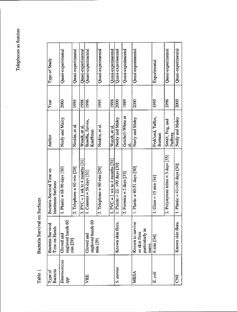
Table 1 is a compilation of our literature review and is reflective of experimental and
quasi-experimental studies dating back to 1989. While some of the studies are dated, in many
cases these studies are either landmark or sole source references. Each of the studies presented
in Table 1 used different inoculum concentrations and techniques, and provided us with evidence
that bacteria might be present on OR telephones. With this evidence we reviewed the literature
on studies involving telephones as a reservoir for bacteria and concluded that time alone would
not eliminate bacteria sufficiently. The importance of hand washing, aseptic technique, and
surface decontamination was evident.
{Please insert "Table 1 Bacterial Survival on Surfaces"} [30] [31] [32] [33] [34] [35] [36]
In one study conducted to document environmental surfaces and hands of healthcare
workers as reservoirs, consisting of 26 telephone cultures in an ICU, the researchers found S.
aureus, Acinetobacter calcoaceticus (A. calcoaceticus), and Pseudomonas spp [34]. In another
study of Non-critical items frequently in contact with hands of staff in the hospital, 20 telephones
from the OR, ICU, Recovery, and emergency room (ER) were cultured and resulted in no gram-
Telephones as Fomites 7
negative bacteria being identified [37]. Cozanitis, Grant, & Makela (1978) cultured 11
telephones in an ICU and identified CNS, Coagulase-positive Staphylococcus, gram-positive
rods and alpha-hemolytic Streptococcus [38]. Lastly, a study conducted to identify the bacteria
on public telephone hand-pieces at a high school showed increasing numbers of bacteria on
telephones from morning to afternoon with CNS being the predominant bacteria identified [39].
The literature review has shown the potential for bacteria to be present on telephones for
variable lengths of time and has demonstrated that there is frequently a lack of hand washing and
decontamination of environmental surfaces by hospital staff. Additionally, inanimate surfaces
have been implicated in infections. In a study conducted by Rusin, Maxwell, and Gerba (2002) a
link is clearly created between the transfer of bacteria from telephones to hands and from hands
to other skin surfaces. Rusin et al. demonstrated that Micrococcus luteus (M. luteus) can be
transferred from telephones to hands with approximately 41% efficiency and from hands to the
mouth at the same rate of 41% [40].
Telephones as Fomites 8
Methods
The purpose of this descriptive study was to determine if the bacteria most frequently
involved in SSI could be found on telephones in the OR of a large teaching medical center. We
focused exclusively on S. aureus, CNS, Enterococcus spp, E. coli, MRSA and VRE. We
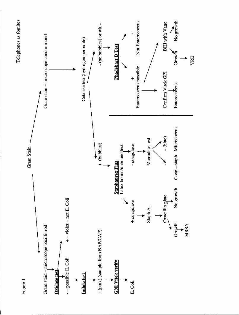
developed an algorithm (see figure 1) and written guidelines for our data collection and analysis
procedures. Data were collected by swab and culture techniques to make observations about
bacteria colony counts. A data collection sheet, was designed based on our algorithm and was
utilized to capture data related to: surgical service, the time of day when cultures were collected,
location of the telephone in the perioperative environment, surgical case number, and OR
temperature and OR humidity at the time of sampling. Our research protocol was approved by
the Institutional Review Boards of the medical center and the Uniformed Services University of
Health Sciences.
Sample and Setting
We used a quota based convenience sample of 30 cultures from telephones within the
ORs at the medical center. Twenty-six cultures were obtained from telephones within 14
operating rooms and two sub-sterile rooms. Two control cultures and two double cultures were
also collected. Specimen collection was divided between two days that were separated by 19
days in order to decrease the likelihood that perioperative personnel might alter hand washing,
aseptic techniques, and environmental disinfection, because they were aware of our study and
data collection [41].
Specimen Collection and Analysis Procedures
In order to ensure precision in the collection of data, we (the principal investigator and
Telephones as Fomites 9
two co-investigators oriented to the medical center's lab and became familiar with testing
supplies and procedures. With the assistance of our Microbiologist Associate Investigator, we
developed a guideline for all testing procedures, based on the algorithm in figure 1. The
guideline was incorporated into a standardized specimen data collection and analysis sheet for
use in recording the identification and interpretation of bacteria. Laboratory personnel evaluated
our skills with a practical exam utilizing eight known bacterial isolates.
Cultures were taken from OR telephones at the end of surgical cases to eliminate
unnecessary traffic through the surgical area and prior to staff cleaning of the surgical suites.
Cultures were taken in the same manner by all three investigators wearing sterile gloves. All
four sides of the telephone hand piece handles were swabbed. The posterior swab path included
one vertical pass from earpiece to mouthpiece while holding the swab on its side and rotating or
rolling it across the surface. Remel Bacti-Swabs with non-nutritive modified Stuart's Medium
were used to obtain bacterial sampling. Validity and reliability testing was obtained by random
double culturing and random control culturing techniques. The double culture technique
included the swabbing of telephones in the manner described previously with two consecutive
culturettes attempting to eliminate path over-run. These culturettes were labeled so that one
investigator was blinded to the source. One control culture was randomiy selected for each
culture batch. The control culturettes were opened and re-sealed without exposure to
contaminants.
After sampling, the swabs were returned aseptically to their cases, labeled and numbered
sequentially. Swabs were carried to the lab within 15 minutes of sampling. All samples were
streaked for isolation onto tripticase soy agar with 5% sheep blood agar (Remel, reference #
01202), chocolate agar (Remel, reference # 01302), and MacConkey agar (Remel reference #
Telephones as Fomites 10
01552), respectively. The agar plates were incubated at 35 degrees Celsius (°C) for 24 hours.
Chocolate and blood agar plates were incubated in 4% carbon dioxide (CO2) while MacConkey
agar plates were incubated in 1.2 % CO2. After the first 24 hours, the entire bacterial floras were
visually quantified into the number of colonies present. If no colonies were present at 24 hours,
confirmation was performed at 48 hours.
Bacteria were initially identified based on growth within the medium. Bacterial growth
on MacConkey was considered gram-negative and was tested to rule out Acinetobacter spp,
Pseudomonas aeruginosa (P. aeruginosa), Haemophilus influenza (H. influenza) and Neisseria
gonorrhea (N. gonorrhea). Bacterial growth on chocolate plates was used to identify H.
influenza and N. gonorrhea, which were ruled out based on gram stain morphology and
microscopic examination. Bacterial growth on blood agar was used to support the growth and
evaluation of S. aureus, MRSA, CNS, Enterococcus spp, VRE, and E. coli.
Bacteria were then identified by: 1) shape- spherical (coccus), rod-like (bacillus), or
spiral (spirochete); and 2) cell wall- gram-positive or gram-negative as seen with gram stain.
Gram-negative rods were tested for oxidase (Remel, ref #425506) and indole (Remel, ref
#21245) reaction. Positive oxidase and negative indole results ruled out E. coli and were tested
further to rule out A. calcoaceticus-baumannii and P. aeruginosa. Gram-positive bacteria were
initially tested using 3% hydrogen peroxide for catalase testing, which was used to differentiate
group 1 (Micrococcus, CNS, S. aureus, and MRSA) from group 2 bacteria (Enterococcus spp.
and other Streptococcaceae). Group 1 bacteria were then tested with Remel Staphaurex Plus (ref
# 30950102) to rule in S. aureus. Bacteria that were negative for Staphaurex Plus were then
tested with the Microdase test (Remel, ref # 21132) to differentiate CNS from Micrococcus spp.
Catalase negative bacteria were analyzed using the Boule Phadebact D test to differentiate
Telephones as Fomites 11
potential Enterococcus from other Streptococcus spp.
The Vitek system version 7.02 was used with BioMerieux Gram Positive Identification
(GPI) and Gram-negative Identification + (GNI+) cards to identify A. calcoaceticus-baumannii
complex, Agrobacterium radiobacter/tumefaciens (A. radiobacter/tumefaciens), and 9 of the 43
isolates of CNS. A single double culture of CNS was also analyzed via GPI card. Identification
of P. aeruginosa was based on the following test results: gram-negative rod, oxidase positive,
catalase positive, presence of motility, and growth at 42°C in tripticase soy broth (TSB). The
methods described above are in compliance with standard culture technique [42] [43].
The counting of colonies was performed by two investigators individually and digital
photos were taken. Additionally, the surface area of the four vertical swabbing paths was
calculated to determine Colony Forming Units (CFU)/centimeter squared (cm). The maximum
swab path width was measured at 3 mm. The length or distance of this path was measured at
95.6 cm. To find the surface area in cm2, the length (95.6 cm) was multiplied by width (0.3 cm)
for a total of 28.7 cm2, which is nearly equivalent to the surface area of a RODAC agar plate
with a 6 cm diameter (3.14 x 32 = 28.26 cm2). Data were entered into a spreadsheet by two
investigators individually, using the completed specimen data collection and analysis sheet. A
test of inter-rater reliability revealed 100% agreement between the two investigators on all data
entered into the two separate spreadsheets.
Laboratory and Equipment
The lab we utilized was accredited by the Commission of Laboratory Accreditation of the
College of American Pathologists (CAP) in 2004. The reliability and validity of the Vitek
system is well established among medical laboratories. Quality Controls (QC) were conducted
on all identification card lots used in this study. Digital photographs were taken using an
Telephones as Fomites 12
Olympus D-380 camera with 2.0 mega pixels effect and five times digital zoom.
Proficiency testing for the Vitek was performed three times during the year of our study.
At the time of preparation of this paper, test results were only available for 2 of the 3 proficiency
tests; these proficiency tests showed > 86 % accuracy of bacterial identification and > 92%
performance satisfaction with 100% antigen detection. This level of testing is in accordance
with CAP accreditation. The Vitek system is approved by the Food and Drug Administration
(FDA) for both gram-positive and gram-negative bacterial identification and sensitivity testing
(1991 &1996).
Phenotypic Testing Agents
We used several phenotypic testing agents while conducting our study. None of the
agents were used beyond their expiration date. We used the same lot numbers among agar plates,
culturettes, Vitek cards and all other supplies. The only exception to this was the Microdase test,
which did change lot numbers during our second batch of testing. We present a brief literature
review of our testing agents here to demonstrate reliability and validity.
Staphaurex Plus in comparison with tube Coagulase test on S. aureus isolates has been
found to have a relative sensitivity of 99.4%, a relative specificity of 95.5% and an overall
agreement of 98.4% [44]. In another study, Staphaurex Plus was found to be 99.6% sensitive
and 93.9% specific[45]. During 510K testing on the Vitek GNI+ card, a Centers for Disease
Control (CDC) gram-negative challenge set was utilized. Gram-negative bacteria were correctly
identified to the species level 85.2% (77.4% to 91.1%) of the time. Correct identifications to the
genus level occurred at 88.7% (81.4% to 93.8%). Misidentifications occurred at 7.8% (3.6% to
14.3%) and no identifications at 7% (3.1% to 13.2%)"[46]. The Microdase test we utilized is
described as "...the simplest and most rapid methods for separating Staphylococci from
Telephones as Fomites 13
Micrococci"[47]. In another evaluation of the Microdase test it was described as "the oxidase
test (Microdase disk; Remel) proved to be the most sensitive (100%) and was sufficiently
specific (99%) for providing a rapid means of accurately differentiating between Staphylococci
and Micrococci"[48]. However, Staphylococcus (S.) lentus, S. sciuri, and S. vitulus can give a
positive Microdase reaction. The impact is probably minimal because in an evaluation of CNS
infections, 86 cultures revealed one S. sciuri and no S. lentus or S. vitulus [49]. The catalase,
Kovacs indole, modified oxidase, and oxidase tests are standard testing agents for the
identification of bacteria [43]. The Phadebact D test was found in one study to be 100%
effective in identifying Group D Streptococcus [50]. Unfortunately, only 80% of Enterococcus
can be identified by group D antigen testing [43].
Statistical Analysis
Data analyses were performed using the Statistical Package for the Social Sciences
(SPSS) version 12.0. A .05 level of probability was chosen to indicate statistical significance.
Descriptive statistics (frequencies and counts) were used to summarize and describe the variables
in the study. Independent sample chi-square tests were used to evaluate the relationship between
bacterial colony counts and surgical service, the time of day when cultures were collected,
location of the telephone in the perioperative environment, surgical case number, and OR
temperature and OR humidity at the time of sampling.
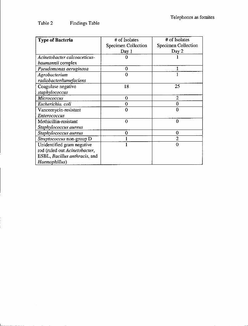
Findings
Of the six bacteria we attempted to identify, only CNS was found. Additionally, A.
calcoaceticus-baumannii complex, P. aeruginosa, A. radiobacter/tumefaciens, Micrococcus,
Streptococcus non-group D, and one unidentified gram-negative rod were found. Table 2
summarizes the types and number of bacterial isolates discovered with comparative separation of
Telephones as Fomites 14
data collection at day one and day two. Only the first culture results for telephones that were
double cultured are included in Table 2 to avoid over-representation of isolates.
{Please insert Table 2 Findings Table}
Table 3 summarizes the five variables that remained in the study after the removal of the
temperature and humidity variables. The humidity and temperature data were not included due
to a lack of standardized measuring instruments. In Table 3, the acronym "BAP" refers to
tripticase soy blood agar plates and the number of colonies identified; "CAP" refers to chocolate
agar plates; and "MAC" refers to MacConkey agar plates. An absence of colonies at both 24 and
48 hours is indicated by "0,0". In Table 3 the word "Total" refers to a cumulative total of all
colonies identified among the three growth mediums of BAP, CAP and MAC. "CFU/cm 2" refers
to the number of colony forming units identified per centimeter squared, as determined by a
swab path surface area of 28.6875 cm2. "Service" refers to the surgical case type that had
occurred in the OR just prior to sampling. "Time of Day" refers to whether specimens were
collected in the a.m. or p.m. "Culture Location" refers to the area that contained the telephone,
which was cultured (i.e., OR Suite or Sub-sterile Room). "Case #" refers to the surgical case
sequence in the room during culturing (i.e. 1st, 2nd or 3rd case of the day).
{Please insert Table 3 Summary Table)
We collected the majority of our samples in the a.m. (61.5%) and 38.5% were collected
in the p.m. The largest numbers of specimens were obtained from the first surgical case of the
day (65.4%), followed by the second case at 30.8%, and the third case at 3.8%. The top five
surgical services operating within the rooms where the telephone cultures were obtained were:
Orthopedics at 42.3%, Ophthalmology at 15.4%, General Surgery at 11.5%, and Cardiothoracic
and Genitourinary Surgery both at 7.7 %. The double cultures from data collection day one and
Telephones as Fomites 15
two revealed CNS of similar quantities and on data collection day two, testing with the Vitek and
GPI cards revealed the same Genus and Species, Staphylococcus epidermidis. Chi-Square
analyses were not significant on any of the variable relationship evaluations. This lack of
significance is most likely due to the small sample size in this study. One telephone (specimen
#9910 in Table 3) was cultured after the room had been cleaned. This specimen had the second
highest number of colony forming units per cm2 at 2.16.
Discussion
In our study we were unable to find S. aureus, Enterococcus spp., and E. coli on
telephones in the OR. The inability to find Enterococcus spp. may be related to the limitations
previously described for the Phadebact D test. Thus, in each of the three isolates that we
recorded as non-group D Streptococcus, we may have missed possible Enterococcus.
Additionally, colony counts for bacteria were low in comparison to levels recorded for public
school telephones [39], horizontal surfaces in OR rooms (5.86-6.98 CFU/cm2) [27], stethoscopes
(158 CFUs) [12], hospital pagers (39-153 CFUs) [51], and telephones in the ICU (7-282 CFU)
[38].
The only environmental surface contamination guidelines that we could find were based
on the use of RODAC plates. The guidance describes that floors with microbial contamination
greater than 50 colonies per plate relate to poor cleanliness [52]. In our study, only four samples
exceeded this amount. Those four samples did not contain the isolates of: Acinetobacter,
Pseudomonas, or Agrobacterium. Again, our findings were generally low in comparison to other
studies. This is largely due to sampling technique variations. For instance, the Yalowitz study
swabbed the entire surface of the telephones and would be expected to have higher colony
forming units. We only counted bacteria at 24 hours and may have missed some slow growing
Telephones as Fomites 16
bacteria. If an agar plate contained no colonies at 24 hours, this was re-evaluated at 48 hours.
Other studies counted bacteria on all plates at 24 and 48 hours [34]. Our inability to find S.
aureus parallels similar difficulties in another study that were remedied by using broth to support
environmental cultures which led to increasing MRSA findings by a factor of two [53].
Similarly, Rafferty and Pancoast (1984) were only able to isolate S. aureus twice out of 114
specimens. Our inability to find E. coli on telephones is consistent with a study by Rafferty and
Pancoast (1984) that found no gram-negative bacteria on 20 telephones in the OR, ICU,
Recovery and ER areas [37]. Additionally, E. coli was found in the literature review to have a
relatively short life span on environmental surfaces [35] [36], which may explain its absence in
our study.
The bacterium most frequently isolated in our study was CNS. CNS and S. aureus are
the most commonly implicated bacteria in surgical site infections, 20% and 14% respectively [3].
Subsequently, CNS are one of the most frequently isolated bacteria in the laboratory [54]. These
bacteria are of little virulence [55] but are frequently implicated as the cause of infections in
patients who are immunocompromised or have medical implants [56] [57] [58] [59]. A high
prevalence of CNS on telephones is consistent with Cozanitis et al. in their finding of CNS on all
the telephones that they cultured [38].
Serendipitous Findings
Our results differ from the findings obtained by Rafferty and Pancoast (1984) in that we
found three other gram-negative bacteria present on telephones, namely: A. calcoaceticus-
baumannii complex, P. aeruginosa, and A. radiobacter/tumefaciens. Similarly, Getchell-White
et al. were also able to find Acinetobacter and Pseudomonas but they were also able to find three
isolates of S. aureus from 26 telephone cultures. They utilized RODAC impression agar plates
Telephones as Fomites 17
which eliminate the number of times that bacteria are transferred (RODAC plates are a direct
transfer technique). Undoubtedly, some of the bacteria remained in our culture swabs and were
not accounted for. Had we utilized RODAC plates our bacterial count could have been higher.
The Acinetobacter (A.) calcoaceticus-baumannii complex is made up of four distinctive
genotypes yet similar phenotypes - all which cause nosocomial infections[60]. For these
reasons, A. calcoaceticus and A. baumannii will be addressed interchangeably. Both, A.
calcoaceticus, a Gram-negative, nonfermentative, coccobacillus and A. baumannii, a Gram-
negative, non motile bacterium can be found as a normal part of skin flora (25%) [61, 62]. A.
calcoaceticus and A. baumannii have been implicated in hospital outbreaks causing patient
colonization, infections, and in some cases mortality [60, 61, 63]. A. calcoaceticus was found on
the skin flora of a respiratory therapist who regularly handled respiratory equipment, thereby
causing respiratory tract colonization and infections in endotracheal intubated patients [61].
However, it is A. baumannii that has been the culprit in most hospital acquired infections [64].
The fact that A. baumannii can survive up to four months on dry surfaces has possible
implications in cross contamination of the hospital environment [65]. And, even after new
cleaning protocols were implemented in a neurosurgical ICU, isolates of A. baumannii were still
colonized from patients [66]. In the bum unit of a public teaching hospital, total hospital costs of
treating a patient with multidrug-resistant A. baumannii (MDRAB) were $98,575 higher than
that of a non-MDRAB patient [67]. Since the advent of using multiple antimicrobial agents in
treating nosocomial infections and ongoing developing isolates, the Acinetobacter spp. has
become more and more resistant to many therapies [62]. For obvious reasons, we need to control
the spread of this bacterium before it becomes a super bug. It is likely that our finding of
Acinetobacter spp. could have been avoided with simple handwashing, surface disinfection and
Telephones as Fomites 18
basic aseptic techniques.
Our finding of Pseudomonas aeruginosa (P. aeruginosa) is not entirely surprising since
this environmental bacteria inhabits soil, water and surfaces with soil or water [68] and has been
previously found on telephones in the ICU [34]. P. aeruginosa can be a part of the normal flora
for some individuals [68] [69]. But, this organism is also an opportunistic pathogen responsible
for many severe nosocomial infections such as septicemia, urinary tract infections, pneumonia,
and osteochondritis [68]. Pseudomonas infections are most commonly found in
immunocompromised patients and patients with cystic fibrosis [68] [69]. For those patients who
become infected, treatment can be complicated by antibiotic resistance [68]. Pseudomonas has
been implicated in eight percent of surgical infections [69] and 10 percent of all hospital-
acquired infections [68]. In 2001, a CDC investigation of a Pseudomonas outbreak revealed that
defective bronchoscopes could be a reservoir for infection [70]. A 2003 study by Leibovitz et al.
found 34 percent of 50 patients fed with nasogastric tubes were colonized with P. aeruginosa in
their oral pharynx [71]. Thus, both nasogastric tubes and bronchoscopes have been implicated as
fomites or reservoirs. The oral cavity may be a potential site of growth for pathogenic organisms
[71]. The presence in the mouth or throat could explain the transmission to the telephone.
Improper hand washing [72] and the handling of an endotracheal tube, oral airway, or some other
contaminated object could also have contributed to this transmission.
Agrobacterium radiobacter/tumefaciens is rarely implicated as an infectious agent. Of
the 42 patients with Agrobacterium infections found in literature by 2003 [73], most involved
immunocompromised hosts [73] [74] or had invasive procedures involving the placement of
plastic implants such as central lines [73] [75] or prosthetic valve [76]. Despite the antibiotic
resistance of Agrobacterium [77], infections from this bacteria are rarely fatal [78]. The apparent
Telephones as Fomites 19
affinity of this bacterium toward plastic may explain its presence on a telephone in our study.
This bacterium was very slow growing and was seen only as a pinpoint colony at 24 hours.
Lastly, we found Micrococcus on telephones in the O.R. "The genus Micrococcus is
currently composed of three species." [43] p. 385. "M. luteus is the most common micrococcal
species found in nature and in clinical specimens" [43] p.385. Micrococci are considered to be
opportunistic pathogens for immunocompromised hosts [43] and have been implicated in
endocarditis [79] and central venous catheter infections [80].
Recommendations for improvement
During the collection of temperature and humidity data within the surgical rooms, we
utilized gauges already present in rooms. Because no standardized method of measurement was
used, these data were removed from our analysis. Future studies could utilize a portable device
to ensure standardized measurement. A larger sample size would help to validate data and
provide a basis for inferential statistics. Additionally, specimen collection may be more efficient
with RODAC impression agar plates, due to the advantages of a direct transfer technique.
Colonies should be counted at both 24 and 48 hours in an attempt to recognize slow growing
bacteria. In our study, morphologic examination was conducted at 24 hours. However, the
separation of colonies based on morphologic examination is much easier at 48 hours. By that
time, hemolytic rings are clearly visible and colonies have had ample time to differentiate
themselves. While some CNS have been shown to be methicillin resistant [81] [3] [82], it may
be beneficial to determine the frequency of this resistance. Finally, adding broth to support
environmental cultures could lead to increased MRSA findings as supported by the study
conducted by Boyce et al. [53].
Telephones as Fomites 20
Implications and Conclusion
We are cautious in drawing conclusions based on a convenience sample from only one
medical center in which surgical technologists and circulators perform cleaning between cases.
Our data may have been impacted by increased workloads. During the time of sampling the
number of surgical cases increased 25.7% between the first and second sampling. During the
entire month of sampling, there was an increase in the number of surgical cases by 37.9% from
the previous year. Additionally, the quantity of bacteria needed to cause disease is unclear.
Hinton, Maltman, and Orr were able to show that fresh Staphylococci intramuscular injections of
100,000 cells could cause infections in 20% of mice while dried Staphylococci injections of
400,000 cells caused infections in 10% of mice [83]. However, our CFUs were low in
comparison to the Hinton, Maltman, and Orr (1960) study and thus we are unable to draw
conclusions related to disease.
Our study does support the need for heightened awareness of cleaning procedures and
standard precautions. It seems reasonable to assume that surface contamination in the form of
Acinetobacter or Pseudomonas in the surgical suite is a risk to both staff and patients. While we
make a case for cleaning, it must be emphasized that cleaning needs to be done correctly. When
bacteria are subjected to sub-lethal levels of disinfectants, they can become resistant to
antibiotics [53] [80]. The current guidance by the Association of periOperative Registered
Nurses (AORN) for environmental cleaning includes terminally cleaning telephones at the end of
the day [18]. Additionally, AORN describes that such cleaning should occur when equipment is
visibly soiled. In our study, one telephone (specimen #9910 in Table 3) was cultured after the
room had been cleaned (between cases). This specimen had the second highest number of
colony forming units per cm2 at 2.16. This finding is particularly disturbing and raises the
Telephones as Fomites 21
question: "Should we be cleaning telephones and other objects frequently contacted by hands in
the perioperative environment between cases rather than at the end of the day?" More
importantly, it is the initial contamination of telephones rather than the cleaning that is
concerning. Standard precautions require workers to wear gloves when the possibility of
exposure to body fluids exists. Upon removing gloves, hands should be washed [75]. The
obvious conundrum for OR personnel follows: I must touch soiled materials, leaving the room
decreases positive air pressure and places patients at risk for infection [18] and running water is
not available in the surgical suite. A possible solution may include waterless hand-cleaner in
surgical suites. Ultimately, the cleanliness of the surgical suite is the responsibility of
perioperative nurses [18]. Perioperative managers in concert with Infection Control Officers
must ensure that Environmental Protection Agency (EPA) approved hospital disinfectants are
both appropriate for emerging resistant bacteria and are being utilized correctly. Close attention
must be applied to these key processes and focused to include aseptic principles and standard
precautions.
Future research may seek to: measure hand washing compliance in the surgical suite;
quantify the number of varying bacteria that can be transferred from surfaces to incisions via
gloved hands and cause infection in mice; frequency with which operating room personnel
contact surfaces with gloves after performing the function requiring the use of those gloves;
and/or quantify contamination of other objects frequently contacted by hands such as door
handles or computer keyboards.
{Please insert Figure 1}
Telephones as Fomites 22
References
1. CDC, National Center for Health Statistics Vital and Health Statistics, Detaileddiagnoses and procedures national hospital discharge survey 1994. Vol. 127. 1997,Hyattsville, MD: Department of Health and Human Services.
2. Weinstein, R.A., Nosocomial infection update, E.I. Diseases, Editor. 1998.3. CDC, Guideline for prevention of surgical site infection, in APIC. 1999.4. Engemann J.J., C.Y., Cosgrove S.E., Fowler V.G., Bronstein M.Z., Trivette S.L., Briggs
J.P., Sexton D.J., and K. K.S., Adverse clinical and economic outcomes attributable tomethicillin resistance among patients with Staphylococcus aureus surgical site infection.Clinical Infectious Diseases, 2003. 36(5): p. 592-598.
5. Altemeier, W.A., W.R. Culbertson, and R.P. Hummel, Surgical considerations ofendogenous infections--sources, types, and methods of control. Surg Clin North Am,1968. 48(1): p. 227-40.
6. Wiley, A.M. and G.B. Ha'eri, Routes of infection. A study of using "tracer particles" inthe orthopedic operating room. Clin Orthop, 1979(139): p. 150-5.
7. APIC, Infection control & applied epidemiology: Principles and practice. 1996, St.Louis, MO: Mosby.
8. Tortora, G.J., Funke, B. R., & Case, C. L., Microbiology an introduction. 1995, RedwoodCity, CA: The Benjamin Cummings Publishing Co. Inc.
9. Gruendemann, B.J., Mangum, S. S.,, Infection Prevention in Surgical Settings. 2001,Philadelphia, PA: Saunders. 387.
10. Rutala, W.A., Disinfection and sterilization ofpatient-care items. Infection Control andHospital Epidemiology, 1996. 17(6): p. 377-384.
11. Scott, E.B., S. F., The survival and transfer of microbial contamination via cloths, handsand utensils. Journal of Applied Bacteriology, 1990. 68: p. 271-278.
12. Marinella, M.A., Pierson, C., & Chenoweh, C., The stethoscope: A potential source ofnosocomial infection. Arch Intern Med, 1997. 157: p. 786-790.
13. Larson, E., A causal link between handwashing and risk of infection? Examination of theevidence. Infection Control and Hospital Epidemiology, 1988. 9(1): p. 28-36.
14. Semmelweiss, I.P., The etiology, the concept and the prophylaxis of childbed fever, inTranslation by Murphy, F. B. 1981. 1861: Pest, Hungary.
15. Bischoff W.E., R.T.M., Sessler C.N., Edmond M.B., Wenzel R.P., Handwashingcompliance by health care workers: The impact of introducing an accessible, alcohol-based hand antiseptic. Archives of Internal Medicine, 2000. 160(7): p. 1017-21.
16. Tait, A.R. and D.B. Tuttle, Preventing perioperative transmission of infection: a surveyof anesthesiology practice. Anesth Analg, 1995. 80(4): p. 764-9.
17. Pittet D, S.F., Hugonnet S, Akakpo C, Souweine B, Clergue F., Hand-cleansing duringpostanesthesia care. Anesthesiology, 2003. 99(3): p. 519-520.
18. A.O.R.N., Standards, Recommended Practices, and Guidlines. 2004, Denver Colorado:AORN. 398.
19. Davies, J., Inactivation of antibiotics and the dissemination of resistance genes. Science,1994. 264(5157): p. 375-82.
20. Rice, L.B., Bacterial monopolists: the bundling and dissemination of antimicrobialresistance genes in gram-positive bacteria. Clin Infect Dis, 2000. 31(3): p. 762-9. Epub2000 Oct 04.
Telephones as Fomiites 23
21. Higgins, N.P., Death and transfiguration among bacteria. Trends Biochem Sci, 1992.17(6): p. 207-11.
22. Cohen, M.L., E.S. Wong, and S. Falkow, Common R-plasmids in Staphylococcus aureusand Staphylococcus epidermidis during a nosocomial Staphylococcus aureus outbreak.Antimicrob Agents Chemother, 1982. 21(2): p. 210-5.
23. Trieu-Cuot, P., E. Derlot, and P. Courvalin, Enhanced conjugative transfer ofplasmidDNA from Escherichia coli to Staphylococcus aureus and Listeria monocytogenes.FEMS Microbiol Lett, 1993. 109(1): p. 19-23.
24. Perry, S.M. and W.P. Monaghan, The prevalence of visible and/or occult blood onanesthesia and monitoring equipment. Aana J, 2001. 69(1): p. 44-8.
25. Devine, J., Cooke, R. P. D., & Wright, E. P., Is methicillin-resistant Staphylococcusaureus (MRSA) contamination of ward-based computer terminals a surrogate marker fornosocomial MRSA transmission and handwashing compliance? Journal of HospitalInfection, 2001(48): p. 72-75.
26. Dharan, S., Mourouga, P., Copin, P., Bessmer, G., Tschanz, B., & Pittet, D., Routinedisinfection of patients' environmental surfaces. Myth or reality? Journal of HospitalInfection, 1999. 42: p. 113-117.
27. Weber, D.O., Gooch, J. J., Walter, R. W., Britt, E. M., & Kraft, R. 0., Influence ofoperating room surface contamination on surgical wounds. Arch Surg, 1976. 111(484-488).
28. Boyce, J.M., Patterns of methicillin-resistant Staphylococcus aureus prevalence. InfectControl Hosp Epidemiol, 1991. 12(2): p. 79-82.
29. Porwancher, R., et al., Epidemiological study of hospital-acquired infection withvancomycin-resistant Enterococcusfaecium: possible transmission by an electronic ear-probe thermometer. Infect Control Hosp Epidemiol, 1997. 18(11): p. 771-3.
30. Noskin, G.A., et al., Recovery of vancomycin-resistant enterococci on fingertips andenvironmental surfaces. Infect Control Hosp Epidemiol, 1995. 16(10): p. 577-8 1.
31. Neely, A.N. and M.P. Maley, Survival of enterococci and staphylococci on hospitalfabrics and plastic. J Clin Microbiol, 2000. 38(2): p. 724-6.
32. Wendt, C., et al., Survival of vancomycin-resistant and vancomycin-susceptibleenterococci on dry surfaces. J Clin Microbiol, 1998. 36(12): p. 3734-6.
33. Bonilla, H.F., M.J. Zervos, and C.A. Kauffman, Long-term survival of vancomycin-resistant Enterococcusfaecium on a contaminated surface. Infect Control HospEpidemiol, 1996. 17(12): p. 770-2.
34. Getchell-White, S.I., L.G. Donowitz, and D.H. Groschel, The inanimate environment ofan intensive care unit as a potential source of nosocomial bacteria: evidence for longsurvival ofAcinetobacter calcoaceticus. Infect Control Hosp Epidemiol, 1989. 10(9): p.402-7.
35. Fryklund, B., K. Tullus, and L.G. Burman, Survival on skin and surfaces of epidemic andnon-epidemic strains of enterobacteria from neonatal special care units. J Hosp Infect,1995. 29(3): p. 201-8.
36. Smith, S.M., R.H. Eng, and F.T. Padberg, Jr., Survival of nosocomial pathogenic bacteriaat ambient temperature. J Med, 1996. 27(5-6): p. 293-302.
37. Rafferty, K.M.P., S. J., Brief report: Bacteriological sampling of telephones and otherhospital staff hand-contact objects. Infection Control, 1984. 5(11): p. 533-535.
38. Cozanitis, D.A., Grant, J., & Makela, P., Bacterial contamination of telephones in an
Telephones as Fomites 24
intensive care unit. Anaesthesist, 1978. 27: p. 439-442.39. Yalowitz, M.B., I., The recovery of bacteria from the handpiece of a high school
telephone. Journal of Environmental Health, 2003. 65(6): p. 18-20.40. Rusin, P., S. Maxwell, and C. Gerba, Comparative surface-to-hand and fingertip-to-
mouth transfer efficiency of gram-positive bacteria, gram-negative bacteria, and phage. JAppl Microbiol, 2002. 93(4): p. 585-92.
41. Harbarth, S., et al., Interventional study to evaluate the impact of an alcohol-based handgel in improving hand hygiene compliance. Pediatr Infect Dis J, 2002. 21(6): p. 489-95.
42. Isenberg, H.D., Clinical microbiology procedures handbook, ed. A.S.f. Microbiology.Vol. 1. 1994.
43. Murray, P.R., Baron, E.J., Pfaller, M.A., Jorgensen, J.H., and Yolken, R.H., Manual ofClinical Microbiology. 8 ed, ed. P.R. Murray. Vol. 1. 2003, Washington D.C.: ASMPress.
44. Remel, Staphaurex Plus ZL33/34. 2002. p. Product insert.45. Wilkerson, M., et al., Comparison offive agglutination tests for identification of
Staphylococcus aureus. J Clin Microbiol, 1997. 35(1): p. 148-5 1.46. bioMerieux, Gram-Negative Identification+ Card for in vitro diagnostic use Pinsert (Rev
0498/I). 1999, bioMerieux Vitek Inc.47. Faller, A. and K.H. Schleifer, Modified oxidase and benzidine tests for separation of
staphylococci from micrococci. J Clin Microbiol, 1981. 13(6): p. 1031-5.48. Baker, J.S., Comparison of various methods for differentiation of staphylococci and
micrococci. J Clin Microbiol, 1984. 19(6): p. 875-9.49. Lang, S., et al., The genomic diversity of coagulase-negative staphylococci associated
with nosocomial infections. J Hosp Infect, 1999. 43(3): p. 187-93.50. Burdash, N.M., et al., Group identification of streptococci. Evaluation of three rapid
agglutination methods. Am J Clin Pathol, 1981. 76(6): p. 819-22.51. Singh, D., Kaur, H., Gardner, W. G., & Treen, L. B., Bacterial contamination of hospital
pagers. Infection Control and Hospital Epidemiology, 2002. 23(5): p. 274-276.52. Pryor, A.K., Veslely, D., & Shaffer, J. G., Cooperative microbial surveys of surfaces in
hospital patient rooms. Health Lab Sciences, 1967. 4: p. 153-159.53. Boyce, J.M., et al., Environmental contamination due to methicillin-resistant
Staphylococcus aureus: possible infection control implications. Infect Control HospEpidemiol, 1997. 18(9): p. 622-7.
54. Patrick, C.C., Coagulase-negative staphylococci: pathogens with increasing clinicalsignificance. J Pediatr, 1990. 116(4): p. 497-507.
55. Rothrock, J.C., Alexander's care of the patient in surgery. 12th ed. ed. 2003, St. Louis:Mosby Inc.
56. Cunha Mde, L., et al., [Clinical significance of coagulase-negative staphylococci isolatedfrom neonates]. J Pediatr (Rio J), 2002. 78(4): p. 279-88.
57. Lesseva, M., Central venous catheter-related bacteraemia in burn patients. Scand JInfect Dis, 1998. 30(6): p. 585-9.
58. al-Rashdan, A., R. Bashir, and F.A. Khan, Staphylococcus capitis causing aortic valveendocarditis. J Heart Valve Dis, 1998. 7(5): p. 518-20.
59. Rupp, M.E. and G.L. Archer, Coagulase-negative staphylococci: pathogens associatedwith medical progress. Clin Infect Dis, 1994. 19(2): p. 23 1-43; quiz 244-5.
60. Gerner-Smidt, P., Ribotyping of the Acinetobacter calcoaceticus-Acinetobacter
Telephones as Fomites 25
baumannii complex. J Clin Microbiol, 1992. 30(10): p. 2680-5.61. Buxton, A.E., Anderson, R. L., Werdegar, D, Atlas, E., Nosocomial respiratory tract
infection and colonization with Acinetobacter calcoaceticus. The American Journal ofMedicine, 1978. 65: p. 507-513.
62. Borgmann, S., et al., Metallo-beta-lactamase expressing multi-resistant Acinetobacterbaumannii transmitted in the operation area. J Hosp Infect, 2004. 57(4): p. 308-15.
63. Scerpella, E.G., Wanger, A. R., Armitige, L., Anderlini, P., & Ericsson, C. D.,Nosocomial outbreak caused by a multiresistant clone of Acinetobacter baumannii:Results of the case-control and molecular epidemiologic investigations. Infect ControlHosp Epidemiol, 1993. 16(2): p. 92-97.
64. Bergogne-Berezin, E., and Towner, K. J., Acinetobacter spp. as nosocomial pathogens:Microbiological, clinical, and epidemiological features. Clinical Microbiology Reviews,1996. 9(2): p. 148-165.
65. Wendt, C., Dietze, B., Dietz, E., & Ruden, H., Survival ofAcinetobacter baumannii ondry surfaces. Journal of Clinical Microbiology, 1997. 35(6): p. 1394-1397.
66. Denton, M., et al., Role of environmental cleaning in controlling an outbreak ofAcinetobacter baumannii on a neurosurgical intensive care unit. J Hosp Infect, 2004.56(2): p. 106-10.
67. Wilson, S.J., Knipe, C. J., Zieger, M. J., Gabehart, K. M., Goodman, J. E., Volk, H. M.,and Sood, R., Direct costs of multidrug-resistant Acinetobacter baumannii in the burnunit of a public teaching hospital. APIC, 2004. 32(6): p. 342-344.
68. Todar, K.U.o.W.-M.D.o.B., Todar's Online Textbook of Bacteriology. 2004, KennethTodar University of Wisconsin-Madison Dept. of Bacterology.
69. Van Delden, C.I., B. H.,, Cell to Cell signaling and Pseudomonas aeruginosa Infections.Emerging Infectious Diseases, 1999. 4(4).
70. CDC, Notice to Readers: Pseudomonas aeruginosa Infections Associated with DefectiveBronchoscopes. 2001, CDC.
71. Leibovitz, A., Dan, M., Zinger,J., Carmeli, Y., Habot, B., Segal, R.,, Pseudomonasaeruginosa and the Oropharyngeal Ecosystem of Tube-Fed Patients. Emerging InfectiousDiseases, 2003. 9(8).
72. Foca M, J.K., Whittier S, Della Latta P, Factor S, Rubenstein D, Saiman L., EndemicPseudomonas aeruginosa infection in a neonatal intensive care unit. N Engl J Med,2000. 343(10): p. 695-700.
73. Amaya, R.A. and M.S. Edwards, Agrobacterium radiobacter bacteremia in pediatricpatients: case report and review. Pediatr Infect Dis J, 2003. 22(2): p. 183-6.
74. Jankauskiene, A., et al., Peritonitis caused by Agrobacterium tumefaciens in a child onperitoneal dialysis. Nephrol Dial Transplant, 2003. 18(11): p. 2456-7.
75. Edmond, M.B., et al., Agrobacterium radiobacter: a recently recognized opportunisticpathogen. Clin Infect Dis, 1993. 16(3): p. 388-91.
76. Plotkin, G.R., Agrobacterium radiobacter prosthetic valve endocarditis. Ann Intern Med,1980. 93(6): p. 839-40.
77. Hulse, M., S. Johnson, and P. Ferrieri, Agrobacterium infections in humans: experienceat one hospital and review. Clin Infect Dis, 1993. 16(1): p. 112-7.
78. Paphitou, N.I. and K.V. Rolston, Catheter-related bacteremia caused by Agrobacteriumradiobacter in a cancer patient: case report and literature review. Infection, 2003. 31(6):p. 421-4.
Telephones as Fomites 26
79. Seifert, H., M. Kaltheuner, and F. Perdreau-Remington, Micrococcus luteus endocarditis:case report and review of the literature. Zentralbl Bakteriol, 1995. 282(4): p. 431-5.
80. Oudiz, R.J., et al., Micrococcus-associated central venous catheter infection in patientswith pulmonary arterial hypertension. Chest, 2004. 126(1): p. 90-4.
81. Hanssen, A.M., G. Kjeldsen, and J.U. Sollid, Local variants of Staphylococcal cassettechromosome mec in sporadic methicillin-resistant Staphylococcus aureus andmethicillin-resistant coagulase-negative Staphylococci: evidence of horizontal genetransfer? Antimicrob Agents Chemother, 2004. 48(1): p. 285-96.
82. Mohanty, S.S. and P.R. Kay, Infection in total joint replacements. Why we screen MRSAwhen MRSE is the problem? J Bone Joint Surg Br, 2004. 86(2): p. 266-8.
83. Hinton, N.A., J.R. Maltman, and J.H. Orr, The effect of desiccation on the ability ofStaphylococcus pyogenes to produce disease in mice. Am J Hyg, 1960. 72: p. 343-50.
o "U0 C .
0 0
•, z
00
r,. 0
-
0,-- It
o•, .. -
o -~ cI"U..- 0 •
'. . o ) ,U) -• C.-." ". 0 _0._, + C.
40.
CIO CC ,3 Cd
C) C)
o4 cd C) Cd m C) m m ) C) 04 2c.)
W)V 00\ Vc W 00 0 Cý 0 f c~ 0
0 ON ON ~ C) C4 C) )
-0 z
N' -5 --=
~ C) ~ 0 )C C *) ~C)< Z Z~~Z ~ZD
CD C,\ C's C
-- ý
cn CdCI ) cc 0~~~ ~c
u-c -o (AO~ -o~C
CO cn C-4t i CIA C) 4 -
\O C)IO ~
rCd - = ý
00> >)
-0 bn. 0 0br
C cd -o~ cc
00
-U COO...CIS z
L pq 4 nEiu
Telephones as fomitesTable 2 Findings Table
Type of Bacteria # of Isolates # of IsolatesSpecimen Collection Specimen Collection
Day 1 Day 2Acinetobacter calcoaceticus- 0 1baumannii complexPseudomonas aeruginosa 0 1Agrobacterium 0 1radiobacter/tumefaciensCoagulase negative 18 25staphylococcusMicrococcus 0 2Escherichia. coli 0 0Vancomycin-resistant 0 0EnterococcusMethicillin-resistant 0 0Staphylococcus aureusStaphylococcus aureus 0 0Streptococcus non-group D 1 2Unidentified gram negative 1 0rod (ruled out Acinetobacter,ESBL, Bacillus anthracis, andHaemophillus)
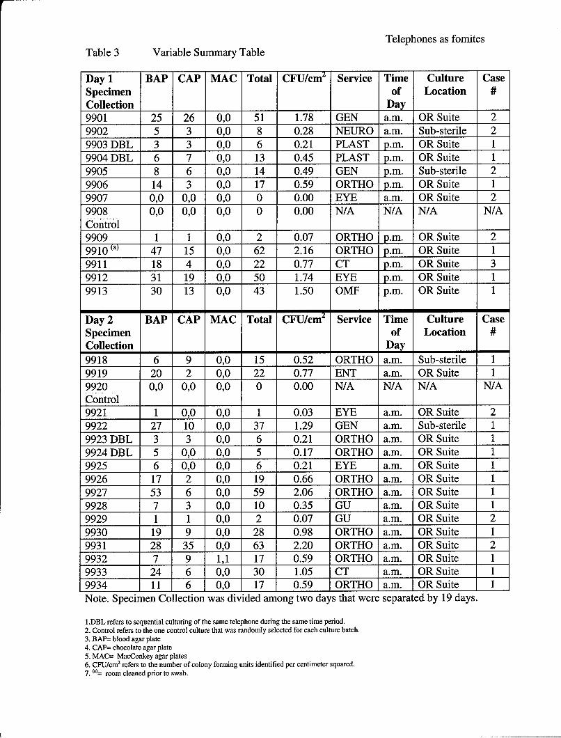
Telephones as fomitesTable 3 Variable Summary Table
Day 1 BAP CAP MAC Total CFU/cm2 Service Time Culture CaseSpecimen of Location #Collection Day9901 25 26 0,0 51 1.78 GEN a.m. OR Suite 29902 5 3 0,0 8 0.28 NEURO a.m. Sub-sterile 29903 DBL 3 3 0,0 6 0.21 PLAST p.m. OR Suite 19904 DBL 6 7 0,0 13 0.45 PLAST p.m. OR Suite 19905 8 6 0,0 14 0.49 GEN p.m. Sub-sterile 29906 14 3 0,0 17 0.59 ORTHO p.m. OR Suite 19907 0,0 0,0 0,0 0 0.00 EYE a.m. OR Suite 29908 0,0 0,0 0,0 0 0.00 N/A N/A N/A N/AControl9909 1 1 0,0 2 0.07 ORTHO p.m. OR Suite 29910,a) 47 15 0,0 62 2.16 ORTHO p.m. OR Suite 19911 18 4 0,0 22 0.77 CT p.m. OR Suite 39912 31 19 0,0 50 1.74 EYE p.m. OR Suite 19913 30 13 0,0 43 1.50 OMF p.m. OR Suite 1
Day 2 BAP CAP MAC Total CFU/cm 2 Service Time Culture CaseSpecimen of Location #Collection Day_9918 6 9 0,0 15 0.52 ORTHO a.m. Sub-sterile 19919 20 2 0,0 22 0.77 ENT a.m. OR Suite 19920 0,0 0,0 0,0 0 0.00 N/A N/A N/A N/AControl9921 1 0,0 0,0 1 0.03 EYE a.m. OR Suite 29922 27 10 0,0 37 1.29 GEN a.m. Sub-sterile 19923 DBL 3 3 0,0 6 0.21 ORTHO a.m. OR Suite 19924 DBL 5 0,0 0,0 5 0.17 ORTHO a.m. OR Suite 19925 6 0,0 0,0 6 0.21 EYE a.m. OR Suite 19926 17 2 0,0 19 0.66 ORTHO a.m. OR Suite 19927 53 6 0,0 59 2.06 ORTHO a.m. OR Suite 19928 7 3 0,0 10 0.35 GU a.m. OR Suite 19929 1 1 0,0 2 0.07 GU a.m. OR Suite 29930 19 9 0,0 28 0.98 ORTHO a.m. OR Suite 19931 28 35 0,0 63 2.20 ORTHO a.m. OR Suite 29932 7 9 1,1 17 0.59 ORTHO a.m. OR Suite 19933 24 6 0,0 30 1.05 CT a.m. OR Suite 19934 11 6 0,0 17 0.59 ORTHO a.m. OR Suite 1Note. Specimen Collection was divided among two days that were separated by 19 days.
1..DBL refers to sequential culturing of the same telephone during the same time period.2. Control refers to the one control culture that was randomly selected for each culture batch.3. BAP= blood agar plate4. CAP= chocolate agar plate5. MAC= MacConkey agar plates6. CFU/cm2 refers to the number of colony forming units identified per centimeter squared.7. (a)= room cleaned prior to swab.