Embed Size (px)
Citation preview
九州大学学術情報リポジトリKyushu University Institutional Repository
Open Pelvic Ring Fracture and MultipleFractures of the Lower Extremities : A CaseReport
Takeuchi, NaohideDepartment of Trauma and Orthopaedic Surgery, Saga-ken Medical Centre Koseikan
Masumoto, KazuyukiDepartment of Plastic and Reconstructive Surgery, Saga-ken Medical Centre Koseikan
Nojiri, JunichiDepartment of Radiology, Saga-ken Medical Centre Koseikan
Akiho, ShunsukeDepartment of Trauma and Orthopaedic Surgery, Saga-ken Medical Centre Koseikan
他
https://doi.org/10.15017/1441361
出版情報:福岡醫學雜誌. 105 (1), pp.22-27, 2014-01-25. 福岡医学会バージョン:published権利関係:
Fukuoka Acta Med. 105(1):22―27,2014
Open Pelvic Ring Fracture and Multiple Fractures
of the Lower Extremities : A Case Report
Naohide TAKEUCHI1), Kazuyuki MASUMOTO2), Junichi NOJIRI3), Shunsuke AKIHO
1),
Shunsuke HOTOKEZAKA1), Kosuke SASAKI1), Kenichi KAWAGUCHI1), Nobuaki TSUKAMOTO1),
Kenta MOMII1), Naohiro FUJITA4) and Takao MAE
1)
1)Department of Trauma and Orthopaedic Surgery, Saga-ken Medical Centre Koseikan, Saga, Japan
2)Department of Plastic and Reconstructive Surgery, Saga-ken Medical Centre Koseikan, Saga, Japan
3)Department of Radiology, Saga-ken Medical Centre Koseikan, Saga, Japan4)Critical Care Center, Saga-ken Medical Centre Koseikan, Saga, Japan
Abstract
A 73-year-old female was hit by a car, and transferred to our hospital. On examination, herconsciousness was alert, but her vital signs were unstable. There are three 10-cm open wounds on herright buttock. X-rays showed an unstable pelvic ring fracture, a right femoral shaft, a right proximaltibia and a right tibial plafond fractures. One hour after the injury, transarterial embolization (TAE)followed by external fixation (EF), and retroperitoneal pelvic packing (RPP) was performed. Two daysand five days after the injury, thorough debridement of the open wounds was performed. The skindefect on the right buttock and the lower abdomen had enlarged to 40 x 35 cm, therefore, negativepressure wound therapy was applied. On the same day, right femur was fixed using a retrogradeintramedullary nailing. 12 days after the injury, the proximal tibial fracture was fixed using a plate, andthe tibial plafond fracture was fixed using screws and external fixators. 28 days after the injury, thesplit-thickness skin graft was performed on the right buttock and the lower abdomen. Seven monthsafter the injury, the open wounds were completely healed without infection. She was able to walksmoothly with a T-cane.For the management of open pelvic ring fractures, it is essential to perform TAE, EF and RPP as
soon as possible. Providing aggressive management, including thorough debridement, is mandatory toprevent severe infection and sepsis. We achieved a good clinical outcome by using a combination ofTAE, EF, RPP and staged surgery, including thorough debridement.
Key words : Open pelvic ring fractures・Transarterial embolization・External fixation・Retroperitoneal pelvic packing
Introduction
Open pelvic ring fractures are one of the most
challenging and life-threatening traumas1)~6). In
open pelvic ring fractures, the pelvic floor is
disrupted, therefore it leads to loss of tamponade
and greater difficulty controlling hemorrhage.
Therefore, the mortality of open pelvic fractures
remains very high due to massive hemorrhage
and sepsis. Reported mortality rates range from
25% to as high as 45%1)~6). We experienced an
unstable open pelvic ring fracture and multiple
fractures of lower extremity. Informed consent
has been obtained from the patient for publica-
tion, including any necessary photographs.
Case report
Case : A 73-year-old female was hit by a car,
and sustained her pelvis and right lower extrem-
ity. On arrival, her consciousness was alert, but
her vital signs were unstable (BP 51/31mmHg,
HR 63 bpm). She had a history of diabetes mellitus
22
Corresponding to : Naohide TAKEUCHI, MD, PhD400 Nakabaru, Kase, Saga City, Saga, 840-8571, JapanTEL : +81-952-24-2171 FAX : +81-952-29-9390E-mail address : takeuchinaohide@gmail. com
and right patella fracture surgery.
On examination, her right pelvis and right thigh
was found to be swollen and deformed. There are
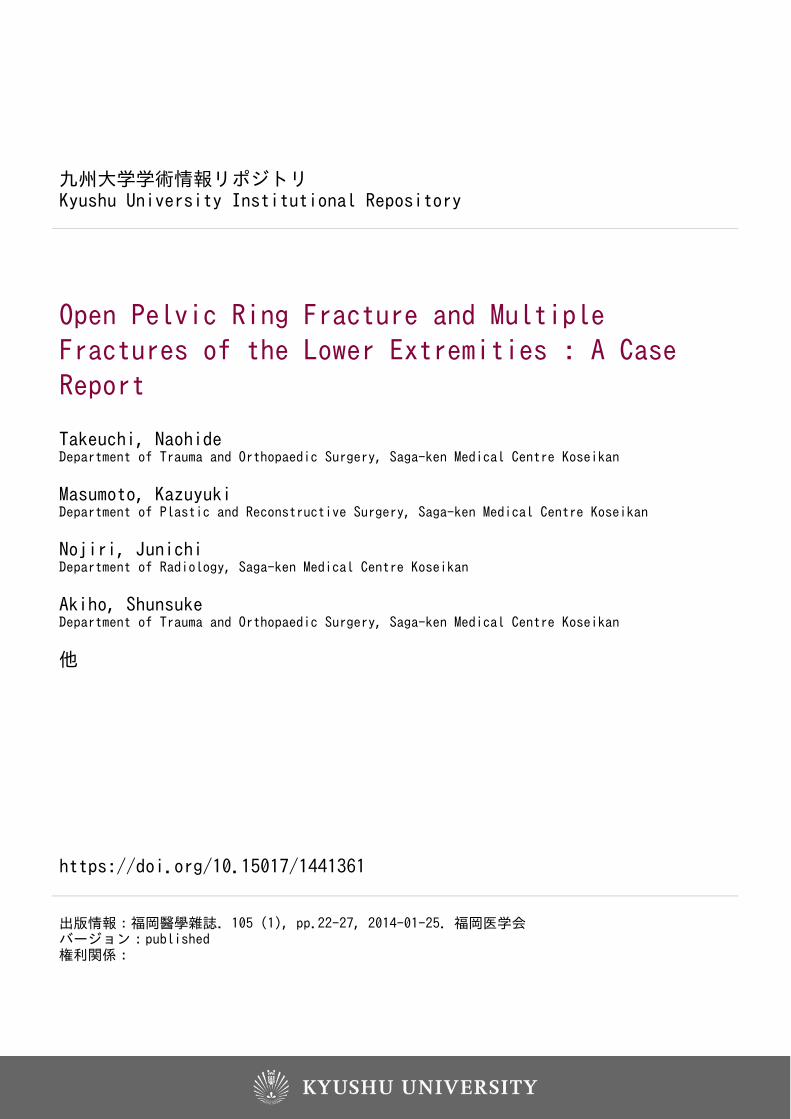
three 10-cm open wounds and degloving injury on
her right buttock (Fig. 1), and a 10-cm open
wound on the right knee and right leg. Chest
X-ray was normal and Focused Assessment with
Sonography for Trauma (FAST) was negative.
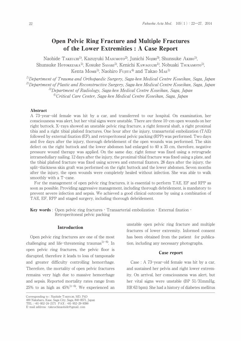
X-rays and CT showed an unstable pelvic ring
fracture including an injury of the left sacroiliac
joint (AO : 61-B2. 2) (Fig. 2a). CT also showed
extravasations and air signs in the pelvis (Fig. 2b).
We diagnosed an open pelvic ring fracture due to
the presence of air signs. The location of
soft-tissue injury was Zone Ⅲ according to the
system described by Faringer4). The injury was a
gradeⅢ A open pelvic ring fracture according to
the classification of Gustilo and Anderson7), and a
class 2 open pelvic ring fracture according to the
Jones-Powell's classification6). The additional
injuries were a right femoral shaft (AO : 32-A2. 3),
a right proximal tibia (AO : 41-B3. 2) and a right
tibial plafond fracture (AO : 43-C2. 3). There was
no rectal or bladder injury. Injury severity score
(ISS) was 26, and revised trauma score (RTS) was
6.38.
One hour after the injury, transarterial embo-
lization (TAE) was performed. Arterial bleeding
was embolized with gelatin. Her blood pressure
was improved (115/81 mmHg). Then, EF was
performed in the operation theater. Before
surgery, her blood pressure was getting worse
(78/55 mmHg), and even after performing EF, her
blood pressure continued to be unstable. There-
fore, retroperitoneal pelvic packing (RPP) was
performed. Surgical laparotomy pads were placed
on each side of the bladder, deep within the
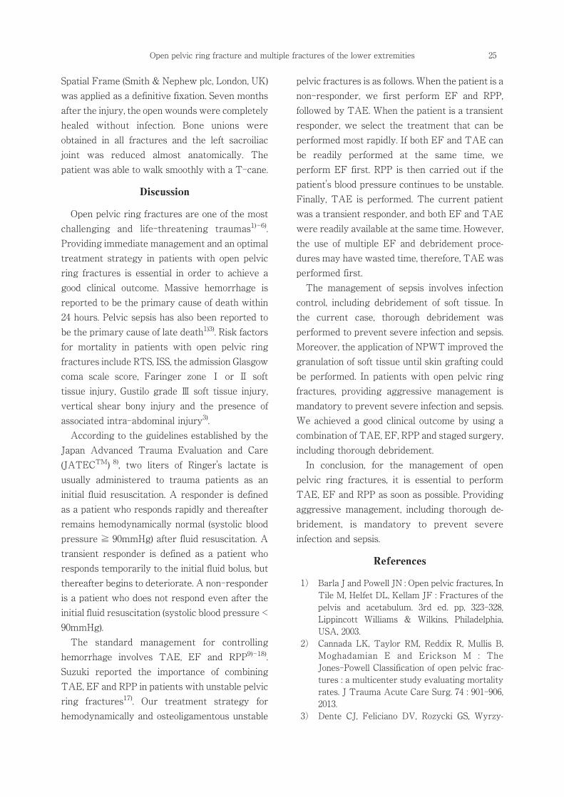
preperitoneal space (Fig. 3). Her blood pressure
was improved (113/59 mmHg). Right femoral and
tibial fractures were also fixed temporally using
external fixators. She received 40 units packed
red blood cells, 35 units fresh frozen plasma, and
30 units platelet transfusion in the first 24 hours.
Two days after the injury, a second-look
surgery was performed. The surgical laparotomy
pads in the pelvis were removed, and thorough
Open pelvic ring fracture and multiple fractures of the lower extremities 23
Fig. 1 There were three 10-cm open wounds anddegloving injury on her right buttock.
Fig. 2 a : A 3D-CT image of the pelvis on the day of admission. The CT showed pubis and ischium fractures and aright iliac fracture.b : A CT image of the pelvis on the day of admission. The axial CT showed an injury of the left sacroiliac jointand air signs around the right ilium.
a
b

debridement of the open wounds on the buttock
was performed. The skin defect on the right
buttock has enlarged to 15 x 15 cm. However, five
days after the injury, the condition of the open
wound was getting worse and infectious (Fig. 4).
Therefore, thorough debridement was performed.
The skin defect has enlarged to 40 x 35 cm (Fig. 5).
On the same day, the right femur was fixed using
a retrograde intramedullary nail (Stryker, Kala-
mazoo, Michigan, USA). Then, negative pressure
wound therapy (NPWT) was performed using the
V.A.C. ATS® system (KCI, San Antonio, Texas,
USA) on the right buttock. Two days after the
second-look surgery, there was no sign of
infection or necrosis on the buttock and lower
abdomen. Therefore, artificial dermis was applied,
and the polyurethane foam of the NPWT was
changed every 48-72 hours.
12 days after the injury, the soft tissues on the
right leg has become necrotic partially, therefore,
debridement of the necrotic soft tissues was
performed. On the same day, the proximal tibial
fracture was fixed using a LCP Medial Proximal
Tibial Plate 3.5 (Synthes GmbH, Oberdorf,
Switzerland), and the tibial plafond fracture was
fixed using screws and external fixators.
28 days after the injury, the split-thickness skin
graft was performed on the right buttock and the
lower abdomen (Fig. 6). For the distal tibial
fracture, skin graft was applied, and a Taylor
N. Takeuchi et al.24
Fig. 3 An X-ray photogragh of the pelvis just aftersurgery on the day of admission. The pelvic ringfracture was fixed using external fixators.Surgical laparotomy pads were placed on eachside of the bladder, deep within the preperi-toneal space.
Fig. 4 The open wound on the right buttock beforesurgery five days after the injury. The condi-tion of the open wound was getting worse andinfectious. The size of the open wound was 15 x15 cm.
Fig. 5 The open wound on the right buttock just aftersurgery five days after the injury. Thoroughdebridement was performed. The skin defecthas enlarged to 40 x 35 cm.
Fig. 6 The split-thickness skin graft was performed onthe right buttock 28 days after the injury.
Spatial Frame (Smith & Nephew plc, London, UK)
was applied as a definitive fixation. Seven months
after the injury, the open wounds were completely
healed without infection. Bone unions were
obtained in all fractures and the left sacroiliac
joint was reduced almost anatomically. The
patient was able to walk smoothly with a T-cane.
Discussion
Open pelvic ring fractures are one of the most
challenging and life-threatening traumas1)~6).
Providing immediate management and an optimal
treatment strategy in patients with open pelvic
ring fractures is essential in order to achieve a
good clinical outcome. Massive hemorrhage is
reported to be the primary cause of death within
24 hours. Pelvic sepsis has also been reported to
be the primary cause of late death1)3). Risk factors
for mortality in patients with open pelvic ring
fractures include RTS, ISS, the admission Glasgow
coma scale score, Faringer zone Ⅰ or Ⅱ soft
tissue injury, Gustilo grade Ⅲ soft tissue injury,
vertical shear bony injury and the presence of
associated intra-abdominal injury3).
According to the guidelines established by the
Japan Advanced Trauma Evaluation and Care
(JATECTM) 8), two liters of Ringer's lactate is
usually administered to trauma patients as an
initial fluid resuscitation. A responder is defined
as a patient who responds rapidly and thereafter
remains hemodynamically normal (systolic blood
pressure≧ 90mmHg) after fluid resuscitation. A
transient responder is defined as a patient who
responds temporarily to the initial fluid bolus, but
thereafter begins to deteriorate. A non-responder
is a patient who does not respond even after the
initial fluid resuscitation (systolic blood pressure <
90mmHg).
The standard management for controlling
hemorrhage involves TAE, EF and RPP9)~18).
Suzuki reported the importance of combining
TAE, EF and RPP in patients with unstable pelvic
ring fractures17). Our treatment strategy for
hemodynamically and osteoligamentous unstable
pelvic fractures is as follows.When the patient is a
non-responder, we first perform EF and RPP,
followed by TAE.When the patient is a transient
responder, we select the treatment that can be
performed most rapidly. If both EF and TAE can
be readily performed at the same time, we
perform EF first. RPP is then carried out if the
patient's blood pressure continues to be unstable.
Finally, TAE is performed. The current patient
was a transient responder, and both EF and TAE
were readily available at the same time. However,
the use of multiple EF and debridement proce-
dures may have wasted time, therefore, TAE was
performed first.
The management of sepsis involves infection
control, including debridement of soft tissue. In
the current case, thorough debridement was
performed to prevent severe infection and sepsis.
Moreover, the application of NPWT improved the
granulation of soft tissue until skin grafting could
be performed. In patients with open pelvic ring
fractures, providing aggressive management is
mandatory to prevent severe infection and sepsis.
We achieved a good clinical outcome by using a
combination of TAE, EF, RPP and staged surgery,
including thorough debridement.
In conclusion, for the management of open
pelvic ring fractures, it is essential to perform
TAE, EF and RPP as soon as possible. Providing
aggressive management, including thorough de-
bridement, is mandatory to prevent severe
infection and sepsis.
References
1) Barla J and Powell JN : Open pelvic fractures, In
Tile M, Helfet DL, Kellam JF : Fractures of the
pelvis and acetabulum. 3rd ed. pp, 323-328,
Lippincott Williams & Wilkins, Philadelphia,
USA, 2003.
2) Cannada LK, Taylor RM, Reddix R, Mullis B,
Moghadamian E and Erickson M : The
Jones-Powell Classification of open pelvic frac-
tures : a multicenter study evaluating mortality
rates. J Trauma Acute Care Surg. 74 : 901-906,
2013.
3) Dente CJ, Feliciano DV, Rozycki GS, Wyrzy-
Open pelvic ring fracture and multiple fractures of the lower extremities 25
kowski AD, Nicholas JM, Salomone JP and
Ingram WL : The outcome of open pelvic
fractures in the modern era. Am J Surg. 190 :
830-835, 2005.
4) Faringer PD, Mullins RJ, Feliciano PD, Duwelius
PJ and Trunkey DD : Selective fecal diversion in
complex open pelvic fractures from blunt
trauma. Arch Surg. 129 : 958-964, 1994.
5) Grotz MR, Allami MK, Harwood P, Pape HC,
Krettek C and Giannoudis OV : Open pelvic
fractures : Epidemiology, current concepts of
management and outcome. Injury. 36 : 1-13,
2005.
6) Jones AL, Powell JN, Kellam JF, McCormack
RG, Dust W and Wimmer P : Open pelvic
fractures : A multicenter retrospective analysis.
Orthop Clin North Am. 28 : 345-350, 1997.
7) Gustilo RB and Anderson JT : Prevention of
infection in the treatment of one thousand and
twenty-five open fractures of long bones :
retrospective and prospective analysis. J Bone
Joint Surg Am. 58 : 453-458, 1976.
8) Yokota J : Japan Advanced Trauma Evaluation
and Care (JATECTM). 4th ed. pp, 45-62, Herusu
Shuppan, Tokyo, Japan, 2012 (in Japanese).
9) Agolini SF, Shah K, Jaffe J, Newcomb J, Rhodes
M and Reed JF 3rd : Arterial embolization is a
rapid and effective technique for controlling
pelvic fracture hemorrhage. J Trauma. 43 :
395-399, 1997.
10) Arvieux C, Thony F, Broux C, Ageron FX,
Rancurel E, Abba J, Faucheron JL, Rambeaud JJ
and Tonetti J : Current management of severe
pelvic and perineal trauma. J Visc Surg. 149 :
227-238, 2012.
11) Burlew CC, Moore EE, Smith WR, Johnson JL,
Biffl WL, Barnett CC and Stahel PF : Preperi-
toneal pelvic packing/external fixation with
secondary angioembolization : optimal care for
life-threatening hemorrhage from unstable
pelvic fractures. J Am Coll Surg. 212 : 628-637,
2011.
12) Cothren CC, Osborn PM, Moore EE, Morgan SJ,
Johnson JL and SmithWR : Preperitoneal pelvic
packing for hemodynamically unstable pelvic
fractures : a paradigm shift. J Trauma. 62 :
834-842, 2007.
13) Flint L, Babikian G, Anders M, Rodriguez J and
Steinberg S : Definitive control of mortality form
severe pelvic fractures. Ann Surg. 211 : 703-706,
1990.
14) Heetveld MJ, Harris I, Schlaphoff G and Sugrue
M : Guidelines for the management of haemody-
namically unstable pelvic fracture patients. Am
J Surg. 74 : 520-529, 2004.
15) Langford JR, Burgess AR, Liporace FA and
Haidukewych GJ : Pelvic fractures : part 1.
Evaluation, classification, and resuscitation. J
Am Acad Orthop Surg. 21 : 448-457, 2013.
16) Smith WR, Moore EE, Osborn P, Agudelo JF,
Morgan SJ, Parekh AA and Cothren C :
Retroperitoneal packing as a resuscitation
technique for hemodynamically unstable pelvic
fractures : report of two cases and description of
technique. J Trauma. 59 : 1510-1514, 2005.
17) Suzuki T, Smith WR and Moore EE : Pelvic
packing or angiography : competitive and com-
plementary? Injury. 40 : 343-353, 2009.
18) Velmahos GC, Toutouzas KG, Vassiliu P,
Sarkisyan G, Chan LS, Hanks SH, Berne TV and
Demetriades D : A prospective study on the
safety and efficacy of angiographic embolization
for pelvic and visceral injuries. J Trauma. 53 :
303-308, 2002.
(Received for publication November 8, 2013)
N. Takeuchi et al.26
(和文抄録)
骨盤輪開放骨折を伴った多発骨折の一例
1)佐賀県医療センター好生館 外傷センター・整形外科2)佐賀県医療センター好生館 形成外科3)佐賀県医療センター好生館 放射線科
4)佐賀県医療センター好生館 救命救急センター
竹 内 直 英1),増 本 和 之2),野 尻 淳 一3),秋 穂 俊 輔1),佛 坂 俊 輔1),佐々木宏介1),
川 口 謙 一1),塚 本 伸 章1),籾 井 健 太1),藤 田 尚 宏4),前 隆 男1)
我々は,骨盤輪開放骨折を伴った多発骨折の一例を経験したので報告する .
症例:73歳女性.歩行中に乗用車にはねられ受傷した.初診時,意識清明であったが,ショック
状態を認めた(血圧:51/31 mmHg,脈拍:63回/分).骨盤と右下肢に腫脹・変形と,右殿部に約
10cm の開放創を 3ヶ所認めた.単純 X線撮影・CT にて,不安定型骨盤輪開放骨折(AO分類:
61-B2.2,Gustilo分類:gradeⅢ A),右大腿骨骨幹部骨折(AO分類:32-A2.3),右脛骨近位端骨
折(AO分類:41-B3.2),右脛骨天蓋開放骨折(AO分類:43-C2.3)と診断した.搬送 1時間後に
経カテーテル的動脈塞栓術(TAE)を施行し,続いて骨盤創外固定術と後腹膜ガーゼパッキングを
施行した.血圧は 113/59 mmHgと改善した.さらに,右大腿骨・脛骨骨折に対して創外固定術を
行った.受傷 2 日目に右殿部開放創のデブリドメント・局所陰圧吸引処置を開始した.受傷 5 日目
に開放創の皮膚壊死を認めたため,デブリドメントを追加した.皮膚欠損は 40 x 35cmとなった.
同日大腿骨骨幹部骨折に対して逆行性髄内釘固定術を施行した.受傷 12 日目に,右脛骨近位端骨
折に対して plate固定術を,右脛骨天蓋開放骨折に対して screw固定と創外固定術を施行した.受
傷 28日目に,右殿部・下腹部の開放創に対して分層植皮術を施行した.受傷 7ヶ月後,開放創は感
染を合併せず治癒し,杖歩行可能となった.
骨盤輪開放骨折は大量出血や感染・敗血症を合併することが多く,死亡率の高い外傷の一つであ
る.大量出血に対しては,TAE・創外固定術・後腹膜ガーゼパッキングを可及的早期に行うことが
必須である.また,開放創の積極的なデブリドメントが感染・敗血症の予防として重要である.本
症例では,TAE・創外固定術・後腹膜ガーゼパッキングと段階的な手術により良好な治療結果を得
ることができた.
Open pelvic ring fracture and multiple fractures of the lower extremities 27