Embed Size (px)
Citation preview
UNITED NATIONS
RC UNEPFAORCCOP4INF16
K0842141 101008
For reasons of economy this document is printed in a limited number Delegates are kindly requested to bring their copies to meetings and not to request additional copies
Rotterdam Convention on the Prior Informed Consent Procedure for Certain Hazardous Chemicals and Pesticides in International Trade Conference of the Parties Fourth meeting Rome 27ndash31 October 2008 Item 5 (e) of the provisional agenda
Implementation of the Convention consideration of a chemical for inclusion in Annex III of the Convention chrysotile asbestos
Report of the World Health Organization workshop on mechanisms of fibre carcinogenesis and assessment of chrysotile asbestos substitutes
Note by the Secretariat The annex to the present note contains the full report of the World Health Organization
workshop on mechanisms of fibre carcinogenesis and assessment of chrysotile asbestos substitutes which was held in Lyon France from 8 to 12 November 2005 A summary consensus report was made available at the third meeting of the Conference of the Parties The full report is presented as received and has not been formally edited by the Secretariat
lowast UNEPFAORCCOP41
United Nations Environment Programme Food and Agriculture Organization of the United Nations
Distr General 9 October 2008 English only
WHO Workshop on Mechanisms of Fibre Carcinogenesis and
Assessment of Chrysotile Asbestos Substitutes
8ndash12 November 2005
Lyon France
Contents Letter from the Secretariat of the Rotterdam Convention to WHO Report of the contact group on chrysotile List of participants List of acronyms Introduction Part 1 Invited Papers on Mechanisms of Fibre Carcinogenesis 11 Uses of chrysotile asbestos and examples of substitutes 12 Human exposure to substitute fibres 13 General physical and chemical characteristics of fibres relevant for their
carcinogenicity 14 Deposition pattern clearance biopersistence changes of fibre characteristics in situ
definition of fibre dose 15 In vitro toxicity tests including genotoxicity and oxygen radical generation 16 Short-term animal tests 17 Dosendashresponse considerations Part 2 Workshop Report 21 General principles for the evaluation of chrysotile asbestos substitutes
211 Key considerations for evaluating epidemiological evidence 212 Significance of short- and long-term studies in experimental animals in the
assessment of the potential hazards of fibres to human health 213 Use of in vitro short-term tests in assessing fibre carcinogenicity 214 Physicochemical properties and biopersistence
22 Assessment of selected chrysotile asbestos substitutes 221 Aramid and para-aramid 222 Attapulgite 223 Carbon fibres 224 Cellulose fibres 225 Graphite whiskers 226 Magnesium sulfate whiskers 227 Polyethylene fibres 228 Polypropylene fibres 229 Polyvinyl alcohol fibres 2210 Polyvinyl chloride fibres 2211 Potassium octatitanate fibres 2212 Synthetic vitreous fibres 2213 Wollastonite
Workshop on Fibre Carcinogenesis and Chrysotile Asbestos Substitutes
Unedited Advance Copy 3
2214 Xonotlite 23 Summary consensus report
231 Introduction 232 Part 1 Methodological aspects 233 Part 2 Hazard assessment
Secretariat for the Rotterdam Convention on thePrior Informed Consent Procedure for Certain Hazardous
Chemicals and Pesticides in International Trade UNEP
ChemicalsUnited Nations Environment Programme
(UNEP)
Plant Protection ServicePlant Production and Protection DivisionFood and Agriculture Organization of the UnitedNations (FAO)
Viale delle Temle di Carncalla00100 Rome Italy
Tel (+3906) 5705 3441Fax (+3906) 5705 6347E-mail picfaoorg
11-13 Chemin des AnemonesCH -1219 ChAtelaine Geneva Switzerland
Tel (+4122)917 8183Fax (+4122) 7973460
E-mail piCunepch
Geneva 25 March 2004
Subject Chrysotile asbestos -assessment of alternatives
Dear Dr Meredith
I refer to the request of the Intergovernmental Negotiation Committee at its tenthsession to the World Health Organisation to conduct an assessment of alternatives to chrysotileasbestos At this meeting the WHO agreed that such an assessment would be able to beconducted however requested that the fifth session of the Interim Chemical Review Committeefor the Rotterdam Convention would consider the alternatives proposed by governments anddevelop a priority risk The Interim Chemicals Review Committee considered the alternativesproposed by governments and developed a priority list for consideration by the WHO They alsodeveloped a list of additional alternatives which were prioritised
These alternatives are identified in the attached document which is an extract from thereport of the Interim Chemical Review Committee I therefore invite the WHO to proceed withthe agreed assessment of the proposed alternatives to chrysotile asbestos If possible it would beappreciated if an update on the progress of the assessment could be provided to theIntergovernmental Negotiating Committee at its eleventh session
Please do not hesitate to contact us should you have any questions in regard to thisletter
Yours sincerely
Address Dr T MeredithCoordinatorIPCS World Health OrganizationCH-1211 Geneva 27 -Switzerland
cc Ms Carolyn Vickers
1
Annex I
Report of the contact group on chrysotile
1 The contact group considered the list of substitutes for chrysotile asbestos proposed byGovernments for assessment by the World Health Organization (WHO) WHO indicated that itwelcomed the guidance provided by the group on important alternatives used by Governments
2 The list was prioritized initially on the basis of the number of Governments which hadnominated the substances Information on which substances had previously been assessed inenvironmental health criteria reports by IPCS was also considered Where possible the groupsknowledge of important uses was also considered
3 The first group of substances are listed on a priority basis in the order in which the contactgroup would like them to be considered by WHO The second group of substances which wereproposed by only one country had undergone no previous assessment by WHO and could beconsidered if resources allowed
Group 1 Substances identified and prioritized for assessment by WHO
Group 2 Substances identified as alternatives to chrysotile to be assessed if resources allow
Aluminium silicates basic magnesium sulphate whisker erionite ductile iron mica phosphatepolyacryl nitryl polytetrafluoroethylene potassium titanate whisker semi-metallics siliconcarbide whisker steel fibres
2
World Health Organization
Workshop on Mechanisms of Fibre Carcinogenesis and Assessment of Chrysotile Asbestos Substitutes
Lyon 8-12 November 2005
LLIISSTT OOFF PPAARRTTIICCIIPPAANNTTSS
Members11
1 Working Group Members and Invited Specialists serve in their individual capacities as scientists and not as representatives of their government or any organization with which they are affiliated Affiliations are provided for identification purposes only
Elke Dopp University Hospital Essen Institute of Hygiene and Occupational Medicine Hufelandstrasse 55 D-45122 Essen Germany Bice Fubini University of Torino Interdepartmental Center lsquoG Scansettirsquo for Studies on Asbestos and other Toxic Particulates Via P Giuria 7 I-10125 Torino Italy Andrea Hartwig Technische Universitaumlt Berlin Fak III ndash Institut fuumlr Lebensmitteltechnologie und Lebensmittelchemie Sekr TIB 43-1 Gustav-Meyer-Allee 25 D-13355 Berlin Germany F Javier Huertas CSIC Estacion Experimental del Zaidin Department of Earth Sciences and Environmental Chemistry Prof Albareda 1 E-18008 Granada Spain Marie-Claude Jaurand Institut National de la Santeacute et de la Recherche
Meacutedicale (INSERM) Geacutenomique fonctionelle des tumeurs solides
U674 - IFR 105 ndash CEPH ndash IUH 27 rue Juliette Dodu F-75010 Paris France Aparna M Koppikar United States Environmental Protection Agency National Center for Environmental Assessment 1200 Pennsylvania Ave NW Washington DC 20460 USA Dr Yasuo Morimoto Department of Occupational Pneumology University of Occupational amp Environmental
Health 1-1 Iseigaoka Yahatanishiku Kitakyushu City 807-8555 Japan Paul A Schulte National Institute for Occupational Safety and Health (NIOSH)CDC Robert A Taft Laboratories 4676 Columbia Parkway Cincinnati OH 45226-1998 USA Leslie Stayner University of Illinois at Chicago School of Public Health (MC 923) 1603 West Taylor Street Room 971 Chicago IL 60612 USA Peter Wardenbach
2
Federal Institute for Occupational Safety and Health (BAuA) Friedrich-Henkel-Weg 1-25 D-44149 Dortmund Germany Invited specialists2 Suresh Moolgavkar3 Fred Hutchinson Cancer Research Center Division of Public Health Sciences Fairview Avenue North M2-B500 PO Box 19024 Seattle Washington DC 98109-1024 USA Hartwig Muhle4 Fraunhofer Institute of Toxicology and Experimental Medicine Nikolai-Fuchs-Strasse 1 D-30625 Hannover Germany Jay Turim5 Sciences International Inc King Street Station 1800 Diagonal Road Suite 500 Alexandria VA 22314 USA Observers Observer for the American Forest amp Paper Association Inc (AFPA)
2NOTE Minor pertinent interests are not listed Examples of minor interests include stock valued at no more than US$10 000 overall research grants that provide no more than 5 of the unitrsquos research budget and consulting and speaking engagements on matters not related to courts or government agencies that do not exceed 2 of time or compensation All consulting or speaking on matters before a court or government agency is listed as a significant pertinent interest 3Is advising a law firm that is representing a corporation with asbestos interests 4 The Fraunhofer Institute is doing contract research for many companies that produce glass wool rock wool or ceramic fibres 5 Is advising a law firm that is representing a corporation with asbestos interests Received research support from the Refractory Ceramic Fibers Coalition an association of producers of refractory ceramic fibres
David Bernstein6 40 chemin de la Petite-Boissiegravere CH-1208 Geneva Switzerland Observer for the European Ceramic Fibres Industry Association (ECFIA) RC Brown7 6 Stocken Hall Mews GB-Stretton LE15 7RL United Kingdom Observer for Health Canada Government of Canada Michel Camus University of Montreal 3875 Rue Saint-Urbain Montreacuteal Queacutebec H2W 1V1 Canada Observer for the Institute of Defense of National Property Brazil Carlos Crespo Avenida 18 No 149 Rio Claro Satildeo Paolo Brazil Observer for NYCO Minerals Inc Christophe de Bellefroid8 NYCO Minerals Inc 45 av de Cicircteaux B-1348 Louvain-la-Neuve Belgium Observer for the Belgian Ministry of Health Freacutedeacuteric Denauw Direction geacuteneacuterale environnement ndash Maicirctrise des risques Place Victor Horta 40 Boicircte 10 B B-1060 Brussels Belgium Observer for the US EPA IRIS Program
6 Consultant to the American forest and paper industry Received research support from the chrysotile industry 7 Consultant to ECFIA Partner owns shares in the Morgan Crucible Company a producer of ceramic fibres 8 Employed by NYCO Minerals a producer of wollastonite
3
Danielle DeVoney United States Environmental Protection Agency National Center for Environmental Assessment 1200 Pennsylvania Avenue NW 8601D Washington DC 20460 USA Observer for Sama Mineraccedilatildeo de Amianto Ltda Milton do Nascimento9 Sama Mineraccedilatildeo de Amianto Ltda Rua Dr Fernandes Coelho 85 2o andar Satildeo Paolo 05404-014 Brazil Observer for the Chrysotile Institute Jacques Dunnigan10 380 Chemin de North-Hatley PO Box 123 Ste-Catherine-de-Hatley QC J0B 1W0 Canada Observer for lrsquoAgence Franccedilaise de Seacutecutiteacute sanitaire de lrsquoenvironnement et du travail (AFSSET) Anne-Marie Fillet AFSSET 27-31 Avenue du Geacuteneacuteral Leclerc F-94704 Maisons Alfort France Observer for International Ban Asbestos (IBAS) Morris Greenberg11 14 North End Road GB-London NW11 7SY United Kingdom Observer for the North American Insulation Manufacturers Association ( NAIMA) John Hadley12 Owens Corning Science and Technology Center Granville Ohio USA
9 Employed by Sama Mineraccedilatildeo de Amianto a producer of asbestos 10 Consultant to the chrysotile industry 11 Has advised plaintiffs seeking compensation for disease related to asbestos or mineral fibres May receive travel support for this meeting from IBAS 12 Employed by Owens Corning a producer of glass fibres
Observer for the Swiss Agency for the Environment Forests and Landscape Bettina Hitzfeld Designated National Authority Rotterdam Convention Swiss Agency for the Environment Forests and Landscape SAEFL Substances Soil Biotechnology Division CH-3003 Berne Switzerland Observer for Future Pipe Industries Llc Mustafa Kabbara13 Future Pipe Industries Llc PO Box 1371 Dubai United Arab Emirates Observer for the Research Institute of Occupational Health of the Russian Academy of Medical Science Evgeny Kovalevskiy State Run Organization Research Institute of Occupational Health Russian Academy of Medical Science The WHO Collaborating Center in Occupational Health 31 Prospect Budennogo 105275 Moscow Russian Federation Observer for the Russian Register of Potentially Hazardous Chemicals Boris Kurlyandskiy 1820 Vadkovskiy per 127994 Moscow Russian Federation Observer for the French Ministry of Health Claude Lambreacute Ministegravere de la Santeacute DGSCAS 14 avenue Duquesne F-75350 Paris SP07 France Observer for the Secretariat of the Rotterdam Convention 13 Employed by the Future Pipe Company a producer of chrysotile cement pipe
4
Sheila Logan Secretariat of the Rotterdam Convention United Nations Environmental Program 11-13 Chemin des Anemones Chatelaine 1219 Geneva Switzerland Observer for Refractory Ceramic Fibers Coalition (RCFC) Daniel Maxim14 15 N Main Street Cranbury NJ 08512 USA Observer for the European Tissue Symposium (ETS) and the Confederation of the European Paper Industry (CEPI) Anthony S Panepinto Procter and Gamble 5299 Spring Grove Avenue Cincinnati OH 45217 USA Observer for the Austrian Federal Ministry for Economic Affairs and Labour Labourinspection Reinhild Puumlrgy Federal Ministry for Economic Affairs and Labour Favoritenstrasse 7 A-1040 Wien Austria Observer for the Finnish Ministry of Social Affairs and Health Antti Tossavainen Finnish Institute of Occupational Health Topeliuksenkatu 41 SF-00250 Helsinki Finland Observer for the International Federation of Building and Wood Workers Lars Vedsmand BAT-Kartellet Kampmannsgade 4 DK-1790 Copenhagen V Denmark 14 Employed by Everest Consultants Inc Consultant to RCFC ECFIA NAIMA and NYCO Minerals
Observer for lrsquoAgence Franccedilaise de Seacutecutiteacute sanitaire de lrsquoenvironnement et du travail (AFSSET) Antoine Villa AFSSET 27-31 Avenue du Geacuteneacuteral Leclerc F-94704 Maisons Alfort France WHO Secretariat Antero Aitio Robert Baan Vincent Cogliano Fatiha El Ghissassi Yann Grosse Beacuteatrice Secretan Kurt Straif Carolyn Vickers Administrative Assistance Sandrine Egraz Helene Lorenzen-Augros
Workshop on Fibre Carcinogenesis and Chrysotile Asbestos Substitutes
Unedited Advance Copy 7
List of acronyms 8-OH-dG 8-hydroxydeoxyguanosine 8-oxo-dG 8-oxo-78-dihydro-2prime-deoxyguanosine AM alveolar macrophages ATP adenosine triphosphate ATPase adenosine triphosphatase BAL bronchoalveolar lavage BET Brunauer Emmett and Teller BHT biological half-time BrdU 5-bromo-2-deoxyuridine CFE colony forming efficiency CI confidence interval CMS calcium magnesium silicate CMZS calcium magnesium zirconium silicate DMPO-OH 55-dimethyl-1-pyrroline N-oxidendashhydroxyl radical DNA deoxyribonucleic acid DNase deoxyribonuclease EC50 median effective concentration ELISA enzyme-linked immunosorbent assay EPR electron paramagnetic resonance FISH fluorescence in situ hybridization GLP Good Laboratory Practice GMD geometric mean diameter GML geometric mean length GSD geometric standard deviation GSH reduced glutathione GSSG oxidized glutathione h-AM alveolar macrophages from humans h-RBC human red blood cells HTE hamster tracheal epithelial h-TII type II pneumocytes from humans IARC International Agency for Research on Cancer IgA immunoglobulin A IL-1 interleukin-1 INC Intergovernmental Negotiating Committee kdis dissolution rate constant L lung LDH lactate dehydrogenase LN lymph nodes MMVF man-made vitreous fibres mRNA messenger ribonucleic acid MTT 3-(45-dimethylthiazol-2-yl)-25-diphenyltetrazolium bromide mvt NIOSH National Institute of Occupational Safety and Health ODC ornithine decarboxylase P particles PAN polyacrylonitrile PCOM phase-contrast optical microscopy PL pleural lavage
Workshop on Fibre Carcinogenesis and Chrysotile Asbestos Substitutes
Unedited Advance Copy 8
PLM polarized light microscopy PMN polymorphonuclear leukocyte PVA polyvinyl alcohol PVC polyvinyl chloride PVC-E PVC emulsion process PVC-S PVC suspension process r-AM alveolar macrophages from rats RCC Research and Consulting Company RCF refractory ceramic fibre RFP respirable-sized fibre-shaped particulates RNA ribonucleic acid RNase ribonuclease RNS reactive nitrogen species ROS reactive oxygen species RSNO nitrosothiol r-TII type II pneumocytes from rats SD standard deviation SDH succinate dehydrogenase SEM scanning electron microscopy SHE Syrian hamster embryo SIR standardized incidence ratio SMR standardized mortality ratio SOD superoxide dismutase TEM transmission electron microscopy TGF transforming growth factor TII type II pneumocytes TLC thin-layer chromatography TNF-α tumour necrosis factor alpha TPA 12-O-tetradecanoyl-phorbol-13-acetate UDS unscheduled DNA synthesis UK United Kingdom USA United States of America VC vinyl chloride VCM vinyl chloride monomer WHO World Health Organization
Part 1
Invited Papers on Mechanisms of Fibre Carcinogenesis
These papers are currently being finalized They were prepared as background for the workshop and are the opinions of the individual authors and not the workshop
Part 2
Workshop Report
This is an unedited advance version of the meeting report being made available for the purpose of providing information to the Conference of the Parties to the Rotterdam Convention
Workshop on Fibre Carcinogenesis and Chrysotile Asbestos Substitutes
Unedited Advance Copy 11
21 General principles for the evaluation of chrysotile asbestos substitutes
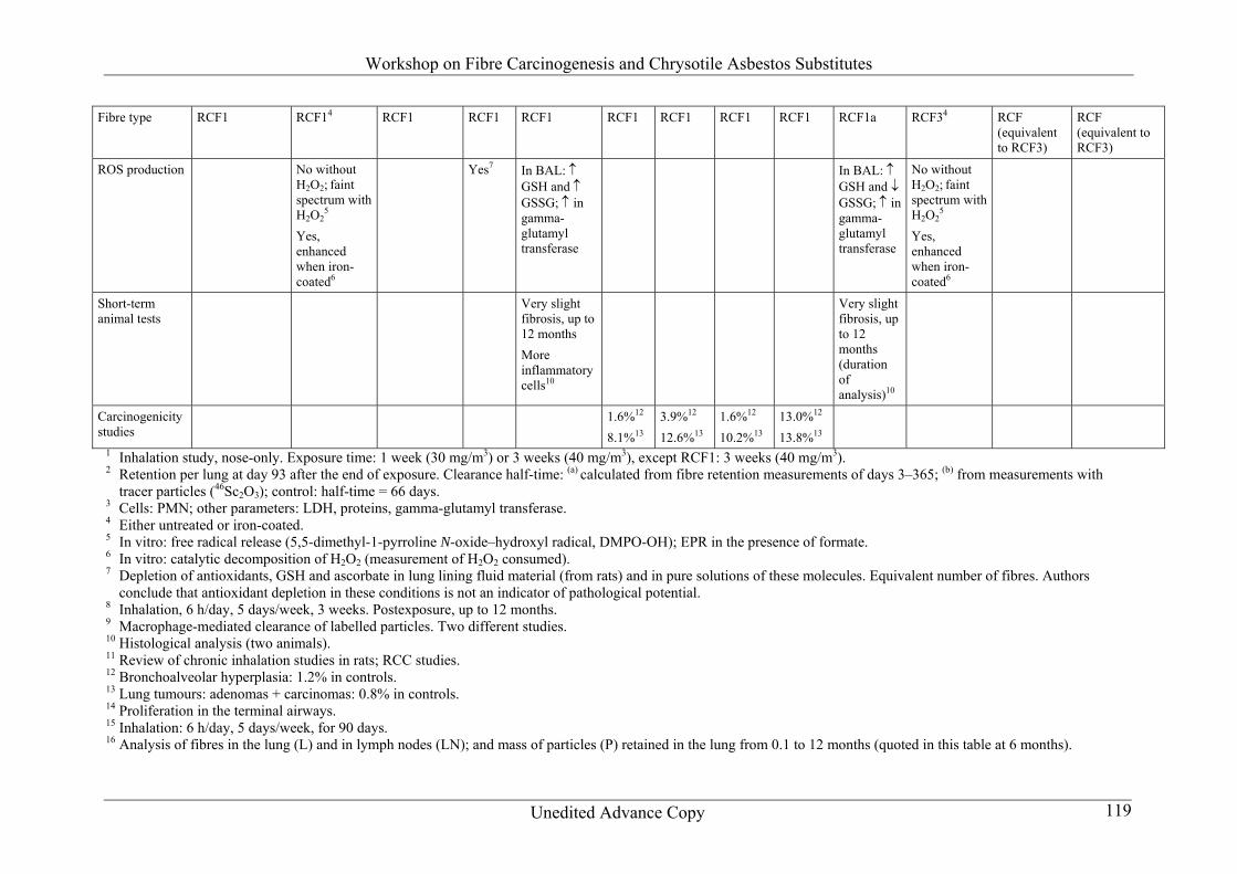
211 Key considerations for evaluating epidemiological evidence Epidemiological studies on fibres have a clear advantage over toxicological studies in that they involve studies of humans They also have the advantage that they study the effects of exposure in the real world where the effects of these exposures may be mitigated or enhanced by other factors For example there is a well recognized synergistic interaction between smoking and asbestos (Vainio amp Boffetta 1994) Despite these obvious advantages the presence or absence of evidence of a risk from epidemiological studies does not always override contrary findings from toxicological studies for a variety of reasons It is frequently difficult to establish causality based solely on epidemiology because of the non-experimental nature of these studies The lack of randomization in epidemiological studies makes it difficult to fully exclude the possibility that confounding or other forms of bias may be responsible for a positive association found in an epidemiological study Negativemdashor what should perhaps be more appropriately referred to as ldquonon-positiverdquomdashstudies rarely if ever provide sufficient evidence for rejecting causality The lack of positive evidence might be explained by limitations of the study design For example for recently introduced fibres studies conducted to evaluate the risk of lung cancer would clearly have inadequate follow-up time and would thus be uninformative Non-positive studies may still be highly informative particularly when they are studies that have adequate statistical power to detect an effect have good characterization of exposures and are unlikely to have substantial potential for confounding or other biases At the very least a well conducted study with a non-positive finding may be used to estimate an upper bound on what risk might be plausible Because of these considerations the interpretation of either positive or non-positive epidemiological findings needs to be carefully considered in light of the strengths or weaknesses of the study design Following is a discussion of some of the key issues that need to be considered particularly with regard to the interpretation of the findings from epidemiological studies of fibres 2111 Statistical power Although well designed epidemiological studies provide the most relevant information for the assessment of risks in human populations the ability of these studies to detect an effect when it is present generally referred to as the power of the study depends on the size of the effect the size of the study and for effects with long latent periods the duration of the study The lessons learnt from occupational cohort studies of asbestos workers have led to much lower fibre exposures in the workplace so that one would expect to detect cancer risks only in large cohorts followed up for a sufficiently long period of time This fact should be kept in mind in the interpretation of occupational cohort studies that do not report increased risks of lung cancer or mesothelioma which is primarily the case for the fibres that are reviewed in this document These studies should be interpreted not as being negative but rather as being non-positive The statistical power of these studies to detect an effect is a critical consideration when interpreting the degree to which the lack of evidence for a carcinogenic effect is
Workshop on Fibre Carcinogenesis and Chrysotile Asbestos Substitutes
Unedited Advance Copy 12
persuasive or not A key example of this concern is the study of refractory ceramic fibre (RCF) workers which is a relatively small cohort (942 workers) that is young (mean age = 51 years) and has been followed for a relatively short period of time (mean length of follow-up of 21 years) (LeMasters 2003) The degree to which this study had adequate power to detect an excess of lung cancer or mesothelioma is a concern which is discussed in section 22121 2112 Adequacy of exposure assessment To estimate exposure usually environmental dust samples are collected In work environments personal monitoring is accomplished by mounting a sampler on a worker in the breathing zone Misclassification of exposure in epidemiological studies of fibres as in all epidemiological studies is potentially a serious problem Errors resulting from misclassification of exposures generally may bias exposurendashresponse relationships Samples are analysed for the number of fibres by different counting methods such as phase-contrast optical microscopy (PCOM) scanning electron microscopy (SEM) polarized light microscopy (PLM) and transmission electron microscopy (TEM) For counting purposes a fibre is defined as a particle gt5 microm in length with a length to diameter ratio of at least 31 and a diameter of lt3 microm this type of fibre is often referred as a World Health Organization (WHO) fibre (WHO 1996) According to National Institute of Occupational Safety and Health (NIOSH) counting rule ldquoBrdquo a fibre is defined as any particle gt5 microm in length with a length to diameter ratio of at least 51 and a diameter of lt3 microm The difference in count between the WHO fibre and the NIOSH counting rule ldquoBrdquo fibre can be significant (Breysse et al 1999) The exposure estimates for individual studies may differ based on which counting rule is used to count fibres Thus it is important to consider these differences in exposure estimates when comparing studies All counting methods (ie TEM PCOM SEM PLM) are unable to distinguish individual organic fibres so these are usually categorized as ldquoother organicrdquo fibres which include polyvinyl alcohol (PVA) fibres cellulose fibres and other textile fibres (clothing bed sheets blankets pillows) In occupational settings exposures are mainly estimated and can be supported by industrial hygiene data The exposure estimations can be based on personal samples area samples job matrices or processes involved Most of the time the samples are dust samples and respirable fibres are part of the samples Thus when epidemiological studies are evaluated the confounding effect of other exposures such as dust particles and other chemicals should be taken into account Most fibre studies indicate that exposures of installers and removal workers are much higher than exposures of workers involved in manufacturing and processing the same fibres Many times direct information about exposure may not be available but biomarkers of exposure if present are a helpful indication of relevant exposure (eg pleural plaques in asbestos exposure) In evaluating carcinogenicity of any substance in humans exposure informationassessment is usually the weakest link and fibres are no exception to this rule 2113 Confounding and multiple exposures Confounding in epidemiological studies is a bias that occurs when the exposed and the non-exposed groups have different disease risks even if they were not exposed to fibres (Pierce amp
Workshop on Fibre Carcinogenesis and Chrysotile Asbestos Substitutes
Unedited Advance Copy 13
Greenland 2004) More generally confounding may occur when the groups to be compared (exposed and non-exposed) are not completely comparable Confounding in studies of fibre-induced lung cancer could be the result of various factors such as cigarette smoking socioeconomic status or exposure to fibres particles or other substances not being studied Confounding can bias an effect in either direction thus leading to the observation of higher or lower risk Confounding can be controlled in the study design analysis or both by means of randomization restriction and matching in the design phase and by stratification or statistical adjustment in the analysis plan Mixed exposures can be taken to mean that a person is exposed to the fibre in question as well as to other fibres particles or substances that produce the same effect It may be that the situation of mixed exposures cannot be treated as a classical problem of confounding because exposure to fibres of concern may be highly correlated with exposures to other fibres particles and substances (Cordier amp Stewart 2004) Stratified analysis and multivariate modelling may not be effective in adverse situations with high colinearity among exposures 212 Significance of short- and long-term studies in experimental animals in the assessment of the potential hazards of fibres to human health End-points in humans that are covered are the long-term effects notably cancer and fibrosis Effects such as skin irritation and byssinosis nylon flock workerrsquos lung are not considered The major route of exposure in humans is inhalation Oral exposure falls outside the scope of this document 2121 Short-term animal experiments
Methodology Fibre dimensions of the aerosol should approach as closely as possible the fibre dimensions encountered in human exposures However as respirability which is mainly dependent on fibre diameter differs between rats and humans special attention should be given to fibre length It makes little sense to increase artificially the fibre length of the aerosol in animal inhalation studies if humans are exposed mainly to much shorter fibres Lung burdennumber and fibre dimensions should be determined at appropriate time points during and after exposure This indicates whether exposure concentrations have been adequately chosen and it gives an impression of the behaviour (disappearance breakage) of the fibres in the lung The adequacy of the study design and performance should be verified by the use of a positive control In studies on fibrosis and inflammation it is important that a recovery group be part of the design
Workshop on Fibre Carcinogenesis and Chrysotile Asbestos Substitutes
Unedited Advance Copy 14
Effects Many end-points have been measured These yield supportive information vis-agrave-vis toxic effects (lung weight bronchoalveolar lavage [BAL]) but the discussion here is limited to those that may have a relation to carcinogenicity or fibrosis Fibrosis (as demonstrated histopathologically in animal experiments) can be determined if exposure is sufficiently long (90 days) especially when a recovery group is examined Fibrosis has not been demonstrated to be a prerequisite to carcinogenesis Epithelial cell proliferation is a repair mechanism after tissue injury but proliferation of target cells for carcinogenesis above background levels can be regarded as an essential step in carcinogenesis as it increases the probability of mutated cell populations Of special concern is increased cell proliferation if it persists after the end of exposure A relationship of epithelial cell proliferation with cancer in experimental animals could be demonstrated for amosite crocidolite MMVF21 and special-purpose glass fibres E and 475 After intraperitoneal injection of fibres proliferative changes might also be studied in peritoneal cells which are the origin of mesothelioma The available data show that proliferation of mesothelial cells is greater with fibrous than with granular dust Analysis of BAL indicates the extent of lung inflammation It is supportive information on fibrosis especially related to effects in silica exposure but less clear-cut in the case of fibres Histopathology of the lung can indicate a carcinogenic potential (especially metaplasia) 2122 Long-term animal experiments (inhalation) The primary aim is to detect fibrosis andor carcinogenic response The predominant tumour type in rats after exposure to certain fibres has been lung cancer but after exposure to amphibole asbestos a low incidence of mesothelioma has also been reported in rats
Methodology This concerns both inhalation and intraperitoneal studies in rats Fibre dimensions of the aerosol should approach as closely as possible the fibre dimensions encountered in human exposures However as respirability which is mainly dependent on fibre diameter differs between rats and humans special attention should be given to fibre length It makes little sense to increase artificially the fibre length of the aerosol in animal inhalation studies if humans are exposed mainly to much shorter fibres Lung burdennumber and fibre dimensions should be determined at appropriate time points during and after exposure This indicates whether exposure concentrations have been adequately chosen and it gives an impression of the behaviours (disappearance breakage) of the fibres in the lung
Workshop on Fibre Carcinogenesis and Chrysotile Asbestos Substitutes
Unedited Advance Copy 15
The adequacy of the study design and performance should be verified by the use of a positive control Exposure concentrations of long-term inhalation studies in rats should be high enough to make it possible to exclude with some confidence a carcinogenic potency similar to the potency of amphibole asbestos Taking into account the existing chronic inhalation data with amphibole asbestos not respirable fibre concentration should at least amount to 1000 fibresml with length gt5 microm 200 fibresml with length gt200 microm and 100 fibresml with length gt20 microm These concentrations represent the lowest aerosol concentrations of amphibole asbestos in rat inhalation studies that resulted in increased incidence in lung tumours If such a minimum exposure concentration is not applied in a chronic rat inhalation study the validity of non-positive results is highly questionable If for various reasons it is not possible to achieve the desired concentrations (eg high mass concentration because fibres are rather thick improved sizing was not successful) exposure of experimental animals by intratracheal or intraperitoneal injection should be performed (Information on the carcinogenicity in experimental animals is derived from studies on natural mineral fibres and on man-made vitreous fibres [MMVF] whereas much less is known on the carcinogenicity of organic fibres and also of carbon fibres)
Sensitivity All fibres that have been shown to cause cancer in epidemiological studies in humans have also been shown to be carcinogenic in animals by inhalation or intraperitoneal injection From studies with asbestos it is apparent that the sensitivity of the rat to fibre-induced lung tumours in inhalation studies is clearly lower than that of humans This holds true when the effect is related to exposure concentrations and lung burdens Differences in size of organs and lifespan might be responsible for this observation The question remains open as to whether this sensitivity difference remains if individual rat or human lung cells are taken as a basis for comparison
Specificity Several fibres that have been shown to be carcinogenic in animals have not been demonstrated to be carcinogenic in epidemiological studies This can be attributed either to the lack of epidemiological investigations or to the various factors that result in non-positive studies (for further details see epidemiology subsections in section 22)
Target organ concordance The main target organs for fibre carcinogenicity in humans are the lung pleura and peritoneum whereas in experimental animals notably rats cancer is mostly seen in the lung (in inhalation exposure) mesothelioma being rare Thus there is a partial target organ non-concordance between humans and rats Target organ non-concordance is more rule than exception in chemical carcinogenesis (IPCS 2005)
Workshop on Fibre Carcinogenesis and Chrysotile Asbestos Substitutes
Unedited Advance Copy 16
Intraperitoneal injection Many more fibres have been tested using intraperitoneal injection compared with inhalation exposure Compared with rat inhalation studies carcinogenicity testing of fibres by intraperitoneal injection represents a sensitive assay With this test system it is easily possible to detect the carcinogenic effects of fibres whose potency is more than 2 orders lower than the potency of crocidolite In studies using intraperitoneal injection there is no indication that granular particles induce tumours themselves thus the confounding by granular materials is excluded in these studies The mechanisms of carcinogenicity in pulmonary cells after inhalation exposure may not be identical to those in peritoneal cells The rank order of potency (per fibre) of those fibres that have been tested by both routes is in general similar Because of the low sensitivity of the rat inhalation model for lung tumours and mesothelioma the intraperitoneal route of administration is a valuable addition to the test battery for fibre carcinogenicity 213 Use of in vitro short-term tests in assessing fibre carcinogenicity Short-term tests are frequently applied in toxicology aiming to provide some information on the genotoxic and transforming potential of chemicals Since fibres are distinctly different from chemical mutagens andor carcinogens this raises the question as to whether and under which circumstances short-term assays particularly cell culture studies are suited to predict fibre genotoxicity and whether the outcome of short-term studies is relevant for fibre-induced carcinogenicity Carcinogenesis is a multistep process involving the generation of deoxyribonucleic acid (DNA) damage by genotoxic agents The cellular response system involves DNA repair systems cell cycle arrest and in the case of heavily damaged cells apoptosis Nevertheless some lesions will lead to mutations growth advantage of initiated cells and increased chromosomal instability are further steps towards tumour formation As best investigated for asbestos after deposition in the different compartments of the respiratory system fibres can act on early and later steps of tumour formation current knowledge on mechanisms involved in asbestos-induced carcinogenicity can be summarized as follows Deposited fibres in the lung may produce reactive oxygen species (ROS) and lipid peroxidation products as a result of their own surface reactivity More ROS and reactive nitrogen species (RNS) are generated because of phagocytosis of fibres by epithelial cells and macrophages in addition fibres may interact with the spindle apparatus in epithelial cells and therefore induce chromosome missegregation and chromosomal damage Further steps include the recruitment of inflammatory cells and the release of mitogenic stimuli ROS and RNS produce DNA and chromosomal damage enhanced mutation rates and increased cell proliferation Therefore exposure to fibres may affect the initiating as well as the promotion step in neoplastic transformation
Workshop on Fibre Carcinogenesis and Chrysotile Asbestos Substitutes
Unedited Advance Copy 17
Identification of fibre genotoxicity in experimental systems can be achieved via cell-free in vitro assays in vitro tests with cultured cells and in vivo studies usually in mice or rats This section gives an overview of the various assays that are available and the suitableoptimal test conditions that should be fulfilled In addition for the in vitro tests with cultured cells and for the short-term in vivo assays an assessment is made of their predictive value with respect to fibre carcinogenicity 2131 In vitro test systems Following the sequence of events in the carcinogenic process a number of end-points can be identified for which an effect after exposure to fibres can be measured in genotoxicity assays DNA damage can be determined as DNA strand breaks and oxidative DNA base modifications (eg 8-oxo-78-dihydro-2prime-deoxyguanosine or 8-oxo-dG) in isolated DNA or in cultured cells Commonly used tests are for example single-cell gel electrophoresis (comet assay) and alkaline DNA unwinding in the presence or absence of DNA repair enzymes to measure DNA breakage and high-performance liquid chromatography in combination with electrochemical detection to measure 8-oxo-dG in plasmid or cellular DNA Cytogenetic effects can be measured in cultured cells in the form of various types of chromosome-type and chromatid-type aberrations in metaphase cells and as anaphase or telophase abnormalities By use of chromosome-specific fluorescent probes in combination with fluorescence in situ hybridization (FISH) information can be obtained on the involvement of individual chromosomes in the cytogenetic effects Micronucleus formation can be determined in dividing cells The micronuclei can be further characterized by use of centromere-specific probes which allow discrimination between clastogenic and aneugenic origins of the micronuclei Mutations can be measured in cell culture systems that are suitable for the detection of large deletions Cell proliferation and cell transformation can be tested in cultured mammalian cells that are suitable for assessment of these end-points (eg Syrian hamster embryo [SHE] cells NIH 3T3 cells BALBc-3T3 cells) 2132 Suitable test conditions and requirements With regard to the design of in vitro tests to determine fibre genotoxicity the following criteria were identified bull Cell type Mammalian cells should be used that are capable of phagocytosis (ie of
engulfing the fibre) Suitable cell lines and cell types are V79 hamster cells Chinese hamster ovary cells human A549 lung cells primary and transformed mesothelial cells lung cells etc Lymphocytes are less well suited since they do not appear to be capable of phagocytosis For mutagenicity studies a cell type that allows detection of large deletions is required
Workshop on Fibre Carcinogenesis and Chrysotile Asbestos Substitutes
Unedited Advance Copy 18
bull Fibre characteristics In a properly described assay the concentration of fibres should be given in μg fibrecm2 and additionally in number of fibres per microgram Information on fibre dimensions chemical composition relative surface area size distribution and behaviour in physiological media (eg sedimentation versus floating in the culture medium) should also be provided
bull Dose range An appropriate fibre dose range should be included in the assay in order to
establish a dosendashresponse relationship bull Phagocytosis and cytotoxicity These two end-points should be assessed to verify that
the fibres have entered into the cells and that cell survival is sufficiently high for the test result to be reliable
bull Incubation time The duration of exposure to the fibres should be sufficient to allow
phagocytosis and subsequent cellular reactions to occur before the assay is performed For measurement of cytogenetic effects a total incubation time that allows 15 cell cycles is required before analysis
bull Controls An appropriate positive control (usually asbestos) should be included in the
study 2133 Predictive value of short-term cell culture tests for fibre carcinogenicity When applying appropriate test conditions as outlined above short-term tests pick up effects related to the initiation of the carcinogenic process including genotoxicity related to surface properties of the fibres phagocytosis as well as genetic changes during mitosis The significance of the respective effects is closely related to the genetic alterations identified For example DNA lesions can still be repaired but mutations are fixed genetic alterations chromosomal instabilities play an important role in tumour development Many genotoxic effects are related to the formation of ROS which are also generated during normal cell metabolism giving rise to measurable background levels of oxidative DNA base modifications nevertheless in the case of (continuous) imbalance between prooxidative and antioxidative effects including DNA repair systems this may result in increased mutation frequencies and genetic instability In contrast to primary genotoxic effects effects related to biopersistence of fibres (eg continuous ldquofrustrated phagocytosisrdquo) will not be picked up Secondary genotoxicity arising from ROS and RNS and mitogen release by macrophages and inflammatory cells are not detected either Therefore negative results indicate a lack of primary genotoxicity but do not exclude effects on later steps of carcinogenesis Furthermore if the fibres under investigation do not sediment but float in the medium short-term genotoxicity tests on adherent cells in culture are likely to be non-informative and negative results do not indicate the absence of genotoxicity Altogether as outlined above fibres may act in principle on all steps in tumour development however of these interactions the in vitro genotoxicity tests are mainly indicative of genotoxic effects involved in the first steps of tumour initiation
Workshop on Fibre Carcinogenesis and Chrysotile Asbestos Substitutes
Unedited Advance Copy 19
2134 Predictive value of short-term in vivo animal studies for fibre carcinogenicity Test systems include conventional animals but also new animal models such as transgenicknockout animals Relevant end-points are DNA damage mutations chromosomal aberrations micronuclei homologous recombination loss of heterozygosity and mitogenesis With respect to genotoxicity short-term in vivo assays may provide an important bridge between short-term cell culture assays and genetic alterations related to carcinogenicity In addition to primary genotoxic effects secondary effects such as mitogenic stimuli macrophage and inflammatory cell interactions may be picked up as well These models may also provide information on mechanistic aspects such as the impact of DNA repair on fibre-induced carcinogenesis 214 Physicochemical properties and biopersistence 2141 Chemical composition origin purity variability of components crystallinity and surface area Fibres proposed as chrysotile asbestos substitutes comprise a chemically and structurally heterogeneous group that can be divided into several sets depending on their nature (organic or inorganic) and origin (natural or artificial) Their chemical composition is a key factor influencing structure and physicochemical properties such as surface area surface reactivity and solubility Attention should be paid not only to the chemical composition of the fibres including their major and trace elements but also to the contaminants or accompanying elements including their speciation The chemical composition of the fibres should be stated very precisely particularly in the presence of elements that may speciate as toxic moieties in vivo (eg arsenic chromium) or that may be associated with carcinogenic effects (eg iron in asbestos) These elements as well as other substances can be incorporated during industrial production of synthetic fibres or may be naturally present in the mineral fibres In the case of natural fibres accessory minerals or contaminants may be associated with carcinogenic potency (eg quartz) Crystallinity influences surface reactivity and solubility because crystalline materials are more stable (less soluble) than their amorphous equivalent Fibrous minerals frequently exhibit a narrow range of chemical composition However potentially hazardous trace elements can be present Silica-based synthetic vitreous fibres are amorphous fibres of variable composition In addition to silica which represents their major component other elements may be present in variable proportions aluminium magnesium calcium sodium potassium iron etc The overall chemical composition is a key parameter controlling solubility and surface reactivity as well as other physical or mechanical properties considered below A number of classification schemes have been proposed based upon such factors as the origin of the material (pure oxides [glass wool] versus minerals [rock wool]) the ratio between silica and alumina or use The great variability in composition complicates a coherent classification Because of the overlap of their constituents and physicochemical
Workshop on Fibre Carcinogenesis and Chrysotile Asbestos Substitutes
Unedited Advance Copy 20
properties silica-based vitreous fibres were treated as a class Recommendations were made about specific members of this class depending upon their compositions physical properties and biopersistence The specific surface of fibre types may differ so that at equal exposuredose expressed in mass different surface areas may be exposed Surface-driven effects have to be compared on a per unit surface basis
Relevance to carcinogenicity The release of carcinogenic elements that speciate and the presence at the fibre surface of elements known to impart carcinogenicity (eg iron in asbestos) are aspects of chemical composition that are relevant to carcinogenicity 2142 Bulk material exposure and material for biological testing For many fibrous materials the bulk material cannot be used for experimental inhalation studies for various reasons For example the bulk fibres may be too thick or too long to be respirable by the test species Therefore milling of the fibres may be necessary Also fibres may contain binders that prevent aerosol generation For organic fibres special treatments such as removal of lignin from cellulose fibres may be necessary to produce respirable fibres
Relevance to carcinogenicity Different preparations may yield different outcomes in in vivo tests 2143 Fibre dimension and deposition The quantity of fibres retained in the lung is the net result of the amount deposited minus the amount cleared The fraction of inhaled particles deposited in the respiratory tract can be calculated from their aerodynamic diameter defined as the geometric diameter of a sphere of unit density that has the same terminal settling particle as the particle in question In general inhaled particles with a large aerodynamic diameter are predominantly deposited in the upper respiratory tract and inhaled particles with a small aerodynamic diameter tend to be deposited in the pulmonary (alveolar) region For fibres the aerodynamic diameter (DA) has been estimated by the formula (Stoumlber 1972)
DA = 13 times p12 times d56 times l16 where p is density d is diameter and l is length Accordingly the fibre diameter determines aerodynamic diameter and hence its regional deposition in the respiratory tract to a significantly greater extent than fibre length Additional modelling of alveolar fibre deposition indicates that an increasing aspect ratio (lengthdiameter) is followed by a decreased deposition fraction As deposition of inhaled fibres also depends on anatomical and physiological parameters of a given species this modelling shows that there is a significant interspecies difference in alveolar deposition more and larger fibres deposit in the respiratory tract of humans than in rats or hamsters For rats and hamsters there is hardly any alveolar deposition when the aerodynamic diameter of
Workshop on Fibre Carcinogenesis and Chrysotile Asbestos Substitutes
Unedited Advance Copy 21
the fibres exceeds 35 microm and the aspect ratio is gt10 In humans considerable alveolar deposition occurs even when the aerodynamic diameter of the fibres approaches 5 microm Taking into account additional physiological parameters (minute volume surface area of the alveolar epithelium) it was estimated that for a given inhaled concentration of fibres with an aerodynamic diameter of 2 microm and an aspect ratio of 20 the dose per unit surface area of the lung is about 10-fold larger in humans than in rats (IARC 2002) Injection and inhalation studies have shown that the longer thinner and more durable fibres show a greater carcinogenic potency (Pott amp Friedrichs 1972 Stanton amp Wrench 1972 Davis et al 1986 Roller et al 1996) However the inhalation studies are far less convincing in this respect (Wardenbach et al 2005) On the basis of human data mesothelioma was concluded to be correlated to the number of fibres gt5 microm in length and lt01 microm in diameter and lung cancer to the number of fibres gt10 microm in length and gt015 microm in diameter (Lippmann 1988) These fibre dimensions were also used to assess the health risks of MMVF (Lippmann 1990) However based on tissue analysis of mesothelioma cases and taking methodological aspects into account (eg detection limit) others argue that asbestos fibres of all length contribute to pathological responses (Suzuki amp Yuen 2001 2002 Dodson et al 2003)
Relevance to carcinogenicity One can assume that there exists a continuous variation in the carcinogenic potency of respirable fibre which increases with length 2144 Solubility and surface reactivity The role of solubility and surface reactivity in fibre carcinogenesis is largely discussed by Kane et al (1996) and in recent reviews (IARC 2002 ILSI 2005) The key points are summarized below
Differences in solubility in water in biological fluids and in vivo Solubility is regulated not only by the solvent but also by the solutes which may adsorb andor selectively remove some fibre components favouring fibre degradation Thus solubility in body fluids and in vivo is often much greater than the chemical solubility in water
Chemical composition and solubility Ionic components of the fibre (eg alkaline and alkaline earth ions in vitreous fibres) favour solubility whereas other components (eg aluminium) decrease solubility Stable polymeric carbon chains yield less soluble materials but in some cases they may undergo enzymatic cleavage in vivo
Surface reactivity in relation to free radical generation surface hydrophilicityhydrophobicity fibre coating and protein adsorption
Surface composition regulates fibre uptake protein adsorption free radical generation and release of metallic ions which are implicated in pathogenic processes (Fubini et al 1998)
Workshop on Fibre Carcinogenesis and Chrysotile Asbestos Substitutes
Unedited Advance Copy 22
The presence of iron at the fibre surface plays a crucial role in most of the above processes (Hardy amp Aust 1995 Kamp amp Weitzman 1999) Iron may be part of the chemical composition of the fibres as it is in most amphibole asbestos and in slag and rock wools may be present as a substitute of similar ions (Mg2+ in chrysotile asbestos) or may be present as an impurity acquired from the environment or endogenously (Fubini amp Otero-Arean 1999) Not all iron species are equally active (Gulumian et al 1999 Fenoglio et al 2001) whereas powdered iron oxides are fully inactive (Fubini amp Mollo 1995) Fibre-derived free radicals are generated at iron centresmdasheven in trace amountsmdashat the fibre surface ROS and RNS are also produced following fibre cell contact in vitro and in vivo Fibre-generated and cell-generated reactive species may subsequently react The degree of surface hydrophilicityhydrophobicity determines wettability and floating and regulates cell surface adhesion protein denaturation and uptake of endogenous molecules which influence toxicity to cells and inflammatory response (Brown et al 1992 Tomatis et al 2002b) The bioactivity of an inhaled fibre is also influenced by the adsorption of proteins and lipids from the fluid lining of the respiratory tract (Wallace amp Keane ) Wettability and floating may influence the results of in vitro studies
Relevance to carcinogenicity Free radical generation favours DNA damage and mutations Surface properties are a determining factor in the inflammatory response 2145 Clearance and biopersistence As discussed above the dose of an inhaled fibre retained in the respiratory tract at any time is equivalent to the deposited dose minus the amount cleared Several physiological clearance mechanisms contribute to a fibrersquos elimination from the lung which include (ILSI 2005) bull removal from the nose and tracheobronchial region by the mucociliary escalator bull phagocytosis by alveolar macrophages in the alveolar region bull interstitial translocation of deposited fibres including translocation to the pleural sites bull clearance via lymphatic channels once fibres have reached the interstitium The biopersistency of a fibre is a measure of its ability to remain in the lungs despite these clearance mechanisms which are mediated by specific fibre properties such as leachability dissolution and breakage A number of test systems including inhalation and intratracheal instillation assays conducted according to well defined protocols and direct measurements of fibre dissolution by recording mass lossunit surface areaunit time in simulated lung fluids have been used to measure a fibrersquos tendency to biopersist In an analysis of a number of inhalation tests conducted on silica-based synthetic vitreous fibres Moolgavkar et al (2001) showed that the carcinogenic potential of a fibre is directly related to its weighted half-life defined as the linear combination of short- and long-term clearance half-lives Specifically it was shown that the unit risk for lung cancer is approximately a linear function of the weighted half-life when the weighted half-life is short
Workshop on Fibre Carcinogenesis and Chrysotile Asbestos Substitutes
Unedited Advance Copy 23
enough that the fibre burden in the lung reaches equilibrium in a time span shorter than the lifespan of the test species
Relevance to carcinogenicity Biopersistence of the fibre increases tissue burden and therefore may increase any toxicity the fibre might possess For synthetic vitreous fibres there is evidence in animals that the potential for carcinogenicity increases with biopersistence This has not been demonstrated for other fibres
Workshop on Fibre Carcinogenesis and Chrysotile Asbestos Substitutes
Unedited Advance Copy 24
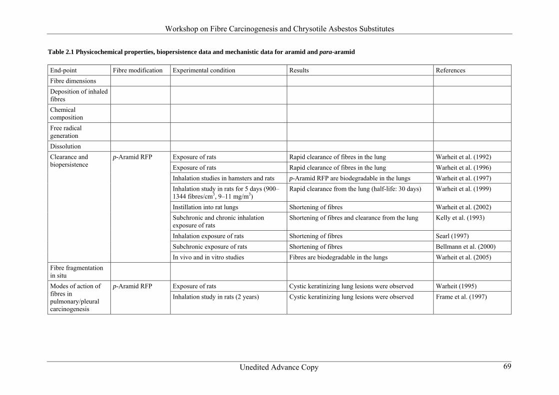
22 Assessment of selected chrysotile asbestos substitutes 221 Aramid and para-aramid (Table 21) 2211 Epidemiological studies No data are available 2212 Animal studies The data presented refer to para-aramid exclusively no data were found on meta-aramid
Carcinogenicity by inhalation Development of keratinizing cysts in the lungs of rats after long-term inhalation exposure was not considered as an indication of carcinogenicity in the IARC (1997) evaluations The study was terminated after 24 months
Carcinogenicity by intraperitoneal injection No data are available
Fibrosis in inhalation exposureintratracheal instillationintraperitoneal injection At 280 fibrescm3 after 2 weeks fibrotic thickening was observed which nearly recovered after 4ndash6 months (Lee 1983 cited in Warheit 2001) In the 2-year study there was also fibrosis after exposure to gt25 fibrescm3 In the Bellman et al (2000) study fibrosis and hyperplastic lesions were observed in the medium- and high-dose (200 and 800 fibresml) groups directly after the end of the exposure
Proliferation No in vivo studies are available
Confidence in database The working group notes that there is an unpublished carcinogenicity study that was not included as it is not publicly available 2213 Mechanistic data
Clearance and changes in situ of the deposited fibresbiopersistence Inhalation toxicity studies with rats comparing the biopersistence of p-aramid fibres (respirable-sized) with asbestos fibres have demonstrated that the aramid fibres were less biopersistent than chrysotile asbestos The longer p-aramid fibres were shortened in the lungs of rats whereas longer chrysotile asbestos fibres were preferentially retained (Warheit et al 1996) Fibre clearance studies of Warheit et al (1992 1997 2002) demonstrated a transient increase in the numbers of retained fibres at 1 week postexposure with a rapid clearance of
Workshop on Fibre Carcinogenesis and Chrysotile Asbestos Substitutes
Unedited Advance Copy 25
fibres thereafter The transient increase in the number of fibres could be due to transverse cleaving of the fibres since the average length of retained fibres continued to decrease over time Also other groups have demonstrated a rapid clearance of the longest fibrils during the first month following subchronic inhalation exposures and this was shown to be associated with a corresponding increase in the number of shorter fibrils (Kelly et al 1993 Searl 1997 Bellmann et al 2000) Warheit et al (1999) reported on a study in which rats were exposed for 5 days to aerosols of p-aramid fibrils (900ndash1344 fibrescm3 9ndash11 mgm3) The number of p-aramid fibrils per lung showed a transient increase at 1 week postexposure and then a rapid decrease thereafter lung clearance half-life was approximately 30 days During the 6 months in the lung the p-aramid fibril mean length progressively decreased from 125 microm to 75 microm In another study mentioned by Warheit et al (1999) rats were exposed to p-aramid fibrils by inhalation for 2 years The initial mean dimensions of the inhaled fibres were 12 microm length and lt03 microm diameter After a 2-year exposure at 25 25 and 100 fibrilscm3 mean lengths of lung-retained fibrils approached 4 microm The time required for the fibrils to be reduced to lt5 microm in the lung was markedly less at lower exposure concentrations Warheit et al (2005) showed in in vivo as well as in vitro experiments that the mean lengths of p-aramid respirable-sized fibre-shaped particulates (RFP) incubated with human or rat BAL fluids were substantially decreased compared with those incubated in phosphate-buffered saline The authors concluded from their investigations that p-aramid RFP are likely to be biodegradable in the lungs of humans
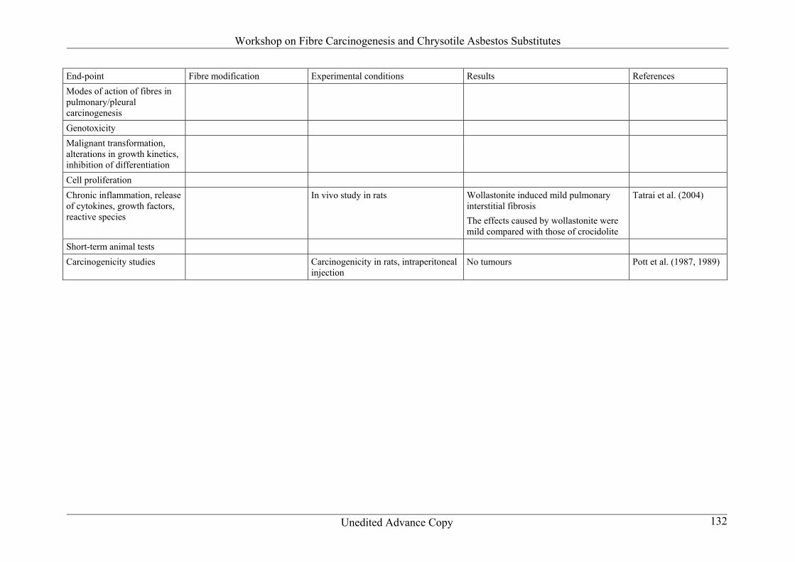
Modes of action of fibres in pulmonarypleural carcinogenesis Cystic keratinizing lung lesions produced following exposure to p-aramid RFP were observed by several authors (reviewed by Warheit 1995) However according to Frame et al (1997) these lesions are not relevant for human risk assessment of pulmonary cancer
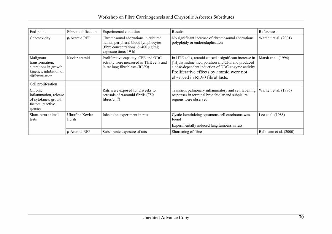
Genotoxicity p-Aramid RFP were tested for induction of chromosomal aberrations in cultured human peripheral blood lymphocytes (fibre concentrations 6ndash400 microgml exposure time 19 h) No significant increase of chromosomal aberrations polyploidy or endoreduplication was found in the exposed cells (Warheit et al 2001)
Malignant transformation alterations in growth kinetics inhibition of differentiation
No data are available
Cell proliferation The proliferative capacity of aramid (Kevlar) as well as colony forming efficiency (CFE) and ornithine decarboxylase (ODC) activity were measured by Marsh et al (1994) in hamster tracheal epithelial (HTE) cells and in rat lung fibroblasts (RL90) In HTE cells aramid
Workshop on Fibre Carcinogenesis and Chrysotile Asbestos Substitutes
Unedited Advance Copy 26
caused a statistically significant increase in [3H]thymidine incorporation and CFE and produced a dose-dependent induction of ODC enzyme activity Proliferative effects by aramid were not observed in RL90 fibroblasts
Chronic inflammation release of cytokines growth factors reactive species In a study by Warheit et al (1996) rats were exposed for 2 weeks to aerosols of p-aramid fibrils (750 fibrescm3) Two weeksrsquo exposure to these fibres produced transient pulmonary inflammatory and cell labelling responses in terminal bronchiolar and subpleural regions
Short-term animal testscarcinogenicity studies In a study by Lee et al (1988) four groups of 100 male and 100 female rats were exposed to ultrafine Kevlar fibrils at concentrations of 0 25 25 and 100 fibrilscm3 for 6 hday 5 daysweek for 2 years One group was exposed to 400 fibrilsml for 1 year and allowed to recover for 1 year At 25 fibrilscm3 the lungs showed a dust cell response slight Type II pneumocyte hyperplasia alveolar bronchiolarization and a negligible amount of collagenized fibrosis in the alveolar duct region At 100 fibrilscm3 the same pulmonary responses were seen as at 25 fibrilscm3 In addition cystic keratinizing squamous cell carcinoma was found in four female rats but not in male rats The lung tumours were derived from metaplastic squamous cells in areas of alveolar bronchiolarization At 400 fibrilscm3 following 1 year of recovery the lung dust content average fibre length and pulmonary lesions were markedly reduced but slight centriacinar emphysema and minimal collagenized fibrosis were found in the alveolar duct region The lung tumours were a unique type of experimentally induced tumours in the rats and have not been seen as spontaneous tumours in humans or experimental animals Therefore the relevance of this type of lung tumour to the human situation is minimal Warheit et al (1999) reported on a study in which rats were exposed to p-aramid RFP for 2 weeks then maintained for a period of postexposure recovery Rats exposed to lower levels (up to 26 fibrescm3) showed only a macrophage response and those exposed to higher levels (ge280 fibrescm3) developed granulomatous lesions at the alveolar duct bifurcations with fibrotic thickening By 6 months postexposure the granulomatous lesions were nearly recovered and the fibrotic lesions were much reduced During lung residence the fibres fragmented and decreased in size at a rapid rate
Summary on the determinants of carcinogenic potency In 1995 the German Research Association (DFG) organized a workshop to reach an agreement on the criteria for the classification of cystic lesions The cystic keratinizing lung lesions produced following exposure to p-aramid and many other dusts appear to be unique to the rat The general opinion was that these lesions are probably not relevant for human risk assessment of pulmonary cancer In 1997 p-aramid fibrils (RFP) were evaluated by the International Agency for Research on Cancer (IARC) and it was judged that they cannot be classified as to their carcinogenicity to humans (Group 3) This decision was based on no data in humans and inadequate evidence in animals to show either the presence or absence of a carcinogenic effect
Workshop on Fibre Carcinogenesis and Chrysotile Asbestos Substitutes
Unedited Advance Copy 27
Warheit et al (2005) concluded from their studies that inhaled p-aramid RFP are likely to be biodegradable in the lungs of humans
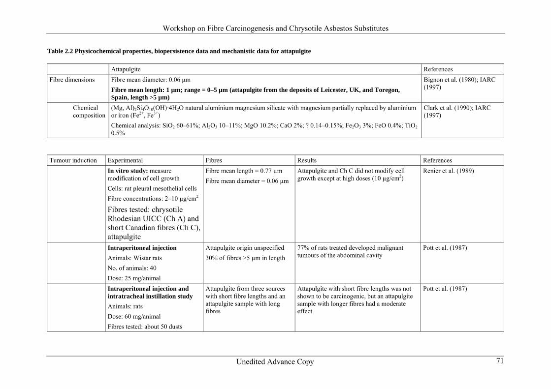
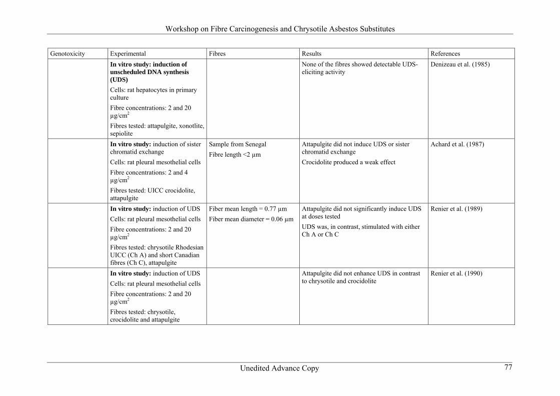
References [References being compiled] 2214 Physicochemical properties and biopersistence p-Aramid releases fibrils with diameter lt1 microm Aramid fibres are approximately 10 microm in length The fibres are respirable Aramid is crystalline Its physicochemical properties are similar to those of known carcinogenic fibres The lung clearance half-life of p-aramid was 30 days after a 5-day inhalation exposure In other experiments clearance was 60ndash170 days after subchronic exposure to 50ndash800 fibrils respectively and 6ndash9 weeks 222 Attapulgite (Table 22) 2221 Epidemiological studies A single cohort study of palygorskite (attapulgite) miners and millers was available It showed small excesses of mortality from lung cancer and stomach cancer but no indications of any exposurendashresponse for either cancer 2222 Animal studies
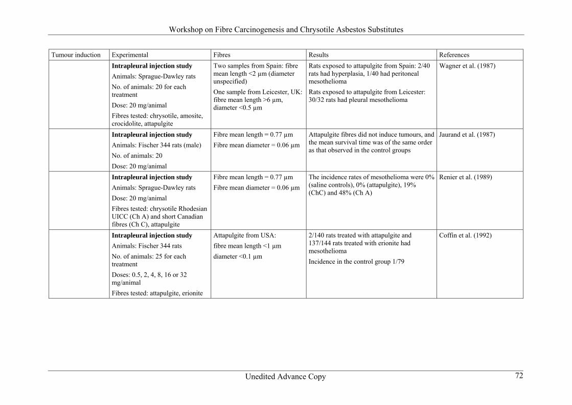
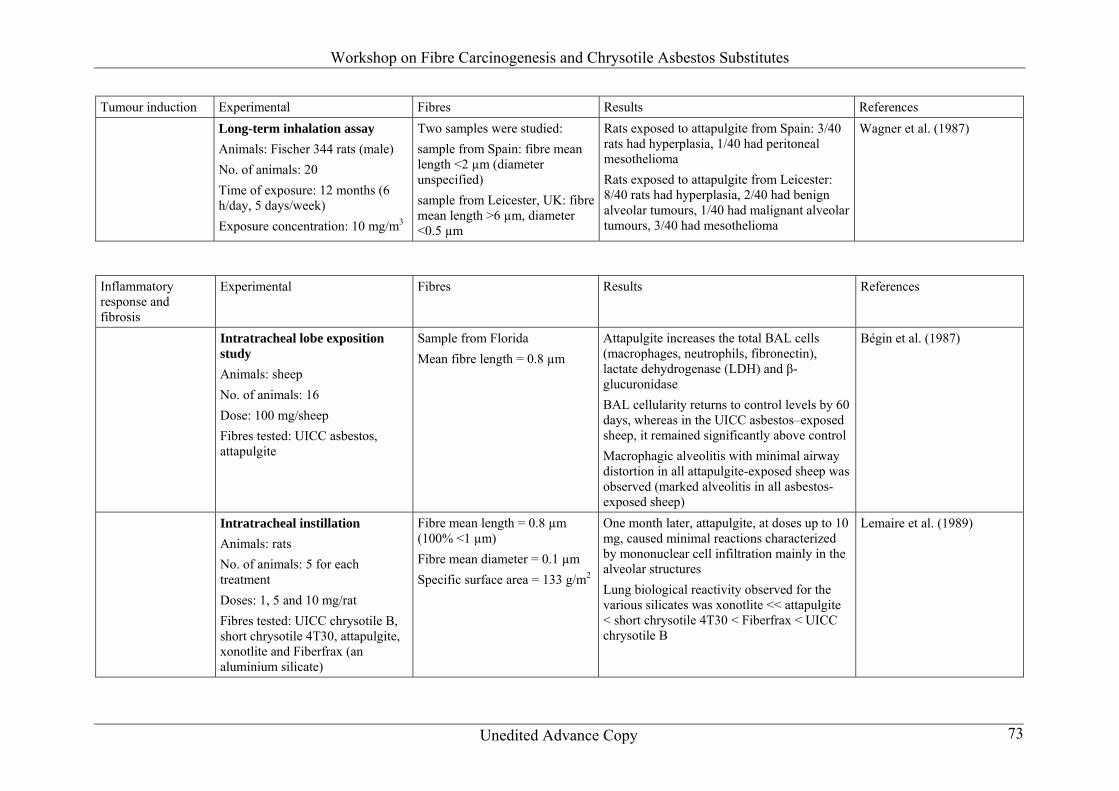
Carcinogenicity by inhalation In one inhalation study in rats with attapulgite from Leicester United Kingdom in which about 20 of the fibres were longer than 6 microm bronchoalveolar hyperplasia and a few benign and malignant alveolar tumours and mesotheliomas were observed In several studies involving exposure of rats by inhalation to short fibres (lt05 longer than or equal to 5 microm) no increase in the incidence of tumours was observed
Carcinogenicity by intraperitonealintrapleural injection The attapulgite sample from Leicester described above also induced a high incidence of pleural mesotheliomas in rats after intrapleural administration One sample of attapulgite in which 05 of the fibres were longer than 6 microm produced a significant increase of pleural mesotheliomas after intrapleural administration Intraperitoneal injection of attapulgite with 30 of the fibres longer than 5 microm and of another attapulgite in which 3 of the fibres were longer than 5 microm induced malignant abdominal tumours in rats
Workshop on Fibre Carcinogenesis and Chrysotile Asbestos Substitutes
Unedited Advance Copy 28
In several studies involving exposure of rats by intrapleural or intraperitoneal administration to short fibres (lt05 longer than or equal to 5 microm) no increase in the incidence of tumours was observed
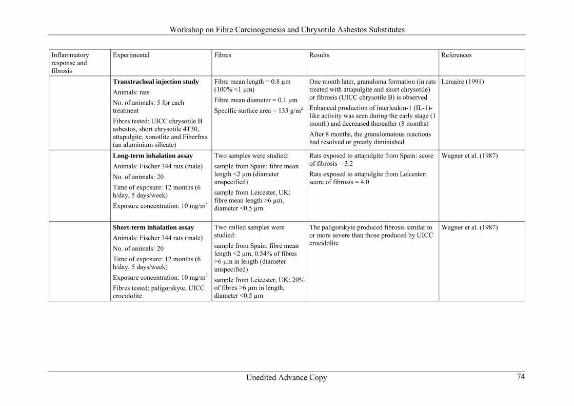
Fibrosis in inhalation exposureintratracheal instillationintraperitoneal injection In the long-term inhalation study of Wagner et al (1987) the fibrosis score was 32 after exposure to lt2 microm attapulgite but 40 after exposure to attapulgite with fibres longer than 6 microm
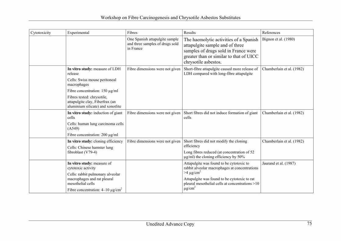
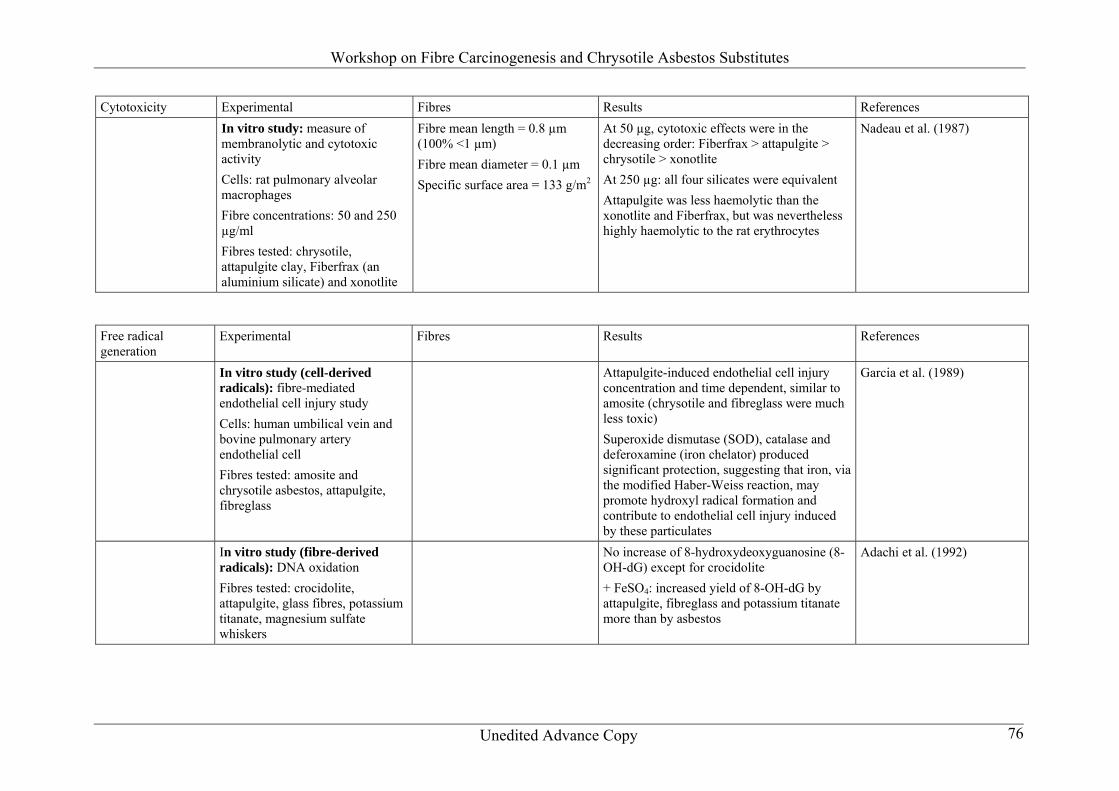
Proliferation No data are available 2223 Mechanistic data There is limited evidence for induction of ROS There are insufficient data to assess genotoxicity 2224 Physicochemical properties and biopersistence Short fibres are lt5 microm in length and long fibres are gt5 microm in length The fibre diameter is 05 microm Mineral impurities (including iron) have been measured There are ambiguous data on free radical generation which depends on the presence of iron in the solution and may not be a property of the fibre 223 Carbon fibres (Table 23) 2231 Epidemiological studies No data are available 2232 Animal studies
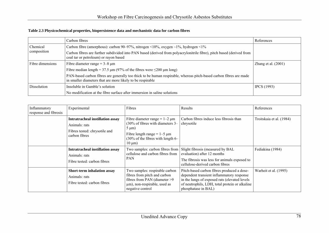
Carcinogenicity by inhalation No valid study is available (in the only existing study no tumours were observed but the fibres were not respirable)
Carcinogenicity by intraperitoneal injection No data are available
Fibrosis in inhalation exposureintratracheal instillationintraperitoneal injection In two 16-week studies with rats no fibrosis was detected at an exposure concentration of 20 mgm3 (fibre number not stated fibre diameters were 35 and 7 microm)
Workshop on Fibre Carcinogenesis and Chrysotile Asbestos Substitutes
Unedited Advance Copy 29
Proliferation No data are available 2233 Mechanistic data No data are available 2234 Physicochemical properties and biopersistence Carbon fibres are lt200 microm in length (median 40 microm) with a diameter between 3 and 8 microm They are amorphous 224 Cellulose fibres (Table 24) 2241 Epidemiological studies A cohort mortality study of 30 157 male workers who had worked for at least 1 year in 14 pulp and paper mill plants in British Columbia Canada (1950ndash1992) was conducted by Band et al (1997) Standardized mortality ratios (SMRs) were computed using Canadian population mortality rates as a comparison Of 4047 deaths 1052 were due to cancer Statistically significant excesses were found for lung cancer (SMR = 132) as well as for cancer of other sites such as pancreas brain liver larynx skin melanoma Hodgkin disease and multiple myeloma in sulfite process workers and kidney cancer in the kraft process workers who had gt15 years of employment For workers who worked in both processes a statistically significant excess was observed for non-Hodgkin lymphoma only Increased pleural cancer mortality was observed in the total cohort (n = 8 SMR = 265 90 confidence interval [CI] = 132ndash478) Of eight pleural cancers five (SMR = 361 90 CI = 142ndash758) were in workers who had ge15 years of employment Duration of employment was used as a surrogate for exposure No specific information for exposure to fibres other chemicals or smoking was provided Band et al (2001) conducted a cancer incidence study in male workers from 14 pulp and paper mill plants in British Columbia (1950ndash1992) Cancer incidence of the cohort was compared with that of the Canadian population to compute standardized incidence ratios (SIRs) A total of 1756 cases were observed in 28 278 workers who had at least 1 year of employment Because the person-year distribution and the case distribution was similar for the workers who had gt15 years and lt15 years from first employment a cut-off of 15 years for latency was selected Significantly increased SIRs were observed for skin melanoma and prostate cancer among kraft workers for cancers of the liver pancreas and lung (SIR = 132) among sulfite workers and for skin melanoma and prostate cancer among workers who worked in both kraft and sulfite processes Ten pleural cancers were reported (SIR = 205) in this population The SIR for mesothelioma was statistically significant when all the workers were considered together but not in individual process groups The authors assert that the mesotheliomas were probably due to past asbestos exposure Length of employment was used as a surrogate for the exposure It is unclear to what extent the cohort was exposed to cellulose fibres and there were confounding exposures to other chemicals as well No information on smoking was available
Workshop on Fibre Carcinogenesis and Chrysotile Asbestos Substitutes
Unedited Advance Copy 30
In a cohort of 63 025 long-term pulp and paper mill workers from 51 plants who had 10 or more years of employment Matanoski et al (1998) found that both the total mortality and the cancer mortality were low Investigators collected employment data for the current workers and created a job dictionary with job titles tasks work areas and subwork areas based on steps in the paper-making process The six work areas included in assigning the workers and for analysis were pulping papermaking finishing powerrecovery mill-wide services and others The mill processes and changes that occurred over the years were described SMRs were computed using United States population mortality rates and 20 county-specific rates where the mills were located Excess lung cancer SMR (135 95 CI = 104ndash175) was observed among employees who worked in the kraft pulping process The adjustments were done for age and calendar time No specific information on exposures to fibres or smoking was available The authors acknowledged the presence of several other chemicals In a Danish paper mill study of 14 362 workers employed any time during the follow-up period from 1943 to 1993 no excess lung cancer incidence was observed (Rix et al 1998) Danish population cancer incidence rates were used to calculate the SIRs The statistically significantly increased SIRs were observed for Hodgkin disease pharyngeal cancer and soft tissue sarcomas No specific information about exposure to fibres was provided 2242 Animal studies There exists a marked heterogeneity within cellulose and cellulose-containing materials (eg bleached kraft cellulose thermomechanical pulp microcrystalline cellulose cellulose for thin-layer chromatography [TLC] insulation cellulose)
Carcinogenicity by inhalation No studies are available
Carcinogenicity by intraperitoneal injection The respirable fraction of cellulose fibre was collected from an aerosol of thermomechanically treated wood pulp which contained besides cellulose other components such as lignin Twice as many cellulose fibres (24) were long (gt15 microm) as compared with the crocidolite fibres the total dose of fibres was 116 mg for cellulose and 18 mg for crocidolite The total doses injected intraperitoneally into rats were 106 107 108 and 109 WHO fibres Crocidolite (108 or 109 WHO fibres) was used as the ldquopositiverdquo control but no tumours were observed in this group Nine of 50 animals of the 109 WHO fibres cellulose group had malignant tumours not derived from mesothelium (sarcomas) two animals in the low dose groups (one in each) developed mesotheliomas (Cullen et al 2002) Cullen et al (2002) concluded that a high dose of cellulose fibres is capable of producing tumours when injected into the abdominal cavity of rats However the non-positive findings after crocidolite injection at high doses as well as the unusual tumour type and their early appearance after cellulose administration render it questionable as to whether the tumours observed were caused by the general mechanisms of fibre carcinogenesis In a long-term study in rats where softwood kraft pulp (lengthdiameter 31 15 times 104 fibres) was injected intraperitoneally into rats no tumours were observed The dose was considerably lower than that in the Cullen et al (2002) study (Rosenbruch et al 1992)
Workshop on Fibre Carcinogenesis and Chrysotile Asbestos Substitutes
Unedited Advance Copy 31
Fibrosis in inhalation exposureintratracheal instillationintraperitoneal injection
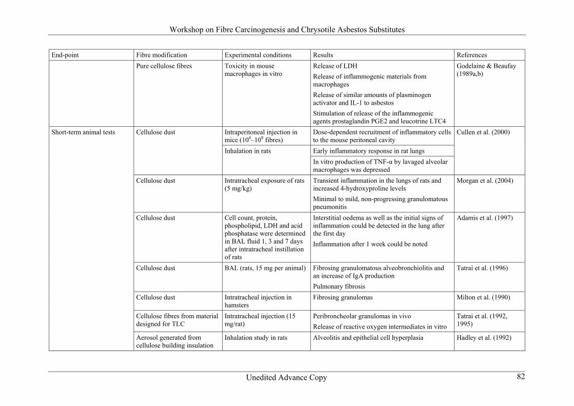
Cellulose after a single intratracheal dose (15 mg per animal) brought about fibrosing granulomatous alveobronchiolitis and an increase of immunoglobulin A (IgA) production in the BAL (Tatrai et al 1996) Fibrosing alveolitis showed moderate progression as a function of time At this dose level almost any material would induce fibrosis
Proliferation No studies are available
Confidence in database Considering the variability of the cellulose products the database to judge cellulose fibres as an entity is weak 2243 Mechanistic data
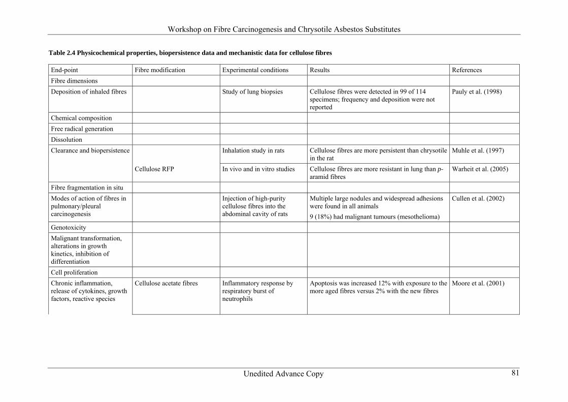
Deposition of inhaled fibres in the different parts of the respiratory tract In a study of lung biopsies Pauly et al (1998) stated that cellulose fibres were detected in 99 of 114 specimens from exposed persons in the United States of America (USA) The fibres were identified by light microscopy but the frequency and deposition of inhaled fibres were not reported
Clearance and changes in situ of the deposited fibresbiopersistence Muhle et al (1997) showed that cellulose fibres are more persistent than chrysotile in the rat but high doses were used and the clearance was probably impaired (Harrison et al 1999) In a newer study by Warheit et al (2005) cellulose RFP was used as a biopersistent reference control fibre to p-aramid The cellulose fibres were not measurably shortened compared with p-aramid
Modes of action of fibres in pulmonarypleural carcinogenesis According to Cullen et al (2002) cellulose fibres are durable and have the potential to persist within the lung These authors injected high-purity cellulose fibres into the abdominal cavity of rats In the highest-dose cellulose group multiple large nodules (granulomas) and widespread adhesions (bands of new tissue connecting organs to each other and to the abdominal wall) were present in all animals Granulomas were not observed in the 109 fibres crocidolite group More than 80 of animals in the 108 and 109 fibres crocidolite asbestos groups had mesotheliomas In contrast there were only two animals in the cellulose groups with mesothelioma tumours However nine (18) animals in the 109 fibres cellulose group had malignant tumours that in contrast to the usual pattern of mesothelioma development following treatment with mineral fibres in rats showed no obvious involvement of mesothelial tissues were not associated with blood-stained ascites fluid and were thus classified as sarcomas Cullen et al (2002) concluded that a high dose of cellulose fibres is capable of producing tumours when injected into the abdominal cavity of rats
Workshop on Fibre Carcinogenesis and Chrysotile Asbestos Substitutes
Unedited Advance Copy 32
Genotoxicity No data are available
Malignant transformation alterations in growth kinetics inhibition of differentiation
No data are available
Cell proliferation No data are available
Chronic inflammation release of cytokines growth factors reactive species Moore et al (2001) investigated the inflammatory response to cellulose acetate fibre materials by the respiratory burst of neutrophils and found positive effects Apoptosis was increased 12 with exposure to the more aged fibres versus 2 with the new fibres The authors suggested that neutrophils are activated by cellulose acetate and display an altered response to more aged fibres Cellulose fibres were found to be toxic to mouse macrophages in vitro (Godelaine amp Beaufay 1989ab) Samples derived from pure cellulose fibre including one where the particle size was in the respirable range caused the release of more LDH than did similar doses of chrysotile or crocidolite Cellulose fibre was also found to stimulate the release of inflammogenic materials from macrophages Cells treated with cellulose and those treated with asbestos released similar amounts of plasminogen activator and IL-1 Cellulose proved more powerful than asbestos in stimulating the release of the inflammogenic agents prostaglandin PGE2 and leucotrine LTC4 Control dust including glass and rock wool preparations produced a much lower response
Short-term animal tests The inflammatory effects of respirable cellulose fibres were studied by Cullen et al (2000) in two short-term animal models intraperitoneal injection in mice and inhalation in rats Both cellulose fibres and the positive control fibre crocidolite asbestos were administered in doses ranging from 104 to 108 fibres and caused marked dose-dependent recruitment of inflammatory cells to the mouse peritoneal cavity which was highest 1 day following injection Crocidolite was much more active than cellulose despite the mass dose of cellulose being 66 times greater for an equivalent number of fibres Inhalation exposure induced an early inflammatory response in rat lungs In vitro production of the proinflammatory cytokine tumour necrosis factor alpha (TNF-α) by lavaged alveolar macrophages was markedly depressed by the end of the exposure period in cellulose-exposed animals The cellulose material studied was less inflammogenic than crocidolite and the extent of the inflammatory response within the lung appeared to decrease with continued exposure over a 14-day period (Cullen et al 2000) Also Morgan et al (2004) reported transient inflammation in the lungs of rats and increased 4-hydroxyproline levels after intratracheal exposure of rats (5 mgkg body weight) to
Workshop on Fibre Carcinogenesis and Chrysotile Asbestos Substitutes
Unedited Advance Copy 33
cellulose fibres Microscopic evaluation revealed a minimal to mild non-progressing granulomatous pneumonitis In a study by Adamis et al (1997) cell count protein phospholipid LDH and acid phosphatase were determined in BAL fluid 1 3 and 7 days after intratracheal instillation of rats Interstitial oedema as well as the initial signs of inflammation could be detected in the lung after the first day Inflammation after 1 week could be noted partly interstitial and partly intra-alveolar and intrabronchial In the BAL fluid protein LDH acid phosphatase phospholipid and cell count were enhanced after days 1 and 3 After 1 month the developing bronchoalveolitis is fibrous in character In vitro cellulose did not damage rat peritoneal macrophages (Adamis et al 1997) Cellulose after a single intratracheal dose (15 mg per animal) brought about fibrosing granulomatous alveobronchiolitis and an increase of IgA production in the BAL (Tatrai et al 1996) Fibrosing alveolitis showed moderate progression as a function of time With different morphological methods injury of type I pneumocytes and the incomplete repair of type II pneumocytes were detected by the investigators The damage of the alveolar epithelium initiated and activated a series of processes that led to definite pulmonary alterations pulmonary fibrosis leading to the disintegration of the alveolo-capillary morphological functional unit (Tatrai et al 1996) Milton et al (1990) studied the ability of cellulose dust to produce emphysema following intratracheal injection in hamsters The results showed fibrosing granulomas and patchy thickening of alveolar septa in cellulose-treated animals The intratracheal injection of cellulose was also used by Tatrai et al (1992 1995) who injected cellulose fibres from material designed for TLC into rats at a dose level of 15 mg Peribroncheolar granulomas developed in the rat lungs which were becoming fibrosed after 3 months As part of these studies human blood leukocytes were treated with cellulose fibres in vitro This material caused significant dose-related release of reactive oxygen intermediates In a study by Hadley et al (1992) rats were treated by inhalation for 28 days with an aerosol generated from cellulose building insulation of which approximately 40 was respirable by rats Target dose levels were 100 500 and 2000 mgm3 animals were sacrified at the end of the inhalation period when dose-related pulmonary changes were found The authors observed diffuse macrophage infiltration throughout the pulmonary parenchyma with macrophages enlarged and with foamy cytoplasms Some areas showed alveolitis and epithelial cell hyperplasia with some areas consolidated with granulation tissue Within granulomas there was evidence of collagen deposition
Carcinogenicity studies Rinsky (1990) reported a casendashcontrol study of 299 malignancies among paperwool workers and found that the only significant excess for neoplasms was for those of the haematopoietic system Solet et al (1989) undertook a proportional mortality analysis of 1010 deaths of workers in the pulp and paper industry These authors reported a general excess of cancer largely lung cancer but did not have information on smoking habits Jarvholm et al (1988) Ericsson et al (1988) and Thoren et al (1989) reported studies on the same soft paper mill production unit The odds ratios for mortality from chronic obstructive pulmonary disease and from asthma among the exposed workers were significantly increased There was no
Workshop on Fibre Carcinogenesis and Chrysotile Asbestos Substitutes
Unedited Advance Copy 34
excess in malignancies A morbidity study demonstrated a dose-related irritation of the upper respiratory tract A decrease in lung vital capacity was associated with long-term exposure Increased elastic recoil pressure and a decreased residual volume were reported among the exposed workers These findings were considered by the authors to be non-specific reactions to the heavy exposure to paper dust in the mill
Summary on the determinants of carcinogenic potency It can be stated that there is evidence that cellulose dust once it reaches the lung tissue has significant biological activity which can include the stimulation of pulmonary fibrosis (Davis 1996) It seems that cellulose fibres are more persistent in the lung than p-aramid RFP
References [References being compiled] 2244 Physicochemical properties and biopersistence Small-diameter cellulose fibres are respirable There is no information available on the chemical properties of these fibres Cellulose fibres are persistent in the lung 225 Graphite whiskers (Table 25) 2251 Epidemiological studies No data are available 2252 Animal studies
Carcinogenicity by inhalation No valid study is available (in the only existing study no tumours were observed but the fibres were not respirable)
Carcinogenicity by intraperitoneal injection No data are available
Fibrosis in inhalation exposureintratracheal instillationintraperitoneal injection In two 16-week studies with rats no fibrosis was detected at an exposure concentration of 20 mgm3 (fibre number not stated fibre diameters were 35 and 7 microm)
Workshop on Fibre Carcinogenesis and Chrysotile Asbestos Substitutes
Unedited Advance Copy 35
Proliferation No data are available 2253 Mechanistic data No data are available 2254 Physicochemical properties and biopersistence Graphite fibres are crystalline and hydrophobic They have a long half-time in the lungs with a clearance of gt1 year 226 Magnesium sulfate whiskers 2261 Epidemiological studies No data are available 2262 Animal studies
Carcinogenicity by inhalation No tumours were induced in a long-term study (1 year of exposure observation period total 2 years no fibre numbers given but the concentration was 14 mgm3) in rats
Carcinogenicity by intraperitoneal injection No data are available
Fibrosis in inhalation exposureintratracheal instillationintraperitoneal injection No fibrosis or metaplasia was observed in the inhalation study described above
Proliferation No data are available 2263 Mechanistic data
Clearance and changes in situ of the deposited fibresbiopersistence According to Fujino et al (1995) magnesium sulfate whiskers are more soluble in vitro than are glass fibres ceramic fibres and potassium octatitanate whiskers
Modes of action of fibres in pulmonarypleural carcinogenesis In a study by Hori et al (1994) Wistar rats were exposed to two types of magnesium sulfate whiskers by inhalation for 6 hday 5 daysweek for 4 weeks (subchronic study) or for 1 year
Workshop on Fibre Carcinogenesis and Chrysotile Asbestos Substitutes
Unedited Advance Copy 36
(chronic study) to clarify the biological effects of the whiskers There were few whiskers detected in the rat lungs even at 1 day after the exposure suggesting that they are dissolved and eliminated rapidly from the lungs To measure the clearance rate of the whiskers from the lungs an intratracheal instillation was performed in golden hamsters The half-life of the whiskers in the lung was determined as 176 min by temporally measuring the magnesium concentration up to 80 min after the instillation A histopathological examination indicated a frequent occurrence of adenoma and carcinoma in the year after chronic exposure but it was not significantly different between exposed and control rats
Genotoxicity No data are available
Malignant transformation alterations in growth kinetics inhibition of differentiation
No data are available
Cell proliferation No data are available
Chronic inflammation release of cytokines growth factors reactive species Culture of alveolar macrophages with fibres at concentrations of 125ndash100 microgml did not induce a significant increase in TNF production and did not significantly affect LDH release (Fujino et al 1995)
Short-term animal tests No data are available
Carcinogenicity studies No data are available
Summary on the determinants of carcinogenic potency There are few data available on the toxicity of magnesium sulfate whiskers It seems that this fibre is easily soluble and not very persistent in the lung and it does not induce increased biological responses compared with the negative control
References [References being compiled] 2264 Physicochemical properties and biopersistence Magnesium sulfate whiskers have a length of gt5 microm and a diameter of lt05 microm They are highly soluble with a half-life of lt1 h They are partially soluble in Gamblersquos solution
Workshop on Fibre Carcinogenesis and Chrysotile Asbestos Substitutes
Unedited Advance Copy 37
227 Polyethylene fibres (Table 27) 2271 Epidemiological studies No data are available 2272 Animal studies
Carcinogenicity by inhalation No data are available
Carcinogenicity by intraperitoneal injection No data are available
Fibrosis in inhalation exposureintratracheal instillationintraperitoneal injection No data are available
Proliferation No data are available 2273 Mechanistic data No data are available on polyethylene fibres Data are available only on polyethylene dust
Clearance and changes in situ of the deposited fibresbiopersistence No data are available
Modes of action of fibres in pulmonarypleural carcinogenesis No data are available
Genotoxicity No data are available
Malignant transformation alterations in growth kinetics inhibition of differentiation
No data are available
Cell proliferation No data are available
Workshop on Fibre Carcinogenesis and Chrysotile Asbestos Substitutes
Unedited Advance Copy 38
Chronic inflammation release of cytokines growth factors reactive species
In an in vitro cytotoxicity study by Styles amp Wilson (1973) non-fibrous polyethylene dust was found to be less toxic to macrophages than was chrysotile
Short-term animal tests No data are available
Carcinogenicity studies No data are available
Summary on the determinants of carcinogenic potency Almost no data are available on the toxicity of polyethylene fibres
References [References being compiled] 2274 Physicochemical properties and biopersistence Polyethylene fibres are produced at lengths above 10ndash15 microm 228 Polypropylene fibres (Table 28) 2281 Epidemiological studies Occupational cohort studies of workers exposed to polypropylene raised concerns regarding colorectal cancer in workers This issue has been addressed in a few epidemiological studies of occupational cohorts engaged in the extrusion of polypropylene fibres Lewis et al (1994) in an update of an earlier investigation that reported a 6-fold excess in colorectal cancer in a polypropylene manufacturing plant in Texas USA concluded that there was no suggestion of an occupationally related risk The cohort was small however consisting of only 412 male workers Another small study (Cowles et al 1994) of a cohort of 257 male employees of a plastics and resins research facility in New Jersey USA reported no increased risk of colorectal cancer although it noted a significant increase in pancreatic cancer and a non-significant increase in lung cancer It is not clear that the workers were exposed to fibres and no information on smoking was available Goldberg amp Theriault (1994) performed a much larger retrospective cohort study in a synthetic textiles factory in Quebec Canada The cohort consisted of 7487 men and 2724 women who had been employed for at least 1 year For men the risk of colon cancer increased with duration of employment in the polypropylene and cellulose triacetate extrusion unit
Workshop on Fibre Carcinogenesis and Chrysotile Asbestos Substitutes
Unedited Advance Copy 39
In a cross-sectional study of respiratory effects among 50 workers exposed to polypropylene flock and 45 controls early signs of interstitial lung disease were noted in high-resolution computed tomography (Atis et al 2005) 2282 Animal studies
Carcinogenicity by inhalation No data are available
Carcinogenicity by intraperitoneal injection No data are available
Fibrosis in inhalation exposureintratracheal instillationintraperitoneal injection The pulmonary toxicity of polypropylene was tested by Hesterberg et al (1992) in male Fischer 344 rats after 90 days of inhalation exposure A recovery group was examined 30 days after the end of the exposure The polypropylene fibres were size-selected to have a geometric mean diameter of 12 microm (geometric standard deviation [GSD] 03) (46 lt1 micromn) and a geometric mean length of 303 microm Three groups of animals were exposed in nose-only inhalation chambers 6 hday 5 daysweek for 90 days to 15 30 or 60 mgm3 of polypropylene or filtered air (negative control) At 90 days the lung burden was 26 times 106 fibreslung No fibrosis was observed in any of the polypropylene-treated groups (Wagner scale 3) in the positive control group treated with RCF the Wagner scale was 40 Warheit et al (1999) described a study in which rats were exposed to polypropylene fibres (average diameter 16 microm average length 20 microm) The rats developed dose- and time-dependent lung changes characterized by increased cellularity and early bronchiolitis but no fibrosis
Proliferation No data are available 2283 Mechanistic data Fibre diameter depends on the manufacturing process and can vary from gt153 microm in monofilament yarn to an average of 1ndash5 microm in microfibres (ICF 1986)
Clearance and changes in situ of the deposited fibresbiopersistence Hesterberg et al (1992) showed that the number of partially degraded (segmented) polypropylene fibres within the lung increased with the exposure concentration and period of exposure as well as with the period of recovery after termination of exposure at 90 days The authors concluded that polypropylene fibres are not fibrogenic to rats after 90 days of inhalation exposure Polypropylene fibres produced dose-dependent minimal or mild increases in cellularity in lungs which appear to be reversible after 90 days especially at the lower dose administered (see above)
Workshop on Fibre Carcinogenesis and Chrysotile Asbestos Substitutes
Unedited Advance Copy 40
Modes of action of fibres in pulmonarypleural carcinogenesis No data are available
Genotoxicity No data are available
Malignant transformation alterations in growth kinetics inhibition of differentiation
No data are available
Cell proliferation No data are available
Chronic inflammation release of cytokines growth factors reactive species The pulmonary toxicity of polypropylene was tested by Hesterberg et al (1992) in male Fischer 344 rats after 90 days of inhalation exposure The polypropylene fibres were size-selected to have a geometric mean diameter of 16 microm (46 lt1 microm) and a geometric mean length of 303 microm Three groups of animals were exposed in nose-only inhalation chambers 6 hday 5 daysweek for 90 days to 15 30 or 60 mgm3 of polypropylene or filtered air (negative control) No fibrosis was observed in any of the groups In an in vitro cytotoxicity study by Styles amp Wilson (1973) non-fibrous polypropylene dust was found to be less toxic to macrophages than was chrysotile
Short-term animal tests Warheit et al (1999) described a study in which rats were exposed to polypropylene fibres (average diameter 16 microm average length 20 microm) The rats developed dose- and time-dependent lung changes characterized by increased cellularity and early bronchiolitis but no fibrosis The cellular changes were considered to be reversible
Carcinogenicity studies No data are available
Summary on the determinants of carcinogenic potency It seems that polypropylene fibres are able to induce lung changes characterized by increased cellularity and early bronchiolitis These changes seem to be reversible after some months No fibrosis was observed in animal experiments
References [References being compiled]
Workshop on Fibre Carcinogenesis and Chrysotile Asbestos Substitutes
Unedited Advance Copy 41
2284 Physicochemical properties and biopersistence In a subchronic toxicity study the diameter of polypropylene fibres was 1ndash5 microm and the length was approximately 30 microm Polypropylene fibres exhibit high biopersistence (200 days) by the intratracheal route (mean diameter 05 microm mean length 12 microm) 229 Polyvinyl alcohol fibres (Table 29) 2291 Epidemiological studies No data are available 2292 Animal studies No data are available 2293 Mechanistic data
Clearance and changes in situ of the deposited fibresbiopersistence The diameter of PVA fibres as manufactured is above the respirable limit and most of them are not inhalable They have a lower density as mineral fibres The fibres are mostly in the range of 10ndash16 microm in diameter There is evidence that they do not fibrillate (Harrison et al 1999)
Modes of action of fibres in pulmonarypleural carcinogenesis Morinaga et al (1999) did not observe an increased lung cancer risk in workers exposed to PVA fibres
Genotoxicity PVA compounds were not genotoxic in a range of in vivo and in vitro studies (Table 210) Table 210 Genotoxicity of PVA compounds in vitro and in vivo
End-point Test system Test material Concentration Results Reference In vitro Reverse mutationa
Salmonella typhimurium TA98 TA100 TA1535 TA1537
PVA 5000 mgplate Negative Huntingdon Life Sciences (2000a)
Reverse mutationb
S typhimurium TA1537
PVA 7500 mgplate Negative Huntingdon Life Sciences (2000a)e
Workshop on Fibre Carcinogenesis and Chrysotile Asbestos Substitutes
Unedited Advance Copy 42
End-point Test system Test material Concentration Results Reference Reverse mutationa
Escherichia coli WPA uvrA pKM101
PVA 5000 mgplate Negative Huntingdon Life Sciences (2000a)c
Cell mutationa Mouse lymphoma L5178Ycells Tk+minus locus
PVA 5000 mgml Negative Huntingdon Life Sciences (2000b)c
In vivo Micronucleus formation
Mouse bone marrow
PVA 2000 mgkg Negative Huntingdon Life Sciences (2000c)c
a In the absence and presence of metabolic activation from S9 liver microsomal preparations b In the presence of metabolic activation from S9 liver microsomal preparations c Study conducted in accordance with United States Department of Agriculture Good Laboratory Practice
(GLP) regulations (Part 58 of 21 Code of Federal Regulations) European Economic Community GLP regulation 9911EEC and Organisation for Economic Co-operation and Development GLP principles
Malignant transformation alterations in growth kinetics inhibition of differentiation
No data are available
Cell proliferation No data are available
Chronic inflammation release of cytokines growth factors reactive species No data are available
Short-term animal tests No data are available
Carcinogenicity studies No data are available
Summary on the determinants of carcinogenic potency PVA fibres as manufactured are above the respirable limit and most of them are not inhalable The only study on lung cancer risk in workers exposed to PVA fibres did not show positive results PVA itself is not genotoxic
References [References being compiled]
Workshop on Fibre Carcinogenesis and Chrysotile Asbestos Substitutes
Unedited Advance Copy 43
2294 Physicochemical properties and biopersistence The diameter of PVA fibres is mostly in the range of 10ndash16 microm They have a lower density as mineral fibres The respirable limit is about 7 microm The fibres do not fibrillate There are no data on biopersistence 2210 Polyvinyl chloride fibres (Table 211) 22101 Epidemiological studies There are no epidemiological studies of polyvinyl chloride (PVC) fibres The studies by Mastrangelo et al (2005) and earlier studies by Wu et al (1989) and Waxweiler et al (1981) reflect exposure not to fibres but rather to granular particles 22102 Animal studies No data are available on fibrous PVC 22103 Mechanistic data No data are available on PVC fibres Data are available only on PVC dust and vinyl chloride monomer (VCM)
Clearance and changes in situ of the deposited fibresbiopersistence No data are available
Modes of action of fibres in pulmonarypleural carcinogenesis No data are available
Genotoxicity Wong et al (2003) investigated genetic polymorphisms and sister chromatid exchanges in VCM-exposed PVC workers and found that workers with susceptible metabolic and DNA repair genotypes experience an increased risk of DNA damage elicited by VCM exposure Lei et al (2004) investigated DNA single strand breaks in PVC monomerndashexposed workers and correlated it to urinary thiodiglycolic acid levels The results of this study showed that levels of DNA single strand breaks were increased with urinary thiodiglycolic acid levels The dosendashresponse relationship of urinary thiodiglycolic acid level and DNA single strand breaks was particularly significant among the workers with a urinary thiodiglycolic acid level of 4 mgg creatinine which is equivalent to a 5 ppm airborne VCM level The authors concluded that airborne VCM exposure greater than 5 ppm could induce DNA damage
Workshop on Fibre Carcinogenesis and Chrysotile Asbestos Substitutes
Unedited Advance Copy 44
Malignant transformation alterations in growth kinetics inhibition of differentiation
No data are available
Cell proliferation Six samples from the normal industrial suspension process (PVC-S) and eight samples from the emulsion process (PVC-E) were studied for cytotoxicity in vitro using the MTT (3-(45-dimethylthiazol-2-yl)-25-diphenyltetrazolium bromide) assay after 20 h of incubation in A549 cells and in primary cultures of alveolar macrophages (AM) and type II pneumocytes (TII) obtained from rats (r-AM r-TII) or from human surgical specimens (h-AM h-TII) Haemolysis was assessed after 2 h of incubation with human erythrocytes (h-RBC) A positive control (crystalline silica Min-U-Sil) and relevant additives of these PVC particles were tested concurrently No toxicity of PVC-S particles could be established up to 5 mgml in the haemolysis test and 25 mgml in the MTT assay In contrast four out of eight PVC-E particles induced significant toxicity with median effective concentration (EC50) values ranging from 07 to 37 mgml in the haemolysis test and from 02 to 12 mgml in primary cells The most toxic particles were those that contained the additives with the highest in vitro cytotoxicity There was a good correlation between EC50 values obtained in relevant bioassays In conclusion PVC-E particles produced a moderate in vitro toxicity for primary rat and human pulmonary cells probably because of the residual presence of additives (Xu et al 2002) To elucidate the role of residual additives two of the emulsion PVC particles were chosen for a follow-up study (Xu et al 2003) Here the authors compared the cytotoxicity and the release of cytokines by PVC particles with their ldquoadditive-freerdquocounterparts and with the pure additives sodium lauryl sulfate and sodium alkylbenzenesulfonate The authors found that unwashed PVC particles exhibited the potential to damage pulmonary cells and to induce the release of inflammatory mediators in vitro These particles might also have hazardous potential for the lung under excessive occupational exposure Xu et al (2003) proposed that the hazardous potential of these PVC particles is related to their residual additives
Chronic inflammation release of cytokines growth factors reactive species Enzymatic and pathomorphological alterations in rat lungs were studied by Agarwal et al (1978) at different time intervals up to 180 days after a single intratracheal administration of 25 mg of PVC dust The activities of two energy-linked enzymes succinate dehydrogenase (SDH) and adenosine triphosphatase (ATPase) and three lysosomal enzymes acid phosphatase β-glucuronidase and ribonuclease were significantly increased in the early period and then started to decline The activities of SDH and ATPase reached control values at 150 days whereas those of the lysosomal enzymes remained significantly higher up to this period Histopathologically the pulmonary response was in the form of acute inflammatory changes during the early stages of dust burden followed by the development of granulomatous lesions containing small amounts of stromal elements
Short-term animal tests Rats guinea-pigs and monkeys were exposed in a study by Groth et al (1981) by inhalation (6 hday 5 daysweek) for up to 22 months to a 13 mgm3 concentration of PVC dust
Workshop on Fibre Carcinogenesis and Chrysotile Asbestos Substitutes
Unedited Advance Copy 45
Autopsies on rats and guinea-pigs were performed after 12 months of exposure and on monkeys after 22 months of exposure Lung function tests were performed on monkeys after 9 14 and 22 months of exposure Aggregates of alveolar macrophages containing PVC particles were found in the lungs of all animals These aggregates were more numerous in the monkey lungs No fibrosis or significant cellular infiltrates were present in or near these cellular aggregates No significant effects on pulmonary function could be demonstrated in the monkeys exposed to PVC Under the conditions of this experiment inhaled PVC produced a benign pneumoconiosis Also Wagner amp Johnson (1981) did not observe carcinogenic effects in rats after inhalation or intrapleural injection of 20 mg PVC dust PVC dust following a single intratracheal instillation (25 mgrat) was substantially cleared through the lymphatic circulation and progressively accumulated in the tracheobronchial lymph nodes in studies by Agarwal et al (1991) The tissue response in tracheobronchial lymph nodes during 60ndash270 days post-instillation of PVC dust was characterized by progressive increase in total organ fresh weight dry weight DNA ribonucleic acid (RNA) and protein contents concurrent with the proliferation of macrophages and hyperplasia of reticular cells Active phagocytosis and enhanced hydrolytic activity in tracheobronchial lymph nodes was evident around 270 days post-instillation by the appearance of PVC-laden macrophages near and within the dust foci and increased activity of acid phosphatase deoxyribonuclease (DNase) ribonuclease (RNase) and β-glucuronidase PVC dust caused degeneration of macrophages and consequent release of hydrolytic enzymes resulted in limited cytotoxicity without inducing reticulination and fibrosis in the tracheobronchial lymph nodes The histology and clinical biochemistry of liver kidney spleen and serum were not altered and there were no detectable PVC particles in these tissues at up to 365 days The authors therefore concluded that lymphatic clearance of intratracheally instilled PVC dust results in its accumulation and mild foreign body reaction in tracheobronchial lymph nodes which is non-fibrogenic at up to 365 days post-instillation (Agarwal et al 1991) In vitro studies of Xu et al (2002 2003) indicated that emulsion PVC particles (PVC-E3) with a mean diameter of 2 microm exhibited a moderate toxicity in different pulmonary cell cultures The in vitro cytotoxicity and proinflammatory potential of PVC-E3 particles were reduced when the additives had been ldquowashed offrdquo (PVC-W3) indicating that PVC particlendashassociated toxicity is probably related to the residual additives The in vivo study of Xu et al (2004a) with male Wistar rats did not confirm the conclusion from the in vitro toxicity tests The pulmonary toxicity of both PVC-E3 and PVC-W3 particles appeared limited In a follow-up study by Xu et al (2004b) rats were repeatedly intratracheally instilled with PVC particles but the results were similar to those with single instillations The examined PVC particles had the potential of inducing a limited and transient acute inflammatory reaction in the lung and possibly a more persistent alteration of pulmonary T lymphocyte subsets towards a high CD4CD8 ratio
Carcinogenicity studies In 1974 vinyl chloride (VC) was first reported in the scientific literature to induce angiosarcoma of the liver both in humans and in animals Additional research has demonstrated the carcinogenicity of VC to other organs and at lower concentrations The target organs for VC now clearly include the liver brain lung and probably the lymphohaematopoietic system The evidence for a carcinogenic risk has been extended to
Workshop on Fibre Carcinogenesis and Chrysotile Asbestos Substitutes
Unedited Advance Copy 46
jobs associated with PVC exposure Cases of liver angiosarcoma have been reported among individuals employed in PVC fabrication facilities and an epidemiological study has demonstrated a significant association between exposure to PVC dust and the risk of lung cancer mortality Cases of angiosarcoma of the liver also have been reported among individuals living in close proximity to VCPVC plants An association between PVC dust and pneumoconiosis has also been demonstrated On the basis of findings prudent control of PVC dust in the industrial setting is indicated (Wagoner 1983) Overall the results of the analysis of 12 studies of VC production and polymerization workers demonstrate an elevated risk of liver malignancies and the possibility of a 2-fold increased risk of brain and central nervous system tumours and perhaps also of malignancies of the lymphatic and haematopoietic system (Nicholson et al 1984) However the role of other agents cannot be excluded in the etiology of non-hepatic malignancies Bronchogenic carcinoma does not appear to be increased from exposures to VCM although a relationship to PVC dust was suggested in one study These conclusions must be considered in light of limited data on workers followed more than 25 years from onset of exposure Considering the numbers of observed and expected deaths in all studies it would appear that the excess of malignancies at non-hepatic sites is less than the excess of liver tumours To the extent that VC exposure is associated with other cancers a similar risk reduction would be expected Raynaudrsquos phenomenon acro-osteolysis scleroderma-like skin lesions hepatomegaly and splenomegaly with non-cirrhotic hepatic fibrosis and severe portal hypertension have been associated with past heavy exposures to VC Evidence exists that the liver disease and portal hypertension may progress following cessation of exposure However all of the above syndromes were found largely in heavily exposed individuals Their occurrence would be much less likely in workers exposed only to concentrations currently allowed Pulmonary deficits X-ray abnormalities and perhaps lung cancer have been associated with VCPVC exposure (Nicholson et al 1984) Increased respiratory cancer was reported by Waxweiler et al (1981) among a cohort of 4806 males employed at a synthetic chemicals plant Upon review of pathologic material the excess was found to be limited to adenocarcinoma and large cell undifferentiated lung cancer Many of the workers had been exposed to VC as well as to chlorinated solvents PVC dust acrylates and acrylonitrile PVC dust appeared to be the most likely etiologic agent Time trends of PVC dust exposure indicated a potential latent period of 5ndash16 years before death (Waxweiler et al 1981) Hsiao et al (2004) concluded from their studies that PVC workers who had high exposure to VCM have an increased risk of developing liver fibrosis Several authors reported that occupational exposure to PVC dust is associated with lung disorders called ldquoPVC pneumoconiosisrdquo (Szende et al 1970 Arnaud et al 1978 Antti-Poika et al 1986 Studnicka et al 1995) Epidemiological studies indicated that occupational PVC dust exposure might affect pulmonary function and lead to a higher prevalence of small opacities on chest X-ray (Soutar amp Gauld 1983 Lloyd et al 1984 Nielsen et al 1989 Lee et al 1991 Ng et al 1991) Pott et al (1989) reported that large amounts of PVC dust led to adhesions of the abdominal organs and fibrosis but a definite carcinogenic effect was not detected
Workshop on Fibre Carcinogenesis and Chrysotile Asbestos Substitutes
Unedited Advance Copy 47
Summary on the determinants of carcinogenic potency It seems likely that workers in the PVC industry who were exposed for a longer time to high PVC levels (gt10 mg PVCm3) have an increased risk of developing pneumoconiosis and decreased lung function Effects were observed just after long-term exposure to PVC dust According to Nicholson et al (1984) the risk of VCPVC-induced malignancies has decreased after 1974 because of improved technologies in the PVC industry In animal experiments with PVC dust most studies did not show significant cellular effects (no fibrosis no significant effects on pulmonary function no carcinogenic effects in rats) However in vitro studies in several cellular systems showed cytotoxic and genotoxic effects It might be that these effects are reversible and non-mutagenic in vivo
References [References being compiled] 22104 Physicochemical properties and biopersistence No data are available 2211 Potassium octatitanate fibres (Table 212) 22111 Epidemiological studies No data are available 22112 Animal studies
Carcinogenicity by inhalation In a 2-year inhalation (up to 200 WHO fibresml) study in rats no tumours were observed The observation period thus was short and the exposure concentration too low for reliable detection of tumour induction In another 2-year study (1-year exposure 1-year additional follow-up) in rats no tumours were observed
Carcinogenicity by intraperitoneal injection There exist four intraperitoneal injection studies one in hamsters and three in rats In all studies a dose-dependent increased incidence of mesothelioma was observed (Stanton 1978 Lee et al 1981 Pott et al 1989 Adachi et al 2001) Up to 80 animals had mesothelioma after exposure to 10 mg
Workshop on Fibre Carcinogenesis and Chrysotile Asbestos Substitutes
Unedited Advance Copy 48
Fibrosis in inhalation exposureintratracheal instillationintraperitoneal injection In the first 2-year inhalation study in rats described above no fibrosis was observed whereas in the other mild fibrosis was reported
Proliferation No data are available 22113 Mechanistic data There is a small database DNA strand breaks and chromosomal aberrations have been observed There is evidence of strong genotoxicity comparable to that of chrysotile 22114 Physicochemical properties and biopersistence The fibres have low solubility and small diameters Iron and cobalt are present as impurities The fibres have a long half-life 2212 Synthetic vitreous fibres (Table 213) 22121 Epidemiological studies
General introduction glass wool continuous glass filament and rock (stone) and slag wool during manufacture
Two large cohort studies and casendashcontrol studies nested within these cohorts from the USA and Europe provide most of the epidemiological evidence concerning potential risk for respiratory and other cancers associated with occupational exposure to glass wool continuous glass filament and rock (stone)slag wool during manufacture The cohort study from the USA included 16 plants extended the follow-up to 1992 and expanded a previous cohort to include women and non-white workers This study included information on smoking habits and a new assessment of historical workplace exposure to respirable fibres and several sources of co-exposure including asbestos formaldehyde and silica The European cohort extended the follow-up to 1990 in 13 plants
Glass wool The findings of the cohort study from the USA provided no evidence of excess mortality from all causes combined or from all cancers combined using local rates A statistically significant 6 excess in respiratory cancer (primarily trachea bronchus and lung) mortality was observed When analysis was restricted to long-term workers the excess was reduced and was no longer statistically significant Adjustment for smoking based on a random sample of workers suggests that smoking may account for the excesses in respiratory cancer observed in the male glass fibre cohort (glass wool and continuous glass filament combined) The SMRs for respiratory cancer were related neither to duration of employment among the total cohort or among long-term workers nor to duration of exposure cumulative exposure or average intensity of exposure to respirable glass fibre (glass wool and continuous glass
Workshop on Fibre Carcinogenesis and Chrysotile Asbestos Substitutes
Unedited Advance Copy 49
filament combined) Analysis by product group showed a statistically significant excess of respiratory cancer for all workers from plants grouped as ldquomostly glass woolrdquo but this excess risk for the ldquomostly glass woolrdquo product group was reduced and no longer statistically significant when the cohort was limited to long-term workers (ge5 years of employment) There was no evidence of an excess of mesothelioma or non-respiratory cancers The casendashcontrol study of respiratory cancer nested within the United States cohort enabled control of plant co-exposure and a more detailed control for confounding by smoking Duration of exposure cumulative exposure average intensity of exposure and the time since first exposure to respirable glass fibre were not associated with an increased risk of respiratory cancer These results were not altered by using different characterizations of categorized respirable fibre exposure or alternative models for continuous exposure data The European cohort study of glass wool workers demonstrated an increased mortality from lung cancer (trachea bronchus and lung) but no trend with time since first hire or duration of employment One death from mesothelioma was observed in this cohort This study did not estimate fibre exposure but used surrogate measures such as ldquotechnological phase at first employmentrdquo No information was available either on co-exposure or on smoking habits
Continuous glass filament Two of the plants of the cohort study from the USA manufactured only continuous glass filament For all workers and for long-term workers from these two plants no evidence of excess mortality from respiratory cancer was found when compared with local rates Adjustment for smoking had little effect on the SMR for respiratory cancer A nested casendashcontrol study that included adjustments for smoking and co-exposure also provided no consistent evidence of excess mortality from respiratory cancer The exposurendashresponse analyses that combined exposures to continuous glass filament and to glass wool are reported in the section on glass wool The European cohort study reported few data to evaluate cancer risks among workers exposed to continuous glass filament This study provided no convincing evidence of an elevated risk for lung cancer Results were also available from two smaller cohort studies in the USA and Canada The cohort study on one continuous glass filament plant in the USA which included a nested casendashcontrol study with information on smoking and co-exposure provided no consistent evidence of an excess risk for lung cancer The Canadian cohort study of one continuous glass filament plant did not include an assessment of smoking or co-exposure This study also provided no consistent evidence of an excess risk for lung cancer
Rock (stone) and slag wool The present evaluation relies mainly on cohort and nested casendashcontrol studies in which exposure to rock (stone) wool and exposure to slag wool were not considered separately The extended follow-up of the rock (stone)slag wool cohort from the USA indicated an overall elevated risk of respiratory cancer when either national or local comparison rates were used However no association was found with duration of exposure or with time since first exposure SMRs were no longer elevated when indirect adjustment for smoking was made The nested casendashcontrol study showed no association between respiratory cancer and
Workshop on Fibre Carcinogenesis and Chrysotile Asbestos Substitutes
Unedited Advance Copy 50
estimated cumulative exposure to respirable fibres with or without adjustment for possible confounding by smoking and other sources of occupational exposure Another nested casendashcontrol study partially overlapping with the study in the USA showed no increased risk for respiratory cancer in association with exposure to slag wool The extended follow-up of the European cohort study indicated an overall elevated risk for lung cancer when national comparison rates were used This study showed an increasing risk with years since first exposure The highest SMR was found among workers with the longest time since first employment and among those first employed in the ldquoearly technological phaserdquo (ie before the introduction of oil and binders and use of the batch processing method) However in a casendashcontrol study that included detailed information on exposure to fibres individual smoking habits and potential occupational confounders no increased risk of lung cancer with increasing fibre exposure was reported The results from these studies provide no evidence of an increased risk of pleural mesotheliomas or any other tumours
Refractory ceramic fibres Preliminary results from a epidemiological mortality study of RCF workers in the USA were available However the limited epidemiological data do not permit an adequate evaluation of the cancer risk associated with exposure to RCF A cohort study of workers at two plants in the USA that produced RCF which included 927 male workers employed for 1 year or more between 1952 and 1997 was described by LeMasters et al (2001) and in a paper addressing risk analysis (Walker et al 2002) The estimated exposure ranged from 10 fibrescm3 (8-h time-weighted average) in the 1950s to lt1 fibrecm3 in the 1990s No significant increase in cancer mortality was reported (The working group noted that neither the observed nor the expected numbers of cancers other than lung cancer were given) Six deaths from lung cancer were observed versus 935 expected (SMR = 064 95 CI = 024ndash127) No cases of mesothelioma were observed (The working group noted that the details of cohort definition and period of follow-up were not clear and there was no analysis of risk in relation to time since first exposure or exposure surrogates The small number of study subjects especially those with adequate latency limits the informativeness of the study) An update of the study of RCF workers in the USA has been published (LeMasters et al 2003) since the completion of the IARC MMVF monograph in 2002 This study included the same group of RCF workers who were first employed between 1952 and 1 June 1987 that was included in the initial study (LeMasters et al 2001) as well as workers who were first employed from 1 June 1987 until 1 January 2000 The study was limited to workers with at least 1 year of employment resulting in a final study size of 942 workers The study extended the follow-up for vital status ascertainment to 31 December 2000 resulting in the identification of 87 deaths from all causes and 29 deaths from lung cancer A deficit in mortality from respiratory cancer was observed based on a life table analysis using either the population of the USA (SMR = 825 95 CI = 377ndash1567) or the New York State population (SMR = 875 95 CI = 377ndash1725) as the reference group An analysis using the Cox proportional hazards model failed to demonstrate a trend between cumulative exposure to RCF fibres and lung cancer risk No cases of either pleural cancer or mesothelioma were identified in this study based on a review of the death certificates Five of the deaths were
Workshop on Fibre Carcinogenesis and Chrysotile Asbestos Substitutes
Unedited Advance Copy 51
known to have pleural plaques and five had interstitial changes based on information collected in a more extensive morbidity study (Lockey et al 2002) A critical issue in interpreting the findings from the LeMasters et al (200) study is whether or not the study had adequate power to detect an excess risk of mesothelioma or lung cancer The authors of the study suggest that their study was limited by its relatively small size relative youth (mean age = 51 years) and possible non-comparability with the general population They suggest that the cohort has achieved ldquoa reasonable period of time since hirerdquo with a mean latency (time since first exposure) of over 21 years However it should be noted that the mean latency implies that a substantial fraction of the cohort would have less than 21 years of follow-up and furthermore that based on our experience with asbestos 21 years may not be an adequate period of time particularly for mesothelioma Based on the study size the authors estimated that this cohort had only a 40 chance (ie statistical power) to detect a 2-fold increase in lung cancer risk In the paper by Walker et al (2002) an analysis of the same cohort was presented in which an attempt was made to test whether or not the findings from the study were statistically inconsistent with what would be expected for asbestos Expected numbers of lung cancer were derived by applying models that were developed by Hodgson amp Darnton (2000) for lung cancer and mesothelioma Background rates of lung cancer in the population of the USA were used with the models to estimate the expected deaths for lung cancer Their findings indicated that the observed lack of mesothelioma deaths in the LeMasters et al (200) study was not statistically inconsistent with what would be expected based on the potency of asbestos For lung cancer they reported that the results from the LeMasters et al (200) study were statistically inconsistent with what would be expected based on the potency for crocidolite or amosite but that an effect consistent with the potency for chrysotile could not be ruled out The working group noted two issues that limit their confidence in the authorsrsquo conclusions with regard to lung cancer First the expected number of deaths for the analysis was based on the population of the USA As LeMasters et al (200) suggested this may be an inappropriate referent group for this study population The SMR for lung cancer was less than 100 for lung cancer (SMR = 825 95 CI = 377ndash1567) when comparisons were made with the population of the USA This may suggest that the RCF workers in this study were a select group who were healthier than the general population (ie healthy worker effect) The other issue that is not addressed by the Walker et al (2002) analysis is the relatively incomplete follow-up of the RCF cohort The models for lung cancer that were used by Walker et al (2002) come from studies that are likely to have had much longer follow-up than the LeMasters et al (200) cohort of RCF workers If this is the case then an adjustment for the shorter follow-up (if this could be accomplished) of the LeMasters et al (200) cohort would result in a smaller number of expected lung cancer deaths and thus the differences between the predicted and observed numbers would be diminished Radiographic evidence indicating pleural plaques has been reported for RCF workers Although the prognostic significance of pleural plaques is unclear such plaques are also a common finding among asbestos-exposed workers
Workshop on Fibre Carcinogenesis and Chrysotile Asbestos Substitutes
Unedited Advance Copy 52
Man-made vitreous fibres (not otherwise specified) A number of studies did not separate exposure to glass wool from exposure to rock (stone) and slag wool or other fibre types or had limited ability to distinguish between these different fibre types Since much more information was available from epidemiological studies in the fibre production industries no separate evaluation is made for the studies of mixed exposure The results of these studies were however taken into consideration for the evaluation of the distinct fibre types A cohort study of Swedish wooden house industry workers exposed to MMVF demonstrated a decreased risk for lung cancer and no positive trend in SMRs for lung cancer with duration of employment An increased risk for stomach cancer was found but the risk did not increase with duration of employment Two population-based casendashcontrol studies in Germany were combined in a pooled analysis that suggested an association between lung cancer and occupational exposure to MMVF Odds ratios were adjusted for smoking and exposure to asbestos but exposure to MMVF and asbestos may not have been separated well enough to rule out residual confounding as an explanation of the results A low response rate in one of the reference groups adds to the uncertainty of the validity of this study A population-based casendashcontrol study from Canada found no association between lung cancer and occupational exposure to glass wool or rock (stone) and slag wool A German casendashcontrol study suggested an association between mesothelioma and exposure to MMVF adjusted for asbestos exposure However several limitations constrain the interpretation of the reported results particularly the potential for misclassification of exposure to asbestos and MMVF and the small number of cases and controls classified as ever having been exposed to MMVF without exposure to asbestos An increased risk for laryngeal and hypopharyngeal cancer in association with exposure to MMVF was reported in a casendashcontrol study from France but this was an isolated finding not observed in other studies 22122 Animal studies From the toxicologicalbiological point of view it makes little sense to distinguish between the fibre types listed above as they are all vitreous and similar in chemical composition Fibre dimensions and the relative amounts of the individual chemical components may vary to a large degree and this may lead to dramatic differences in the pattern of deposition and biological and toxicological response This can most easily be shown by differences in the biopersistence of the different fibres
Carcinogenicity by inhalation Some of the fibres tested showed a clear-cut positive response (E-glass RCF1)mdashthat is they induced lung tumours in rats Both E-glass and RCF1 contained granular particles and it cannot be excluded that these granular particles contributed to the induction of the lung tumours It was also possible to get an indication of differing tumorigenic potency (E-glass more potent than 475) Other fibres (eg MMVF10 MMVF11 MMVF21 MMVF22) did not
Workshop on Fibre Carcinogenesis and Chrysotile Asbestos Substitutes
Unedited Advance Copy 53
induce tumours but in many of the studies the exposure in regard to fibre concentration (but not necessarily total mass concentration) was low
Carcinogenicity by intraperitoneal injection Compared with rat inhalation studies carcinogenicity testing of fibres by intraperitoneal injection represents a sensitive assay With this system it is possible to detect the carcinogenic effect of fibres whose potency is more than 3 orders of magnitude lower than the potency of crocidolite such as B-01-09 There was a close association between the fibre biopersistence in the lung and the carcinogenic potency in the peritoneum A ranking order of potency (number of fibres with length gt5 microm) required to induce tumours at the 25 level was as follows crocidolite ceramic basalt (stone wool) MMVF21 (stone wool) B-20-20 (experimental stone wool) M-stone (stone wool) MMVF11 (glass wool) M-slag (slag wool) B-09-20 (experimental glass wool) R-stone-E3 (experimental stone wool) B-01-09 (experimental glass wool) (Roller amp Pott 1996 TRGS 1999)
Fibrosis in inhalation exposureintratracheal instillationintraperitoneal injection Similar biopersistence-related variation in response as seen with the carcinogenic potency was also observed with fibrotic changes in the lung after inhalation exposure
Proliferation Proliferation of terminal bronchial epithelium and alveolar parenchymal cells has been observed after 3 monthsrsquo inhalation exposure to E-glass and MMVF21 and the rock wools RIF 41001 and RIF 42020-6 (Bellmann amp Muhle 2003 Kamstrup 2004) 22123 Mechanistic data
Introduction Table 214 summarizes results collected in the latest IARC Monograph (IARC 2002) results reported herein and information in Table 213 Table 214 Summary of information on clearance biopersistence and mode of action of synthetic vitreous fibres Fibre type Fibrous glass Mineral wool Ceramic fibres Clearancebiopersistence Mucociliary mvt Transport (half-time days)1 2ndash80 5ndash60 6ndash50
Pleural translocation + + Dissolution in situ Fibre fragmentation in situ Mode of action Phagocytosis + + + Point mutations (+)2 (minus)2 3 (+) (minus) Base DNA damage + + +
Workshop on Fibre Carcinogenesis and Chrysotile Asbestos Substitutes
Unedited Advance Copy 54
Fibre type Fibrous glass Mineral wool Ceramic fibres Deletions Effects on mitosis + + + Chromosome damage +4 + +4
Gene transfer (minus) Malignant transformation5 + Growth alteration Altered differentiation Activation of growth factors (GFs) Activation of growth factor receptor (GFR) Signalling pathways Death apoptosis necrosis Cell influx in BAL Cytokine release Release of growth factors ROS production6 + + +7
Short-term animal tests Carcinogenicity studies + + +
1 From McConnell (2000) 2 In parentheses one experiment found 3 In vivo assay in Big Blue rats 5 daysrsquo exposure to short fibres 4 Dependence on fibre dimensions 5 In vivo assays with cells in culture (morphological transformation anchorage independence) 6 In vivo assays with cells in culture and in vitro assays 7 Several assays concluded to low effect Since the last IARC monograph publication (IARC 2002) most of the literature on fibrous glass mineral wool and ceramic fibres has been based on analyses of nose-only inhalation studies conducted at the Research and Consulting Company (RCC) These papers mainly focused on risk assessment for lung cancer As far as RCC findings are concerned results lead to the conclusion of an absence of statistically significant carcinogenic potency of different glass wools (glass fibres MMVF10 MMVF12 slag wool MMVF21) and rock wool MMVF21 and of the carcinogenicity of ceramics RCF1 RCF2 and RCF3 but not RCF4 (Rossiter amp Chase 1995) While the exploitation of such a large number of data is of great interest to formulate hypotheses on the mechanisms of fibre deposition and clearance and of carcinogenesis several comments can be made bull Under different experimental conditions other studies demonstrated carcinogenic potency
with other samples of fibrous glass and mineral wool (IARC 2002) bull Interpretation of RCC data have been taken from results obtained in one animal species
(rat) bull Nose-only exposure is not similar to the situation encountered in human exposure bull Moreover it must be emphasized that the risk for mesothelioma a major concern in the
field of fibre exposure was not taken into consideration regarding the low rate of mesothelioma observed by inhalation in rats and the absence of data on pleural burden
In the recently published papers it has been proposed that the half-time of fibres longer than 20 microm is a good indicator of lung cancer risk for inhaled fibres (Moolgavkar et al 2001
Workshop on Fibre Carcinogenesis and Chrysotile Asbestos Substitutes
Unedited Advance Copy 55
Daniel Maxim et al 2002 Turim amp Brown 2003) The cut-off of 20 microm of length for fibres presenting the most biologically active potential is rather hypothetical It is based on the assumption that macrophages do not ingest longer fibres While it is well demonstrated that the biological effects of ldquolongrdquo fibres are higher than those of ldquoshortrdquo fibres it does not imply that short fibres have no effect Fibres 20 microm in length appear to be retained in the nasal compartment When studying the maximal tolerated dose of RCF in rats Mast et al (2000) reported that the mean sizes of RCF fibres were 203 microm 813 microm and 762 microm for fibres deposited in the nose tracheobronchial and pulmonary compartments respectively for an aerosol with mean fibre length of 159 plusmn 24 microm This study indicates that by inhalation fibres longer than 20 microm might be more likely to pose a problem to the nasal compartment in rats and that smaller fibres should be considered to study pulmonary effects Interestingly Dorger et al (2000) found that MMVF10 and MMVF21 fibres phagocytized by rat and hamster alveolar macrophages corresponded to these dimensions as the median length was about 10 microm in both species In this assay the median lengths of the fibres to which cells were exposed were 163 microm for MMVF10 and 194 microm for MMVF21 Several models have been applied to interpret RCC data using quantitative risk analysis A synthesis of quantitative risk analyses for RCF fibres is given by Daniel Maxim et al (2003) Using a two-stage clonal expansion model Moolgavkar et al (1999 2000) found that a model positing these RCF fibres as an initiator had the highest likelihood Several years ago Lu et al (1988) formulated similar conclusions using a two-step transformation assay with cells in culture With that in mind studies investigating genotoxicity are of particular interest Hence tumour development in animals seems to be the consequence of fibresrsquo genotoxicity and of inflammatory reactions (otherwise considered a secondary mechanism) andor of a complete carcinogenic potential of the fibres Investigation of inflammation is based on the hypothesis that molecules released during the inflammatory process are taken as the main process accounting for the carcinogenicity of particles Based on this assumption the persistence of the particles in the lung is considered as the major relevant parameter to evaluate the carcinogenic potential of fibres While inflammation contributes to the carcinogenic process biopersistence of an agent is not a prerequisite for its carcinogenic potency and it is well evident that carcinogens may be non-persistent agents Biopersistence can be taken as important as it determines the dose accumulating in the lung tissue but it should not be taken as the discriminative parameter of carcinogenicity It is now established that cancer is a multistep process inflammation may be one of them but it is not unique The long delay between the beginning of exposure and the revelation of the disease is consistent with this mechanism In that respect it is well established that interaction with the genetic material (DNA and chromosomes) is one major critical parameter to account for carcinogenicity Interaction may take place at the time or early after fibre deposition leaving a susceptible cell that will exhibit fully transformed phenotype and cancer development later with the occurrence of other damage and cellular changes Investigations related to genetic damage provoked by MMVFs are poorly developed The few studies carried out so far encourage the development of these investigations In a recent paper Speit (2002) proposed pertinent assays for genotoxicity testing of fibres In a previous IARC issue (IARC 2002) results obtained with glass fibres were reported A positive response in genotoxicity tests in non-human mammalian cells was found with several samples (induction of DNA damage formation of micronucleus and of binucleated and
Workshop on Fibre Carcinogenesis and Chrysotile Asbestos Substitutes
Unedited Advance Copy 56
multinucleated cells) depending on fibre dimensions Few data were available on the genetic effects of rock wool and slag wool but some mutagenic effect was found in Salmonella typhimurium as well as DNA and chromosome damage in hamster or human cells Concerning ceramic fibres genetic alterations have been detected with some samples in different tests using S typhimurium Drosophila melanogaster and hamster and human cells
Fibrous glass (glass fibres glass wool) Transport of fibres Adhesion of fibres to cells is a parameter playing a role in the movement of fibres in the respiratory system An adhesion of MMVF10 to tracheal explants was found to be enhanced with high doses of TNF-α a cytokine produced during the inflammatory process (Xie et al 2000) Glass fibre type B-0109 (Bayer fibre) has shown a clearance half-time of 37 days following nose-only exposure of rats (Creutzenberg et al 1997) Clearance half-time was also calculated from measurements with tracer particles of 46Sc2O3 it was not different from that of control clean airndashexposed animals Pleural translocation Experiments carried out by Bermudez et al (2003) demonstrated that MMVF10a fibreglass was deposited in the lung and translocated to the pleura of both Fischer 344 rats and Syrian golden hamsters after 4 weeksrsquo exposure by nose-only inhalation at nominal aerosol mass concentrations of 45 mgm3 (610 WHO fibresm3) Pleural burden was significantly greater in hamsters than in rats Importantly fibres gt8 microm in length were detected in both lung and pleura demonstrating that fibres other than ldquoshortrdquo fibres are found in these tissues after up to 12 weeks of recovery following 12 weeks of exposure In the pleura this size category was the most frequent in both species MMVF10a is a slightly thinner and longer fraction of MMVF10 Phagocytosis Internalization of a glass wool sample by MeT-5A mesothelial cells in culture was reported by Cavallo et al (2004) MMVF10 fibres were reported to be phagocytized by rat and hamster alveolar macrophages (Dorger et al 2000) The median sizes of the internalized fibres were 104 microm and 88 microm in rat and hamster macrophages respectively A low cytotoxicity in rat macrophages resulted but no toxicity was found in hamster macrophages In animals alveolar macrophages showed phagocytosis in transgenic Big Blue rats exposed by inhalation to CM44 (similar to ldquoCrdquo fibres) (Bottin et al 2003) DNA damage Using a comet assay Cavallo et al (2004) found slight direct and oxidative DNA damage in cultured MeT-5A mesothelial cells exposed to a glass wool sample Nevertheless the enhancement was not significant (Note It is however apparent from the figures that the cell response does not greatly differ from that of other samplesmdashfor instance Danish rock wool) One study focused on DNA damage in mesothelial cells following exposure of rats to MMVF11 A significant enhancement of 8-OH-dG in mesothelial cell DNA was detected
Workshop on Fibre Carcinogenesis and Chrysotile Asbestos Substitutes
Unedited Advance Copy 57
10 days postexposure with the lowest doses tested and decreased after 20 weeks Nevertheless at 20 weeks a dosendashresponse increase in 8-OH-dG formation was found (Schuumlrkes et al 2004) Mutation No mutation has been found in transgenic Big Blue rats exposed to CM44 (similar to ldquoCrdquo fibres) (Bottin et al 2003) Inflammation Inoculation of MMVF11 in the peritoneal cavity of rats resulted in a significant macrophage influx 10ndash20 weeksrsquo postexposure and significant TNF-α release (Schuumlrkes et al 2004) In their comparative study of the response of hamsters and rats to MMVF10a Bermudez et al (2003) found minimal pleural inflammation in both species An increase in the number of macrophages and lymphocytes was found in hamsters and an increase in the number of macrophages was observed in rats No increases in BAL parameters such as polymorphonuclear leukocyte (PMN) influx release of LDH and proteins and level of gamma-glutamyl transferase were observed in rats exposed nose-only to glass fibre type B-0109 (Creutzenberg et al 1997) ROS production In an in vitro assay investigating the depletion of antioxidants reduced glutathione (GSH) and ascorbate Brown et al (2000) observed ROS production with MMVF10 Similar results were obtained with code 104475 at an equivalent number of fibres Using cell systems ROS production has been investigated with RAW2647 and J774 cells exposed to a glass wool sample (Nishiike et al 2005) The authors found production of nitroxide derivatives but no oxygen radical generation Another cell type differentiated HL-60-M was found to produce ROS as detected by chemiluminescence when exposed to glass wool code A Fibre preincubation in unbuffered physiological saline resulted in a decrease of this production (Zoller amp Zeller 2000) Carcinogenicity studies Alveolar fibrosis and bronchoalveolar hyperplasia were observed in rats exposed to 104E fibres and at a lower rate to 104475 fibres in an inhalation study (Cullen et al 2000) Lung tumours and pleural mesotheliomas were found following exposure to 104E fibres and only lung tumours were detected after exposure to 104475 fibres It can be noted that at the end of exposure time (12 months) the number of fibres gt15 microm in length was higher with the most carcinogenic fibre 104E in comparison with 104475 This contrasted with data after 12 months of recovery as the opposite was found suggesting that this time is not the best indicator to correlate with the carcinogenic potency According to the authors 104E and 104475 fibres had similar kdis (dissolution rate constant) values but different chemical stabilities in the lung This property could account for the observed changes in the relative number of fibres with exposure conditions In the same study intraperitoneal inoculation of 109 fibres resulted in 875 and 333 mesotheliomas with 104E and 104475 fibres respectively
Workshop on Fibre Carcinogenesis and Chrysotile Asbestos Substitutes
Unedited Advance Copy 58
A glass fibre type has been found to be carcinogenic in rats after intrapleural inoculation of 20 mg of fibres (Fukada et al 1987)
Mineral wool (rock wool slag wool) Transport of fibres MMVF21 fibres showed a clearance half-time of 92 days (WHO fibres) following nose-only exposure of rats (Creutzenberg et al 1997) The clearance half-time calculated from measurements with tracer particles of 46Sc2O3 showed an enhancement when compared with controls (102 versus 66 days) Using the same assay clearance of labelled particles did not significantly differ from that of controls in rats exposed to high-temperature insulation wools CMS a calcium magnesium silicate and CMZS a calcium magnesium zirconium silicate Half-times of fibres were approximately 50 and 90 days (WHO fibres) and 10 and 50 days (fibres gt20 microm in length) for CMS and CMZS respectively (Bellmann et al 2002) In this study the amount of fibres was measured in the lung and in the lymph nodes Numbers of fibres found in the lung 3 months after the end of exposure were of the same level (10 times 106 WHO fibres per lung) but more CMS fibres were detected in the lymph nodes Non-fibrous particles were 05 mglung Phagocytosis Internalization of two rock wool samples by MeT-5A mesothelial cells in culture was reported by Cavallo et al (2004) MMVF21 fibres were reported to be phagocytized by rat and hamster alveolar macrophages (Droger et al 2000) The median size of the internalized fibres was 77 microm in both rat and hamster macrophages Cytotoxicity resulted in rat macrophages but not in hamster macrophages Different samples of stone wools (A B1 B2 and C) were phagocytized by differentiated U937 cells (Dika Ngua et al 2005) Electron microscopic studies revealed that rock wool fibres were phagocytized by rat alveolar macrophages in culture (Kudo et al 2003) DNA damage With their comet assay Cavallo et al (2004) found slight direct DNA damage in cultured MeT-5A mesothelial cells exposed to a rock wool sample and a slight direct and oxidative enhancement with a Danish rock wool sample (Note It is however apparent from the figures that the cell response does not greatly differ from that of other samples) Growth alteration Neither of the high-temperature insulation wools CMS and CMZS induced cell proliferation in the terminal airways by inhalation in rats (Brown et al 2002) Inflammation BAL parameters (PMN influx release of LDH and proteins and level of gamma-glutamyl transferase) were increased in rats exposed nose-only to MMVF21 (Creutzenberg et al 1997)
Workshop on Fibre Carcinogenesis and Chrysotile Asbestos Substitutes
Unedited Advance Copy 59
The high-temperature insulation wools CMS and CMZS did not modify BAL parameters (cell influx and total proteins) by inhalation in rats (Brown et al 2002) Apoptosis A rock wool sample did not produce DNA fragmentation in cultures of alveolar macrophages from Fischer rats (Kudo et al 2003) Apoptosis was found in U937 cells treated with different samples of stone wools (A B1 B2 and C) The enhancement was significant with the A sample compared with controls (Dika Ngua et al 2005) ROS production ROS production has been investigated with RAW2647 and J774 cells exposed to a rock wool sample (Nishiike et al 2005) The authors found oxygen radical generation and no production of nitroxide derivatives HL-60-M cells were found to produce ROS detected by chemiluminescence when exposed to stone wool code G HT-N and MMVF21 Fibre preincubation in unbuffered physiological saline resulted in an enhancement of this production except with stone wool code G (Zoller amp Zeller 2000) Reduction of oxygen consumption an effect resulting from cytotoxicity was reported by Kim et al (2001) in rat alveolar macrophages exposed to rock wool fibres Carcinogenicity studies One rock wool sample D6 (equivalent to MMVF21) has produced 56 mesotheliomas in rats exposed by intraperitoneal inoculation to 05 times 109 WHO fibres (no tumours in control rats) In contrast no tumours were found in rats exposed to HT Roxulreg (MMVF34) (Kamstrup et al 2002)
Ceramic fibres Transport of fibres RCF1 sample has been used in several experiments Creutzenberg et al (1997) examined the half-time of tracer-labelled particles in rats The half-time was found to increase from 66 days in clean air controls to 1200 days in RCF-exposed animals 93 days after a short exposure (3 months) The corresponding half-time for fibres was 113 days An RCF sample (equivalent to RCF3) exhibited a high enhancement of tracer half-time and a fibre half-time of about 320 days (WHO fibres) and 15 days (fibres gt20 microm in length) (Bellmann et al 2002) A comparison between RCF1 and RCF1a samples was carried out in rats (Bellmann et al 2001) Short-term exposure resulted in greater effects with the RCF1 sample (Brown et al 2005) The aerosol concentration of fibres gt20 microm in length was similar between the two samples (130 fibresml) nevertheless the concentration of WHO fibres and of all fibres was greater in the RCF1 sample compared with RCF1a (679 plusmn 149 versus 481 plusmn 162 for WHO fibres) The difference in effects between the two samples does not seem to be related to different half-times of the fibres gt20 microm in length as they were similar moreover the lung
Workshop on Fibre Carcinogenesis and Chrysotile Asbestos Substitutes
Unedited Advance Copy 60
burden of these fibres was even lower in the RCF1a sample (Bellmann et al 2001 Brown et al 2005) In contrast clearance half-times of fibres were enhanced and lung burden was greater in animals exposed to RCF1 when other size ranges of fibres were considered Clearance impairment is assumed to result from the presence of a high content of non-fibrous particles in this sample in comparison to RCF1a (Brown et al 2005) Accordingly retardation of macrophage-mediated clearance of labelled particles was observed in animals exposed to RCF1 (Note Data on clearance were obtained from two different experiments) (It may be discussed whether clearance is preferentially retarded in the RCF1 sample for ldquomacrophage fibresrdquo in comparison with long fibres as for each group of fibres [gt20 microm WHO and all fibres] the half-time increase was similar about 20 as compared to RCF1a) Histological analyses showed very slight fibrosis with both samples and more inflammatory cells with the RCF1 sample Phagocytosis Internalization of an RCF sample by MeT-5A mesothelial cells in culture was reported by Cavallo et al (2004) Alveolar macrophages of rats exposed by inhalation for 2 weeks to a ceramic sample demonstrated an increased phagocytic activity towards heat-inactivated yeast at the end of the exposure time up to 4 weeks of recovery time (Morimoto et al 1994) DNA damage Direct DNA damage in MeT-5A mesothelial cells exposed to an RCF sample and no oxidative DNA damage were found with the comet assay (Cavallo et al 2004) (Note It seems that only the lower concentration produced significant enhancement However it is apparent from the figures that the cell response does not greatly differ from that of other samples) Growth alteration A cell proliferation in the terminal airways was detected in rats exposed by inhalation to RCF (equivalent to RCF3) ( Brown et al 2002) Chromosome damage Chromosome breakage and hyperploidy were detected in cultures of human amniotic fluid cells exposed to an RCF sample (Dopp et al 1997) Transformation RCF1 RCF3 and RCF4 samples produced transformation of SHE cells RCF3 was the most active followed by RCF1 and RCF4 which was weakly active Iron coating of RCF1 and RCF3 attenuated the level of transformation but the low transforming potency of RCF4 was slightly affected (Elias et al 2002) Inflammation In rats exposed by inhalation RCF1 fibres produced an increase in BAL cell influx and biochemical parameters (Creutzenberg et al 1997) and RCF (equivalent to RCF3) produced a slight increase in cell influx and proteins (Brown et al 2002)
Workshop on Fibre Carcinogenesis and Chrysotile Asbestos Substitutes
Unedited Advance Copy 61
No mRNA expression of cytokines (TNF-α and IL-6) but enhancement of transforming growth factor-beta (TGF-β) expression was detected in lungs of rats 1 year after exposure to ceramic fibres (Morimoto et al 2001) ROS production In an in vitro assay investigating the depletion of antioxidants GSH and ascorbate ROS production was detected with the RCF1 and RCF4 samples (Brown et al 2000) Using another type of in vitro assay Elias et al (2002) did not find ROS production with RCF1 RCF3 or RCF4 samples a faint electron paramagnetic resonance (EPR) spectrum was observed in the presence of hydrogen peroxide In this study catalytic decomposition of hydrogen peroxide by the iron-coated RCF samples was found ROS production based on the levels of GSH and oxidized glutathione (GSSG) and no oxygen radical generation were found when RAW2647 and J774 cells were exposed to RCF1 (Nishiike et al 2005) In this study production of nitroxide derivatives was enhanced Reduction of oxygen consumption an effect resulting from cytotoxicity was reported by Kim et al (2001) in rat alveolar macrophages exposed to ceramic fibres Short-term experiment Histological analyses of lungs of rats exposed to RCF1 and RCF1a samples showed very slight fibrosis with both samples and more inflammatory cells with the RCF1 sample (Bellmann et al 2001) Carcinogenicity studies Based on retention and clearance data reviewed in Mast et al (2000) for an RCF1 sample several authors consider that the tumour rate observed with the highest dose (30 mgm3) likely resulted from an overload effect
Summary on the determinants of carcinogenic potency The major determinants of carcinogenicity are biopersistence fibre dimensions and physicochemical properties The available epidemiological data are not informative for this decision but they are also not inconsistent In each class (fibrous wools ceramic) there is a range of characteristics This cannot be summarized in a table as depending on the characteristics the potential for carcinogenicity will vary The working group did not feel comfortable making the same type of evalution
References [References being compiled] 22124 Physicochemical properties and biopersistence Iron (trace amounts to 9) is part of the composition of glass fibres Their diameter is rather small but wools have a wide diameter range Free radical release occurs Biopersistence is low for glass wool higher for rock wool and much higher for RCF No information is available for mineral wools and ceramic fibres
Workshop on Fibre Carcinogenesis and Chrysotile Asbestos Substitutes
Unedited Advance Copy 62
2213 Wollastonite (Table 215) 22131 Epidemiological studies In the only available small cohort mortality study of workers in a wollastonite quarry the observed numbers of deaths from all cancers combined and lung cancer were lower than expected A case of a rare malignant mesenchymal tumour in the retroperitoneum of a 73-year-old woman was noted to have features similar to a mesothelioma Pleural plaques were detected in nine (18) workers and four of these lacked any indication of asbestos exposure 22132 Animal studies
Carcinogenicity by inhalation No tumours were observed in rats exposed to up to 55 WHO fibresml corresponding to 10 mgm3 The concentration of WHO fibres in the study was thus low The exposure duration was 20 months The follow-up was extended to the natural death of the animals
Carcinogenicity by intraperitoneal injection When rats were injected with wollastonite intraperitoneally no abdominal tumours were found (Pott et al 1987 1989 IARC 1997) Similarly in another intraperitoneal injection study (Rittinghausen et al 1991 1992 IARC 1997) in which rats were given 30 mg wollastonite no abdominal tumours were observed The animals were killed 130 weeks after the start of the treatment Additional negative intraperitoneal studies exist (IARC 1997)
Fibrosis in inhalation exposureintratracheal instillationintraperitoneal injection No fibrosis was observed in the inhalation carcinogenicity study after 1 year (06 studied) after 2 years 16 animals showed interstitial fibrosis
Proliferation Intraperitoneal injection of wollastonite induced an early (inflammatory and) proliferative response (5-bromo-2-deoxyuridine [BrdU] incorporation) in mice which subsided in 21 days (McDonald et al 1997) Two per cent of the fibres were left after 6 months 22133 Mechanistic data
Clearance and changes in situ of the deposited fibresbiopersistence Warheit et al (1999) reported on a study in which rats were exposed for 5 days to aerosols of wollastonite fibres (800 fibrescm3 115 mgm3) The wollastonite fibres cleared from the lung rapidly with a clearance half-life of less than 1 week During 1 month in the lung wollastonite mean fibre length progressively decreased from 11 to 6 microm
Workshop on Fibre Carcinogenesis and Chrysotile Asbestos Substitutes
Unedited Advance Copy 63
Modes of action of fibres in pulmonarypleural carcinogenesis No data are available
Genotoxicity No data are available
Malignant transformation alterations in growth kinetics inhibition of differentiation
No data are available
Cell proliferation No data are available
Chronic inflammation release of cytokines growth factors reactive species Pulmonary toxicity of wollastonite has been studied by Tatrai et al (2004) using both in vivo long-term sequential and in vitro methods in Sprague-Dawley rats By the end of 6 months wollastonite had induced mild pulmonary interstitial fibrosis whereas crocidolite induced progressive interstitial fibrosis as a function of time Wollastonite decreased the activity of gamma-glutamyl transpeptidase and glutathione peroxidase and increased expression of proinflammatory peptides The effects caused by wollastonite were mild compared with those of crocidolite
Short-term animal tests No data are available
Carcinogenicity studies Pott et al (1987 1989) showed that intraperitoneal injection of 05 mg wollastonite did not induce lung tumours in rats
Summary on the determinants of carcinogenic potency It seems that wollastonite induces pulmonary interstitial fibrosis in rats but not lung tumours
References [References being compiled] 22134 Physicochemical properties and biopersistence Data on free radical release are ambiguous Fibres are lt1 microm in diameter and gt5 microm in length
Workshop on Fibre Carcinogenesis and Chrysotile Asbestos Substitutes
Unedited Advance Copy 64
Biopersistence is low but there is an indication from the IARC Monograph that wollasonite may be contaminated with carcinogenic minerals Biopersistence is low without contaminants and medium with contaminants 2214 Xonotlite (Table 216) 22141 Epidemiological studies No data are available 22142 Animal studies
Carcinogenicity by inhalation No data are available
Carcinogenicity by intraperitonealintrapleural injection Calcium silicate (Syn-Xo-1) at a dose of 1 mg did not induce intrapleural tumours in rats after intrapleural administration in a 25-month study in this study two chrysotile samples (of five) and one sepiolite sample (of two) induced mesotheliomas (Fukuda et al 1985ndash87)
Fibrosis in inhalation exposureintratracheal instillationintraperitoneal injection No fibrosis occurred after intratracheal instillation of 10 mg or 5 mg (Lemaire et al 1989) Calcium silicate (Syn-Xo-1) did not induce fibrosis in rats (25-month experiment) or mice (18-month experiment) after intraperitoneal administration
Proliferation No data are available 22143 Mechanistic data Xonotlite is negative in most test systems there is no oxidative DNA damage and it did not induce free radicals There was a positive result in Chinese hamster ovary cells 22144 Physicochemical properties and biopersistence Xonotlite fibres are small in diameter (lt1 microm) and length (lt2 microm) They are very soluble Xonotlite does not induce free radicals
Workshop on Fibre Carcinogenesis and Chrysotile Asbestos Substitutes
Unedited Advance Copy 65
23 Summary consensus report1 231 Introduction The WHO Workshop on Mechanisms of Fibre Carcinogenesis and Assessment of Chrysotile Asbestos Substitutes was convened at IARC in Lyon in response to a request from the Intergovernmental Negotiating Committee (INC) for the Rotterdam Convention on the Prior Informed Consent Procedure for Certain Hazardous Chemicals and Pesticides in International Trade (see page XX) The substitutes considered by the WHO workshop included the 12 chrysotile substitutes identified by the INC for priority assessment by WHO 2 substances from a second list provided by the INC to be assessed if resources allow and 1 further substance for which data were submitted in response to WHOrsquos public ldquocall for datardquo for the workshop The workshop opened on 8 November with a one-day session devoted to taking statements from observers mainly representing various commercial interests along with some government observers and the Rotterdam Convention Secretariat In addition a statement submitted by a labour organization was read by the workshop Secretariat Observers were invited to submit any comments on the pre-workshop working drafts in writing Invited specialists and observers did not participate in the evaluations of the substitutes (see section 233) or the final agreement of Part 1 (see section 232) A list of participants appears on page XX 232 Part 1 Methodological aspects The workshop considered the mode(s) of action of fibre carcinogenesis and the developments in the field after the IARC (1996) report but did not produce a formal assessment of the state of the art The workshop established a framework for hazard assessment based on epidemiological data (whether data are sufficient to determine carcinogenicity) in vivo animal data (whether there is an indication of carcinogenicity or lung fibrosis) mechanistic information (whether critical indicators of carcinogenicity exist eg positive results for genotoxicity in in vitro tests) and physicochemical and biopersistence data as determinants of dose at the target site and possible indicators of carcinogenic potential The workshop conclusions on each of these factors appear in the following paragraphs In light of the workshop scope to assess fibrous forms of the substitutes the workshop confined its considerations to effects related to cancer focusing on lung cancer mesothelioma and lung fibrosis Further noting that substitutes may be used in a variety of applications with different exposure potential either alone or in combination with other substances the workshop did not embark on risk assessment but rather limited its work to assessing the hazard Epidemiological studies on fibres have a clear advantage over toxicological studies in that they involve studies of humans They also have the advantage that they study the 1 This report contains the collective views of an international group of experts and does not necessarily represent the decisions or the stated policy of the World Health Organization
Workshop on Fibre Carcinogenesis and Chrysotile Asbestos Substitutes
Unedited Advance Copy 66
effects of exposure in the real world where the effects of these exposures may be mitigated or enhanced by other factors Despite these obvious advantages the presence or absence of evidence of risk from epidemiological studies does not always override contrary findings from toxicological studies The interpretation of either positive or non-positive epidemiological findings needs to be carefully considered in light of the strengths and weaknesses of the study design In in vivo animal studies carcinogenic response (lung cancer mesothelioma) and fibrosis were considered to be the key effects epithelial cell proliferation and inflammation were not regarded to be equally important indicators of human health hazard From studies with asbestos it is apparent that the sensitivity of the rat to fibre-induced lung tumours in inhalation studies is clearly lower than that of humans This holds true when the effect is related to exposure concentrations and lung burdens The workshop discussed the hypothesis that differences in sensitivity could be due to greater lung mass andor longer lifespan in humans however the question remains open as to whether this sensitivity difference remains if individual rat or human lung cells are taken as a basis for comparison In comparison testing of fibres by intraperitoneal injection represents a useful and sensitive assay which also avoids confounding effects of granular dusts Genotoxic potential in experimental systems can be assessed via cell-free in vitro assays in vitro tests with cultured cells and in vivo studies usually in mice or rats Fibres may act in principle on all steps in tumour development However of these interactions the in vitro genotoxicity tests are mainly indicative of genotoxic effects involved in the first steps of tumour initiation Effects related to biopersistence of fibres (such as continuous ldquofrustrated phagocytosisrdquo) and secondary genotoxicity arising from ROS and RNS and mitogen release by macrophages and inflammatory cells are not detected in routinely used genotoxicity tests Therefore negative results indicate a lack of primary genotoxicity but do not exclude effects on later steps of carcinogenesis A completely inert fibre that could be used as a negative control in the above-mentioned assays has not been identified The chemical composition of the substitutes is a key factor influencing structure and physicochemical properties such as surface area surface reactivity and solubility Attention should be paid not only to the chemical composition of the fibres including their major and trace elements but also to contaminants or accompanying elements including their speciation Fibre-derived free radical generation favours DNA damage and mutations Surface properties are a determining factor in the inflammatory response In relation to fibre dimension and deposition one can assume that there exists a continuous variation in the carcinogenic potency of respirable fibres which increases with length Biopersistence of a fibre increases tissue burden and therefore may increase any toxicity the fibre might possess For synthetic vitreous fibres there is evidence in animals that the potential for carcinogenicity increases with biopersistence This has not been demonstrated however for other fibres For all fibres the fibres must be respirable to pose an appreciable hazard Respirability is mainly determined by diameter and density thus with a given fibre diameter a higher specific density is associated with lower respirability (note that the specific density of most organic fibres is lower than the specific density of inorganic fibres)
Workshop on Fibre Carcinogenesis and Chrysotile Asbestos Substitutes
Unedited Advance Copy 67
233 Part 2 Hazard assessment The workshop decided to group substitutes roughly into hazard groupings of high medium and low However for some substitutes there was insufficient information to draw any conclusion on hazard and in this case the workshop categorized the hazard as indeterminate (a category that is not comparable to the other groupings) The hazard groups high medium and low should be considered in relation to each other and did not have reference to formal criteria or definitions as such For details of each substance the reader is referred to section 22 It is important to note that for each substitute the fibre dimensions of commercially available products may vary and the workshop did not assess this variation The substitutes are listed below in alphabetical order para-Aramid releases respirable fibres with dimensions similar to those of known carcinogenic fibres p-Aramid fibres have induced pulmonary effects in animal inhalation studies Biopersistence was noted The workshop considered the human health hazard to be medium Most natural deposits contain attapulgite fibres that are lt5 microm in length and at workplaces the mean fibre length was lt04 microm The hazard from exposure to respirable attapulgite is likely to be high for long fibres and low for short fibres This assessment is mainly based on findings in long-term inhalation experiments in animals in which tumours were seen with long fibres no tumours were seen in studies with short fibres The nominal diameter of carbon fibres ranges from 5 to 15 microm Workplace exposure in production and processing is mostly to non-respirable fibres The workshop considered the hazard from inhalation exposure to these fibres to be low Most cellulose fibres are not respirable for these the hazard is low For respirable fibres the available data do not allow the evaluation of the hazard the hazard is thus indeterminate The dimensions of graphite whiskers indicate high respirability and they have a long half-time in the lungs However in the absence of any further useful information the hazard from inhalation exposure was considered to be indeterminate Magnesium sulfate whiskers did not induce tumours in limited inhalation and intratracheal administration studies were negative in limited short-term tests and are very quickly eliminated from the lung It was discussed whether the hazard grouping should be low or indeterminate On the basis of the data available in the time available consensus was not reached For respirable polyethylene polyvinyl chloride and polyvinyl alcohol fibres the data were insufficient for hazard classification and the working group thus considered the hazard indeterminate
Workshop on Fibre Carcinogenesis and Chrysotile Asbestos Substitutes
Unedited Advance Copy 68
In facilities producing polypropylene fibres exposure to respirable fibres occurs After intratracheal administration respirable polypropylene fibres were highly biopersistent however no fibrosis was reported in a subchronic animal study However the data are sparse and the human health hazard potential was considered to be indeterminate The workshop considered that respirable potassium octatitanate fibres are likely to pose a high hazard to humans after inhalation exposure At workplaces there is exposure to respirable fibres There was a high and partly dose-dependent incidence of mesothelioma after intraperitoneal injection in two species (high incidence indicating high potency) There is evidence of genotoxicity Biopersistence was noted Wool-like synthetic vitreous fibres (including glass woolfibrous glass mineral wool special-purpose vitreous silicates and RCF) contain respirable fibres For these fibres the major determinants of hazard are biopersistence fibre dimensions and physicochemical properties It was noted that the available epidemiological data are not informative due to mixed (vitreous fibre) exposures or other design limitations Based on inhalation exposure studies intraperitoneal injection studies and biopersistence studies it was concluded that the carcinogenic hazard could vary from high to low with high for the biopersistent fibres and low for non-biopersistent fibres Natural wollastonite contains respirable fibres In occupational settings exposure is mainly to short fibres In chronic studies wollastonite did not induce tumours after intraperitoneal injection in animals however samples of wollastonite were active in different studies for genotoxicity After considering this apparent discrepancy it was concluded that the hazard was likely to be low In a limited study with intraperitoneal implantation xonotlite did not induce tumours After intratracheal injection in a chronic study no inflammatory or fibrotic reaction of the lung was observed The chemical composition of xonotlite is similar to that of wollastonite but it is more rapidly eliminated from the lung The workshop considered the human health hazard to be low
Workshop on Fibre Carcinogenesis and Chrysotile Asbestos Substitutes
Unedited Advance Copy 69
Table 21 Physicochemical properties biopersistence data and mechanistic data for aramid and para-aramid End-point Fibre modification Experimental condition Results References Fibre dimensions Deposition of inhaled fibres
Chemical composition
Free radical generation
Dissolution p-Aramid RFP Exposure of rats Rapid clearance of fibres in the lung Warheit et al (1992) Exposure of rats Rapid clearance of fibres in the lung Warheit et al (1996) Inhalation studies in hamsters and rats p-Aramid RFP are biodegradable in the lungs Warheit et al (1997) Inhalation study in rats for 5 days (900ndash
1344 fibrescm3 9ndash11 mgm3) Rapid clearance from the lung (half-life 30 days) Warheit et al (1999)
Instillation into rat lungs Shortening of fibres Warheit et al (2002) Subchronic and chronic inhalation
exposure of rats Shortening of fibres and clearance from the lung Kelly et al (1993)
Inhalation exposure of rats Shortening of fibres Searl (1997) Subchronic exposure of rats Shortening of fibres Bellmann et al (2000)
Clearance and biopersistence
In vivo and in vitro studies Fibres are biodegradable in the lungs Warheit et al (2005) Fibre fragmentation in situ
p-Aramid RFP Exposure of rats Cystic keratinizing lung lesions were observed Warheit (1995) Modes of action of fibres in pulmonarypleural carcinogenesis
Inhalation study in rats (2 years) Cystic keratinizing lung lesions were observed Frame et al (1997)
Workshop on Fibre Carcinogenesis and Chrysotile Asbestos Substitutes
Unedited Advance Copy 70
End-point Fibre modification Experimental condition Results References Genotoxicity
p-Aramid RFP Chromosomal aberrations in cultured human peripheral blood lymphocytes (fibre concentrations 6ndash400 microgml exposure time 19 h)
No significant increase of chromosomal aberrations polyploidy or endoreduplication
Warheit et al (2001)
Malignant transformation alterations in growth kinetics inhibition of differentiation
Kevlar aramid Proliferative capacity CFE and ODC activity were measured in THE cells and in rat lung fibroblasts (RL90)
In HTE cells aramid caused a significant increase in [3H]thymidine incorporation and CFE and produced a dose-dependent induction of ODC enzyme activity Proliferative effects by aramid were not observed in RL90 fibroblasts
Marsh et al (1994)
Cell proliferation Chronic inflammation release of cytokines growth factors reactive species
Rats were exposed for 2 weeks to aerosols of p-aramid fibrils (750 fibrescm3)
Transient pulmonary inflammatory and cell labelling responses in terminal bronchiolar and subpleural regions were observed
Warheit et al (1996)
Short-term animal tests
Ultrafine Kevlar fibrils
Inhalation experiment in rats Cystic keratinizing squamous cell carcinoma was found Experimentally induced lung tumours in rats
Lee et al (1988)
p-Aramid RFP Subchronic exposure of rats Shortening of fibres Bellmann et al (2000)
Workshop on Fibre Carcinogenesis and Chrysotile Asbestos Substitutes
Unedited Advance Copy 71
Table 22 Physicochemical properties biopersistence data and mechanistic data for attapulgite Attapulgite References Fibre dimensions Fibre mean diameter 006 μm
Fibre mean length 1 microm range = 0ndash5 microm (attapulgite from the deposits of Leicester UK and Toregon Spain length gt5 microm)
Bignon et al (1980) IARC (1997)
Chemical composition
(Mg Al)2Si4O10(OH)4H2O natural aluminium magnesium silicate with magnesium partially replaced by aluminium or iron (Fe2+ Fe3+) Chemical analysis SiO2 60ndash61 Al2O3 10ndash11 MgO 102 CaO 2 014ndash015 Fe2O3 3 FeO 04 TiO2 05
Clark et al (1990) IARC (1997)
Tumour induction Experimental Fibres Results References In vitro study measure
modification of cell growth Cells rat pleural mesothelial cells Fibre concentrations 2ndash10 microgcm2
Fibres tested chrysotile Rhodesian UICC (Ch A) and short Canadian fibres (Ch C) attapulgite
Fibre mean length = 077 microm Fibre mean diameter = 006 microm
Attapulgite and Ch C did not modify cell growth except at high doses (10 microgcm2)
Renier et al (1989)
Intraperitoneal injection Animals Wistar rats No of animals 40 Dose 25 mganimal
Attapulgite origin unspecified 30 of fibres gt5 microm in length
77 of rats treated developed malignant tumours of the abdominal cavity
Pott et al (1987)
Intraperitoneal injection and intratracheal instillation study Animals rats Dose 60 mganimal Fibres tested about 50 dusts
Attapulgite from three sources with short fibre lengths and an attapulgite sample with long fibres
Attapulgite with short fibre lengths was not shown to be carcinogenic but an attapulgite sample with longer fibres had a moderate effect
Pott et al (1987)
Workshop on Fibre Carcinogenesis and Chrysotile Asbestos Substitutes
Unedited Advance Copy 72
Tumour induction Experimental Fibres Results References Intrapleural injection study
Animals Sprague-Dawley rats No of animals 20 for each treatment Dose 20 mganimal Fibres tested chrysotile amosite crocidolite attapulgite
Two samples from Spain fibre mean length lt2 microm (diameter unspecified) One sample from Leicester UK fibre mean length gt6 microm diameter lt05 microm
Rats exposed to attapulgite from Spain 240 rats had hyperplasia 140 had peritoneal mesothelioma Rats exposed to attapulgite from Leicester 3032 rats had pleural mesothelioma
Wagner et al (1987)
Intrapleural injection study Animals Fischer 344 rats (male) No of animals 20 Dose 20 mganimal
Fibre mean length = 077 microm Fibre mean diameter = 006 microm
Attapulgite fibres did not induce tumours and the mean survival time was of the same order as that observed in the control groups
Jaurand et al (1987)
Intrapleural injection study Animals Sprague-Dawley rats Dose 20 mganimal Fibres tested chrysotile Rhodesian UICC (Ch A) and short Canadian fibres (Ch C) attapulgite
Fibre mean length = 077 microm Fibre mean diameter = 006 microm
The incidence rates of mesothelioma were 0 (saline controls) 0 (attapulgite) 19 (ChC) and 48 (Ch A)
Renier et al (1989)
Intrapleural injection study Animals Fischer 344 rats No of animals 25 for each treatment Doses 05 2 4 8 16 or 32 mganimal Fibres tested attapulgite erionite
Attapulgite from USA fibre mean length lt1 microm diameter lt01 microm
2140 rats treated with attapulgite and 137144 rats treated with erionite had mesothelioma Incidence in the control group 179
Coffin et al (1992)
Workshop on Fibre Carcinogenesis and Chrysotile Asbestos Substitutes
Unedited Advance Copy 73
Tumour induction Experimental Fibres Results References Long-term inhalation assay
Animals Fischer 344 rats (male) No of animals 20 Time of exposure 12 months (6 hday 5 daysweek) Exposure concentration 10 mgm3
Two samples were studied sample from Spain fibre mean length lt2 microm (diameter unspecified) sample from Leicester UK fibre mean length gt6 microm diameter lt05 microm
Rats exposed to attapulgite from Spain 340 rats had hyperplasia 140 had peritoneal mesothelioma Rats exposed to attapulgite from Leicester 840 rats had hyperplasia 240 had benign alveolar tumours 140 had malignant alveolar tumours 340 had mesothelioma
Wagner et al (1987)
Inflammatory response and fibrosis
Experimental Fibres Results References
Intratracheal lobe exposition study Animals sheep No of animals 16 Dose 100 mgsheep Fibres tested UICC asbestos attapulgite
Sample from Florida Mean fibre length = 08 microm
Attapulgite increases the total BAL cells (macrophages neutrophils fibronectin) lactate dehydrogenase (LDH) and β-glucuronidase BAL cellularity returns to control levels by 60 days whereas in the UICC asbestosndashexposed sheep it remained significantly above control Macrophagic alveolitis with minimal airway distortion in all attapulgite-exposed sheep was observed (marked alveolitis in all asbestos-exposed sheep)
Beacutegin et al (1987)
Intratracheal instillation Animals rats No of animals 5 for each treatment Doses 1 5 and 10 mgrat Fibres tested UICC chrysotile B short chrysotile 4T30 attapulgite xonotlite and Fiberfrax (an aluminium silicate)
Fibre mean length = 08 microm (100 lt1 microm) Fibre mean diameter = 01 microm Specific surface area = 133 gm2
One month later attapulgite at doses up to 10 mg caused minimal reactions characterized by mononuclear cell infiltration mainly in the alveolar structures Lung biological reactivity observed for the various silicates was xonotlite ltlt attapulgite lt short chrysotile 4T30 lt Fiberfrax lt UICC chrysotile B
Lemaire et al (1989)
Workshop on Fibre Carcinogenesis and Chrysotile Asbestos Substitutes
Unedited Advance Copy 74
Inflammatory response and fibrosis
Experimental Fibres Results References
Transtracheal injection study Animals rats No of animals 5 for each treatment Fibres tested UICC chrysotile B asbestos short chrysotile 4T30 attapulgite xonotlite and Fiberfrax (an aluminium silicate)
Fibre mean length = 08 microm (100 lt1 microm) Fibre mean diameter = 01 microm Specific surface area = 133 gm2
One month later granuloma formation (in rats treated with attapulgite and short chrysotile) or fibrosis (UICC chrysotile B) is observed Enhanced production of interleukin-1 (IL-1)-like activity was seen during the early stage (1 month) and decreased thereafter (8 months) After 8 months the granulomatous reactions had resolved or greatly diminished
Lemaire (1991)
Long-term inhalation assay Animals Fischer 344 rats (male) No of animals 20 Time of exposure 12 months (6 hday 5 daysweek) Exposure concentration 10 mgm3
Two samples were studied sample from Spain fibre mean length lt2 microm (diameter unspecified) sample from Leicester UK fibre mean length gt6 microm diameter lt05 microm
Rats exposed to attapulgite from Spain score of fibrosis = 32 Rats exposed to attapulgite from Leicester score of fibrosis = 40
Wagner et al (1987)
Short-term inhalation assay Animals Fischer 344 rats (male) No of animals 20 Time of exposure 12 months (6 hday 5 daysweek) Exposure concentration 10 mgm3 Fibres tested paligorskyte UICC crocidolite
Two milled samples were studied sample from Spain fibre mean length lt2 microm 054 of fibres gt6 microm in length (diameter unspecified) sample from Leicester UK 20 of fibres gt6 microm in length diameter lt05 microm
The paligorskyte produced fibrosis similar to or more severe than those produced by UICC crocidolite
Wagner et al (1987)
Workshop on Fibre Carcinogenesis and Chrysotile Asbestos Substitutes
Unedited Advance Copy 75
Cytotoxicity Experimental Fibres Results References One Spanish attapulgite sample
and three samples of drugs sold in France
The haemolytic activities of a Spanish attapulgite sample and of three samples of drugs sold in France were greater than or similar to that of UICC chrysotile asbestos
Bignon et al (1980)
In vitro study measure of LDH release Cells Swiss mouse peritoneal macrophages Fibre concentration 150 microgml Fibres tested chrysotile attapulgite clay Fiberfrax (an aluminium silicate) and xonotlite
Fibre dimensions were not given Short-fibre attapulgite caused more release of LDH compared with long-fibre attapulgite
Chamberlain et al (1982)
In vitro study induction of giant cells Cells human lung carcinoma cells (A549) Fibre concentration 200 microgml
Fibre dimensions were not given Short fibres did not induce formation of giant cells
Chamberlain et al (1982)
In vitro study cloning efficiency Cells Chinese hamster lung fibroblast (V79-4)
Fibre dimensions were not given Short fibres did not modify the cloning efficiency Long fibres reduced (at concentration of 52 microgml) the cloning efficiency by 50
Chamberlain et al (1982)
In vitro study measure of cytotoxic activity Cells rabbit pulmonary alveolar macrophages and rat pleural mesothelial cells Fibre concentration 4ndash10 microgcm2
Attapulgite was found to be cytotoxic to rabbit alveolar macrophages at concentrations gt4 microgcm2
Attapulgite was found to be cytotoxic to rat pleural mesothelial cells at concentrations gt10 microgcm2
Jaurand et al (1987)
Workshop on Fibre Carcinogenesis and Chrysotile Asbestos Substitutes
Unedited Advance Copy 76
Cytotoxicity Experimental Fibres Results References In vitro study measure of
membranolytic and cytotoxic activity Cells rat pulmonary alveolar macrophages Fibre concentrations 50 and 250 microgml Fibres tested chrysotile attapulgite clay Fiberfrax (an aluminium silicate) and xonotlite
Fibre mean length = 08 microm (100 lt1 microm) Fibre mean diameter = 01 microm Specific surface area = 133 gm2
At 50 microg cytotoxic effects were in the decreasing order Fiberfrax gt attapulgite gt chrysotile gt xonotlite At 250 microg all four silicates were equivalent Attapulgite was less haemolytic than the xonotlite and Fiberfrax but was nevertheless highly haemolytic to the rat erythrocytes
Nadeau et al (1987)
Free radical generation
Experimental Fibres Results References
In vitro study (cell-derived radicals) fibre-mediated endothelial cell injury study Cells human umbilical vein and bovine pulmonary artery endothelial cell Fibres tested amosite and chrysotile asbestos attapulgite fibreglass
Attapulgite-induced endothelial cell injury concentration and time dependent similar to amosite (chrysotile and fibreglass were much less toxic) Superoxide dismutase (SOD) catalase and deferoxamine (iron chelator) produced significant protection suggesting that iron via the modified Haber-Weiss reaction may promote hydroxyl radical formation and contribute to endothelial cell injury induced by these particulates
Garcia et al (1989)
In vitro study (fibre-derived radicals) DNA oxidation Fibres tested crocidolite attapulgite glass fibres potassium titanate magnesium sulfate whiskers
No increase of 8-hydroxydeoxyguanosine (8-OH-dG) except for crocidolite + FeSO4 increased yield of 8-OH-dG by attapulgite fibreglass and potassium titanate more than by asbestos
Adachi et al (1992)
Workshop on Fibre Carcinogenesis and Chrysotile Asbestos Substitutes
Unedited Advance Copy 77
Genotoxicity Experimental Fibres Results References In vitro study induction of
unscheduled DNA synthesis (UDS) Cells rat hepatocytes in primary culture Fibre concentrations 2 and 20 microgcm2
Fibres tested attapulgite xonotlite sepiolite
None of the fibres showed detectable UDS-eliciting activity
Denizeau et al (1985)
In vitro study induction of sister chromatid exchange Cells rat pleural mesothelial cells Fibre concentrations 2 and 4 microgcm2
Fibres tested UICC crocidolite attapulgite
Sample from Senegal Fibre length lt2 microm
Attapulgite did not induce UDS or sister chromatid exchange Crocidolite produced a weak effect
Achard et al (1987)
In vitro study induction of UDS Cells rat pleural mesothelial cells Fibre concentrations 2 and 20 microgcm2
Fibres tested chrysotile Rhodesian UICC (Ch A) and short Canadian fibres (Ch C) attapulgite
Fiber mean length = 077 microm Fiber mean diameter = 006 microm
Attapulgite did not significantly induce UDS at doses tested UDS was in contrast stimulated with either Ch A or Ch C
Renier et al (1989)
In vitro study induction of UDS Cells rat pleural mesothelial cells Fibre concentrations 2 and 20 microgcm2
Fibres tested chrysotile crocidolite and attapulgite
Attapulgite did not enhance UDS in contrast to chrysotile and crocidolite
Renier et al (1990)
Workshop on Fibre Carcinogenesis and Chrysotile Asbestos Substitutes
Unedited Advance Copy 78
Table 23 Physicochemical properties biopersistence data and mechanistic data for carbon fibres Carbon fibres References Chemical composition
Carbon fibre (amorphous) carbon 90ndash97 nitrogen lt10 oxygen ~1 hydrogen lt1 Carbon fibres are further subdivided into PAN based (derived from polyacrylonitrile fibre) pitch based (derived from coal tar or petroleum) or rayon based
Fibre dimensions
Fibre diameter range = 3ndash8 μm Fibre median length = 375 μm (97 of the fibres were le200 μm long) PAN-based carbon fibres are generally too thick to be human respirable whereas pitch-based carbon fibres are made in smaller diameters that are more likely to be respirable
Zhang et al (2001)
Dissolution Insoluble in Gamblersquos solution No modification at the fibre surface after immersion in saline solutions
IPCS (1993)
Inflammatory response and fibrosis
Experimental Fibres Results References
Intratracheal instillation assay Animals rats Fibres tested chrysotile and carbon fibres
Fibre diameter range = 1ndash2 μm (30 of fibres with diameters 3ndash5 μm) Fibre length range = 1ndash5 μm (30 of the fibres with length 6ndash10 μm)
Carbon fibres induce less fibrosis than chrysotile
Troitskaia et al (1984)
Intratracheal instillation assay Animals rats Fibre tested carbon fibres
Two samples carbon fibres from cellulose and carbon fibres from PAN
Slight fibrosis (measured by BAL evaluation) after 12 months The fibrosis was less for animals exposed to cellulose-derived carbon fibres
Fediakina (1984)
Short-term inhalation assay Animals rats Fibre tested carbon fibres
Two samples respirable carbon fibres from pitch and carbon fibres from PAN (diameter gt9 microm) non-respirable used as negative control
Pitch-based carbon fibres produced a dose-dependent transient inflammatory response in the lungs of exposed rats (elevated levels of neutrophils LDH total protein or alkaline phosphatase in BAL)
Warheit et al (1995)
Workshop on Fibre Carcinogenesis and Chrysotile Asbestos Substitutes
Unedited Advance Copy 79
Inflammatory response and fibrosis
Experimental Fibres Results References
Long-term inhalation assay Animals rats Exposure concentration 20 mgm3
Time of esposure 16 weeks (6 hday 5 daysweek) Fibre tested carbon fibres
Fibre from PAN non-respirable (mean fibre diameter 7 μm)
No inflammatory or fibrogenic response Owen et al (1986)
Long-term inhalation assay Animals rats Time of exposure 16 weeks (6 hday 5 daysweek) Fibre tested carbon fibre
Fibre from PAN non-respirable (mean fibre diameter 35 μm)
No inflammatory or fibrogenic response Waritz et al (1998)
Cytotoxicity Experimental Fibres Results References In vitro study measure of
cytotoxic activity Cells rabbit pulmonary alveolar macrophages Dust tested carbon fibres quartz and alumina particles
74 of fibres with diameter lt3 microm 25 of carbon fibre samples were cytotoxic for macrophages
Martin et al (1989)
Intratracheal instillation assayAnimals rats Dusts tested carbon fibres quartz and alumina particles
74 of fibres with diameter lt3 microm The cytotoxicity increases in the order alumina carbon fibres quartz
Martin et al (1989)
Genotoxicity Experimental Fibres Results References
Workshop on Fibre Carcinogenesis and Chrysotile Asbestos Substitutes
Unedited Advance Copy 80
Genotoxicity Experimental Fibres Results References In vitro study measure of
cytotoxic activity Cells rabbit pulmonary alveolar macrophages Dusts tested carbon fibres quartz and alumina particles
74 of fibres with diameter lt3 microm 25 of carbon fibre samples were cytotoxic for macrophages
Martin et al (1989)
Workshop on Fibre Carcinogenesis and Chrysotile Asbestos Substitutes
Unedited Advance Copy 81
Table 24 Physicochemical properties biopersistence data and mechanistic data for cellulose fibres End-point Fibre modification Experimental conditions Results References Fibre dimensions Deposition of inhaled fibres
Study of lung biopsies Cellulose fibres were detected in 99 of 114 specimens frequency and deposition were not reported
Pauly et al (1998)
Chemical composition Free radical generation Dissolution Clearance and biopersistence Inhalation study in rats Cellulose fibres are more persistent than chrysotile
in the rat Muhle et al (1997)
Cellulose RFP In vivo and in vitro studies Cellulose fibres are more resistant in lung than p-aramid fibres
Warheit et al (2005)
Fibre fragmentation in situ Modes of action of fibres in pulmonarypleural carcinogenesis
Injection of high-purity cellulose fibres into the abdominal cavity of rats
Multiple large nodules and widespread adhesions were found in all animals 9 (18) had malignant tumours (mesothelioma)
Cullen et al (2002)
Genotoxicity Malignant transformation alterations in growth kinetics inhibition of differentiation
Cell proliferation Chronic inflammation release of cytokines growth factors reactive species
Cellulose acetate fibres Inflammatory response by respiratory burst of neutrophils
Apoptosis was increased 12 with exposure to the more aged fibres versus 2 with the new fibres
Moore et al (2001)
Workshop on Fibre Carcinogenesis and Chrysotile Asbestos Substitutes
Unedited Advance Copy 82
End-point Fibre modification Experimental conditions Results References Pure cellulose fibres Toxicity in mouse
macrophages in vitro Release of LDH Release of inflammogenic materials from macrophages Release of similar amounts of plasminogen activator and IL-1 to asbestos Stimulation of release of the inflammogenic agents prostaglandin PGE2 and leucotrine LTC4
Godelaine amp Beaufay (1989ab)
Short-term animal tests Cellulose dust Intraperitoneal injection in mice (104ndash108 fibres)
Dose-dependent recruitment of inflammatory cells to the mouse peritoneal cavity
Cullen et al (2000)
Inhalation in rats Early inflammatory response in rat lungs In vitro production of TNF-α by lavaged alveolar
macrophages was depressed
Cellulose dust Intratracheal exposure of rats (5 mgkg)
Transient inflammation in the lungs of rats and increased 4-hydroxyproline levels Minimal to mild non-progressing granulomatous pneumonitis
Morgan et al (2004)
Cellulose dust Cell count protein phospholipid LDH and acid phosphatase were determined in BAL fluid 1 3 and 7 days after intratracheal instillation of rats
Interstitial oedema as well as the initial signs of inflammation could be detected in the lung after the first day Inflammation after 1 week could be noted
Adamis et al (1997)
Cellulose dust BAL (rats 15 mg per animal) Fibrosing granulomatous alveobronchiolitis and an increase of IgA production Pulmonary fibrosis
Tatrai et al (1996)
Cellulose dust Intratracheal injection in hamsters
Fibrosing granulomas Milton et al (1990)
Cellulose fibres from material designed for TLC
Intratracheal injection (15 mgrat)
Peribroncheolar granulomas in vivo Release of reactive oxygen intermediates in vitro
Tatrai et al (1992 1995)
Aerosol generated from cellulose building insulation
Inhalation study in rats Alveolitis and epithelial cell hyperplasia Hadley et al (1992)
Workshop on Fibre Carcinogenesis and Chrysotile Asbestos Substitutes
Unedited Advance Copy 83
End-point Fibre modification Experimental conditions Results References Carcinogenicity studies Casendashcontrol study of 299 malignancies among paperwool
workers Only neoplasms were found Rinsky (1990)
Mortality analysis of 1010 deaths of workers in the pulp and paper industry
Excess of cancer largely lung cancer but no information on smoking habits
Solet et al (1989)
Mortalities from chronic obstructive pulmonary disease and from asthma were increased No excess in malignancies
Jarvholm et al (1988)
Dose-related irritation of the upper respiratory tract
Ericsson et al (1988)
Mortality studies on soft paper mill production unit in Sweden
Nonspecific reactions to the heavy exposure Thoren et al (1989)
Workshop on Fibre Carcinogenesis and Chrysotile Asbestos Substitutes
Unedited Advance Copy 84
Table 25 Mechanistic data physicochemical properties and biopersistence data for graphite whiskers Graphite whiskers References Chemical composition
Graphite whiskers (crystalline made by further heating amorphous carbon fibres) carbon 99 nitrogen oxygen hydrogen lt
Fibre dimensions
Geometric mean diameter = 09 microm Geometric mean length = 7 microm
Zhang et al (2001)
Dissolution Insoluble in Gamblersquos solution No modification at the fibre surface after immersion in saline solutions
IPCS (1993)
Biopersistence Experimental Fibres Results References Short-term inhalation assay
Animals Wistar rats Exposure concentration 1517 plusmn 788 fibresml (83 plusmn 22 mgm3) Time of exposure 4 weeks (6 hday 5 daysweek) Fibre tested graphite whiskers
Geometric mean diameter = 086 microm Geometric mean length = 68 microm
The longer whiskers (gt20 microm) tended to remain selectively in the lung after the 12-month clearance The whiskers were broken down in the lung The surface of the whiskers peeled away in the lung as a result of their stratified crystalline structure so thinner and longer whiskers would increase after an extended clearance period The clearance rate tends to decrease with time which is consistent with the fact that the percentage of graphite whiskers longer than 20 microm increased during the 12-month clearance period
Ishimatsu et al (2003)
Workshop on Fibre Carcinogenesis and Chrysotile Asbestos Substitutes
Unedited Advance Copy 85
Table 26 Physicochemical properties biopersistence data and mechanistic data for magnesium sulfate whiskers Magesium sulfate whiskers References Fibre dimensions and aerodynamic diameter
Fibre diameter 035 microm range 030ndash046 microm Fiber mean length 110 microm range = 0ndash55 microm
UBE industry Hori et al (1994) Fujino et al (1995)
Chemical composition
MgSO4 5Mg(OH)23 H2O whisker manufactured by hydrothermal synthesis from magnesium sulfate and magnesium hydroxide Chemical analysis MgO 66ndash665 CaO 055ndash057 SiO2 014ndash015 Fe2O3 004ndash005 Al2O3 004 B2O3 021 Metal traces (microgg) Mn (41) Zn (9) Ni (5) Sr (4) As (068) Pb (050) Cd (3)
UBE industry
Dissolution Solubility (37 degC) in water 003 gl Gamble 046 gl physiological solution 0047 gl Hori et al (1994) Biopersistence Experimental Fibres Results References Short- and long-term inhalation
assay Animals Wistar rats (male) No of animals 42 for each study Time of exposure 4 weeks or 1 year (6 hday 5 daysweek) Exposure concentration (mgm3) 40 (short-term assay) and 18 (long-term assay)
Two types of magnesium sulfate whiskers of different lengths (long and short) Fibre diameter (SD) μm long = 044 (16) short = 031 (13) Fibre length (SD) μm long = 12 (23) short = 12 (21) The whiskers had large aspect ratios with a few non-fibrous particles
Magnesium sulfate whiskers are dissolved and eliminated rapidly from the lungs (few whiskers detected in the rat lungs even at 1 day after the exposure) Biological half-time (BHT) = 176 min
Hori et al (1994)
Intratracheal instillation assay Animals golden hamsters No of animals 30 Whiskers concentration 11 mg
The same as the previous experiment
High solubility in lung fluids Hori et al (1994)
Workshop on Fibre Carcinogenesis and Chrysotile Asbestos Substitutes
Unedited Advance Copy 86
Tumour induction Experimental Fibres Results References Intratracheal injection
Animals Syrian hamsters No of animals 20 for each treatment Dose 20 mganimal each week for 5 weeks total 10 mganimal Fibres tested rock wool fibreglass potassium titanate fibre calcium sulfate basic fibre magnesium sulfate whiskers
Fibre diameter = 045 microm Fiber length = 224 microm
Tumours in hamsters that had received basic magnesium sulfate fibre Incidence basic magnesium sulfate (920) gt calcium sulfate fibre (320) gt fibreglass (220) The primary sites of the tumours were not only in the pleural cavity but also in the intratrachial organs kidney adrenal gland bladder and uterus
Adachi et al (1991)
Short- and long-term inhalation assay Animals Wistar rats (male) No of animals 42 for each study Time of exposure 4 weeks or 1 year (6 hday 5 daysweek) Exposure concentration (mgm3) 40 (short-term assay) and 18 (long-term assay)
Two types of magnesium sulfate whiskers of different lengths (long and short) Fibre diameter (SD) μm long = 044 (16) short = 031 (13) Fibre length (SD) μm long = 12 (23) short = 12 (21) The whiskers had large aspect ratios with a few non-fibrous particles
Adenomas were observed in the exposed group but the number was not significantly greater than that of the control group Hepatocellular adenoma and carcinoma occurred somewhat more often in the long whisker group than in the control groups
Hori et al (1994)
Workshop on Fibre Carcinogenesis and Chrysotile Asbestos Substitutes
Unedited Advance Copy 87
Inflammatory response and fibrosis
Experimental Fibres Results References
In vitro study measure of cytokines cytoplasmic and lysosomal enzymes released from alveolar macrophages Cells alveolar macrophages Fibres tested crocidolite amosite chrysotile anthophyllite ceramic fibre erionite potassium titanate fibre magnesium sulfate whiskers
Two types of magnesium sulfate whiskers of different lengths (long and short) Fibre diameter (SD) μm long = 044 (16) short = 031 (13) Fibre length (SD) μm long = 12 (23) short = 12 (21)
Magnesium sulfate whiskers induce a low increase of TNF and β-glucuronidase No significant differences were observed between long and short magnesium sulfate whiskers Activity potassium titanate ge crocidolite chrysotile amosite gt anthophyllite gt erionite ceramic fibre magnesium sulfate whiskers
Fujino et al (1995)
Cytotoxicity Experimental Fibres Results References In vitro study measure of LDH
release from alveolar macrophagesCells alveolar macrophages Fibres tested crocidolite amosite chrysotile anthophyllite ceramic fibre erionite potassium titanate fibre magnesium sulfate whiskers
Two types of magnesium sulfate whiskers of different lengths (long and short) Fibre diameter (SD) μm long = 044 (16) short = 031 (13) Fibre length (SD) μm long = 12 (23) short = 12(21)
Magnesium sulfate whiskers do not induce LDH release
Fujino et al (1995)
Free radical generation
Experimental Fibres Results References
In vitro study (fibre-derived radicals) DNA oxidation Fibres tested crocidolite attapulgite glass fibres potassium titanate magnesium sulfate whiskers
No increase of 8-OH-dG except for crocidolite Mannitol increases the yield of 8-OH-dG for magnesium sulfate whiskers and fibreglass
Adachi et al (1992)
End-point Fibre modification Experimental conditions Results References Fibre dimensions
Workshop on Fibre Carcinogenesis and Chrysotile Asbestos Substitutes
Unedited Advance Copy 88
End-point Fibre modification Experimental conditions Results References Deposition of inhaled fibres
Chemical composition
Free radical generation
Dissolution Solubility study in vitro Most soluble in vitro compared with glass fibres ceramic fibres and potassium octatitanate whiskers
Fujino et al (1995)
Clearance and biopersistence Subchronic and chronic inhalation study in rats Intratracheal instillation in hamsters
Rapid clearance from the lung Hori et al (1994)
Fibre fragmentation in situ Modes of action of fibres in pulmonarypleural carcinogenesis
Genotoxicity Malignant transformation alterations in growth kinetics inhibition of differentiation
Cell proliferation Chronic inflammation release of cytokines growth factors reactive species
In vitro study No increase in TNF production No significant release in LDH
Fujino et al (1995)
Short-term animal tests
Workshop on Fibre Carcinogenesis and Chrysotile Asbestos Substitutes
Unedited Advance Copy 89
Table 27 Physicochemical properties biopersistence data and mechanistic data for polyethylene fibres End-point Fibre modification Experimental conditions Results References Fibre dimensions Deposition of inhaled fibres Chemical composition Free radical generation Dissolution Clearance and biopersistence Fibre fragmentation in situ Modes of action of fibres in pulmonarypleural carcinogenesis
Genotoxicity Malignant transformation alterations in growth kinetics inhibition of differentiation
Cell proliferation Chronic inflammation release of cytokines growth factors reactive species
In vitro cytotoxicity in macrophages
Less toxic than chrysotile Styles amp Wilson (1973)
Short-term animal tests
Workshop on Fibre Carcinogenesis and Chrysotile Asbestos Substitutes
Unedited Advance Copy 90
Table 28 Physicochemical properties biopersistence data and mechanistic data for polypropylene fibres End-point Fibre modification Experimental conditions Results References Fibre dimensions Deposition of inhaled fibres Chemical composition Free radical generation Dissolution Clearance and biopersistence Inhalation study in rats Number of fibres increases in
the lung (segmentation) Hesterberg et al (1992)
Fibre fragmentation in situ Modes of action of fibres in pulmonarypleural carcinogenesis
Inhalation study in rats Dose-dependent minimal or mild increases in cellularity in lungs which appear to be reversible after 90 days especially at the lower dose
Hesterberg et al (1992)
Genotoxicity Malignant transformation alterations in growth kinetics inhibition of differentiation
Cell proliferation In vitro cytotoxicity in
macrophages Less toxic than chrysotile Styles amp Wilson (1973) Chronic inflammation
release of cytokines growth factors reactive species Nose-only inhalation study in
rats No fibrosis Hesterberg et al (1992)
Short-term animal tests Inhalation study in rats No fibrosis Warheit et al (1999)
Workshop on Fibre Carcinogenesis and Chrysotile Asbestos Substitutes
Unedited Advance Copy 91
Table 29 Physicochemical properties biopersistence data and mechanistic data for polyvinyl alcohol fibres End-point Fibre modification Experimental conditions Results References Fibre dimensions PVA fibres Range of 10ndash16 microm in
diameter they do not fibrillateHarrison et al (1999)
Deposition of inhaled fibres Chemical composition Free radical generation Dissolution Clearance and biopersistence Fibre fragmentation in situ Modes of action of fibres in pulmonarypleural carcinogenesis
Reverse mutation analysis in S typhimurium
No effect
Reverse mutation analysis in E coli
No effect
Huntingdon Life Sciences (2000a)
Reverse mutation analysis in mouse lymphoma L5178Ycells
No effect Huntingdon Life Sciences (2000b)
Genotoxicity PVA
Micronucleus formation analysis in mouse bone marrow
No effect Huntingdon Life Sciences (2000c)
Malignant transformation alterations in growth kinetics inhibition of differentiation
Cell proliferation Chronic inflammation release of cytokines growth factors reactive species
Workshop on Fibre Carcinogenesis and Chrysotile Asbestos Substitutes
Unedited Advance Copy 92
End-point Fibre modification Experimental conditions Results References Short-term animal tests
Workshop on Fibre Carcinogenesis and Chrysotile Asbestos Substitutes
Unedited Advance Copy 93
Table 211 Physicochemical properties biopersistence data and mechanistic data for polyvinyl chloride fibres End-point Fibre modification Experimental conditions Results References Fibre dimensions Deposition of inhaled fibres Chemical composition Free radical generation Dissolution Clearance and biopersistence Fibre fragmentation in situ Modes of action of fibres in pulmonarypleural carcinogenesis
VCM Genetic polymorphisms and sister chromatid exchanges in VCM-exposed PVC workers
Workers with susceptible metabolic and DNA repair genotypes experience an increased risk of DNA damage elicited by VCM exposure
Wong et al (2003) Genotoxicity
DNA single strand breaks in PVC-exposed workers
Increased DNA single strand breaks at 5 ppm airborne VCM level
Lei et al (2004)
PVC-S (industrial suspension process) PVC-E (emulsion process)
Cytotoxicity study in vitro No toxicity of PVC-S particles PVC-E particles induced significant toxicity with EC50 values ranging from 07 to 37 mgml in the haemolysis test Most toxic particles were those that contained additives
Xu et al (2002) Malignant transformation alterations in growth kinetics inhibition of differentiation
PVC particles with their ldquoadditive-freerdquo counterparts and with the pure additives sodium lauryl sulfate and sodium alkylbenzenesulfonate
Cytotoxicity and release of cytokines Unwashed PVC particles induced damage of pulmonary cells Release of inflammatory mediators in vitro
Xu et al (2003)
Cell proliferation
Workshop on Fibre Carcinogenesis and Chrysotile Asbestos Substitutes
Unedited Advance Copy 94
End-point Fibre modification Experimental conditions Results References Chronic inflammation release of cytokines growth factors reactive species
PVC dust Inhalation study in rats Acute inflammation Granulomatous lesions
Agarwal et al (1978)
Short-term animal tests PVC dust Inhalation study in rats guinea-pigs and monkeys
No fibrosis No significant effects on pulmonary function Benign pneumoconiosis
Groth et al (1981)
PVC dust Inhalation or intrapleural injection (20 mgrat)
No carcinogenic effects Wagner amp Johnson (1981)
PVC dust Intratracheal instillation (25 mgrat) No histological and clinical changes of liver kidney spleen until 365 days post-instillation
Agarwal et al (1991)
Emulsion PVC particles (PVC-E3)
In vitro studies Moderate toxicity in different pulmonary cell cultures Toxicity is probably related to the residual additives
Xu et al (2002 2003)
PVC-E3 and PVC-W3 particles
Inhalation study with rats Pulmonary toxicity of both PVC-E3 and PVC-W3 particles appeared limited
Xu et al (2004a)
PVC particles Intratracheal instillation of rats Limited potential of PVC particles in causing acute inflammatory reaction in the lung
Xu et al (2004b)
PVC dust Intraperitoneal injection of rats No tumours Pott et al (1989) Analysis of 12 studies of VC production and polymerization workers
Elevated risk of liver malignancies the possibility of a 2-fold increased risk of brain and central nervous system tumours and perhaps also of malignancies of the lymphatic and haematopoietic system
Nicholson et al (1984) Carcinogenicity studies
Cohort study of 4806 males employed at a synthetic chemicals plant
Excess was found to be limited to adenocarcinoma and large cell undifferentiated lung cancer
Waxweiler et al (1981)
Workshop on Fibre Carcinogenesis and Chrysotile Asbestos Substitutes
Unedited Advance Copy 95
End-point Fibre modification Experimental conditions Results References A total of 347 workers with occupational exposure to VCM were systemically examined using liver ultrasonography and routine liver function tests
Significantly increased risks of developing liver fibrosis
Hsiao et al (2004)
Occupational exposure to PVC dust is associated with lung disorders called ldquoPVC pneumoconiosisrdquo
Szende et al (1970) Arnaud et al (1978) Antti-Poika et al (1986) Studnicka et al (1995)
Epidemiological studies
Occupational PVC dust exposure might affect pulmonary function and lead to a higher prevalence of small opacities on chest X-ray
Soutar amp Gauld (1983) Nielsen et al (1989) Lee et al (1991) Ng et al (1991)
Workshop on Fibre Carcinogenesis and Chrysotile Asbestos Substitutes
Unedited Advance Copy 96
Table 212 Physicochemical properties biopersistence data and mechanistic data for potassium octatitanate fibres Potassium titanate whiskers References Fibre dimensions and aerodynamic diameter
Fibre mean diameter (SD) μm 035 (15) range 05ndash100 Fibre geometric mean length (SD) μm PT1 = 60 (204) range = 005ndash1
Kohyama et al (1997) Ishihara et al (1998)
Deposition of inhaled fibres in the different parts of the respiratory tract
Aggregated dust cells were observed in the subpleural region and around the bronchioles Oyabu et al (2004)
Chemical composition
K2Ti8O17 No impurities were identified in the samples at a level of 1 Fe2O3 was 02 Co lt01 B 01
Fujino et al (1995) Kohyama et al (1997) Ohyama et al (2001)
Different kinds of potassium titanate
Potassium octatitanate K2Ti8O17 (reference sample PT1) rough surface Potassium hexatitanate K2Ti6O13 (PT2) smooth surface Similar fibre dimensions
Ishihara et al (2002)
Surface reactivity PT1 (rough surface) increase significantly BAL total protein BAL cytokine-induced neutrophil attractantsgrowth-related gene product and fucose compared with PT2 (smooth surface) at the same dose level
Ishihara et al (2002)
Biopersistence Experimental Fibres Results References Dissolution study in vitro High persistence Shutou et al (1992) Short-term inhalation assay
Animals Wistar rats (male) Time of exposure 4 weeks (6 hday 5 daysweek) Exposure concentration (mgm3) PT1 = 11 TW = 17 Exposure concentration (fibresml) PT1 = 137 plusmn 53 TW = 73 plusmn 16
TW and PT1 are potassium octatitanate whiskers with the same crystalline structure but from different factories Chemical composition 100 K2Omiddot8TiO2 Fibre diameter (SD) μm PT1 = 044 (15) TW = 039 (15) Fibre length (SD) μm PT1 = 34 (27) TW = 56 (23)
No differences between two whiskers BHT = 33 months No decrease in diameter and no surface change due to dissolution were detected over the 12-month observation period Short fibres were cleared more rapidly than the longer ones (20 microm) Macrophage-mediated clearance was the main mechanism for the clearance
Yamato et al (2002)
Workshop on Fibre Carcinogenesis and Chrysotile Asbestos Substitutes
Unedited Advance Copy 97
Biopersistence Experimental Fibres Results References Long-term inhalation assay
Animals male Wistar rats No of animals 30 Time of exposure 1 year (6 hday 5 daysweek) Exposure concentration (mgm3) 22 Exposure concentrations (fibresml) 111 plusmn 34
Chemical composition 100 K2Omiddot8TiO2 Fibre diameter (SD) μm = 044 (15) Fibre length (SD) μm = 34 (27)
The lung burden was 24ndash07 mg after 1 year The clearance of PT1 after 1-year inhalation was prolonged so that the BHT was difficult to estimate
Yamato et al (2003)
Long-term inhalation assay Animals male Fischer 344 rats No of animals 135 Time of exposure 3 6 12 18 and 24 months (6 hday 5 daysweek) Exposure concentration 20 60 or 200 WHO fibresml
With 20 WHO fibresml no accumulation in the lung With 200 WHO fibresml rapid increase in lung burden with saturation of lung clearance mechanism (overloading)
Ikegami et al (2004)
Inflammatory response and fibrosis
Experimental Fibres Results References
In vitro study measure of TNF Cells alveolar macrophages
Fibre mean diameter (SD) microm 041 (15) Fibre mean length (SD) microm 28 (20)
Potassium titanate whiskers increase TNF level Potassium titanate whiskers caused the highest level of TNF production among fibres
Fujino et al (1995)
Workshop on Fibre Carcinogenesis and Chrysotile Asbestos Substitutes
Unedited Advance Copy 98
Inflammatory response and fibrosis
Experimental Fibres Results References
In vitro study release of intracellular IL-1 Cells RAW2647 murine macrophage cell line Fibres tested silicon carbide whisker titanium oxide whisker potassium titanate whisker UICC crocidolite and titanium dioxide (rutile) particles
Potassium titanate whiskers from Japanese Standard Materials Fibre diameter (SD) μm 035 (151) range 05ndash100 Fibre length (SD) μm 60 (20) range 05ndash1
No whiskers cause a marked increase of intracellular IL-1 whereas crocidolite induced a significant increase Potassium titanate whiskers silicon carbide whiskers crocidolite and titanium dioxide particles induce similar production of TNF
Ishihara et al (1998)
Intratracheal instillation assay Animals male Wistar rats No of animals 5 per treatment Fibres tested crocidolite RCF1 chrysotile
Fibre mean diameter (SD) microm 041 (SD) Fibre mean length (SD) microm 28 (20)
Potassium titanate whiskers produce marked pulmonary fibrosis in rats Potassium titanate whiskers produce pulmonary inflammation (increased numbers of BAL fluid neutrophils and macrophages increased expression of TNF mRNA peak after 3 days and of IL-1 mRNA peak after 1 month) The increase of gene expression is higher for potassium titanate whiskers than for crocidolite
Tsuda et al (1997)
Intratracheal instillation assay Animals rats Time of exposure 4 weeks
IL-1 and IL-6 gene expression was accelerated in alveolar macrophages and lung tissue The effect of the potassium octatitanate whiskers was similar to the effect of crocidolite Potassium octatitanate whiskers exhibit high fibrotic potential
Morimoto et al (1999)
Crocidolite was the most potent fibrogenic agent and was 10 times more fibrogenic than potassium octatitanate (Fybex) in terms of exposure
Lee et al (1981)
Workshop on Fibre Carcinogenesis and Chrysotile Asbestos Substitutes
Unedited Advance Copy 99
Inflammatory response and fibrosis
Experimental Fibres Results References
Intratracheal instillation assay Animals male Wistar rats No of animals 20 for each treatment Fibres tested silicon carbide whisker titanium oxide whisker potassium titanate whisker UICC crocidolite and titanium dioxide (rutile) particles
Potassium titanate whiskers from Japanese Standard Materials Fibre diameter (SD) μm 035 (151) range 05ndash100 Fibre length (SD) μm 60 (20) range 05ndash1
At 2 months all whiskers cause very slight fibrous reactions with inflammatory cell aggregation around the resident fibres
Ishihara et al (1998)
Intratracheal instillation assay Animals male Wistar rats No of animals 4 for each treatment Doses 02 and 10 mg
Potassium octatitanate K2Ti8O17 (PT1 from Japanese Standard Materials) and potassium hexatitanate K2Ti6O13 (PT2) PT1 has an irregular and rough surface whereas PT2 has a smooth surface Geometric mean length (microm) PT1 60 PT2 50 Geometric mean width (microm) PT1 035 PT2 031
Total protein concentration in BAL increased significantly from day 1 BAL total protein and fucose in group PT1 increased significantly compared with those in group PT2 at a dose level of 10 mg A dose-independent increase of gamma-glucuronidase activity and decrease of SOD activity were observed in both fibres BAL tumour necrosis factor-f (TNF-f) increased significantly in animals treated with 10 mg dosage of PT1 and PT2 on day 1 On day 1 BAL cytokine-induced neutrophil attractantsgrowth-related gene product increased significantly in the PT1 group treated with 02 and 10 mg dosages
Ishihara et al (2002)
Long-term inhalation assay Animals male Wistar rats No of animals 30 Time of exposure 1 year (6 hday 5 daysweek) Exposure concentration (mgm3) 22 Exposure concentration (fibresml) 111 plusmn 34
Increase of IL-6 and TNF gene expression in lung tissue Mild fibrotic change around fibre-laden macrophages in the rat lungs
Tsuda et al (1998)
Workshop on Fibre Carcinogenesis and Chrysotile Asbestos Substitutes
Unedited Advance Copy 100
Inflammatory response and fibrosis
Experimental Fibres Results References
Long-term inhalation assay Animals Wistar rats No of animals 35 for each treatment Time of exposure 13 weeks (6 hday 5 daysweek) Exposure concentration (mgm3) 01 10 100 Exposure concentration (fibresml) 1700 (123 WHO) 5900 (952 WHO) 112 000 (7440 WHO)
Fibre mean diameter (SD) microm 048 (15) Fibre mean length (SD) microm 28 (20)
No effect for 01 mg Slight fibrotic thickening in the alveolar ducts and adjoining alveoli with proliferating fibroblasts and microgranulomas dose dependent (100 gtgt 10 mgm3)
Ikegami et al ()
Cytoxicity Experimental Fibres Results References In vitro study measure of cytokines
cytoplasmic and lysosomal enzymes released from alveolar macrophages Cells alveolar macrophages Fibres tested crocidolite amosite chrysotile anthophyllite ceramic fibre erionite potassium titanate fibre magnesium sulfate whiskers
Fibre length = 28 microm Potassium titanate induces LDH release Activity potassium titanate = crocidolite chrysotile amosite gt anthophyllite gt erionite ceramic fibre magnesium sulfate whiskers (no activity)
Fujino et al (1995)
In vitro study measure of LDH release and cell viability Cells RAW2647 murine macrophage cell line Fibres tested silicon carbide whisker titanium oxide whisker potassium titanate whisker UICC crocidolite and titanium dioxide (rutile) particles
Fibre diameter (SD) μm 035 (151) range 05ndash100 Fibre length (SD) μm 60 (20) range 05ndash1
The three types of whiskers caused an increase in LDH transudation and a decrease of cell viability at 24 h of incubation in a dose-dependent manner There was no significant difference found among the three kinds of whiskers Activity lt crocidolite
Ishihara et al (1998)
Workshop on Fibre Carcinogenesis and Chrysotile Asbestos Substitutes
Unedited Advance Copy 101
Tumour induction Experimental Fibres Results References Intraperitoneal injection
Animals hamsters High incidence of mesothelioma Lee et al (1981)
Intraperitoneal injection assay Animals F344 rats No of animals 13 for each treatment Doses 5 and 10 mg Fibres tested chrysotile glass wool rock wool refractory fibre silicon carbide whisker titanium oxide (rutile) whisker wollastonite
Incidence of peritoneal mesothelioma at the end of the experiment was 85 for 10 mg UICC chrysotile B 77 for 10 mg of potassium titanate whisker 70 for 5 mg of silicon carbide whisker 20 for 5 mg of potassium titanate whisker 20 for 20 mg of refractory fibre 2 and 10 for 20 mg of refractory fibre 1
Adachi et al (2001)
Long-term inhalation assay Animals male Fischer 344 rats No of animals 135 Time of exposure 3 6 12 18 and 24 months (6 hday 5 daysweek)
Neither exposure-related pulmonary neoplasm nor mesothelioma was observed in 24 months of exposure
Ikegami et al (2004)
Free radical release and genotoxicity
Experimental Fibre Results References
In vitro study (fibre-derived radicals) DNA oxidation Fibres tested crocidolite glass fibres potassium titanate
No formation of 8-OH-dG except for crocidolite
Nejjari et al (1993)
Workshop on Fibre Carcinogenesis and Chrysotile Asbestos Substitutes
Unedited Advance Copy 102
Free radical release and genotoxicity
Experimental Fibre Results References
In vitro study (cell-derived radicals) DNA oxidation Cells J774 cell line (murine reticulum cell sarcoma) Fibres tested crocidolite amosite chrysotile ceramic fibres glass fibres Concentration 100 microgml
No increase of 8-OH-dG except for crocidolite and amosite
Murata-Kamiya et al (1997)
In vitro study DNA oxidation Fibres tested crocidolite attapulgite glass fibres
No increase of 8-OH-dG except for crocidolite + FeSO4 increases the yield of 8-OH-dG by attapulgite fibreglass potassium titanate more than by asbestos
Adachi et al (1992)
In vitro study (cell-derived radicals) release of superoxide Cells human monocyte-derived macrophages Fibres tested glass wool rock wool micro glass fibre refractory ceramic fibre 11 87 refractory mullite fibre potassium titanium whisker silicon carbide whisker titanium oxide whisker wollastonite Concentration 10 mgml
Potassium titanate whiskers from Japanese Standard Materials Fibre diameter (SD) μm 035 (151) range 05ndash100 Fibre length (SD) μm 60 (20) range 05ndash1
All fibres (except wollastonite) induced release of superoxide Each response was almost completely inhibited by SOD The release of superoxide occurs nonspecifically for various types of mineral fibres depending on fibre length
Ohyama et al (2001)
Workshop on Fibre Carcinogenesis and Chrysotile Asbestos Substitutes
Unedited Advance Copy 103
Table 213 Physicochemical properties biopersistence data and mechanistic data for synthetic vitreous fibres 1a Fibrous glass (all papers) MMVF10 MMVF10 MMVF10 MMVF10a MMVF10a B-0109 (GF) CM44 (similar to
ldquoCrdquo fibres) References Xie et al (2000) Brown et al (2000) Droger et al (2000) Bermudez et al
(2003) Bermudez et al (2003)
Creutzenberg et al (1997)
Bottin et al (2003)
Mean length microm 8524 gt10 6717 gt20
Median 163 (18ndash740)
Geometric mean length (GML) = 125 plusmn 25
GML = 125 plusmn 25 50 gt865
902 WHO fibresml5 1324 particlesml5
GML = 38 plusmn 197
Mean diameter microm Geometric mean diameter (GMD) = 093 plusmn 15
GMD = 093 plusmn 15 50 lt099 GMD = 034 plusmn 153
Cell typeanimals Rat tracheal explant In vitro Rat Syrian hamster alveolar macrophages
F344 rat 4 Hamster Syrian golden5
Female Wistar rats [Crl(WI)BR]6
Transgenic Big Blue rats male LacI F344 rats exposed to aerosols9
Clearance biopersistence
Mucociliary mvt Transport Yes but with
higher concentrations of TNF-α1
GML = 69 microm GMD = 071 microm Total no = 501 times 106 (76 times 103cm2) down to 56 Length ratio gt5lt5 microm =12 constant with time
GML = 69 microm GMD = 068 microm Total no = 64 times 106 (23 times 103cm2) down to 10 Length ratio gt5lt5 microm =12 goes to shorter and thinner with time
161 times 106 WHO fibres7
37(a)57(b)
50 fibres gt20 microm cleared in 128 days Lung burden 90 lt128 microm (1 day) lt79 microm (90 days)
Workshop on Fibre Carcinogenesis and Chrysotile Asbestos Substitutes
Unedited Advance Copy 104
MMVF10 MMVF10 MMVF10 MMVF10a MMVF10a B-0109 (GF) CM44 (similar to ldquoCrdquo fibres)
Pleural translocation
GML = 83 plusmn 16 microm GMD = 063 plusmn 15 microm Total no = 12 times 103
GML = 103 plusmn 19 microm GMD = 067 plusmn 15 microm Total no = 14 times 103
Dissolution in situ Fibre fragmentation in situ
Mode of action Phagocytosis Yes 104 microm (rat)
88 microm (hamster)3
Fibre-free alveolar macrophages 14 (rat) 22 (hamster)3
Yes found in alveolar macrophages
Point mutations No mutation in lacI and cII genes
Base DNA damage Deletions Effects on mitosis Chromosome damage
Gene transfer Malignant transformation
Growth alteration Altered differentiation
Workshop on Fibre Carcinogenesis and Chrysotile Asbestos Substitutes
Unedited Advance Copy 105
MMVF10 MMVF10 MMVF10 MMVF10a MMVF10a B-0109 (GF) CM44 (similar to ldquoCrdquo fibres)
Activation of GFs Activation of GFR Signalling pathways
Death apoptosis necrosis
Low uarr LDH release (rat) No uarr LDH release (hamster)
Cell influx in BALBAL parameters
BAL = macrophages neutrophils lymphocytes Pleural lavage (PL) = macrophages few neutrophils lymphocytes
Macrophage (asymp rats) neutrophils lymphocytes lower than in rats PL = similar to rats
No increase8
Cytokine release Enzyme release No release
No release No release
Release of growth factors
ROS production Yes greater than with RCF and amosite2
Short-term animal tests
Yes (diaphragm at 4 weeks)
Yes (costal wall at 4 weeks)
Carcinogenicity studies
1 Adhesion to tracheal explant Role of TNF-α studied by the addition of increased concentrations of TNF-α 2 Depletion of antioxidants GSH and ascorbate in lung lining fluid material (from rats) and in pure solutions of these molecules Equivalent number of fibres Authors
conclude that antioxidant depletion in these conditions is not an indicator of pathological potential 3 Videomicroscopy Median size of completely phagocytized fibres
Workshop on Fibre Carcinogenesis and Chrysotile Asbestos Substitutes
Unedited Advance Copy 106
4 0 4 and 12 weeks of exposure and 12 weeks postexposure Aerosol 44 plusmn 16 mgm3 761 fibrescm3 13 non-fibrous (number) 5 Values for 30 mgm3 6 Inhalation study nose-only Exposure time 1 week (30 mgm3) or 3 weeks (40 mgm3) except RCF1 3 weeks (40 mgm3) 7 Retention per lung at day 93 after the end of exposure Clearance half-time (a) calculated from fibre retention measurements of days 3ndash365 (b) from measurements with
tracer particles (46Sc2O3) control half-time = 66 days 8 Cells PMN other parameters LDH proteins gamma-glutamyl transferase 9 63 plusmn 27 mgm3 (601 plusmn 338 WHO fibrescm3 (PCOM) 72 plusmn 54 fibrescm3 gt20 microm of length) 1b Fibrous glass continued (all papers) Fibre type Code 100475 1044752 104E GW Code A Glass wool MN100 (glass
fibre) Glass wool
References Brown et al (2000)
Cullen et al (2000) Cullen et al (2000) Zoller amp Zeller (2000)
Cavallo et al (2004)
Fukada et al (1987)
Nishiike et al (2005)
Mean length microm 50 gt10 1932 gt20
2911 fibresml Length lt5 3977 total fibresml (58 fibres 15ndash20 microm in length)
1381 fibresml length lt5 2354 total fibresml (76 fibres 15ndash20 microm in length)
865 gt5 (arithmetic) 573 200 plusmn 258
Mean diameter microm 84 lt1 (arithmetic) 43 088 plusmn 310 Cell typeanimals In vitro1 Wistar rats Wistar rats HL-60-M7 MeT-5A Rats11 RAW2647 and
J774 Clearance biopersistence
Mucociliary mvt Transport 14 982 times 106 fibres (95
fibres 15ndash20 microm in length) (106 fibres gt15 microm in length)5
4244 fibres (58 fibres gt15 microm in length)6
Clearance half-time = 71 months 11 028 times 106 fibres (167 fibres 15ndash20 microm in length 250 fibres gt15 microm in length)5
3271 fibres (35 fibres gt15 microm in length)6
Workshop on Fibre Carcinogenesis and Chrysotile Asbestos Substitutes
Unedited Advance Copy 107
Fibre type Code 100475 1044752 104E GW Code A Glass wool MN100 (glass fibre)
Glass wool
Pleural translocation
Dissolution in situ Substantial chemical changes after 12 monthsrsquo recovery
No chemical changes after 12 monthsrsquo recovery
Fibre fragmentation in situ
Mode of action Phagocytosis Yes (SEM) Point mutations Base DNA damage Slight direct and
oxidative not significant10
Deletions Effects on mitosis Chromosome damage
Gene transfer Malignant transformation
Growth alteration Altered differentiation
Activation of GFs Activation of GFR Signalling pathways
Death apoptosis necrosis
Workshop on Fibre Carcinogenesis and Chrysotile Asbestos Substitutes
Unedited Advance Copy 108
Fibre type Code 100475 1044752 104E GW Code A Glass wool MN100 (glass fibre)
Glass wool
Cell influx in BALBAL parameters
Cytokine release Release of growth factors
ROS production Yes greater than with RCF and amosite
Yes darr after preincubation89
NO2minus production
(RAW2647 and J774)12
darr GSH uarrGSSG12
uarr RSNO (RAW2647 and J774)12
No O2bull
generation12
Short-term animal tests
Carcinogenicity studies
Fibrosis much lower than with 104E3
105 (438) lung tumours3 0 mesotheliomas3
824 mesotheliomas4
Alveolar fibrosis bronchoalveolar hyperplasia3
232 (743) lung tumours3 47 mesotheliomas3
2124 mesotheliomas4
Fibrosis = 87 (223) Hyperplasia = 87 (223) Mesothelioma = 217 (523)
1 Depletion of antioxidants GSH and ascorbate in lung lining fluid material (from rats) and in pure solutions of these molecules Equivalent number of fibres 2 Inclusion of published data (Davis et al 1996) 3 Inhalation chamber 1000 fibresml gt5 microm in length for 12 months recovery period = 12 months Lung tumours in controls = 53 (238) mesotheliomas 0 4 Intraperitoneal injection studies (109 fibres) No mesothelioma in controls 5 Total fibres deposited per lung after 12 monthsrsquo exposure 6 Total fibres deposited per lung after 12 monthsrsquo exposure + 12 monthsrsquo recovery 7 Differentiated promyelocytic cell line (125-dihydroxyvitamin D3)
Workshop on Fibre Carcinogenesis and Chrysotile Asbestos Substitutes
Unedited Advance Copy 109
8 In vivo cells in culture chemoluminescence Positive control = quartz (22 m2g) Assay with and without preincubation in unbuffered physiological saline for 4 weeks 9 When decreased after preincubation basic pH of solutes When enhanced after preincubation acid pH of solutes 10 Comment From the figures direct breakage enhancement with all particles 11 Intrapleural inoculation of 20 mg Data 25 monthsrsquo postexposure 12 NO2
minus release (Greiss reagent spectrophotometry) in both cell types GSH and GSSG measurement in RAW2647 measurement of S-nitrosothiols (RSNO) measurement of O2
bull cytochrome C reduction with and without SOD in RAW2647 2a Mineral wool (all papers)
Fibre type MMVF21 MMVF21 D6 (equivalent to MMVF21)
CMS8 CMS8 CMZS8 CMZS8 Stone wool A (Saint Gobain)
Stone wool B1 (Saint Gobain)
Stone wool B2 (Saint Gobain)
Stone wool C (Saint Gobain)
References Creutzenberg et al (1997)
Droger et al (2000)
Kamstrup et al (2002)
Bellmann et al (2002)
Brown et al (2002)
Bellmann et al (2002)
Brown et al (2002)
Dika Ngua et al (2005)
Dika Ngua et al (2005)
Dika Ngua et al (2005)
Dika Ngua et al (2005)
Mean length microm
50 gt761
879 WHO fibresml1
1423 particlesml1
GML = 85 median = 78 WHO gt20 microm in length = 18
644 WHO fibresml 152 fibresml gt20 microm in length Particles = 707 mgm3
535 WHO fibresml 152 fibresml gt20 microm in length Particles = 498 mgm3
GML = 1454 GML = 1130 GML = 1110 GML = 1166
Mean diameter microm
50 lt095 GMD = 08 median = 087
GMD = 085 GMD = 062 GMD = 088 GMD = 092
Cell type animals
Female Wistar rats [Crl(WI)BR]2
Rat Syrian hamster alveolar macrophages
Female Wistar rats1
Male Fischer 344 rats
Rats Male Fischer 344 rats
Rats U93713 U93713 U93713
U93713
Clearance biopersistence
Mucociliary mvt
Workshop on Fibre Carcinogenesis and Chrysotile Asbestos Substitutes
Unedited Advance Copy 110
Fibre type MMVF21 MMVF21 D6 (equivalent to MMVF21)
CMS8 CMS8 CMZS8 CMZS8 Stone wool A (Saint Gobain)
Stone wool B1 (Saint Gobain)
Stone wool B2 (Saint Gobain)
Stone wool C (Saint Gobain)
Transport 882 times 106 WHO fibres3
92(a)102(b)
Half-time = 6 days Traditional MMVF21 = 65 days (WHO fibres) 92 days (long fibres)5
WHO L = 92 LN = 00639
gt20 microm in length L = 001 LN = 09
P = 0543 mglung9
No uarr10
WHO ~5011
gt20 microm in length ~9011
WHO L = 50 LN = 00169
gt20 microm in length L = 023 LN = 0019
P = 0181 mglung9
No uarr10
WHO ~1011
gt20 microm in length ~5011
Pleural translocation
Dissolution in situ
Fibre fragmentation in situ
Mode of action
Phagocytosis Yes 77 microm (both rat and hamster)5 Fibre-free alveolar macrophages 8 (rat) 12 (hamster)
Yes (SEM) Yes (SEM) Yes (SEM) Yes (SEM)
Workshop on Fibre Carcinogenesis and Chrysotile Asbestos Substitutes
Unedited Advance Copy 111
Fibre type MMVF21 MMVF21 D6 (equivalent to MMVF21)
CMS8 CMS8 CMZS8 CMZS8 Stone wool A (Saint Gobain)
Stone wool B1 (Saint Gobain)
Stone wool B2 (Saint Gobain)
Stone wool C (Saint Gobain)
Point mutations
Base DNA damage
Deletions
Effects on mitosis
Chromosome damage
Gene transfer Malignant transformation
Growth alteration
No No
Altered differentiation
Activation of GFs
Activation of GFR
Signalling pathways
Death apoptosis necrosis
uarr LDH release (rat)
No uarr LDH release (hamster)
Significantly enhanced14
Enhanced14 Enhanced14 Enhanced14
Cell influx in BALBAL parameters
Increase4 No increase12
No increase12
Cytokine release
Workshop on Fibre Carcinogenesis and Chrysotile Asbestos Substitutes
Unedited Advance Copy 112
Fibre type MMVF21 MMVF21 D6 (equivalent to MMVF21)
CMS8 CMS8 CMZS8 CMZS8 Stone wool A (Saint Gobain)
Stone wool B1 (Saint Gobain)
Stone wool B2 (Saint Gobain)
Stone wool C (Saint Gobain)
Release of growth factors
ROS production
Short-term animal tests
Carcinogenicity studies
Mean survival = 474 days (controls = 536 days)7
56 mesotheli-omas (3257) (none in controls)7
1 Values for 30 mgm3 2 Inhalation study nose-only Exposure time 1 week (30 mgm3) or 3 weeks (40 mgm3) except RCF1 3 weeks (40 mgm3) 3 Retention per lung at day 93 after the end of exposure Clearance half-time (a) calculated from fibre retention measurements of days 3ndash365 (b) from measurements with
tracer particles (46Sc2O3) control half-time = 66 days 4 Cells PMN other parameters LDH proteins gamma-glutamyl transferase 5 Videomicroscopy Median size of phagocytized fibres 6 Inhalationbiopersistence assay (Kamstrup et al 1998) 7 A single intraperitoneal injection of 05 times 109 WHO fibres Control = saline Delay = until survival (about 50 rats per group) 8 High-temperature insulation wool CMS = calcium magnesium silicate CMZS = calcium magnesium zirconium silicate 9 Inhalation 6 hday 5 daysweek for 90 days Analysis of fibres in the lung (L) and in lymph nodes (LN) and mass of particles (P) retained in the lung from 01 to 12
months (quoted in this table at 6 months) 10 Clearance half-time of labelled particles (46Sc2O3) after 5 days and 6 months 11 Clearance half-time of WHO fibres and fibres gt20 microm in length 12 Cell influx and total proteins in BAL 13 Monocytic cell line (histiocytic lymphoma) differentiated with 12-O-tetradecanoyl-phorbol-13-acetate (TPA) 14 Enzyme-linked immunosorbent assay (ELISA) for apoptosis (Roche)
Workshop on Fibre Carcinogenesis and Chrysotile Asbestos Substitutes
Unedited Advance Copy 113
2b Mineral wool continued (all papers) Fibre type HT (Roxulreg)
(MMVF34) Stone wool code G
Stone wool HT-N Rock wool Danish rock wool Rock wool Japan RW1
References Kamstrup et al (2002)
Zoller amp Zeller (2000)
Zoller amp Zeller (2000)
Cavallo et al (2004)
Cavallo et al (2004) Kudo et al (2003) Kim et al (2001)
Mean length microm GML = 107 median = 99 WHO fibres gt20 microm in length = 28
(arithmetic) 521 969 165 plusmn 251 165 plusmn 25 (17 times 103 fibresmicrog)
Mean diameter microm GMD = 065 median = 067
(arithmetic) 23 37 18 plusmn 23
Cell typeanimals Female Wistar rats
HL-60-M3 HL-60-M3 MeT-5A MeT-5A Fischer rat alveolar macrophages
Rat alveolar macrophages
Clearance biopersistence
Mucociliary mvt Transport Half-time
HT fibres 25 days (WHO fibres) and 6 days (fibres gt20 microm in length)1
Pleural translocation Dissolution in situ Fibre fragmentation in situ
Mode of action Phagocytosis Yes (SEM) Yes (SEM) Yes (SEM TEM) Point mutations
Workshop on Fibre Carcinogenesis and Chrysotile Asbestos Substitutes
Unedited Advance Copy 114
Fibre type HT (Roxulreg) (MMVF34)
Stone wool code G
Stone wool HT-N Rock wool Danish rock wool Rock wool Japan RW1
Base DNA damage Slight direct6 Significant oxidative
Slight direct and oxidative not significant6
Deletions Effects on mitosis Chromosome damage
Gene transfer Malignant transformation
Growth alteration Altered differentiation
Activation of GFs Activation of GFR Signalling pathways Death apoptosis necrosis
Cytotoxicity no modification of relaxation no LDH release Apoptosis No DNA fragmentation
Cytotoxicity7 yes smaller than RF2 ATP reduction greater than RF2 Reduction of oxygen consumption by mitochondria
Cell influx in BALBAL parameters
Cytokine release Release of growth factors
Workshop on Fibre Carcinogenesis and Chrysotile Asbestos Substitutes
Unedited Advance Copy 115
Fibre type HT (Roxulreg) (MMVF34)
Stone wool code G
Stone wool HT-N Rock wool Danish rock wool Rock wool Japan RW1
ROS production Yes darr after preincubation45
Yes uarr after preincubation45
Short-term animal tests
Carcinogenicity studies
Mean survival = 646 days (controls = 672 days)2
0 mesotheliomas (050) (none in controls)
1 Inhalation biopersistence assay (Kamstrup et al 1998) 2 A single intraperitoneal injection of 05 times 109 WHO fibres Control = saline Delay = until survival (about 50 rats per group) 3 Differentiated promyelocytic cell line (125-dihydroxyvitamin D3) 4 In vivo cells in culture chemoluminescence Positive control = quartz (22 m2g) Assay with and without preincubation in unbuffered physiological saline for 4 weeks 5 When decreased after preincubation basic pH of solutes When enhanced after preincubation acid pH of solutes 6 Comment From the figures direct breakage enhancement with all particles 7 MTT assay LDH release ATP level mitochondrial activity (oxygen consumption) 3a Ceramic fibres (all papers)
Fibre type RCF1 RCF14 RCF1 RCF1 RCF1 RCF1 RCF1 RCF1 RCF1 RCF1a RCF34 RCF (equivalent to RCF3)
RCF (equivalent to RCF3)
Reference Creutzenberg et al (1997)
Elias et al (2002)
Creutzenberg et al (1997)
Brown et al (2000)
Bellmann et al (2001)
Mast et al (2000)1
1
Mast et al (2000)11
Mast et al (2000)11
Mast et al (2000)11
Bellmann et al (2001)
Elias et al (2002)
Brown et al (2002)
Bellmann et al (2002)
Workshop on Fibre Carcinogenesis and Chrysotile Asbestos Substitutes
Unedited Advance Copy 116
Fibre type RCF1 RCF14 RCF1 RCF1 RCF1 RCF1 RCF1 RCF1 RCF1 RCF1a RCF34 RCF (equivalent to RCF3)
RCF (equivalent to RCF3)
Mean length microm
50 gt63 microm 679 WHO fibresml 1298 particlesml
GML 732 plusmn 301 (143 times 106 fibresmg 407 gt10 microm in length)
50 gt63 679 WHO fibresml 1298 particlesml
7736 gt10 4527 gt20
130 plusmn 24 fibresml gt20 microm in length 682 plusmn 171 fibresml gt5 microm in length 552 plusmn 147 WHO fibresml Particles 512 plusmn 81 mgm3
GML 135 plusmn 26 Total fibres 36ml WHO fibres 26 plusmn 12ml
GML 139 plusmn 25 Total fibres 91ml WHO fibres 75 plusmn 35ml
GML 138 plusmn 25 Total fibres 162ml WHO fibres 120 plusmn 135ml
GML 1359 plusmn 24 Total fibres 234ml WHO fibres 187 plusmn 53ml
125 plusmn 48 fibresml gt20 microm in length 488 fibresml gt5 microm in length 363 plusmn 122 WHO fibresml Particles 258 plusmn 58 mgm3
GML 852 plusmn 339 (955 times 106 fibresmg 447 gt10 microm in length)
515 WHO fibresml 137 fibresml gt20 microm in length Particles 586 mgm3
Mean diameter microm
50 lt082 GMD 060 plusmn 226
50 lt082 080 plusmn 206
080 plusmn 203
080 plusmn 199
082 plusmn 189
GMD 054 plusmn 234
Cell typeanimals
Female Wistar rats [Crl(WI)BR]1
SHE Female Wistar rats [Crl(WI)BR]1
In vitro Female Wistar rats8
Rats Rats Rats Rats Female Wistar rats8
SHE Rats Male Fischer 344 rats15
Clearance biopersistence
Mucociliary mvt
Workshop on Fibre Carcinogenesis and Chrysotile Asbestos Substitutes
Unedited Advance Copy 117
Fibre type RCF1 RCF14 RCF1 RCF1 RCF1 RCF1 RCF1 RCF1 RCF1 RCF1a RCF34 RCF (equivalent to RCF3)
RCF (equivalent to RCF3)
Transport 1308 times 106 WHO fibres2
113(a)1200(b)
1308 times 106 WHO fibres2
113(a)1200(b)
Half-time = 1200 days (control = 66 days)9
Charge of lung-associated lymph nodes = 0306 times 106 WHO fibres 12 months postexposure
Half-time = 60 days (control = 60 days)9
Charge of lung-associated lymph nodes in WHO fibres = 0017 times 106rat 12 months post-exposure)
WHO fibres L = 228 LN = 061216
gt20 microm in length L = 315 LN = 001316
P = 2199 mglung16
High uarr17
WHO ~32018
gt20 microm length ~15018
Pleural translocation
Dissolution in situ
Fibre fragmentation in situ
Mode of action
Phagocytosis
Point mutations
Base DNA damage
Deletions
Effects on mitosis
Chromosome damage
Gene transfer
Workshop on Fibre Carcinogenesis and Chrysotile Asbestos Substitutes
Unedited Advance Copy 118
Fibre type RCF1 RCF14 RCF1 RCF1 RCF1 RCF1 RCF1 RCF1 RCF1 RCF1a RCF34 RCF (equivalent to RCF3)
RCF (equivalent to RCF3)
Malignant transformation
Yes attenuated by iron coating
Yes the most active attenuated by iron coating
Growth alteration
Yes14
Altered differentiation
Activation of GFs
Activation of GFR
Signalling pathways
Death apoptosis necrosis
Cytotoxicity yes
In BAL uarr LDH
In BAL uarr LDH
Cytotoxicity yes the most active
Cell influx in BALBAL parameters
Increase3 Increase3 PMNs influx 189 to 34 between 3 days and 12 months post-exposure
PMNs influx 152 to 18 between 3 days and 12 months post-exposure
Slight increase
Cytokine release
Release of growth factors
Workshop on Fibre Carcinogenesis and Chrysotile Asbestos Substitutes
Unedited Advance Copy 119
Fibre type RCF1 RCF14 RCF1 RCF1 RCF1 RCF1 RCF1 RCF1 RCF1 RCF1a RCF34 RCF (equivalent to RCF3)
RCF (equivalent to RCF3)
ROS production No without H2O2 faint spectrum with H2O2
5
Yes enhanced when iron-coated6
Yes7 In BAL uarr GSH and uarr GSSG uarr in gamma-glutamyl transferase
In BAL uarr GSH and darr GSSG uarr in gamma-glutamyl transferase
No without H2O2 faint spectrum with H2O2
5
Yes enhanced when iron-coated6
Short-term animal tests
Very slight fibrosis up to 12 months More inflammatory cells10
Very slight fibrosis up to 12 months (duration of analysis)10
Carcinogenicity studies
1612
8113
3912
12613
1612
10213
13012
13813
1 Inhalation study nose-only Exposure time 1 week (30 mgm3) or 3 weeks (40 mgm3) except RCF1 3 weeks (40 mgm3) 2 Retention per lung at day 93 after the end of exposure Clearance half-time (a) calculated from fibre retention measurements of days 3ndash365 (b) from measurements with
tracer particles (46Sc2O3) control half-time = 66 days 3 Cells PMN other parameters LDH proteins gamma-glutamyl transferase 4 Either untreated or iron-coated 5 In vitro free radical release (55-dimethyl-1-pyrroline N-oxidendashhydroxyl radical DMPO-OH) EPR in the presence of formate 6 In vitro catalytic decomposition of H2O2 (measurement of H2O2 consumed) 7 Depletion of antioxidants GSH and ascorbate in lung lining fluid material (from rats) and in pure solutions of these molecules Equivalent number of fibres Authors
conclude that antioxidant depletion in these conditions is not an indicator of pathological potential 8 Inhalation 6 hday 5 daysweek 3 weeks Postexposure up to 12 months 9 Macrophage-mediated clearance of labelled particles Two different studies 10 Histological analysis (two animals) 11 Review of chronic inhalation studies in rats RCC studies 12 Bronchoalveolar hyperplasia 12 in controls 13 Lung tumours adenomas + carcinomas 08 in controls 14 Proliferation in the terminal airways 15 Inhalation 6 hday 5 daysweek for 90 days 16 Analysis of fibres in the lung (L) and in lymph nodes (LN) and mass of particles (P) retained in the lung from 01 to 12 months (quoted in this table at 6 months)
Workshop on Fibre Carcinogenesis and Chrysotile Asbestos Substitutes
Unedited Advance Copy 120
17 Clearance half-time of labelled particles (46Sc2O3) after 5 days and 6 months 18 Clearance half-time of WHO fibres and fibres gt20 microm in length 3b Ceramic fibres continued (all papers)
Fibre type RCF4 RCF4 RCF42 RF1 (Japan) RF1 RCF Ceramic RCF Japan RF2 Reference Brown et al
(2000) Brown et al (2000)
Elias et al (2002)
Motimoto et al (2001)
Nishiike et al (2005)
Cavallo et al (2004)
Morimoto et al (1994)
Dopp et al (1997) Kim et al (2001)
Mean length microm
5935 gt10 1796 gt20
5935 gt10 1796 gt20
GML 554 plusmn 211 (1555 times 106 fibresmg 215 gt10 microm in length)
GML 120 plusmn 236 (29 gt20 microm in length)
120 plusmn 236 445 (average) 1203
110 plusmn 196 (87 times 103 fibresmicrog)
Mean diameter microm
GMD 088 plusmn 178
GMD 077 plusmn 253 (51 lt1 microm in diameter)
077 plusmn 253 33 Mass median aerodynamic = 44 plusmn 20
(average) 090
Cell typeanimals
In vitro In vitro SHE Male Wistar rats5
RAW2647 and J7746
MeT-5A Male Wistar rats
Human amniotic fluid cells
Rat alveolar macrophages
Clearance biopersistence
Mucociliary mvt
Transport Increased phagocytic activity of alveolar macrophages towards heat-inactivated yeasts
Pleural translocation
Workshop on Fibre Carcinogenesis and Chrysotile Asbestos Substitutes
Unedited Advance Copy 121
Fibre type RCF4 RCF4 RCF42 RF1 (Japan) RF1 RCF Ceramic RCF Japan RF2 Dissolution in situ
Fibre fragmentation in situ
Mode of action Phagocytosis Yes (SEM) Point mutations Base DNA damage
Direct significant (only at low dose decrease at highest doses) no oxidative7
Deletions Effects on mitosis
Chromosome damage
Breakage and hyperploidy8
Gene transfer
Malignant transformation
Weakly active
Growth alteration
Altered differentiation
Activation of GFs
Workshop on Fibre Carcinogenesis and Chrysotile Asbestos Substitutes
Unedited Advance Copy 122
Fibre type RCF4 RCF4 RCF42 RF1 (Japan) RF1 RCF Ceramic RCF Japan RF2 Activation of GFR
Signalling pathways
Death apoptosis necrosis
Cytotoxicity yes
Cytotoxicity9 yes greater than RW1 ATP reduction smaller than RW1 Reduction of oxygen consumption by mitochondria
Cell influx in BALBAL parameters
Cytokine release
No release of mRNA of TNF-α IL-6 enhancement of TGF-β No histological changes
Release of growth factors
Workshop on Fibre Carcinogenesis and Chrysotile Asbestos Substitutes
Unedited Advance Copy 123
Fibre type RCF4 RCF4 RCF42 RF1 (Japan) RF1 RCF Ceramic RCF Japan RF2 ROS production
Yes1 Yes1 No without H2O2 faint spectrum with H2O2
3
Yes enhanced when iron-coated4
NO2minus
production (RAW2647 and J774) uarr GSH uarrGSSG (not significant) uarr RSNO (RAW2647 and J774) No O2
bull
generation
Short-term animal tests
Carcinogenicity studies
1 Depletion of antioxidants GSH and ascorbate in lung lining fluid material (from rats) and in pure solutions of these molecules Equivalent number of fibres Authors conclude that antioxidant depletion in these conditions is not an indicator of pathological potential
2 Either untreated or iron-coated 3 In vitro free radical release (DMPO-OH) EPR in the presence of formate 4 In vitro catalytic decomposition of H2O2 (measurement of H2O2 consumed) 5 Intratracheal 2 mg Measurements 4 weeks after instillation Inhalation 6 hday 5 daysweek 1 year (28 plusmn 10 mgm3 123 plusmn 26 fibresml) Measurement of expression
in the lung after 1 year inhalation 6 NO2
minus release (Greiss reagent spectrophotometry) in both cell types GSH and GSSG measurement in RAW2647 measurement of S-nitrosothiols measurement of O2bull
cytochrome C reduction with and without SOD in RAW2647 7 Comet assay in alkaline conditions 8 Micronucleus test with CREST serum FISH chromosomes 1 and 9 9 MTT assay LDH release ATP level mitochondrial activity (oxygen consumption)
Workshop on Fibre Carcinogenesis and Chrysotile Asbestos Substitutes
Unedited Advance Copy 124
Table 215 Mechanistic data physicochemical properties and biopersistence data for wollastonite Wollastonite References Fibre dimensions
Mean fibre length = 60 microm Mean fibre diameter = 07 microm
Fujino et al (1995)
Chemical composition
CaSiO3 Wollastonite is an acicular mineral that occurs in triclinic and monoclinic varieties Chemical analysis SiO2 49ndash51 CaO 44ndash46 MgO 03ndash1 Al2O3 03ndash08 Fe2O3 02ndash08 Metal traces (microgg) Mn Deposits of natural wollastonite typically contain other minerals or contaminants which may include toxic contaminants such as crystalline silica and various types of asbestos Contamination has occurred even in the New York wollastonite deposits where traces of tremolite were found in a localized area in this deposit
IARC (1997)
Biopersistence Experimental Fibres Results References Short- and long-term inhalation
assay Animals Wistar rats (male) No of animals 3ndash4 Time of exposure 5 days (6 hday)Exposure concentration (mgm3) 115 (800 fibrescm3)
NYAD-G from NYCO Minerals Wollastonite fibres were cleared rapidly with a retention half-time of lt1 week Mean fibre lengths decreased from 11 microm to 6 microm over a 1-month period and fibre diameters increased from 05 microm to 10 microm in the same time
Warheit et al (1994)
Intratracheal instillation assay Animals Wistar rat No of animals 5 Dose 20 mgrat Fibres tested xonotilite wollastonite crocidolite
Four samples (from NYCO) with different mean length and diameter Fibre length range = 17ndash43 Fibre diameter range = 010ndash085
Estimated half-times for the wollastonite fibres (length lt5 microm) ranged from 15 to 21 days
Bellman amp Muhle (1994)
Workshop on Fibre Carcinogenesis and Chrysotile Asbestos Substitutes
Unedited Advance Copy 125
Biopersistence Experimental Fibres Results References Intratracheal instillation assay
Animals Wistar rat Dose 20 mgrat Fibres tested various coated and uncoated wollastonites
Three sources including India Eternit and NYCO Minerals
Estimated half-times for the wollastonite fibres ranged from 10 to 18 days
Muhle et al (1991 1994)
Tumour induction Experimental Fibres Results References Intraperitoneal injection assay
Animals Wistar rats Doses 001ndash025 mganimal Fibres tested 11 fibrous and 3 granular dusts
Wollastonite fibres were not carcinogenic The authors ascribed the lack of response to wollastonite to its low durability
Pott et al (1989)
Intraperitoneal injection assay Animals Wistar rats No of animals 50 Dose 30 mganimal Fibres tested wollastonite and crocidolite
Mean fibre length = 56 microm Mean fibre diameter = 07 micromf
130 weeks after the start of the treatment no abdominal tumours were observed among animals treated with wollastonite In a positive control group treated with 3 mg crocidolite abdominal tumours were observed in 3250 rats
Muhle et al (1991) Rittinghausen et al (1991)
Intraperitoneal injection assay Animals F344 rats No of animals 13 for each treatment Doses 5 and 10 mgrat Fibres tested chrysotile glass wool rock wool refractory fibres silicon carbide whisker titanium oxide (rutile) whisker wollastonite
Wollastonite from China Mean fibre length = 105 microm Mean fibre diameter = 10 micromf
After 2 years no mesotheliomas were found in groups of rats exposed to glass wool rock wool or wollastonite but the article is not explicit on this point
Adachi et al (2001)
Workshop on Fibre Carcinogenesis and Chrysotile Asbestos Substitutes
Unedited Advance Copy 126
Tumour induction Experimental Fibres Results References Long-term inhalation assay
Animals F344 rats No of animals 78 Time of exposure 1 or 2 years (6 hday 5 daysweek) Exposure concentration 10 mgm3 (360 total fibrescm3 or 55 WHO fibrescm3)
Mean fibre diameter = 01ndash10 micromf
Wollastonite did not cause an increased tumour rate compared with controls The authors ascribed the lack of response to wollastonite to its low biopersistence
McConnell et al (1991)
Inflammatory response and fibrosis
Experimental Fibres Results References
Intraperitoneal injection assay Animals rats Doses 001 and 100 mgrat Fibres tested crocidolite chrysotile actinolite erionite wollastonite glass fibres 104475 polypropylene aramide fibres
After 15ndash28 months association of fibrosis and local reactive hyperplasia of partly atypical proliferation of rat omentum mesothelium was observed
Friemann et al (1990)
Instillation in the rat lung assay Fibres tested wollastonite and amosite
Instillation (into the lung) of wollastonite long and short fibres did not significantly influence the cellular parameters studied in contrast to amosite (particularly the longer fibres)
Hurbaacutenkovaacute et al (1995)
Workshop on Fibre Carcinogenesis and Chrysotile Asbestos Substitutes
Unedited Advance Copy 127
Inflammatory response and fibrosis
Experimental Fibres Results References
Intratracheal injection assay Animals rats Dose 25 mgrat Dusts tested crocidolite quartz wollastonite
Three wollastonite samples from China (mean fibre length 116 microm mean fibre diameter 13 microm) Two samples from NYCO Minerals (mean fibre length 92 microm mean fibre diameter 12 microm)
Wollastonite from China induced a significant increase in the indices of fibrogenic effects (total hydroxyproline content wet weight of the lung total lipid content in the lung) in comparison with the controls NYAD wollastonite produced only a small but significant increase in hydroxyproline levels The fibrogenicity was considerably less than that of crocidolite and quartz
Cambelova amp Juck (1994)
Intratracheal instillation assay mesothelial cell proliferation evaluation Animals Sprague-Dawley rats Fibres tested wollastonite and UICC crocidolite
NYAD 1250 NYCO Minerals Mean fibre length 18 microm mean fibre diameter 044 microm
Intraperitoneal injection of wollastonite fibres induced an early inflammatory and proliferative response that subsided after 21 days In contrast the number of fibres recovered from tissue digests had not declined 6 months after injection of crocidolite asbestos
Macdonald amp Kane (1997)
Intratracheal instillation assay Animals Sprague-Dawley rats Dose 1 mganimal Fibres tested Chinese wollastonite and crocidolite
44 fibres gt20 microm in length and lt1 microm in diameter
After 1 month the wollastonite-exposed animals developed minimal fibrosis This inflammation did not progress among those animals examined after 3 and 6 monthsIn contrast after 1 month the crocidolite-exposed animals developed a very intense multifocal inflammation which became more intense at 3 and 6 months The authors concluded that wollastonite is less cytotoxic than crocidolite and attributed the ldquovery mild histological reactionrdquo to the short half-time
Taacutetrai et al (2004)
Workshop on Fibre Carcinogenesis and Chrysotile Asbestos Substitutes
Unedited Advance Copy 128
Inflammatory response and fibrosis
Experimental Fibres Results References
Short-term inhalation assay study of combined effect of cigarette smoke and fibrous mineral Animals Fischer 344 rats Doses 30 or 60 mgm3 combined with daily exposure to cigarette smoke for 175 days Fibres tested amosite and wollastonite
Significant rise in binucleated cells after inhalation of wollastonite but only marginal changes in multinucleated cells and other inflammation parameters Wollastonite at the same inhalation exposure concentration caused less toxic effects in rats than amosite
Beno et al (2005)
Long-term inhalation assay Animals F344 rats No of animals 78 Time of exposure 1 or 2 years (6 hday 5 daysweek) Exposure concentration 10 mgm3 (360 total fibrescm3 or 55 WHO fibrescm3)
Wollastonite did not induce interstitial fibrosis
McConnell et al (1991)
Short-term inhalation assay Animals rats Time of exposure 3 or 5 days (6 hday) Exposure concentration 50 or 100 mgm3 (123 or 835 fibrescm3) Fibres tested wollastonite and crocidolite
NYAD-G from NYCO Minerals
Wollastonite produced transient pulmonary inflammatory responses and increased the enzyme and protein levels in BAL fluid only if the mass median aerodynamic diameter was 26 microm and the exposure concentration exceeded 500 fibresml The severity and duration of the response to wollastonite were less than those observed with crocidolite
Warheit et al (1991)
Cytotoxicity Experimental Fibres Results References
Workshop on Fibre Carcinogenesis and Chrysotile Asbestos Substitutes
Unedited Advance Copy 129
Cytotoxicity Experimental Fibres Results References In vitro study measure of
membranolytic activity Cells human red blood cells Fibres tested chrysotile wollastonite (three varieties)
All wollastonite varieties were found to have smaller haemolytic potential in human red blood cells than that of chrysotile in vitro
Aslam et al (1995)
Short-term inhalation assay study of combined effect of cigarette smoke and fibrous mineral Animals Fischer 344 rats Doses 30 or 60 mgm3 combined with daily exposure to cigarette smoke for 175 days Fibres tested amosite and wollastonite
Exposure to wollastonite did not affect cell viability of alveolar macrophages in contrast to amosite Exposure to wollastonite significantly increased only the cathepsin D activity in BAL cells Smoking significantly enhanced only the effect of amosite
Cerna et al (2004)
Free radical generation
Experimental Characteristics of the fibres Results References
In vitro study (fibre-derived radicals) DNA oxidation Fibres tested crocidolite glass fibres potassium titanate
No formation of 8-OH-dG was detected
Nejjari et al (1993)
In vitro study fibre-derived radicals using a cell-free reactive mixture containing deoxyribose and cell-derived radicals using PMN suspension
NYAD from NYCO Minerals (length lt5 microm)
Wollastonite fibres produced higher ROS levels both in the PMN suspensions and in the cell-free reactive mixtures These ROS were not hydroxyl radicals The authors concluded ldquothat wollastonite fibers are probably less toxic than asbestos fibersrdquo
Governa et al (1998)
Workshop on Fibre Carcinogenesis and Chrysotile Asbestos Substitutes
Unedited Advance Copy 130
Free radical generation
Experimental Characteristics of the fibres Results References
In vitro study (cell-derived radicals) measure of release of superoxide Cells human monocyte-derived macrophage Fibres tested glass wool rock wool micro glass fibre two types of refractory ceramic fibre refractory mullite fibre potassium titanium whisker silicon carbide whisker titanium oxide whisker and wollastonite
Japan Fibrous Material standard reference
Wollastonite did not induce release of superoxide Release of superoxide from macrophages occurs nonspecifically for various types of mineral fibres depending on fibre length
Ohyama et al (2001)
Genotoxicity Experimental Fibres Results References In vitro study induction of DNA
damage (strand breaks) by the fibres alone or in combination with cigarette smoke Cells Alveolar macrophages and type II cells isolated from Fischer 344 rats Fibres tested amosite wollastonite rock wool or glass fibres
The number of DNA strand breaks in both cell types was enhanced after exposure to all types of fibre The enhancement was dose dependent The highest level of strand breaks was observed after amosite exposure The combined exposure to mineral fibres and cigarette smoke showed synergic effects on the level of the strand breaks
Kovacikova et al (2004)
Apoptosis Experimental Fibres Results References
Workshop on Fibre Carcinogenesis and Chrysotile Asbestos Substitutes
Unedited Advance Copy 131
Apoptosis Experimental Fibres Results References In vitro study
Cells Pleural mesothelial cells (rabbit or human) (1ndash10 microgcm2) Fibre concentration 1ndash10 microgcm2 Fibres tested crocidolite amosite chrysotile and wollastonite
Wollastanite did not induce apoptosis compared with control
Broaddus et al (1996)
In vitro study Cells Pleural mesothelial cells (rabbit) Fibre concentration 1ndash10 microgcm2 Fibres tested crocidolite and wollastonite as a control
Wollastanite did not induce apoptosis compared with control
Marchi et al (2000)
Intrapleural instillation Animals rabbits Fibres tested crocidolite and wollastonite as a control
Wollastanite did not induce apoptosis of mesothelial cells compared with control
Marchi et al (2000)
End-point Fibre modification Experimental conditions Results References Fibre dimensions Deposition of inhaled fibres Chemical composition Free radical generation Dissolution Clearance and biopersistence Inhalation study in rats for 5 days (800
fibrescm3 115 mgmsup3) Rapid clearance from the lung (half-life lt1 week)
Warheit et al (1999)
Fibre fragmentation in situ
Workshop on Fibre Carcinogenesis and Chrysotile Asbestos Substitutes
Unedited Advance Copy 132
End-point Fibre modification Experimental conditions Results References Modes of action of fibres in pulmonarypleural carcinogenesis
Genotoxicity Malignant transformation alterations in growth kinetics inhibition of differentiation
Cell proliferation Chronic inflammation release of cytokines growth factors reactive species
In vivo study in rats Wollastonite induced mild pulmonary interstitial fibrosis The effects caused by wollastonite were mild compared with those of crocidolite
Tatrai et al (2004)
Short-term animal tests Carcinogenicity studies Carcinogenicity in rats intraperitoneal
injection No tumours Pott et al (1987 1989)
Workshop on Fibre Carcinogenesis and Chrysotile Asbestos Substitutes
Unedited Advance Copy 133
Table 216 Physicochemical properties biopersistence data and mechanistic data for xonotlite Xonotlite References Fibre dimensions and surface area
Fibre diameter microm = 01ndash05 Fiber mean length microm 15 range = 10ndash50 Aspect ratio = 10 Powder single crystals of acicular morphology and of agglomerates of these crystals Surface area (BET) = 30ndash60 gm2
UBE industries
Chemical composition
6CaO 6SiO2 H2O manufactured by hydrothermal synthesis from hydrated lime and silica Chemical analysis SiO2 45ndash49 CaO 420ndash435 MgO 01ndash023 Al2O3 015ndash115 Fe2O3 011 Metal traces (microgg) Sr (177) Mn (21) Pb(95) Cd (2)
UBE industries
Dissolution Solubility in water 003 gl Gamblersquos solution 024 gl UBE industries Biopersistence Experimental Fibres Results References Intratracheal instillation assay
Animals Wistar rat No of animals 5 Dose 20 mgrat Fibres tested xonotlite wollastonite crocidolite
Single crystals of acicular morphology with a median length of 13 microm
More than 99 of single crystals and about 85ndash89 of the agglomerates were eliminated 2 days after instillation
Bellman amp Muhle (1994)
Inflammatory response
Experimental Fibres Results References
Workshop on Fibre Carcinogenesis and Chrysotile Asbestos Substitutes
Unedited Advance Copy 134
Inflammatory response
Experimental Fibres Results References
Intratracheal instillation Animals rats No of animals 5 for each treatment Doses 1 5 and 10 mgrat Fibres tested UICC chrysotile B asbestos short chrysotile 4T30 attapulgite xonotlite and Fiberfrax (an aluminium silicate)
Fibre mean diameter μm = 02 Fibre mean length μm 12 range = 10ndash50 Aspect ratio = 6
Xonotlite caused minimal inflammatory reactions detectable only at high dose (10 mg) and by bronchoalveolar analysis (chrysotile at all doses tested induced fibrotic lesions and inflammatory response)
Lemaire et al (1989)
Cytotoxicity Experimental Fibres Results References In vitro study measure of membranolytic
and cytotoxic activity Cells rat pulmonary alveolar macrophages Fibre concentrations 50 and 250 microgml Fibres tested chrysotile attapulgite clay Fiberfrax (an aluminium silicate) and xonotlite
Fibre mean diameter μm = 02 Fibre mean length μm 12 range = 10ndash50 Aspect ratio = 6
At 50 microg cytotoxic effect was in the decreasing order Fiberfrax gt attapulgite gt chrysotile gt xonotlite At 250 microg all four silicates were equivalent Xonotlite had a strong haemolysis potential very close to that of chrysotile
Nadeau et al (1987)
Free radical generation
Experimental Fibres Results References
In vitro study (fibre-derived radicals) DNA oxidation Fibres tested crocidolite glass fibres potassium titanate xonotlite
No formation of 8-OH-dG except for crocidolite
Nejjari et al (1993)
Workshop on Fibre Carcinogenesis and Chrysotile Asbestos Substitutes
Unedited Advance Copy 135
Genotoxicity Experimental Fibres Results References In vitro study induction of UDS
Cells rat hepatocytes in primary culture Fibre concentrations 1 and 10 microgml Fibres tested attapulgite xonotlite sepiolite
None of the fibres showed detectable UDS-eliciting activity
Denizeau et al (1985)
WHO Workshop on Mechanisms of Fibre Carcinogenesis and
Assessment of Chrysotile Asbestos Substitutes
8ndash12 November 2005
Lyon France
Contents Letter from the Secretariat of the Rotterdam Convention to WHO Report of the contact group on chrysotile List of participants List of acronyms Introduction Part 1 Invited Papers on Mechanisms of Fibre Carcinogenesis 11 Uses of chrysotile asbestos and examples of substitutes 12 Human exposure to substitute fibres 13 General physical and chemical characteristics of fibres relevant for their
carcinogenicity 14 Deposition pattern clearance biopersistence changes of fibre characteristics in situ
definition of fibre dose 15 In vitro toxicity tests including genotoxicity and oxygen radical generation 16 Short-term animal tests 17 Dosendashresponse considerations Part 2 Workshop Report 21 General principles for the evaluation of chrysotile asbestos substitutes
211 Key considerations for evaluating epidemiological evidence 212 Significance of short- and long-term studies in experimental animals in the
assessment of the potential hazards of fibres to human health 213 Use of in vitro short-term tests in assessing fibre carcinogenicity 214 Physicochemical properties and biopersistence
22 Assessment of selected chrysotile asbestos substitutes 221 Aramid and para-aramid 222 Attapulgite 223 Carbon fibres 224 Cellulose fibres 225 Graphite whiskers 226 Magnesium sulfate whiskers 227 Polyethylene fibres 228 Polypropylene fibres 229 Polyvinyl alcohol fibres 2210 Polyvinyl chloride fibres 2211 Potassium octatitanate fibres 2212 Synthetic vitreous fibres 2213 Wollastonite
Workshop on Fibre Carcinogenesis and Chrysotile Asbestos Substitutes
Unedited Advance Copy 3
2214 Xonotlite 23 Summary consensus report
231 Introduction 232 Part 1 Methodological aspects 233 Part 2 Hazard assessment
Secretariat for the Rotterdam Convention on thePrior Informed Consent Procedure for Certain Hazardous
Chemicals and Pesticides in International Trade UNEP
ChemicalsUnited Nations Environment Programme
(UNEP)
Plant Protection ServicePlant Production and Protection DivisionFood and Agriculture Organization of the UnitedNations (FAO)
Viale delle Temle di Carncalla00100 Rome Italy
Tel (+3906) 5705 3441Fax (+3906) 5705 6347E-mail picfaoorg
11-13 Chemin des AnemonesCH -1219 ChAtelaine Geneva Switzerland
Tel (+4122)917 8183Fax (+4122) 7973460
E-mail piCunepch
Geneva 25 March 2004
Subject Chrysotile asbestos -assessment of alternatives
Dear Dr Meredith
I refer to the request of the Intergovernmental Negotiation Committee at its tenthsession to the World Health Organisation to conduct an assessment of alternatives to chrysotileasbestos At this meeting the WHO agreed that such an assessment would be able to beconducted however requested that the fifth session of the Interim Chemical Review Committeefor the Rotterdam Convention would consider the alternatives proposed by governments anddevelop a priority risk The Interim Chemicals Review Committee considered the alternativesproposed by governments and developed a priority list for consideration by the WHO They alsodeveloped a list of additional alternatives which were prioritised
These alternatives are identified in the attached document which is an extract from thereport of the Interim Chemical Review Committee I therefore invite the WHO to proceed withthe agreed assessment of the proposed alternatives to chrysotile asbestos If possible it would beappreciated if an update on the progress of the assessment could be provided to theIntergovernmental Negotiating Committee at its eleventh session
Please do not hesitate to contact us should you have any questions in regard to thisletter
Yours sincerely
Address Dr T MeredithCoordinatorIPCS World Health OrganizationCH-1211 Geneva 27 -Switzerland
cc Ms Carolyn Vickers
1
Annex I
Report of the contact group on chrysotile
1 The contact group considered the list of substitutes for chrysotile asbestos proposed byGovernments for assessment by the World Health Organization (WHO) WHO indicated that itwelcomed the guidance provided by the group on important alternatives used by Governments
2 The list was prioritized initially on the basis of the number of Governments which hadnominated the substances Information on which substances had previously been assessed inenvironmental health criteria reports by IPCS was also considered Where possible the groupsknowledge of important uses was also considered
3 The first group of substances are listed on a priority basis in the order in which the contactgroup would like them to be considered by WHO The second group of substances which wereproposed by only one country had undergone no previous assessment by WHO and could beconsidered if resources allowed
Group 1 Substances identified and prioritized for assessment by WHO
Group 2 Substances identified as alternatives to chrysotile to be assessed if resources allow
Aluminium silicates basic magnesium sulphate whisker erionite ductile iron mica phosphatepolyacryl nitryl polytetrafluoroethylene potassium titanate whisker semi-metallics siliconcarbide whisker steel fibres
2
World Health Organization
Workshop on Mechanisms of Fibre Carcinogenesis and Assessment of Chrysotile Asbestos Substitutes
Lyon 8-12 November 2005
LLIISSTT OOFF PPAARRTTIICCIIPPAANNTTSS
Members11
1 Working Group Members and Invited Specialists serve in their individual capacities as scientists and not as representatives of their government or any organization with which they are affiliated Affiliations are provided for identification purposes only
Elke Dopp University Hospital Essen Institute of Hygiene and Occupational Medicine Hufelandstrasse 55 D-45122 Essen Germany Bice Fubini University of Torino Interdepartmental Center lsquoG Scansettirsquo for Studies on Asbestos and other Toxic Particulates Via P Giuria 7 I-10125 Torino Italy Andrea Hartwig Technische Universitaumlt Berlin Fak III ndash Institut fuumlr Lebensmitteltechnologie und Lebensmittelchemie Sekr TIB 43-1 Gustav-Meyer-Allee 25 D-13355 Berlin Germany F Javier Huertas CSIC Estacion Experimental del Zaidin Department of Earth Sciences and Environmental Chemistry Prof Albareda 1 E-18008 Granada Spain Marie-Claude Jaurand Institut National de la Santeacute et de la Recherche
Meacutedicale (INSERM) Geacutenomique fonctionelle des tumeurs solides
U674 - IFR 105 ndash CEPH ndash IUH 27 rue Juliette Dodu F-75010 Paris France Aparna M Koppikar United States Environmental Protection Agency National Center for Environmental Assessment 1200 Pennsylvania Ave NW Washington DC 20460 USA Dr Yasuo Morimoto Department of Occupational Pneumology University of Occupational amp Environmental
Health 1-1 Iseigaoka Yahatanishiku Kitakyushu City 807-8555 Japan Paul A Schulte National Institute for Occupational Safety and Health (NIOSH)CDC Robert A Taft Laboratories 4676 Columbia Parkway Cincinnati OH 45226-1998 USA Leslie Stayner University of Illinois at Chicago School of Public Health (MC 923) 1603 West Taylor Street Room 971 Chicago IL 60612 USA Peter Wardenbach
2
Federal Institute for Occupational Safety and Health (BAuA) Friedrich-Henkel-Weg 1-25 D-44149 Dortmund Germany Invited specialists2 Suresh Moolgavkar3 Fred Hutchinson Cancer Research Center Division of Public Health Sciences Fairview Avenue North M2-B500 PO Box 19024 Seattle Washington DC 98109-1024 USA Hartwig Muhle4 Fraunhofer Institute of Toxicology and Experimental Medicine Nikolai-Fuchs-Strasse 1 D-30625 Hannover Germany Jay Turim5 Sciences International Inc King Street Station 1800 Diagonal Road Suite 500 Alexandria VA 22314 USA Observers Observer for the American Forest amp Paper Association Inc (AFPA)
2NOTE Minor pertinent interests are not listed Examples of minor interests include stock valued at no more than US$10 000 overall research grants that provide no more than 5 of the unitrsquos research budget and consulting and speaking engagements on matters not related to courts or government agencies that do not exceed 2 of time or compensation All consulting or speaking on matters before a court or government agency is listed as a significant pertinent interest 3Is advising a law firm that is representing a corporation with asbestos interests 4 The Fraunhofer Institute is doing contract research for many companies that produce glass wool rock wool or ceramic fibres 5 Is advising a law firm that is representing a corporation with asbestos interests Received research support from the Refractory Ceramic Fibers Coalition an association of producers of refractory ceramic fibres
David Bernstein6 40 chemin de la Petite-Boissiegravere CH-1208 Geneva Switzerland Observer for the European Ceramic Fibres Industry Association (ECFIA) RC Brown7 6 Stocken Hall Mews GB-Stretton LE15 7RL United Kingdom Observer for Health Canada Government of Canada Michel Camus University of Montreal 3875 Rue Saint-Urbain Montreacuteal Queacutebec H2W 1V1 Canada Observer for the Institute of Defense of National Property Brazil Carlos Crespo Avenida 18 No 149 Rio Claro Satildeo Paolo Brazil Observer for NYCO Minerals Inc Christophe de Bellefroid8 NYCO Minerals Inc 45 av de Cicircteaux B-1348 Louvain-la-Neuve Belgium Observer for the Belgian Ministry of Health Freacutedeacuteric Denauw Direction geacuteneacuterale environnement ndash Maicirctrise des risques Place Victor Horta 40 Boicircte 10 B B-1060 Brussels Belgium Observer for the US EPA IRIS Program
6 Consultant to the American forest and paper industry Received research support from the chrysotile industry 7 Consultant to ECFIA Partner owns shares in the Morgan Crucible Company a producer of ceramic fibres 8 Employed by NYCO Minerals a producer of wollastonite
3
Danielle DeVoney United States Environmental Protection Agency National Center for Environmental Assessment 1200 Pennsylvania Avenue NW 8601D Washington DC 20460 USA Observer for Sama Mineraccedilatildeo de Amianto Ltda Milton do Nascimento9 Sama Mineraccedilatildeo de Amianto Ltda Rua Dr Fernandes Coelho 85 2o andar Satildeo Paolo 05404-014 Brazil Observer for the Chrysotile Institute Jacques Dunnigan10 380 Chemin de North-Hatley PO Box 123 Ste-Catherine-de-Hatley QC J0B 1W0 Canada Observer for lrsquoAgence Franccedilaise de Seacutecutiteacute sanitaire de lrsquoenvironnement et du travail (AFSSET) Anne-Marie Fillet AFSSET 27-31 Avenue du Geacuteneacuteral Leclerc F-94704 Maisons Alfort France Observer for International Ban Asbestos (IBAS) Morris Greenberg11 14 North End Road GB-London NW11 7SY United Kingdom Observer for the North American Insulation Manufacturers Association ( NAIMA) John Hadley12 Owens Corning Science and Technology Center Granville Ohio USA
9 Employed by Sama Mineraccedilatildeo de Amianto a producer of asbestos 10 Consultant to the chrysotile industry 11 Has advised plaintiffs seeking compensation for disease related to asbestos or mineral fibres May receive travel support for this meeting from IBAS 12 Employed by Owens Corning a producer of glass fibres
Observer for the Swiss Agency for the Environment Forests and Landscape Bettina Hitzfeld Designated National Authority Rotterdam Convention Swiss Agency for the Environment Forests and Landscape SAEFL Substances Soil Biotechnology Division CH-3003 Berne Switzerland Observer for Future Pipe Industries Llc Mustafa Kabbara13 Future Pipe Industries Llc PO Box 1371 Dubai United Arab Emirates Observer for the Research Institute of Occupational Health of the Russian Academy of Medical Science Evgeny Kovalevskiy State Run Organization Research Institute of Occupational Health Russian Academy of Medical Science The WHO Collaborating Center in Occupational Health 31 Prospect Budennogo 105275 Moscow Russian Federation Observer for the Russian Register of Potentially Hazardous Chemicals Boris Kurlyandskiy 1820 Vadkovskiy per 127994 Moscow Russian Federation Observer for the French Ministry of Health Claude Lambreacute Ministegravere de la Santeacute DGSCAS 14 avenue Duquesne F-75350 Paris SP07 France Observer for the Secretariat of the Rotterdam Convention 13 Employed by the Future Pipe Company a producer of chrysotile cement pipe
4
Sheila Logan Secretariat of the Rotterdam Convention United Nations Environmental Program 11-13 Chemin des Anemones Chatelaine 1219 Geneva Switzerland Observer for Refractory Ceramic Fibers Coalition (RCFC) Daniel Maxim14 15 N Main Street Cranbury NJ 08512 USA Observer for the European Tissue Symposium (ETS) and the Confederation of the European Paper Industry (CEPI) Anthony S Panepinto Procter and Gamble 5299 Spring Grove Avenue Cincinnati OH 45217 USA Observer for the Austrian Federal Ministry for Economic Affairs and Labour Labourinspection Reinhild Puumlrgy Federal Ministry for Economic Affairs and Labour Favoritenstrasse 7 A-1040 Wien Austria Observer for the Finnish Ministry of Social Affairs and Health Antti Tossavainen Finnish Institute of Occupational Health Topeliuksenkatu 41 SF-00250 Helsinki Finland Observer for the International Federation of Building and Wood Workers Lars Vedsmand BAT-Kartellet Kampmannsgade 4 DK-1790 Copenhagen V Denmark 14 Employed by Everest Consultants Inc Consultant to RCFC ECFIA NAIMA and NYCO Minerals
Observer for lrsquoAgence Franccedilaise de Seacutecutiteacute sanitaire de lrsquoenvironnement et du travail (AFSSET) Antoine Villa AFSSET 27-31 Avenue du Geacuteneacuteral Leclerc F-94704 Maisons Alfort France WHO Secretariat Antero Aitio Robert Baan Vincent Cogliano Fatiha El Ghissassi Yann Grosse Beacuteatrice Secretan Kurt Straif Carolyn Vickers Administrative Assistance Sandrine Egraz Helene Lorenzen-Augros
Workshop on Fibre Carcinogenesis and Chrysotile Asbestos Substitutes
Unedited Advance Copy 7
List of acronyms 8-OH-dG 8-hydroxydeoxyguanosine 8-oxo-dG 8-oxo-78-dihydro-2prime-deoxyguanosine AM alveolar macrophages ATP adenosine triphosphate ATPase adenosine triphosphatase BAL bronchoalveolar lavage BET Brunauer Emmett and Teller BHT biological half-time BrdU 5-bromo-2-deoxyuridine CFE colony forming efficiency CI confidence interval CMS calcium magnesium silicate CMZS calcium magnesium zirconium silicate DMPO-OH 55-dimethyl-1-pyrroline N-oxidendashhydroxyl radical DNA deoxyribonucleic acid DNase deoxyribonuclease EC50 median effective concentration ELISA enzyme-linked immunosorbent assay EPR electron paramagnetic resonance FISH fluorescence in situ hybridization GLP Good Laboratory Practice GMD geometric mean diameter GML geometric mean length GSD geometric standard deviation GSH reduced glutathione GSSG oxidized glutathione h-AM alveolar macrophages from humans h-RBC human red blood cells HTE hamster tracheal epithelial h-TII type II pneumocytes from humans IARC International Agency for Research on Cancer IgA immunoglobulin A IL-1 interleukin-1 INC Intergovernmental Negotiating Committee kdis dissolution rate constant L lung LDH lactate dehydrogenase LN lymph nodes MMVF man-made vitreous fibres mRNA messenger ribonucleic acid MTT 3-(45-dimethylthiazol-2-yl)-25-diphenyltetrazolium bromide mvt NIOSH National Institute of Occupational Safety and Health ODC ornithine decarboxylase P particles PAN polyacrylonitrile PCOM phase-contrast optical microscopy PL pleural lavage
Workshop on Fibre Carcinogenesis and Chrysotile Asbestos Substitutes
Unedited Advance Copy 8
PLM polarized light microscopy PMN polymorphonuclear leukocyte PVA polyvinyl alcohol PVC polyvinyl chloride PVC-E PVC emulsion process PVC-S PVC suspension process r-AM alveolar macrophages from rats RCC Research and Consulting Company RCF refractory ceramic fibre RFP respirable-sized fibre-shaped particulates RNA ribonucleic acid RNase ribonuclease RNS reactive nitrogen species ROS reactive oxygen species RSNO nitrosothiol r-TII type II pneumocytes from rats SD standard deviation SDH succinate dehydrogenase SEM scanning electron microscopy SHE Syrian hamster embryo SIR standardized incidence ratio SMR standardized mortality ratio SOD superoxide dismutase TEM transmission electron microscopy TGF transforming growth factor TII type II pneumocytes TLC thin-layer chromatography TNF-α tumour necrosis factor alpha TPA 12-O-tetradecanoyl-phorbol-13-acetate UDS unscheduled DNA synthesis UK United Kingdom USA United States of America VC vinyl chloride VCM vinyl chloride monomer WHO World Health Organization
Part 1
Invited Papers on Mechanisms of Fibre Carcinogenesis
These papers are currently being finalized They were prepared as background for the workshop and are the opinions of the individual authors and not the workshop
Part 2
Workshop Report
This is an unedited advance version of the meeting report being made available for the purpose of providing information to the Conference of the Parties to the Rotterdam Convention
Workshop on Fibre Carcinogenesis and Chrysotile Asbestos Substitutes
Unedited Advance Copy 11
21 General principles for the evaluation of chrysotile asbestos substitutes
211 Key considerations for evaluating epidemiological evidence Epidemiological studies on fibres have a clear advantage over toxicological studies in that they involve studies of humans They also have the advantage that they study the effects of exposure in the real world where the effects of these exposures may be mitigated or enhanced by other factors For example there is a well recognized synergistic interaction between smoking and asbestos (Vainio amp Boffetta 1994) Despite these obvious advantages the presence or absence of evidence of a risk from epidemiological studies does not always override contrary findings from toxicological studies for a variety of reasons It is frequently difficult to establish causality based solely on epidemiology because of the non-experimental nature of these studies The lack of randomization in epidemiological studies makes it difficult to fully exclude the possibility that confounding or other forms of bias may be responsible for a positive association found in an epidemiological study Negativemdashor what should perhaps be more appropriately referred to as ldquonon-positiverdquomdashstudies rarely if ever provide sufficient evidence for rejecting causality The lack of positive evidence might be explained by limitations of the study design For example for recently introduced fibres studies conducted to evaluate the risk of lung cancer would clearly have inadequate follow-up time and would thus be uninformative Non-positive studies may still be highly informative particularly when they are studies that have adequate statistical power to detect an effect have good characterization of exposures and are unlikely to have substantial potential for confounding or other biases At the very least a well conducted study with a non-positive finding may be used to estimate an upper bound on what risk might be plausible Because of these considerations the interpretation of either positive or non-positive epidemiological findings needs to be carefully considered in light of the strengths or weaknesses of the study design Following is a discussion of some of the key issues that need to be considered particularly with regard to the interpretation of the findings from epidemiological studies of fibres 2111 Statistical power Although well designed epidemiological studies provide the most relevant information for the assessment of risks in human populations the ability of these studies to detect an effect when it is present generally referred to as the power of the study depends on the size of the effect the size of the study and for effects with long latent periods the duration of the study The lessons learnt from occupational cohort studies of asbestos workers have led to much lower fibre exposures in the workplace so that one would expect to detect cancer risks only in large cohorts followed up for a sufficiently long period of time This fact should be kept in mind in the interpretation of occupational cohort studies that do not report increased risks of lung cancer or mesothelioma which is primarily the case for the fibres that are reviewed in this document These studies should be interpreted not as being negative but rather as being non-positive The statistical power of these studies to detect an effect is a critical consideration when interpreting the degree to which the lack of evidence for a carcinogenic effect is
Workshop on Fibre Carcinogenesis and Chrysotile Asbestos Substitutes
Unedited Advance Copy 12
persuasive or not A key example of this concern is the study of refractory ceramic fibre (RCF) workers which is a relatively small cohort (942 workers) that is young (mean age = 51 years) and has been followed for a relatively short period of time (mean length of follow-up of 21 years) (LeMasters 2003) The degree to which this study had adequate power to detect an excess of lung cancer or mesothelioma is a concern which is discussed in section 22121 2112 Adequacy of exposure assessment To estimate exposure usually environmental dust samples are collected In work environments personal monitoring is accomplished by mounting a sampler on a worker in the breathing zone Misclassification of exposure in epidemiological studies of fibres as in all epidemiological studies is potentially a serious problem Errors resulting from misclassification of exposures generally may bias exposurendashresponse relationships Samples are analysed for the number of fibres by different counting methods such as phase-contrast optical microscopy (PCOM) scanning electron microscopy (SEM) polarized light microscopy (PLM) and transmission electron microscopy (TEM) For counting purposes a fibre is defined as a particle gt5 microm in length with a length to diameter ratio of at least 31 and a diameter of lt3 microm this type of fibre is often referred as a World Health Organization (WHO) fibre (WHO 1996) According to National Institute of Occupational Safety and Health (NIOSH) counting rule ldquoBrdquo a fibre is defined as any particle gt5 microm in length with a length to diameter ratio of at least 51 and a diameter of lt3 microm The difference in count between the WHO fibre and the NIOSH counting rule ldquoBrdquo fibre can be significant (Breysse et al 1999) The exposure estimates for individual studies may differ based on which counting rule is used to count fibres Thus it is important to consider these differences in exposure estimates when comparing studies All counting methods (ie TEM PCOM SEM PLM) are unable to distinguish individual organic fibres so these are usually categorized as ldquoother organicrdquo fibres which include polyvinyl alcohol (PVA) fibres cellulose fibres and other textile fibres (clothing bed sheets blankets pillows) In occupational settings exposures are mainly estimated and can be supported by industrial hygiene data The exposure estimations can be based on personal samples area samples job matrices or processes involved Most of the time the samples are dust samples and respirable fibres are part of the samples Thus when epidemiological studies are evaluated the confounding effect of other exposures such as dust particles and other chemicals should be taken into account Most fibre studies indicate that exposures of installers and removal workers are much higher than exposures of workers involved in manufacturing and processing the same fibres Many times direct information about exposure may not be available but biomarkers of exposure if present are a helpful indication of relevant exposure (eg pleural plaques in asbestos exposure) In evaluating carcinogenicity of any substance in humans exposure informationassessment is usually the weakest link and fibres are no exception to this rule 2113 Confounding and multiple exposures Confounding in epidemiological studies is a bias that occurs when the exposed and the non-exposed groups have different disease risks even if they were not exposed to fibres (Pierce amp
Workshop on Fibre Carcinogenesis and Chrysotile Asbestos Substitutes
Unedited Advance Copy 13
Greenland 2004) More generally confounding may occur when the groups to be compared (exposed and non-exposed) are not completely comparable Confounding in studies of fibre-induced lung cancer could be the result of various factors such as cigarette smoking socioeconomic status or exposure to fibres particles or other substances not being studied Confounding can bias an effect in either direction thus leading to the observation of higher or lower risk Confounding can be controlled in the study design analysis or both by means of randomization restriction and matching in the design phase and by stratification or statistical adjustment in the analysis plan Mixed exposures can be taken to mean that a person is exposed to the fibre in question as well as to other fibres particles or substances that produce the same effect It may be that the situation of mixed exposures cannot be treated as a classical problem of confounding because exposure to fibres of concern may be highly correlated with exposures to other fibres particles and substances (Cordier amp Stewart 2004) Stratified analysis and multivariate modelling may not be effective in adverse situations with high colinearity among exposures 212 Significance of short- and long-term studies in experimental animals in the assessment of the potential hazards of fibres to human health End-points in humans that are covered are the long-term effects notably cancer and fibrosis Effects such as skin irritation and byssinosis nylon flock workerrsquos lung are not considered The major route of exposure in humans is inhalation Oral exposure falls outside the scope of this document 2121 Short-term animal experiments
Methodology Fibre dimensions of the aerosol should approach as closely as possible the fibre dimensions encountered in human exposures However as respirability which is mainly dependent on fibre diameter differs between rats and humans special attention should be given to fibre length It makes little sense to increase artificially the fibre length of the aerosol in animal inhalation studies if humans are exposed mainly to much shorter fibres Lung burdennumber and fibre dimensions should be determined at appropriate time points during and after exposure This indicates whether exposure concentrations have been adequately chosen and it gives an impression of the behaviour (disappearance breakage) of the fibres in the lung The adequacy of the study design and performance should be verified by the use of a positive control In studies on fibrosis and inflammation it is important that a recovery group be part of the design
Workshop on Fibre Carcinogenesis and Chrysotile Asbestos Substitutes
Unedited Advance Copy 14
Effects Many end-points have been measured These yield supportive information vis-agrave-vis toxic effects (lung weight bronchoalveolar lavage [BAL]) but the discussion here is limited to those that may have a relation to carcinogenicity or fibrosis Fibrosis (as demonstrated histopathologically in animal experiments) can be determined if exposure is sufficiently long (90 days) especially when a recovery group is examined Fibrosis has not been demonstrated to be a prerequisite to carcinogenesis Epithelial cell proliferation is a repair mechanism after tissue injury but proliferation of target cells for carcinogenesis above background levels can be regarded as an essential step in carcinogenesis as it increases the probability of mutated cell populations Of special concern is increased cell proliferation if it persists after the end of exposure A relationship of epithelial cell proliferation with cancer in experimental animals could be demonstrated for amosite crocidolite MMVF21 and special-purpose glass fibres E and 475 After intraperitoneal injection of fibres proliferative changes might also be studied in peritoneal cells which are the origin of mesothelioma The available data show that proliferation of mesothelial cells is greater with fibrous than with granular dust Analysis of BAL indicates the extent of lung inflammation It is supportive information on fibrosis especially related to effects in silica exposure but less clear-cut in the case of fibres Histopathology of the lung can indicate a carcinogenic potential (especially metaplasia) 2122 Long-term animal experiments (inhalation) The primary aim is to detect fibrosis andor carcinogenic response The predominant tumour type in rats after exposure to certain fibres has been lung cancer but after exposure to amphibole asbestos a low incidence of mesothelioma has also been reported in rats
Methodology This concerns both inhalation and intraperitoneal studies in rats Fibre dimensions of the aerosol should approach as closely as possible the fibre dimensions encountered in human exposures However as respirability which is mainly dependent on fibre diameter differs between rats and humans special attention should be given to fibre length It makes little sense to increase artificially the fibre length of the aerosol in animal inhalation studies if humans are exposed mainly to much shorter fibres Lung burdennumber and fibre dimensions should be determined at appropriate time points during and after exposure This indicates whether exposure concentrations have been adequately chosen and it gives an impression of the behaviours (disappearance breakage) of the fibres in the lung
Workshop on Fibre Carcinogenesis and Chrysotile Asbestos Substitutes
Unedited Advance Copy 15
The adequacy of the study design and performance should be verified by the use of a positive control Exposure concentrations of long-term inhalation studies in rats should be high enough to make it possible to exclude with some confidence a carcinogenic potency similar to the potency of amphibole asbestos Taking into account the existing chronic inhalation data with amphibole asbestos not respirable fibre concentration should at least amount to 1000 fibresml with length gt5 microm 200 fibresml with length gt200 microm and 100 fibresml with length gt20 microm These concentrations represent the lowest aerosol concentrations of amphibole asbestos in rat inhalation studies that resulted in increased incidence in lung tumours If such a minimum exposure concentration is not applied in a chronic rat inhalation study the validity of non-positive results is highly questionable If for various reasons it is not possible to achieve the desired concentrations (eg high mass concentration because fibres are rather thick improved sizing was not successful) exposure of experimental animals by intratracheal or intraperitoneal injection should be performed (Information on the carcinogenicity in experimental animals is derived from studies on natural mineral fibres and on man-made vitreous fibres [MMVF] whereas much less is known on the carcinogenicity of organic fibres and also of carbon fibres)
Sensitivity All fibres that have been shown to cause cancer in epidemiological studies in humans have also been shown to be carcinogenic in animals by inhalation or intraperitoneal injection From studies with asbestos it is apparent that the sensitivity of the rat to fibre-induced lung tumours in inhalation studies is clearly lower than that of humans This holds true when the effect is related to exposure concentrations and lung burdens Differences in size of organs and lifespan might be responsible for this observation The question remains open as to whether this sensitivity difference remains if individual rat or human lung cells are taken as a basis for comparison
Specificity Several fibres that have been shown to be carcinogenic in animals have not been demonstrated to be carcinogenic in epidemiological studies This can be attributed either to the lack of epidemiological investigations or to the various factors that result in non-positive studies (for further details see epidemiology subsections in section 22)
Target organ concordance The main target organs for fibre carcinogenicity in humans are the lung pleura and peritoneum whereas in experimental animals notably rats cancer is mostly seen in the lung (in inhalation exposure) mesothelioma being rare Thus there is a partial target organ non-concordance between humans and rats Target organ non-concordance is more rule than exception in chemical carcinogenesis (IPCS 2005)
Workshop on Fibre Carcinogenesis and Chrysotile Asbestos Substitutes
Unedited Advance Copy 16
Intraperitoneal injection Many more fibres have been tested using intraperitoneal injection compared with inhalation exposure Compared with rat inhalation studies carcinogenicity testing of fibres by intraperitoneal injection represents a sensitive assay With this test system it is easily possible to detect the carcinogenic effects of fibres whose potency is more than 2 orders lower than the potency of crocidolite In studies using intraperitoneal injection there is no indication that granular particles induce tumours themselves thus the confounding by granular materials is excluded in these studies The mechanisms of carcinogenicity in pulmonary cells after inhalation exposure may not be identical to those in peritoneal cells The rank order of potency (per fibre) of those fibres that have been tested by both routes is in general similar Because of the low sensitivity of the rat inhalation model for lung tumours and mesothelioma the intraperitoneal route of administration is a valuable addition to the test battery for fibre carcinogenicity 213 Use of in vitro short-term tests in assessing fibre carcinogenicity Short-term tests are frequently applied in toxicology aiming to provide some information on the genotoxic and transforming potential of chemicals Since fibres are distinctly different from chemical mutagens andor carcinogens this raises the question as to whether and under which circumstances short-term assays particularly cell culture studies are suited to predict fibre genotoxicity and whether the outcome of short-term studies is relevant for fibre-induced carcinogenicity Carcinogenesis is a multistep process involving the generation of deoxyribonucleic acid (DNA) damage by genotoxic agents The cellular response system involves DNA repair systems cell cycle arrest and in the case of heavily damaged cells apoptosis Nevertheless some lesions will lead to mutations growth advantage of initiated cells and increased chromosomal instability are further steps towards tumour formation As best investigated for asbestos after deposition in the different compartments of the respiratory system fibres can act on early and later steps of tumour formation current knowledge on mechanisms involved in asbestos-induced carcinogenicity can be summarized as follows Deposited fibres in the lung may produce reactive oxygen species (ROS) and lipid peroxidation products as a result of their own surface reactivity More ROS and reactive nitrogen species (RNS) are generated because of phagocytosis of fibres by epithelial cells and macrophages in addition fibres may interact with the spindle apparatus in epithelial cells and therefore induce chromosome missegregation and chromosomal damage Further steps include the recruitment of inflammatory cells and the release of mitogenic stimuli ROS and RNS produce DNA and chromosomal damage enhanced mutation rates and increased cell proliferation Therefore exposure to fibres may affect the initiating as well as the promotion step in neoplastic transformation
Workshop on Fibre Carcinogenesis and Chrysotile Asbestos Substitutes
Unedited Advance Copy 17
Identification of fibre genotoxicity in experimental systems can be achieved via cell-free in vitro assays in vitro tests with cultured cells and in vivo studies usually in mice or rats This section gives an overview of the various assays that are available and the suitableoptimal test conditions that should be fulfilled In addition for the in vitro tests with cultured cells and for the short-term in vivo assays an assessment is made of their predictive value with respect to fibre carcinogenicity 2131 In vitro test systems Following the sequence of events in the carcinogenic process a number of end-points can be identified for which an effect after exposure to fibres can be measured in genotoxicity assays DNA damage can be determined as DNA strand breaks and oxidative DNA base modifications (eg 8-oxo-78-dihydro-2prime-deoxyguanosine or 8-oxo-dG) in isolated DNA or in cultured cells Commonly used tests are for example single-cell gel electrophoresis (comet assay) and alkaline DNA unwinding in the presence or absence of DNA repair enzymes to measure DNA breakage and high-performance liquid chromatography in combination with electrochemical detection to measure 8-oxo-dG in plasmid or cellular DNA Cytogenetic effects can be measured in cultured cells in the form of various types of chromosome-type and chromatid-type aberrations in metaphase cells and as anaphase or telophase abnormalities By use of chromosome-specific fluorescent probes in combination with fluorescence in situ hybridization (FISH) information can be obtained on the involvement of individual chromosomes in the cytogenetic effects Micronucleus formation can be determined in dividing cells The micronuclei can be further characterized by use of centromere-specific probes which allow discrimination between clastogenic and aneugenic origins of the micronuclei Mutations can be measured in cell culture systems that are suitable for the detection of large deletions Cell proliferation and cell transformation can be tested in cultured mammalian cells that are suitable for assessment of these end-points (eg Syrian hamster embryo [SHE] cells NIH 3T3 cells BALBc-3T3 cells) 2132 Suitable test conditions and requirements With regard to the design of in vitro tests to determine fibre genotoxicity the following criteria were identified bull Cell type Mammalian cells should be used that are capable of phagocytosis (ie of
engulfing the fibre) Suitable cell lines and cell types are V79 hamster cells Chinese hamster ovary cells human A549 lung cells primary and transformed mesothelial cells lung cells etc Lymphocytes are less well suited since they do not appear to be capable of phagocytosis For mutagenicity studies a cell type that allows detection of large deletions is required
Workshop on Fibre Carcinogenesis and Chrysotile Asbestos Substitutes
Unedited Advance Copy 18
bull Fibre characteristics In a properly described assay the concentration of fibres should be given in μg fibrecm2 and additionally in number of fibres per microgram Information on fibre dimensions chemical composition relative surface area size distribution and behaviour in physiological media (eg sedimentation versus floating in the culture medium) should also be provided
bull Dose range An appropriate fibre dose range should be included in the assay in order to
establish a dosendashresponse relationship bull Phagocytosis and cytotoxicity These two end-points should be assessed to verify that
the fibres have entered into the cells and that cell survival is sufficiently high for the test result to be reliable
bull Incubation time The duration of exposure to the fibres should be sufficient to allow
phagocytosis and subsequent cellular reactions to occur before the assay is performed For measurement of cytogenetic effects a total incubation time that allows 15 cell cycles is required before analysis
bull Controls An appropriate positive control (usually asbestos) should be included in the
study 2133 Predictive value of short-term cell culture tests for fibre carcinogenicity When applying appropriate test conditions as outlined above short-term tests pick up effects related to the initiation of the carcinogenic process including genotoxicity related to surface properties of the fibres phagocytosis as well as genetic changes during mitosis The significance of the respective effects is closely related to the genetic alterations identified For example DNA lesions can still be repaired but mutations are fixed genetic alterations chromosomal instabilities play an important role in tumour development Many genotoxic effects are related to the formation of ROS which are also generated during normal cell metabolism giving rise to measurable background levels of oxidative DNA base modifications nevertheless in the case of (continuous) imbalance between prooxidative and antioxidative effects including DNA repair systems this may result in increased mutation frequencies and genetic instability In contrast to primary genotoxic effects effects related to biopersistence of fibres (eg continuous ldquofrustrated phagocytosisrdquo) will not be picked up Secondary genotoxicity arising from ROS and RNS and mitogen release by macrophages and inflammatory cells are not detected either Therefore negative results indicate a lack of primary genotoxicity but do not exclude effects on later steps of carcinogenesis Furthermore if the fibres under investigation do not sediment but float in the medium short-term genotoxicity tests on adherent cells in culture are likely to be non-informative and negative results do not indicate the absence of genotoxicity Altogether as outlined above fibres may act in principle on all steps in tumour development however of these interactions the in vitro genotoxicity tests are mainly indicative of genotoxic effects involved in the first steps of tumour initiation
Workshop on Fibre Carcinogenesis and Chrysotile Asbestos Substitutes
Unedited Advance Copy 19
2134 Predictive value of short-term in vivo animal studies for fibre carcinogenicity Test systems include conventional animals but also new animal models such as transgenicknockout animals Relevant end-points are DNA damage mutations chromosomal aberrations micronuclei homologous recombination loss of heterozygosity and mitogenesis With respect to genotoxicity short-term in vivo assays may provide an important bridge between short-term cell culture assays and genetic alterations related to carcinogenicity In addition to primary genotoxic effects secondary effects such as mitogenic stimuli macrophage and inflammatory cell interactions may be picked up as well These models may also provide information on mechanistic aspects such as the impact of DNA repair on fibre-induced carcinogenesis 214 Physicochemical properties and biopersistence 2141 Chemical composition origin purity variability of components crystallinity and surface area Fibres proposed as chrysotile asbestos substitutes comprise a chemically and structurally heterogeneous group that can be divided into several sets depending on their nature (organic or inorganic) and origin (natural or artificial) Their chemical composition is a key factor influencing structure and physicochemical properties such as surface area surface reactivity and solubility Attention should be paid not only to the chemical composition of the fibres including their major and trace elements but also to the contaminants or accompanying elements including their speciation The chemical composition of the fibres should be stated very precisely particularly in the presence of elements that may speciate as toxic moieties in vivo (eg arsenic chromium) or that may be associated with carcinogenic effects (eg iron in asbestos) These elements as well as other substances can be incorporated during industrial production of synthetic fibres or may be naturally present in the mineral fibres In the case of natural fibres accessory minerals or contaminants may be associated with carcinogenic potency (eg quartz) Crystallinity influences surface reactivity and solubility because crystalline materials are more stable (less soluble) than their amorphous equivalent Fibrous minerals frequently exhibit a narrow range of chemical composition However potentially hazardous trace elements can be present Silica-based synthetic vitreous fibres are amorphous fibres of variable composition In addition to silica which represents their major component other elements may be present in variable proportions aluminium magnesium calcium sodium potassium iron etc The overall chemical composition is a key parameter controlling solubility and surface reactivity as well as other physical or mechanical properties considered below A number of classification schemes have been proposed based upon such factors as the origin of the material (pure oxides [glass wool] versus minerals [rock wool]) the ratio between silica and alumina or use The great variability in composition complicates a coherent classification Because of the overlap of their constituents and physicochemical
Workshop on Fibre Carcinogenesis and Chrysotile Asbestos Substitutes
Unedited Advance Copy 20
properties silica-based vitreous fibres were treated as a class Recommendations were made about specific members of this class depending upon their compositions physical properties and biopersistence The specific surface of fibre types may differ so that at equal exposuredose expressed in mass different surface areas may be exposed Surface-driven effects have to be compared on a per unit surface basis
Relevance to carcinogenicity The release of carcinogenic elements that speciate and the presence at the fibre surface of elements known to impart carcinogenicity (eg iron in asbestos) are aspects of chemical composition that are relevant to carcinogenicity 2142 Bulk material exposure and material for biological testing For many fibrous materials the bulk material cannot be used for experimental inhalation studies for various reasons For example the bulk fibres may be too thick or too long to be respirable by the test species Therefore milling of the fibres may be necessary Also fibres may contain binders that prevent aerosol generation For organic fibres special treatments such as removal of lignin from cellulose fibres may be necessary to produce respirable fibres
Relevance to carcinogenicity Different preparations may yield different outcomes in in vivo tests 2143 Fibre dimension and deposition The quantity of fibres retained in the lung is the net result of the amount deposited minus the amount cleared The fraction of inhaled particles deposited in the respiratory tract can be calculated from their aerodynamic diameter defined as the geometric diameter of a sphere of unit density that has the same terminal settling particle as the particle in question In general inhaled particles with a large aerodynamic diameter are predominantly deposited in the upper respiratory tract and inhaled particles with a small aerodynamic diameter tend to be deposited in the pulmonary (alveolar) region For fibres the aerodynamic diameter (DA) has been estimated by the formula (Stoumlber 1972)
DA = 13 times p12 times d56 times l16 where p is density d is diameter and l is length Accordingly the fibre diameter determines aerodynamic diameter and hence its regional deposition in the respiratory tract to a significantly greater extent than fibre length Additional modelling of alveolar fibre deposition indicates that an increasing aspect ratio (lengthdiameter) is followed by a decreased deposition fraction As deposition of inhaled fibres also depends on anatomical and physiological parameters of a given species this modelling shows that there is a significant interspecies difference in alveolar deposition more and larger fibres deposit in the respiratory tract of humans than in rats or hamsters For rats and hamsters there is hardly any alveolar deposition when the aerodynamic diameter of
Workshop on Fibre Carcinogenesis and Chrysotile Asbestos Substitutes
Unedited Advance Copy 21
the fibres exceeds 35 microm and the aspect ratio is gt10 In humans considerable alveolar deposition occurs even when the aerodynamic diameter of the fibres approaches 5 microm Taking into account additional physiological parameters (minute volume surface area of the alveolar epithelium) it was estimated that for a given inhaled concentration of fibres with an aerodynamic diameter of 2 microm and an aspect ratio of 20 the dose per unit surface area of the lung is about 10-fold larger in humans than in rats (IARC 2002) Injection and inhalation studies have shown that the longer thinner and more durable fibres show a greater carcinogenic potency (Pott amp Friedrichs 1972 Stanton amp Wrench 1972 Davis et al 1986 Roller et al 1996) However the inhalation studies are far less convincing in this respect (Wardenbach et al 2005) On the basis of human data mesothelioma was concluded to be correlated to the number of fibres gt5 microm in length and lt01 microm in diameter and lung cancer to the number of fibres gt10 microm in length and gt015 microm in diameter (Lippmann 1988) These fibre dimensions were also used to assess the health risks of MMVF (Lippmann 1990) However based on tissue analysis of mesothelioma cases and taking methodological aspects into account (eg detection limit) others argue that asbestos fibres of all length contribute to pathological responses (Suzuki amp Yuen 2001 2002 Dodson et al 2003)
Relevance to carcinogenicity One can assume that there exists a continuous variation in the carcinogenic potency of respirable fibre which increases with length 2144 Solubility and surface reactivity The role of solubility and surface reactivity in fibre carcinogenesis is largely discussed by Kane et al (1996) and in recent reviews (IARC 2002 ILSI 2005) The key points are summarized below
Differences in solubility in water in biological fluids and in vivo Solubility is regulated not only by the solvent but also by the solutes which may adsorb andor selectively remove some fibre components favouring fibre degradation Thus solubility in body fluids and in vivo is often much greater than the chemical solubility in water
Chemical composition and solubility Ionic components of the fibre (eg alkaline and alkaline earth ions in vitreous fibres) favour solubility whereas other components (eg aluminium) decrease solubility Stable polymeric carbon chains yield less soluble materials but in some cases they may undergo enzymatic cleavage in vivo
Surface reactivity in relation to free radical generation surface hydrophilicityhydrophobicity fibre coating and protein adsorption
Surface composition regulates fibre uptake protein adsorption free radical generation and release of metallic ions which are implicated in pathogenic processes (Fubini et al 1998)
Workshop on Fibre Carcinogenesis and Chrysotile Asbestos Substitutes
Unedited Advance Copy 22
The presence of iron at the fibre surface plays a crucial role in most of the above processes (Hardy amp Aust 1995 Kamp amp Weitzman 1999) Iron may be part of the chemical composition of the fibres as it is in most amphibole asbestos and in slag and rock wools may be present as a substitute of similar ions (Mg2+ in chrysotile asbestos) or may be present as an impurity acquired from the environment or endogenously (Fubini amp Otero-Arean 1999) Not all iron species are equally active (Gulumian et al 1999 Fenoglio et al 2001) whereas powdered iron oxides are fully inactive (Fubini amp Mollo 1995) Fibre-derived free radicals are generated at iron centresmdasheven in trace amountsmdashat the fibre surface ROS and RNS are also produced following fibre cell contact in vitro and in vivo Fibre-generated and cell-generated reactive species may subsequently react The degree of surface hydrophilicityhydrophobicity determines wettability and floating and regulates cell surface adhesion protein denaturation and uptake of endogenous molecules which influence toxicity to cells and inflammatory response (Brown et al 1992 Tomatis et al 2002b) The bioactivity of an inhaled fibre is also influenced by the adsorption of proteins and lipids from the fluid lining of the respiratory tract (Wallace amp Keane ) Wettability and floating may influence the results of in vitro studies
Relevance to carcinogenicity Free radical generation favours DNA damage and mutations Surface properties are a determining factor in the inflammatory response 2145 Clearance and biopersistence As discussed above the dose of an inhaled fibre retained in the respiratory tract at any time is equivalent to the deposited dose minus the amount cleared Several physiological clearance mechanisms contribute to a fibrersquos elimination from the lung which include (ILSI 2005) bull removal from the nose and tracheobronchial region by the mucociliary escalator bull phagocytosis by alveolar macrophages in the alveolar region bull interstitial translocation of deposited fibres including translocation to the pleural sites bull clearance via lymphatic channels once fibres have reached the interstitium The biopersistency of a fibre is a measure of its ability to remain in the lungs despite these clearance mechanisms which are mediated by specific fibre properties such as leachability dissolution and breakage A number of test systems including inhalation and intratracheal instillation assays conducted according to well defined protocols and direct measurements of fibre dissolution by recording mass lossunit surface areaunit time in simulated lung fluids have been used to measure a fibrersquos tendency to biopersist In an analysis of a number of inhalation tests conducted on silica-based synthetic vitreous fibres Moolgavkar et al (2001) showed that the carcinogenic potential of a fibre is directly related to its weighted half-life defined as the linear combination of short- and long-term clearance half-lives Specifically it was shown that the unit risk for lung cancer is approximately a linear function of the weighted half-life when the weighted half-life is short
Workshop on Fibre Carcinogenesis and Chrysotile Asbestos Substitutes
Unedited Advance Copy 23
enough that the fibre burden in the lung reaches equilibrium in a time span shorter than the lifespan of the test species
Relevance to carcinogenicity Biopersistence of the fibre increases tissue burden and therefore may increase any toxicity the fibre might possess For synthetic vitreous fibres there is evidence in animals that the potential for carcinogenicity increases with biopersistence This has not been demonstrated for other fibres
Workshop on Fibre Carcinogenesis and Chrysotile Asbestos Substitutes
Unedited Advance Copy 24
22 Assessment of selected chrysotile asbestos substitutes 221 Aramid and para-aramid (Table 21) 2211 Epidemiological studies No data are available 2212 Animal studies The data presented refer to para-aramid exclusively no data were found on meta-aramid
Carcinogenicity by inhalation Development of keratinizing cysts in the lungs of rats after long-term inhalation exposure was not considered as an indication of carcinogenicity in the IARC (1997) evaluations The study was terminated after 24 months
Carcinogenicity by intraperitoneal injection No data are available
Fibrosis in inhalation exposureintratracheal instillationintraperitoneal injection At 280 fibrescm3 after 2 weeks fibrotic thickening was observed which nearly recovered after 4ndash6 months (Lee 1983 cited in Warheit 2001) In the 2-year study there was also fibrosis after exposure to gt25 fibrescm3 In the Bellman et al (2000) study fibrosis and hyperplastic lesions were observed in the medium- and high-dose (200 and 800 fibresml) groups directly after the end of the exposure
Proliferation No in vivo studies are available
Confidence in database The working group notes that there is an unpublished carcinogenicity study that was not included as it is not publicly available 2213 Mechanistic data
Clearance and changes in situ of the deposited fibresbiopersistence Inhalation toxicity studies with rats comparing the biopersistence of p-aramid fibres (respirable-sized) with asbestos fibres have demonstrated that the aramid fibres were less biopersistent than chrysotile asbestos The longer p-aramid fibres were shortened in the lungs of rats whereas longer chrysotile asbestos fibres were preferentially retained (Warheit et al 1996) Fibre clearance studies of Warheit et al (1992 1997 2002) demonstrated a transient increase in the numbers of retained fibres at 1 week postexposure with a rapid clearance of
Workshop on Fibre Carcinogenesis and Chrysotile Asbestos Substitutes
Unedited Advance Copy 25
fibres thereafter The transient increase in the number of fibres could be due to transverse cleaving of the fibres since the average length of retained fibres continued to decrease over time Also other groups have demonstrated a rapid clearance of the longest fibrils during the first month following subchronic inhalation exposures and this was shown to be associated with a corresponding increase in the number of shorter fibrils (Kelly et al 1993 Searl 1997 Bellmann et al 2000) Warheit et al (1999) reported on a study in which rats were exposed for 5 days to aerosols of p-aramid fibrils (900ndash1344 fibrescm3 9ndash11 mgm3) The number of p-aramid fibrils per lung showed a transient increase at 1 week postexposure and then a rapid decrease thereafter lung clearance half-life was approximately 30 days During the 6 months in the lung the p-aramid fibril mean length progressively decreased from 125 microm to 75 microm In another study mentioned by Warheit et al (1999) rats were exposed to p-aramid fibrils by inhalation for 2 years The initial mean dimensions of the inhaled fibres were 12 microm length and lt03 microm diameter After a 2-year exposure at 25 25 and 100 fibrilscm3 mean lengths of lung-retained fibrils approached 4 microm The time required for the fibrils to be reduced to lt5 microm in the lung was markedly less at lower exposure concentrations Warheit et al (2005) showed in in vivo as well as in vitro experiments that the mean lengths of p-aramid respirable-sized fibre-shaped particulates (RFP) incubated with human or rat BAL fluids were substantially decreased compared with those incubated in phosphate-buffered saline The authors concluded from their investigations that p-aramid RFP are likely to be biodegradable in the lungs of humans
Modes of action of fibres in pulmonarypleural carcinogenesis Cystic keratinizing lung lesions produced following exposure to p-aramid RFP were observed by several authors (reviewed by Warheit 1995) However according to Frame et al (1997) these lesions are not relevant for human risk assessment of pulmonary cancer
Genotoxicity p-Aramid RFP were tested for induction of chromosomal aberrations in cultured human peripheral blood lymphocytes (fibre concentrations 6ndash400 microgml exposure time 19 h) No significant increase of chromosomal aberrations polyploidy or endoreduplication was found in the exposed cells (Warheit et al 2001)
Malignant transformation alterations in growth kinetics inhibition of differentiation
No data are available
Cell proliferation The proliferative capacity of aramid (Kevlar) as well as colony forming efficiency (CFE) and ornithine decarboxylase (ODC) activity were measured by Marsh et al (1994) in hamster tracheal epithelial (HTE) cells and in rat lung fibroblasts (RL90) In HTE cells aramid
Workshop on Fibre Carcinogenesis and Chrysotile Asbestos Substitutes
Unedited Advance Copy 26
caused a statistically significant increase in [3H]thymidine incorporation and CFE and produced a dose-dependent induction of ODC enzyme activity Proliferative effects by aramid were not observed in RL90 fibroblasts
Chronic inflammation release of cytokines growth factors reactive species In a study by Warheit et al (1996) rats were exposed for 2 weeks to aerosols of p-aramid fibrils (750 fibrescm3) Two weeksrsquo exposure to these fibres produced transient pulmonary inflammatory and cell labelling responses in terminal bronchiolar and subpleural regions
Short-term animal testscarcinogenicity studies In a study by Lee et al (1988) four groups of 100 male and 100 female rats were exposed to ultrafine Kevlar fibrils at concentrations of 0 25 25 and 100 fibrilscm3 for 6 hday 5 daysweek for 2 years One group was exposed to 400 fibrilsml for 1 year and allowed to recover for 1 year At 25 fibrilscm3 the lungs showed a dust cell response slight Type II pneumocyte hyperplasia alveolar bronchiolarization and a negligible amount of collagenized fibrosis in the alveolar duct region At 100 fibrilscm3 the same pulmonary responses were seen as at 25 fibrilscm3 In addition cystic keratinizing squamous cell carcinoma was found in four female rats but not in male rats The lung tumours were derived from metaplastic squamous cells in areas of alveolar bronchiolarization At 400 fibrilscm3 following 1 year of recovery the lung dust content average fibre length and pulmonary lesions were markedly reduced but slight centriacinar emphysema and minimal collagenized fibrosis were found in the alveolar duct region The lung tumours were a unique type of experimentally induced tumours in the rats and have not been seen as spontaneous tumours in humans or experimental animals Therefore the relevance of this type of lung tumour to the human situation is minimal Warheit et al (1999) reported on a study in which rats were exposed to p-aramid RFP for 2 weeks then maintained for a period of postexposure recovery Rats exposed to lower levels (up to 26 fibrescm3) showed only a macrophage response and those exposed to higher levels (ge280 fibrescm3) developed granulomatous lesions at the alveolar duct bifurcations with fibrotic thickening By 6 months postexposure the granulomatous lesions were nearly recovered and the fibrotic lesions were much reduced During lung residence the fibres fragmented and decreased in size at a rapid rate
Summary on the determinants of carcinogenic potency In 1995 the German Research Association (DFG) organized a workshop to reach an agreement on the criteria for the classification of cystic lesions The cystic keratinizing lung lesions produced following exposure to p-aramid and many other dusts appear to be unique to the rat The general opinion was that these lesions are probably not relevant for human risk assessment of pulmonary cancer In 1997 p-aramid fibrils (RFP) were evaluated by the International Agency for Research on Cancer (IARC) and it was judged that they cannot be classified as to their carcinogenicity to humans (Group 3) This decision was based on no data in humans and inadequate evidence in animals to show either the presence or absence of a carcinogenic effect
Workshop on Fibre Carcinogenesis and Chrysotile Asbestos Substitutes
Unedited Advance Copy 27
Warheit et al (2005) concluded from their studies that inhaled p-aramid RFP are likely to be biodegradable in the lungs of humans
References [References being compiled] 2214 Physicochemical properties and biopersistence p-Aramid releases fibrils with diameter lt1 microm Aramid fibres are approximately 10 microm in length The fibres are respirable Aramid is crystalline Its physicochemical properties are similar to those of known carcinogenic fibres The lung clearance half-life of p-aramid was 30 days after a 5-day inhalation exposure In other experiments clearance was 60ndash170 days after subchronic exposure to 50ndash800 fibrils respectively and 6ndash9 weeks 222 Attapulgite (Table 22) 2221 Epidemiological studies A single cohort study of palygorskite (attapulgite) miners and millers was available It showed small excesses of mortality from lung cancer and stomach cancer but no indications of any exposurendashresponse for either cancer 2222 Animal studies
Carcinogenicity by inhalation In one inhalation study in rats with attapulgite from Leicester United Kingdom in which about 20 of the fibres were longer than 6 microm bronchoalveolar hyperplasia and a few benign and malignant alveolar tumours and mesotheliomas were observed In several studies involving exposure of rats by inhalation to short fibres (lt05 longer than or equal to 5 microm) no increase in the incidence of tumours was observed
Carcinogenicity by intraperitonealintrapleural injection The attapulgite sample from Leicester described above also induced a high incidence of pleural mesotheliomas in rats after intrapleural administration One sample of attapulgite in which 05 of the fibres were longer than 6 microm produced a significant increase of pleural mesotheliomas after intrapleural administration Intraperitoneal injection of attapulgite with 30 of the fibres longer than 5 microm and of another attapulgite in which 3 of the fibres were longer than 5 microm induced malignant abdominal tumours in rats
Workshop on Fibre Carcinogenesis and Chrysotile Asbestos Substitutes
Unedited Advance Copy 28
In several studies involving exposure of rats by intrapleural or intraperitoneal administration to short fibres (lt05 longer than or equal to 5 microm) no increase in the incidence of tumours was observed
Fibrosis in inhalation exposureintratracheal instillationintraperitoneal injection In the long-term inhalation study of Wagner et al (1987) the fibrosis score was 32 after exposure to lt2 microm attapulgite but 40 after exposure to attapulgite with fibres longer than 6 microm
Proliferation No data are available 2223 Mechanistic data There is limited evidence for induction of ROS There are insufficient data to assess genotoxicity 2224 Physicochemical properties and biopersistence Short fibres are lt5 microm in length and long fibres are gt5 microm in length The fibre diameter is 05 microm Mineral impurities (including iron) have been measured There are ambiguous data on free radical generation which depends on the presence of iron in the solution and may not be a property of the fibre 223 Carbon fibres (Table 23) 2231 Epidemiological studies No data are available 2232 Animal studies
Carcinogenicity by inhalation No valid study is available (in the only existing study no tumours were observed but the fibres were not respirable)
Carcinogenicity by intraperitoneal injection No data are available
Fibrosis in inhalation exposureintratracheal instillationintraperitoneal injection In two 16-week studies with rats no fibrosis was detected at an exposure concentration of 20 mgm3 (fibre number not stated fibre diameters were 35 and 7 microm)
Workshop on Fibre Carcinogenesis and Chrysotile Asbestos Substitutes
Unedited Advance Copy 29
Proliferation No data are available 2233 Mechanistic data No data are available 2234 Physicochemical properties and biopersistence Carbon fibres are lt200 microm in length (median 40 microm) with a diameter between 3 and 8 microm They are amorphous 224 Cellulose fibres (Table 24) 2241 Epidemiological studies A cohort mortality study of 30 157 male workers who had worked for at least 1 year in 14 pulp and paper mill plants in British Columbia Canada (1950ndash1992) was conducted by Band et al (1997) Standardized mortality ratios (SMRs) were computed using Canadian population mortality rates as a comparison Of 4047 deaths 1052 were due to cancer Statistically significant excesses were found for lung cancer (SMR = 132) as well as for cancer of other sites such as pancreas brain liver larynx skin melanoma Hodgkin disease and multiple myeloma in sulfite process workers and kidney cancer in the kraft process workers who had gt15 years of employment For workers who worked in both processes a statistically significant excess was observed for non-Hodgkin lymphoma only Increased pleural cancer mortality was observed in the total cohort (n = 8 SMR = 265 90 confidence interval [CI] = 132ndash478) Of eight pleural cancers five (SMR = 361 90 CI = 142ndash758) were in workers who had ge15 years of employment Duration of employment was used as a surrogate for exposure No specific information for exposure to fibres other chemicals or smoking was provided Band et al (2001) conducted a cancer incidence study in male workers from 14 pulp and paper mill plants in British Columbia (1950ndash1992) Cancer incidence of the cohort was compared with that of the Canadian population to compute standardized incidence ratios (SIRs) A total of 1756 cases were observed in 28 278 workers who had at least 1 year of employment Because the person-year distribution and the case distribution was similar for the workers who had gt15 years and lt15 years from first employment a cut-off of 15 years for latency was selected Significantly increased SIRs were observed for skin melanoma and prostate cancer among kraft workers for cancers of the liver pancreas and lung (SIR = 132) among sulfite workers and for skin melanoma and prostate cancer among workers who worked in both kraft and sulfite processes Ten pleural cancers were reported (SIR = 205) in this population The SIR for mesothelioma was statistically significant when all the workers were considered together but not in individual process groups The authors assert that the mesotheliomas were probably due to past asbestos exposure Length of employment was used as a surrogate for the exposure It is unclear to what extent the cohort was exposed to cellulose fibres and there were confounding exposures to other chemicals as well No information on smoking was available
Workshop on Fibre Carcinogenesis and Chrysotile Asbestos Substitutes
Unedited Advance Copy 30
In a cohort of 63 025 long-term pulp and paper mill workers from 51 plants who had 10 or more years of employment Matanoski et al (1998) found that both the total mortality and the cancer mortality were low Investigators collected employment data for the current workers and created a job dictionary with job titles tasks work areas and subwork areas based on steps in the paper-making process The six work areas included in assigning the workers and for analysis were pulping papermaking finishing powerrecovery mill-wide services and others The mill processes and changes that occurred over the years were described SMRs were computed using United States population mortality rates and 20 county-specific rates where the mills were located Excess lung cancer SMR (135 95 CI = 104ndash175) was observed among employees who worked in the kraft pulping process The adjustments were done for age and calendar time No specific information on exposures to fibres or smoking was available The authors acknowledged the presence of several other chemicals In a Danish paper mill study of 14 362 workers employed any time during the follow-up period from 1943 to 1993 no excess lung cancer incidence was observed (Rix et al 1998) Danish population cancer incidence rates were used to calculate the SIRs The statistically significantly increased SIRs were observed for Hodgkin disease pharyngeal cancer and soft tissue sarcomas No specific information about exposure to fibres was provided 2242 Animal studies There exists a marked heterogeneity within cellulose and cellulose-containing materials (eg bleached kraft cellulose thermomechanical pulp microcrystalline cellulose cellulose for thin-layer chromatography [TLC] insulation cellulose)
Carcinogenicity by inhalation No studies are available
Carcinogenicity by intraperitoneal injection The respirable fraction of cellulose fibre was collected from an aerosol of thermomechanically treated wood pulp which contained besides cellulose other components such as lignin Twice as many cellulose fibres (24) were long (gt15 microm) as compared with the crocidolite fibres the total dose of fibres was 116 mg for cellulose and 18 mg for crocidolite The total doses injected intraperitoneally into rats were 106 107 108 and 109 WHO fibres Crocidolite (108 or 109 WHO fibres) was used as the ldquopositiverdquo control but no tumours were observed in this group Nine of 50 animals of the 109 WHO fibres cellulose group had malignant tumours not derived from mesothelium (sarcomas) two animals in the low dose groups (one in each) developed mesotheliomas (Cullen et al 2002) Cullen et al (2002) concluded that a high dose of cellulose fibres is capable of producing tumours when injected into the abdominal cavity of rats However the non-positive findings after crocidolite injection at high doses as well as the unusual tumour type and their early appearance after cellulose administration render it questionable as to whether the tumours observed were caused by the general mechanisms of fibre carcinogenesis In a long-term study in rats where softwood kraft pulp (lengthdiameter 31 15 times 104 fibres) was injected intraperitoneally into rats no tumours were observed The dose was considerably lower than that in the Cullen et al (2002) study (Rosenbruch et al 1992)
Workshop on Fibre Carcinogenesis and Chrysotile Asbestos Substitutes
Unedited Advance Copy 31
Fibrosis in inhalation exposureintratracheal instillationintraperitoneal injection
Cellulose after a single intratracheal dose (15 mg per animal) brought about fibrosing granulomatous alveobronchiolitis and an increase of immunoglobulin A (IgA) production in the BAL (Tatrai et al 1996) Fibrosing alveolitis showed moderate progression as a function of time At this dose level almost any material would induce fibrosis
Proliferation No studies are available
Confidence in database Considering the variability of the cellulose products the database to judge cellulose fibres as an entity is weak 2243 Mechanistic data
Deposition of inhaled fibres in the different parts of the respiratory tract In a study of lung biopsies Pauly et al (1998) stated that cellulose fibres were detected in 99 of 114 specimens from exposed persons in the United States of America (USA) The fibres were identified by light microscopy but the frequency and deposition of inhaled fibres were not reported
Clearance and changes in situ of the deposited fibresbiopersistence Muhle et al (1997) showed that cellulose fibres are more persistent than chrysotile in the rat but high doses were used and the clearance was probably impaired (Harrison et al 1999) In a newer study by Warheit et al (2005) cellulose RFP was used as a biopersistent reference control fibre to p-aramid The cellulose fibres were not measurably shortened compared with p-aramid
Modes of action of fibres in pulmonarypleural carcinogenesis According to Cullen et al (2002) cellulose fibres are durable and have the potential to persist within the lung These authors injected high-purity cellulose fibres into the abdominal cavity of rats In the highest-dose cellulose group multiple large nodules (granulomas) and widespread adhesions (bands of new tissue connecting organs to each other and to the abdominal wall) were present in all animals Granulomas were not observed in the 109 fibres crocidolite group More than 80 of animals in the 108 and 109 fibres crocidolite asbestos groups had mesotheliomas In contrast there were only two animals in the cellulose groups with mesothelioma tumours However nine (18) animals in the 109 fibres cellulose group had malignant tumours that in contrast to the usual pattern of mesothelioma development following treatment with mineral fibres in rats showed no obvious involvement of mesothelial tissues were not associated with blood-stained ascites fluid and were thus classified as sarcomas Cullen et al (2002) concluded that a high dose of cellulose fibres is capable of producing tumours when injected into the abdominal cavity of rats
Workshop on Fibre Carcinogenesis and Chrysotile Asbestos Substitutes
Unedited Advance Copy 32
Genotoxicity No data are available
Malignant transformation alterations in growth kinetics inhibition of differentiation
No data are available
Cell proliferation No data are available
Chronic inflammation release of cytokines growth factors reactive species Moore et al (2001) investigated the inflammatory response to cellulose acetate fibre materials by the respiratory burst of neutrophils and found positive effects Apoptosis was increased 12 with exposure to the more aged fibres versus 2 with the new fibres The authors suggested that neutrophils are activated by cellulose acetate and display an altered response to more aged fibres Cellulose fibres were found to be toxic to mouse macrophages in vitro (Godelaine amp Beaufay 1989ab) Samples derived from pure cellulose fibre including one where the particle size was in the respirable range caused the release of more LDH than did similar doses of chrysotile or crocidolite Cellulose fibre was also found to stimulate the release of inflammogenic materials from macrophages Cells treated with cellulose and those treated with asbestos released similar amounts of plasminogen activator and IL-1 Cellulose proved more powerful than asbestos in stimulating the release of the inflammogenic agents prostaglandin PGE2 and leucotrine LTC4 Control dust including glass and rock wool preparations produced a much lower response
Short-term animal tests The inflammatory effects of respirable cellulose fibres were studied by Cullen et al (2000) in two short-term animal models intraperitoneal injection in mice and inhalation in rats Both cellulose fibres and the positive control fibre crocidolite asbestos were administered in doses ranging from 104 to 108 fibres and caused marked dose-dependent recruitment of inflammatory cells to the mouse peritoneal cavity which was highest 1 day following injection Crocidolite was much more active than cellulose despite the mass dose of cellulose being 66 times greater for an equivalent number of fibres Inhalation exposure induced an early inflammatory response in rat lungs In vitro production of the proinflammatory cytokine tumour necrosis factor alpha (TNF-α) by lavaged alveolar macrophages was markedly depressed by the end of the exposure period in cellulose-exposed animals The cellulose material studied was less inflammogenic than crocidolite and the extent of the inflammatory response within the lung appeared to decrease with continued exposure over a 14-day period (Cullen et al 2000) Also Morgan et al (2004) reported transient inflammation in the lungs of rats and increased 4-hydroxyproline levels after intratracheal exposure of rats (5 mgkg body weight) to
Workshop on Fibre Carcinogenesis and Chrysotile Asbestos Substitutes
Unedited Advance Copy 33
cellulose fibres Microscopic evaluation revealed a minimal to mild non-progressing granulomatous pneumonitis In a study by Adamis et al (1997) cell count protein phospholipid LDH and acid phosphatase were determined in BAL fluid 1 3 and 7 days after intratracheal instillation of rats Interstitial oedema as well as the initial signs of inflammation could be detected in the lung after the first day Inflammation after 1 week could be noted partly interstitial and partly intra-alveolar and intrabronchial In the BAL fluid protein LDH acid phosphatase phospholipid and cell count were enhanced after days 1 and 3 After 1 month the developing bronchoalveolitis is fibrous in character In vitro cellulose did not damage rat peritoneal macrophages (Adamis et al 1997) Cellulose after a single intratracheal dose (15 mg per animal) brought about fibrosing granulomatous alveobronchiolitis and an increase of IgA production in the BAL (Tatrai et al 1996) Fibrosing alveolitis showed moderate progression as a function of time With different morphological methods injury of type I pneumocytes and the incomplete repair of type II pneumocytes were detected by the investigators The damage of the alveolar epithelium initiated and activated a series of processes that led to definite pulmonary alterations pulmonary fibrosis leading to the disintegration of the alveolo-capillary morphological functional unit (Tatrai et al 1996) Milton et al (1990) studied the ability of cellulose dust to produce emphysema following intratracheal injection in hamsters The results showed fibrosing granulomas and patchy thickening of alveolar septa in cellulose-treated animals The intratracheal injection of cellulose was also used by Tatrai et al (1992 1995) who injected cellulose fibres from material designed for TLC into rats at a dose level of 15 mg Peribroncheolar granulomas developed in the rat lungs which were becoming fibrosed after 3 months As part of these studies human blood leukocytes were treated with cellulose fibres in vitro This material caused significant dose-related release of reactive oxygen intermediates In a study by Hadley et al (1992) rats were treated by inhalation for 28 days with an aerosol generated from cellulose building insulation of which approximately 40 was respirable by rats Target dose levels were 100 500 and 2000 mgm3 animals were sacrified at the end of the inhalation period when dose-related pulmonary changes were found The authors observed diffuse macrophage infiltration throughout the pulmonary parenchyma with macrophages enlarged and with foamy cytoplasms Some areas showed alveolitis and epithelial cell hyperplasia with some areas consolidated with granulation tissue Within granulomas there was evidence of collagen deposition
Carcinogenicity studies Rinsky (1990) reported a casendashcontrol study of 299 malignancies among paperwool workers and found that the only significant excess for neoplasms was for those of the haematopoietic system Solet et al (1989) undertook a proportional mortality analysis of 1010 deaths of workers in the pulp and paper industry These authors reported a general excess of cancer largely lung cancer but did not have information on smoking habits Jarvholm et al (1988) Ericsson et al (1988) and Thoren et al (1989) reported studies on the same soft paper mill production unit The odds ratios for mortality from chronic obstructive pulmonary disease and from asthma among the exposed workers were significantly increased There was no
Workshop on Fibre Carcinogenesis and Chrysotile Asbestos Substitutes
Unedited Advance Copy 34
excess in malignancies A morbidity study demonstrated a dose-related irritation of the upper respiratory tract A decrease in lung vital capacity was associated with long-term exposure Increased elastic recoil pressure and a decreased residual volume were reported among the exposed workers These findings were considered by the authors to be non-specific reactions to the heavy exposure to paper dust in the mill
Summary on the determinants of carcinogenic potency It can be stated that there is evidence that cellulose dust once it reaches the lung tissue has significant biological activity which can include the stimulation of pulmonary fibrosis (Davis 1996) It seems that cellulose fibres are more persistent in the lung than p-aramid RFP
References [References being compiled] 2244 Physicochemical properties and biopersistence Small-diameter cellulose fibres are respirable There is no information available on the chemical properties of these fibres Cellulose fibres are persistent in the lung 225 Graphite whiskers (Table 25) 2251 Epidemiological studies No data are available 2252 Animal studies
Carcinogenicity by inhalation No valid study is available (in the only existing study no tumours were observed but the fibres were not respirable)
Carcinogenicity by intraperitoneal injection No data are available
Fibrosis in inhalation exposureintratracheal instillationintraperitoneal injection In two 16-week studies with rats no fibrosis was detected at an exposure concentration of 20 mgm3 (fibre number not stated fibre diameters were 35 and 7 microm)
Workshop on Fibre Carcinogenesis and Chrysotile Asbestos Substitutes
Unedited Advance Copy 35
Proliferation No data are available 2253 Mechanistic data No data are available 2254 Physicochemical properties and biopersistence Graphite fibres are crystalline and hydrophobic They have a long half-time in the lungs with a clearance of gt1 year 226 Magnesium sulfate whiskers 2261 Epidemiological studies No data are available 2262 Animal studies
Carcinogenicity by inhalation No tumours were induced in a long-term study (1 year of exposure observation period total 2 years no fibre numbers given but the concentration was 14 mgm3) in rats
Carcinogenicity by intraperitoneal injection No data are available
Fibrosis in inhalation exposureintratracheal instillationintraperitoneal injection No fibrosis or metaplasia was observed in the inhalation study described above
Proliferation No data are available 2263 Mechanistic data
Clearance and changes in situ of the deposited fibresbiopersistence According to Fujino et al (1995) magnesium sulfate whiskers are more soluble in vitro than are glass fibres ceramic fibres and potassium octatitanate whiskers
Modes of action of fibres in pulmonarypleural carcinogenesis In a study by Hori et al (1994) Wistar rats were exposed to two types of magnesium sulfate whiskers by inhalation for 6 hday 5 daysweek for 4 weeks (subchronic study) or for 1 year
Workshop on Fibre Carcinogenesis and Chrysotile Asbestos Substitutes
Unedited Advance Copy 36
(chronic study) to clarify the biological effects of the whiskers There were few whiskers detected in the rat lungs even at 1 day after the exposure suggesting that they are dissolved and eliminated rapidly from the lungs To measure the clearance rate of the whiskers from the lungs an intratracheal instillation was performed in golden hamsters The half-life of the whiskers in the lung was determined as 176 min by temporally measuring the magnesium concentration up to 80 min after the instillation A histopathological examination indicated a frequent occurrence of adenoma and carcinoma in the year after chronic exposure but it was not significantly different between exposed and control rats
Genotoxicity No data are available
Malignant transformation alterations in growth kinetics inhibition of differentiation
No data are available
Cell proliferation No data are available
Chronic inflammation release of cytokines growth factors reactive species Culture of alveolar macrophages with fibres at concentrations of 125ndash100 microgml did not induce a significant increase in TNF production and did not significantly affect LDH release (Fujino et al 1995)
Short-term animal tests No data are available
Carcinogenicity studies No data are available
Summary on the determinants of carcinogenic potency There are few data available on the toxicity of magnesium sulfate whiskers It seems that this fibre is easily soluble and not very persistent in the lung and it does not induce increased biological responses compared with the negative control
References [References being compiled] 2264 Physicochemical properties and biopersistence Magnesium sulfate whiskers have a length of gt5 microm and a diameter of lt05 microm They are highly soluble with a half-life of lt1 h They are partially soluble in Gamblersquos solution
Workshop on Fibre Carcinogenesis and Chrysotile Asbestos Substitutes
Unedited Advance Copy 37
227 Polyethylene fibres (Table 27) 2271 Epidemiological studies No data are available 2272 Animal studies
Carcinogenicity by inhalation No data are available
Carcinogenicity by intraperitoneal injection No data are available
Fibrosis in inhalation exposureintratracheal instillationintraperitoneal injection No data are available
Proliferation No data are available 2273 Mechanistic data No data are available on polyethylene fibres Data are available only on polyethylene dust
Clearance and changes in situ of the deposited fibresbiopersistence No data are available
Modes of action of fibres in pulmonarypleural carcinogenesis No data are available
Genotoxicity No data are available
Malignant transformation alterations in growth kinetics inhibition of differentiation
No data are available
Cell proliferation No data are available
Workshop on Fibre Carcinogenesis and Chrysotile Asbestos Substitutes
Unedited Advance Copy 38
Chronic inflammation release of cytokines growth factors reactive species
In an in vitro cytotoxicity study by Styles amp Wilson (1973) non-fibrous polyethylene dust was found to be less toxic to macrophages than was chrysotile
Short-term animal tests No data are available
Carcinogenicity studies No data are available
Summary on the determinants of carcinogenic potency Almost no data are available on the toxicity of polyethylene fibres
References [References being compiled] 2274 Physicochemical properties and biopersistence Polyethylene fibres are produced at lengths above 10ndash15 microm 228 Polypropylene fibres (Table 28) 2281 Epidemiological studies Occupational cohort studies of workers exposed to polypropylene raised concerns regarding colorectal cancer in workers This issue has been addressed in a few epidemiological studies of occupational cohorts engaged in the extrusion of polypropylene fibres Lewis et al (1994) in an update of an earlier investigation that reported a 6-fold excess in colorectal cancer in a polypropylene manufacturing plant in Texas USA concluded that there was no suggestion of an occupationally related risk The cohort was small however consisting of only 412 male workers Another small study (Cowles et al 1994) of a cohort of 257 male employees of a plastics and resins research facility in New Jersey USA reported no increased risk of colorectal cancer although it noted a significant increase in pancreatic cancer and a non-significant increase in lung cancer It is not clear that the workers were exposed to fibres and no information on smoking was available Goldberg amp Theriault (1994) performed a much larger retrospective cohort study in a synthetic textiles factory in Quebec Canada The cohort consisted of 7487 men and 2724 women who had been employed for at least 1 year For men the risk of colon cancer increased with duration of employment in the polypropylene and cellulose triacetate extrusion unit
Workshop on Fibre Carcinogenesis and Chrysotile Asbestos Substitutes
Unedited Advance Copy 39
In a cross-sectional study of respiratory effects among 50 workers exposed to polypropylene flock and 45 controls early signs of interstitial lung disease were noted in high-resolution computed tomography (Atis et al 2005) 2282 Animal studies
Carcinogenicity by inhalation No data are available
Carcinogenicity by intraperitoneal injection No data are available
Fibrosis in inhalation exposureintratracheal instillationintraperitoneal injection The pulmonary toxicity of polypropylene was tested by Hesterberg et al (1992) in male Fischer 344 rats after 90 days of inhalation exposure A recovery group was examined 30 days after the end of the exposure The polypropylene fibres were size-selected to have a geometric mean diameter of 12 microm (geometric standard deviation [GSD] 03) (46 lt1 micromn) and a geometric mean length of 303 microm Three groups of animals were exposed in nose-only inhalation chambers 6 hday 5 daysweek for 90 days to 15 30 or 60 mgm3 of polypropylene or filtered air (negative control) At 90 days the lung burden was 26 times 106 fibreslung No fibrosis was observed in any of the polypropylene-treated groups (Wagner scale 3) in the positive control group treated with RCF the Wagner scale was 40 Warheit et al (1999) described a study in which rats were exposed to polypropylene fibres (average diameter 16 microm average length 20 microm) The rats developed dose- and time-dependent lung changes characterized by increased cellularity and early bronchiolitis but no fibrosis
Proliferation No data are available 2283 Mechanistic data Fibre diameter depends on the manufacturing process and can vary from gt153 microm in monofilament yarn to an average of 1ndash5 microm in microfibres (ICF 1986)
Clearance and changes in situ of the deposited fibresbiopersistence Hesterberg et al (1992) showed that the number of partially degraded (segmented) polypropylene fibres within the lung increased with the exposure concentration and period of exposure as well as with the period of recovery after termination of exposure at 90 days The authors concluded that polypropylene fibres are not fibrogenic to rats after 90 days of inhalation exposure Polypropylene fibres produced dose-dependent minimal or mild increases in cellularity in lungs which appear to be reversible after 90 days especially at the lower dose administered (see above)
Workshop on Fibre Carcinogenesis and Chrysotile Asbestos Substitutes
Unedited Advance Copy 40
Modes of action of fibres in pulmonarypleural carcinogenesis No data are available
Genotoxicity No data are available
Malignant transformation alterations in growth kinetics inhibition of differentiation
No data are available
Cell proliferation No data are available
Chronic inflammation release of cytokines growth factors reactive species The pulmonary toxicity of polypropylene was tested by Hesterberg et al (1992) in male Fischer 344 rats after 90 days of inhalation exposure The polypropylene fibres were size-selected to have a geometric mean diameter of 16 microm (46 lt1 microm) and a geometric mean length of 303 microm Three groups of animals were exposed in nose-only inhalation chambers 6 hday 5 daysweek for 90 days to 15 30 or 60 mgm3 of polypropylene or filtered air (negative control) No fibrosis was observed in any of the groups In an in vitro cytotoxicity study by Styles amp Wilson (1973) non-fibrous polypropylene dust was found to be less toxic to macrophages than was chrysotile
Short-term animal tests Warheit et al (1999) described a study in which rats were exposed to polypropylene fibres (average diameter 16 microm average length 20 microm) The rats developed dose- and time-dependent lung changes characterized by increased cellularity and early bronchiolitis but no fibrosis The cellular changes were considered to be reversible
Carcinogenicity studies No data are available
Summary on the determinants of carcinogenic potency It seems that polypropylene fibres are able to induce lung changes characterized by increased cellularity and early bronchiolitis These changes seem to be reversible after some months No fibrosis was observed in animal experiments
References [References being compiled]
Workshop on Fibre Carcinogenesis and Chrysotile Asbestos Substitutes
Unedited Advance Copy 41
2284 Physicochemical properties and biopersistence In a subchronic toxicity study the diameter of polypropylene fibres was 1ndash5 microm and the length was approximately 30 microm Polypropylene fibres exhibit high biopersistence (200 days) by the intratracheal route (mean diameter 05 microm mean length 12 microm) 229 Polyvinyl alcohol fibres (Table 29) 2291 Epidemiological studies No data are available 2292 Animal studies No data are available 2293 Mechanistic data
Clearance and changes in situ of the deposited fibresbiopersistence The diameter of PVA fibres as manufactured is above the respirable limit and most of them are not inhalable They have a lower density as mineral fibres The fibres are mostly in the range of 10ndash16 microm in diameter There is evidence that they do not fibrillate (Harrison et al 1999)
Modes of action of fibres in pulmonarypleural carcinogenesis Morinaga et al (1999) did not observe an increased lung cancer risk in workers exposed to PVA fibres
Genotoxicity PVA compounds were not genotoxic in a range of in vivo and in vitro studies (Table 210) Table 210 Genotoxicity of PVA compounds in vitro and in vivo
End-point Test system Test material Concentration Results Reference In vitro Reverse mutationa
Salmonella typhimurium TA98 TA100 TA1535 TA1537
PVA 5000 mgplate Negative Huntingdon Life Sciences (2000a)
Reverse mutationb
S typhimurium TA1537
PVA 7500 mgplate Negative Huntingdon Life Sciences (2000a)e
Workshop on Fibre Carcinogenesis and Chrysotile Asbestos Substitutes
Unedited Advance Copy 42
End-point Test system Test material Concentration Results Reference Reverse mutationa
Escherichia coli WPA uvrA pKM101
PVA 5000 mgplate Negative Huntingdon Life Sciences (2000a)c
Cell mutationa Mouse lymphoma L5178Ycells Tk+minus locus
PVA 5000 mgml Negative Huntingdon Life Sciences (2000b)c
In vivo Micronucleus formation
Mouse bone marrow
PVA 2000 mgkg Negative Huntingdon Life Sciences (2000c)c
a In the absence and presence of metabolic activation from S9 liver microsomal preparations b In the presence of metabolic activation from S9 liver microsomal preparations c Study conducted in accordance with United States Department of Agriculture Good Laboratory Practice
(GLP) regulations (Part 58 of 21 Code of Federal Regulations) European Economic Community GLP regulation 9911EEC and Organisation for Economic Co-operation and Development GLP principles
Malignant transformation alterations in growth kinetics inhibition of differentiation
No data are available
Cell proliferation No data are available
Chronic inflammation release of cytokines growth factors reactive species No data are available
Short-term animal tests No data are available
Carcinogenicity studies No data are available
Summary on the determinants of carcinogenic potency PVA fibres as manufactured are above the respirable limit and most of them are not inhalable The only study on lung cancer risk in workers exposed to PVA fibres did not show positive results PVA itself is not genotoxic
References [References being compiled]
Workshop on Fibre Carcinogenesis and Chrysotile Asbestos Substitutes
Unedited Advance Copy 43
2294 Physicochemical properties and biopersistence The diameter of PVA fibres is mostly in the range of 10ndash16 microm They have a lower density as mineral fibres The respirable limit is about 7 microm The fibres do not fibrillate There are no data on biopersistence 2210 Polyvinyl chloride fibres (Table 211) 22101 Epidemiological studies There are no epidemiological studies of polyvinyl chloride (PVC) fibres The studies by Mastrangelo et al (2005) and earlier studies by Wu et al (1989) and Waxweiler et al (1981) reflect exposure not to fibres but rather to granular particles 22102 Animal studies No data are available on fibrous PVC 22103 Mechanistic data No data are available on PVC fibres Data are available only on PVC dust and vinyl chloride monomer (VCM)
Clearance and changes in situ of the deposited fibresbiopersistence No data are available
Modes of action of fibres in pulmonarypleural carcinogenesis No data are available
Genotoxicity Wong et al (2003) investigated genetic polymorphisms and sister chromatid exchanges in VCM-exposed PVC workers and found that workers with susceptible metabolic and DNA repair genotypes experience an increased risk of DNA damage elicited by VCM exposure Lei et al (2004) investigated DNA single strand breaks in PVC monomerndashexposed workers and correlated it to urinary thiodiglycolic acid levels The results of this study showed that levels of DNA single strand breaks were increased with urinary thiodiglycolic acid levels The dosendashresponse relationship of urinary thiodiglycolic acid level and DNA single strand breaks was particularly significant among the workers with a urinary thiodiglycolic acid level of 4 mgg creatinine which is equivalent to a 5 ppm airborne VCM level The authors concluded that airborne VCM exposure greater than 5 ppm could induce DNA damage
Workshop on Fibre Carcinogenesis and Chrysotile Asbestos Substitutes
Unedited Advance Copy 44
Malignant transformation alterations in growth kinetics inhibition of differentiation
No data are available
Cell proliferation Six samples from the normal industrial suspension process (PVC-S) and eight samples from the emulsion process (PVC-E) were studied for cytotoxicity in vitro using the MTT (3-(45-dimethylthiazol-2-yl)-25-diphenyltetrazolium bromide) assay after 20 h of incubation in A549 cells and in primary cultures of alveolar macrophages (AM) and type II pneumocytes (TII) obtained from rats (r-AM r-TII) or from human surgical specimens (h-AM h-TII) Haemolysis was assessed after 2 h of incubation with human erythrocytes (h-RBC) A positive control (crystalline silica Min-U-Sil) and relevant additives of these PVC particles were tested concurrently No toxicity of PVC-S particles could be established up to 5 mgml in the haemolysis test and 25 mgml in the MTT assay In contrast four out of eight PVC-E particles induced significant toxicity with median effective concentration (EC50) values ranging from 07 to 37 mgml in the haemolysis test and from 02 to 12 mgml in primary cells The most toxic particles were those that contained the additives with the highest in vitro cytotoxicity There was a good correlation between EC50 values obtained in relevant bioassays In conclusion PVC-E particles produced a moderate in vitro toxicity for primary rat and human pulmonary cells probably because of the residual presence of additives (Xu et al 2002) To elucidate the role of residual additives two of the emulsion PVC particles were chosen for a follow-up study (Xu et al 2003) Here the authors compared the cytotoxicity and the release of cytokines by PVC particles with their ldquoadditive-freerdquocounterparts and with the pure additives sodium lauryl sulfate and sodium alkylbenzenesulfonate The authors found that unwashed PVC particles exhibited the potential to damage pulmonary cells and to induce the release of inflammatory mediators in vitro These particles might also have hazardous potential for the lung under excessive occupational exposure Xu et al (2003) proposed that the hazardous potential of these PVC particles is related to their residual additives
Chronic inflammation release of cytokines growth factors reactive species Enzymatic and pathomorphological alterations in rat lungs were studied by Agarwal et al (1978) at different time intervals up to 180 days after a single intratracheal administration of 25 mg of PVC dust The activities of two energy-linked enzymes succinate dehydrogenase (SDH) and adenosine triphosphatase (ATPase) and three lysosomal enzymes acid phosphatase β-glucuronidase and ribonuclease were significantly increased in the early period and then started to decline The activities of SDH and ATPase reached control values at 150 days whereas those of the lysosomal enzymes remained significantly higher up to this period Histopathologically the pulmonary response was in the form of acute inflammatory changes during the early stages of dust burden followed by the development of granulomatous lesions containing small amounts of stromal elements
Short-term animal tests Rats guinea-pigs and monkeys were exposed in a study by Groth et al (1981) by inhalation (6 hday 5 daysweek) for up to 22 months to a 13 mgm3 concentration of PVC dust
Workshop on Fibre Carcinogenesis and Chrysotile Asbestos Substitutes
Unedited Advance Copy 45
Autopsies on rats and guinea-pigs were performed after 12 months of exposure and on monkeys after 22 months of exposure Lung function tests were performed on monkeys after 9 14 and 22 months of exposure Aggregates of alveolar macrophages containing PVC particles were found in the lungs of all animals These aggregates were more numerous in the monkey lungs No fibrosis or significant cellular infiltrates were present in or near these cellular aggregates No significant effects on pulmonary function could be demonstrated in the monkeys exposed to PVC Under the conditions of this experiment inhaled PVC produced a benign pneumoconiosis Also Wagner amp Johnson (1981) did not observe carcinogenic effects in rats after inhalation or intrapleural injection of 20 mg PVC dust PVC dust following a single intratracheal instillation (25 mgrat) was substantially cleared through the lymphatic circulation and progressively accumulated in the tracheobronchial lymph nodes in studies by Agarwal et al (1991) The tissue response in tracheobronchial lymph nodes during 60ndash270 days post-instillation of PVC dust was characterized by progressive increase in total organ fresh weight dry weight DNA ribonucleic acid (RNA) and protein contents concurrent with the proliferation of macrophages and hyperplasia of reticular cells Active phagocytosis and enhanced hydrolytic activity in tracheobronchial lymph nodes was evident around 270 days post-instillation by the appearance of PVC-laden macrophages near and within the dust foci and increased activity of acid phosphatase deoxyribonuclease (DNase) ribonuclease (RNase) and β-glucuronidase PVC dust caused degeneration of macrophages and consequent release of hydrolytic enzymes resulted in limited cytotoxicity without inducing reticulination and fibrosis in the tracheobronchial lymph nodes The histology and clinical biochemistry of liver kidney spleen and serum were not altered and there were no detectable PVC particles in these tissues at up to 365 days The authors therefore concluded that lymphatic clearance of intratracheally instilled PVC dust results in its accumulation and mild foreign body reaction in tracheobronchial lymph nodes which is non-fibrogenic at up to 365 days post-instillation (Agarwal et al 1991) In vitro studies of Xu et al (2002 2003) indicated that emulsion PVC particles (PVC-E3) with a mean diameter of 2 microm exhibited a moderate toxicity in different pulmonary cell cultures The in vitro cytotoxicity and proinflammatory potential of PVC-E3 particles were reduced when the additives had been ldquowashed offrdquo (PVC-W3) indicating that PVC particlendashassociated toxicity is probably related to the residual additives The in vivo study of Xu et al (2004a) with male Wistar rats did not confirm the conclusion from the in vitro toxicity tests The pulmonary toxicity of both PVC-E3 and PVC-W3 particles appeared limited In a follow-up study by Xu et al (2004b) rats were repeatedly intratracheally instilled with PVC particles but the results were similar to those with single instillations The examined PVC particles had the potential of inducing a limited and transient acute inflammatory reaction in the lung and possibly a more persistent alteration of pulmonary T lymphocyte subsets towards a high CD4CD8 ratio
Carcinogenicity studies In 1974 vinyl chloride (VC) was first reported in the scientific literature to induce angiosarcoma of the liver both in humans and in animals Additional research has demonstrated the carcinogenicity of VC to other organs and at lower concentrations The target organs for VC now clearly include the liver brain lung and probably the lymphohaematopoietic system The evidence for a carcinogenic risk has been extended to
Workshop on Fibre Carcinogenesis and Chrysotile Asbestos Substitutes
Unedited Advance Copy 46
jobs associated with PVC exposure Cases of liver angiosarcoma have been reported among individuals employed in PVC fabrication facilities and an epidemiological study has demonstrated a significant association between exposure to PVC dust and the risk of lung cancer mortality Cases of angiosarcoma of the liver also have been reported among individuals living in close proximity to VCPVC plants An association between PVC dust and pneumoconiosis has also been demonstrated On the basis of findings prudent control of PVC dust in the industrial setting is indicated (Wagoner 1983) Overall the results of the analysis of 12 studies of VC production and polymerization workers demonstrate an elevated risk of liver malignancies and the possibility of a 2-fold increased risk of brain and central nervous system tumours and perhaps also of malignancies of the lymphatic and haematopoietic system (Nicholson et al 1984) However the role of other agents cannot be excluded in the etiology of non-hepatic malignancies Bronchogenic carcinoma does not appear to be increased from exposures to VCM although a relationship to PVC dust was suggested in one study These conclusions must be considered in light of limited data on workers followed more than 25 years from onset of exposure Considering the numbers of observed and expected deaths in all studies it would appear that the excess of malignancies at non-hepatic sites is less than the excess of liver tumours To the extent that VC exposure is associated with other cancers a similar risk reduction would be expected Raynaudrsquos phenomenon acro-osteolysis scleroderma-like skin lesions hepatomegaly and splenomegaly with non-cirrhotic hepatic fibrosis and severe portal hypertension have been associated with past heavy exposures to VC Evidence exists that the liver disease and portal hypertension may progress following cessation of exposure However all of the above syndromes were found largely in heavily exposed individuals Their occurrence would be much less likely in workers exposed only to concentrations currently allowed Pulmonary deficits X-ray abnormalities and perhaps lung cancer have been associated with VCPVC exposure (Nicholson et al 1984) Increased respiratory cancer was reported by Waxweiler et al (1981) among a cohort of 4806 males employed at a synthetic chemicals plant Upon review of pathologic material the excess was found to be limited to adenocarcinoma and large cell undifferentiated lung cancer Many of the workers had been exposed to VC as well as to chlorinated solvents PVC dust acrylates and acrylonitrile PVC dust appeared to be the most likely etiologic agent Time trends of PVC dust exposure indicated a potential latent period of 5ndash16 years before death (Waxweiler et al 1981) Hsiao et al (2004) concluded from their studies that PVC workers who had high exposure to VCM have an increased risk of developing liver fibrosis Several authors reported that occupational exposure to PVC dust is associated with lung disorders called ldquoPVC pneumoconiosisrdquo (Szende et al 1970 Arnaud et al 1978 Antti-Poika et al 1986 Studnicka et al 1995) Epidemiological studies indicated that occupational PVC dust exposure might affect pulmonary function and lead to a higher prevalence of small opacities on chest X-ray (Soutar amp Gauld 1983 Lloyd et al 1984 Nielsen et al 1989 Lee et al 1991 Ng et al 1991) Pott et al (1989) reported that large amounts of PVC dust led to adhesions of the abdominal organs and fibrosis but a definite carcinogenic effect was not detected
Workshop on Fibre Carcinogenesis and Chrysotile Asbestos Substitutes
Unedited Advance Copy 47
Summary on the determinants of carcinogenic potency It seems likely that workers in the PVC industry who were exposed for a longer time to high PVC levels (gt10 mg PVCm3) have an increased risk of developing pneumoconiosis and decreased lung function Effects were observed just after long-term exposure to PVC dust According to Nicholson et al (1984) the risk of VCPVC-induced malignancies has decreased after 1974 because of improved technologies in the PVC industry In animal experiments with PVC dust most studies did not show significant cellular effects (no fibrosis no significant effects on pulmonary function no carcinogenic effects in rats) However in vitro studies in several cellular systems showed cytotoxic and genotoxic effects It might be that these effects are reversible and non-mutagenic in vivo
References [References being compiled] 22104 Physicochemical properties and biopersistence No data are available 2211 Potassium octatitanate fibres (Table 212) 22111 Epidemiological studies No data are available 22112 Animal studies
Carcinogenicity by inhalation In a 2-year inhalation (up to 200 WHO fibresml) study in rats no tumours were observed The observation period thus was short and the exposure concentration too low for reliable detection of tumour induction In another 2-year study (1-year exposure 1-year additional follow-up) in rats no tumours were observed
Carcinogenicity by intraperitoneal injection There exist four intraperitoneal injection studies one in hamsters and three in rats In all studies a dose-dependent increased incidence of mesothelioma was observed (Stanton 1978 Lee et al 1981 Pott et al 1989 Adachi et al 2001) Up to 80 animals had mesothelioma after exposure to 10 mg
Workshop on Fibre Carcinogenesis and Chrysotile Asbestos Substitutes
Unedited Advance Copy 48
Fibrosis in inhalation exposureintratracheal instillationintraperitoneal injection In the first 2-year inhalation study in rats described above no fibrosis was observed whereas in the other mild fibrosis was reported
Proliferation No data are available 22113 Mechanistic data There is a small database DNA strand breaks and chromosomal aberrations have been observed There is evidence of strong genotoxicity comparable to that of chrysotile 22114 Physicochemical properties and biopersistence The fibres have low solubility and small diameters Iron and cobalt are present as impurities The fibres have a long half-life 2212 Synthetic vitreous fibres (Table 213) 22121 Epidemiological studies
General introduction glass wool continuous glass filament and rock (stone) and slag wool during manufacture
Two large cohort studies and casendashcontrol studies nested within these cohorts from the USA and Europe provide most of the epidemiological evidence concerning potential risk for respiratory and other cancers associated with occupational exposure to glass wool continuous glass filament and rock (stone)slag wool during manufacture The cohort study from the USA included 16 plants extended the follow-up to 1992 and expanded a previous cohort to include women and non-white workers This study included information on smoking habits and a new assessment of historical workplace exposure to respirable fibres and several sources of co-exposure including asbestos formaldehyde and silica The European cohort extended the follow-up to 1990 in 13 plants
Glass wool The findings of the cohort study from the USA provided no evidence of excess mortality from all causes combined or from all cancers combined using local rates A statistically significant 6 excess in respiratory cancer (primarily trachea bronchus and lung) mortality was observed When analysis was restricted to long-term workers the excess was reduced and was no longer statistically significant Adjustment for smoking based on a random sample of workers suggests that smoking may account for the excesses in respiratory cancer observed in the male glass fibre cohort (glass wool and continuous glass filament combined) The SMRs for respiratory cancer were related neither to duration of employment among the total cohort or among long-term workers nor to duration of exposure cumulative exposure or average intensity of exposure to respirable glass fibre (glass wool and continuous glass
Workshop on Fibre Carcinogenesis and Chrysotile Asbestos Substitutes
Unedited Advance Copy 49
filament combined) Analysis by product group showed a statistically significant excess of respiratory cancer for all workers from plants grouped as ldquomostly glass woolrdquo but this excess risk for the ldquomostly glass woolrdquo product group was reduced and no longer statistically significant when the cohort was limited to long-term workers (ge5 years of employment) There was no evidence of an excess of mesothelioma or non-respiratory cancers The casendashcontrol study of respiratory cancer nested within the United States cohort enabled control of plant co-exposure and a more detailed control for confounding by smoking Duration of exposure cumulative exposure average intensity of exposure and the time since first exposure to respirable glass fibre were not associated with an increased risk of respiratory cancer These results were not altered by using different characterizations of categorized respirable fibre exposure or alternative models for continuous exposure data The European cohort study of glass wool workers demonstrated an increased mortality from lung cancer (trachea bronchus and lung) but no trend with time since first hire or duration of employment One death from mesothelioma was observed in this cohort This study did not estimate fibre exposure but used surrogate measures such as ldquotechnological phase at first employmentrdquo No information was available either on co-exposure or on smoking habits
Continuous glass filament Two of the plants of the cohort study from the USA manufactured only continuous glass filament For all workers and for long-term workers from these two plants no evidence of excess mortality from respiratory cancer was found when compared with local rates Adjustment for smoking had little effect on the SMR for respiratory cancer A nested casendashcontrol study that included adjustments for smoking and co-exposure also provided no consistent evidence of excess mortality from respiratory cancer The exposurendashresponse analyses that combined exposures to continuous glass filament and to glass wool are reported in the section on glass wool The European cohort study reported few data to evaluate cancer risks among workers exposed to continuous glass filament This study provided no convincing evidence of an elevated risk for lung cancer Results were also available from two smaller cohort studies in the USA and Canada The cohort study on one continuous glass filament plant in the USA which included a nested casendashcontrol study with information on smoking and co-exposure provided no consistent evidence of an excess risk for lung cancer The Canadian cohort study of one continuous glass filament plant did not include an assessment of smoking or co-exposure This study also provided no consistent evidence of an excess risk for lung cancer
Rock (stone) and slag wool The present evaluation relies mainly on cohort and nested casendashcontrol studies in which exposure to rock (stone) wool and exposure to slag wool were not considered separately The extended follow-up of the rock (stone)slag wool cohort from the USA indicated an overall elevated risk of respiratory cancer when either national or local comparison rates were used However no association was found with duration of exposure or with time since first exposure SMRs were no longer elevated when indirect adjustment for smoking was made The nested casendashcontrol study showed no association between respiratory cancer and
Workshop on Fibre Carcinogenesis and Chrysotile Asbestos Substitutes
Unedited Advance Copy 50
estimated cumulative exposure to respirable fibres with or without adjustment for possible confounding by smoking and other sources of occupational exposure Another nested casendashcontrol study partially overlapping with the study in the USA showed no increased risk for respiratory cancer in association with exposure to slag wool The extended follow-up of the European cohort study indicated an overall elevated risk for lung cancer when national comparison rates were used This study showed an increasing risk with years since first exposure The highest SMR was found among workers with the longest time since first employment and among those first employed in the ldquoearly technological phaserdquo (ie before the introduction of oil and binders and use of the batch processing method) However in a casendashcontrol study that included detailed information on exposure to fibres individual smoking habits and potential occupational confounders no increased risk of lung cancer with increasing fibre exposure was reported The results from these studies provide no evidence of an increased risk of pleural mesotheliomas or any other tumours
Refractory ceramic fibres Preliminary results from a epidemiological mortality study of RCF workers in the USA were available However the limited epidemiological data do not permit an adequate evaluation of the cancer risk associated with exposure to RCF A cohort study of workers at two plants in the USA that produced RCF which included 927 male workers employed for 1 year or more between 1952 and 1997 was described by LeMasters et al (2001) and in a paper addressing risk analysis (Walker et al 2002) The estimated exposure ranged from 10 fibrescm3 (8-h time-weighted average) in the 1950s to lt1 fibrecm3 in the 1990s No significant increase in cancer mortality was reported (The working group noted that neither the observed nor the expected numbers of cancers other than lung cancer were given) Six deaths from lung cancer were observed versus 935 expected (SMR = 064 95 CI = 024ndash127) No cases of mesothelioma were observed (The working group noted that the details of cohort definition and period of follow-up were not clear and there was no analysis of risk in relation to time since first exposure or exposure surrogates The small number of study subjects especially those with adequate latency limits the informativeness of the study) An update of the study of RCF workers in the USA has been published (LeMasters et al 2003) since the completion of the IARC MMVF monograph in 2002 This study included the same group of RCF workers who were first employed between 1952 and 1 June 1987 that was included in the initial study (LeMasters et al 2001) as well as workers who were first employed from 1 June 1987 until 1 January 2000 The study was limited to workers with at least 1 year of employment resulting in a final study size of 942 workers The study extended the follow-up for vital status ascertainment to 31 December 2000 resulting in the identification of 87 deaths from all causes and 29 deaths from lung cancer A deficit in mortality from respiratory cancer was observed based on a life table analysis using either the population of the USA (SMR = 825 95 CI = 377ndash1567) or the New York State population (SMR = 875 95 CI = 377ndash1725) as the reference group An analysis using the Cox proportional hazards model failed to demonstrate a trend between cumulative exposure to RCF fibres and lung cancer risk No cases of either pleural cancer or mesothelioma were identified in this study based on a review of the death certificates Five of the deaths were
Workshop on Fibre Carcinogenesis and Chrysotile Asbestos Substitutes
Unedited Advance Copy 51
known to have pleural plaques and five had interstitial changes based on information collected in a more extensive morbidity study (Lockey et al 2002) A critical issue in interpreting the findings from the LeMasters et al (200) study is whether or not the study had adequate power to detect an excess risk of mesothelioma or lung cancer The authors of the study suggest that their study was limited by its relatively small size relative youth (mean age = 51 years) and possible non-comparability with the general population They suggest that the cohort has achieved ldquoa reasonable period of time since hirerdquo with a mean latency (time since first exposure) of over 21 years However it should be noted that the mean latency implies that a substantial fraction of the cohort would have less than 21 years of follow-up and furthermore that based on our experience with asbestos 21 years may not be an adequate period of time particularly for mesothelioma Based on the study size the authors estimated that this cohort had only a 40 chance (ie statistical power) to detect a 2-fold increase in lung cancer risk In the paper by Walker et al (2002) an analysis of the same cohort was presented in which an attempt was made to test whether or not the findings from the study were statistically inconsistent with what would be expected for asbestos Expected numbers of lung cancer were derived by applying models that were developed by Hodgson amp Darnton (2000) for lung cancer and mesothelioma Background rates of lung cancer in the population of the USA were used with the models to estimate the expected deaths for lung cancer Their findings indicated that the observed lack of mesothelioma deaths in the LeMasters et al (200) study was not statistically inconsistent with what would be expected based on the potency of asbestos For lung cancer they reported that the results from the LeMasters et al (200) study were statistically inconsistent with what would be expected based on the potency for crocidolite or amosite but that an effect consistent with the potency for chrysotile could not be ruled out The working group noted two issues that limit their confidence in the authorsrsquo conclusions with regard to lung cancer First the expected number of deaths for the analysis was based on the population of the USA As LeMasters et al (200) suggested this may be an inappropriate referent group for this study population The SMR for lung cancer was less than 100 for lung cancer (SMR = 825 95 CI = 377ndash1567) when comparisons were made with the population of the USA This may suggest that the RCF workers in this study were a select group who were healthier than the general population (ie healthy worker effect) The other issue that is not addressed by the Walker et al (2002) analysis is the relatively incomplete follow-up of the RCF cohort The models for lung cancer that were used by Walker et al (2002) come from studies that are likely to have had much longer follow-up than the LeMasters et al (200) cohort of RCF workers If this is the case then an adjustment for the shorter follow-up (if this could be accomplished) of the LeMasters et al (200) cohort would result in a smaller number of expected lung cancer deaths and thus the differences between the predicted and observed numbers would be diminished Radiographic evidence indicating pleural plaques has been reported for RCF workers Although the prognostic significance of pleural plaques is unclear such plaques are also a common finding among asbestos-exposed workers
Workshop on Fibre Carcinogenesis and Chrysotile Asbestos Substitutes
Unedited Advance Copy 52
Man-made vitreous fibres (not otherwise specified) A number of studies did not separate exposure to glass wool from exposure to rock (stone) and slag wool or other fibre types or had limited ability to distinguish between these different fibre types Since much more information was available from epidemiological studies in the fibre production industries no separate evaluation is made for the studies of mixed exposure The results of these studies were however taken into consideration for the evaluation of the distinct fibre types A cohort study of Swedish wooden house industry workers exposed to MMVF demonstrated a decreased risk for lung cancer and no positive trend in SMRs for lung cancer with duration of employment An increased risk for stomach cancer was found but the risk did not increase with duration of employment Two population-based casendashcontrol studies in Germany were combined in a pooled analysis that suggested an association between lung cancer and occupational exposure to MMVF Odds ratios were adjusted for smoking and exposure to asbestos but exposure to MMVF and asbestos may not have been separated well enough to rule out residual confounding as an explanation of the results A low response rate in one of the reference groups adds to the uncertainty of the validity of this study A population-based casendashcontrol study from Canada found no association between lung cancer and occupational exposure to glass wool or rock (stone) and slag wool A German casendashcontrol study suggested an association between mesothelioma and exposure to MMVF adjusted for asbestos exposure However several limitations constrain the interpretation of the reported results particularly the potential for misclassification of exposure to asbestos and MMVF and the small number of cases and controls classified as ever having been exposed to MMVF without exposure to asbestos An increased risk for laryngeal and hypopharyngeal cancer in association with exposure to MMVF was reported in a casendashcontrol study from France but this was an isolated finding not observed in other studies 22122 Animal studies From the toxicologicalbiological point of view it makes little sense to distinguish between the fibre types listed above as they are all vitreous and similar in chemical composition Fibre dimensions and the relative amounts of the individual chemical components may vary to a large degree and this may lead to dramatic differences in the pattern of deposition and biological and toxicological response This can most easily be shown by differences in the biopersistence of the different fibres
Carcinogenicity by inhalation Some of the fibres tested showed a clear-cut positive response (E-glass RCF1)mdashthat is they induced lung tumours in rats Both E-glass and RCF1 contained granular particles and it cannot be excluded that these granular particles contributed to the induction of the lung tumours It was also possible to get an indication of differing tumorigenic potency (E-glass more potent than 475) Other fibres (eg MMVF10 MMVF11 MMVF21 MMVF22) did not
Workshop on Fibre Carcinogenesis and Chrysotile Asbestos Substitutes
Unedited Advance Copy 53
induce tumours but in many of the studies the exposure in regard to fibre concentration (but not necessarily total mass concentration) was low
Carcinogenicity by intraperitoneal injection Compared with rat inhalation studies carcinogenicity testing of fibres by intraperitoneal injection represents a sensitive assay With this system it is possible to detect the carcinogenic effect of fibres whose potency is more than 3 orders of magnitude lower than the potency of crocidolite such as B-01-09 There was a close association between the fibre biopersistence in the lung and the carcinogenic potency in the peritoneum A ranking order of potency (number of fibres with length gt5 microm) required to induce tumours at the 25 level was as follows crocidolite ceramic basalt (stone wool) MMVF21 (stone wool) B-20-20 (experimental stone wool) M-stone (stone wool) MMVF11 (glass wool) M-slag (slag wool) B-09-20 (experimental glass wool) R-stone-E3 (experimental stone wool) B-01-09 (experimental glass wool) (Roller amp Pott 1996 TRGS 1999)
Fibrosis in inhalation exposureintratracheal instillationintraperitoneal injection Similar biopersistence-related variation in response as seen with the carcinogenic potency was also observed with fibrotic changes in the lung after inhalation exposure
Proliferation Proliferation of terminal bronchial epithelium and alveolar parenchymal cells has been observed after 3 monthsrsquo inhalation exposure to E-glass and MMVF21 and the rock wools RIF 41001 and RIF 42020-6 (Bellmann amp Muhle 2003 Kamstrup 2004) 22123 Mechanistic data
Introduction Table 214 summarizes results collected in the latest IARC Monograph (IARC 2002) results reported herein and information in Table 213 Table 214 Summary of information on clearance biopersistence and mode of action of synthetic vitreous fibres Fibre type Fibrous glass Mineral wool Ceramic fibres Clearancebiopersistence Mucociliary mvt Transport (half-time days)1 2ndash80 5ndash60 6ndash50
Pleural translocation + + Dissolution in situ Fibre fragmentation in situ Mode of action Phagocytosis + + + Point mutations (+)2 (minus)2 3 (+) (minus) Base DNA damage + + +
Workshop on Fibre Carcinogenesis and Chrysotile Asbestos Substitutes
Unedited Advance Copy 54
Fibre type Fibrous glass Mineral wool Ceramic fibres Deletions Effects on mitosis + + + Chromosome damage +4 + +4
Gene transfer (minus) Malignant transformation5 + Growth alteration Altered differentiation Activation of growth factors (GFs) Activation of growth factor receptor (GFR) Signalling pathways Death apoptosis necrosis Cell influx in BAL Cytokine release Release of growth factors ROS production6 + + +7
Short-term animal tests Carcinogenicity studies + + +
1 From McConnell (2000) 2 In parentheses one experiment found 3 In vivo assay in Big Blue rats 5 daysrsquo exposure to short fibres 4 Dependence on fibre dimensions 5 In vivo assays with cells in culture (morphological transformation anchorage independence) 6 In vivo assays with cells in culture and in vitro assays 7 Several assays concluded to low effect Since the last IARC monograph publication (IARC 2002) most of the literature on fibrous glass mineral wool and ceramic fibres has been based on analyses of nose-only inhalation studies conducted at the Research and Consulting Company (RCC) These papers mainly focused on risk assessment for lung cancer As far as RCC findings are concerned results lead to the conclusion of an absence of statistically significant carcinogenic potency of different glass wools (glass fibres MMVF10 MMVF12 slag wool MMVF21) and rock wool MMVF21 and of the carcinogenicity of ceramics RCF1 RCF2 and RCF3 but not RCF4 (Rossiter amp Chase 1995) While the exploitation of such a large number of data is of great interest to formulate hypotheses on the mechanisms of fibre deposition and clearance and of carcinogenesis several comments can be made bull Under different experimental conditions other studies demonstrated carcinogenic potency
with other samples of fibrous glass and mineral wool (IARC 2002) bull Interpretation of RCC data have been taken from results obtained in one animal species
(rat) bull Nose-only exposure is not similar to the situation encountered in human exposure bull Moreover it must be emphasized that the risk for mesothelioma a major concern in the
field of fibre exposure was not taken into consideration regarding the low rate of mesothelioma observed by inhalation in rats and the absence of data on pleural burden
In the recently published papers it has been proposed that the half-time of fibres longer than 20 microm is a good indicator of lung cancer risk for inhaled fibres (Moolgavkar et al 2001
Workshop on Fibre Carcinogenesis and Chrysotile Asbestos Substitutes
Unedited Advance Copy 55
Daniel Maxim et al 2002 Turim amp Brown 2003) The cut-off of 20 microm of length for fibres presenting the most biologically active potential is rather hypothetical It is based on the assumption that macrophages do not ingest longer fibres While it is well demonstrated that the biological effects of ldquolongrdquo fibres are higher than those of ldquoshortrdquo fibres it does not imply that short fibres have no effect Fibres 20 microm in length appear to be retained in the nasal compartment When studying the maximal tolerated dose of RCF in rats Mast et al (2000) reported that the mean sizes of RCF fibres were 203 microm 813 microm and 762 microm for fibres deposited in the nose tracheobronchial and pulmonary compartments respectively for an aerosol with mean fibre length of 159 plusmn 24 microm This study indicates that by inhalation fibres longer than 20 microm might be more likely to pose a problem to the nasal compartment in rats and that smaller fibres should be considered to study pulmonary effects Interestingly Dorger et al (2000) found that MMVF10 and MMVF21 fibres phagocytized by rat and hamster alveolar macrophages corresponded to these dimensions as the median length was about 10 microm in both species In this assay the median lengths of the fibres to which cells were exposed were 163 microm for MMVF10 and 194 microm for MMVF21 Several models have been applied to interpret RCC data using quantitative risk analysis A synthesis of quantitative risk analyses for RCF fibres is given by Daniel Maxim et al (2003) Using a two-stage clonal expansion model Moolgavkar et al (1999 2000) found that a model positing these RCF fibres as an initiator had the highest likelihood Several years ago Lu et al (1988) formulated similar conclusions using a two-step transformation assay with cells in culture With that in mind studies investigating genotoxicity are of particular interest Hence tumour development in animals seems to be the consequence of fibresrsquo genotoxicity and of inflammatory reactions (otherwise considered a secondary mechanism) andor of a complete carcinogenic potential of the fibres Investigation of inflammation is based on the hypothesis that molecules released during the inflammatory process are taken as the main process accounting for the carcinogenicity of particles Based on this assumption the persistence of the particles in the lung is considered as the major relevant parameter to evaluate the carcinogenic potential of fibres While inflammation contributes to the carcinogenic process biopersistence of an agent is not a prerequisite for its carcinogenic potency and it is well evident that carcinogens may be non-persistent agents Biopersistence can be taken as important as it determines the dose accumulating in the lung tissue but it should not be taken as the discriminative parameter of carcinogenicity It is now established that cancer is a multistep process inflammation may be one of them but it is not unique The long delay between the beginning of exposure and the revelation of the disease is consistent with this mechanism In that respect it is well established that interaction with the genetic material (DNA and chromosomes) is one major critical parameter to account for carcinogenicity Interaction may take place at the time or early after fibre deposition leaving a susceptible cell that will exhibit fully transformed phenotype and cancer development later with the occurrence of other damage and cellular changes Investigations related to genetic damage provoked by MMVFs are poorly developed The few studies carried out so far encourage the development of these investigations In a recent paper Speit (2002) proposed pertinent assays for genotoxicity testing of fibres In a previous IARC issue (IARC 2002) results obtained with glass fibres were reported A positive response in genotoxicity tests in non-human mammalian cells was found with several samples (induction of DNA damage formation of micronucleus and of binucleated and
Workshop on Fibre Carcinogenesis and Chrysotile Asbestos Substitutes
Unedited Advance Copy 56
multinucleated cells) depending on fibre dimensions Few data were available on the genetic effects of rock wool and slag wool but some mutagenic effect was found in Salmonella typhimurium as well as DNA and chromosome damage in hamster or human cells Concerning ceramic fibres genetic alterations have been detected with some samples in different tests using S typhimurium Drosophila melanogaster and hamster and human cells
Fibrous glass (glass fibres glass wool) Transport of fibres Adhesion of fibres to cells is a parameter playing a role in the movement of fibres in the respiratory system An adhesion of MMVF10 to tracheal explants was found to be enhanced with high doses of TNF-α a cytokine produced during the inflammatory process (Xie et al 2000) Glass fibre type B-0109 (Bayer fibre) has shown a clearance half-time of 37 days following nose-only exposure of rats (Creutzenberg et al 1997) Clearance half-time was also calculated from measurements with tracer particles of 46Sc2O3 it was not different from that of control clean airndashexposed animals Pleural translocation Experiments carried out by Bermudez et al (2003) demonstrated that MMVF10a fibreglass was deposited in the lung and translocated to the pleura of both Fischer 344 rats and Syrian golden hamsters after 4 weeksrsquo exposure by nose-only inhalation at nominal aerosol mass concentrations of 45 mgm3 (610 WHO fibresm3) Pleural burden was significantly greater in hamsters than in rats Importantly fibres gt8 microm in length were detected in both lung and pleura demonstrating that fibres other than ldquoshortrdquo fibres are found in these tissues after up to 12 weeks of recovery following 12 weeks of exposure In the pleura this size category was the most frequent in both species MMVF10a is a slightly thinner and longer fraction of MMVF10 Phagocytosis Internalization of a glass wool sample by MeT-5A mesothelial cells in culture was reported by Cavallo et al (2004) MMVF10 fibres were reported to be phagocytized by rat and hamster alveolar macrophages (Dorger et al 2000) The median sizes of the internalized fibres were 104 microm and 88 microm in rat and hamster macrophages respectively A low cytotoxicity in rat macrophages resulted but no toxicity was found in hamster macrophages In animals alveolar macrophages showed phagocytosis in transgenic Big Blue rats exposed by inhalation to CM44 (similar to ldquoCrdquo fibres) (Bottin et al 2003) DNA damage Using a comet assay Cavallo et al (2004) found slight direct and oxidative DNA damage in cultured MeT-5A mesothelial cells exposed to a glass wool sample Nevertheless the enhancement was not significant (Note It is however apparent from the figures that the cell response does not greatly differ from that of other samplesmdashfor instance Danish rock wool) One study focused on DNA damage in mesothelial cells following exposure of rats to MMVF11 A significant enhancement of 8-OH-dG in mesothelial cell DNA was detected
Workshop on Fibre Carcinogenesis and Chrysotile Asbestos Substitutes
Unedited Advance Copy 57
10 days postexposure with the lowest doses tested and decreased after 20 weeks Nevertheless at 20 weeks a dosendashresponse increase in 8-OH-dG formation was found (Schuumlrkes et al 2004) Mutation No mutation has been found in transgenic Big Blue rats exposed to CM44 (similar to ldquoCrdquo fibres) (Bottin et al 2003) Inflammation Inoculation of MMVF11 in the peritoneal cavity of rats resulted in a significant macrophage influx 10ndash20 weeksrsquo postexposure and significant TNF-α release (Schuumlrkes et al 2004) In their comparative study of the response of hamsters and rats to MMVF10a Bermudez et al (2003) found minimal pleural inflammation in both species An increase in the number of macrophages and lymphocytes was found in hamsters and an increase in the number of macrophages was observed in rats No increases in BAL parameters such as polymorphonuclear leukocyte (PMN) influx release of LDH and proteins and level of gamma-glutamyl transferase were observed in rats exposed nose-only to glass fibre type B-0109 (Creutzenberg et al 1997) ROS production In an in vitro assay investigating the depletion of antioxidants reduced glutathione (GSH) and ascorbate Brown et al (2000) observed ROS production with MMVF10 Similar results were obtained with code 104475 at an equivalent number of fibres Using cell systems ROS production has been investigated with RAW2647 and J774 cells exposed to a glass wool sample (Nishiike et al 2005) The authors found production of nitroxide derivatives but no oxygen radical generation Another cell type differentiated HL-60-M was found to produce ROS as detected by chemiluminescence when exposed to glass wool code A Fibre preincubation in unbuffered physiological saline resulted in a decrease of this production (Zoller amp Zeller 2000) Carcinogenicity studies Alveolar fibrosis and bronchoalveolar hyperplasia were observed in rats exposed to 104E fibres and at a lower rate to 104475 fibres in an inhalation study (Cullen et al 2000) Lung tumours and pleural mesotheliomas were found following exposure to 104E fibres and only lung tumours were detected after exposure to 104475 fibres It can be noted that at the end of exposure time (12 months) the number of fibres gt15 microm in length was higher with the most carcinogenic fibre 104E in comparison with 104475 This contrasted with data after 12 months of recovery as the opposite was found suggesting that this time is not the best indicator to correlate with the carcinogenic potency According to the authors 104E and 104475 fibres had similar kdis (dissolution rate constant) values but different chemical stabilities in the lung This property could account for the observed changes in the relative number of fibres with exposure conditions In the same study intraperitoneal inoculation of 109 fibres resulted in 875 and 333 mesotheliomas with 104E and 104475 fibres respectively
Workshop on Fibre Carcinogenesis and Chrysotile Asbestos Substitutes
Unedited Advance Copy 58
A glass fibre type has been found to be carcinogenic in rats after intrapleural inoculation of 20 mg of fibres (Fukada et al 1987)
Mineral wool (rock wool slag wool) Transport of fibres MMVF21 fibres showed a clearance half-time of 92 days (WHO fibres) following nose-only exposure of rats (Creutzenberg et al 1997) The clearance half-time calculated from measurements with tracer particles of 46Sc2O3 showed an enhancement when compared with controls (102 versus 66 days) Using the same assay clearance of labelled particles did not significantly differ from that of controls in rats exposed to high-temperature insulation wools CMS a calcium magnesium silicate and CMZS a calcium magnesium zirconium silicate Half-times of fibres were approximately 50 and 90 days (WHO fibres) and 10 and 50 days (fibres gt20 microm in length) for CMS and CMZS respectively (Bellmann et al 2002) In this study the amount of fibres was measured in the lung and in the lymph nodes Numbers of fibres found in the lung 3 months after the end of exposure were of the same level (10 times 106 WHO fibres per lung) but more CMS fibres were detected in the lymph nodes Non-fibrous particles were 05 mglung Phagocytosis Internalization of two rock wool samples by MeT-5A mesothelial cells in culture was reported by Cavallo et al (2004) MMVF21 fibres were reported to be phagocytized by rat and hamster alveolar macrophages (Droger et al 2000) The median size of the internalized fibres was 77 microm in both rat and hamster macrophages Cytotoxicity resulted in rat macrophages but not in hamster macrophages Different samples of stone wools (A B1 B2 and C) were phagocytized by differentiated U937 cells (Dika Ngua et al 2005) Electron microscopic studies revealed that rock wool fibres were phagocytized by rat alveolar macrophages in culture (Kudo et al 2003) DNA damage With their comet assay Cavallo et al (2004) found slight direct DNA damage in cultured MeT-5A mesothelial cells exposed to a rock wool sample and a slight direct and oxidative enhancement with a Danish rock wool sample (Note It is however apparent from the figures that the cell response does not greatly differ from that of other samples) Growth alteration Neither of the high-temperature insulation wools CMS and CMZS induced cell proliferation in the terminal airways by inhalation in rats (Brown et al 2002) Inflammation BAL parameters (PMN influx release of LDH and proteins and level of gamma-glutamyl transferase) were increased in rats exposed nose-only to MMVF21 (Creutzenberg et al 1997)
Workshop on Fibre Carcinogenesis and Chrysotile Asbestos Substitutes
Unedited Advance Copy 59
The high-temperature insulation wools CMS and CMZS did not modify BAL parameters (cell influx and total proteins) by inhalation in rats (Brown et al 2002) Apoptosis A rock wool sample did not produce DNA fragmentation in cultures of alveolar macrophages from Fischer rats (Kudo et al 2003) Apoptosis was found in U937 cells treated with different samples of stone wools (A B1 B2 and C) The enhancement was significant with the A sample compared with controls (Dika Ngua et al 2005) ROS production ROS production has been investigated with RAW2647 and J774 cells exposed to a rock wool sample (Nishiike et al 2005) The authors found oxygen radical generation and no production of nitroxide derivatives HL-60-M cells were found to produce ROS detected by chemiluminescence when exposed to stone wool code G HT-N and MMVF21 Fibre preincubation in unbuffered physiological saline resulted in an enhancement of this production except with stone wool code G (Zoller amp Zeller 2000) Reduction of oxygen consumption an effect resulting from cytotoxicity was reported by Kim et al (2001) in rat alveolar macrophages exposed to rock wool fibres Carcinogenicity studies One rock wool sample D6 (equivalent to MMVF21) has produced 56 mesotheliomas in rats exposed by intraperitoneal inoculation to 05 times 109 WHO fibres (no tumours in control rats) In contrast no tumours were found in rats exposed to HT Roxulreg (MMVF34) (Kamstrup et al 2002)
Ceramic fibres Transport of fibres RCF1 sample has been used in several experiments Creutzenberg et al (1997) examined the half-time of tracer-labelled particles in rats The half-time was found to increase from 66 days in clean air controls to 1200 days in RCF-exposed animals 93 days after a short exposure (3 months) The corresponding half-time for fibres was 113 days An RCF sample (equivalent to RCF3) exhibited a high enhancement of tracer half-time and a fibre half-time of about 320 days (WHO fibres) and 15 days (fibres gt20 microm in length) (Bellmann et al 2002) A comparison between RCF1 and RCF1a samples was carried out in rats (Bellmann et al 2001) Short-term exposure resulted in greater effects with the RCF1 sample (Brown et al 2005) The aerosol concentration of fibres gt20 microm in length was similar between the two samples (130 fibresml) nevertheless the concentration of WHO fibres and of all fibres was greater in the RCF1 sample compared with RCF1a (679 plusmn 149 versus 481 plusmn 162 for WHO fibres) The difference in effects between the two samples does not seem to be related to different half-times of the fibres gt20 microm in length as they were similar moreover the lung
Workshop on Fibre Carcinogenesis and Chrysotile Asbestos Substitutes
Unedited Advance Copy 60
burden of these fibres was even lower in the RCF1a sample (Bellmann et al 2001 Brown et al 2005) In contrast clearance half-times of fibres were enhanced and lung burden was greater in animals exposed to RCF1 when other size ranges of fibres were considered Clearance impairment is assumed to result from the presence of a high content of non-fibrous particles in this sample in comparison to RCF1a (Brown et al 2005) Accordingly retardation of macrophage-mediated clearance of labelled particles was observed in animals exposed to RCF1 (Note Data on clearance were obtained from two different experiments) (It may be discussed whether clearance is preferentially retarded in the RCF1 sample for ldquomacrophage fibresrdquo in comparison with long fibres as for each group of fibres [gt20 microm WHO and all fibres] the half-time increase was similar about 20 as compared to RCF1a) Histological analyses showed very slight fibrosis with both samples and more inflammatory cells with the RCF1 sample Phagocytosis Internalization of an RCF sample by MeT-5A mesothelial cells in culture was reported by Cavallo et al (2004) Alveolar macrophages of rats exposed by inhalation for 2 weeks to a ceramic sample demonstrated an increased phagocytic activity towards heat-inactivated yeast at the end of the exposure time up to 4 weeks of recovery time (Morimoto et al 1994) DNA damage Direct DNA damage in MeT-5A mesothelial cells exposed to an RCF sample and no oxidative DNA damage were found with the comet assay (Cavallo et al 2004) (Note It seems that only the lower concentration produced significant enhancement However it is apparent from the figures that the cell response does not greatly differ from that of other samples) Growth alteration A cell proliferation in the terminal airways was detected in rats exposed by inhalation to RCF (equivalent to RCF3) ( Brown et al 2002) Chromosome damage Chromosome breakage and hyperploidy were detected in cultures of human amniotic fluid cells exposed to an RCF sample (Dopp et al 1997) Transformation RCF1 RCF3 and RCF4 samples produced transformation of SHE cells RCF3 was the most active followed by RCF1 and RCF4 which was weakly active Iron coating of RCF1 and RCF3 attenuated the level of transformation but the low transforming potency of RCF4 was slightly affected (Elias et al 2002) Inflammation In rats exposed by inhalation RCF1 fibres produced an increase in BAL cell influx and biochemical parameters (Creutzenberg et al 1997) and RCF (equivalent to RCF3) produced a slight increase in cell influx and proteins (Brown et al 2002)
Workshop on Fibre Carcinogenesis and Chrysotile Asbestos Substitutes
Unedited Advance Copy 61
No mRNA expression of cytokines (TNF-α and IL-6) but enhancement of transforming growth factor-beta (TGF-β) expression was detected in lungs of rats 1 year after exposure to ceramic fibres (Morimoto et al 2001) ROS production In an in vitro assay investigating the depletion of antioxidants GSH and ascorbate ROS production was detected with the RCF1 and RCF4 samples (Brown et al 2000) Using another type of in vitro assay Elias et al (2002) did not find ROS production with RCF1 RCF3 or RCF4 samples a faint electron paramagnetic resonance (EPR) spectrum was observed in the presence of hydrogen peroxide In this study catalytic decomposition of hydrogen peroxide by the iron-coated RCF samples was found ROS production based on the levels of GSH and oxidized glutathione (GSSG) and no oxygen radical generation were found when RAW2647 and J774 cells were exposed to RCF1 (Nishiike et al 2005) In this study production of nitroxide derivatives was enhanced Reduction of oxygen consumption an effect resulting from cytotoxicity was reported by Kim et al (2001) in rat alveolar macrophages exposed to ceramic fibres Short-term experiment Histological analyses of lungs of rats exposed to RCF1 and RCF1a samples showed very slight fibrosis with both samples and more inflammatory cells with the RCF1 sample (Bellmann et al 2001) Carcinogenicity studies Based on retention and clearance data reviewed in Mast et al (2000) for an RCF1 sample several authors consider that the tumour rate observed with the highest dose (30 mgm3) likely resulted from an overload effect
Summary on the determinants of carcinogenic potency The major determinants of carcinogenicity are biopersistence fibre dimensions and physicochemical properties The available epidemiological data are not informative for this decision but they are also not inconsistent In each class (fibrous wools ceramic) there is a range of characteristics This cannot be summarized in a table as depending on the characteristics the potential for carcinogenicity will vary The working group did not feel comfortable making the same type of evalution
References [References being compiled] 22124 Physicochemical properties and biopersistence Iron (trace amounts to 9) is part of the composition of glass fibres Their diameter is rather small but wools have a wide diameter range Free radical release occurs Biopersistence is low for glass wool higher for rock wool and much higher for RCF No information is available for mineral wools and ceramic fibres
Workshop on Fibre Carcinogenesis and Chrysotile Asbestos Substitutes
Unedited Advance Copy 62
2213 Wollastonite (Table 215) 22131 Epidemiological studies In the only available small cohort mortality study of workers in a wollastonite quarry the observed numbers of deaths from all cancers combined and lung cancer were lower than expected A case of a rare malignant mesenchymal tumour in the retroperitoneum of a 73-year-old woman was noted to have features similar to a mesothelioma Pleural plaques were detected in nine (18) workers and four of these lacked any indication of asbestos exposure 22132 Animal studies
Carcinogenicity by inhalation No tumours were observed in rats exposed to up to 55 WHO fibresml corresponding to 10 mgm3 The concentration of WHO fibres in the study was thus low The exposure duration was 20 months The follow-up was extended to the natural death of the animals
Carcinogenicity by intraperitoneal injection When rats were injected with wollastonite intraperitoneally no abdominal tumours were found (Pott et al 1987 1989 IARC 1997) Similarly in another intraperitoneal injection study (Rittinghausen et al 1991 1992 IARC 1997) in which rats were given 30 mg wollastonite no abdominal tumours were observed The animals were killed 130 weeks after the start of the treatment Additional negative intraperitoneal studies exist (IARC 1997)
Fibrosis in inhalation exposureintratracheal instillationintraperitoneal injection No fibrosis was observed in the inhalation carcinogenicity study after 1 year (06 studied) after 2 years 16 animals showed interstitial fibrosis
Proliferation Intraperitoneal injection of wollastonite induced an early (inflammatory and) proliferative response (5-bromo-2-deoxyuridine [BrdU] incorporation) in mice which subsided in 21 days (McDonald et al 1997) Two per cent of the fibres were left after 6 months 22133 Mechanistic data
Clearance and changes in situ of the deposited fibresbiopersistence Warheit et al (1999) reported on a study in which rats were exposed for 5 days to aerosols of wollastonite fibres (800 fibrescm3 115 mgm3) The wollastonite fibres cleared from the lung rapidly with a clearance half-life of less than 1 week During 1 month in the lung wollastonite mean fibre length progressively decreased from 11 to 6 microm
Workshop on Fibre Carcinogenesis and Chrysotile Asbestos Substitutes
Unedited Advance Copy 63
Modes of action of fibres in pulmonarypleural carcinogenesis No data are available
Genotoxicity No data are available
Malignant transformation alterations in growth kinetics inhibition of differentiation
No data are available
Cell proliferation No data are available
Chronic inflammation release of cytokines growth factors reactive species Pulmonary toxicity of wollastonite has been studied by Tatrai et al (2004) using both in vivo long-term sequential and in vitro methods in Sprague-Dawley rats By the end of 6 months wollastonite had induced mild pulmonary interstitial fibrosis whereas crocidolite induced progressive interstitial fibrosis as a function of time Wollastonite decreased the activity of gamma-glutamyl transpeptidase and glutathione peroxidase and increased expression of proinflammatory peptides The effects caused by wollastonite were mild compared with those of crocidolite
Short-term animal tests No data are available
Carcinogenicity studies Pott et al (1987 1989) showed that intraperitoneal injection of 05 mg wollastonite did not induce lung tumours in rats
Summary on the determinants of carcinogenic potency It seems that wollastonite induces pulmonary interstitial fibrosis in rats but not lung tumours
References [References being compiled] 22134 Physicochemical properties and biopersistence Data on free radical release are ambiguous Fibres are lt1 microm in diameter and gt5 microm in length
Workshop on Fibre Carcinogenesis and Chrysotile Asbestos Substitutes
Unedited Advance Copy 64
Biopersistence is low but there is an indication from the IARC Monograph that wollasonite may be contaminated with carcinogenic minerals Biopersistence is low without contaminants and medium with contaminants 2214 Xonotlite (Table 216) 22141 Epidemiological studies No data are available 22142 Animal studies
Carcinogenicity by inhalation No data are available
Carcinogenicity by intraperitonealintrapleural injection Calcium silicate (Syn-Xo-1) at a dose of 1 mg did not induce intrapleural tumours in rats after intrapleural administration in a 25-month study in this study two chrysotile samples (of five) and one sepiolite sample (of two) induced mesotheliomas (Fukuda et al 1985ndash87)
Fibrosis in inhalation exposureintratracheal instillationintraperitoneal injection No fibrosis occurred after intratracheal instillation of 10 mg or 5 mg (Lemaire et al 1989) Calcium silicate (Syn-Xo-1) did not induce fibrosis in rats (25-month experiment) or mice (18-month experiment) after intraperitoneal administration
Proliferation No data are available 22143 Mechanistic data Xonotlite is negative in most test systems there is no oxidative DNA damage and it did not induce free radicals There was a positive result in Chinese hamster ovary cells 22144 Physicochemical properties and biopersistence Xonotlite fibres are small in diameter (lt1 microm) and length (lt2 microm) They are very soluble Xonotlite does not induce free radicals
Workshop on Fibre Carcinogenesis and Chrysotile Asbestos Substitutes
Unedited Advance Copy 65
23 Summary consensus report1 231 Introduction The WHO Workshop on Mechanisms of Fibre Carcinogenesis and Assessment of Chrysotile Asbestos Substitutes was convened at IARC in Lyon in response to a request from the Intergovernmental Negotiating Committee (INC) for the Rotterdam Convention on the Prior Informed Consent Procedure for Certain Hazardous Chemicals and Pesticides in International Trade (see page XX) The substitutes considered by the WHO workshop included the 12 chrysotile substitutes identified by the INC for priority assessment by WHO 2 substances from a second list provided by the INC to be assessed if resources allow and 1 further substance for which data were submitted in response to WHOrsquos public ldquocall for datardquo for the workshop The workshop opened on 8 November with a one-day session devoted to taking statements from observers mainly representing various commercial interests along with some government observers and the Rotterdam Convention Secretariat In addition a statement submitted by a labour organization was read by the workshop Secretariat Observers were invited to submit any comments on the pre-workshop working drafts in writing Invited specialists and observers did not participate in the evaluations of the substitutes (see section 233) or the final agreement of Part 1 (see section 232) A list of participants appears on page XX 232 Part 1 Methodological aspects The workshop considered the mode(s) of action of fibre carcinogenesis and the developments in the field after the IARC (1996) report but did not produce a formal assessment of the state of the art The workshop established a framework for hazard assessment based on epidemiological data (whether data are sufficient to determine carcinogenicity) in vivo animal data (whether there is an indication of carcinogenicity or lung fibrosis) mechanistic information (whether critical indicators of carcinogenicity exist eg positive results for genotoxicity in in vitro tests) and physicochemical and biopersistence data as determinants of dose at the target site and possible indicators of carcinogenic potential The workshop conclusions on each of these factors appear in the following paragraphs In light of the workshop scope to assess fibrous forms of the substitutes the workshop confined its considerations to effects related to cancer focusing on lung cancer mesothelioma and lung fibrosis Further noting that substitutes may be used in a variety of applications with different exposure potential either alone or in combination with other substances the workshop did not embark on risk assessment but rather limited its work to assessing the hazard Epidemiological studies on fibres have a clear advantage over toxicological studies in that they involve studies of humans They also have the advantage that they study the 1 This report contains the collective views of an international group of experts and does not necessarily represent the decisions or the stated policy of the World Health Organization
Workshop on Fibre Carcinogenesis and Chrysotile Asbestos Substitutes
Unedited Advance Copy 66
effects of exposure in the real world where the effects of these exposures may be mitigated or enhanced by other factors Despite these obvious advantages the presence or absence of evidence of risk from epidemiological studies does not always override contrary findings from toxicological studies The interpretation of either positive or non-positive epidemiological findings needs to be carefully considered in light of the strengths and weaknesses of the study design In in vivo animal studies carcinogenic response (lung cancer mesothelioma) and fibrosis were considered to be the key effects epithelial cell proliferation and inflammation were not regarded to be equally important indicators of human health hazard From studies with asbestos it is apparent that the sensitivity of the rat to fibre-induced lung tumours in inhalation studies is clearly lower than that of humans This holds true when the effect is related to exposure concentrations and lung burdens The workshop discussed the hypothesis that differences in sensitivity could be due to greater lung mass andor longer lifespan in humans however the question remains open as to whether this sensitivity difference remains if individual rat or human lung cells are taken as a basis for comparison In comparison testing of fibres by intraperitoneal injection represents a useful and sensitive assay which also avoids confounding effects of granular dusts Genotoxic potential in experimental systems can be assessed via cell-free in vitro assays in vitro tests with cultured cells and in vivo studies usually in mice or rats Fibres may act in principle on all steps in tumour development However of these interactions the in vitro genotoxicity tests are mainly indicative of genotoxic effects involved in the first steps of tumour initiation Effects related to biopersistence of fibres (such as continuous ldquofrustrated phagocytosisrdquo) and secondary genotoxicity arising from ROS and RNS and mitogen release by macrophages and inflammatory cells are not detected in routinely used genotoxicity tests Therefore negative results indicate a lack of primary genotoxicity but do not exclude effects on later steps of carcinogenesis A completely inert fibre that could be used as a negative control in the above-mentioned assays has not been identified The chemical composition of the substitutes is a key factor influencing structure and physicochemical properties such as surface area surface reactivity and solubility Attention should be paid not only to the chemical composition of the fibres including their major and trace elements but also to contaminants or accompanying elements including their speciation Fibre-derived free radical generation favours DNA damage and mutations Surface properties are a determining factor in the inflammatory response In relation to fibre dimension and deposition one can assume that there exists a continuous variation in the carcinogenic potency of respirable fibres which increases with length Biopersistence of a fibre increases tissue burden and therefore may increase any toxicity the fibre might possess For synthetic vitreous fibres there is evidence in animals that the potential for carcinogenicity increases with biopersistence This has not been demonstrated however for other fibres For all fibres the fibres must be respirable to pose an appreciable hazard Respirability is mainly determined by diameter and density thus with a given fibre diameter a higher specific density is associated with lower respirability (note that the specific density of most organic fibres is lower than the specific density of inorganic fibres)
Workshop on Fibre Carcinogenesis and Chrysotile Asbestos Substitutes
Unedited Advance Copy 67
233 Part 2 Hazard assessment The workshop decided to group substitutes roughly into hazard groupings of high medium and low However for some substitutes there was insufficient information to draw any conclusion on hazard and in this case the workshop categorized the hazard as indeterminate (a category that is not comparable to the other groupings) The hazard groups high medium and low should be considered in relation to each other and did not have reference to formal criteria or definitions as such For details of each substance the reader is referred to section 22 It is important to note that for each substitute the fibre dimensions of commercially available products may vary and the workshop did not assess this variation The substitutes are listed below in alphabetical order para-Aramid releases respirable fibres with dimensions similar to those of known carcinogenic fibres p-Aramid fibres have induced pulmonary effects in animal inhalation studies Biopersistence was noted The workshop considered the human health hazard to be medium Most natural deposits contain attapulgite fibres that are lt5 microm in length and at workplaces the mean fibre length was lt04 microm The hazard from exposure to respirable attapulgite is likely to be high for long fibres and low for short fibres This assessment is mainly based on findings in long-term inhalation experiments in animals in which tumours were seen with long fibres no tumours were seen in studies with short fibres The nominal diameter of carbon fibres ranges from 5 to 15 microm Workplace exposure in production and processing is mostly to non-respirable fibres The workshop considered the hazard from inhalation exposure to these fibres to be low Most cellulose fibres are not respirable for these the hazard is low For respirable fibres the available data do not allow the evaluation of the hazard the hazard is thus indeterminate The dimensions of graphite whiskers indicate high respirability and they have a long half-time in the lungs However in the absence of any further useful information the hazard from inhalation exposure was considered to be indeterminate Magnesium sulfate whiskers did not induce tumours in limited inhalation and intratracheal administration studies were negative in limited short-term tests and are very quickly eliminated from the lung It was discussed whether the hazard grouping should be low or indeterminate On the basis of the data available in the time available consensus was not reached For respirable polyethylene polyvinyl chloride and polyvinyl alcohol fibres the data were insufficient for hazard classification and the working group thus considered the hazard indeterminate
Workshop on Fibre Carcinogenesis and Chrysotile Asbestos Substitutes
Unedited Advance Copy 68
In facilities producing polypropylene fibres exposure to respirable fibres occurs After intratracheal administration respirable polypropylene fibres were highly biopersistent however no fibrosis was reported in a subchronic animal study However the data are sparse and the human health hazard potential was considered to be indeterminate The workshop considered that respirable potassium octatitanate fibres are likely to pose a high hazard to humans after inhalation exposure At workplaces there is exposure to respirable fibres There was a high and partly dose-dependent incidence of mesothelioma after intraperitoneal injection in two species (high incidence indicating high potency) There is evidence of genotoxicity Biopersistence was noted Wool-like synthetic vitreous fibres (including glass woolfibrous glass mineral wool special-purpose vitreous silicates and RCF) contain respirable fibres For these fibres the major determinants of hazard are biopersistence fibre dimensions and physicochemical properties It was noted that the available epidemiological data are not informative due to mixed (vitreous fibre) exposures or other design limitations Based on inhalation exposure studies intraperitoneal injection studies and biopersistence studies it was concluded that the carcinogenic hazard could vary from high to low with high for the biopersistent fibres and low for non-biopersistent fibres Natural wollastonite contains respirable fibres In occupational settings exposure is mainly to short fibres In chronic studies wollastonite did not induce tumours after intraperitoneal injection in animals however samples of wollastonite were active in different studies for genotoxicity After considering this apparent discrepancy it was concluded that the hazard was likely to be low In a limited study with intraperitoneal implantation xonotlite did not induce tumours After intratracheal injection in a chronic study no inflammatory or fibrotic reaction of the lung was observed The chemical composition of xonotlite is similar to that of wollastonite but it is more rapidly eliminated from the lung The workshop considered the human health hazard to be low
Workshop on Fibre Carcinogenesis and Chrysotile Asbestos Substitutes
Unedited Advance Copy 69
Table 21 Physicochemical properties biopersistence data and mechanistic data for aramid and para-aramid End-point Fibre modification Experimental condition Results References Fibre dimensions Deposition of inhaled fibres
Chemical composition
Free radical generation
Dissolution p-Aramid RFP Exposure of rats Rapid clearance of fibres in the lung Warheit et al (1992) Exposure of rats Rapid clearance of fibres in the lung Warheit et al (1996) Inhalation studies in hamsters and rats p-Aramid RFP are biodegradable in the lungs Warheit et al (1997) Inhalation study in rats for 5 days (900ndash
1344 fibrescm3 9ndash11 mgm3) Rapid clearance from the lung (half-life 30 days) Warheit et al (1999)
Instillation into rat lungs Shortening of fibres Warheit et al (2002) Subchronic and chronic inhalation
exposure of rats Shortening of fibres and clearance from the lung Kelly et al (1993)
Inhalation exposure of rats Shortening of fibres Searl (1997) Subchronic exposure of rats Shortening of fibres Bellmann et al (2000)
Clearance and biopersistence
In vivo and in vitro studies Fibres are biodegradable in the lungs Warheit et al (2005) Fibre fragmentation in situ
p-Aramid RFP Exposure of rats Cystic keratinizing lung lesions were observed Warheit (1995) Modes of action of fibres in pulmonarypleural carcinogenesis
Inhalation study in rats (2 years) Cystic keratinizing lung lesions were observed Frame et al (1997)
Workshop on Fibre Carcinogenesis and Chrysotile Asbestos Substitutes
Unedited Advance Copy 70
End-point Fibre modification Experimental condition Results References Genotoxicity
p-Aramid RFP Chromosomal aberrations in cultured human peripheral blood lymphocytes (fibre concentrations 6ndash400 microgml exposure time 19 h)
No significant increase of chromosomal aberrations polyploidy or endoreduplication
Warheit et al (2001)
Malignant transformation alterations in growth kinetics inhibition of differentiation
Kevlar aramid Proliferative capacity CFE and ODC activity were measured in THE cells and in rat lung fibroblasts (RL90)
In HTE cells aramid caused a significant increase in [3H]thymidine incorporation and CFE and produced a dose-dependent induction of ODC enzyme activity Proliferative effects by aramid were not observed in RL90 fibroblasts
Marsh et al (1994)
Cell proliferation Chronic inflammation release of cytokines growth factors reactive species
Rats were exposed for 2 weeks to aerosols of p-aramid fibrils (750 fibrescm3)
Transient pulmonary inflammatory and cell labelling responses in terminal bronchiolar and subpleural regions were observed
Warheit et al (1996)
Short-term animal tests
Ultrafine Kevlar fibrils
Inhalation experiment in rats Cystic keratinizing squamous cell carcinoma was found Experimentally induced lung tumours in rats
Lee et al (1988)
p-Aramid RFP Subchronic exposure of rats Shortening of fibres Bellmann et al (2000)
Workshop on Fibre Carcinogenesis and Chrysotile Asbestos Substitutes
Unedited Advance Copy 71
Table 22 Physicochemical properties biopersistence data and mechanistic data for attapulgite Attapulgite References Fibre dimensions Fibre mean diameter 006 μm
Fibre mean length 1 microm range = 0ndash5 microm (attapulgite from the deposits of Leicester UK and Toregon Spain length gt5 microm)
Bignon et al (1980) IARC (1997)
Chemical composition
(Mg Al)2Si4O10(OH)4H2O natural aluminium magnesium silicate with magnesium partially replaced by aluminium or iron (Fe2+ Fe3+) Chemical analysis SiO2 60ndash61 Al2O3 10ndash11 MgO 102 CaO 2 014ndash015 Fe2O3 3 FeO 04 TiO2 05
Clark et al (1990) IARC (1997)
Tumour induction Experimental Fibres Results References In vitro study measure
modification of cell growth Cells rat pleural mesothelial cells Fibre concentrations 2ndash10 microgcm2
Fibres tested chrysotile Rhodesian UICC (Ch A) and short Canadian fibres (Ch C) attapulgite
Fibre mean length = 077 microm Fibre mean diameter = 006 microm
Attapulgite and Ch C did not modify cell growth except at high doses (10 microgcm2)
Renier et al (1989)
Intraperitoneal injection Animals Wistar rats No of animals 40 Dose 25 mganimal
Attapulgite origin unspecified 30 of fibres gt5 microm in length
77 of rats treated developed malignant tumours of the abdominal cavity
Pott et al (1987)
Intraperitoneal injection and intratracheal instillation study Animals rats Dose 60 mganimal Fibres tested about 50 dusts
Attapulgite from three sources with short fibre lengths and an attapulgite sample with long fibres
Attapulgite with short fibre lengths was not shown to be carcinogenic but an attapulgite sample with longer fibres had a moderate effect
Pott et al (1987)
Workshop on Fibre Carcinogenesis and Chrysotile Asbestos Substitutes
Unedited Advance Copy 72
Tumour induction Experimental Fibres Results References Intrapleural injection study
Animals Sprague-Dawley rats No of animals 20 for each treatment Dose 20 mganimal Fibres tested chrysotile amosite crocidolite attapulgite
Two samples from Spain fibre mean length lt2 microm (diameter unspecified) One sample from Leicester UK fibre mean length gt6 microm diameter lt05 microm
Rats exposed to attapulgite from Spain 240 rats had hyperplasia 140 had peritoneal mesothelioma Rats exposed to attapulgite from Leicester 3032 rats had pleural mesothelioma
Wagner et al (1987)
Intrapleural injection study Animals Fischer 344 rats (male) No of animals 20 Dose 20 mganimal
Fibre mean length = 077 microm Fibre mean diameter = 006 microm
Attapulgite fibres did not induce tumours and the mean survival time was of the same order as that observed in the control groups
Jaurand et al (1987)
Intrapleural injection study Animals Sprague-Dawley rats Dose 20 mganimal Fibres tested chrysotile Rhodesian UICC (Ch A) and short Canadian fibres (Ch C) attapulgite
Fibre mean length = 077 microm Fibre mean diameter = 006 microm
The incidence rates of mesothelioma were 0 (saline controls) 0 (attapulgite) 19 (ChC) and 48 (Ch A)
Renier et al (1989)
Intrapleural injection study Animals Fischer 344 rats No of animals 25 for each treatment Doses 05 2 4 8 16 or 32 mganimal Fibres tested attapulgite erionite
Attapulgite from USA fibre mean length lt1 microm diameter lt01 microm
2140 rats treated with attapulgite and 137144 rats treated with erionite had mesothelioma Incidence in the control group 179
Coffin et al (1992)
Workshop on Fibre Carcinogenesis and Chrysotile Asbestos Substitutes
Unedited Advance Copy 73
Tumour induction Experimental Fibres Results References Long-term inhalation assay
Animals Fischer 344 rats (male) No of animals 20 Time of exposure 12 months (6 hday 5 daysweek) Exposure concentration 10 mgm3
Two samples were studied sample from Spain fibre mean length lt2 microm (diameter unspecified) sample from Leicester UK fibre mean length gt6 microm diameter lt05 microm
Rats exposed to attapulgite from Spain 340 rats had hyperplasia 140 had peritoneal mesothelioma Rats exposed to attapulgite from Leicester 840 rats had hyperplasia 240 had benign alveolar tumours 140 had malignant alveolar tumours 340 had mesothelioma
Wagner et al (1987)
Inflammatory response and fibrosis
Experimental Fibres Results References
Intratracheal lobe exposition study Animals sheep No of animals 16 Dose 100 mgsheep Fibres tested UICC asbestos attapulgite
Sample from Florida Mean fibre length = 08 microm
Attapulgite increases the total BAL cells (macrophages neutrophils fibronectin) lactate dehydrogenase (LDH) and β-glucuronidase BAL cellularity returns to control levels by 60 days whereas in the UICC asbestosndashexposed sheep it remained significantly above control Macrophagic alveolitis with minimal airway distortion in all attapulgite-exposed sheep was observed (marked alveolitis in all asbestos-exposed sheep)
Beacutegin et al (1987)
Intratracheal instillation Animals rats No of animals 5 for each treatment Doses 1 5 and 10 mgrat Fibres tested UICC chrysotile B short chrysotile 4T30 attapulgite xonotlite and Fiberfrax (an aluminium silicate)
Fibre mean length = 08 microm (100 lt1 microm) Fibre mean diameter = 01 microm Specific surface area = 133 gm2
One month later attapulgite at doses up to 10 mg caused minimal reactions characterized by mononuclear cell infiltration mainly in the alveolar structures Lung biological reactivity observed for the various silicates was xonotlite ltlt attapulgite lt short chrysotile 4T30 lt Fiberfrax lt UICC chrysotile B
Lemaire et al (1989)
Workshop on Fibre Carcinogenesis and Chrysotile Asbestos Substitutes
Unedited Advance Copy 74
Inflammatory response and fibrosis
Experimental Fibres Results References
Transtracheal injection study Animals rats No of animals 5 for each treatment Fibres tested UICC chrysotile B asbestos short chrysotile 4T30 attapulgite xonotlite and Fiberfrax (an aluminium silicate)
Fibre mean length = 08 microm (100 lt1 microm) Fibre mean diameter = 01 microm Specific surface area = 133 gm2
One month later granuloma formation (in rats treated with attapulgite and short chrysotile) or fibrosis (UICC chrysotile B) is observed Enhanced production of interleukin-1 (IL-1)-like activity was seen during the early stage (1 month) and decreased thereafter (8 months) After 8 months the granulomatous reactions had resolved or greatly diminished
Lemaire (1991)
Long-term inhalation assay Animals Fischer 344 rats (male) No of animals 20 Time of exposure 12 months (6 hday 5 daysweek) Exposure concentration 10 mgm3
Two samples were studied sample from Spain fibre mean length lt2 microm (diameter unspecified) sample from Leicester UK fibre mean length gt6 microm diameter lt05 microm
Rats exposed to attapulgite from Spain score of fibrosis = 32 Rats exposed to attapulgite from Leicester score of fibrosis = 40
Wagner et al (1987)
Short-term inhalation assay Animals Fischer 344 rats (male) No of animals 20 Time of exposure 12 months (6 hday 5 daysweek) Exposure concentration 10 mgm3 Fibres tested paligorskyte UICC crocidolite
Two milled samples were studied sample from Spain fibre mean length lt2 microm 054 of fibres gt6 microm in length (diameter unspecified) sample from Leicester UK 20 of fibres gt6 microm in length diameter lt05 microm
The paligorskyte produced fibrosis similar to or more severe than those produced by UICC crocidolite
Wagner et al (1987)
Workshop on Fibre Carcinogenesis and Chrysotile Asbestos Substitutes
Unedited Advance Copy 75
Cytotoxicity Experimental Fibres Results References One Spanish attapulgite sample
and three samples of drugs sold in France
The haemolytic activities of a Spanish attapulgite sample and of three samples of drugs sold in France were greater than or similar to that of UICC chrysotile asbestos
Bignon et al (1980)
In vitro study measure of LDH release Cells Swiss mouse peritoneal macrophages Fibre concentration 150 microgml Fibres tested chrysotile attapulgite clay Fiberfrax (an aluminium silicate) and xonotlite
Fibre dimensions were not given Short-fibre attapulgite caused more release of LDH compared with long-fibre attapulgite
Chamberlain et al (1982)
In vitro study induction of giant cells Cells human lung carcinoma cells (A549) Fibre concentration 200 microgml
Fibre dimensions were not given Short fibres did not induce formation of giant cells
Chamberlain et al (1982)
In vitro study cloning efficiency Cells Chinese hamster lung fibroblast (V79-4)
Fibre dimensions were not given Short fibres did not modify the cloning efficiency Long fibres reduced (at concentration of 52 microgml) the cloning efficiency by 50
Chamberlain et al (1982)
In vitro study measure of cytotoxic activity Cells rabbit pulmonary alveolar macrophages and rat pleural mesothelial cells Fibre concentration 4ndash10 microgcm2
Attapulgite was found to be cytotoxic to rabbit alveolar macrophages at concentrations gt4 microgcm2
Attapulgite was found to be cytotoxic to rat pleural mesothelial cells at concentrations gt10 microgcm2
Jaurand et al (1987)
Workshop on Fibre Carcinogenesis and Chrysotile Asbestos Substitutes
Unedited Advance Copy 76
Cytotoxicity Experimental Fibres Results References In vitro study measure of
membranolytic and cytotoxic activity Cells rat pulmonary alveolar macrophages Fibre concentrations 50 and 250 microgml Fibres tested chrysotile attapulgite clay Fiberfrax (an aluminium silicate) and xonotlite
Fibre mean length = 08 microm (100 lt1 microm) Fibre mean diameter = 01 microm Specific surface area = 133 gm2
At 50 microg cytotoxic effects were in the decreasing order Fiberfrax gt attapulgite gt chrysotile gt xonotlite At 250 microg all four silicates were equivalent Attapulgite was less haemolytic than the xonotlite and Fiberfrax but was nevertheless highly haemolytic to the rat erythrocytes
Nadeau et al (1987)
Free radical generation
Experimental Fibres Results References
In vitro study (cell-derived radicals) fibre-mediated endothelial cell injury study Cells human umbilical vein and bovine pulmonary artery endothelial cell Fibres tested amosite and chrysotile asbestos attapulgite fibreglass
Attapulgite-induced endothelial cell injury concentration and time dependent similar to amosite (chrysotile and fibreglass were much less toxic) Superoxide dismutase (SOD) catalase and deferoxamine (iron chelator) produced significant protection suggesting that iron via the modified Haber-Weiss reaction may promote hydroxyl radical formation and contribute to endothelial cell injury induced by these particulates
Garcia et al (1989)
In vitro study (fibre-derived radicals) DNA oxidation Fibres tested crocidolite attapulgite glass fibres potassium titanate magnesium sulfate whiskers
No increase of 8-hydroxydeoxyguanosine (8-OH-dG) except for crocidolite + FeSO4 increased yield of 8-OH-dG by attapulgite fibreglass and potassium titanate more than by asbestos
Adachi et al (1992)
Workshop on Fibre Carcinogenesis and Chrysotile Asbestos Substitutes
Unedited Advance Copy 77
Genotoxicity Experimental Fibres Results References In vitro study induction of
unscheduled DNA synthesis (UDS) Cells rat hepatocytes in primary culture Fibre concentrations 2 and 20 microgcm2
Fibres tested attapulgite xonotlite sepiolite
None of the fibres showed detectable UDS-eliciting activity
Denizeau et al (1985)
In vitro study induction of sister chromatid exchange Cells rat pleural mesothelial cells Fibre concentrations 2 and 4 microgcm2
Fibres tested UICC crocidolite attapulgite
Sample from Senegal Fibre length lt2 microm
Attapulgite did not induce UDS or sister chromatid exchange Crocidolite produced a weak effect
Achard et al (1987)
In vitro study induction of UDS Cells rat pleural mesothelial cells Fibre concentrations 2 and 20 microgcm2
Fibres tested chrysotile Rhodesian UICC (Ch A) and short Canadian fibres (Ch C) attapulgite
Fiber mean length = 077 microm Fiber mean diameter = 006 microm
Attapulgite did not significantly induce UDS at doses tested UDS was in contrast stimulated with either Ch A or Ch C
Renier et al (1989)
In vitro study induction of UDS Cells rat pleural mesothelial cells Fibre concentrations 2 and 20 microgcm2
Fibres tested chrysotile crocidolite and attapulgite
Attapulgite did not enhance UDS in contrast to chrysotile and crocidolite
Renier et al (1990)
Workshop on Fibre Carcinogenesis and Chrysotile Asbestos Substitutes
Unedited Advance Copy 78
Table 23 Physicochemical properties biopersistence data and mechanistic data for carbon fibres Carbon fibres References Chemical composition
Carbon fibre (amorphous) carbon 90ndash97 nitrogen lt10 oxygen ~1 hydrogen lt1 Carbon fibres are further subdivided into PAN based (derived from polyacrylonitrile fibre) pitch based (derived from coal tar or petroleum) or rayon based
Fibre dimensions
Fibre diameter range = 3ndash8 μm Fibre median length = 375 μm (97 of the fibres were le200 μm long) PAN-based carbon fibres are generally too thick to be human respirable whereas pitch-based carbon fibres are made in smaller diameters that are more likely to be respirable
Zhang et al (2001)
Dissolution Insoluble in Gamblersquos solution No modification at the fibre surface after immersion in saline solutions
IPCS (1993)
Inflammatory response and fibrosis
Experimental Fibres Results References
Intratracheal instillation assay Animals rats Fibres tested chrysotile and carbon fibres
Fibre diameter range = 1ndash2 μm (30 of fibres with diameters 3ndash5 μm) Fibre length range = 1ndash5 μm (30 of the fibres with length 6ndash10 μm)
Carbon fibres induce less fibrosis than chrysotile
Troitskaia et al (1984)
Intratracheal instillation assay Animals rats Fibre tested carbon fibres
Two samples carbon fibres from cellulose and carbon fibres from PAN
Slight fibrosis (measured by BAL evaluation) after 12 months The fibrosis was less for animals exposed to cellulose-derived carbon fibres
Fediakina (1984)
Short-term inhalation assay Animals rats Fibre tested carbon fibres
Two samples respirable carbon fibres from pitch and carbon fibres from PAN (diameter gt9 microm) non-respirable used as negative control
Pitch-based carbon fibres produced a dose-dependent transient inflammatory response in the lungs of exposed rats (elevated levels of neutrophils LDH total protein or alkaline phosphatase in BAL)
Warheit et al (1995)
Workshop on Fibre Carcinogenesis and Chrysotile Asbestos Substitutes
Unedited Advance Copy 79
Inflammatory response and fibrosis
Experimental Fibres Results References
Long-term inhalation assay Animals rats Exposure concentration 20 mgm3
Time of esposure 16 weeks (6 hday 5 daysweek) Fibre tested carbon fibres
Fibre from PAN non-respirable (mean fibre diameter 7 μm)
No inflammatory or fibrogenic response Owen et al (1986)
Long-term inhalation assay Animals rats Time of exposure 16 weeks (6 hday 5 daysweek) Fibre tested carbon fibre
Fibre from PAN non-respirable (mean fibre diameter 35 μm)
No inflammatory or fibrogenic response Waritz et al (1998)
Cytotoxicity Experimental Fibres Results References In vitro study measure of
cytotoxic activity Cells rabbit pulmonary alveolar macrophages Dust tested carbon fibres quartz and alumina particles
74 of fibres with diameter lt3 microm 25 of carbon fibre samples were cytotoxic for macrophages
Martin et al (1989)
Intratracheal instillation assayAnimals rats Dusts tested carbon fibres quartz and alumina particles
74 of fibres with diameter lt3 microm The cytotoxicity increases in the order alumina carbon fibres quartz
Martin et al (1989)
Genotoxicity Experimental Fibres Results References
Workshop on Fibre Carcinogenesis and Chrysotile Asbestos Substitutes
Unedited Advance Copy 80
Genotoxicity Experimental Fibres Results References In vitro study measure of
cytotoxic activity Cells rabbit pulmonary alveolar macrophages Dusts tested carbon fibres quartz and alumina particles
74 of fibres with diameter lt3 microm 25 of carbon fibre samples were cytotoxic for macrophages
Martin et al (1989)
Workshop on Fibre Carcinogenesis and Chrysotile Asbestos Substitutes
Unedited Advance Copy 81
Table 24 Physicochemical properties biopersistence data and mechanistic data for cellulose fibres End-point Fibre modification Experimental conditions Results References Fibre dimensions Deposition of inhaled fibres
Study of lung biopsies Cellulose fibres were detected in 99 of 114 specimens frequency and deposition were not reported
Pauly et al (1998)
Chemical composition Free radical generation Dissolution Clearance and biopersistence Inhalation study in rats Cellulose fibres are more persistent than chrysotile
in the rat Muhle et al (1997)
Cellulose RFP In vivo and in vitro studies Cellulose fibres are more resistant in lung than p-aramid fibres
Warheit et al (2005)
Fibre fragmentation in situ Modes of action of fibres in pulmonarypleural carcinogenesis
Injection of high-purity cellulose fibres into the abdominal cavity of rats
Multiple large nodules and widespread adhesions were found in all animals 9 (18) had malignant tumours (mesothelioma)
Cullen et al (2002)
Genotoxicity Malignant transformation alterations in growth kinetics inhibition of differentiation
Cell proliferation Chronic inflammation release of cytokines growth factors reactive species
Cellulose acetate fibres Inflammatory response by respiratory burst of neutrophils
Apoptosis was increased 12 with exposure to the more aged fibres versus 2 with the new fibres
Moore et al (2001)
Workshop on Fibre Carcinogenesis and Chrysotile Asbestos Substitutes
Unedited Advance Copy 82
End-point Fibre modification Experimental conditions Results References Pure cellulose fibres Toxicity in mouse
macrophages in vitro Release of LDH Release of inflammogenic materials from macrophages Release of similar amounts of plasminogen activator and IL-1 to asbestos Stimulation of release of the inflammogenic agents prostaglandin PGE2 and leucotrine LTC4
Godelaine amp Beaufay (1989ab)
Short-term animal tests Cellulose dust Intraperitoneal injection in mice (104ndash108 fibres)
Dose-dependent recruitment of inflammatory cells to the mouse peritoneal cavity
Cullen et al (2000)
Inhalation in rats Early inflammatory response in rat lungs In vitro production of TNF-α by lavaged alveolar
macrophages was depressed
Cellulose dust Intratracheal exposure of rats (5 mgkg)
Transient inflammation in the lungs of rats and increased 4-hydroxyproline levels Minimal to mild non-progressing granulomatous pneumonitis
Morgan et al (2004)
Cellulose dust Cell count protein phospholipid LDH and acid phosphatase were determined in BAL fluid 1 3 and 7 days after intratracheal instillation of rats
Interstitial oedema as well as the initial signs of inflammation could be detected in the lung after the first day Inflammation after 1 week could be noted
Adamis et al (1997)
Cellulose dust BAL (rats 15 mg per animal) Fibrosing granulomatous alveobronchiolitis and an increase of IgA production Pulmonary fibrosis
Tatrai et al (1996)
Cellulose dust Intratracheal injection in hamsters
Fibrosing granulomas Milton et al (1990)
Cellulose fibres from material designed for TLC
Intratracheal injection (15 mgrat)
Peribroncheolar granulomas in vivo Release of reactive oxygen intermediates in vitro
Tatrai et al (1992 1995)
Aerosol generated from cellulose building insulation
Inhalation study in rats Alveolitis and epithelial cell hyperplasia Hadley et al (1992)
Workshop on Fibre Carcinogenesis and Chrysotile Asbestos Substitutes
Unedited Advance Copy 83
End-point Fibre modification Experimental conditions Results References Carcinogenicity studies Casendashcontrol study of 299 malignancies among paperwool
workers Only neoplasms were found Rinsky (1990)
Mortality analysis of 1010 deaths of workers in the pulp and paper industry
Excess of cancer largely lung cancer but no information on smoking habits
Solet et al (1989)
Mortalities from chronic obstructive pulmonary disease and from asthma were increased No excess in malignancies
Jarvholm et al (1988)
Dose-related irritation of the upper respiratory tract
Ericsson et al (1988)
Mortality studies on soft paper mill production unit in Sweden
Nonspecific reactions to the heavy exposure Thoren et al (1989)
Workshop on Fibre Carcinogenesis and Chrysotile Asbestos Substitutes
Unedited Advance Copy 84
Table 25 Mechanistic data physicochemical properties and biopersistence data for graphite whiskers Graphite whiskers References Chemical composition
Graphite whiskers (crystalline made by further heating amorphous carbon fibres) carbon 99 nitrogen oxygen hydrogen lt
Fibre dimensions
Geometric mean diameter = 09 microm Geometric mean length = 7 microm
Zhang et al (2001)
Dissolution Insoluble in Gamblersquos solution No modification at the fibre surface after immersion in saline solutions
IPCS (1993)
Biopersistence Experimental Fibres Results References Short-term inhalation assay
Animals Wistar rats Exposure concentration 1517 plusmn 788 fibresml (83 plusmn 22 mgm3) Time of exposure 4 weeks (6 hday 5 daysweek) Fibre tested graphite whiskers
Geometric mean diameter = 086 microm Geometric mean length = 68 microm
The longer whiskers (gt20 microm) tended to remain selectively in the lung after the 12-month clearance The whiskers were broken down in the lung The surface of the whiskers peeled away in the lung as a result of their stratified crystalline structure so thinner and longer whiskers would increase after an extended clearance period The clearance rate tends to decrease with time which is consistent with the fact that the percentage of graphite whiskers longer than 20 microm increased during the 12-month clearance period
Ishimatsu et al (2003)
Workshop on Fibre Carcinogenesis and Chrysotile Asbestos Substitutes
Unedited Advance Copy 85
Table 26 Physicochemical properties biopersistence data and mechanistic data for magnesium sulfate whiskers Magesium sulfate whiskers References Fibre dimensions and aerodynamic diameter
Fibre diameter 035 microm range 030ndash046 microm Fiber mean length 110 microm range = 0ndash55 microm
UBE industry Hori et al (1994) Fujino et al (1995)
Chemical composition
MgSO4 5Mg(OH)23 H2O whisker manufactured by hydrothermal synthesis from magnesium sulfate and magnesium hydroxide Chemical analysis MgO 66ndash665 CaO 055ndash057 SiO2 014ndash015 Fe2O3 004ndash005 Al2O3 004 B2O3 021 Metal traces (microgg) Mn (41) Zn (9) Ni (5) Sr (4) As (068) Pb (050) Cd (3)
UBE industry
Dissolution Solubility (37 degC) in water 003 gl Gamble 046 gl physiological solution 0047 gl Hori et al (1994) Biopersistence Experimental Fibres Results References Short- and long-term inhalation
assay Animals Wistar rats (male) No of animals 42 for each study Time of exposure 4 weeks or 1 year (6 hday 5 daysweek) Exposure concentration (mgm3) 40 (short-term assay) and 18 (long-term assay)
Two types of magnesium sulfate whiskers of different lengths (long and short) Fibre diameter (SD) μm long = 044 (16) short = 031 (13) Fibre length (SD) μm long = 12 (23) short = 12 (21) The whiskers had large aspect ratios with a few non-fibrous particles
Magnesium sulfate whiskers are dissolved and eliminated rapidly from the lungs (few whiskers detected in the rat lungs even at 1 day after the exposure) Biological half-time (BHT) = 176 min
Hori et al (1994)
Intratracheal instillation assay Animals golden hamsters No of animals 30 Whiskers concentration 11 mg
The same as the previous experiment
High solubility in lung fluids Hori et al (1994)
Workshop on Fibre Carcinogenesis and Chrysotile Asbestos Substitutes
Unedited Advance Copy 86
Tumour induction Experimental Fibres Results References Intratracheal injection
Animals Syrian hamsters No of animals 20 for each treatment Dose 20 mganimal each week for 5 weeks total 10 mganimal Fibres tested rock wool fibreglass potassium titanate fibre calcium sulfate basic fibre magnesium sulfate whiskers
Fibre diameter = 045 microm Fiber length = 224 microm
Tumours in hamsters that had received basic magnesium sulfate fibre Incidence basic magnesium sulfate (920) gt calcium sulfate fibre (320) gt fibreglass (220) The primary sites of the tumours were not only in the pleural cavity but also in the intratrachial organs kidney adrenal gland bladder and uterus
Adachi et al (1991)
Short- and long-term inhalation assay Animals Wistar rats (male) No of animals 42 for each study Time of exposure 4 weeks or 1 year (6 hday 5 daysweek) Exposure concentration (mgm3) 40 (short-term assay) and 18 (long-term assay)
Two types of magnesium sulfate whiskers of different lengths (long and short) Fibre diameter (SD) μm long = 044 (16) short = 031 (13) Fibre length (SD) μm long = 12 (23) short = 12 (21) The whiskers had large aspect ratios with a few non-fibrous particles
Adenomas were observed in the exposed group but the number was not significantly greater than that of the control group Hepatocellular adenoma and carcinoma occurred somewhat more often in the long whisker group than in the control groups
Hori et al (1994)
Workshop on Fibre Carcinogenesis and Chrysotile Asbestos Substitutes
Unedited Advance Copy 87
Inflammatory response and fibrosis
Experimental Fibres Results References
In vitro study measure of cytokines cytoplasmic and lysosomal enzymes released from alveolar macrophages Cells alveolar macrophages Fibres tested crocidolite amosite chrysotile anthophyllite ceramic fibre erionite potassium titanate fibre magnesium sulfate whiskers
Two types of magnesium sulfate whiskers of different lengths (long and short) Fibre diameter (SD) μm long = 044 (16) short = 031 (13) Fibre length (SD) μm long = 12 (23) short = 12 (21)
Magnesium sulfate whiskers induce a low increase of TNF and β-glucuronidase No significant differences were observed between long and short magnesium sulfate whiskers Activity potassium titanate ge crocidolite chrysotile amosite gt anthophyllite gt erionite ceramic fibre magnesium sulfate whiskers
Fujino et al (1995)
Cytotoxicity Experimental Fibres Results References In vitro study measure of LDH
release from alveolar macrophagesCells alveolar macrophages Fibres tested crocidolite amosite chrysotile anthophyllite ceramic fibre erionite potassium titanate fibre magnesium sulfate whiskers
Two types of magnesium sulfate whiskers of different lengths (long and short) Fibre diameter (SD) μm long = 044 (16) short = 031 (13) Fibre length (SD) μm long = 12 (23) short = 12(21)
Magnesium sulfate whiskers do not induce LDH release
Fujino et al (1995)
Free radical generation
Experimental Fibres Results References
In vitro study (fibre-derived radicals) DNA oxidation Fibres tested crocidolite attapulgite glass fibres potassium titanate magnesium sulfate whiskers
No increase of 8-OH-dG except for crocidolite Mannitol increases the yield of 8-OH-dG for magnesium sulfate whiskers and fibreglass
Adachi et al (1992)
End-point Fibre modification Experimental conditions Results References Fibre dimensions
Workshop on Fibre Carcinogenesis and Chrysotile Asbestos Substitutes
Unedited Advance Copy 88
End-point Fibre modification Experimental conditions Results References Deposition of inhaled fibres
Chemical composition
Free radical generation
Dissolution Solubility study in vitro Most soluble in vitro compared with glass fibres ceramic fibres and potassium octatitanate whiskers
Fujino et al (1995)
Clearance and biopersistence Subchronic and chronic inhalation study in rats Intratracheal instillation in hamsters
Rapid clearance from the lung Hori et al (1994)
Fibre fragmentation in situ Modes of action of fibres in pulmonarypleural carcinogenesis
Genotoxicity Malignant transformation alterations in growth kinetics inhibition of differentiation
Cell proliferation Chronic inflammation release of cytokines growth factors reactive species
In vitro study No increase in TNF production No significant release in LDH
Fujino et al (1995)
Short-term animal tests
Workshop on Fibre Carcinogenesis and Chrysotile Asbestos Substitutes
Unedited Advance Copy 89
Table 27 Physicochemical properties biopersistence data and mechanistic data for polyethylene fibres End-point Fibre modification Experimental conditions Results References Fibre dimensions Deposition of inhaled fibres Chemical composition Free radical generation Dissolution Clearance and biopersistence Fibre fragmentation in situ Modes of action of fibres in pulmonarypleural carcinogenesis
Genotoxicity Malignant transformation alterations in growth kinetics inhibition of differentiation
Cell proliferation Chronic inflammation release of cytokines growth factors reactive species
In vitro cytotoxicity in macrophages
Less toxic than chrysotile Styles amp Wilson (1973)
Short-term animal tests
Workshop on Fibre Carcinogenesis and Chrysotile Asbestos Substitutes
Unedited Advance Copy 90
Table 28 Physicochemical properties biopersistence data and mechanistic data for polypropylene fibres End-point Fibre modification Experimental conditions Results References Fibre dimensions Deposition of inhaled fibres Chemical composition Free radical generation Dissolution Clearance and biopersistence Inhalation study in rats Number of fibres increases in
the lung (segmentation) Hesterberg et al (1992)
Fibre fragmentation in situ Modes of action of fibres in pulmonarypleural carcinogenesis
Inhalation study in rats Dose-dependent minimal or mild increases in cellularity in lungs which appear to be reversible after 90 days especially at the lower dose
Hesterberg et al (1992)
Genotoxicity Malignant transformation alterations in growth kinetics inhibition of differentiation
Cell proliferation In vitro cytotoxicity in
macrophages Less toxic than chrysotile Styles amp Wilson (1973) Chronic inflammation
release of cytokines growth factors reactive species Nose-only inhalation study in
rats No fibrosis Hesterberg et al (1992)
Short-term animal tests Inhalation study in rats No fibrosis Warheit et al (1999)
Workshop on Fibre Carcinogenesis and Chrysotile Asbestos Substitutes
Unedited Advance Copy 91
Table 29 Physicochemical properties biopersistence data and mechanistic data for polyvinyl alcohol fibres End-point Fibre modification Experimental conditions Results References Fibre dimensions PVA fibres Range of 10ndash16 microm in
diameter they do not fibrillateHarrison et al (1999)
Deposition of inhaled fibres Chemical composition Free radical generation Dissolution Clearance and biopersistence Fibre fragmentation in situ Modes of action of fibres in pulmonarypleural carcinogenesis
Reverse mutation analysis in S typhimurium
No effect
Reverse mutation analysis in E coli
No effect
Huntingdon Life Sciences (2000a)
Reverse mutation analysis in mouse lymphoma L5178Ycells
No effect Huntingdon Life Sciences (2000b)
Genotoxicity PVA
Micronucleus formation analysis in mouse bone marrow
No effect Huntingdon Life Sciences (2000c)
Malignant transformation alterations in growth kinetics inhibition of differentiation
Cell proliferation Chronic inflammation release of cytokines growth factors reactive species
Workshop on Fibre Carcinogenesis and Chrysotile Asbestos Substitutes
Unedited Advance Copy 92
End-point Fibre modification Experimental conditions Results References Short-term animal tests
Workshop on Fibre Carcinogenesis and Chrysotile Asbestos Substitutes
Unedited Advance Copy 93
Table 211 Physicochemical properties biopersistence data and mechanistic data for polyvinyl chloride fibres End-point Fibre modification Experimental conditions Results References Fibre dimensions Deposition of inhaled fibres Chemical composition Free radical generation Dissolution Clearance and biopersistence Fibre fragmentation in situ Modes of action of fibres in pulmonarypleural carcinogenesis
VCM Genetic polymorphisms and sister chromatid exchanges in VCM-exposed PVC workers
Workers with susceptible metabolic and DNA repair genotypes experience an increased risk of DNA damage elicited by VCM exposure
Wong et al (2003) Genotoxicity
DNA single strand breaks in PVC-exposed workers
Increased DNA single strand breaks at 5 ppm airborne VCM level
Lei et al (2004)
PVC-S (industrial suspension process) PVC-E (emulsion process)
Cytotoxicity study in vitro No toxicity of PVC-S particles PVC-E particles induced significant toxicity with EC50 values ranging from 07 to 37 mgml in the haemolysis test Most toxic particles were those that contained additives
Xu et al (2002) Malignant transformation alterations in growth kinetics inhibition of differentiation
PVC particles with their ldquoadditive-freerdquo counterparts and with the pure additives sodium lauryl sulfate and sodium alkylbenzenesulfonate
Cytotoxicity and release of cytokines Unwashed PVC particles induced damage of pulmonary cells Release of inflammatory mediators in vitro
Xu et al (2003)
Cell proliferation
Workshop on Fibre Carcinogenesis and Chrysotile Asbestos Substitutes
Unedited Advance Copy 94
End-point Fibre modification Experimental conditions Results References Chronic inflammation release of cytokines growth factors reactive species
PVC dust Inhalation study in rats Acute inflammation Granulomatous lesions
Agarwal et al (1978)
Short-term animal tests PVC dust Inhalation study in rats guinea-pigs and monkeys
No fibrosis No significant effects on pulmonary function Benign pneumoconiosis
Groth et al (1981)
PVC dust Inhalation or intrapleural injection (20 mgrat)
No carcinogenic effects Wagner amp Johnson (1981)
PVC dust Intratracheal instillation (25 mgrat) No histological and clinical changes of liver kidney spleen until 365 days post-instillation
Agarwal et al (1991)
Emulsion PVC particles (PVC-E3)
In vitro studies Moderate toxicity in different pulmonary cell cultures Toxicity is probably related to the residual additives
Xu et al (2002 2003)
PVC-E3 and PVC-W3 particles
Inhalation study with rats Pulmonary toxicity of both PVC-E3 and PVC-W3 particles appeared limited
Xu et al (2004a)
PVC particles Intratracheal instillation of rats Limited potential of PVC particles in causing acute inflammatory reaction in the lung
Xu et al (2004b)
PVC dust Intraperitoneal injection of rats No tumours Pott et al (1989) Analysis of 12 studies of VC production and polymerization workers
Elevated risk of liver malignancies the possibility of a 2-fold increased risk of brain and central nervous system tumours and perhaps also of malignancies of the lymphatic and haematopoietic system
Nicholson et al (1984) Carcinogenicity studies
Cohort study of 4806 males employed at a synthetic chemicals plant
Excess was found to be limited to adenocarcinoma and large cell undifferentiated lung cancer
Waxweiler et al (1981)
Workshop on Fibre Carcinogenesis and Chrysotile Asbestos Substitutes
Unedited Advance Copy 95
End-point Fibre modification Experimental conditions Results References A total of 347 workers with occupational exposure to VCM were systemically examined using liver ultrasonography and routine liver function tests
Significantly increased risks of developing liver fibrosis
Hsiao et al (2004)
Occupational exposure to PVC dust is associated with lung disorders called ldquoPVC pneumoconiosisrdquo
Szende et al (1970) Arnaud et al (1978) Antti-Poika et al (1986) Studnicka et al (1995)
Epidemiological studies
Occupational PVC dust exposure might affect pulmonary function and lead to a higher prevalence of small opacities on chest X-ray
Soutar amp Gauld (1983) Nielsen et al (1989) Lee et al (1991) Ng et al (1991)
Workshop on Fibre Carcinogenesis and Chrysotile Asbestos Substitutes
Unedited Advance Copy 96
Table 212 Physicochemical properties biopersistence data and mechanistic data for potassium octatitanate fibres Potassium titanate whiskers References Fibre dimensions and aerodynamic diameter
Fibre mean diameter (SD) μm 035 (15) range 05ndash100 Fibre geometric mean length (SD) μm PT1 = 60 (204) range = 005ndash1
Kohyama et al (1997) Ishihara et al (1998)
Deposition of inhaled fibres in the different parts of the respiratory tract
Aggregated dust cells were observed in the subpleural region and around the bronchioles Oyabu et al (2004)
Chemical composition
K2Ti8O17 No impurities were identified in the samples at a level of 1 Fe2O3 was 02 Co lt01 B 01
Fujino et al (1995) Kohyama et al (1997) Ohyama et al (2001)
Different kinds of potassium titanate
Potassium octatitanate K2Ti8O17 (reference sample PT1) rough surface Potassium hexatitanate K2Ti6O13 (PT2) smooth surface Similar fibre dimensions
Ishihara et al (2002)
Surface reactivity PT1 (rough surface) increase significantly BAL total protein BAL cytokine-induced neutrophil attractantsgrowth-related gene product and fucose compared with PT2 (smooth surface) at the same dose level
Ishihara et al (2002)
Biopersistence Experimental Fibres Results References Dissolution study in vitro High persistence Shutou et al (1992) Short-term inhalation assay
Animals Wistar rats (male) Time of exposure 4 weeks (6 hday 5 daysweek) Exposure concentration (mgm3) PT1 = 11 TW = 17 Exposure concentration (fibresml) PT1 = 137 plusmn 53 TW = 73 plusmn 16
TW and PT1 are potassium octatitanate whiskers with the same crystalline structure but from different factories Chemical composition 100 K2Omiddot8TiO2 Fibre diameter (SD) μm PT1 = 044 (15) TW = 039 (15) Fibre length (SD) μm PT1 = 34 (27) TW = 56 (23)
No differences between two whiskers BHT = 33 months No decrease in diameter and no surface change due to dissolution were detected over the 12-month observation period Short fibres were cleared more rapidly than the longer ones (20 microm) Macrophage-mediated clearance was the main mechanism for the clearance
Yamato et al (2002)
Workshop on Fibre Carcinogenesis and Chrysotile Asbestos Substitutes
Unedited Advance Copy 97
Biopersistence Experimental Fibres Results References Long-term inhalation assay
Animals male Wistar rats No of animals 30 Time of exposure 1 year (6 hday 5 daysweek) Exposure concentration (mgm3) 22 Exposure concentrations (fibresml) 111 plusmn 34
Chemical composition 100 K2Omiddot8TiO2 Fibre diameter (SD) μm = 044 (15) Fibre length (SD) μm = 34 (27)
The lung burden was 24ndash07 mg after 1 year The clearance of PT1 after 1-year inhalation was prolonged so that the BHT was difficult to estimate
Yamato et al (2003)
Long-term inhalation assay Animals male Fischer 344 rats No of animals 135 Time of exposure 3 6 12 18 and 24 months (6 hday 5 daysweek) Exposure concentration 20 60 or 200 WHO fibresml
With 20 WHO fibresml no accumulation in the lung With 200 WHO fibresml rapid increase in lung burden with saturation of lung clearance mechanism (overloading)
Ikegami et al (2004)
Inflammatory response and fibrosis
Experimental Fibres Results References
In vitro study measure of TNF Cells alveolar macrophages
Fibre mean diameter (SD) microm 041 (15) Fibre mean length (SD) microm 28 (20)
Potassium titanate whiskers increase TNF level Potassium titanate whiskers caused the highest level of TNF production among fibres
Fujino et al (1995)
Workshop on Fibre Carcinogenesis and Chrysotile Asbestos Substitutes
Unedited Advance Copy 98
Inflammatory response and fibrosis
Experimental Fibres Results References
In vitro study release of intracellular IL-1 Cells RAW2647 murine macrophage cell line Fibres tested silicon carbide whisker titanium oxide whisker potassium titanate whisker UICC crocidolite and titanium dioxide (rutile) particles
Potassium titanate whiskers from Japanese Standard Materials Fibre diameter (SD) μm 035 (151) range 05ndash100 Fibre length (SD) μm 60 (20) range 05ndash1
No whiskers cause a marked increase of intracellular IL-1 whereas crocidolite induced a significant increase Potassium titanate whiskers silicon carbide whiskers crocidolite and titanium dioxide particles induce similar production of TNF
Ishihara et al (1998)
Intratracheal instillation assay Animals male Wistar rats No of animals 5 per treatment Fibres tested crocidolite RCF1 chrysotile
Fibre mean diameter (SD) microm 041 (SD) Fibre mean length (SD) microm 28 (20)
Potassium titanate whiskers produce marked pulmonary fibrosis in rats Potassium titanate whiskers produce pulmonary inflammation (increased numbers of BAL fluid neutrophils and macrophages increased expression of TNF mRNA peak after 3 days and of IL-1 mRNA peak after 1 month) The increase of gene expression is higher for potassium titanate whiskers than for crocidolite
Tsuda et al (1997)
Intratracheal instillation assay Animals rats Time of exposure 4 weeks
IL-1 and IL-6 gene expression was accelerated in alveolar macrophages and lung tissue The effect of the potassium octatitanate whiskers was similar to the effect of crocidolite Potassium octatitanate whiskers exhibit high fibrotic potential
Morimoto et al (1999)
Crocidolite was the most potent fibrogenic agent and was 10 times more fibrogenic than potassium octatitanate (Fybex) in terms of exposure
Lee et al (1981)
Workshop on Fibre Carcinogenesis and Chrysotile Asbestos Substitutes
Unedited Advance Copy 99
Inflammatory response and fibrosis
Experimental Fibres Results References
Intratracheal instillation assay Animals male Wistar rats No of animals 20 for each treatment Fibres tested silicon carbide whisker titanium oxide whisker potassium titanate whisker UICC crocidolite and titanium dioxide (rutile) particles
Potassium titanate whiskers from Japanese Standard Materials Fibre diameter (SD) μm 035 (151) range 05ndash100 Fibre length (SD) μm 60 (20) range 05ndash1
At 2 months all whiskers cause very slight fibrous reactions with inflammatory cell aggregation around the resident fibres
Ishihara et al (1998)
Intratracheal instillation assay Animals male Wistar rats No of animals 4 for each treatment Doses 02 and 10 mg
Potassium octatitanate K2Ti8O17 (PT1 from Japanese Standard Materials) and potassium hexatitanate K2Ti6O13 (PT2) PT1 has an irregular and rough surface whereas PT2 has a smooth surface Geometric mean length (microm) PT1 60 PT2 50 Geometric mean width (microm) PT1 035 PT2 031
Total protein concentration in BAL increased significantly from day 1 BAL total protein and fucose in group PT1 increased significantly compared with those in group PT2 at a dose level of 10 mg A dose-independent increase of gamma-glucuronidase activity and decrease of SOD activity were observed in both fibres BAL tumour necrosis factor-f (TNF-f) increased significantly in animals treated with 10 mg dosage of PT1 and PT2 on day 1 On day 1 BAL cytokine-induced neutrophil attractantsgrowth-related gene product increased significantly in the PT1 group treated with 02 and 10 mg dosages
Ishihara et al (2002)
Long-term inhalation assay Animals male Wistar rats No of animals 30 Time of exposure 1 year (6 hday 5 daysweek) Exposure concentration (mgm3) 22 Exposure concentration (fibresml) 111 plusmn 34
Increase of IL-6 and TNF gene expression in lung tissue Mild fibrotic change around fibre-laden macrophages in the rat lungs
Tsuda et al (1998)
Workshop on Fibre Carcinogenesis and Chrysotile Asbestos Substitutes
Unedited Advance Copy 100
Inflammatory response and fibrosis
Experimental Fibres Results References
Long-term inhalation assay Animals Wistar rats No of animals 35 for each treatment Time of exposure 13 weeks (6 hday 5 daysweek) Exposure concentration (mgm3) 01 10 100 Exposure concentration (fibresml) 1700 (123 WHO) 5900 (952 WHO) 112 000 (7440 WHO)
Fibre mean diameter (SD) microm 048 (15) Fibre mean length (SD) microm 28 (20)
No effect for 01 mg Slight fibrotic thickening in the alveolar ducts and adjoining alveoli with proliferating fibroblasts and microgranulomas dose dependent (100 gtgt 10 mgm3)
Ikegami et al ()
Cytoxicity Experimental Fibres Results References In vitro study measure of cytokines
cytoplasmic and lysosomal enzymes released from alveolar macrophages Cells alveolar macrophages Fibres tested crocidolite amosite chrysotile anthophyllite ceramic fibre erionite potassium titanate fibre magnesium sulfate whiskers
Fibre length = 28 microm Potassium titanate induces LDH release Activity potassium titanate = crocidolite chrysotile amosite gt anthophyllite gt erionite ceramic fibre magnesium sulfate whiskers (no activity)
Fujino et al (1995)
In vitro study measure of LDH release and cell viability Cells RAW2647 murine macrophage cell line Fibres tested silicon carbide whisker titanium oxide whisker potassium titanate whisker UICC crocidolite and titanium dioxide (rutile) particles
Fibre diameter (SD) μm 035 (151) range 05ndash100 Fibre length (SD) μm 60 (20) range 05ndash1
The three types of whiskers caused an increase in LDH transudation and a decrease of cell viability at 24 h of incubation in a dose-dependent manner There was no significant difference found among the three kinds of whiskers Activity lt crocidolite
Ishihara et al (1998)
Workshop on Fibre Carcinogenesis and Chrysotile Asbestos Substitutes
Unedited Advance Copy 101
Tumour induction Experimental Fibres Results References Intraperitoneal injection
Animals hamsters High incidence of mesothelioma Lee et al (1981)
Intraperitoneal injection assay Animals F344 rats No of animals 13 for each treatment Doses 5 and 10 mg Fibres tested chrysotile glass wool rock wool refractory fibre silicon carbide whisker titanium oxide (rutile) whisker wollastonite
Incidence of peritoneal mesothelioma at the end of the experiment was 85 for 10 mg UICC chrysotile B 77 for 10 mg of potassium titanate whisker 70 for 5 mg of silicon carbide whisker 20 for 5 mg of potassium titanate whisker 20 for 20 mg of refractory fibre 2 and 10 for 20 mg of refractory fibre 1
Adachi et al (2001)
Long-term inhalation assay Animals male Fischer 344 rats No of animals 135 Time of exposure 3 6 12 18 and 24 months (6 hday 5 daysweek)
Neither exposure-related pulmonary neoplasm nor mesothelioma was observed in 24 months of exposure
Ikegami et al (2004)
Free radical release and genotoxicity
Experimental Fibre Results References
In vitro study (fibre-derived radicals) DNA oxidation Fibres tested crocidolite glass fibres potassium titanate
No formation of 8-OH-dG except for crocidolite
Nejjari et al (1993)
Workshop on Fibre Carcinogenesis and Chrysotile Asbestos Substitutes
Unedited Advance Copy 102
Free radical release and genotoxicity
Experimental Fibre Results References
In vitro study (cell-derived radicals) DNA oxidation Cells J774 cell line (murine reticulum cell sarcoma) Fibres tested crocidolite amosite chrysotile ceramic fibres glass fibres Concentration 100 microgml
No increase of 8-OH-dG except for crocidolite and amosite
Murata-Kamiya et al (1997)
In vitro study DNA oxidation Fibres tested crocidolite attapulgite glass fibres
No increase of 8-OH-dG except for crocidolite + FeSO4 increases the yield of 8-OH-dG by attapulgite fibreglass potassium titanate more than by asbestos
Adachi et al (1992)
In vitro study (cell-derived radicals) release of superoxide Cells human monocyte-derived macrophages Fibres tested glass wool rock wool micro glass fibre refractory ceramic fibre 11 87 refractory mullite fibre potassium titanium whisker silicon carbide whisker titanium oxide whisker wollastonite Concentration 10 mgml
Potassium titanate whiskers from Japanese Standard Materials Fibre diameter (SD) μm 035 (151) range 05ndash100 Fibre length (SD) μm 60 (20) range 05ndash1
All fibres (except wollastonite) induced release of superoxide Each response was almost completely inhibited by SOD The release of superoxide occurs nonspecifically for various types of mineral fibres depending on fibre length
Ohyama et al (2001)
Workshop on Fibre Carcinogenesis and Chrysotile Asbestos Substitutes
Unedited Advance Copy 103
Table 213 Physicochemical properties biopersistence data and mechanistic data for synthetic vitreous fibres 1a Fibrous glass (all papers) MMVF10 MMVF10 MMVF10 MMVF10a MMVF10a B-0109 (GF) CM44 (similar to
ldquoCrdquo fibres) References Xie et al (2000) Brown et al (2000) Droger et al (2000) Bermudez et al
(2003) Bermudez et al (2003)
Creutzenberg et al (1997)
Bottin et al (2003)
Mean length microm 8524 gt10 6717 gt20
Median 163 (18ndash740)
Geometric mean length (GML) = 125 plusmn 25
GML = 125 plusmn 25 50 gt865
902 WHO fibresml5 1324 particlesml5
GML = 38 plusmn 197
Mean diameter microm Geometric mean diameter (GMD) = 093 plusmn 15
GMD = 093 plusmn 15 50 lt099 GMD = 034 plusmn 153
Cell typeanimals Rat tracheal explant In vitro Rat Syrian hamster alveolar macrophages
F344 rat 4 Hamster Syrian golden5
Female Wistar rats [Crl(WI)BR]6
Transgenic Big Blue rats male LacI F344 rats exposed to aerosols9
Clearance biopersistence
Mucociliary mvt Transport Yes but with
higher concentrations of TNF-α1
GML = 69 microm GMD = 071 microm Total no = 501 times 106 (76 times 103cm2) down to 56 Length ratio gt5lt5 microm =12 constant with time
GML = 69 microm GMD = 068 microm Total no = 64 times 106 (23 times 103cm2) down to 10 Length ratio gt5lt5 microm =12 goes to shorter and thinner with time
161 times 106 WHO fibres7
37(a)57(b)
50 fibres gt20 microm cleared in 128 days Lung burden 90 lt128 microm (1 day) lt79 microm (90 days)
Workshop on Fibre Carcinogenesis and Chrysotile Asbestos Substitutes
Unedited Advance Copy 104
MMVF10 MMVF10 MMVF10 MMVF10a MMVF10a B-0109 (GF) CM44 (similar to ldquoCrdquo fibres)
Pleural translocation
GML = 83 plusmn 16 microm GMD = 063 plusmn 15 microm Total no = 12 times 103
GML = 103 plusmn 19 microm GMD = 067 plusmn 15 microm Total no = 14 times 103
Dissolution in situ Fibre fragmentation in situ
Mode of action Phagocytosis Yes 104 microm (rat)
88 microm (hamster)3
Fibre-free alveolar macrophages 14 (rat) 22 (hamster)3
Yes found in alveolar macrophages
Point mutations No mutation in lacI and cII genes
Base DNA damage Deletions Effects on mitosis Chromosome damage
Gene transfer Malignant transformation
Growth alteration Altered differentiation
Workshop on Fibre Carcinogenesis and Chrysotile Asbestos Substitutes
Unedited Advance Copy 105
MMVF10 MMVF10 MMVF10 MMVF10a MMVF10a B-0109 (GF) CM44 (similar to ldquoCrdquo fibres)
Activation of GFs Activation of GFR Signalling pathways
Death apoptosis necrosis
Low uarr LDH release (rat) No uarr LDH release (hamster)
Cell influx in BALBAL parameters
BAL = macrophages neutrophils lymphocytes Pleural lavage (PL) = macrophages few neutrophils lymphocytes
Macrophage (asymp rats) neutrophils lymphocytes lower than in rats PL = similar to rats
No increase8
Cytokine release Enzyme release No release
No release No release
Release of growth factors
ROS production Yes greater than with RCF and amosite2
Short-term animal tests
Yes (diaphragm at 4 weeks)
Yes (costal wall at 4 weeks)
Carcinogenicity studies
1 Adhesion to tracheal explant Role of TNF-α studied by the addition of increased concentrations of TNF-α 2 Depletion of antioxidants GSH and ascorbate in lung lining fluid material (from rats) and in pure solutions of these molecules Equivalent number of fibres Authors
conclude that antioxidant depletion in these conditions is not an indicator of pathological potential 3 Videomicroscopy Median size of completely phagocytized fibres
Workshop on Fibre Carcinogenesis and Chrysotile Asbestos Substitutes
Unedited Advance Copy 106
4 0 4 and 12 weeks of exposure and 12 weeks postexposure Aerosol 44 plusmn 16 mgm3 761 fibrescm3 13 non-fibrous (number) 5 Values for 30 mgm3 6 Inhalation study nose-only Exposure time 1 week (30 mgm3) or 3 weeks (40 mgm3) except RCF1 3 weeks (40 mgm3) 7 Retention per lung at day 93 after the end of exposure Clearance half-time (a) calculated from fibre retention measurements of days 3ndash365 (b) from measurements with
tracer particles (46Sc2O3) control half-time = 66 days 8 Cells PMN other parameters LDH proteins gamma-glutamyl transferase 9 63 plusmn 27 mgm3 (601 plusmn 338 WHO fibrescm3 (PCOM) 72 plusmn 54 fibrescm3 gt20 microm of length) 1b Fibrous glass continued (all papers) Fibre type Code 100475 1044752 104E GW Code A Glass wool MN100 (glass
fibre) Glass wool
References Brown et al (2000)
Cullen et al (2000) Cullen et al (2000) Zoller amp Zeller (2000)
Cavallo et al (2004)
Fukada et al (1987)
Nishiike et al (2005)
Mean length microm 50 gt10 1932 gt20
2911 fibresml Length lt5 3977 total fibresml (58 fibres 15ndash20 microm in length)
1381 fibresml length lt5 2354 total fibresml (76 fibres 15ndash20 microm in length)
865 gt5 (arithmetic) 573 200 plusmn 258
Mean diameter microm 84 lt1 (arithmetic) 43 088 plusmn 310 Cell typeanimals In vitro1 Wistar rats Wistar rats HL-60-M7 MeT-5A Rats11 RAW2647 and
J774 Clearance biopersistence
Mucociliary mvt Transport 14 982 times 106 fibres (95
fibres 15ndash20 microm in length) (106 fibres gt15 microm in length)5
4244 fibres (58 fibres gt15 microm in length)6
Clearance half-time = 71 months 11 028 times 106 fibres (167 fibres 15ndash20 microm in length 250 fibres gt15 microm in length)5
3271 fibres (35 fibres gt15 microm in length)6
Workshop on Fibre Carcinogenesis and Chrysotile Asbestos Substitutes
Unedited Advance Copy 107
Fibre type Code 100475 1044752 104E GW Code A Glass wool MN100 (glass fibre)
Glass wool
Pleural translocation
Dissolution in situ Substantial chemical changes after 12 monthsrsquo recovery
No chemical changes after 12 monthsrsquo recovery
Fibre fragmentation in situ
Mode of action Phagocytosis Yes (SEM) Point mutations Base DNA damage Slight direct and
oxidative not significant10
Deletions Effects on mitosis Chromosome damage
Gene transfer Malignant transformation
Growth alteration Altered differentiation
Activation of GFs Activation of GFR Signalling pathways
Death apoptosis necrosis
Workshop on Fibre Carcinogenesis and Chrysotile Asbestos Substitutes
Unedited Advance Copy 108
Fibre type Code 100475 1044752 104E GW Code A Glass wool MN100 (glass fibre)
Glass wool
Cell influx in BALBAL parameters
Cytokine release Release of growth factors
ROS production Yes greater than with RCF and amosite
Yes darr after preincubation89
NO2minus production
(RAW2647 and J774)12
darr GSH uarrGSSG12
uarr RSNO (RAW2647 and J774)12
No O2bull
generation12
Short-term animal tests
Carcinogenicity studies
Fibrosis much lower than with 104E3
105 (438) lung tumours3 0 mesotheliomas3
824 mesotheliomas4
Alveolar fibrosis bronchoalveolar hyperplasia3
232 (743) lung tumours3 47 mesotheliomas3
2124 mesotheliomas4
Fibrosis = 87 (223) Hyperplasia = 87 (223) Mesothelioma = 217 (523)
1 Depletion of antioxidants GSH and ascorbate in lung lining fluid material (from rats) and in pure solutions of these molecules Equivalent number of fibres 2 Inclusion of published data (Davis et al 1996) 3 Inhalation chamber 1000 fibresml gt5 microm in length for 12 months recovery period = 12 months Lung tumours in controls = 53 (238) mesotheliomas 0 4 Intraperitoneal injection studies (109 fibres) No mesothelioma in controls 5 Total fibres deposited per lung after 12 monthsrsquo exposure 6 Total fibres deposited per lung after 12 monthsrsquo exposure + 12 monthsrsquo recovery 7 Differentiated promyelocytic cell line (125-dihydroxyvitamin D3)
Workshop on Fibre Carcinogenesis and Chrysotile Asbestos Substitutes
Unedited Advance Copy 109
8 In vivo cells in culture chemoluminescence Positive control = quartz (22 m2g) Assay with and without preincubation in unbuffered physiological saline for 4 weeks 9 When decreased after preincubation basic pH of solutes When enhanced after preincubation acid pH of solutes 10 Comment From the figures direct breakage enhancement with all particles 11 Intrapleural inoculation of 20 mg Data 25 monthsrsquo postexposure 12 NO2
minus release (Greiss reagent spectrophotometry) in both cell types GSH and GSSG measurement in RAW2647 measurement of S-nitrosothiols (RSNO) measurement of O2
bull cytochrome C reduction with and without SOD in RAW2647 2a Mineral wool (all papers)
Fibre type MMVF21 MMVF21 D6 (equivalent to MMVF21)
CMS8 CMS8 CMZS8 CMZS8 Stone wool A (Saint Gobain)
Stone wool B1 (Saint Gobain)
Stone wool B2 (Saint Gobain)
Stone wool C (Saint Gobain)
References Creutzenberg et al (1997)
Droger et al (2000)
Kamstrup et al (2002)
Bellmann et al (2002)
Brown et al (2002)
Bellmann et al (2002)
Brown et al (2002)
Dika Ngua et al (2005)
Dika Ngua et al (2005)
Dika Ngua et al (2005)
Dika Ngua et al (2005)
Mean length microm
50 gt761
879 WHO fibresml1
1423 particlesml1
GML = 85 median = 78 WHO gt20 microm in length = 18
644 WHO fibresml 152 fibresml gt20 microm in length Particles = 707 mgm3
535 WHO fibresml 152 fibresml gt20 microm in length Particles = 498 mgm3
GML = 1454 GML = 1130 GML = 1110 GML = 1166
Mean diameter microm
50 lt095 GMD = 08 median = 087
GMD = 085 GMD = 062 GMD = 088 GMD = 092
Cell type animals
Female Wistar rats [Crl(WI)BR]2
Rat Syrian hamster alveolar macrophages
Female Wistar rats1
Male Fischer 344 rats
Rats Male Fischer 344 rats
Rats U93713 U93713 U93713
U93713
Clearance biopersistence
Mucociliary mvt
Workshop on Fibre Carcinogenesis and Chrysotile Asbestos Substitutes
Unedited Advance Copy 110
Fibre type MMVF21 MMVF21 D6 (equivalent to MMVF21)
CMS8 CMS8 CMZS8 CMZS8 Stone wool A (Saint Gobain)
Stone wool B1 (Saint Gobain)
Stone wool B2 (Saint Gobain)
Stone wool C (Saint Gobain)
Transport 882 times 106 WHO fibres3
92(a)102(b)
Half-time = 6 days Traditional MMVF21 = 65 days (WHO fibres) 92 days (long fibres)5
WHO L = 92 LN = 00639
gt20 microm in length L = 001 LN = 09
P = 0543 mglung9
No uarr10
WHO ~5011
gt20 microm in length ~9011
WHO L = 50 LN = 00169
gt20 microm in length L = 023 LN = 0019
P = 0181 mglung9
No uarr10
WHO ~1011
gt20 microm in length ~5011
Pleural translocation
Dissolution in situ
Fibre fragmentation in situ
Mode of action
Phagocytosis Yes 77 microm (both rat and hamster)5 Fibre-free alveolar macrophages 8 (rat) 12 (hamster)
Yes (SEM) Yes (SEM) Yes (SEM) Yes (SEM)
Workshop on Fibre Carcinogenesis and Chrysotile Asbestos Substitutes
Unedited Advance Copy 111
Fibre type MMVF21 MMVF21 D6 (equivalent to MMVF21)
CMS8 CMS8 CMZS8 CMZS8 Stone wool A (Saint Gobain)
Stone wool B1 (Saint Gobain)
Stone wool B2 (Saint Gobain)
Stone wool C (Saint Gobain)
Point mutations
Base DNA damage
Deletions
Effects on mitosis
Chromosome damage
Gene transfer Malignant transformation
Growth alteration
No No
Altered differentiation
Activation of GFs
Activation of GFR
Signalling pathways
Death apoptosis necrosis
uarr LDH release (rat)
No uarr LDH release (hamster)
Significantly enhanced14
Enhanced14 Enhanced14 Enhanced14
Cell influx in BALBAL parameters
Increase4 No increase12
No increase12
Cytokine release
Workshop on Fibre Carcinogenesis and Chrysotile Asbestos Substitutes
Unedited Advance Copy 112
Fibre type MMVF21 MMVF21 D6 (equivalent to MMVF21)
CMS8 CMS8 CMZS8 CMZS8 Stone wool A (Saint Gobain)
Stone wool B1 (Saint Gobain)
Stone wool B2 (Saint Gobain)
Stone wool C (Saint Gobain)
Release of growth factors
ROS production
Short-term animal tests
Carcinogenicity studies
Mean survival = 474 days (controls = 536 days)7
56 mesotheli-omas (3257) (none in controls)7
1 Values for 30 mgm3 2 Inhalation study nose-only Exposure time 1 week (30 mgm3) or 3 weeks (40 mgm3) except RCF1 3 weeks (40 mgm3) 3 Retention per lung at day 93 after the end of exposure Clearance half-time (a) calculated from fibre retention measurements of days 3ndash365 (b) from measurements with
tracer particles (46Sc2O3) control half-time = 66 days 4 Cells PMN other parameters LDH proteins gamma-glutamyl transferase 5 Videomicroscopy Median size of phagocytized fibres 6 Inhalationbiopersistence assay (Kamstrup et al 1998) 7 A single intraperitoneal injection of 05 times 109 WHO fibres Control = saline Delay = until survival (about 50 rats per group) 8 High-temperature insulation wool CMS = calcium magnesium silicate CMZS = calcium magnesium zirconium silicate 9 Inhalation 6 hday 5 daysweek for 90 days Analysis of fibres in the lung (L) and in lymph nodes (LN) and mass of particles (P) retained in the lung from 01 to 12
months (quoted in this table at 6 months) 10 Clearance half-time of labelled particles (46Sc2O3) after 5 days and 6 months 11 Clearance half-time of WHO fibres and fibres gt20 microm in length 12 Cell influx and total proteins in BAL 13 Monocytic cell line (histiocytic lymphoma) differentiated with 12-O-tetradecanoyl-phorbol-13-acetate (TPA) 14 Enzyme-linked immunosorbent assay (ELISA) for apoptosis (Roche)
Workshop on Fibre Carcinogenesis and Chrysotile Asbestos Substitutes
Unedited Advance Copy 113
2b Mineral wool continued (all papers) Fibre type HT (Roxulreg)
(MMVF34) Stone wool code G
Stone wool HT-N Rock wool Danish rock wool Rock wool Japan RW1
References Kamstrup et al (2002)
Zoller amp Zeller (2000)
Zoller amp Zeller (2000)
Cavallo et al (2004)
Cavallo et al (2004) Kudo et al (2003) Kim et al (2001)
Mean length microm GML = 107 median = 99 WHO fibres gt20 microm in length = 28
(arithmetic) 521 969 165 plusmn 251 165 plusmn 25 (17 times 103 fibresmicrog)
Mean diameter microm GMD = 065 median = 067
(arithmetic) 23 37 18 plusmn 23
Cell typeanimals Female Wistar rats
HL-60-M3 HL-60-M3 MeT-5A MeT-5A Fischer rat alveolar macrophages
Rat alveolar macrophages
Clearance biopersistence
Mucociliary mvt Transport Half-time
HT fibres 25 days (WHO fibres) and 6 days (fibres gt20 microm in length)1
Pleural translocation Dissolution in situ Fibre fragmentation in situ
Mode of action Phagocytosis Yes (SEM) Yes (SEM) Yes (SEM TEM) Point mutations
Workshop on Fibre Carcinogenesis and Chrysotile Asbestos Substitutes
Unedited Advance Copy 114
Fibre type HT (Roxulreg) (MMVF34)
Stone wool code G
Stone wool HT-N Rock wool Danish rock wool Rock wool Japan RW1
Base DNA damage Slight direct6 Significant oxidative
Slight direct and oxidative not significant6
Deletions Effects on mitosis Chromosome damage
Gene transfer Malignant transformation
Growth alteration Altered differentiation
Activation of GFs Activation of GFR Signalling pathways Death apoptosis necrosis
Cytotoxicity no modification of relaxation no LDH release Apoptosis No DNA fragmentation
Cytotoxicity7 yes smaller than RF2 ATP reduction greater than RF2 Reduction of oxygen consumption by mitochondria
Cell influx in BALBAL parameters
Cytokine release Release of growth factors
Workshop on Fibre Carcinogenesis and Chrysotile Asbestos Substitutes
Unedited Advance Copy 115
Fibre type HT (Roxulreg) (MMVF34)
Stone wool code G
Stone wool HT-N Rock wool Danish rock wool Rock wool Japan RW1
ROS production Yes darr after preincubation45
Yes uarr after preincubation45
Short-term animal tests
Carcinogenicity studies
Mean survival = 646 days (controls = 672 days)2
0 mesotheliomas (050) (none in controls)
1 Inhalation biopersistence assay (Kamstrup et al 1998) 2 A single intraperitoneal injection of 05 times 109 WHO fibres Control = saline Delay = until survival (about 50 rats per group) 3 Differentiated promyelocytic cell line (125-dihydroxyvitamin D3) 4 In vivo cells in culture chemoluminescence Positive control = quartz (22 m2g) Assay with and without preincubation in unbuffered physiological saline for 4 weeks 5 When decreased after preincubation basic pH of solutes When enhanced after preincubation acid pH of solutes 6 Comment From the figures direct breakage enhancement with all particles 7 MTT assay LDH release ATP level mitochondrial activity (oxygen consumption) 3a Ceramic fibres (all papers)
Fibre type RCF1 RCF14 RCF1 RCF1 RCF1 RCF1 RCF1 RCF1 RCF1 RCF1a RCF34 RCF (equivalent to RCF3)
RCF (equivalent to RCF3)
Reference Creutzenberg et al (1997)
Elias et al (2002)
Creutzenberg et al (1997)
Brown et al (2000)
Bellmann et al (2001)
Mast et al (2000)1
1
Mast et al (2000)11
Mast et al (2000)11
Mast et al (2000)11
Bellmann et al (2001)
Elias et al (2002)
Brown et al (2002)
Bellmann et al (2002)
Workshop on Fibre Carcinogenesis and Chrysotile Asbestos Substitutes
Unedited Advance Copy 116
Fibre type RCF1 RCF14 RCF1 RCF1 RCF1 RCF1 RCF1 RCF1 RCF1 RCF1a RCF34 RCF (equivalent to RCF3)
RCF (equivalent to RCF3)
Mean length microm
50 gt63 microm 679 WHO fibresml 1298 particlesml
GML 732 plusmn 301 (143 times 106 fibresmg 407 gt10 microm in length)
50 gt63 679 WHO fibresml 1298 particlesml
7736 gt10 4527 gt20
130 plusmn 24 fibresml gt20 microm in length 682 plusmn 171 fibresml gt5 microm in length 552 plusmn 147 WHO fibresml Particles 512 plusmn 81 mgm3
GML 135 plusmn 26 Total fibres 36ml WHO fibres 26 plusmn 12ml
GML 139 plusmn 25 Total fibres 91ml WHO fibres 75 plusmn 35ml
GML 138 plusmn 25 Total fibres 162ml WHO fibres 120 plusmn 135ml
GML 1359 plusmn 24 Total fibres 234ml WHO fibres 187 plusmn 53ml
125 plusmn 48 fibresml gt20 microm in length 488 fibresml gt5 microm in length 363 plusmn 122 WHO fibresml Particles 258 plusmn 58 mgm3
GML 852 plusmn 339 (955 times 106 fibresmg 447 gt10 microm in length)
515 WHO fibresml 137 fibresml gt20 microm in length Particles 586 mgm3
Mean diameter microm
50 lt082 GMD 060 plusmn 226
50 lt082 080 plusmn 206
080 plusmn 203
080 plusmn 199
082 plusmn 189
GMD 054 plusmn 234
Cell typeanimals
Female Wistar rats [Crl(WI)BR]1
SHE Female Wistar rats [Crl(WI)BR]1
In vitro Female Wistar rats8
Rats Rats Rats Rats Female Wistar rats8
SHE Rats Male Fischer 344 rats15
Clearance biopersistence
Mucociliary mvt
Workshop on Fibre Carcinogenesis and Chrysotile Asbestos Substitutes
Unedited Advance Copy 117
Fibre type RCF1 RCF14 RCF1 RCF1 RCF1 RCF1 RCF1 RCF1 RCF1 RCF1a RCF34 RCF (equivalent to RCF3)
RCF (equivalent to RCF3)
Transport 1308 times 106 WHO fibres2
113(a)1200(b)
1308 times 106 WHO fibres2
113(a)1200(b)
Half-time = 1200 days (control = 66 days)9
Charge of lung-associated lymph nodes = 0306 times 106 WHO fibres 12 months postexposure
Half-time = 60 days (control = 60 days)9
Charge of lung-associated lymph nodes in WHO fibres = 0017 times 106rat 12 months post-exposure)
WHO fibres L = 228 LN = 061216
gt20 microm in length L = 315 LN = 001316
P = 2199 mglung16
High uarr17
WHO ~32018
gt20 microm length ~15018
Pleural translocation
Dissolution in situ
Fibre fragmentation in situ
Mode of action
Phagocytosis
Point mutations
Base DNA damage
Deletions
Effects on mitosis
Chromosome damage
Gene transfer
Workshop on Fibre Carcinogenesis and Chrysotile Asbestos Substitutes
Unedited Advance Copy 118
Fibre type RCF1 RCF14 RCF1 RCF1 RCF1 RCF1 RCF1 RCF1 RCF1 RCF1a RCF34 RCF (equivalent to RCF3)
RCF (equivalent to RCF3)
Malignant transformation
Yes attenuated by iron coating
Yes the most active attenuated by iron coating
Growth alteration
Yes14
Altered differentiation
Activation of GFs
Activation of GFR
Signalling pathways
Death apoptosis necrosis
Cytotoxicity yes
In BAL uarr LDH
In BAL uarr LDH
Cytotoxicity yes the most active
Cell influx in BALBAL parameters
Increase3 Increase3 PMNs influx 189 to 34 between 3 days and 12 months post-exposure
PMNs influx 152 to 18 between 3 days and 12 months post-exposure
Slight increase
Cytokine release
Release of growth factors
Workshop on Fibre Carcinogenesis and Chrysotile Asbestos Substitutes
Unedited Advance Copy 119
Fibre type RCF1 RCF14 RCF1 RCF1 RCF1 RCF1 RCF1 RCF1 RCF1 RCF1a RCF34 RCF (equivalent to RCF3)
RCF (equivalent to RCF3)
ROS production No without H2O2 faint spectrum with H2O2
5
Yes enhanced when iron-coated6
Yes7 In BAL uarr GSH and uarr GSSG uarr in gamma-glutamyl transferase
In BAL uarr GSH and darr GSSG uarr in gamma-glutamyl transferase
No without H2O2 faint spectrum with H2O2
5
Yes enhanced when iron-coated6
Short-term animal tests
Very slight fibrosis up to 12 months More inflammatory cells10
Very slight fibrosis up to 12 months (duration of analysis)10
Carcinogenicity studies
1612
8113
3912
12613
1612
10213
13012
13813
1 Inhalation study nose-only Exposure time 1 week (30 mgm3) or 3 weeks (40 mgm3) except RCF1 3 weeks (40 mgm3) 2 Retention per lung at day 93 after the end of exposure Clearance half-time (a) calculated from fibre retention measurements of days 3ndash365 (b) from measurements with
tracer particles (46Sc2O3) control half-time = 66 days 3 Cells PMN other parameters LDH proteins gamma-glutamyl transferase 4 Either untreated or iron-coated 5 In vitro free radical release (55-dimethyl-1-pyrroline N-oxidendashhydroxyl radical DMPO-OH) EPR in the presence of formate 6 In vitro catalytic decomposition of H2O2 (measurement of H2O2 consumed) 7 Depletion of antioxidants GSH and ascorbate in lung lining fluid material (from rats) and in pure solutions of these molecules Equivalent number of fibres Authors
conclude that antioxidant depletion in these conditions is not an indicator of pathological potential 8 Inhalation 6 hday 5 daysweek 3 weeks Postexposure up to 12 months 9 Macrophage-mediated clearance of labelled particles Two different studies 10 Histological analysis (two animals) 11 Review of chronic inhalation studies in rats RCC studies 12 Bronchoalveolar hyperplasia 12 in controls 13 Lung tumours adenomas + carcinomas 08 in controls 14 Proliferation in the terminal airways 15 Inhalation 6 hday 5 daysweek for 90 days 16 Analysis of fibres in the lung (L) and in lymph nodes (LN) and mass of particles (P) retained in the lung from 01 to 12 months (quoted in this table at 6 months)
Workshop on Fibre Carcinogenesis and Chrysotile Asbestos Substitutes
Unedited Advance Copy 120
17 Clearance half-time of labelled particles (46Sc2O3) after 5 days and 6 months 18 Clearance half-time of WHO fibres and fibres gt20 microm in length 3b Ceramic fibres continued (all papers)
Fibre type RCF4 RCF4 RCF42 RF1 (Japan) RF1 RCF Ceramic RCF Japan RF2 Reference Brown et al
(2000) Brown et al (2000)
Elias et al (2002)
Motimoto et al (2001)
Nishiike et al (2005)
Cavallo et al (2004)
Morimoto et al (1994)
Dopp et al (1997) Kim et al (2001)
Mean length microm
5935 gt10 1796 gt20
5935 gt10 1796 gt20
GML 554 plusmn 211 (1555 times 106 fibresmg 215 gt10 microm in length)
GML 120 plusmn 236 (29 gt20 microm in length)
120 plusmn 236 445 (average) 1203
110 plusmn 196 (87 times 103 fibresmicrog)
Mean diameter microm
GMD 088 plusmn 178
GMD 077 plusmn 253 (51 lt1 microm in diameter)
077 plusmn 253 33 Mass median aerodynamic = 44 plusmn 20
(average) 090
Cell typeanimals
In vitro In vitro SHE Male Wistar rats5
RAW2647 and J7746
MeT-5A Male Wistar rats
Human amniotic fluid cells
Rat alveolar macrophages
Clearance biopersistence
Mucociliary mvt
Transport Increased phagocytic activity of alveolar macrophages towards heat-inactivated yeasts
Pleural translocation
Workshop on Fibre Carcinogenesis and Chrysotile Asbestos Substitutes
Unedited Advance Copy 121
Fibre type RCF4 RCF4 RCF42 RF1 (Japan) RF1 RCF Ceramic RCF Japan RF2 Dissolution in situ
Fibre fragmentation in situ
Mode of action Phagocytosis Yes (SEM) Point mutations Base DNA damage
Direct significant (only at low dose decrease at highest doses) no oxidative7
Deletions Effects on mitosis
Chromosome damage
Breakage and hyperploidy8
Gene transfer
Malignant transformation
Weakly active
Growth alteration
Altered differentiation
Activation of GFs
Workshop on Fibre Carcinogenesis and Chrysotile Asbestos Substitutes
Unedited Advance Copy 122
Fibre type RCF4 RCF4 RCF42 RF1 (Japan) RF1 RCF Ceramic RCF Japan RF2 Activation of GFR
Signalling pathways
Death apoptosis necrosis
Cytotoxicity yes
Cytotoxicity9 yes greater than RW1 ATP reduction smaller than RW1 Reduction of oxygen consumption by mitochondria
Cell influx in BALBAL parameters
Cytokine release
No release of mRNA of TNF-α IL-6 enhancement of TGF-β No histological changes
Release of growth factors
Workshop on Fibre Carcinogenesis and Chrysotile Asbestos Substitutes
Unedited Advance Copy 123
Fibre type RCF4 RCF4 RCF42 RF1 (Japan) RF1 RCF Ceramic RCF Japan RF2 ROS production
Yes1 Yes1 No without H2O2 faint spectrum with H2O2
3
Yes enhanced when iron-coated4
NO2minus
production (RAW2647 and J774) uarr GSH uarrGSSG (not significant) uarr RSNO (RAW2647 and J774) No O2
bull
generation
Short-term animal tests
Carcinogenicity studies
1 Depletion of antioxidants GSH and ascorbate in lung lining fluid material (from rats) and in pure solutions of these molecules Equivalent number of fibres Authors conclude that antioxidant depletion in these conditions is not an indicator of pathological potential
2 Either untreated or iron-coated 3 In vitro free radical release (DMPO-OH) EPR in the presence of formate 4 In vitro catalytic decomposition of H2O2 (measurement of H2O2 consumed) 5 Intratracheal 2 mg Measurements 4 weeks after instillation Inhalation 6 hday 5 daysweek 1 year (28 plusmn 10 mgm3 123 plusmn 26 fibresml) Measurement of expression
in the lung after 1 year inhalation 6 NO2
minus release (Greiss reagent spectrophotometry) in both cell types GSH and GSSG measurement in RAW2647 measurement of S-nitrosothiols measurement of O2bull
cytochrome C reduction with and without SOD in RAW2647 7 Comet assay in alkaline conditions 8 Micronucleus test with CREST serum FISH chromosomes 1 and 9 9 MTT assay LDH release ATP level mitochondrial activity (oxygen consumption)
Workshop on Fibre Carcinogenesis and Chrysotile Asbestos Substitutes
Unedited Advance Copy 124
Table 215 Mechanistic data physicochemical properties and biopersistence data for wollastonite Wollastonite References Fibre dimensions
Mean fibre length = 60 microm Mean fibre diameter = 07 microm
Fujino et al (1995)
Chemical composition
CaSiO3 Wollastonite is an acicular mineral that occurs in triclinic and monoclinic varieties Chemical analysis SiO2 49ndash51 CaO 44ndash46 MgO 03ndash1 Al2O3 03ndash08 Fe2O3 02ndash08 Metal traces (microgg) Mn Deposits of natural wollastonite typically contain other minerals or contaminants which may include toxic contaminants such as crystalline silica and various types of asbestos Contamination has occurred even in the New York wollastonite deposits where traces of tremolite were found in a localized area in this deposit
IARC (1997)
Biopersistence Experimental Fibres Results References Short- and long-term inhalation
assay Animals Wistar rats (male) No of animals 3ndash4 Time of exposure 5 days (6 hday)Exposure concentration (mgm3) 115 (800 fibrescm3)
NYAD-G from NYCO Minerals Wollastonite fibres were cleared rapidly with a retention half-time of lt1 week Mean fibre lengths decreased from 11 microm to 6 microm over a 1-month period and fibre diameters increased from 05 microm to 10 microm in the same time
Warheit et al (1994)
Intratracheal instillation assay Animals Wistar rat No of animals 5 Dose 20 mgrat Fibres tested xonotilite wollastonite crocidolite
Four samples (from NYCO) with different mean length and diameter Fibre length range = 17ndash43 Fibre diameter range = 010ndash085
Estimated half-times for the wollastonite fibres (length lt5 microm) ranged from 15 to 21 days
Bellman amp Muhle (1994)
Workshop on Fibre Carcinogenesis and Chrysotile Asbestos Substitutes
Unedited Advance Copy 125
Biopersistence Experimental Fibres Results References Intratracheal instillation assay
Animals Wistar rat Dose 20 mgrat Fibres tested various coated and uncoated wollastonites
Three sources including India Eternit and NYCO Minerals
Estimated half-times for the wollastonite fibres ranged from 10 to 18 days
Muhle et al (1991 1994)
Tumour induction Experimental Fibres Results References Intraperitoneal injection assay
Animals Wistar rats Doses 001ndash025 mganimal Fibres tested 11 fibrous and 3 granular dusts
Wollastonite fibres were not carcinogenic The authors ascribed the lack of response to wollastonite to its low durability
Pott et al (1989)
Intraperitoneal injection assay Animals Wistar rats No of animals 50 Dose 30 mganimal Fibres tested wollastonite and crocidolite
Mean fibre length = 56 microm Mean fibre diameter = 07 micromf
130 weeks after the start of the treatment no abdominal tumours were observed among animals treated with wollastonite In a positive control group treated with 3 mg crocidolite abdominal tumours were observed in 3250 rats
Muhle et al (1991) Rittinghausen et al (1991)
Intraperitoneal injection assay Animals F344 rats No of animals 13 for each treatment Doses 5 and 10 mgrat Fibres tested chrysotile glass wool rock wool refractory fibres silicon carbide whisker titanium oxide (rutile) whisker wollastonite
Wollastonite from China Mean fibre length = 105 microm Mean fibre diameter = 10 micromf
After 2 years no mesotheliomas were found in groups of rats exposed to glass wool rock wool or wollastonite but the article is not explicit on this point
Adachi et al (2001)
Workshop on Fibre Carcinogenesis and Chrysotile Asbestos Substitutes
Unedited Advance Copy 126
Tumour induction Experimental Fibres Results References Long-term inhalation assay
Animals F344 rats No of animals 78 Time of exposure 1 or 2 years (6 hday 5 daysweek) Exposure concentration 10 mgm3 (360 total fibrescm3 or 55 WHO fibrescm3)
Mean fibre diameter = 01ndash10 micromf
Wollastonite did not cause an increased tumour rate compared with controls The authors ascribed the lack of response to wollastonite to its low biopersistence
McConnell et al (1991)
Inflammatory response and fibrosis
Experimental Fibres Results References
Intraperitoneal injection assay Animals rats Doses 001 and 100 mgrat Fibres tested crocidolite chrysotile actinolite erionite wollastonite glass fibres 104475 polypropylene aramide fibres
After 15ndash28 months association of fibrosis and local reactive hyperplasia of partly atypical proliferation of rat omentum mesothelium was observed
Friemann et al (1990)
Instillation in the rat lung assay Fibres tested wollastonite and amosite
Instillation (into the lung) of wollastonite long and short fibres did not significantly influence the cellular parameters studied in contrast to amosite (particularly the longer fibres)
Hurbaacutenkovaacute et al (1995)
Workshop on Fibre Carcinogenesis and Chrysotile Asbestos Substitutes
Unedited Advance Copy 127
Inflammatory response and fibrosis
Experimental Fibres Results References
Intratracheal injection assay Animals rats Dose 25 mgrat Dusts tested crocidolite quartz wollastonite
Three wollastonite samples from China (mean fibre length 116 microm mean fibre diameter 13 microm) Two samples from NYCO Minerals (mean fibre length 92 microm mean fibre diameter 12 microm)
Wollastonite from China induced a significant increase in the indices of fibrogenic effects (total hydroxyproline content wet weight of the lung total lipid content in the lung) in comparison with the controls NYAD wollastonite produced only a small but significant increase in hydroxyproline levels The fibrogenicity was considerably less than that of crocidolite and quartz
Cambelova amp Juck (1994)
Intratracheal instillation assay mesothelial cell proliferation evaluation Animals Sprague-Dawley rats Fibres tested wollastonite and UICC crocidolite
NYAD 1250 NYCO Minerals Mean fibre length 18 microm mean fibre diameter 044 microm
Intraperitoneal injection of wollastonite fibres induced an early inflammatory and proliferative response that subsided after 21 days In contrast the number of fibres recovered from tissue digests had not declined 6 months after injection of crocidolite asbestos
Macdonald amp Kane (1997)
Intratracheal instillation assay Animals Sprague-Dawley rats Dose 1 mganimal Fibres tested Chinese wollastonite and crocidolite
44 fibres gt20 microm in length and lt1 microm in diameter
After 1 month the wollastonite-exposed animals developed minimal fibrosis This inflammation did not progress among those animals examined after 3 and 6 monthsIn contrast after 1 month the crocidolite-exposed animals developed a very intense multifocal inflammation which became more intense at 3 and 6 months The authors concluded that wollastonite is less cytotoxic than crocidolite and attributed the ldquovery mild histological reactionrdquo to the short half-time
Taacutetrai et al (2004)
Workshop on Fibre Carcinogenesis and Chrysotile Asbestos Substitutes
Unedited Advance Copy 128
Inflammatory response and fibrosis
Experimental Fibres Results References
Short-term inhalation assay study of combined effect of cigarette smoke and fibrous mineral Animals Fischer 344 rats Doses 30 or 60 mgm3 combined with daily exposure to cigarette smoke for 175 days Fibres tested amosite and wollastonite
Significant rise in binucleated cells after inhalation of wollastonite but only marginal changes in multinucleated cells and other inflammation parameters Wollastonite at the same inhalation exposure concentration caused less toxic effects in rats than amosite
Beno et al (2005)
Long-term inhalation assay Animals F344 rats No of animals 78 Time of exposure 1 or 2 years (6 hday 5 daysweek) Exposure concentration 10 mgm3 (360 total fibrescm3 or 55 WHO fibrescm3)
Wollastonite did not induce interstitial fibrosis
McConnell et al (1991)
Short-term inhalation assay Animals rats Time of exposure 3 or 5 days (6 hday) Exposure concentration 50 or 100 mgm3 (123 or 835 fibrescm3) Fibres tested wollastonite and crocidolite
NYAD-G from NYCO Minerals
Wollastonite produced transient pulmonary inflammatory responses and increased the enzyme and protein levels in BAL fluid only if the mass median aerodynamic diameter was 26 microm and the exposure concentration exceeded 500 fibresml The severity and duration of the response to wollastonite were less than those observed with crocidolite
Warheit et al (1991)
Cytotoxicity Experimental Fibres Results References
Workshop on Fibre Carcinogenesis and Chrysotile Asbestos Substitutes
Unedited Advance Copy 129
Cytotoxicity Experimental Fibres Results References In vitro study measure of
membranolytic activity Cells human red blood cells Fibres tested chrysotile wollastonite (three varieties)
All wollastonite varieties were found to have smaller haemolytic potential in human red blood cells than that of chrysotile in vitro
Aslam et al (1995)
Short-term inhalation assay study of combined effect of cigarette smoke and fibrous mineral Animals Fischer 344 rats Doses 30 or 60 mgm3 combined with daily exposure to cigarette smoke for 175 days Fibres tested amosite and wollastonite
Exposure to wollastonite did not affect cell viability of alveolar macrophages in contrast to amosite Exposure to wollastonite significantly increased only the cathepsin D activity in BAL cells Smoking significantly enhanced only the effect of amosite
Cerna et al (2004)
Free radical generation
Experimental Characteristics of the fibres Results References
In vitro study (fibre-derived radicals) DNA oxidation Fibres tested crocidolite glass fibres potassium titanate
No formation of 8-OH-dG was detected
Nejjari et al (1993)
In vitro study fibre-derived radicals using a cell-free reactive mixture containing deoxyribose and cell-derived radicals using PMN suspension
NYAD from NYCO Minerals (length lt5 microm)
Wollastonite fibres produced higher ROS levels both in the PMN suspensions and in the cell-free reactive mixtures These ROS were not hydroxyl radicals The authors concluded ldquothat wollastonite fibers are probably less toxic than asbestos fibersrdquo
Governa et al (1998)
Workshop on Fibre Carcinogenesis and Chrysotile Asbestos Substitutes
Unedited Advance Copy 130
Free radical generation
Experimental Characteristics of the fibres Results References
In vitro study (cell-derived radicals) measure of release of superoxide Cells human monocyte-derived macrophage Fibres tested glass wool rock wool micro glass fibre two types of refractory ceramic fibre refractory mullite fibre potassium titanium whisker silicon carbide whisker titanium oxide whisker and wollastonite
Japan Fibrous Material standard reference
Wollastonite did not induce release of superoxide Release of superoxide from macrophages occurs nonspecifically for various types of mineral fibres depending on fibre length
Ohyama et al (2001)
Genotoxicity Experimental Fibres Results References In vitro study induction of DNA
damage (strand breaks) by the fibres alone or in combination with cigarette smoke Cells Alveolar macrophages and type II cells isolated from Fischer 344 rats Fibres tested amosite wollastonite rock wool or glass fibres
The number of DNA strand breaks in both cell types was enhanced after exposure to all types of fibre The enhancement was dose dependent The highest level of strand breaks was observed after amosite exposure The combined exposure to mineral fibres and cigarette smoke showed synergic effects on the level of the strand breaks
Kovacikova et al (2004)
Apoptosis Experimental Fibres Results References
Workshop on Fibre Carcinogenesis and Chrysotile Asbestos Substitutes
Unedited Advance Copy 131
Apoptosis Experimental Fibres Results References In vitro study
Cells Pleural mesothelial cells (rabbit or human) (1ndash10 microgcm2) Fibre concentration 1ndash10 microgcm2 Fibres tested crocidolite amosite chrysotile and wollastonite
Wollastanite did not induce apoptosis compared with control
Broaddus et al (1996)
In vitro study Cells Pleural mesothelial cells (rabbit) Fibre concentration 1ndash10 microgcm2 Fibres tested crocidolite and wollastonite as a control
Wollastanite did not induce apoptosis compared with control
Marchi et al (2000)
Intrapleural instillation Animals rabbits Fibres tested crocidolite and wollastonite as a control
Wollastanite did not induce apoptosis of mesothelial cells compared with control
Marchi et al (2000)
End-point Fibre modification Experimental conditions Results References Fibre dimensions Deposition of inhaled fibres Chemical composition Free radical generation Dissolution Clearance and biopersistence Inhalation study in rats for 5 days (800
fibrescm3 115 mgmsup3) Rapid clearance from the lung (half-life lt1 week)
Warheit et al (1999)
Fibre fragmentation in situ
Workshop on Fibre Carcinogenesis and Chrysotile Asbestos Substitutes
Unedited Advance Copy 132
End-point Fibre modification Experimental conditions Results References Modes of action of fibres in pulmonarypleural carcinogenesis
Genotoxicity Malignant transformation alterations in growth kinetics inhibition of differentiation
Cell proliferation Chronic inflammation release of cytokines growth factors reactive species
In vivo study in rats Wollastonite induced mild pulmonary interstitial fibrosis The effects caused by wollastonite were mild compared with those of crocidolite
Tatrai et al (2004)
Short-term animal tests Carcinogenicity studies Carcinogenicity in rats intraperitoneal
injection No tumours Pott et al (1987 1989)
Workshop on Fibre Carcinogenesis and Chrysotile Asbestos Substitutes
Unedited Advance Copy 133
Table 216 Physicochemical properties biopersistence data and mechanistic data for xonotlite Xonotlite References Fibre dimensions and surface area
Fibre diameter microm = 01ndash05 Fiber mean length microm 15 range = 10ndash50 Aspect ratio = 10 Powder single crystals of acicular morphology and of agglomerates of these crystals Surface area (BET) = 30ndash60 gm2
UBE industries
Chemical composition
6CaO 6SiO2 H2O manufactured by hydrothermal synthesis from hydrated lime and silica Chemical analysis SiO2 45ndash49 CaO 420ndash435 MgO 01ndash023 Al2O3 015ndash115 Fe2O3 011 Metal traces (microgg) Sr (177) Mn (21) Pb(95) Cd (2)
UBE industries
Dissolution Solubility in water 003 gl Gamblersquos solution 024 gl UBE industries Biopersistence Experimental Fibres Results References Intratracheal instillation assay
Animals Wistar rat No of animals 5 Dose 20 mgrat Fibres tested xonotlite wollastonite crocidolite
Single crystals of acicular morphology with a median length of 13 microm
More than 99 of single crystals and about 85ndash89 of the agglomerates were eliminated 2 days after instillation
Bellman amp Muhle (1994)
Inflammatory response
Experimental Fibres Results References
Workshop on Fibre Carcinogenesis and Chrysotile Asbestos Substitutes
Unedited Advance Copy 134
Inflammatory response
Experimental Fibres Results References
Intratracheal instillation Animals rats No of animals 5 for each treatment Doses 1 5 and 10 mgrat Fibres tested UICC chrysotile B asbestos short chrysotile 4T30 attapulgite xonotlite and Fiberfrax (an aluminium silicate)
Fibre mean diameter μm = 02 Fibre mean length μm 12 range = 10ndash50 Aspect ratio = 6
Xonotlite caused minimal inflammatory reactions detectable only at high dose (10 mg) and by bronchoalveolar analysis (chrysotile at all doses tested induced fibrotic lesions and inflammatory response)
Lemaire et al (1989)
Cytotoxicity Experimental Fibres Results References In vitro study measure of membranolytic
and cytotoxic activity Cells rat pulmonary alveolar macrophages Fibre concentrations 50 and 250 microgml Fibres tested chrysotile attapulgite clay Fiberfrax (an aluminium silicate) and xonotlite
Fibre mean diameter μm = 02 Fibre mean length μm 12 range = 10ndash50 Aspect ratio = 6
At 50 microg cytotoxic effect was in the decreasing order Fiberfrax gt attapulgite gt chrysotile gt xonotlite At 250 microg all four silicates were equivalent Xonotlite had a strong haemolysis potential very close to that of chrysotile
Nadeau et al (1987)
Free radical generation
Experimental Fibres Results References
In vitro study (fibre-derived radicals) DNA oxidation Fibres tested crocidolite glass fibres potassium titanate xonotlite
No formation of 8-OH-dG except for crocidolite
Nejjari et al (1993)
Workshop on Fibre Carcinogenesis and Chrysotile Asbestos Substitutes
Unedited Advance Copy 135
Genotoxicity Experimental Fibres Results References In vitro study induction of UDS
Cells rat hepatocytes in primary culture Fibre concentrations 1 and 10 microgml Fibres tested attapulgite xonotlite sepiolite
None of the fibres showed detectable UDS-eliciting activity
Denizeau et al (1985)
Workshop on Fibre Carcinogenesis and Chrysotile Asbestos Substitutes
Unedited Advance Copy 3
2214 Xonotlite 23 Summary consensus report
231 Introduction 232 Part 1 Methodological aspects 233 Part 2 Hazard assessment
Secretariat for the Rotterdam Convention on thePrior Informed Consent Procedure for Certain Hazardous
Chemicals and Pesticides in International Trade UNEP
ChemicalsUnited Nations Environment Programme
(UNEP)
Plant Protection ServicePlant Production and Protection DivisionFood and Agriculture Organization of the UnitedNations (FAO)
Viale delle Temle di Carncalla00100 Rome Italy
Tel (+3906) 5705 3441Fax (+3906) 5705 6347E-mail picfaoorg
11-13 Chemin des AnemonesCH -1219 ChAtelaine Geneva Switzerland
Tel (+4122)917 8183Fax (+4122) 7973460
E-mail piCunepch
Geneva 25 March 2004
Subject Chrysotile asbestos -assessment of alternatives
Dear Dr Meredith
I refer to the request of the Intergovernmental Negotiation Committee at its tenthsession to the World Health Organisation to conduct an assessment of alternatives to chrysotileasbestos At this meeting the WHO agreed that such an assessment would be able to beconducted however requested that the fifth session of the Interim Chemical Review Committeefor the Rotterdam Convention would consider the alternatives proposed by governments anddevelop a priority risk The Interim Chemicals Review Committee considered the alternativesproposed by governments and developed a priority list for consideration by the WHO They alsodeveloped a list of additional alternatives which were prioritised
These alternatives are identified in the attached document which is an extract from thereport of the Interim Chemical Review Committee I therefore invite the WHO to proceed withthe agreed assessment of the proposed alternatives to chrysotile asbestos If possible it would beappreciated if an update on the progress of the assessment could be provided to theIntergovernmental Negotiating Committee at its eleventh session
Please do not hesitate to contact us should you have any questions in regard to thisletter
Yours sincerely
Address Dr T MeredithCoordinatorIPCS World Health OrganizationCH-1211 Geneva 27 -Switzerland
cc Ms Carolyn Vickers
1
Annex I
Report of the contact group on chrysotile
1 The contact group considered the list of substitutes for chrysotile asbestos proposed byGovernments for assessment by the World Health Organization (WHO) WHO indicated that itwelcomed the guidance provided by the group on important alternatives used by Governments
2 The list was prioritized initially on the basis of the number of Governments which hadnominated the substances Information on which substances had previously been assessed inenvironmental health criteria reports by IPCS was also considered Where possible the groupsknowledge of important uses was also considered
3 The first group of substances are listed on a priority basis in the order in which the contactgroup would like them to be considered by WHO The second group of substances which wereproposed by only one country had undergone no previous assessment by WHO and could beconsidered if resources allowed
Group 1 Substances identified and prioritized for assessment by WHO
Group 2 Substances identified as alternatives to chrysotile to be assessed if resources allow
Aluminium silicates basic magnesium sulphate whisker erionite ductile iron mica phosphatepolyacryl nitryl polytetrafluoroethylene potassium titanate whisker semi-metallics siliconcarbide whisker steel fibres
2
World Health Organization
Workshop on Mechanisms of Fibre Carcinogenesis and Assessment of Chrysotile Asbestos Substitutes
Lyon 8-12 November 2005
LLIISSTT OOFF PPAARRTTIICCIIPPAANNTTSS
Members11
1 Working Group Members and Invited Specialists serve in their individual capacities as scientists and not as representatives of their government or any organization with which they are affiliated Affiliations are provided for identification purposes only
Elke Dopp University Hospital Essen Institute of Hygiene and Occupational Medicine Hufelandstrasse 55 D-45122 Essen Germany Bice Fubini University of Torino Interdepartmental Center lsquoG Scansettirsquo for Studies on Asbestos and other Toxic Particulates Via P Giuria 7 I-10125 Torino Italy Andrea Hartwig Technische Universitaumlt Berlin Fak III ndash Institut fuumlr Lebensmitteltechnologie und Lebensmittelchemie Sekr TIB 43-1 Gustav-Meyer-Allee 25 D-13355 Berlin Germany F Javier Huertas CSIC Estacion Experimental del Zaidin Department of Earth Sciences and Environmental Chemistry Prof Albareda 1 E-18008 Granada Spain Marie-Claude Jaurand Institut National de la Santeacute et de la Recherche
Meacutedicale (INSERM) Geacutenomique fonctionelle des tumeurs solides
U674 - IFR 105 ndash CEPH ndash IUH 27 rue Juliette Dodu F-75010 Paris France Aparna M Koppikar United States Environmental Protection Agency National Center for Environmental Assessment 1200 Pennsylvania Ave NW Washington DC 20460 USA Dr Yasuo Morimoto Department of Occupational Pneumology University of Occupational amp Environmental
Health 1-1 Iseigaoka Yahatanishiku Kitakyushu City 807-8555 Japan Paul A Schulte National Institute for Occupational Safety and Health (NIOSH)CDC Robert A Taft Laboratories 4676 Columbia Parkway Cincinnati OH 45226-1998 USA Leslie Stayner University of Illinois at Chicago School of Public Health (MC 923) 1603 West Taylor Street Room 971 Chicago IL 60612 USA Peter Wardenbach
2
Federal Institute for Occupational Safety and Health (BAuA) Friedrich-Henkel-Weg 1-25 D-44149 Dortmund Germany Invited specialists2 Suresh Moolgavkar3 Fred Hutchinson Cancer Research Center Division of Public Health Sciences Fairview Avenue North M2-B500 PO Box 19024 Seattle Washington DC 98109-1024 USA Hartwig Muhle4 Fraunhofer Institute of Toxicology and Experimental Medicine Nikolai-Fuchs-Strasse 1 D-30625 Hannover Germany Jay Turim5 Sciences International Inc King Street Station 1800 Diagonal Road Suite 500 Alexandria VA 22314 USA Observers Observer for the American Forest amp Paper Association Inc (AFPA)
2NOTE Minor pertinent interests are not listed Examples of minor interests include stock valued at no more than US$10 000 overall research grants that provide no more than 5 of the unitrsquos research budget and consulting and speaking engagements on matters not related to courts or government agencies that do not exceed 2 of time or compensation All consulting or speaking on matters before a court or government agency is listed as a significant pertinent interest 3Is advising a law firm that is representing a corporation with asbestos interests 4 The Fraunhofer Institute is doing contract research for many companies that produce glass wool rock wool or ceramic fibres 5 Is advising a law firm that is representing a corporation with asbestos interests Received research support from the Refractory Ceramic Fibers Coalition an association of producers of refractory ceramic fibres
David Bernstein6 40 chemin de la Petite-Boissiegravere CH-1208 Geneva Switzerland Observer for the European Ceramic Fibres Industry Association (ECFIA) RC Brown7 6 Stocken Hall Mews GB-Stretton LE15 7RL United Kingdom Observer for Health Canada Government of Canada Michel Camus University of Montreal 3875 Rue Saint-Urbain Montreacuteal Queacutebec H2W 1V1 Canada Observer for the Institute of Defense of National Property Brazil Carlos Crespo Avenida 18 No 149 Rio Claro Satildeo Paolo Brazil Observer for NYCO Minerals Inc Christophe de Bellefroid8 NYCO Minerals Inc 45 av de Cicircteaux B-1348 Louvain-la-Neuve Belgium Observer for the Belgian Ministry of Health Freacutedeacuteric Denauw Direction geacuteneacuterale environnement ndash Maicirctrise des risques Place Victor Horta 40 Boicircte 10 B B-1060 Brussels Belgium Observer for the US EPA IRIS Program
6 Consultant to the American forest and paper industry Received research support from the chrysotile industry 7 Consultant to ECFIA Partner owns shares in the Morgan Crucible Company a producer of ceramic fibres 8 Employed by NYCO Minerals a producer of wollastonite
3
Danielle DeVoney United States Environmental Protection Agency National Center for Environmental Assessment 1200 Pennsylvania Avenue NW 8601D Washington DC 20460 USA Observer for Sama Mineraccedilatildeo de Amianto Ltda Milton do Nascimento9 Sama Mineraccedilatildeo de Amianto Ltda Rua Dr Fernandes Coelho 85 2o andar Satildeo Paolo 05404-014 Brazil Observer for the Chrysotile Institute Jacques Dunnigan10 380 Chemin de North-Hatley PO Box 123 Ste-Catherine-de-Hatley QC J0B 1W0 Canada Observer for lrsquoAgence Franccedilaise de Seacutecutiteacute sanitaire de lrsquoenvironnement et du travail (AFSSET) Anne-Marie Fillet AFSSET 27-31 Avenue du Geacuteneacuteral Leclerc F-94704 Maisons Alfort France Observer for International Ban Asbestos (IBAS) Morris Greenberg11 14 North End Road GB-London NW11 7SY United Kingdom Observer for the North American Insulation Manufacturers Association ( NAIMA) John Hadley12 Owens Corning Science and Technology Center Granville Ohio USA
9 Employed by Sama Mineraccedilatildeo de Amianto a producer of asbestos 10 Consultant to the chrysotile industry 11 Has advised plaintiffs seeking compensation for disease related to asbestos or mineral fibres May receive travel support for this meeting from IBAS 12 Employed by Owens Corning a producer of glass fibres
Observer for the Swiss Agency for the Environment Forests and Landscape Bettina Hitzfeld Designated National Authority Rotterdam Convention Swiss Agency for the Environment Forests and Landscape SAEFL Substances Soil Biotechnology Division CH-3003 Berne Switzerland Observer for Future Pipe Industries Llc Mustafa Kabbara13 Future Pipe Industries Llc PO Box 1371 Dubai United Arab Emirates Observer for the Research Institute of Occupational Health of the Russian Academy of Medical Science Evgeny Kovalevskiy State Run Organization Research Institute of Occupational Health Russian Academy of Medical Science The WHO Collaborating Center in Occupational Health 31 Prospect Budennogo 105275 Moscow Russian Federation Observer for the Russian Register of Potentially Hazardous Chemicals Boris Kurlyandskiy 1820 Vadkovskiy per 127994 Moscow Russian Federation Observer for the French Ministry of Health Claude Lambreacute Ministegravere de la Santeacute DGSCAS 14 avenue Duquesne F-75350 Paris SP07 France Observer for the Secretariat of the Rotterdam Convention 13 Employed by the Future Pipe Company a producer of chrysotile cement pipe
4
Sheila Logan Secretariat of the Rotterdam Convention United Nations Environmental Program 11-13 Chemin des Anemones Chatelaine 1219 Geneva Switzerland Observer for Refractory Ceramic Fibers Coalition (RCFC) Daniel Maxim14 15 N Main Street Cranbury NJ 08512 USA Observer for the European Tissue Symposium (ETS) and the Confederation of the European Paper Industry (CEPI) Anthony S Panepinto Procter and Gamble 5299 Spring Grove Avenue Cincinnati OH 45217 USA Observer for the Austrian Federal Ministry for Economic Affairs and Labour Labourinspection Reinhild Puumlrgy Federal Ministry for Economic Affairs and Labour Favoritenstrasse 7 A-1040 Wien Austria Observer for the Finnish Ministry of Social Affairs and Health Antti Tossavainen Finnish Institute of Occupational Health Topeliuksenkatu 41 SF-00250 Helsinki Finland Observer for the International Federation of Building and Wood Workers Lars Vedsmand BAT-Kartellet Kampmannsgade 4 DK-1790 Copenhagen V Denmark 14 Employed by Everest Consultants Inc Consultant to RCFC ECFIA NAIMA and NYCO Minerals
Observer for lrsquoAgence Franccedilaise de Seacutecutiteacute sanitaire de lrsquoenvironnement et du travail (AFSSET) Antoine Villa AFSSET 27-31 Avenue du Geacuteneacuteral Leclerc F-94704 Maisons Alfort France WHO Secretariat Antero Aitio Robert Baan Vincent Cogliano Fatiha El Ghissassi Yann Grosse Beacuteatrice Secretan Kurt Straif Carolyn Vickers Administrative Assistance Sandrine Egraz Helene Lorenzen-Augros
Workshop on Fibre Carcinogenesis and Chrysotile Asbestos Substitutes
Unedited Advance Copy 7
List of acronyms 8-OH-dG 8-hydroxydeoxyguanosine 8-oxo-dG 8-oxo-78-dihydro-2prime-deoxyguanosine AM alveolar macrophages ATP adenosine triphosphate ATPase adenosine triphosphatase BAL bronchoalveolar lavage BET Brunauer Emmett and Teller BHT biological half-time BrdU 5-bromo-2-deoxyuridine CFE colony forming efficiency CI confidence interval CMS calcium magnesium silicate CMZS calcium magnesium zirconium silicate DMPO-OH 55-dimethyl-1-pyrroline N-oxidendashhydroxyl radical DNA deoxyribonucleic acid DNase deoxyribonuclease EC50 median effective concentration ELISA enzyme-linked immunosorbent assay EPR electron paramagnetic resonance FISH fluorescence in situ hybridization GLP Good Laboratory Practice GMD geometric mean diameter GML geometric mean length GSD geometric standard deviation GSH reduced glutathione GSSG oxidized glutathione h-AM alveolar macrophages from humans h-RBC human red blood cells HTE hamster tracheal epithelial h-TII type II pneumocytes from humans IARC International Agency for Research on Cancer IgA immunoglobulin A IL-1 interleukin-1 INC Intergovernmental Negotiating Committee kdis dissolution rate constant L lung LDH lactate dehydrogenase LN lymph nodes MMVF man-made vitreous fibres mRNA messenger ribonucleic acid MTT 3-(45-dimethylthiazol-2-yl)-25-diphenyltetrazolium bromide mvt NIOSH National Institute of Occupational Safety and Health ODC ornithine decarboxylase P particles PAN polyacrylonitrile PCOM phase-contrast optical microscopy PL pleural lavage
Workshop on Fibre Carcinogenesis and Chrysotile Asbestos Substitutes
Unedited Advance Copy 8
PLM polarized light microscopy PMN polymorphonuclear leukocyte PVA polyvinyl alcohol PVC polyvinyl chloride PVC-E PVC emulsion process PVC-S PVC suspension process r-AM alveolar macrophages from rats RCC Research and Consulting Company RCF refractory ceramic fibre RFP respirable-sized fibre-shaped particulates RNA ribonucleic acid RNase ribonuclease RNS reactive nitrogen species ROS reactive oxygen species RSNO nitrosothiol r-TII type II pneumocytes from rats SD standard deviation SDH succinate dehydrogenase SEM scanning electron microscopy SHE Syrian hamster embryo SIR standardized incidence ratio SMR standardized mortality ratio SOD superoxide dismutase TEM transmission electron microscopy TGF transforming growth factor TII type II pneumocytes TLC thin-layer chromatography TNF-α tumour necrosis factor alpha TPA 12-O-tetradecanoyl-phorbol-13-acetate UDS unscheduled DNA synthesis UK United Kingdom USA United States of America VC vinyl chloride VCM vinyl chloride monomer WHO World Health Organization
Part 1
Invited Papers on Mechanisms of Fibre Carcinogenesis
These papers are currently being finalized They were prepared as background for the workshop and are the opinions of the individual authors and not the workshop
Part 2
Workshop Report
This is an unedited advance version of the meeting report being made available for the purpose of providing information to the Conference of the Parties to the Rotterdam Convention
Workshop on Fibre Carcinogenesis and Chrysotile Asbestos Substitutes
Unedited Advance Copy 11
21 General principles for the evaluation of chrysotile asbestos substitutes
211 Key considerations for evaluating epidemiological evidence Epidemiological studies on fibres have a clear advantage over toxicological studies in that they involve studies of humans They also have the advantage that they study the effects of exposure in the real world where the effects of these exposures may be mitigated or enhanced by other factors For example there is a well recognized synergistic interaction between smoking and asbestos (Vainio amp Boffetta 1994) Despite these obvious advantages the presence or absence of evidence of a risk from epidemiological studies does not always override contrary findings from toxicological studies for a variety of reasons It is frequently difficult to establish causality based solely on epidemiology because of the non-experimental nature of these studies The lack of randomization in epidemiological studies makes it difficult to fully exclude the possibility that confounding or other forms of bias may be responsible for a positive association found in an epidemiological study Negativemdashor what should perhaps be more appropriately referred to as ldquonon-positiverdquomdashstudies rarely if ever provide sufficient evidence for rejecting causality The lack of positive evidence might be explained by limitations of the study design For example for recently introduced fibres studies conducted to evaluate the risk of lung cancer would clearly have inadequate follow-up time and would thus be uninformative Non-positive studies may still be highly informative particularly when they are studies that have adequate statistical power to detect an effect have good characterization of exposures and are unlikely to have substantial potential for confounding or other biases At the very least a well conducted study with a non-positive finding may be used to estimate an upper bound on what risk might be plausible Because of these considerations the interpretation of either positive or non-positive epidemiological findings needs to be carefully considered in light of the strengths or weaknesses of the study design Following is a discussion of some of the key issues that need to be considered particularly with regard to the interpretation of the findings from epidemiological studies of fibres 2111 Statistical power Although well designed epidemiological studies provide the most relevant information for the assessment of risks in human populations the ability of these studies to detect an effect when it is present generally referred to as the power of the study depends on the size of the effect the size of the study and for effects with long latent periods the duration of the study The lessons learnt from occupational cohort studies of asbestos workers have led to much lower fibre exposures in the workplace so that one would expect to detect cancer risks only in large cohorts followed up for a sufficiently long period of time This fact should be kept in mind in the interpretation of occupational cohort studies that do not report increased risks of lung cancer or mesothelioma which is primarily the case for the fibres that are reviewed in this document These studies should be interpreted not as being negative but rather as being non-positive The statistical power of these studies to detect an effect is a critical consideration when interpreting the degree to which the lack of evidence for a carcinogenic effect is
Workshop on Fibre Carcinogenesis and Chrysotile Asbestos Substitutes
Unedited Advance Copy 12
persuasive or not A key example of this concern is the study of refractory ceramic fibre (RCF) workers which is a relatively small cohort (942 workers) that is young (mean age = 51 years) and has been followed for a relatively short period of time (mean length of follow-up of 21 years) (LeMasters 2003) The degree to which this study had adequate power to detect an excess of lung cancer or mesothelioma is a concern which is discussed in section 22121 2112 Adequacy of exposure assessment To estimate exposure usually environmental dust samples are collected In work environments personal monitoring is accomplished by mounting a sampler on a worker in the breathing zone Misclassification of exposure in epidemiological studies of fibres as in all epidemiological studies is potentially a serious problem Errors resulting from misclassification of exposures generally may bias exposurendashresponse relationships Samples are analysed for the number of fibres by different counting methods such as phase-contrast optical microscopy (PCOM) scanning electron microscopy (SEM) polarized light microscopy (PLM) and transmission electron microscopy (TEM) For counting purposes a fibre is defined as a particle gt5 microm in length with a length to diameter ratio of at least 31 and a diameter of lt3 microm this type of fibre is often referred as a World Health Organization (WHO) fibre (WHO 1996) According to National Institute of Occupational Safety and Health (NIOSH) counting rule ldquoBrdquo a fibre is defined as any particle gt5 microm in length with a length to diameter ratio of at least 51 and a diameter of lt3 microm The difference in count between the WHO fibre and the NIOSH counting rule ldquoBrdquo fibre can be significant (Breysse et al 1999) The exposure estimates for individual studies may differ based on which counting rule is used to count fibres Thus it is important to consider these differences in exposure estimates when comparing studies All counting methods (ie TEM PCOM SEM PLM) are unable to distinguish individual organic fibres so these are usually categorized as ldquoother organicrdquo fibres which include polyvinyl alcohol (PVA) fibres cellulose fibres and other textile fibres (clothing bed sheets blankets pillows) In occupational settings exposures are mainly estimated and can be supported by industrial hygiene data The exposure estimations can be based on personal samples area samples job matrices or processes involved Most of the time the samples are dust samples and respirable fibres are part of the samples Thus when epidemiological studies are evaluated the confounding effect of other exposures such as dust particles and other chemicals should be taken into account Most fibre studies indicate that exposures of installers and removal workers are much higher than exposures of workers involved in manufacturing and processing the same fibres Many times direct information about exposure may not be available but biomarkers of exposure if present are a helpful indication of relevant exposure (eg pleural plaques in asbestos exposure) In evaluating carcinogenicity of any substance in humans exposure informationassessment is usually the weakest link and fibres are no exception to this rule 2113 Confounding and multiple exposures Confounding in epidemiological studies is a bias that occurs when the exposed and the non-exposed groups have different disease risks even if they were not exposed to fibres (Pierce amp
Workshop on Fibre Carcinogenesis and Chrysotile Asbestos Substitutes
Unedited Advance Copy 13
Greenland 2004) More generally confounding may occur when the groups to be compared (exposed and non-exposed) are not completely comparable Confounding in studies of fibre-induced lung cancer could be the result of various factors such as cigarette smoking socioeconomic status or exposure to fibres particles or other substances not being studied Confounding can bias an effect in either direction thus leading to the observation of higher or lower risk Confounding can be controlled in the study design analysis or both by means of randomization restriction and matching in the design phase and by stratification or statistical adjustment in the analysis plan Mixed exposures can be taken to mean that a person is exposed to the fibre in question as well as to other fibres particles or substances that produce the same effect It may be that the situation of mixed exposures cannot be treated as a classical problem of confounding because exposure to fibres of concern may be highly correlated with exposures to other fibres particles and substances (Cordier amp Stewart 2004) Stratified analysis and multivariate modelling may not be effective in adverse situations with high colinearity among exposures 212 Significance of short- and long-term studies in experimental animals in the assessment of the potential hazards of fibres to human health End-points in humans that are covered are the long-term effects notably cancer and fibrosis Effects such as skin irritation and byssinosis nylon flock workerrsquos lung are not considered The major route of exposure in humans is inhalation Oral exposure falls outside the scope of this document 2121 Short-term animal experiments
Methodology Fibre dimensions of the aerosol should approach as closely as possible the fibre dimensions encountered in human exposures However as respirability which is mainly dependent on fibre diameter differs between rats and humans special attention should be given to fibre length It makes little sense to increase artificially the fibre length of the aerosol in animal inhalation studies if humans are exposed mainly to much shorter fibres Lung burdennumber and fibre dimensions should be determined at appropriate time points during and after exposure This indicates whether exposure concentrations have been adequately chosen and it gives an impression of the behaviour (disappearance breakage) of the fibres in the lung The adequacy of the study design and performance should be verified by the use of a positive control In studies on fibrosis and inflammation it is important that a recovery group be part of the design
Workshop on Fibre Carcinogenesis and Chrysotile Asbestos Substitutes
Unedited Advance Copy 14
Effects Many end-points have been measured These yield supportive information vis-agrave-vis toxic effects (lung weight bronchoalveolar lavage [BAL]) but the discussion here is limited to those that may have a relation to carcinogenicity or fibrosis Fibrosis (as demonstrated histopathologically in animal experiments) can be determined if exposure is sufficiently long (90 days) especially when a recovery group is examined Fibrosis has not been demonstrated to be a prerequisite to carcinogenesis Epithelial cell proliferation is a repair mechanism after tissue injury but proliferation of target cells for carcinogenesis above background levels can be regarded as an essential step in carcinogenesis as it increases the probability of mutated cell populations Of special concern is increased cell proliferation if it persists after the end of exposure A relationship of epithelial cell proliferation with cancer in experimental animals could be demonstrated for amosite crocidolite MMVF21 and special-purpose glass fibres E and 475 After intraperitoneal injection of fibres proliferative changes might also be studied in peritoneal cells which are the origin of mesothelioma The available data show that proliferation of mesothelial cells is greater with fibrous than with granular dust Analysis of BAL indicates the extent of lung inflammation It is supportive information on fibrosis especially related to effects in silica exposure but less clear-cut in the case of fibres Histopathology of the lung can indicate a carcinogenic potential (especially metaplasia) 2122 Long-term animal experiments (inhalation) The primary aim is to detect fibrosis andor carcinogenic response The predominant tumour type in rats after exposure to certain fibres has been lung cancer but after exposure to amphibole asbestos a low incidence of mesothelioma has also been reported in rats
Methodology This concerns both inhalation and intraperitoneal studies in rats Fibre dimensions of the aerosol should approach as closely as possible the fibre dimensions encountered in human exposures However as respirability which is mainly dependent on fibre diameter differs between rats and humans special attention should be given to fibre length It makes little sense to increase artificially the fibre length of the aerosol in animal inhalation studies if humans are exposed mainly to much shorter fibres Lung burdennumber and fibre dimensions should be determined at appropriate time points during and after exposure This indicates whether exposure concentrations have been adequately chosen and it gives an impression of the behaviours (disappearance breakage) of the fibres in the lung
Workshop on Fibre Carcinogenesis and Chrysotile Asbestos Substitutes
Unedited Advance Copy 15
The adequacy of the study design and performance should be verified by the use of a positive control Exposure concentrations of long-term inhalation studies in rats should be high enough to make it possible to exclude with some confidence a carcinogenic potency similar to the potency of amphibole asbestos Taking into account the existing chronic inhalation data with amphibole asbestos not respirable fibre concentration should at least amount to 1000 fibresml with length gt5 microm 200 fibresml with length gt200 microm and 100 fibresml with length gt20 microm These concentrations represent the lowest aerosol concentrations of amphibole asbestos in rat inhalation studies that resulted in increased incidence in lung tumours If such a minimum exposure concentration is not applied in a chronic rat inhalation study the validity of non-positive results is highly questionable If for various reasons it is not possible to achieve the desired concentrations (eg high mass concentration because fibres are rather thick improved sizing was not successful) exposure of experimental animals by intratracheal or intraperitoneal injection should be performed (Information on the carcinogenicity in experimental animals is derived from studies on natural mineral fibres and on man-made vitreous fibres [MMVF] whereas much less is known on the carcinogenicity of organic fibres and also of carbon fibres)
Sensitivity All fibres that have been shown to cause cancer in epidemiological studies in humans have also been shown to be carcinogenic in animals by inhalation or intraperitoneal injection From studies with asbestos it is apparent that the sensitivity of the rat to fibre-induced lung tumours in inhalation studies is clearly lower than that of humans This holds true when the effect is related to exposure concentrations and lung burdens Differences in size of organs and lifespan might be responsible for this observation The question remains open as to whether this sensitivity difference remains if individual rat or human lung cells are taken as a basis for comparison
Specificity Several fibres that have been shown to be carcinogenic in animals have not been demonstrated to be carcinogenic in epidemiological studies This can be attributed either to the lack of epidemiological investigations or to the various factors that result in non-positive studies (for further details see epidemiology subsections in section 22)
Target organ concordance The main target organs for fibre carcinogenicity in humans are the lung pleura and peritoneum whereas in experimental animals notably rats cancer is mostly seen in the lung (in inhalation exposure) mesothelioma being rare Thus there is a partial target organ non-concordance between humans and rats Target organ non-concordance is more rule than exception in chemical carcinogenesis (IPCS 2005)
Workshop on Fibre Carcinogenesis and Chrysotile Asbestos Substitutes
Unedited Advance Copy 16
Intraperitoneal injection Many more fibres have been tested using intraperitoneal injection compared with inhalation exposure Compared with rat inhalation studies carcinogenicity testing of fibres by intraperitoneal injection represents a sensitive assay With this test system it is easily possible to detect the carcinogenic effects of fibres whose potency is more than 2 orders lower than the potency of crocidolite In studies using intraperitoneal injection there is no indication that granular particles induce tumours themselves thus the confounding by granular materials is excluded in these studies The mechanisms of carcinogenicity in pulmonary cells after inhalation exposure may not be identical to those in peritoneal cells The rank order of potency (per fibre) of those fibres that have been tested by both routes is in general similar Because of the low sensitivity of the rat inhalation model for lung tumours and mesothelioma the intraperitoneal route of administration is a valuable addition to the test battery for fibre carcinogenicity 213 Use of in vitro short-term tests in assessing fibre carcinogenicity Short-term tests are frequently applied in toxicology aiming to provide some information on the genotoxic and transforming potential of chemicals Since fibres are distinctly different from chemical mutagens andor carcinogens this raises the question as to whether and under which circumstances short-term assays particularly cell culture studies are suited to predict fibre genotoxicity and whether the outcome of short-term studies is relevant for fibre-induced carcinogenicity Carcinogenesis is a multistep process involving the generation of deoxyribonucleic acid (DNA) damage by genotoxic agents The cellular response system involves DNA repair systems cell cycle arrest and in the case of heavily damaged cells apoptosis Nevertheless some lesions will lead to mutations growth advantage of initiated cells and increased chromosomal instability are further steps towards tumour formation As best investigated for asbestos after deposition in the different compartments of the respiratory system fibres can act on early and later steps of tumour formation current knowledge on mechanisms involved in asbestos-induced carcinogenicity can be summarized as follows Deposited fibres in the lung may produce reactive oxygen species (ROS) and lipid peroxidation products as a result of their own surface reactivity More ROS and reactive nitrogen species (RNS) are generated because of phagocytosis of fibres by epithelial cells and macrophages in addition fibres may interact with the spindle apparatus in epithelial cells and therefore induce chromosome missegregation and chromosomal damage Further steps include the recruitment of inflammatory cells and the release of mitogenic stimuli ROS and RNS produce DNA and chromosomal damage enhanced mutation rates and increased cell proliferation Therefore exposure to fibres may affect the initiating as well as the promotion step in neoplastic transformation
Workshop on Fibre Carcinogenesis and Chrysotile Asbestos Substitutes
Unedited Advance Copy 17
Identification of fibre genotoxicity in experimental systems can be achieved via cell-free in vitro assays in vitro tests with cultured cells and in vivo studies usually in mice or rats This section gives an overview of the various assays that are available and the suitableoptimal test conditions that should be fulfilled In addition for the in vitro tests with cultured cells and for the short-term in vivo assays an assessment is made of their predictive value with respect to fibre carcinogenicity 2131 In vitro test systems Following the sequence of events in the carcinogenic process a number of end-points can be identified for which an effect after exposure to fibres can be measured in genotoxicity assays DNA damage can be determined as DNA strand breaks and oxidative DNA base modifications (eg 8-oxo-78-dihydro-2prime-deoxyguanosine or 8-oxo-dG) in isolated DNA or in cultured cells Commonly used tests are for example single-cell gel electrophoresis (comet assay) and alkaline DNA unwinding in the presence or absence of DNA repair enzymes to measure DNA breakage and high-performance liquid chromatography in combination with electrochemical detection to measure 8-oxo-dG in plasmid or cellular DNA Cytogenetic effects can be measured in cultured cells in the form of various types of chromosome-type and chromatid-type aberrations in metaphase cells and as anaphase or telophase abnormalities By use of chromosome-specific fluorescent probes in combination with fluorescence in situ hybridization (FISH) information can be obtained on the involvement of individual chromosomes in the cytogenetic effects Micronucleus formation can be determined in dividing cells The micronuclei can be further characterized by use of centromere-specific probes which allow discrimination between clastogenic and aneugenic origins of the micronuclei Mutations can be measured in cell culture systems that are suitable for the detection of large deletions Cell proliferation and cell transformation can be tested in cultured mammalian cells that are suitable for assessment of these end-points (eg Syrian hamster embryo [SHE] cells NIH 3T3 cells BALBc-3T3 cells) 2132 Suitable test conditions and requirements With regard to the design of in vitro tests to determine fibre genotoxicity the following criteria were identified bull Cell type Mammalian cells should be used that are capable of phagocytosis (ie of
engulfing the fibre) Suitable cell lines and cell types are V79 hamster cells Chinese hamster ovary cells human A549 lung cells primary and transformed mesothelial cells lung cells etc Lymphocytes are less well suited since they do not appear to be capable of phagocytosis For mutagenicity studies a cell type that allows detection of large deletions is required
Workshop on Fibre Carcinogenesis and Chrysotile Asbestos Substitutes
Unedited Advance Copy 18
bull Fibre characteristics In a properly described assay the concentration of fibres should be given in μg fibrecm2 and additionally in number of fibres per microgram Information on fibre dimensions chemical composition relative surface area size distribution and behaviour in physiological media (eg sedimentation versus floating in the culture medium) should also be provided
bull Dose range An appropriate fibre dose range should be included in the assay in order to
establish a dosendashresponse relationship bull Phagocytosis and cytotoxicity These two end-points should be assessed to verify that
the fibres have entered into the cells and that cell survival is sufficiently high for the test result to be reliable
bull Incubation time The duration of exposure to the fibres should be sufficient to allow
phagocytosis and subsequent cellular reactions to occur before the assay is performed For measurement of cytogenetic effects a total incubation time that allows 15 cell cycles is required before analysis
bull Controls An appropriate positive control (usually asbestos) should be included in the
study 2133 Predictive value of short-term cell culture tests for fibre carcinogenicity When applying appropriate test conditions as outlined above short-term tests pick up effects related to the initiation of the carcinogenic process including genotoxicity related to surface properties of the fibres phagocytosis as well as genetic changes during mitosis The significance of the respective effects is closely related to the genetic alterations identified For example DNA lesions can still be repaired but mutations are fixed genetic alterations chromosomal instabilities play an important role in tumour development Many genotoxic effects are related to the formation of ROS which are also generated during normal cell metabolism giving rise to measurable background levels of oxidative DNA base modifications nevertheless in the case of (continuous) imbalance between prooxidative and antioxidative effects including DNA repair systems this may result in increased mutation frequencies and genetic instability In contrast to primary genotoxic effects effects related to biopersistence of fibres (eg continuous ldquofrustrated phagocytosisrdquo) will not be picked up Secondary genotoxicity arising from ROS and RNS and mitogen release by macrophages and inflammatory cells are not detected either Therefore negative results indicate a lack of primary genotoxicity but do not exclude effects on later steps of carcinogenesis Furthermore if the fibres under investigation do not sediment but float in the medium short-term genotoxicity tests on adherent cells in culture are likely to be non-informative and negative results do not indicate the absence of genotoxicity Altogether as outlined above fibres may act in principle on all steps in tumour development however of these interactions the in vitro genotoxicity tests are mainly indicative of genotoxic effects involved in the first steps of tumour initiation
Workshop on Fibre Carcinogenesis and Chrysotile Asbestos Substitutes
Unedited Advance Copy 19
2134 Predictive value of short-term in vivo animal studies for fibre carcinogenicity Test systems include conventional animals but also new animal models such as transgenicknockout animals Relevant end-points are DNA damage mutations chromosomal aberrations micronuclei homologous recombination loss of heterozygosity and mitogenesis With respect to genotoxicity short-term in vivo assays may provide an important bridge between short-term cell culture assays and genetic alterations related to carcinogenicity In addition to primary genotoxic effects secondary effects such as mitogenic stimuli macrophage and inflammatory cell interactions may be picked up as well These models may also provide information on mechanistic aspects such as the impact of DNA repair on fibre-induced carcinogenesis 214 Physicochemical properties and biopersistence 2141 Chemical composition origin purity variability of components crystallinity and surface area Fibres proposed as chrysotile asbestos substitutes comprise a chemically and structurally heterogeneous group that can be divided into several sets depending on their nature (organic or inorganic) and origin (natural or artificial) Their chemical composition is a key factor influencing structure and physicochemical properties such as surface area surface reactivity and solubility Attention should be paid not only to the chemical composition of the fibres including their major and trace elements but also to the contaminants or accompanying elements including their speciation The chemical composition of the fibres should be stated very precisely particularly in the presence of elements that may speciate as toxic moieties in vivo (eg arsenic chromium) or that may be associated with carcinogenic effects (eg iron in asbestos) These elements as well as other substances can be incorporated during industrial production of synthetic fibres or may be naturally present in the mineral fibres In the case of natural fibres accessory minerals or contaminants may be associated with carcinogenic potency (eg quartz) Crystallinity influences surface reactivity and solubility because crystalline materials are more stable (less soluble) than their amorphous equivalent Fibrous minerals frequently exhibit a narrow range of chemical composition However potentially hazardous trace elements can be present Silica-based synthetic vitreous fibres are amorphous fibres of variable composition In addition to silica which represents their major component other elements may be present in variable proportions aluminium magnesium calcium sodium potassium iron etc The overall chemical composition is a key parameter controlling solubility and surface reactivity as well as other physical or mechanical properties considered below A number of classification schemes have been proposed based upon such factors as the origin of the material (pure oxides [glass wool] versus minerals [rock wool]) the ratio between silica and alumina or use The great variability in composition complicates a coherent classification Because of the overlap of their constituents and physicochemical
Workshop on Fibre Carcinogenesis and Chrysotile Asbestos Substitutes
Unedited Advance Copy 20
properties silica-based vitreous fibres were treated as a class Recommendations were made about specific members of this class depending upon their compositions physical properties and biopersistence The specific surface of fibre types may differ so that at equal exposuredose expressed in mass different surface areas may be exposed Surface-driven effects have to be compared on a per unit surface basis
Relevance to carcinogenicity The release of carcinogenic elements that speciate and the presence at the fibre surface of elements known to impart carcinogenicity (eg iron in asbestos) are aspects of chemical composition that are relevant to carcinogenicity 2142 Bulk material exposure and material for biological testing For many fibrous materials the bulk material cannot be used for experimental inhalation studies for various reasons For example the bulk fibres may be too thick or too long to be respirable by the test species Therefore milling of the fibres may be necessary Also fibres may contain binders that prevent aerosol generation For organic fibres special treatments such as removal of lignin from cellulose fibres may be necessary to produce respirable fibres
Relevance to carcinogenicity Different preparations may yield different outcomes in in vivo tests 2143 Fibre dimension and deposition The quantity of fibres retained in the lung is the net result of the amount deposited minus the amount cleared The fraction of inhaled particles deposited in the respiratory tract can be calculated from their aerodynamic diameter defined as the geometric diameter of a sphere of unit density that has the same terminal settling particle as the particle in question In general inhaled particles with a large aerodynamic diameter are predominantly deposited in the upper respiratory tract and inhaled particles with a small aerodynamic diameter tend to be deposited in the pulmonary (alveolar) region For fibres the aerodynamic diameter (DA) has been estimated by the formula (Stoumlber 1972)
DA = 13 times p12 times d56 times l16 where p is density d is diameter and l is length Accordingly the fibre diameter determines aerodynamic diameter and hence its regional deposition in the respiratory tract to a significantly greater extent than fibre length Additional modelling of alveolar fibre deposition indicates that an increasing aspect ratio (lengthdiameter) is followed by a decreased deposition fraction As deposition of inhaled fibres also depends on anatomical and physiological parameters of a given species this modelling shows that there is a significant interspecies difference in alveolar deposition more and larger fibres deposit in the respiratory tract of humans than in rats or hamsters For rats and hamsters there is hardly any alveolar deposition when the aerodynamic diameter of
Workshop on Fibre Carcinogenesis and Chrysotile Asbestos Substitutes
Unedited Advance Copy 21
the fibres exceeds 35 microm and the aspect ratio is gt10 In humans considerable alveolar deposition occurs even when the aerodynamic diameter of the fibres approaches 5 microm Taking into account additional physiological parameters (minute volume surface area of the alveolar epithelium) it was estimated that for a given inhaled concentration of fibres with an aerodynamic diameter of 2 microm and an aspect ratio of 20 the dose per unit surface area of the lung is about 10-fold larger in humans than in rats (IARC 2002) Injection and inhalation studies have shown that the longer thinner and more durable fibres show a greater carcinogenic potency (Pott amp Friedrichs 1972 Stanton amp Wrench 1972 Davis et al 1986 Roller et al 1996) However the inhalation studies are far less convincing in this respect (Wardenbach et al 2005) On the basis of human data mesothelioma was concluded to be correlated to the number of fibres gt5 microm in length and lt01 microm in diameter and lung cancer to the number of fibres gt10 microm in length and gt015 microm in diameter (Lippmann 1988) These fibre dimensions were also used to assess the health risks of MMVF (Lippmann 1990) However based on tissue analysis of mesothelioma cases and taking methodological aspects into account (eg detection limit) others argue that asbestos fibres of all length contribute to pathological responses (Suzuki amp Yuen 2001 2002 Dodson et al 2003)
Relevance to carcinogenicity One can assume that there exists a continuous variation in the carcinogenic potency of respirable fibre which increases with length 2144 Solubility and surface reactivity The role of solubility and surface reactivity in fibre carcinogenesis is largely discussed by Kane et al (1996) and in recent reviews (IARC 2002 ILSI 2005) The key points are summarized below
Differences in solubility in water in biological fluids and in vivo Solubility is regulated not only by the solvent but also by the solutes which may adsorb andor selectively remove some fibre components favouring fibre degradation Thus solubility in body fluids and in vivo is often much greater than the chemical solubility in water
Chemical composition and solubility Ionic components of the fibre (eg alkaline and alkaline earth ions in vitreous fibres) favour solubility whereas other components (eg aluminium) decrease solubility Stable polymeric carbon chains yield less soluble materials but in some cases they may undergo enzymatic cleavage in vivo
Surface reactivity in relation to free radical generation surface hydrophilicityhydrophobicity fibre coating and protein adsorption
Surface composition regulates fibre uptake protein adsorption free radical generation and release of metallic ions which are implicated in pathogenic processes (Fubini et al 1998)
Workshop on Fibre Carcinogenesis and Chrysotile Asbestos Substitutes
Unedited Advance Copy 22
The presence of iron at the fibre surface plays a crucial role in most of the above processes (Hardy amp Aust 1995 Kamp amp Weitzman 1999) Iron may be part of the chemical composition of the fibres as it is in most amphibole asbestos and in slag and rock wools may be present as a substitute of similar ions (Mg2+ in chrysotile asbestos) or may be present as an impurity acquired from the environment or endogenously (Fubini amp Otero-Arean 1999) Not all iron species are equally active (Gulumian et al 1999 Fenoglio et al 2001) whereas powdered iron oxides are fully inactive (Fubini amp Mollo 1995) Fibre-derived free radicals are generated at iron centresmdasheven in trace amountsmdashat the fibre surface ROS and RNS are also produced following fibre cell contact in vitro and in vivo Fibre-generated and cell-generated reactive species may subsequently react The degree of surface hydrophilicityhydrophobicity determines wettability and floating and regulates cell surface adhesion protein denaturation and uptake of endogenous molecules which influence toxicity to cells and inflammatory response (Brown et al 1992 Tomatis et al 2002b) The bioactivity of an inhaled fibre is also influenced by the adsorption of proteins and lipids from the fluid lining of the respiratory tract (Wallace amp Keane ) Wettability and floating may influence the results of in vitro studies
Relevance to carcinogenicity Free radical generation favours DNA damage and mutations Surface properties are a determining factor in the inflammatory response 2145 Clearance and biopersistence As discussed above the dose of an inhaled fibre retained in the respiratory tract at any time is equivalent to the deposited dose minus the amount cleared Several physiological clearance mechanisms contribute to a fibrersquos elimination from the lung which include (ILSI 2005) bull removal from the nose and tracheobronchial region by the mucociliary escalator bull phagocytosis by alveolar macrophages in the alveolar region bull interstitial translocation of deposited fibres including translocation to the pleural sites bull clearance via lymphatic channels once fibres have reached the interstitium The biopersistency of a fibre is a measure of its ability to remain in the lungs despite these clearance mechanisms which are mediated by specific fibre properties such as leachability dissolution and breakage A number of test systems including inhalation and intratracheal instillation assays conducted according to well defined protocols and direct measurements of fibre dissolution by recording mass lossunit surface areaunit time in simulated lung fluids have been used to measure a fibrersquos tendency to biopersist In an analysis of a number of inhalation tests conducted on silica-based synthetic vitreous fibres Moolgavkar et al (2001) showed that the carcinogenic potential of a fibre is directly related to its weighted half-life defined as the linear combination of short- and long-term clearance half-lives Specifically it was shown that the unit risk for lung cancer is approximately a linear function of the weighted half-life when the weighted half-life is short
Workshop on Fibre Carcinogenesis and Chrysotile Asbestos Substitutes
Unedited Advance Copy 23
enough that the fibre burden in the lung reaches equilibrium in a time span shorter than the lifespan of the test species
Relevance to carcinogenicity Biopersistence of the fibre increases tissue burden and therefore may increase any toxicity the fibre might possess For synthetic vitreous fibres there is evidence in animals that the potential for carcinogenicity increases with biopersistence This has not been demonstrated for other fibres
Workshop on Fibre Carcinogenesis and Chrysotile Asbestos Substitutes
Unedited Advance Copy 24
22 Assessment of selected chrysotile asbestos substitutes 221 Aramid and para-aramid (Table 21) 2211 Epidemiological studies No data are available 2212 Animal studies The data presented refer to para-aramid exclusively no data were found on meta-aramid
Carcinogenicity by inhalation Development of keratinizing cysts in the lungs of rats after long-term inhalation exposure was not considered as an indication of carcinogenicity in the IARC (1997) evaluations The study was terminated after 24 months
Carcinogenicity by intraperitoneal injection No data are available
Fibrosis in inhalation exposureintratracheal instillationintraperitoneal injection At 280 fibrescm3 after 2 weeks fibrotic thickening was observed which nearly recovered after 4ndash6 months (Lee 1983 cited in Warheit 2001) In the 2-year study there was also fibrosis after exposure to gt25 fibrescm3 In the Bellman et al (2000) study fibrosis and hyperplastic lesions were observed in the medium- and high-dose (200 and 800 fibresml) groups directly after the end of the exposure
Proliferation No in vivo studies are available
Confidence in database The working group notes that there is an unpublished carcinogenicity study that was not included as it is not publicly available 2213 Mechanistic data
Clearance and changes in situ of the deposited fibresbiopersistence Inhalation toxicity studies with rats comparing the biopersistence of p-aramid fibres (respirable-sized) with asbestos fibres have demonstrated that the aramid fibres were less biopersistent than chrysotile asbestos The longer p-aramid fibres were shortened in the lungs of rats whereas longer chrysotile asbestos fibres were preferentially retained (Warheit et al 1996) Fibre clearance studies of Warheit et al (1992 1997 2002) demonstrated a transient increase in the numbers of retained fibres at 1 week postexposure with a rapid clearance of
Workshop on Fibre Carcinogenesis and Chrysotile Asbestos Substitutes
Unedited Advance Copy 25
fibres thereafter The transient increase in the number of fibres could be due to transverse cleaving of the fibres since the average length of retained fibres continued to decrease over time Also other groups have demonstrated a rapid clearance of the longest fibrils during the first month following subchronic inhalation exposures and this was shown to be associated with a corresponding increase in the number of shorter fibrils (Kelly et al 1993 Searl 1997 Bellmann et al 2000) Warheit et al (1999) reported on a study in which rats were exposed for 5 days to aerosols of p-aramid fibrils (900ndash1344 fibrescm3 9ndash11 mgm3) The number of p-aramid fibrils per lung showed a transient increase at 1 week postexposure and then a rapid decrease thereafter lung clearance half-life was approximately 30 days During the 6 months in the lung the p-aramid fibril mean length progressively decreased from 125 microm to 75 microm In another study mentioned by Warheit et al (1999) rats were exposed to p-aramid fibrils by inhalation for 2 years The initial mean dimensions of the inhaled fibres were 12 microm length and lt03 microm diameter After a 2-year exposure at 25 25 and 100 fibrilscm3 mean lengths of lung-retained fibrils approached 4 microm The time required for the fibrils to be reduced to lt5 microm in the lung was markedly less at lower exposure concentrations Warheit et al (2005) showed in in vivo as well as in vitro experiments that the mean lengths of p-aramid respirable-sized fibre-shaped particulates (RFP) incubated with human or rat BAL fluids were substantially decreased compared with those incubated in phosphate-buffered saline The authors concluded from their investigations that p-aramid RFP are likely to be biodegradable in the lungs of humans
Modes of action of fibres in pulmonarypleural carcinogenesis Cystic keratinizing lung lesions produced following exposure to p-aramid RFP were observed by several authors (reviewed by Warheit 1995) However according to Frame et al (1997) these lesions are not relevant for human risk assessment of pulmonary cancer
Genotoxicity p-Aramid RFP were tested for induction of chromosomal aberrations in cultured human peripheral blood lymphocytes (fibre concentrations 6ndash400 microgml exposure time 19 h) No significant increase of chromosomal aberrations polyploidy or endoreduplication was found in the exposed cells (Warheit et al 2001)
Malignant transformation alterations in growth kinetics inhibition of differentiation
No data are available
Cell proliferation The proliferative capacity of aramid (Kevlar) as well as colony forming efficiency (CFE) and ornithine decarboxylase (ODC) activity were measured by Marsh et al (1994) in hamster tracheal epithelial (HTE) cells and in rat lung fibroblasts (RL90) In HTE cells aramid
Workshop on Fibre Carcinogenesis and Chrysotile Asbestos Substitutes
Unedited Advance Copy 26
caused a statistically significant increase in [3H]thymidine incorporation and CFE and produced a dose-dependent induction of ODC enzyme activity Proliferative effects by aramid were not observed in RL90 fibroblasts
Chronic inflammation release of cytokines growth factors reactive species In a study by Warheit et al (1996) rats were exposed for 2 weeks to aerosols of p-aramid fibrils (750 fibrescm3) Two weeksrsquo exposure to these fibres produced transient pulmonary inflammatory and cell labelling responses in terminal bronchiolar and subpleural regions
Short-term animal testscarcinogenicity studies In a study by Lee et al (1988) four groups of 100 male and 100 female rats were exposed to ultrafine Kevlar fibrils at concentrations of 0 25 25 and 100 fibrilscm3 for 6 hday 5 daysweek for 2 years One group was exposed to 400 fibrilsml for 1 year and allowed to recover for 1 year At 25 fibrilscm3 the lungs showed a dust cell response slight Type II pneumocyte hyperplasia alveolar bronchiolarization and a negligible amount of collagenized fibrosis in the alveolar duct region At 100 fibrilscm3 the same pulmonary responses were seen as at 25 fibrilscm3 In addition cystic keratinizing squamous cell carcinoma was found in four female rats but not in male rats The lung tumours were derived from metaplastic squamous cells in areas of alveolar bronchiolarization At 400 fibrilscm3 following 1 year of recovery the lung dust content average fibre length and pulmonary lesions were markedly reduced but slight centriacinar emphysema and minimal collagenized fibrosis were found in the alveolar duct region The lung tumours were a unique type of experimentally induced tumours in the rats and have not been seen as spontaneous tumours in humans or experimental animals Therefore the relevance of this type of lung tumour to the human situation is minimal Warheit et al (1999) reported on a study in which rats were exposed to p-aramid RFP for 2 weeks then maintained for a period of postexposure recovery Rats exposed to lower levels (up to 26 fibrescm3) showed only a macrophage response and those exposed to higher levels (ge280 fibrescm3) developed granulomatous lesions at the alveolar duct bifurcations with fibrotic thickening By 6 months postexposure the granulomatous lesions were nearly recovered and the fibrotic lesions were much reduced During lung residence the fibres fragmented and decreased in size at a rapid rate
Summary on the determinants of carcinogenic potency In 1995 the German Research Association (DFG) organized a workshop to reach an agreement on the criteria for the classification of cystic lesions The cystic keratinizing lung lesions produced following exposure to p-aramid and many other dusts appear to be unique to the rat The general opinion was that these lesions are probably not relevant for human risk assessment of pulmonary cancer In 1997 p-aramid fibrils (RFP) were evaluated by the International Agency for Research on Cancer (IARC) and it was judged that they cannot be classified as to their carcinogenicity to humans (Group 3) This decision was based on no data in humans and inadequate evidence in animals to show either the presence or absence of a carcinogenic effect
Workshop on Fibre Carcinogenesis and Chrysotile Asbestos Substitutes
Unedited Advance Copy 27
Warheit et al (2005) concluded from their studies that inhaled p-aramid RFP are likely to be biodegradable in the lungs of humans
References [References being compiled] 2214 Physicochemical properties and biopersistence p-Aramid releases fibrils with diameter lt1 microm Aramid fibres are approximately 10 microm in length The fibres are respirable Aramid is crystalline Its physicochemical properties are similar to those of known carcinogenic fibres The lung clearance half-life of p-aramid was 30 days after a 5-day inhalation exposure In other experiments clearance was 60ndash170 days after subchronic exposure to 50ndash800 fibrils respectively and 6ndash9 weeks 222 Attapulgite (Table 22) 2221 Epidemiological studies A single cohort study of palygorskite (attapulgite) miners and millers was available It showed small excesses of mortality from lung cancer and stomach cancer but no indications of any exposurendashresponse for either cancer 2222 Animal studies
Carcinogenicity by inhalation In one inhalation study in rats with attapulgite from Leicester United Kingdom in which about 20 of the fibres were longer than 6 microm bronchoalveolar hyperplasia and a few benign and malignant alveolar tumours and mesotheliomas were observed In several studies involving exposure of rats by inhalation to short fibres (lt05 longer than or equal to 5 microm) no increase in the incidence of tumours was observed
Carcinogenicity by intraperitonealintrapleural injection The attapulgite sample from Leicester described above also induced a high incidence of pleural mesotheliomas in rats after intrapleural administration One sample of attapulgite in which 05 of the fibres were longer than 6 microm produced a significant increase of pleural mesotheliomas after intrapleural administration Intraperitoneal injection of attapulgite with 30 of the fibres longer than 5 microm and of another attapulgite in which 3 of the fibres were longer than 5 microm induced malignant abdominal tumours in rats
Workshop on Fibre Carcinogenesis and Chrysotile Asbestos Substitutes
Unedited Advance Copy 28
In several studies involving exposure of rats by intrapleural or intraperitoneal administration to short fibres (lt05 longer than or equal to 5 microm) no increase in the incidence of tumours was observed
Fibrosis in inhalation exposureintratracheal instillationintraperitoneal injection In the long-term inhalation study of Wagner et al (1987) the fibrosis score was 32 after exposure to lt2 microm attapulgite but 40 after exposure to attapulgite with fibres longer than 6 microm
Proliferation No data are available 2223 Mechanistic data There is limited evidence for induction of ROS There are insufficient data to assess genotoxicity 2224 Physicochemical properties and biopersistence Short fibres are lt5 microm in length and long fibres are gt5 microm in length The fibre diameter is 05 microm Mineral impurities (including iron) have been measured There are ambiguous data on free radical generation which depends on the presence of iron in the solution and may not be a property of the fibre 223 Carbon fibres (Table 23) 2231 Epidemiological studies No data are available 2232 Animal studies
Carcinogenicity by inhalation No valid study is available (in the only existing study no tumours were observed but the fibres were not respirable)
Carcinogenicity by intraperitoneal injection No data are available
Fibrosis in inhalation exposureintratracheal instillationintraperitoneal injection In two 16-week studies with rats no fibrosis was detected at an exposure concentration of 20 mgm3 (fibre number not stated fibre diameters were 35 and 7 microm)
Workshop on Fibre Carcinogenesis and Chrysotile Asbestos Substitutes
Unedited Advance Copy 29
Proliferation No data are available 2233 Mechanistic data No data are available 2234 Physicochemical properties and biopersistence Carbon fibres are lt200 microm in length (median 40 microm) with a diameter between 3 and 8 microm They are amorphous 224 Cellulose fibres (Table 24) 2241 Epidemiological studies A cohort mortality study of 30 157 male workers who had worked for at least 1 year in 14 pulp and paper mill plants in British Columbia Canada (1950ndash1992) was conducted by Band et al (1997) Standardized mortality ratios (SMRs) were computed using Canadian population mortality rates as a comparison Of 4047 deaths 1052 were due to cancer Statistically significant excesses were found for lung cancer (SMR = 132) as well as for cancer of other sites such as pancreas brain liver larynx skin melanoma Hodgkin disease and multiple myeloma in sulfite process workers and kidney cancer in the kraft process workers who had gt15 years of employment For workers who worked in both processes a statistically significant excess was observed for non-Hodgkin lymphoma only Increased pleural cancer mortality was observed in the total cohort (n = 8 SMR = 265 90 confidence interval [CI] = 132ndash478) Of eight pleural cancers five (SMR = 361 90 CI = 142ndash758) were in workers who had ge15 years of employment Duration of employment was used as a surrogate for exposure No specific information for exposure to fibres other chemicals or smoking was provided Band et al (2001) conducted a cancer incidence study in male workers from 14 pulp and paper mill plants in British Columbia (1950ndash1992) Cancer incidence of the cohort was compared with that of the Canadian population to compute standardized incidence ratios (SIRs) A total of 1756 cases were observed in 28 278 workers who had at least 1 year of employment Because the person-year distribution and the case distribution was similar for the workers who had gt15 years and lt15 years from first employment a cut-off of 15 years for latency was selected Significantly increased SIRs were observed for skin melanoma and prostate cancer among kraft workers for cancers of the liver pancreas and lung (SIR = 132) among sulfite workers and for skin melanoma and prostate cancer among workers who worked in both kraft and sulfite processes Ten pleural cancers were reported (SIR = 205) in this population The SIR for mesothelioma was statistically significant when all the workers were considered together but not in individual process groups The authors assert that the mesotheliomas were probably due to past asbestos exposure Length of employment was used as a surrogate for the exposure It is unclear to what extent the cohort was exposed to cellulose fibres and there were confounding exposures to other chemicals as well No information on smoking was available
Workshop on Fibre Carcinogenesis and Chrysotile Asbestos Substitutes
Unedited Advance Copy 30
In a cohort of 63 025 long-term pulp and paper mill workers from 51 plants who had 10 or more years of employment Matanoski et al (1998) found that both the total mortality and the cancer mortality were low Investigators collected employment data for the current workers and created a job dictionary with job titles tasks work areas and subwork areas based on steps in the paper-making process The six work areas included in assigning the workers and for analysis were pulping papermaking finishing powerrecovery mill-wide services and others The mill processes and changes that occurred over the years were described SMRs were computed using United States population mortality rates and 20 county-specific rates where the mills were located Excess lung cancer SMR (135 95 CI = 104ndash175) was observed among employees who worked in the kraft pulping process The adjustments were done for age and calendar time No specific information on exposures to fibres or smoking was available The authors acknowledged the presence of several other chemicals In a Danish paper mill study of 14 362 workers employed any time during the follow-up period from 1943 to 1993 no excess lung cancer incidence was observed (Rix et al 1998) Danish population cancer incidence rates were used to calculate the SIRs The statistically significantly increased SIRs were observed for Hodgkin disease pharyngeal cancer and soft tissue sarcomas No specific information about exposure to fibres was provided 2242 Animal studies There exists a marked heterogeneity within cellulose and cellulose-containing materials (eg bleached kraft cellulose thermomechanical pulp microcrystalline cellulose cellulose for thin-layer chromatography [TLC] insulation cellulose)
Carcinogenicity by inhalation No studies are available
Carcinogenicity by intraperitoneal injection The respirable fraction of cellulose fibre was collected from an aerosol of thermomechanically treated wood pulp which contained besides cellulose other components such as lignin Twice as many cellulose fibres (24) were long (gt15 microm) as compared with the crocidolite fibres the total dose of fibres was 116 mg for cellulose and 18 mg for crocidolite The total doses injected intraperitoneally into rats were 106 107 108 and 109 WHO fibres Crocidolite (108 or 109 WHO fibres) was used as the ldquopositiverdquo control but no tumours were observed in this group Nine of 50 animals of the 109 WHO fibres cellulose group had malignant tumours not derived from mesothelium (sarcomas) two animals in the low dose groups (one in each) developed mesotheliomas (Cullen et al 2002) Cullen et al (2002) concluded that a high dose of cellulose fibres is capable of producing tumours when injected into the abdominal cavity of rats However the non-positive findings after crocidolite injection at high doses as well as the unusual tumour type and their early appearance after cellulose administration render it questionable as to whether the tumours observed were caused by the general mechanisms of fibre carcinogenesis In a long-term study in rats where softwood kraft pulp (lengthdiameter 31 15 times 104 fibres) was injected intraperitoneally into rats no tumours were observed The dose was considerably lower than that in the Cullen et al (2002) study (Rosenbruch et al 1992)
Workshop on Fibre Carcinogenesis and Chrysotile Asbestos Substitutes
Unedited Advance Copy 31
Fibrosis in inhalation exposureintratracheal instillationintraperitoneal injection
Cellulose after a single intratracheal dose (15 mg per animal) brought about fibrosing granulomatous alveobronchiolitis and an increase of immunoglobulin A (IgA) production in the BAL (Tatrai et al 1996) Fibrosing alveolitis showed moderate progression as a function of time At this dose level almost any material would induce fibrosis
Proliferation No studies are available
Confidence in database Considering the variability of the cellulose products the database to judge cellulose fibres as an entity is weak 2243 Mechanistic data
Deposition of inhaled fibres in the different parts of the respiratory tract In a study of lung biopsies Pauly et al (1998) stated that cellulose fibres were detected in 99 of 114 specimens from exposed persons in the United States of America (USA) The fibres were identified by light microscopy but the frequency and deposition of inhaled fibres were not reported
Clearance and changes in situ of the deposited fibresbiopersistence Muhle et al (1997) showed that cellulose fibres are more persistent than chrysotile in the rat but high doses were used and the clearance was probably impaired (Harrison et al 1999) In a newer study by Warheit et al (2005) cellulose RFP was used as a biopersistent reference control fibre to p-aramid The cellulose fibres were not measurably shortened compared with p-aramid
Modes of action of fibres in pulmonarypleural carcinogenesis According to Cullen et al (2002) cellulose fibres are durable and have the potential to persist within the lung These authors injected high-purity cellulose fibres into the abdominal cavity of rats In the highest-dose cellulose group multiple large nodules (granulomas) and widespread adhesions (bands of new tissue connecting organs to each other and to the abdominal wall) were present in all animals Granulomas were not observed in the 109 fibres crocidolite group More than 80 of animals in the 108 and 109 fibres crocidolite asbestos groups had mesotheliomas In contrast there were only two animals in the cellulose groups with mesothelioma tumours However nine (18) animals in the 109 fibres cellulose group had malignant tumours that in contrast to the usual pattern of mesothelioma development following treatment with mineral fibres in rats showed no obvious involvement of mesothelial tissues were not associated with blood-stained ascites fluid and were thus classified as sarcomas Cullen et al (2002) concluded that a high dose of cellulose fibres is capable of producing tumours when injected into the abdominal cavity of rats
Workshop on Fibre Carcinogenesis and Chrysotile Asbestos Substitutes
Unedited Advance Copy 32
Genotoxicity No data are available
Malignant transformation alterations in growth kinetics inhibition of differentiation
No data are available
Cell proliferation No data are available
Chronic inflammation release of cytokines growth factors reactive species Moore et al (2001) investigated the inflammatory response to cellulose acetate fibre materials by the respiratory burst of neutrophils and found positive effects Apoptosis was increased 12 with exposure to the more aged fibres versus 2 with the new fibres The authors suggested that neutrophils are activated by cellulose acetate and display an altered response to more aged fibres Cellulose fibres were found to be toxic to mouse macrophages in vitro (Godelaine amp Beaufay 1989ab) Samples derived from pure cellulose fibre including one where the particle size was in the respirable range caused the release of more LDH than did similar doses of chrysotile or crocidolite Cellulose fibre was also found to stimulate the release of inflammogenic materials from macrophages Cells treated with cellulose and those treated with asbestos released similar amounts of plasminogen activator and IL-1 Cellulose proved more powerful than asbestos in stimulating the release of the inflammogenic agents prostaglandin PGE2 and leucotrine LTC4 Control dust including glass and rock wool preparations produced a much lower response
Short-term animal tests The inflammatory effects of respirable cellulose fibres were studied by Cullen et al (2000) in two short-term animal models intraperitoneal injection in mice and inhalation in rats Both cellulose fibres and the positive control fibre crocidolite asbestos were administered in doses ranging from 104 to 108 fibres and caused marked dose-dependent recruitment of inflammatory cells to the mouse peritoneal cavity which was highest 1 day following injection Crocidolite was much more active than cellulose despite the mass dose of cellulose being 66 times greater for an equivalent number of fibres Inhalation exposure induced an early inflammatory response in rat lungs In vitro production of the proinflammatory cytokine tumour necrosis factor alpha (TNF-α) by lavaged alveolar macrophages was markedly depressed by the end of the exposure period in cellulose-exposed animals The cellulose material studied was less inflammogenic than crocidolite and the extent of the inflammatory response within the lung appeared to decrease with continued exposure over a 14-day period (Cullen et al 2000) Also Morgan et al (2004) reported transient inflammation in the lungs of rats and increased 4-hydroxyproline levels after intratracheal exposure of rats (5 mgkg body weight) to
Workshop on Fibre Carcinogenesis and Chrysotile Asbestos Substitutes
Unedited Advance Copy 33
cellulose fibres Microscopic evaluation revealed a minimal to mild non-progressing granulomatous pneumonitis In a study by Adamis et al (1997) cell count protein phospholipid LDH and acid phosphatase were determined in BAL fluid 1 3 and 7 days after intratracheal instillation of rats Interstitial oedema as well as the initial signs of inflammation could be detected in the lung after the first day Inflammation after 1 week could be noted partly interstitial and partly intra-alveolar and intrabronchial In the BAL fluid protein LDH acid phosphatase phospholipid and cell count were enhanced after days 1 and 3 After 1 month the developing bronchoalveolitis is fibrous in character In vitro cellulose did not damage rat peritoneal macrophages (Adamis et al 1997) Cellulose after a single intratracheal dose (15 mg per animal) brought about fibrosing granulomatous alveobronchiolitis and an increase of IgA production in the BAL (Tatrai et al 1996) Fibrosing alveolitis showed moderate progression as a function of time With different morphological methods injury of type I pneumocytes and the incomplete repair of type II pneumocytes were detected by the investigators The damage of the alveolar epithelium initiated and activated a series of processes that led to definite pulmonary alterations pulmonary fibrosis leading to the disintegration of the alveolo-capillary morphological functional unit (Tatrai et al 1996) Milton et al (1990) studied the ability of cellulose dust to produce emphysema following intratracheal injection in hamsters The results showed fibrosing granulomas and patchy thickening of alveolar septa in cellulose-treated animals The intratracheal injection of cellulose was also used by Tatrai et al (1992 1995) who injected cellulose fibres from material designed for TLC into rats at a dose level of 15 mg Peribroncheolar granulomas developed in the rat lungs which were becoming fibrosed after 3 months As part of these studies human blood leukocytes were treated with cellulose fibres in vitro This material caused significant dose-related release of reactive oxygen intermediates In a study by Hadley et al (1992) rats were treated by inhalation for 28 days with an aerosol generated from cellulose building insulation of which approximately 40 was respirable by rats Target dose levels were 100 500 and 2000 mgm3 animals were sacrified at the end of the inhalation period when dose-related pulmonary changes were found The authors observed diffuse macrophage infiltration throughout the pulmonary parenchyma with macrophages enlarged and with foamy cytoplasms Some areas showed alveolitis and epithelial cell hyperplasia with some areas consolidated with granulation tissue Within granulomas there was evidence of collagen deposition
Carcinogenicity studies Rinsky (1990) reported a casendashcontrol study of 299 malignancies among paperwool workers and found that the only significant excess for neoplasms was for those of the haematopoietic system Solet et al (1989) undertook a proportional mortality analysis of 1010 deaths of workers in the pulp and paper industry These authors reported a general excess of cancer largely lung cancer but did not have information on smoking habits Jarvholm et al (1988) Ericsson et al (1988) and Thoren et al (1989) reported studies on the same soft paper mill production unit The odds ratios for mortality from chronic obstructive pulmonary disease and from asthma among the exposed workers were significantly increased There was no
Workshop on Fibre Carcinogenesis and Chrysotile Asbestos Substitutes
Unedited Advance Copy 34
excess in malignancies A morbidity study demonstrated a dose-related irritation of the upper respiratory tract A decrease in lung vital capacity was associated with long-term exposure Increased elastic recoil pressure and a decreased residual volume were reported among the exposed workers These findings were considered by the authors to be non-specific reactions to the heavy exposure to paper dust in the mill
Summary on the determinants of carcinogenic potency It can be stated that there is evidence that cellulose dust once it reaches the lung tissue has significant biological activity which can include the stimulation of pulmonary fibrosis (Davis 1996) It seems that cellulose fibres are more persistent in the lung than p-aramid RFP
References [References being compiled] 2244 Physicochemical properties and biopersistence Small-diameter cellulose fibres are respirable There is no information available on the chemical properties of these fibres Cellulose fibres are persistent in the lung 225 Graphite whiskers (Table 25) 2251 Epidemiological studies No data are available 2252 Animal studies
Carcinogenicity by inhalation No valid study is available (in the only existing study no tumours were observed but the fibres were not respirable)
Carcinogenicity by intraperitoneal injection No data are available
Fibrosis in inhalation exposureintratracheal instillationintraperitoneal injection In two 16-week studies with rats no fibrosis was detected at an exposure concentration of 20 mgm3 (fibre number not stated fibre diameters were 35 and 7 microm)
Workshop on Fibre Carcinogenesis and Chrysotile Asbestos Substitutes
Unedited Advance Copy 35
Proliferation No data are available 2253 Mechanistic data No data are available 2254 Physicochemical properties and biopersistence Graphite fibres are crystalline and hydrophobic They have a long half-time in the lungs with a clearance of gt1 year 226 Magnesium sulfate whiskers 2261 Epidemiological studies No data are available 2262 Animal studies
Carcinogenicity by inhalation No tumours were induced in a long-term study (1 year of exposure observation period total 2 years no fibre numbers given but the concentration was 14 mgm3) in rats
Carcinogenicity by intraperitoneal injection No data are available
Fibrosis in inhalation exposureintratracheal instillationintraperitoneal injection No fibrosis or metaplasia was observed in the inhalation study described above
Proliferation No data are available 2263 Mechanistic data
Clearance and changes in situ of the deposited fibresbiopersistence According to Fujino et al (1995) magnesium sulfate whiskers are more soluble in vitro than are glass fibres ceramic fibres and potassium octatitanate whiskers
Modes of action of fibres in pulmonarypleural carcinogenesis In a study by Hori et al (1994) Wistar rats were exposed to two types of magnesium sulfate whiskers by inhalation for 6 hday 5 daysweek for 4 weeks (subchronic study) or for 1 year
Workshop on Fibre Carcinogenesis and Chrysotile Asbestos Substitutes
Unedited Advance Copy 36
(chronic study) to clarify the biological effects of the whiskers There were few whiskers detected in the rat lungs even at 1 day after the exposure suggesting that they are dissolved and eliminated rapidly from the lungs To measure the clearance rate of the whiskers from the lungs an intratracheal instillation was performed in golden hamsters The half-life of the whiskers in the lung was determined as 176 min by temporally measuring the magnesium concentration up to 80 min after the instillation A histopathological examination indicated a frequent occurrence of adenoma and carcinoma in the year after chronic exposure but it was not significantly different between exposed and control rats
Genotoxicity No data are available
Malignant transformation alterations in growth kinetics inhibition of differentiation
No data are available
Cell proliferation No data are available
Chronic inflammation release of cytokines growth factors reactive species Culture of alveolar macrophages with fibres at concentrations of 125ndash100 microgml did not induce a significant increase in TNF production and did not significantly affect LDH release (Fujino et al 1995)
Short-term animal tests No data are available
Carcinogenicity studies No data are available
Summary on the determinants of carcinogenic potency There are few data available on the toxicity of magnesium sulfate whiskers It seems that this fibre is easily soluble and not very persistent in the lung and it does not induce increased biological responses compared with the negative control
References [References being compiled] 2264 Physicochemical properties and biopersistence Magnesium sulfate whiskers have a length of gt5 microm and a diameter of lt05 microm They are highly soluble with a half-life of lt1 h They are partially soluble in Gamblersquos solution
Workshop on Fibre Carcinogenesis and Chrysotile Asbestos Substitutes
Unedited Advance Copy 37
227 Polyethylene fibres (Table 27) 2271 Epidemiological studies No data are available 2272 Animal studies
Carcinogenicity by inhalation No data are available
Carcinogenicity by intraperitoneal injection No data are available
Fibrosis in inhalation exposureintratracheal instillationintraperitoneal injection No data are available
Proliferation No data are available 2273 Mechanistic data No data are available on polyethylene fibres Data are available only on polyethylene dust
Clearance and changes in situ of the deposited fibresbiopersistence No data are available
Modes of action of fibres in pulmonarypleural carcinogenesis No data are available
Genotoxicity No data are available
Malignant transformation alterations in growth kinetics inhibition of differentiation
No data are available
Cell proliferation No data are available
Workshop on Fibre Carcinogenesis and Chrysotile Asbestos Substitutes
Unedited Advance Copy 38
Chronic inflammation release of cytokines growth factors reactive species
In an in vitro cytotoxicity study by Styles amp Wilson (1973) non-fibrous polyethylene dust was found to be less toxic to macrophages than was chrysotile
Short-term animal tests No data are available
Carcinogenicity studies No data are available
Summary on the determinants of carcinogenic potency Almost no data are available on the toxicity of polyethylene fibres
References [References being compiled] 2274 Physicochemical properties and biopersistence Polyethylene fibres are produced at lengths above 10ndash15 microm 228 Polypropylene fibres (Table 28) 2281 Epidemiological studies Occupational cohort studies of workers exposed to polypropylene raised concerns regarding colorectal cancer in workers This issue has been addressed in a few epidemiological studies of occupational cohorts engaged in the extrusion of polypropylene fibres Lewis et al (1994) in an update of an earlier investigation that reported a 6-fold excess in colorectal cancer in a polypropylene manufacturing plant in Texas USA concluded that there was no suggestion of an occupationally related risk The cohort was small however consisting of only 412 male workers Another small study (Cowles et al 1994) of a cohort of 257 male employees of a plastics and resins research facility in New Jersey USA reported no increased risk of colorectal cancer although it noted a significant increase in pancreatic cancer and a non-significant increase in lung cancer It is not clear that the workers were exposed to fibres and no information on smoking was available Goldberg amp Theriault (1994) performed a much larger retrospective cohort study in a synthetic textiles factory in Quebec Canada The cohort consisted of 7487 men and 2724 women who had been employed for at least 1 year For men the risk of colon cancer increased with duration of employment in the polypropylene and cellulose triacetate extrusion unit
Workshop on Fibre Carcinogenesis and Chrysotile Asbestos Substitutes
Unedited Advance Copy 39
In a cross-sectional study of respiratory effects among 50 workers exposed to polypropylene flock and 45 controls early signs of interstitial lung disease were noted in high-resolution computed tomography (Atis et al 2005) 2282 Animal studies
Carcinogenicity by inhalation No data are available
Carcinogenicity by intraperitoneal injection No data are available
Fibrosis in inhalation exposureintratracheal instillationintraperitoneal injection The pulmonary toxicity of polypropylene was tested by Hesterberg et al (1992) in male Fischer 344 rats after 90 days of inhalation exposure A recovery group was examined 30 days after the end of the exposure The polypropylene fibres were size-selected to have a geometric mean diameter of 12 microm (geometric standard deviation [GSD] 03) (46 lt1 micromn) and a geometric mean length of 303 microm Three groups of animals were exposed in nose-only inhalation chambers 6 hday 5 daysweek for 90 days to 15 30 or 60 mgm3 of polypropylene or filtered air (negative control) At 90 days the lung burden was 26 times 106 fibreslung No fibrosis was observed in any of the polypropylene-treated groups (Wagner scale 3) in the positive control group treated with RCF the Wagner scale was 40 Warheit et al (1999) described a study in which rats were exposed to polypropylene fibres (average diameter 16 microm average length 20 microm) The rats developed dose- and time-dependent lung changes characterized by increased cellularity and early bronchiolitis but no fibrosis
Proliferation No data are available 2283 Mechanistic data Fibre diameter depends on the manufacturing process and can vary from gt153 microm in monofilament yarn to an average of 1ndash5 microm in microfibres (ICF 1986)
Clearance and changes in situ of the deposited fibresbiopersistence Hesterberg et al (1992) showed that the number of partially degraded (segmented) polypropylene fibres within the lung increased with the exposure concentration and period of exposure as well as with the period of recovery after termination of exposure at 90 days The authors concluded that polypropylene fibres are not fibrogenic to rats after 90 days of inhalation exposure Polypropylene fibres produced dose-dependent minimal or mild increases in cellularity in lungs which appear to be reversible after 90 days especially at the lower dose administered (see above)
Workshop on Fibre Carcinogenesis and Chrysotile Asbestos Substitutes
Unedited Advance Copy 40
Modes of action of fibres in pulmonarypleural carcinogenesis No data are available
Genotoxicity No data are available
Malignant transformation alterations in growth kinetics inhibition of differentiation
No data are available
Cell proliferation No data are available
Chronic inflammation release of cytokines growth factors reactive species The pulmonary toxicity of polypropylene was tested by Hesterberg et al (1992) in male Fischer 344 rats after 90 days of inhalation exposure The polypropylene fibres were size-selected to have a geometric mean diameter of 16 microm (46 lt1 microm) and a geometric mean length of 303 microm Three groups of animals were exposed in nose-only inhalation chambers 6 hday 5 daysweek for 90 days to 15 30 or 60 mgm3 of polypropylene or filtered air (negative control) No fibrosis was observed in any of the groups In an in vitro cytotoxicity study by Styles amp Wilson (1973) non-fibrous polypropylene dust was found to be less toxic to macrophages than was chrysotile
Short-term animal tests Warheit et al (1999) described a study in which rats were exposed to polypropylene fibres (average diameter 16 microm average length 20 microm) The rats developed dose- and time-dependent lung changes characterized by increased cellularity and early bronchiolitis but no fibrosis The cellular changes were considered to be reversible
Carcinogenicity studies No data are available
Summary on the determinants of carcinogenic potency It seems that polypropylene fibres are able to induce lung changes characterized by increased cellularity and early bronchiolitis These changes seem to be reversible after some months No fibrosis was observed in animal experiments
References [References being compiled]
Workshop on Fibre Carcinogenesis and Chrysotile Asbestos Substitutes
Unedited Advance Copy 41
2284 Physicochemical properties and biopersistence In a subchronic toxicity study the diameter of polypropylene fibres was 1ndash5 microm and the length was approximately 30 microm Polypropylene fibres exhibit high biopersistence (200 days) by the intratracheal route (mean diameter 05 microm mean length 12 microm) 229 Polyvinyl alcohol fibres (Table 29) 2291 Epidemiological studies No data are available 2292 Animal studies No data are available 2293 Mechanistic data
Clearance and changes in situ of the deposited fibresbiopersistence The diameter of PVA fibres as manufactured is above the respirable limit and most of them are not inhalable They have a lower density as mineral fibres The fibres are mostly in the range of 10ndash16 microm in diameter There is evidence that they do not fibrillate (Harrison et al 1999)
Modes of action of fibres in pulmonarypleural carcinogenesis Morinaga et al (1999) did not observe an increased lung cancer risk in workers exposed to PVA fibres
Genotoxicity PVA compounds were not genotoxic in a range of in vivo and in vitro studies (Table 210) Table 210 Genotoxicity of PVA compounds in vitro and in vivo
End-point Test system Test material Concentration Results Reference In vitro Reverse mutationa
Salmonella typhimurium TA98 TA100 TA1535 TA1537
PVA 5000 mgplate Negative Huntingdon Life Sciences (2000a)
Reverse mutationb
S typhimurium TA1537
PVA 7500 mgplate Negative Huntingdon Life Sciences (2000a)e
Workshop on Fibre Carcinogenesis and Chrysotile Asbestos Substitutes
Unedited Advance Copy 42
End-point Test system Test material Concentration Results Reference Reverse mutationa
Escherichia coli WPA uvrA pKM101
PVA 5000 mgplate Negative Huntingdon Life Sciences (2000a)c
Cell mutationa Mouse lymphoma L5178Ycells Tk+minus locus
PVA 5000 mgml Negative Huntingdon Life Sciences (2000b)c
In vivo Micronucleus formation
Mouse bone marrow
PVA 2000 mgkg Negative Huntingdon Life Sciences (2000c)c
a In the absence and presence of metabolic activation from S9 liver microsomal preparations b In the presence of metabolic activation from S9 liver microsomal preparations c Study conducted in accordance with United States Department of Agriculture Good Laboratory Practice
(GLP) regulations (Part 58 of 21 Code of Federal Regulations) European Economic Community GLP regulation 9911EEC and Organisation for Economic Co-operation and Development GLP principles
Malignant transformation alterations in growth kinetics inhibition of differentiation
No data are available
Cell proliferation No data are available
Chronic inflammation release of cytokines growth factors reactive species No data are available
Short-term animal tests No data are available
Carcinogenicity studies No data are available
Summary on the determinants of carcinogenic potency PVA fibres as manufactured are above the respirable limit and most of them are not inhalable The only study on lung cancer risk in workers exposed to PVA fibres did not show positive results PVA itself is not genotoxic
References [References being compiled]
Workshop on Fibre Carcinogenesis and Chrysotile Asbestos Substitutes
Unedited Advance Copy 43
2294 Physicochemical properties and biopersistence The diameter of PVA fibres is mostly in the range of 10ndash16 microm They have a lower density as mineral fibres The respirable limit is about 7 microm The fibres do not fibrillate There are no data on biopersistence 2210 Polyvinyl chloride fibres (Table 211) 22101 Epidemiological studies There are no epidemiological studies of polyvinyl chloride (PVC) fibres The studies by Mastrangelo et al (2005) and earlier studies by Wu et al (1989) and Waxweiler et al (1981) reflect exposure not to fibres but rather to granular particles 22102 Animal studies No data are available on fibrous PVC 22103 Mechanistic data No data are available on PVC fibres Data are available only on PVC dust and vinyl chloride monomer (VCM)
Clearance and changes in situ of the deposited fibresbiopersistence No data are available
Modes of action of fibres in pulmonarypleural carcinogenesis No data are available
Genotoxicity Wong et al (2003) investigated genetic polymorphisms and sister chromatid exchanges in VCM-exposed PVC workers and found that workers with susceptible metabolic and DNA repair genotypes experience an increased risk of DNA damage elicited by VCM exposure Lei et al (2004) investigated DNA single strand breaks in PVC monomerndashexposed workers and correlated it to urinary thiodiglycolic acid levels The results of this study showed that levels of DNA single strand breaks were increased with urinary thiodiglycolic acid levels The dosendashresponse relationship of urinary thiodiglycolic acid level and DNA single strand breaks was particularly significant among the workers with a urinary thiodiglycolic acid level of 4 mgg creatinine which is equivalent to a 5 ppm airborne VCM level The authors concluded that airborne VCM exposure greater than 5 ppm could induce DNA damage
Workshop on Fibre Carcinogenesis and Chrysotile Asbestos Substitutes
Unedited Advance Copy 44
Malignant transformation alterations in growth kinetics inhibition of differentiation
No data are available
Cell proliferation Six samples from the normal industrial suspension process (PVC-S) and eight samples from the emulsion process (PVC-E) were studied for cytotoxicity in vitro using the MTT (3-(45-dimethylthiazol-2-yl)-25-diphenyltetrazolium bromide) assay after 20 h of incubation in A549 cells and in primary cultures of alveolar macrophages (AM) and type II pneumocytes (TII) obtained from rats (r-AM r-TII) or from human surgical specimens (h-AM h-TII) Haemolysis was assessed after 2 h of incubation with human erythrocytes (h-RBC) A positive control (crystalline silica Min-U-Sil) and relevant additives of these PVC particles were tested concurrently No toxicity of PVC-S particles could be established up to 5 mgml in the haemolysis test and 25 mgml in the MTT assay In contrast four out of eight PVC-E particles induced significant toxicity with median effective concentration (EC50) values ranging from 07 to 37 mgml in the haemolysis test and from 02 to 12 mgml in primary cells The most toxic particles were those that contained the additives with the highest in vitro cytotoxicity There was a good correlation between EC50 values obtained in relevant bioassays In conclusion PVC-E particles produced a moderate in vitro toxicity for primary rat and human pulmonary cells probably because of the residual presence of additives (Xu et al 2002) To elucidate the role of residual additives two of the emulsion PVC particles were chosen for a follow-up study (Xu et al 2003) Here the authors compared the cytotoxicity and the release of cytokines by PVC particles with their ldquoadditive-freerdquocounterparts and with the pure additives sodium lauryl sulfate and sodium alkylbenzenesulfonate The authors found that unwashed PVC particles exhibited the potential to damage pulmonary cells and to induce the release of inflammatory mediators in vitro These particles might also have hazardous potential for the lung under excessive occupational exposure Xu et al (2003) proposed that the hazardous potential of these PVC particles is related to their residual additives
Chronic inflammation release of cytokines growth factors reactive species Enzymatic and pathomorphological alterations in rat lungs were studied by Agarwal et al (1978) at different time intervals up to 180 days after a single intratracheal administration of 25 mg of PVC dust The activities of two energy-linked enzymes succinate dehydrogenase (SDH) and adenosine triphosphatase (ATPase) and three lysosomal enzymes acid phosphatase β-glucuronidase and ribonuclease were significantly increased in the early period and then started to decline The activities of SDH and ATPase reached control values at 150 days whereas those of the lysosomal enzymes remained significantly higher up to this period Histopathologically the pulmonary response was in the form of acute inflammatory changes during the early stages of dust burden followed by the development of granulomatous lesions containing small amounts of stromal elements
Short-term animal tests Rats guinea-pigs and monkeys were exposed in a study by Groth et al (1981) by inhalation (6 hday 5 daysweek) for up to 22 months to a 13 mgm3 concentration of PVC dust
Workshop on Fibre Carcinogenesis and Chrysotile Asbestos Substitutes
Unedited Advance Copy 45
Autopsies on rats and guinea-pigs were performed after 12 months of exposure and on monkeys after 22 months of exposure Lung function tests were performed on monkeys after 9 14 and 22 months of exposure Aggregates of alveolar macrophages containing PVC particles were found in the lungs of all animals These aggregates were more numerous in the monkey lungs No fibrosis or significant cellular infiltrates were present in or near these cellular aggregates No significant effects on pulmonary function could be demonstrated in the monkeys exposed to PVC Under the conditions of this experiment inhaled PVC produced a benign pneumoconiosis Also Wagner amp Johnson (1981) did not observe carcinogenic effects in rats after inhalation or intrapleural injection of 20 mg PVC dust PVC dust following a single intratracheal instillation (25 mgrat) was substantially cleared through the lymphatic circulation and progressively accumulated in the tracheobronchial lymph nodes in studies by Agarwal et al (1991) The tissue response in tracheobronchial lymph nodes during 60ndash270 days post-instillation of PVC dust was characterized by progressive increase in total organ fresh weight dry weight DNA ribonucleic acid (RNA) and protein contents concurrent with the proliferation of macrophages and hyperplasia of reticular cells Active phagocytosis and enhanced hydrolytic activity in tracheobronchial lymph nodes was evident around 270 days post-instillation by the appearance of PVC-laden macrophages near and within the dust foci and increased activity of acid phosphatase deoxyribonuclease (DNase) ribonuclease (RNase) and β-glucuronidase PVC dust caused degeneration of macrophages and consequent release of hydrolytic enzymes resulted in limited cytotoxicity without inducing reticulination and fibrosis in the tracheobronchial lymph nodes The histology and clinical biochemistry of liver kidney spleen and serum were not altered and there were no detectable PVC particles in these tissues at up to 365 days The authors therefore concluded that lymphatic clearance of intratracheally instilled PVC dust results in its accumulation and mild foreign body reaction in tracheobronchial lymph nodes which is non-fibrogenic at up to 365 days post-instillation (Agarwal et al 1991) In vitro studies of Xu et al (2002 2003) indicated that emulsion PVC particles (PVC-E3) with a mean diameter of 2 microm exhibited a moderate toxicity in different pulmonary cell cultures The in vitro cytotoxicity and proinflammatory potential of PVC-E3 particles were reduced when the additives had been ldquowashed offrdquo (PVC-W3) indicating that PVC particlendashassociated toxicity is probably related to the residual additives The in vivo study of Xu et al (2004a) with male Wistar rats did not confirm the conclusion from the in vitro toxicity tests The pulmonary toxicity of both PVC-E3 and PVC-W3 particles appeared limited In a follow-up study by Xu et al (2004b) rats were repeatedly intratracheally instilled with PVC particles but the results were similar to those with single instillations The examined PVC particles had the potential of inducing a limited and transient acute inflammatory reaction in the lung and possibly a more persistent alteration of pulmonary T lymphocyte subsets towards a high CD4CD8 ratio
Carcinogenicity studies In 1974 vinyl chloride (VC) was first reported in the scientific literature to induce angiosarcoma of the liver both in humans and in animals Additional research has demonstrated the carcinogenicity of VC to other organs and at lower concentrations The target organs for VC now clearly include the liver brain lung and probably the lymphohaematopoietic system The evidence for a carcinogenic risk has been extended to
Workshop on Fibre Carcinogenesis and Chrysotile Asbestos Substitutes
Unedited Advance Copy 46
jobs associated with PVC exposure Cases of liver angiosarcoma have been reported among individuals employed in PVC fabrication facilities and an epidemiological study has demonstrated a significant association between exposure to PVC dust and the risk of lung cancer mortality Cases of angiosarcoma of the liver also have been reported among individuals living in close proximity to VCPVC plants An association between PVC dust and pneumoconiosis has also been demonstrated On the basis of findings prudent control of PVC dust in the industrial setting is indicated (Wagoner 1983) Overall the results of the analysis of 12 studies of VC production and polymerization workers demonstrate an elevated risk of liver malignancies and the possibility of a 2-fold increased risk of brain and central nervous system tumours and perhaps also of malignancies of the lymphatic and haematopoietic system (Nicholson et al 1984) However the role of other agents cannot be excluded in the etiology of non-hepatic malignancies Bronchogenic carcinoma does not appear to be increased from exposures to VCM although a relationship to PVC dust was suggested in one study These conclusions must be considered in light of limited data on workers followed more than 25 years from onset of exposure Considering the numbers of observed and expected deaths in all studies it would appear that the excess of malignancies at non-hepatic sites is less than the excess of liver tumours To the extent that VC exposure is associated with other cancers a similar risk reduction would be expected Raynaudrsquos phenomenon acro-osteolysis scleroderma-like skin lesions hepatomegaly and splenomegaly with non-cirrhotic hepatic fibrosis and severe portal hypertension have been associated with past heavy exposures to VC Evidence exists that the liver disease and portal hypertension may progress following cessation of exposure However all of the above syndromes were found largely in heavily exposed individuals Their occurrence would be much less likely in workers exposed only to concentrations currently allowed Pulmonary deficits X-ray abnormalities and perhaps lung cancer have been associated with VCPVC exposure (Nicholson et al 1984) Increased respiratory cancer was reported by Waxweiler et al (1981) among a cohort of 4806 males employed at a synthetic chemicals plant Upon review of pathologic material the excess was found to be limited to adenocarcinoma and large cell undifferentiated lung cancer Many of the workers had been exposed to VC as well as to chlorinated solvents PVC dust acrylates and acrylonitrile PVC dust appeared to be the most likely etiologic agent Time trends of PVC dust exposure indicated a potential latent period of 5ndash16 years before death (Waxweiler et al 1981) Hsiao et al (2004) concluded from their studies that PVC workers who had high exposure to VCM have an increased risk of developing liver fibrosis Several authors reported that occupational exposure to PVC dust is associated with lung disorders called ldquoPVC pneumoconiosisrdquo (Szende et al 1970 Arnaud et al 1978 Antti-Poika et al 1986 Studnicka et al 1995) Epidemiological studies indicated that occupational PVC dust exposure might affect pulmonary function and lead to a higher prevalence of small opacities on chest X-ray (Soutar amp Gauld 1983 Lloyd et al 1984 Nielsen et al 1989 Lee et al 1991 Ng et al 1991) Pott et al (1989) reported that large amounts of PVC dust led to adhesions of the abdominal organs and fibrosis but a definite carcinogenic effect was not detected
Workshop on Fibre Carcinogenesis and Chrysotile Asbestos Substitutes
Unedited Advance Copy 47
Summary on the determinants of carcinogenic potency It seems likely that workers in the PVC industry who were exposed for a longer time to high PVC levels (gt10 mg PVCm3) have an increased risk of developing pneumoconiosis and decreased lung function Effects were observed just after long-term exposure to PVC dust According to Nicholson et al (1984) the risk of VCPVC-induced malignancies has decreased after 1974 because of improved technologies in the PVC industry In animal experiments with PVC dust most studies did not show significant cellular effects (no fibrosis no significant effects on pulmonary function no carcinogenic effects in rats) However in vitro studies in several cellular systems showed cytotoxic and genotoxic effects It might be that these effects are reversible and non-mutagenic in vivo
References [References being compiled] 22104 Physicochemical properties and biopersistence No data are available 2211 Potassium octatitanate fibres (Table 212) 22111 Epidemiological studies No data are available 22112 Animal studies
Carcinogenicity by inhalation In a 2-year inhalation (up to 200 WHO fibresml) study in rats no tumours were observed The observation period thus was short and the exposure concentration too low for reliable detection of tumour induction In another 2-year study (1-year exposure 1-year additional follow-up) in rats no tumours were observed
Carcinogenicity by intraperitoneal injection There exist four intraperitoneal injection studies one in hamsters and three in rats In all studies a dose-dependent increased incidence of mesothelioma was observed (Stanton 1978 Lee et al 1981 Pott et al 1989 Adachi et al 2001) Up to 80 animals had mesothelioma after exposure to 10 mg
Workshop on Fibre Carcinogenesis and Chrysotile Asbestos Substitutes
Unedited Advance Copy 48
Fibrosis in inhalation exposureintratracheal instillationintraperitoneal injection In the first 2-year inhalation study in rats described above no fibrosis was observed whereas in the other mild fibrosis was reported
Proliferation No data are available 22113 Mechanistic data There is a small database DNA strand breaks and chromosomal aberrations have been observed There is evidence of strong genotoxicity comparable to that of chrysotile 22114 Physicochemical properties and biopersistence The fibres have low solubility and small diameters Iron and cobalt are present as impurities The fibres have a long half-life 2212 Synthetic vitreous fibres (Table 213) 22121 Epidemiological studies
General introduction glass wool continuous glass filament and rock (stone) and slag wool during manufacture
Two large cohort studies and casendashcontrol studies nested within these cohorts from the USA and Europe provide most of the epidemiological evidence concerning potential risk for respiratory and other cancers associated with occupational exposure to glass wool continuous glass filament and rock (stone)slag wool during manufacture The cohort study from the USA included 16 plants extended the follow-up to 1992 and expanded a previous cohort to include women and non-white workers This study included information on smoking habits and a new assessment of historical workplace exposure to respirable fibres and several sources of co-exposure including asbestos formaldehyde and silica The European cohort extended the follow-up to 1990 in 13 plants
Glass wool The findings of the cohort study from the USA provided no evidence of excess mortality from all causes combined or from all cancers combined using local rates A statistically significant 6 excess in respiratory cancer (primarily trachea bronchus and lung) mortality was observed When analysis was restricted to long-term workers the excess was reduced and was no longer statistically significant Adjustment for smoking based on a random sample of workers suggests that smoking may account for the excesses in respiratory cancer observed in the male glass fibre cohort (glass wool and continuous glass filament combined) The SMRs for respiratory cancer were related neither to duration of employment among the total cohort or among long-term workers nor to duration of exposure cumulative exposure or average intensity of exposure to respirable glass fibre (glass wool and continuous glass
Workshop on Fibre Carcinogenesis and Chrysotile Asbestos Substitutes
Unedited Advance Copy 49
filament combined) Analysis by product group showed a statistically significant excess of respiratory cancer for all workers from plants grouped as ldquomostly glass woolrdquo but this excess risk for the ldquomostly glass woolrdquo product group was reduced and no longer statistically significant when the cohort was limited to long-term workers (ge5 years of employment) There was no evidence of an excess of mesothelioma or non-respiratory cancers The casendashcontrol study of respiratory cancer nested within the United States cohort enabled control of plant co-exposure and a more detailed control for confounding by smoking Duration of exposure cumulative exposure average intensity of exposure and the time since first exposure to respirable glass fibre were not associated with an increased risk of respiratory cancer These results were not altered by using different characterizations of categorized respirable fibre exposure or alternative models for continuous exposure data The European cohort study of glass wool workers demonstrated an increased mortality from lung cancer (trachea bronchus and lung) but no trend with time since first hire or duration of employment One death from mesothelioma was observed in this cohort This study did not estimate fibre exposure but used surrogate measures such as ldquotechnological phase at first employmentrdquo No information was available either on co-exposure or on smoking habits
Continuous glass filament Two of the plants of the cohort study from the USA manufactured only continuous glass filament For all workers and for long-term workers from these two plants no evidence of excess mortality from respiratory cancer was found when compared with local rates Adjustment for smoking had little effect on the SMR for respiratory cancer A nested casendashcontrol study that included adjustments for smoking and co-exposure also provided no consistent evidence of excess mortality from respiratory cancer The exposurendashresponse analyses that combined exposures to continuous glass filament and to glass wool are reported in the section on glass wool The European cohort study reported few data to evaluate cancer risks among workers exposed to continuous glass filament This study provided no convincing evidence of an elevated risk for lung cancer Results were also available from two smaller cohort studies in the USA and Canada The cohort study on one continuous glass filament plant in the USA which included a nested casendashcontrol study with information on smoking and co-exposure provided no consistent evidence of an excess risk for lung cancer The Canadian cohort study of one continuous glass filament plant did not include an assessment of smoking or co-exposure This study also provided no consistent evidence of an excess risk for lung cancer
Rock (stone) and slag wool The present evaluation relies mainly on cohort and nested casendashcontrol studies in which exposure to rock (stone) wool and exposure to slag wool were not considered separately The extended follow-up of the rock (stone)slag wool cohort from the USA indicated an overall elevated risk of respiratory cancer when either national or local comparison rates were used However no association was found with duration of exposure or with time since first exposure SMRs were no longer elevated when indirect adjustment for smoking was made The nested casendashcontrol study showed no association between respiratory cancer and
Workshop on Fibre Carcinogenesis and Chrysotile Asbestos Substitutes
Unedited Advance Copy 50
estimated cumulative exposure to respirable fibres with or without adjustment for possible confounding by smoking and other sources of occupational exposure Another nested casendashcontrol study partially overlapping with the study in the USA showed no increased risk for respiratory cancer in association with exposure to slag wool The extended follow-up of the European cohort study indicated an overall elevated risk for lung cancer when national comparison rates were used This study showed an increasing risk with years since first exposure The highest SMR was found among workers with the longest time since first employment and among those first employed in the ldquoearly technological phaserdquo (ie before the introduction of oil and binders and use of the batch processing method) However in a casendashcontrol study that included detailed information on exposure to fibres individual smoking habits and potential occupational confounders no increased risk of lung cancer with increasing fibre exposure was reported The results from these studies provide no evidence of an increased risk of pleural mesotheliomas or any other tumours
Refractory ceramic fibres Preliminary results from a epidemiological mortality study of RCF workers in the USA were available However the limited epidemiological data do not permit an adequate evaluation of the cancer risk associated with exposure to RCF A cohort study of workers at two plants in the USA that produced RCF which included 927 male workers employed for 1 year or more between 1952 and 1997 was described by LeMasters et al (2001) and in a paper addressing risk analysis (Walker et al 2002) The estimated exposure ranged from 10 fibrescm3 (8-h time-weighted average) in the 1950s to lt1 fibrecm3 in the 1990s No significant increase in cancer mortality was reported (The working group noted that neither the observed nor the expected numbers of cancers other than lung cancer were given) Six deaths from lung cancer were observed versus 935 expected (SMR = 064 95 CI = 024ndash127) No cases of mesothelioma were observed (The working group noted that the details of cohort definition and period of follow-up were not clear and there was no analysis of risk in relation to time since first exposure or exposure surrogates The small number of study subjects especially those with adequate latency limits the informativeness of the study) An update of the study of RCF workers in the USA has been published (LeMasters et al 2003) since the completion of the IARC MMVF monograph in 2002 This study included the same group of RCF workers who were first employed between 1952 and 1 June 1987 that was included in the initial study (LeMasters et al 2001) as well as workers who were first employed from 1 June 1987 until 1 January 2000 The study was limited to workers with at least 1 year of employment resulting in a final study size of 942 workers The study extended the follow-up for vital status ascertainment to 31 December 2000 resulting in the identification of 87 deaths from all causes and 29 deaths from lung cancer A deficit in mortality from respiratory cancer was observed based on a life table analysis using either the population of the USA (SMR = 825 95 CI = 377ndash1567) or the New York State population (SMR = 875 95 CI = 377ndash1725) as the reference group An analysis using the Cox proportional hazards model failed to demonstrate a trend between cumulative exposure to RCF fibres and lung cancer risk No cases of either pleural cancer or mesothelioma were identified in this study based on a review of the death certificates Five of the deaths were
Workshop on Fibre Carcinogenesis and Chrysotile Asbestos Substitutes
Unedited Advance Copy 51
known to have pleural plaques and five had interstitial changes based on information collected in a more extensive morbidity study (Lockey et al 2002) A critical issue in interpreting the findings from the LeMasters et al (200) study is whether or not the study had adequate power to detect an excess risk of mesothelioma or lung cancer The authors of the study suggest that their study was limited by its relatively small size relative youth (mean age = 51 years) and possible non-comparability with the general population They suggest that the cohort has achieved ldquoa reasonable period of time since hirerdquo with a mean latency (time since first exposure) of over 21 years However it should be noted that the mean latency implies that a substantial fraction of the cohort would have less than 21 years of follow-up and furthermore that based on our experience with asbestos 21 years may not be an adequate period of time particularly for mesothelioma Based on the study size the authors estimated that this cohort had only a 40 chance (ie statistical power) to detect a 2-fold increase in lung cancer risk In the paper by Walker et al (2002) an analysis of the same cohort was presented in which an attempt was made to test whether or not the findings from the study were statistically inconsistent with what would be expected for asbestos Expected numbers of lung cancer were derived by applying models that were developed by Hodgson amp Darnton (2000) for lung cancer and mesothelioma Background rates of lung cancer in the population of the USA were used with the models to estimate the expected deaths for lung cancer Their findings indicated that the observed lack of mesothelioma deaths in the LeMasters et al (200) study was not statistically inconsistent with what would be expected based on the potency of asbestos For lung cancer they reported that the results from the LeMasters et al (200) study were statistically inconsistent with what would be expected based on the potency for crocidolite or amosite but that an effect consistent with the potency for chrysotile could not be ruled out The working group noted two issues that limit their confidence in the authorsrsquo conclusions with regard to lung cancer First the expected number of deaths for the analysis was based on the population of the USA As LeMasters et al (200) suggested this may be an inappropriate referent group for this study population The SMR for lung cancer was less than 100 for lung cancer (SMR = 825 95 CI = 377ndash1567) when comparisons were made with the population of the USA This may suggest that the RCF workers in this study were a select group who were healthier than the general population (ie healthy worker effect) The other issue that is not addressed by the Walker et al (2002) analysis is the relatively incomplete follow-up of the RCF cohort The models for lung cancer that were used by Walker et al (2002) come from studies that are likely to have had much longer follow-up than the LeMasters et al (200) cohort of RCF workers If this is the case then an adjustment for the shorter follow-up (if this could be accomplished) of the LeMasters et al (200) cohort would result in a smaller number of expected lung cancer deaths and thus the differences between the predicted and observed numbers would be diminished Radiographic evidence indicating pleural plaques has been reported for RCF workers Although the prognostic significance of pleural plaques is unclear such plaques are also a common finding among asbestos-exposed workers
Workshop on Fibre Carcinogenesis and Chrysotile Asbestos Substitutes
Unedited Advance Copy 52
Man-made vitreous fibres (not otherwise specified) A number of studies did not separate exposure to glass wool from exposure to rock (stone) and slag wool or other fibre types or had limited ability to distinguish between these different fibre types Since much more information was available from epidemiological studies in the fibre production industries no separate evaluation is made for the studies of mixed exposure The results of these studies were however taken into consideration for the evaluation of the distinct fibre types A cohort study of Swedish wooden house industry workers exposed to MMVF demonstrated a decreased risk for lung cancer and no positive trend in SMRs for lung cancer with duration of employment An increased risk for stomach cancer was found but the risk did not increase with duration of employment Two population-based casendashcontrol studies in Germany were combined in a pooled analysis that suggested an association between lung cancer and occupational exposure to MMVF Odds ratios were adjusted for smoking and exposure to asbestos but exposure to MMVF and asbestos may not have been separated well enough to rule out residual confounding as an explanation of the results A low response rate in one of the reference groups adds to the uncertainty of the validity of this study A population-based casendashcontrol study from Canada found no association between lung cancer and occupational exposure to glass wool or rock (stone) and slag wool A German casendashcontrol study suggested an association between mesothelioma and exposure to MMVF adjusted for asbestos exposure However several limitations constrain the interpretation of the reported results particularly the potential for misclassification of exposure to asbestos and MMVF and the small number of cases and controls classified as ever having been exposed to MMVF without exposure to asbestos An increased risk for laryngeal and hypopharyngeal cancer in association with exposure to MMVF was reported in a casendashcontrol study from France but this was an isolated finding not observed in other studies 22122 Animal studies From the toxicologicalbiological point of view it makes little sense to distinguish between the fibre types listed above as they are all vitreous and similar in chemical composition Fibre dimensions and the relative amounts of the individual chemical components may vary to a large degree and this may lead to dramatic differences in the pattern of deposition and biological and toxicological response This can most easily be shown by differences in the biopersistence of the different fibres
Carcinogenicity by inhalation Some of the fibres tested showed a clear-cut positive response (E-glass RCF1)mdashthat is they induced lung tumours in rats Both E-glass and RCF1 contained granular particles and it cannot be excluded that these granular particles contributed to the induction of the lung tumours It was also possible to get an indication of differing tumorigenic potency (E-glass more potent than 475) Other fibres (eg MMVF10 MMVF11 MMVF21 MMVF22) did not
Workshop on Fibre Carcinogenesis and Chrysotile Asbestos Substitutes
Unedited Advance Copy 53
induce tumours but in many of the studies the exposure in regard to fibre concentration (but not necessarily total mass concentration) was low
Carcinogenicity by intraperitoneal injection Compared with rat inhalation studies carcinogenicity testing of fibres by intraperitoneal injection represents a sensitive assay With this system it is possible to detect the carcinogenic effect of fibres whose potency is more than 3 orders of magnitude lower than the potency of crocidolite such as B-01-09 There was a close association between the fibre biopersistence in the lung and the carcinogenic potency in the peritoneum A ranking order of potency (number of fibres with length gt5 microm) required to induce tumours at the 25 level was as follows crocidolite ceramic basalt (stone wool) MMVF21 (stone wool) B-20-20 (experimental stone wool) M-stone (stone wool) MMVF11 (glass wool) M-slag (slag wool) B-09-20 (experimental glass wool) R-stone-E3 (experimental stone wool) B-01-09 (experimental glass wool) (Roller amp Pott 1996 TRGS 1999)
Fibrosis in inhalation exposureintratracheal instillationintraperitoneal injection Similar biopersistence-related variation in response as seen with the carcinogenic potency was also observed with fibrotic changes in the lung after inhalation exposure
Proliferation Proliferation of terminal bronchial epithelium and alveolar parenchymal cells has been observed after 3 monthsrsquo inhalation exposure to E-glass and MMVF21 and the rock wools RIF 41001 and RIF 42020-6 (Bellmann amp Muhle 2003 Kamstrup 2004) 22123 Mechanistic data
Introduction Table 214 summarizes results collected in the latest IARC Monograph (IARC 2002) results reported herein and information in Table 213 Table 214 Summary of information on clearance biopersistence and mode of action of synthetic vitreous fibres Fibre type Fibrous glass Mineral wool Ceramic fibres Clearancebiopersistence Mucociliary mvt Transport (half-time days)1 2ndash80 5ndash60 6ndash50
Pleural translocation + + Dissolution in situ Fibre fragmentation in situ Mode of action Phagocytosis + + + Point mutations (+)2 (minus)2 3 (+) (minus) Base DNA damage + + +
Workshop on Fibre Carcinogenesis and Chrysotile Asbestos Substitutes
Unedited Advance Copy 54
Fibre type Fibrous glass Mineral wool Ceramic fibres Deletions Effects on mitosis + + + Chromosome damage +4 + +4
Gene transfer (minus) Malignant transformation5 + Growth alteration Altered differentiation Activation of growth factors (GFs) Activation of growth factor receptor (GFR) Signalling pathways Death apoptosis necrosis Cell influx in BAL Cytokine release Release of growth factors ROS production6 + + +7
Short-term animal tests Carcinogenicity studies + + +
1 From McConnell (2000) 2 In parentheses one experiment found 3 In vivo assay in Big Blue rats 5 daysrsquo exposure to short fibres 4 Dependence on fibre dimensions 5 In vivo assays with cells in culture (morphological transformation anchorage independence) 6 In vivo assays with cells in culture and in vitro assays 7 Several assays concluded to low effect Since the last IARC monograph publication (IARC 2002) most of the literature on fibrous glass mineral wool and ceramic fibres has been based on analyses of nose-only inhalation studies conducted at the Research and Consulting Company (RCC) These papers mainly focused on risk assessment for lung cancer As far as RCC findings are concerned results lead to the conclusion of an absence of statistically significant carcinogenic potency of different glass wools (glass fibres MMVF10 MMVF12 slag wool MMVF21) and rock wool MMVF21 and of the carcinogenicity of ceramics RCF1 RCF2 and RCF3 but not RCF4 (Rossiter amp Chase 1995) While the exploitation of such a large number of data is of great interest to formulate hypotheses on the mechanisms of fibre deposition and clearance and of carcinogenesis several comments can be made bull Under different experimental conditions other studies demonstrated carcinogenic potency
with other samples of fibrous glass and mineral wool (IARC 2002) bull Interpretation of RCC data have been taken from results obtained in one animal species
(rat) bull Nose-only exposure is not similar to the situation encountered in human exposure bull Moreover it must be emphasized that the risk for mesothelioma a major concern in the
field of fibre exposure was not taken into consideration regarding the low rate of mesothelioma observed by inhalation in rats and the absence of data on pleural burden
In the recently published papers it has been proposed that the half-time of fibres longer than 20 microm is a good indicator of lung cancer risk for inhaled fibres (Moolgavkar et al 2001
Workshop on Fibre Carcinogenesis and Chrysotile Asbestos Substitutes
Unedited Advance Copy 55
Daniel Maxim et al 2002 Turim amp Brown 2003) The cut-off of 20 microm of length for fibres presenting the most biologically active potential is rather hypothetical It is based on the assumption that macrophages do not ingest longer fibres While it is well demonstrated that the biological effects of ldquolongrdquo fibres are higher than those of ldquoshortrdquo fibres it does not imply that short fibres have no effect Fibres 20 microm in length appear to be retained in the nasal compartment When studying the maximal tolerated dose of RCF in rats Mast et al (2000) reported that the mean sizes of RCF fibres were 203 microm 813 microm and 762 microm for fibres deposited in the nose tracheobronchial and pulmonary compartments respectively for an aerosol with mean fibre length of 159 plusmn 24 microm This study indicates that by inhalation fibres longer than 20 microm might be more likely to pose a problem to the nasal compartment in rats and that smaller fibres should be considered to study pulmonary effects Interestingly Dorger et al (2000) found that MMVF10 and MMVF21 fibres phagocytized by rat and hamster alveolar macrophages corresponded to these dimensions as the median length was about 10 microm in both species In this assay the median lengths of the fibres to which cells were exposed were 163 microm for MMVF10 and 194 microm for MMVF21 Several models have been applied to interpret RCC data using quantitative risk analysis A synthesis of quantitative risk analyses for RCF fibres is given by Daniel Maxim et al (2003) Using a two-stage clonal expansion model Moolgavkar et al (1999 2000) found that a model positing these RCF fibres as an initiator had the highest likelihood Several years ago Lu et al (1988) formulated similar conclusions using a two-step transformation assay with cells in culture With that in mind studies investigating genotoxicity are of particular interest Hence tumour development in animals seems to be the consequence of fibresrsquo genotoxicity and of inflammatory reactions (otherwise considered a secondary mechanism) andor of a complete carcinogenic potential of the fibres Investigation of inflammation is based on the hypothesis that molecules released during the inflammatory process are taken as the main process accounting for the carcinogenicity of particles Based on this assumption the persistence of the particles in the lung is considered as the major relevant parameter to evaluate the carcinogenic potential of fibres While inflammation contributes to the carcinogenic process biopersistence of an agent is not a prerequisite for its carcinogenic potency and it is well evident that carcinogens may be non-persistent agents Biopersistence can be taken as important as it determines the dose accumulating in the lung tissue but it should not be taken as the discriminative parameter of carcinogenicity It is now established that cancer is a multistep process inflammation may be one of them but it is not unique The long delay between the beginning of exposure and the revelation of the disease is consistent with this mechanism In that respect it is well established that interaction with the genetic material (DNA and chromosomes) is one major critical parameter to account for carcinogenicity Interaction may take place at the time or early after fibre deposition leaving a susceptible cell that will exhibit fully transformed phenotype and cancer development later with the occurrence of other damage and cellular changes Investigations related to genetic damage provoked by MMVFs are poorly developed The few studies carried out so far encourage the development of these investigations In a recent paper Speit (2002) proposed pertinent assays for genotoxicity testing of fibres In a previous IARC issue (IARC 2002) results obtained with glass fibres were reported A positive response in genotoxicity tests in non-human mammalian cells was found with several samples (induction of DNA damage formation of micronucleus and of binucleated and
Workshop on Fibre Carcinogenesis and Chrysotile Asbestos Substitutes
Unedited Advance Copy 56
multinucleated cells) depending on fibre dimensions Few data were available on the genetic effects of rock wool and slag wool but some mutagenic effect was found in Salmonella typhimurium as well as DNA and chromosome damage in hamster or human cells Concerning ceramic fibres genetic alterations have been detected with some samples in different tests using S typhimurium Drosophila melanogaster and hamster and human cells
Fibrous glass (glass fibres glass wool) Transport of fibres Adhesion of fibres to cells is a parameter playing a role in the movement of fibres in the respiratory system An adhesion of MMVF10 to tracheal explants was found to be enhanced with high doses of TNF-α a cytokine produced during the inflammatory process (Xie et al 2000) Glass fibre type B-0109 (Bayer fibre) has shown a clearance half-time of 37 days following nose-only exposure of rats (Creutzenberg et al 1997) Clearance half-time was also calculated from measurements with tracer particles of 46Sc2O3 it was not different from that of control clean airndashexposed animals Pleural translocation Experiments carried out by Bermudez et al (2003) demonstrated that MMVF10a fibreglass was deposited in the lung and translocated to the pleura of both Fischer 344 rats and Syrian golden hamsters after 4 weeksrsquo exposure by nose-only inhalation at nominal aerosol mass concentrations of 45 mgm3 (610 WHO fibresm3) Pleural burden was significantly greater in hamsters than in rats Importantly fibres gt8 microm in length were detected in both lung and pleura demonstrating that fibres other than ldquoshortrdquo fibres are found in these tissues after up to 12 weeks of recovery following 12 weeks of exposure In the pleura this size category was the most frequent in both species MMVF10a is a slightly thinner and longer fraction of MMVF10 Phagocytosis Internalization of a glass wool sample by MeT-5A mesothelial cells in culture was reported by Cavallo et al (2004) MMVF10 fibres were reported to be phagocytized by rat and hamster alveolar macrophages (Dorger et al 2000) The median sizes of the internalized fibres were 104 microm and 88 microm in rat and hamster macrophages respectively A low cytotoxicity in rat macrophages resulted but no toxicity was found in hamster macrophages In animals alveolar macrophages showed phagocytosis in transgenic Big Blue rats exposed by inhalation to CM44 (similar to ldquoCrdquo fibres) (Bottin et al 2003) DNA damage Using a comet assay Cavallo et al (2004) found slight direct and oxidative DNA damage in cultured MeT-5A mesothelial cells exposed to a glass wool sample Nevertheless the enhancement was not significant (Note It is however apparent from the figures that the cell response does not greatly differ from that of other samplesmdashfor instance Danish rock wool) One study focused on DNA damage in mesothelial cells following exposure of rats to MMVF11 A significant enhancement of 8-OH-dG in mesothelial cell DNA was detected
Workshop on Fibre Carcinogenesis and Chrysotile Asbestos Substitutes
Unedited Advance Copy 57
10 days postexposure with the lowest doses tested and decreased after 20 weeks Nevertheless at 20 weeks a dosendashresponse increase in 8-OH-dG formation was found (Schuumlrkes et al 2004) Mutation No mutation has been found in transgenic Big Blue rats exposed to CM44 (similar to ldquoCrdquo fibres) (Bottin et al 2003) Inflammation Inoculation of MMVF11 in the peritoneal cavity of rats resulted in a significant macrophage influx 10ndash20 weeksrsquo postexposure and significant TNF-α release (Schuumlrkes et al 2004) In their comparative study of the response of hamsters and rats to MMVF10a Bermudez et al (2003) found minimal pleural inflammation in both species An increase in the number of macrophages and lymphocytes was found in hamsters and an increase in the number of macrophages was observed in rats No increases in BAL parameters such as polymorphonuclear leukocyte (PMN) influx release of LDH and proteins and level of gamma-glutamyl transferase were observed in rats exposed nose-only to glass fibre type B-0109 (Creutzenberg et al 1997) ROS production In an in vitro assay investigating the depletion of antioxidants reduced glutathione (GSH) and ascorbate Brown et al (2000) observed ROS production with MMVF10 Similar results were obtained with code 104475 at an equivalent number of fibres Using cell systems ROS production has been investigated with RAW2647 and J774 cells exposed to a glass wool sample (Nishiike et al 2005) The authors found production of nitroxide derivatives but no oxygen radical generation Another cell type differentiated HL-60-M was found to produce ROS as detected by chemiluminescence when exposed to glass wool code A Fibre preincubation in unbuffered physiological saline resulted in a decrease of this production (Zoller amp Zeller 2000) Carcinogenicity studies Alveolar fibrosis and bronchoalveolar hyperplasia were observed in rats exposed to 104E fibres and at a lower rate to 104475 fibres in an inhalation study (Cullen et al 2000) Lung tumours and pleural mesotheliomas were found following exposure to 104E fibres and only lung tumours were detected after exposure to 104475 fibres It can be noted that at the end of exposure time (12 months) the number of fibres gt15 microm in length was higher with the most carcinogenic fibre 104E in comparison with 104475 This contrasted with data after 12 months of recovery as the opposite was found suggesting that this time is not the best indicator to correlate with the carcinogenic potency According to the authors 104E and 104475 fibres had similar kdis (dissolution rate constant) values but different chemical stabilities in the lung This property could account for the observed changes in the relative number of fibres with exposure conditions In the same study intraperitoneal inoculation of 109 fibres resulted in 875 and 333 mesotheliomas with 104E and 104475 fibres respectively
Workshop on Fibre Carcinogenesis and Chrysotile Asbestos Substitutes
Unedited Advance Copy 58
A glass fibre type has been found to be carcinogenic in rats after intrapleural inoculation of 20 mg of fibres (Fukada et al 1987)
Mineral wool (rock wool slag wool) Transport of fibres MMVF21 fibres showed a clearance half-time of 92 days (WHO fibres) following nose-only exposure of rats (Creutzenberg et al 1997) The clearance half-time calculated from measurements with tracer particles of 46Sc2O3 showed an enhancement when compared with controls (102 versus 66 days) Using the same assay clearance of labelled particles did not significantly differ from that of controls in rats exposed to high-temperature insulation wools CMS a calcium magnesium silicate and CMZS a calcium magnesium zirconium silicate Half-times of fibres were approximately 50 and 90 days (WHO fibres) and 10 and 50 days (fibres gt20 microm in length) for CMS and CMZS respectively (Bellmann et al 2002) In this study the amount of fibres was measured in the lung and in the lymph nodes Numbers of fibres found in the lung 3 months after the end of exposure were of the same level (10 times 106 WHO fibres per lung) but more CMS fibres were detected in the lymph nodes Non-fibrous particles were 05 mglung Phagocytosis Internalization of two rock wool samples by MeT-5A mesothelial cells in culture was reported by Cavallo et al (2004) MMVF21 fibres were reported to be phagocytized by rat and hamster alveolar macrophages (Droger et al 2000) The median size of the internalized fibres was 77 microm in both rat and hamster macrophages Cytotoxicity resulted in rat macrophages but not in hamster macrophages Different samples of stone wools (A B1 B2 and C) were phagocytized by differentiated U937 cells (Dika Ngua et al 2005) Electron microscopic studies revealed that rock wool fibres were phagocytized by rat alveolar macrophages in culture (Kudo et al 2003) DNA damage With their comet assay Cavallo et al (2004) found slight direct DNA damage in cultured MeT-5A mesothelial cells exposed to a rock wool sample and a slight direct and oxidative enhancement with a Danish rock wool sample (Note It is however apparent from the figures that the cell response does not greatly differ from that of other samples) Growth alteration Neither of the high-temperature insulation wools CMS and CMZS induced cell proliferation in the terminal airways by inhalation in rats (Brown et al 2002) Inflammation BAL parameters (PMN influx release of LDH and proteins and level of gamma-glutamyl transferase) were increased in rats exposed nose-only to MMVF21 (Creutzenberg et al 1997)
Workshop on Fibre Carcinogenesis and Chrysotile Asbestos Substitutes
Unedited Advance Copy 59
The high-temperature insulation wools CMS and CMZS did not modify BAL parameters (cell influx and total proteins) by inhalation in rats (Brown et al 2002) Apoptosis A rock wool sample did not produce DNA fragmentation in cultures of alveolar macrophages from Fischer rats (Kudo et al 2003) Apoptosis was found in U937 cells treated with different samples of stone wools (A B1 B2 and C) The enhancement was significant with the A sample compared with controls (Dika Ngua et al 2005) ROS production ROS production has been investigated with RAW2647 and J774 cells exposed to a rock wool sample (Nishiike et al 2005) The authors found oxygen radical generation and no production of nitroxide derivatives HL-60-M cells were found to produce ROS detected by chemiluminescence when exposed to stone wool code G HT-N and MMVF21 Fibre preincubation in unbuffered physiological saline resulted in an enhancement of this production except with stone wool code G (Zoller amp Zeller 2000) Reduction of oxygen consumption an effect resulting from cytotoxicity was reported by Kim et al (2001) in rat alveolar macrophages exposed to rock wool fibres Carcinogenicity studies One rock wool sample D6 (equivalent to MMVF21) has produced 56 mesotheliomas in rats exposed by intraperitoneal inoculation to 05 times 109 WHO fibres (no tumours in control rats) In contrast no tumours were found in rats exposed to HT Roxulreg (MMVF34) (Kamstrup et al 2002)
Ceramic fibres Transport of fibres RCF1 sample has been used in several experiments Creutzenberg et al (1997) examined the half-time of tracer-labelled particles in rats The half-time was found to increase from 66 days in clean air controls to 1200 days in RCF-exposed animals 93 days after a short exposure (3 months) The corresponding half-time for fibres was 113 days An RCF sample (equivalent to RCF3) exhibited a high enhancement of tracer half-time and a fibre half-time of about 320 days (WHO fibres) and 15 days (fibres gt20 microm in length) (Bellmann et al 2002) A comparison between RCF1 and RCF1a samples was carried out in rats (Bellmann et al 2001) Short-term exposure resulted in greater effects with the RCF1 sample (Brown et al 2005) The aerosol concentration of fibres gt20 microm in length was similar between the two samples (130 fibresml) nevertheless the concentration of WHO fibres and of all fibres was greater in the RCF1 sample compared with RCF1a (679 plusmn 149 versus 481 plusmn 162 for WHO fibres) The difference in effects between the two samples does not seem to be related to different half-times of the fibres gt20 microm in length as they were similar moreover the lung
Workshop on Fibre Carcinogenesis and Chrysotile Asbestos Substitutes
Unedited Advance Copy 60
burden of these fibres was even lower in the RCF1a sample (Bellmann et al 2001 Brown et al 2005) In contrast clearance half-times of fibres were enhanced and lung burden was greater in animals exposed to RCF1 when other size ranges of fibres were considered Clearance impairment is assumed to result from the presence of a high content of non-fibrous particles in this sample in comparison to RCF1a (Brown et al 2005) Accordingly retardation of macrophage-mediated clearance of labelled particles was observed in animals exposed to RCF1 (Note Data on clearance were obtained from two different experiments) (It may be discussed whether clearance is preferentially retarded in the RCF1 sample for ldquomacrophage fibresrdquo in comparison with long fibres as for each group of fibres [gt20 microm WHO and all fibres] the half-time increase was similar about 20 as compared to RCF1a) Histological analyses showed very slight fibrosis with both samples and more inflammatory cells with the RCF1 sample Phagocytosis Internalization of an RCF sample by MeT-5A mesothelial cells in culture was reported by Cavallo et al (2004) Alveolar macrophages of rats exposed by inhalation for 2 weeks to a ceramic sample demonstrated an increased phagocytic activity towards heat-inactivated yeast at the end of the exposure time up to 4 weeks of recovery time (Morimoto et al 1994) DNA damage Direct DNA damage in MeT-5A mesothelial cells exposed to an RCF sample and no oxidative DNA damage were found with the comet assay (Cavallo et al 2004) (Note It seems that only the lower concentration produced significant enhancement However it is apparent from the figures that the cell response does not greatly differ from that of other samples) Growth alteration A cell proliferation in the terminal airways was detected in rats exposed by inhalation to RCF (equivalent to RCF3) ( Brown et al 2002) Chromosome damage Chromosome breakage and hyperploidy were detected in cultures of human amniotic fluid cells exposed to an RCF sample (Dopp et al 1997) Transformation RCF1 RCF3 and RCF4 samples produced transformation of SHE cells RCF3 was the most active followed by RCF1 and RCF4 which was weakly active Iron coating of RCF1 and RCF3 attenuated the level of transformation but the low transforming potency of RCF4 was slightly affected (Elias et al 2002) Inflammation In rats exposed by inhalation RCF1 fibres produced an increase in BAL cell influx and biochemical parameters (Creutzenberg et al 1997) and RCF (equivalent to RCF3) produced a slight increase in cell influx and proteins (Brown et al 2002)
Workshop on Fibre Carcinogenesis and Chrysotile Asbestos Substitutes
Unedited Advance Copy 61
No mRNA expression of cytokines (TNF-α and IL-6) but enhancement of transforming growth factor-beta (TGF-β) expression was detected in lungs of rats 1 year after exposure to ceramic fibres (Morimoto et al 2001) ROS production In an in vitro assay investigating the depletion of antioxidants GSH and ascorbate ROS production was detected with the RCF1 and RCF4 samples (Brown et al 2000) Using another type of in vitro assay Elias et al (2002) did not find ROS production with RCF1 RCF3 or RCF4 samples a faint electron paramagnetic resonance (EPR) spectrum was observed in the presence of hydrogen peroxide In this study catalytic decomposition of hydrogen peroxide by the iron-coated RCF samples was found ROS production based on the levels of GSH and oxidized glutathione (GSSG) and no oxygen radical generation were found when RAW2647 and J774 cells were exposed to RCF1 (Nishiike et al 2005) In this study production of nitroxide derivatives was enhanced Reduction of oxygen consumption an effect resulting from cytotoxicity was reported by Kim et al (2001) in rat alveolar macrophages exposed to ceramic fibres Short-term experiment Histological analyses of lungs of rats exposed to RCF1 and RCF1a samples showed very slight fibrosis with both samples and more inflammatory cells with the RCF1 sample (Bellmann et al 2001) Carcinogenicity studies Based on retention and clearance data reviewed in Mast et al (2000) for an RCF1 sample several authors consider that the tumour rate observed with the highest dose (30 mgm3) likely resulted from an overload effect
Summary on the determinants of carcinogenic potency The major determinants of carcinogenicity are biopersistence fibre dimensions and physicochemical properties The available epidemiological data are not informative for this decision but they are also not inconsistent In each class (fibrous wools ceramic) there is a range of characteristics This cannot be summarized in a table as depending on the characteristics the potential for carcinogenicity will vary The working group did not feel comfortable making the same type of evalution
References [References being compiled] 22124 Physicochemical properties and biopersistence Iron (trace amounts to 9) is part of the composition of glass fibres Their diameter is rather small but wools have a wide diameter range Free radical release occurs Biopersistence is low for glass wool higher for rock wool and much higher for RCF No information is available for mineral wools and ceramic fibres
Workshop on Fibre Carcinogenesis and Chrysotile Asbestos Substitutes
Unedited Advance Copy 62
2213 Wollastonite (Table 215) 22131 Epidemiological studies In the only available small cohort mortality study of workers in a wollastonite quarry the observed numbers of deaths from all cancers combined and lung cancer were lower than expected A case of a rare malignant mesenchymal tumour in the retroperitoneum of a 73-year-old woman was noted to have features similar to a mesothelioma Pleural plaques were detected in nine (18) workers and four of these lacked any indication of asbestos exposure 22132 Animal studies
Carcinogenicity by inhalation No tumours were observed in rats exposed to up to 55 WHO fibresml corresponding to 10 mgm3 The concentration of WHO fibres in the study was thus low The exposure duration was 20 months The follow-up was extended to the natural death of the animals
Carcinogenicity by intraperitoneal injection When rats were injected with wollastonite intraperitoneally no abdominal tumours were found (Pott et al 1987 1989 IARC 1997) Similarly in another intraperitoneal injection study (Rittinghausen et al 1991 1992 IARC 1997) in which rats were given 30 mg wollastonite no abdominal tumours were observed The animals were killed 130 weeks after the start of the treatment Additional negative intraperitoneal studies exist (IARC 1997)
Fibrosis in inhalation exposureintratracheal instillationintraperitoneal injection No fibrosis was observed in the inhalation carcinogenicity study after 1 year (06 studied) after 2 years 16 animals showed interstitial fibrosis
Proliferation Intraperitoneal injection of wollastonite induced an early (inflammatory and) proliferative response (5-bromo-2-deoxyuridine [BrdU] incorporation) in mice which subsided in 21 days (McDonald et al 1997) Two per cent of the fibres were left after 6 months 22133 Mechanistic data
Clearance and changes in situ of the deposited fibresbiopersistence Warheit et al (1999) reported on a study in which rats were exposed for 5 days to aerosols of wollastonite fibres (800 fibrescm3 115 mgm3) The wollastonite fibres cleared from the lung rapidly with a clearance half-life of less than 1 week During 1 month in the lung wollastonite mean fibre length progressively decreased from 11 to 6 microm
Workshop on Fibre Carcinogenesis and Chrysotile Asbestos Substitutes
Unedited Advance Copy 63
Modes of action of fibres in pulmonarypleural carcinogenesis No data are available
Genotoxicity No data are available
Malignant transformation alterations in growth kinetics inhibition of differentiation
No data are available
Cell proliferation No data are available
Chronic inflammation release of cytokines growth factors reactive species Pulmonary toxicity of wollastonite has been studied by Tatrai et al (2004) using both in vivo long-term sequential and in vitro methods in Sprague-Dawley rats By the end of 6 months wollastonite had induced mild pulmonary interstitial fibrosis whereas crocidolite induced progressive interstitial fibrosis as a function of time Wollastonite decreased the activity of gamma-glutamyl transpeptidase and glutathione peroxidase and increased expression of proinflammatory peptides The effects caused by wollastonite were mild compared with those of crocidolite
Short-term animal tests No data are available
Carcinogenicity studies Pott et al (1987 1989) showed that intraperitoneal injection of 05 mg wollastonite did not induce lung tumours in rats
Summary on the determinants of carcinogenic potency It seems that wollastonite induces pulmonary interstitial fibrosis in rats but not lung tumours
References [References being compiled] 22134 Physicochemical properties and biopersistence Data on free radical release are ambiguous Fibres are lt1 microm in diameter and gt5 microm in length
Workshop on Fibre Carcinogenesis and Chrysotile Asbestos Substitutes
Unedited Advance Copy 64
Biopersistence is low but there is an indication from the IARC Monograph that wollasonite may be contaminated with carcinogenic minerals Biopersistence is low without contaminants and medium with contaminants 2214 Xonotlite (Table 216) 22141 Epidemiological studies No data are available 22142 Animal studies
Carcinogenicity by inhalation No data are available
Carcinogenicity by intraperitonealintrapleural injection Calcium silicate (Syn-Xo-1) at a dose of 1 mg did not induce intrapleural tumours in rats after intrapleural administration in a 25-month study in this study two chrysotile samples (of five) and one sepiolite sample (of two) induced mesotheliomas (Fukuda et al 1985ndash87)
Fibrosis in inhalation exposureintratracheal instillationintraperitoneal injection No fibrosis occurred after intratracheal instillation of 10 mg or 5 mg (Lemaire et al 1989) Calcium silicate (Syn-Xo-1) did not induce fibrosis in rats (25-month experiment) or mice (18-month experiment) after intraperitoneal administration
Proliferation No data are available 22143 Mechanistic data Xonotlite is negative in most test systems there is no oxidative DNA damage and it did not induce free radicals There was a positive result in Chinese hamster ovary cells 22144 Physicochemical properties and biopersistence Xonotlite fibres are small in diameter (lt1 microm) and length (lt2 microm) They are very soluble Xonotlite does not induce free radicals
Workshop on Fibre Carcinogenesis and Chrysotile Asbestos Substitutes
Unedited Advance Copy 65
23 Summary consensus report1 231 Introduction The WHO Workshop on Mechanisms of Fibre Carcinogenesis and Assessment of Chrysotile Asbestos Substitutes was convened at IARC in Lyon in response to a request from the Intergovernmental Negotiating Committee (INC) for the Rotterdam Convention on the Prior Informed Consent Procedure for Certain Hazardous Chemicals and Pesticides in International Trade (see page XX) The substitutes considered by the WHO workshop included the 12 chrysotile substitutes identified by the INC for priority assessment by WHO 2 substances from a second list provided by the INC to be assessed if resources allow and 1 further substance for which data were submitted in response to WHOrsquos public ldquocall for datardquo for the workshop The workshop opened on 8 November with a one-day session devoted to taking statements from observers mainly representing various commercial interests along with some government observers and the Rotterdam Convention Secretariat In addition a statement submitted by a labour organization was read by the workshop Secretariat Observers were invited to submit any comments on the pre-workshop working drafts in writing Invited specialists and observers did not participate in the evaluations of the substitutes (see section 233) or the final agreement of Part 1 (see section 232) A list of participants appears on page XX 232 Part 1 Methodological aspects The workshop considered the mode(s) of action of fibre carcinogenesis and the developments in the field after the IARC (1996) report but did not produce a formal assessment of the state of the art The workshop established a framework for hazard assessment based on epidemiological data (whether data are sufficient to determine carcinogenicity) in vivo animal data (whether there is an indication of carcinogenicity or lung fibrosis) mechanistic information (whether critical indicators of carcinogenicity exist eg positive results for genotoxicity in in vitro tests) and physicochemical and biopersistence data as determinants of dose at the target site and possible indicators of carcinogenic potential The workshop conclusions on each of these factors appear in the following paragraphs In light of the workshop scope to assess fibrous forms of the substitutes the workshop confined its considerations to effects related to cancer focusing on lung cancer mesothelioma and lung fibrosis Further noting that substitutes may be used in a variety of applications with different exposure potential either alone or in combination with other substances the workshop did not embark on risk assessment but rather limited its work to assessing the hazard Epidemiological studies on fibres have a clear advantage over toxicological studies in that they involve studies of humans They also have the advantage that they study the 1 This report contains the collective views of an international group of experts and does not necessarily represent the decisions or the stated policy of the World Health Organization
Workshop on Fibre Carcinogenesis and Chrysotile Asbestos Substitutes
Unedited Advance Copy 66
effects of exposure in the real world where the effects of these exposures may be mitigated or enhanced by other factors Despite these obvious advantages the presence or absence of evidence of risk from epidemiological studies does not always override contrary findings from toxicological studies The interpretation of either positive or non-positive epidemiological findings needs to be carefully considered in light of the strengths and weaknesses of the study design In in vivo animal studies carcinogenic response (lung cancer mesothelioma) and fibrosis were considered to be the key effects epithelial cell proliferation and inflammation were not regarded to be equally important indicators of human health hazard From studies with asbestos it is apparent that the sensitivity of the rat to fibre-induced lung tumours in inhalation studies is clearly lower than that of humans This holds true when the effect is related to exposure concentrations and lung burdens The workshop discussed the hypothesis that differences in sensitivity could be due to greater lung mass andor longer lifespan in humans however the question remains open as to whether this sensitivity difference remains if individual rat or human lung cells are taken as a basis for comparison In comparison testing of fibres by intraperitoneal injection represents a useful and sensitive assay which also avoids confounding effects of granular dusts Genotoxic potential in experimental systems can be assessed via cell-free in vitro assays in vitro tests with cultured cells and in vivo studies usually in mice or rats Fibres may act in principle on all steps in tumour development However of these interactions the in vitro genotoxicity tests are mainly indicative of genotoxic effects involved in the first steps of tumour initiation Effects related to biopersistence of fibres (such as continuous ldquofrustrated phagocytosisrdquo) and secondary genotoxicity arising from ROS and RNS and mitogen release by macrophages and inflammatory cells are not detected in routinely used genotoxicity tests Therefore negative results indicate a lack of primary genotoxicity but do not exclude effects on later steps of carcinogenesis A completely inert fibre that could be used as a negative control in the above-mentioned assays has not been identified The chemical composition of the substitutes is a key factor influencing structure and physicochemical properties such as surface area surface reactivity and solubility Attention should be paid not only to the chemical composition of the fibres including their major and trace elements but also to contaminants or accompanying elements including their speciation Fibre-derived free radical generation favours DNA damage and mutations Surface properties are a determining factor in the inflammatory response In relation to fibre dimension and deposition one can assume that there exists a continuous variation in the carcinogenic potency of respirable fibres which increases with length Biopersistence of a fibre increases tissue burden and therefore may increase any toxicity the fibre might possess For synthetic vitreous fibres there is evidence in animals that the potential for carcinogenicity increases with biopersistence This has not been demonstrated however for other fibres For all fibres the fibres must be respirable to pose an appreciable hazard Respirability is mainly determined by diameter and density thus with a given fibre diameter a higher specific density is associated with lower respirability (note that the specific density of most organic fibres is lower than the specific density of inorganic fibres)
Workshop on Fibre Carcinogenesis and Chrysotile Asbestos Substitutes
Unedited Advance Copy 67
233 Part 2 Hazard assessment The workshop decided to group substitutes roughly into hazard groupings of high medium and low However for some substitutes there was insufficient information to draw any conclusion on hazard and in this case the workshop categorized the hazard as indeterminate (a category that is not comparable to the other groupings) The hazard groups high medium and low should be considered in relation to each other and did not have reference to formal criteria or definitions as such For details of each substance the reader is referred to section 22 It is important to note that for each substitute the fibre dimensions of commercially available products may vary and the workshop did not assess this variation The substitutes are listed below in alphabetical order para-Aramid releases respirable fibres with dimensions similar to those of known carcinogenic fibres p-Aramid fibres have induced pulmonary effects in animal inhalation studies Biopersistence was noted The workshop considered the human health hazard to be medium Most natural deposits contain attapulgite fibres that are lt5 microm in length and at workplaces the mean fibre length was lt04 microm The hazard from exposure to respirable attapulgite is likely to be high for long fibres and low for short fibres This assessment is mainly based on findings in long-term inhalation experiments in animals in which tumours were seen with long fibres no tumours were seen in studies with short fibres The nominal diameter of carbon fibres ranges from 5 to 15 microm Workplace exposure in production and processing is mostly to non-respirable fibres The workshop considered the hazard from inhalation exposure to these fibres to be low Most cellulose fibres are not respirable for these the hazard is low For respirable fibres the available data do not allow the evaluation of the hazard the hazard is thus indeterminate The dimensions of graphite whiskers indicate high respirability and they have a long half-time in the lungs However in the absence of any further useful information the hazard from inhalation exposure was considered to be indeterminate Magnesium sulfate whiskers did not induce tumours in limited inhalation and intratracheal administration studies were negative in limited short-term tests and are very quickly eliminated from the lung It was discussed whether the hazard grouping should be low or indeterminate On the basis of the data available in the time available consensus was not reached For respirable polyethylene polyvinyl chloride and polyvinyl alcohol fibres the data were insufficient for hazard classification and the working group thus considered the hazard indeterminate
Workshop on Fibre Carcinogenesis and Chrysotile Asbestos Substitutes
Unedited Advance Copy 68
In facilities producing polypropylene fibres exposure to respirable fibres occurs After intratracheal administration respirable polypropylene fibres were highly biopersistent however no fibrosis was reported in a subchronic animal study However the data are sparse and the human health hazard potential was considered to be indeterminate The workshop considered that respirable potassium octatitanate fibres are likely to pose a high hazard to humans after inhalation exposure At workplaces there is exposure to respirable fibres There was a high and partly dose-dependent incidence of mesothelioma after intraperitoneal injection in two species (high incidence indicating high potency) There is evidence of genotoxicity Biopersistence was noted Wool-like synthetic vitreous fibres (including glass woolfibrous glass mineral wool special-purpose vitreous silicates and RCF) contain respirable fibres For these fibres the major determinants of hazard are biopersistence fibre dimensions and physicochemical properties It was noted that the available epidemiological data are not informative due to mixed (vitreous fibre) exposures or other design limitations Based on inhalation exposure studies intraperitoneal injection studies and biopersistence studies it was concluded that the carcinogenic hazard could vary from high to low with high for the biopersistent fibres and low for non-biopersistent fibres Natural wollastonite contains respirable fibres In occupational settings exposure is mainly to short fibres In chronic studies wollastonite did not induce tumours after intraperitoneal injection in animals however samples of wollastonite were active in different studies for genotoxicity After considering this apparent discrepancy it was concluded that the hazard was likely to be low In a limited study with intraperitoneal implantation xonotlite did not induce tumours After intratracheal injection in a chronic study no inflammatory or fibrotic reaction of the lung was observed The chemical composition of xonotlite is similar to that of wollastonite but it is more rapidly eliminated from the lung The workshop considered the human health hazard to be low
Workshop on Fibre Carcinogenesis and Chrysotile Asbestos Substitutes
Unedited Advance Copy 69
Table 21 Physicochemical properties biopersistence data and mechanistic data for aramid and para-aramid End-point Fibre modification Experimental condition Results References Fibre dimensions Deposition of inhaled fibres
Chemical composition
Free radical generation
Dissolution p-Aramid RFP Exposure of rats Rapid clearance of fibres in the lung Warheit et al (1992) Exposure of rats Rapid clearance of fibres in the lung Warheit et al (1996) Inhalation studies in hamsters and rats p-Aramid RFP are biodegradable in the lungs Warheit et al (1997) Inhalation study in rats for 5 days (900ndash
1344 fibrescm3 9ndash11 mgm3) Rapid clearance from the lung (half-life 30 days) Warheit et al (1999)
Instillation into rat lungs Shortening of fibres Warheit et al (2002) Subchronic and chronic inhalation
exposure of rats Shortening of fibres and clearance from the lung Kelly et al (1993)
Inhalation exposure of rats Shortening of fibres Searl (1997) Subchronic exposure of rats Shortening of fibres Bellmann et al (2000)
Clearance and biopersistence
In vivo and in vitro studies Fibres are biodegradable in the lungs Warheit et al (2005) Fibre fragmentation in situ
p-Aramid RFP Exposure of rats Cystic keratinizing lung lesions were observed Warheit (1995) Modes of action of fibres in pulmonarypleural carcinogenesis
Inhalation study in rats (2 years) Cystic keratinizing lung lesions were observed Frame et al (1997)
Workshop on Fibre Carcinogenesis and Chrysotile Asbestos Substitutes
Unedited Advance Copy 70
End-point Fibre modification Experimental condition Results References Genotoxicity
p-Aramid RFP Chromosomal aberrations in cultured human peripheral blood lymphocytes (fibre concentrations 6ndash400 microgml exposure time 19 h)
No significant increase of chromosomal aberrations polyploidy or endoreduplication
Warheit et al (2001)
Malignant transformation alterations in growth kinetics inhibition of differentiation
Kevlar aramid Proliferative capacity CFE and ODC activity were measured in THE cells and in rat lung fibroblasts (RL90)
In HTE cells aramid caused a significant increase in [3H]thymidine incorporation and CFE and produced a dose-dependent induction of ODC enzyme activity Proliferative effects by aramid were not observed in RL90 fibroblasts
Marsh et al (1994)
Cell proliferation Chronic inflammation release of cytokines growth factors reactive species
Rats were exposed for 2 weeks to aerosols of p-aramid fibrils (750 fibrescm3)
Transient pulmonary inflammatory and cell labelling responses in terminal bronchiolar and subpleural regions were observed
Warheit et al (1996)
Short-term animal tests
Ultrafine Kevlar fibrils
Inhalation experiment in rats Cystic keratinizing squamous cell carcinoma was found Experimentally induced lung tumours in rats
Lee et al (1988)
p-Aramid RFP Subchronic exposure of rats Shortening of fibres Bellmann et al (2000)
Workshop on Fibre Carcinogenesis and Chrysotile Asbestos Substitutes
Unedited Advance Copy 71
Table 22 Physicochemical properties biopersistence data and mechanistic data for attapulgite Attapulgite References Fibre dimensions Fibre mean diameter 006 μm
Fibre mean length 1 microm range = 0ndash5 microm (attapulgite from the deposits of Leicester UK and Toregon Spain length gt5 microm)
Bignon et al (1980) IARC (1997)
Chemical composition
(Mg Al)2Si4O10(OH)4H2O natural aluminium magnesium silicate with magnesium partially replaced by aluminium or iron (Fe2+ Fe3+) Chemical analysis SiO2 60ndash61 Al2O3 10ndash11 MgO 102 CaO 2 014ndash015 Fe2O3 3 FeO 04 TiO2 05
Clark et al (1990) IARC (1997)
Tumour induction Experimental Fibres Results References In vitro study measure
modification of cell growth Cells rat pleural mesothelial cells Fibre concentrations 2ndash10 microgcm2
Fibres tested chrysotile Rhodesian UICC (Ch A) and short Canadian fibres (Ch C) attapulgite
Fibre mean length = 077 microm Fibre mean diameter = 006 microm
Attapulgite and Ch C did not modify cell growth except at high doses (10 microgcm2)
Renier et al (1989)
Intraperitoneal injection Animals Wistar rats No of animals 40 Dose 25 mganimal
Attapulgite origin unspecified 30 of fibres gt5 microm in length
77 of rats treated developed malignant tumours of the abdominal cavity
Pott et al (1987)
Intraperitoneal injection and intratracheal instillation study Animals rats Dose 60 mganimal Fibres tested about 50 dusts
Attapulgite from three sources with short fibre lengths and an attapulgite sample with long fibres
Attapulgite with short fibre lengths was not shown to be carcinogenic but an attapulgite sample with longer fibres had a moderate effect
Pott et al (1987)
Workshop on Fibre Carcinogenesis and Chrysotile Asbestos Substitutes
Unedited Advance Copy 72
Tumour induction Experimental Fibres Results References Intrapleural injection study
Animals Sprague-Dawley rats No of animals 20 for each treatment Dose 20 mganimal Fibres tested chrysotile amosite crocidolite attapulgite
Two samples from Spain fibre mean length lt2 microm (diameter unspecified) One sample from Leicester UK fibre mean length gt6 microm diameter lt05 microm
Rats exposed to attapulgite from Spain 240 rats had hyperplasia 140 had peritoneal mesothelioma Rats exposed to attapulgite from Leicester 3032 rats had pleural mesothelioma
Wagner et al (1987)
Intrapleural injection study Animals Fischer 344 rats (male) No of animals 20 Dose 20 mganimal
Fibre mean length = 077 microm Fibre mean diameter = 006 microm
Attapulgite fibres did not induce tumours and the mean survival time was of the same order as that observed in the control groups
Jaurand et al (1987)
Intrapleural injection study Animals Sprague-Dawley rats Dose 20 mganimal Fibres tested chrysotile Rhodesian UICC (Ch A) and short Canadian fibres (Ch C) attapulgite
Fibre mean length = 077 microm Fibre mean diameter = 006 microm
The incidence rates of mesothelioma were 0 (saline controls) 0 (attapulgite) 19 (ChC) and 48 (Ch A)
Renier et al (1989)
Intrapleural injection study Animals Fischer 344 rats No of animals 25 for each treatment Doses 05 2 4 8 16 or 32 mganimal Fibres tested attapulgite erionite
Attapulgite from USA fibre mean length lt1 microm diameter lt01 microm
2140 rats treated with attapulgite and 137144 rats treated with erionite had mesothelioma Incidence in the control group 179
Coffin et al (1992)
Workshop on Fibre Carcinogenesis and Chrysotile Asbestos Substitutes
Unedited Advance Copy 73
Tumour induction Experimental Fibres Results References Long-term inhalation assay
Animals Fischer 344 rats (male) No of animals 20 Time of exposure 12 months (6 hday 5 daysweek) Exposure concentration 10 mgm3
Two samples were studied sample from Spain fibre mean length lt2 microm (diameter unspecified) sample from Leicester UK fibre mean length gt6 microm diameter lt05 microm
Rats exposed to attapulgite from Spain 340 rats had hyperplasia 140 had peritoneal mesothelioma Rats exposed to attapulgite from Leicester 840 rats had hyperplasia 240 had benign alveolar tumours 140 had malignant alveolar tumours 340 had mesothelioma
Wagner et al (1987)
Inflammatory response and fibrosis
Experimental Fibres Results References
Intratracheal lobe exposition study Animals sheep No of animals 16 Dose 100 mgsheep Fibres tested UICC asbestos attapulgite
Sample from Florida Mean fibre length = 08 microm
Attapulgite increases the total BAL cells (macrophages neutrophils fibronectin) lactate dehydrogenase (LDH) and β-glucuronidase BAL cellularity returns to control levels by 60 days whereas in the UICC asbestosndashexposed sheep it remained significantly above control Macrophagic alveolitis with minimal airway distortion in all attapulgite-exposed sheep was observed (marked alveolitis in all asbestos-exposed sheep)
Beacutegin et al (1987)
Intratracheal instillation Animals rats No of animals 5 for each treatment Doses 1 5 and 10 mgrat Fibres tested UICC chrysotile B short chrysotile 4T30 attapulgite xonotlite and Fiberfrax (an aluminium silicate)
Fibre mean length = 08 microm (100 lt1 microm) Fibre mean diameter = 01 microm Specific surface area = 133 gm2
One month later attapulgite at doses up to 10 mg caused minimal reactions characterized by mononuclear cell infiltration mainly in the alveolar structures Lung biological reactivity observed for the various silicates was xonotlite ltlt attapulgite lt short chrysotile 4T30 lt Fiberfrax lt UICC chrysotile B
Lemaire et al (1989)
Workshop on Fibre Carcinogenesis and Chrysotile Asbestos Substitutes
Unedited Advance Copy 74
Inflammatory response and fibrosis
Experimental Fibres Results References
Transtracheal injection study Animals rats No of animals 5 for each treatment Fibres tested UICC chrysotile B asbestos short chrysotile 4T30 attapulgite xonotlite and Fiberfrax (an aluminium silicate)
Fibre mean length = 08 microm (100 lt1 microm) Fibre mean diameter = 01 microm Specific surface area = 133 gm2
One month later granuloma formation (in rats treated with attapulgite and short chrysotile) or fibrosis (UICC chrysotile B) is observed Enhanced production of interleukin-1 (IL-1)-like activity was seen during the early stage (1 month) and decreased thereafter (8 months) After 8 months the granulomatous reactions had resolved or greatly diminished
Lemaire (1991)
Long-term inhalation assay Animals Fischer 344 rats (male) No of animals 20 Time of exposure 12 months (6 hday 5 daysweek) Exposure concentration 10 mgm3
Two samples were studied sample from Spain fibre mean length lt2 microm (diameter unspecified) sample from Leicester UK fibre mean length gt6 microm diameter lt05 microm
Rats exposed to attapulgite from Spain score of fibrosis = 32 Rats exposed to attapulgite from Leicester score of fibrosis = 40
Wagner et al (1987)
Short-term inhalation assay Animals Fischer 344 rats (male) No of animals 20 Time of exposure 12 months (6 hday 5 daysweek) Exposure concentration 10 mgm3 Fibres tested paligorskyte UICC crocidolite
Two milled samples were studied sample from Spain fibre mean length lt2 microm 054 of fibres gt6 microm in length (diameter unspecified) sample from Leicester UK 20 of fibres gt6 microm in length diameter lt05 microm
The paligorskyte produced fibrosis similar to or more severe than those produced by UICC crocidolite
Wagner et al (1987)
Workshop on Fibre Carcinogenesis and Chrysotile Asbestos Substitutes
Unedited Advance Copy 75
Cytotoxicity Experimental Fibres Results References One Spanish attapulgite sample
and three samples of drugs sold in France
The haemolytic activities of a Spanish attapulgite sample and of three samples of drugs sold in France were greater than or similar to that of UICC chrysotile asbestos
Bignon et al (1980)
In vitro study measure of LDH release Cells Swiss mouse peritoneal macrophages Fibre concentration 150 microgml Fibres tested chrysotile attapulgite clay Fiberfrax (an aluminium silicate) and xonotlite
Fibre dimensions were not given Short-fibre attapulgite caused more release of LDH compared with long-fibre attapulgite
Chamberlain et al (1982)
In vitro study induction of giant cells Cells human lung carcinoma cells (A549) Fibre concentration 200 microgml
Fibre dimensions were not given Short fibres did not induce formation of giant cells
Chamberlain et al (1982)
In vitro study cloning efficiency Cells Chinese hamster lung fibroblast (V79-4)
Fibre dimensions were not given Short fibres did not modify the cloning efficiency Long fibres reduced (at concentration of 52 microgml) the cloning efficiency by 50
Chamberlain et al (1982)
In vitro study measure of cytotoxic activity Cells rabbit pulmonary alveolar macrophages and rat pleural mesothelial cells Fibre concentration 4ndash10 microgcm2
Attapulgite was found to be cytotoxic to rabbit alveolar macrophages at concentrations gt4 microgcm2
Attapulgite was found to be cytotoxic to rat pleural mesothelial cells at concentrations gt10 microgcm2
Jaurand et al (1987)
Workshop on Fibre Carcinogenesis and Chrysotile Asbestos Substitutes
Unedited Advance Copy 76
Cytotoxicity Experimental Fibres Results References In vitro study measure of
membranolytic and cytotoxic activity Cells rat pulmonary alveolar macrophages Fibre concentrations 50 and 250 microgml Fibres tested chrysotile attapulgite clay Fiberfrax (an aluminium silicate) and xonotlite
Fibre mean length = 08 microm (100 lt1 microm) Fibre mean diameter = 01 microm Specific surface area = 133 gm2
At 50 microg cytotoxic effects were in the decreasing order Fiberfrax gt attapulgite gt chrysotile gt xonotlite At 250 microg all four silicates were equivalent Attapulgite was less haemolytic than the xonotlite and Fiberfrax but was nevertheless highly haemolytic to the rat erythrocytes
Nadeau et al (1987)
Free radical generation
Experimental Fibres Results References
In vitro study (cell-derived radicals) fibre-mediated endothelial cell injury study Cells human umbilical vein and bovine pulmonary artery endothelial cell Fibres tested amosite and chrysotile asbestos attapulgite fibreglass
Attapulgite-induced endothelial cell injury concentration and time dependent similar to amosite (chrysotile and fibreglass were much less toxic) Superoxide dismutase (SOD) catalase and deferoxamine (iron chelator) produced significant protection suggesting that iron via the modified Haber-Weiss reaction may promote hydroxyl radical formation and contribute to endothelial cell injury induced by these particulates
Garcia et al (1989)
In vitro study (fibre-derived radicals) DNA oxidation Fibres tested crocidolite attapulgite glass fibres potassium titanate magnesium sulfate whiskers
No increase of 8-hydroxydeoxyguanosine (8-OH-dG) except for crocidolite + FeSO4 increased yield of 8-OH-dG by attapulgite fibreglass and potassium titanate more than by asbestos
Adachi et al (1992)
Workshop on Fibre Carcinogenesis and Chrysotile Asbestos Substitutes
Unedited Advance Copy 77
Genotoxicity Experimental Fibres Results References In vitro study induction of
unscheduled DNA synthesis (UDS) Cells rat hepatocytes in primary culture Fibre concentrations 2 and 20 microgcm2
Fibres tested attapulgite xonotlite sepiolite
None of the fibres showed detectable UDS-eliciting activity
Denizeau et al (1985)
In vitro study induction of sister chromatid exchange Cells rat pleural mesothelial cells Fibre concentrations 2 and 4 microgcm2
Fibres tested UICC crocidolite attapulgite
Sample from Senegal Fibre length lt2 microm
Attapulgite did not induce UDS or sister chromatid exchange Crocidolite produced a weak effect
Achard et al (1987)
In vitro study induction of UDS Cells rat pleural mesothelial cells Fibre concentrations 2 and 20 microgcm2
Fibres tested chrysotile Rhodesian UICC (Ch A) and short Canadian fibres (Ch C) attapulgite
Fiber mean length = 077 microm Fiber mean diameter = 006 microm
Attapulgite did not significantly induce UDS at doses tested UDS was in contrast stimulated with either Ch A or Ch C
Renier et al (1989)
In vitro study induction of UDS Cells rat pleural mesothelial cells Fibre concentrations 2 and 20 microgcm2
Fibres tested chrysotile crocidolite and attapulgite
Attapulgite did not enhance UDS in contrast to chrysotile and crocidolite
Renier et al (1990)
Workshop on Fibre Carcinogenesis and Chrysotile Asbestos Substitutes
Unedited Advance Copy 78
Table 23 Physicochemical properties biopersistence data and mechanistic data for carbon fibres Carbon fibres References Chemical composition
Carbon fibre (amorphous) carbon 90ndash97 nitrogen lt10 oxygen ~1 hydrogen lt1 Carbon fibres are further subdivided into PAN based (derived from polyacrylonitrile fibre) pitch based (derived from coal tar or petroleum) or rayon based
Fibre dimensions
Fibre diameter range = 3ndash8 μm Fibre median length = 375 μm (97 of the fibres were le200 μm long) PAN-based carbon fibres are generally too thick to be human respirable whereas pitch-based carbon fibres are made in smaller diameters that are more likely to be respirable
Zhang et al (2001)
Dissolution Insoluble in Gamblersquos solution No modification at the fibre surface after immersion in saline solutions
IPCS (1993)
Inflammatory response and fibrosis
Experimental Fibres Results References
Intratracheal instillation assay Animals rats Fibres tested chrysotile and carbon fibres
Fibre diameter range = 1ndash2 μm (30 of fibres with diameters 3ndash5 μm) Fibre length range = 1ndash5 μm (30 of the fibres with length 6ndash10 μm)
Carbon fibres induce less fibrosis than chrysotile
Troitskaia et al (1984)
Intratracheal instillation assay Animals rats Fibre tested carbon fibres
Two samples carbon fibres from cellulose and carbon fibres from PAN
Slight fibrosis (measured by BAL evaluation) after 12 months The fibrosis was less for animals exposed to cellulose-derived carbon fibres
Fediakina (1984)
Short-term inhalation assay Animals rats Fibre tested carbon fibres
Two samples respirable carbon fibres from pitch and carbon fibres from PAN (diameter gt9 microm) non-respirable used as negative control
Pitch-based carbon fibres produced a dose-dependent transient inflammatory response in the lungs of exposed rats (elevated levels of neutrophils LDH total protein or alkaline phosphatase in BAL)
Warheit et al (1995)
Workshop on Fibre Carcinogenesis and Chrysotile Asbestos Substitutes
Unedited Advance Copy 79
Inflammatory response and fibrosis
Experimental Fibres Results References
Long-term inhalation assay Animals rats Exposure concentration 20 mgm3
Time of esposure 16 weeks (6 hday 5 daysweek) Fibre tested carbon fibres
Fibre from PAN non-respirable (mean fibre diameter 7 μm)
No inflammatory or fibrogenic response Owen et al (1986)
Long-term inhalation assay Animals rats Time of exposure 16 weeks (6 hday 5 daysweek) Fibre tested carbon fibre
Fibre from PAN non-respirable (mean fibre diameter 35 μm)
No inflammatory or fibrogenic response Waritz et al (1998)
Cytotoxicity Experimental Fibres Results References In vitro study measure of
cytotoxic activity Cells rabbit pulmonary alveolar macrophages Dust tested carbon fibres quartz and alumina particles
74 of fibres with diameter lt3 microm 25 of carbon fibre samples were cytotoxic for macrophages
Martin et al (1989)
Intratracheal instillation assayAnimals rats Dusts tested carbon fibres quartz and alumina particles
74 of fibres with diameter lt3 microm The cytotoxicity increases in the order alumina carbon fibres quartz
Martin et al (1989)
Genotoxicity Experimental Fibres Results References
Workshop on Fibre Carcinogenesis and Chrysotile Asbestos Substitutes
Unedited Advance Copy 80
Genotoxicity Experimental Fibres Results References In vitro study measure of
cytotoxic activity Cells rabbit pulmonary alveolar macrophages Dusts tested carbon fibres quartz and alumina particles
74 of fibres with diameter lt3 microm 25 of carbon fibre samples were cytotoxic for macrophages
Martin et al (1989)
Workshop on Fibre Carcinogenesis and Chrysotile Asbestos Substitutes
Unedited Advance Copy 81
Table 24 Physicochemical properties biopersistence data and mechanistic data for cellulose fibres End-point Fibre modification Experimental conditions Results References Fibre dimensions Deposition of inhaled fibres
Study of lung biopsies Cellulose fibres were detected in 99 of 114 specimens frequency and deposition were not reported
Pauly et al (1998)
Chemical composition Free radical generation Dissolution Clearance and biopersistence Inhalation study in rats Cellulose fibres are more persistent than chrysotile
in the rat Muhle et al (1997)
Cellulose RFP In vivo and in vitro studies Cellulose fibres are more resistant in lung than p-aramid fibres
Warheit et al (2005)
Fibre fragmentation in situ Modes of action of fibres in pulmonarypleural carcinogenesis
Injection of high-purity cellulose fibres into the abdominal cavity of rats
Multiple large nodules and widespread adhesions were found in all animals 9 (18) had malignant tumours (mesothelioma)
Cullen et al (2002)
Genotoxicity Malignant transformation alterations in growth kinetics inhibition of differentiation
Cell proliferation Chronic inflammation release of cytokines growth factors reactive species
Cellulose acetate fibres Inflammatory response by respiratory burst of neutrophils
Apoptosis was increased 12 with exposure to the more aged fibres versus 2 with the new fibres
Moore et al (2001)
Workshop on Fibre Carcinogenesis and Chrysotile Asbestos Substitutes
Unedited Advance Copy 82
End-point Fibre modification Experimental conditions Results References Pure cellulose fibres Toxicity in mouse
macrophages in vitro Release of LDH Release of inflammogenic materials from macrophages Release of similar amounts of plasminogen activator and IL-1 to asbestos Stimulation of release of the inflammogenic agents prostaglandin PGE2 and leucotrine LTC4
Godelaine amp Beaufay (1989ab)
Short-term animal tests Cellulose dust Intraperitoneal injection in mice (104ndash108 fibres)
Dose-dependent recruitment of inflammatory cells to the mouse peritoneal cavity
Cullen et al (2000)
Inhalation in rats Early inflammatory response in rat lungs In vitro production of TNF-α by lavaged alveolar
macrophages was depressed
Cellulose dust Intratracheal exposure of rats (5 mgkg)
Transient inflammation in the lungs of rats and increased 4-hydroxyproline levels Minimal to mild non-progressing granulomatous pneumonitis
Morgan et al (2004)
Cellulose dust Cell count protein phospholipid LDH and acid phosphatase were determined in BAL fluid 1 3 and 7 days after intratracheal instillation of rats
Interstitial oedema as well as the initial signs of inflammation could be detected in the lung after the first day Inflammation after 1 week could be noted
Adamis et al (1997)
Cellulose dust BAL (rats 15 mg per animal) Fibrosing granulomatous alveobronchiolitis and an increase of IgA production Pulmonary fibrosis
Tatrai et al (1996)
Cellulose dust Intratracheal injection in hamsters
Fibrosing granulomas Milton et al (1990)
Cellulose fibres from material designed for TLC
Intratracheal injection (15 mgrat)
Peribroncheolar granulomas in vivo Release of reactive oxygen intermediates in vitro
Tatrai et al (1992 1995)
Aerosol generated from cellulose building insulation
Inhalation study in rats Alveolitis and epithelial cell hyperplasia Hadley et al (1992)
Workshop on Fibre Carcinogenesis and Chrysotile Asbestos Substitutes
Unedited Advance Copy 83
End-point Fibre modification Experimental conditions Results References Carcinogenicity studies Casendashcontrol study of 299 malignancies among paperwool
workers Only neoplasms were found Rinsky (1990)
Mortality analysis of 1010 deaths of workers in the pulp and paper industry
Excess of cancer largely lung cancer but no information on smoking habits
Solet et al (1989)
Mortalities from chronic obstructive pulmonary disease and from asthma were increased No excess in malignancies
Jarvholm et al (1988)
Dose-related irritation of the upper respiratory tract
Ericsson et al (1988)
Mortality studies on soft paper mill production unit in Sweden
Nonspecific reactions to the heavy exposure Thoren et al (1989)
Workshop on Fibre Carcinogenesis and Chrysotile Asbestos Substitutes
Unedited Advance Copy 84
Table 25 Mechanistic data physicochemical properties and biopersistence data for graphite whiskers Graphite whiskers References Chemical composition
Graphite whiskers (crystalline made by further heating amorphous carbon fibres) carbon 99 nitrogen oxygen hydrogen lt
Fibre dimensions
Geometric mean diameter = 09 microm Geometric mean length = 7 microm
Zhang et al (2001)
Dissolution Insoluble in Gamblersquos solution No modification at the fibre surface after immersion in saline solutions
IPCS (1993)
Biopersistence Experimental Fibres Results References Short-term inhalation assay
Animals Wistar rats Exposure concentration 1517 plusmn 788 fibresml (83 plusmn 22 mgm3) Time of exposure 4 weeks (6 hday 5 daysweek) Fibre tested graphite whiskers
Geometric mean diameter = 086 microm Geometric mean length = 68 microm
The longer whiskers (gt20 microm) tended to remain selectively in the lung after the 12-month clearance The whiskers were broken down in the lung The surface of the whiskers peeled away in the lung as a result of their stratified crystalline structure so thinner and longer whiskers would increase after an extended clearance period The clearance rate tends to decrease with time which is consistent with the fact that the percentage of graphite whiskers longer than 20 microm increased during the 12-month clearance period
Ishimatsu et al (2003)
Workshop on Fibre Carcinogenesis and Chrysotile Asbestos Substitutes
Unedited Advance Copy 85
Table 26 Physicochemical properties biopersistence data and mechanistic data for magnesium sulfate whiskers Magesium sulfate whiskers References Fibre dimensions and aerodynamic diameter
Fibre diameter 035 microm range 030ndash046 microm Fiber mean length 110 microm range = 0ndash55 microm
UBE industry Hori et al (1994) Fujino et al (1995)
Chemical composition
MgSO4 5Mg(OH)23 H2O whisker manufactured by hydrothermal synthesis from magnesium sulfate and magnesium hydroxide Chemical analysis MgO 66ndash665 CaO 055ndash057 SiO2 014ndash015 Fe2O3 004ndash005 Al2O3 004 B2O3 021 Metal traces (microgg) Mn (41) Zn (9) Ni (5) Sr (4) As (068) Pb (050) Cd (3)
UBE industry
Dissolution Solubility (37 degC) in water 003 gl Gamble 046 gl physiological solution 0047 gl Hori et al (1994) Biopersistence Experimental Fibres Results References Short- and long-term inhalation
assay Animals Wistar rats (male) No of animals 42 for each study Time of exposure 4 weeks or 1 year (6 hday 5 daysweek) Exposure concentration (mgm3) 40 (short-term assay) and 18 (long-term assay)
Two types of magnesium sulfate whiskers of different lengths (long and short) Fibre diameter (SD) μm long = 044 (16) short = 031 (13) Fibre length (SD) μm long = 12 (23) short = 12 (21) The whiskers had large aspect ratios with a few non-fibrous particles
Magnesium sulfate whiskers are dissolved and eliminated rapidly from the lungs (few whiskers detected in the rat lungs even at 1 day after the exposure) Biological half-time (BHT) = 176 min
Hori et al (1994)
Intratracheal instillation assay Animals golden hamsters No of animals 30 Whiskers concentration 11 mg
The same as the previous experiment
High solubility in lung fluids Hori et al (1994)
Workshop on Fibre Carcinogenesis and Chrysotile Asbestos Substitutes
Unedited Advance Copy 86
Tumour induction Experimental Fibres Results References Intratracheal injection
Animals Syrian hamsters No of animals 20 for each treatment Dose 20 mganimal each week for 5 weeks total 10 mganimal Fibres tested rock wool fibreglass potassium titanate fibre calcium sulfate basic fibre magnesium sulfate whiskers
Fibre diameter = 045 microm Fiber length = 224 microm
Tumours in hamsters that had received basic magnesium sulfate fibre Incidence basic magnesium sulfate (920) gt calcium sulfate fibre (320) gt fibreglass (220) The primary sites of the tumours were not only in the pleural cavity but also in the intratrachial organs kidney adrenal gland bladder and uterus
Adachi et al (1991)
Short- and long-term inhalation assay Animals Wistar rats (male) No of animals 42 for each study Time of exposure 4 weeks or 1 year (6 hday 5 daysweek) Exposure concentration (mgm3) 40 (short-term assay) and 18 (long-term assay)
Two types of magnesium sulfate whiskers of different lengths (long and short) Fibre diameter (SD) μm long = 044 (16) short = 031 (13) Fibre length (SD) μm long = 12 (23) short = 12 (21) The whiskers had large aspect ratios with a few non-fibrous particles
Adenomas were observed in the exposed group but the number was not significantly greater than that of the control group Hepatocellular adenoma and carcinoma occurred somewhat more often in the long whisker group than in the control groups
Hori et al (1994)
Workshop on Fibre Carcinogenesis and Chrysotile Asbestos Substitutes
Unedited Advance Copy 87
Inflammatory response and fibrosis
Experimental Fibres Results References
In vitro study measure of cytokines cytoplasmic and lysosomal enzymes released from alveolar macrophages Cells alveolar macrophages Fibres tested crocidolite amosite chrysotile anthophyllite ceramic fibre erionite potassium titanate fibre magnesium sulfate whiskers
Two types of magnesium sulfate whiskers of different lengths (long and short) Fibre diameter (SD) μm long = 044 (16) short = 031 (13) Fibre length (SD) μm long = 12 (23) short = 12 (21)
Magnesium sulfate whiskers induce a low increase of TNF and β-glucuronidase No significant differences were observed between long and short magnesium sulfate whiskers Activity potassium titanate ge crocidolite chrysotile amosite gt anthophyllite gt erionite ceramic fibre magnesium sulfate whiskers
Fujino et al (1995)
Cytotoxicity Experimental Fibres Results References In vitro study measure of LDH
release from alveolar macrophagesCells alveolar macrophages Fibres tested crocidolite amosite chrysotile anthophyllite ceramic fibre erionite potassium titanate fibre magnesium sulfate whiskers
Two types of magnesium sulfate whiskers of different lengths (long and short) Fibre diameter (SD) μm long = 044 (16) short = 031 (13) Fibre length (SD) μm long = 12 (23) short = 12(21)
Magnesium sulfate whiskers do not induce LDH release
Fujino et al (1995)
Free radical generation
Experimental Fibres Results References
In vitro study (fibre-derived radicals) DNA oxidation Fibres tested crocidolite attapulgite glass fibres potassium titanate magnesium sulfate whiskers
No increase of 8-OH-dG except for crocidolite Mannitol increases the yield of 8-OH-dG for magnesium sulfate whiskers and fibreglass
Adachi et al (1992)
End-point Fibre modification Experimental conditions Results References Fibre dimensions
Workshop on Fibre Carcinogenesis and Chrysotile Asbestos Substitutes
Unedited Advance Copy 88
End-point Fibre modification Experimental conditions Results References Deposition of inhaled fibres
Chemical composition
Free radical generation
Dissolution Solubility study in vitro Most soluble in vitro compared with glass fibres ceramic fibres and potassium octatitanate whiskers
Fujino et al (1995)
Clearance and biopersistence Subchronic and chronic inhalation study in rats Intratracheal instillation in hamsters
Rapid clearance from the lung Hori et al (1994)
Fibre fragmentation in situ Modes of action of fibres in pulmonarypleural carcinogenesis
Genotoxicity Malignant transformation alterations in growth kinetics inhibition of differentiation
Cell proliferation Chronic inflammation release of cytokines growth factors reactive species
In vitro study No increase in TNF production No significant release in LDH
Fujino et al (1995)
Short-term animal tests
Workshop on Fibre Carcinogenesis and Chrysotile Asbestos Substitutes
Unedited Advance Copy 89
Table 27 Physicochemical properties biopersistence data and mechanistic data for polyethylene fibres End-point Fibre modification Experimental conditions Results References Fibre dimensions Deposition of inhaled fibres Chemical composition Free radical generation Dissolution Clearance and biopersistence Fibre fragmentation in situ Modes of action of fibres in pulmonarypleural carcinogenesis
Genotoxicity Malignant transformation alterations in growth kinetics inhibition of differentiation
Cell proliferation Chronic inflammation release of cytokines growth factors reactive species
In vitro cytotoxicity in macrophages
Less toxic than chrysotile Styles amp Wilson (1973)
Short-term animal tests
Workshop on Fibre Carcinogenesis and Chrysotile Asbestos Substitutes
Unedited Advance Copy 90
Table 28 Physicochemical properties biopersistence data and mechanistic data for polypropylene fibres End-point Fibre modification Experimental conditions Results References Fibre dimensions Deposition of inhaled fibres Chemical composition Free radical generation Dissolution Clearance and biopersistence Inhalation study in rats Number of fibres increases in
the lung (segmentation) Hesterberg et al (1992)
Fibre fragmentation in situ Modes of action of fibres in pulmonarypleural carcinogenesis
Inhalation study in rats Dose-dependent minimal or mild increases in cellularity in lungs which appear to be reversible after 90 days especially at the lower dose
Hesterberg et al (1992)
Genotoxicity Malignant transformation alterations in growth kinetics inhibition of differentiation
Cell proliferation In vitro cytotoxicity in
macrophages Less toxic than chrysotile Styles amp Wilson (1973) Chronic inflammation
release of cytokines growth factors reactive species Nose-only inhalation study in
rats No fibrosis Hesterberg et al (1992)
Short-term animal tests Inhalation study in rats No fibrosis Warheit et al (1999)
Workshop on Fibre Carcinogenesis and Chrysotile Asbestos Substitutes
Unedited Advance Copy 91
Table 29 Physicochemical properties biopersistence data and mechanistic data for polyvinyl alcohol fibres End-point Fibre modification Experimental conditions Results References Fibre dimensions PVA fibres Range of 10ndash16 microm in
diameter they do not fibrillateHarrison et al (1999)
Deposition of inhaled fibres Chemical composition Free radical generation Dissolution Clearance and biopersistence Fibre fragmentation in situ Modes of action of fibres in pulmonarypleural carcinogenesis
Reverse mutation analysis in S typhimurium
No effect
Reverse mutation analysis in E coli
No effect
Huntingdon Life Sciences (2000a)
Reverse mutation analysis in mouse lymphoma L5178Ycells
No effect Huntingdon Life Sciences (2000b)
Genotoxicity PVA
Micronucleus formation analysis in mouse bone marrow
No effect Huntingdon Life Sciences (2000c)
Malignant transformation alterations in growth kinetics inhibition of differentiation
Cell proliferation Chronic inflammation release of cytokines growth factors reactive species
Workshop on Fibre Carcinogenesis and Chrysotile Asbestos Substitutes
Unedited Advance Copy 92
End-point Fibre modification Experimental conditions Results References Short-term animal tests
Workshop on Fibre Carcinogenesis and Chrysotile Asbestos Substitutes
Unedited Advance Copy 93
Table 211 Physicochemical properties biopersistence data and mechanistic data for polyvinyl chloride fibres End-point Fibre modification Experimental conditions Results References Fibre dimensions Deposition of inhaled fibres Chemical composition Free radical generation Dissolution Clearance and biopersistence Fibre fragmentation in situ Modes of action of fibres in pulmonarypleural carcinogenesis
VCM Genetic polymorphisms and sister chromatid exchanges in VCM-exposed PVC workers
Workers with susceptible metabolic and DNA repair genotypes experience an increased risk of DNA damage elicited by VCM exposure
Wong et al (2003) Genotoxicity
DNA single strand breaks in PVC-exposed workers
Increased DNA single strand breaks at 5 ppm airborne VCM level
Lei et al (2004)
PVC-S (industrial suspension process) PVC-E (emulsion process)
Cytotoxicity study in vitro No toxicity of PVC-S particles PVC-E particles induced significant toxicity with EC50 values ranging from 07 to 37 mgml in the haemolysis test Most toxic particles were those that contained additives
Xu et al (2002) Malignant transformation alterations in growth kinetics inhibition of differentiation
PVC particles with their ldquoadditive-freerdquo counterparts and with the pure additives sodium lauryl sulfate and sodium alkylbenzenesulfonate
Cytotoxicity and release of cytokines Unwashed PVC particles induced damage of pulmonary cells Release of inflammatory mediators in vitro
Xu et al (2003)
Cell proliferation
Workshop on Fibre Carcinogenesis and Chrysotile Asbestos Substitutes
Unedited Advance Copy 94
End-point Fibre modification Experimental conditions Results References Chronic inflammation release of cytokines growth factors reactive species
PVC dust Inhalation study in rats Acute inflammation Granulomatous lesions
Agarwal et al (1978)
Short-term animal tests PVC dust Inhalation study in rats guinea-pigs and monkeys
No fibrosis No significant effects on pulmonary function Benign pneumoconiosis
Groth et al (1981)
PVC dust Inhalation or intrapleural injection (20 mgrat)
No carcinogenic effects Wagner amp Johnson (1981)
PVC dust Intratracheal instillation (25 mgrat) No histological and clinical changes of liver kidney spleen until 365 days post-instillation
Agarwal et al (1991)
Emulsion PVC particles (PVC-E3)
In vitro studies Moderate toxicity in different pulmonary cell cultures Toxicity is probably related to the residual additives
Xu et al (2002 2003)
PVC-E3 and PVC-W3 particles
Inhalation study with rats Pulmonary toxicity of both PVC-E3 and PVC-W3 particles appeared limited
Xu et al (2004a)
PVC particles Intratracheal instillation of rats Limited potential of PVC particles in causing acute inflammatory reaction in the lung
Xu et al (2004b)
PVC dust Intraperitoneal injection of rats No tumours Pott et al (1989) Analysis of 12 studies of VC production and polymerization workers
Elevated risk of liver malignancies the possibility of a 2-fold increased risk of brain and central nervous system tumours and perhaps also of malignancies of the lymphatic and haematopoietic system
Nicholson et al (1984) Carcinogenicity studies
Cohort study of 4806 males employed at a synthetic chemicals plant
Excess was found to be limited to adenocarcinoma and large cell undifferentiated lung cancer
Waxweiler et al (1981)
Workshop on Fibre Carcinogenesis and Chrysotile Asbestos Substitutes
Unedited Advance Copy 95
End-point Fibre modification Experimental conditions Results References A total of 347 workers with occupational exposure to VCM were systemically examined using liver ultrasonography and routine liver function tests
Significantly increased risks of developing liver fibrosis
Hsiao et al (2004)
Occupational exposure to PVC dust is associated with lung disorders called ldquoPVC pneumoconiosisrdquo
Szende et al (1970) Arnaud et al (1978) Antti-Poika et al (1986) Studnicka et al (1995)
Epidemiological studies
Occupational PVC dust exposure might affect pulmonary function and lead to a higher prevalence of small opacities on chest X-ray
Soutar amp Gauld (1983) Nielsen et al (1989) Lee et al (1991) Ng et al (1991)
Workshop on Fibre Carcinogenesis and Chrysotile Asbestos Substitutes
Unedited Advance Copy 96
Table 212 Physicochemical properties biopersistence data and mechanistic data for potassium octatitanate fibres Potassium titanate whiskers References Fibre dimensions and aerodynamic diameter
Fibre mean diameter (SD) μm 035 (15) range 05ndash100 Fibre geometric mean length (SD) μm PT1 = 60 (204) range = 005ndash1
Kohyama et al (1997) Ishihara et al (1998)
Deposition of inhaled fibres in the different parts of the respiratory tract
Aggregated dust cells were observed in the subpleural region and around the bronchioles Oyabu et al (2004)
Chemical composition
K2Ti8O17 No impurities were identified in the samples at a level of 1 Fe2O3 was 02 Co lt01 B 01
Fujino et al (1995) Kohyama et al (1997) Ohyama et al (2001)
Different kinds of potassium titanate
Potassium octatitanate K2Ti8O17 (reference sample PT1) rough surface Potassium hexatitanate K2Ti6O13 (PT2) smooth surface Similar fibre dimensions
Ishihara et al (2002)
Surface reactivity PT1 (rough surface) increase significantly BAL total protein BAL cytokine-induced neutrophil attractantsgrowth-related gene product and fucose compared with PT2 (smooth surface) at the same dose level
Ishihara et al (2002)
Biopersistence Experimental Fibres Results References Dissolution study in vitro High persistence Shutou et al (1992) Short-term inhalation assay
Animals Wistar rats (male) Time of exposure 4 weeks (6 hday 5 daysweek) Exposure concentration (mgm3) PT1 = 11 TW = 17 Exposure concentration (fibresml) PT1 = 137 plusmn 53 TW = 73 plusmn 16
TW and PT1 are potassium octatitanate whiskers with the same crystalline structure but from different factories Chemical composition 100 K2Omiddot8TiO2 Fibre diameter (SD) μm PT1 = 044 (15) TW = 039 (15) Fibre length (SD) μm PT1 = 34 (27) TW = 56 (23)
No differences between two whiskers BHT = 33 months No decrease in diameter and no surface change due to dissolution were detected over the 12-month observation period Short fibres were cleared more rapidly than the longer ones (20 microm) Macrophage-mediated clearance was the main mechanism for the clearance
Yamato et al (2002)
Workshop on Fibre Carcinogenesis and Chrysotile Asbestos Substitutes
Unedited Advance Copy 97
Biopersistence Experimental Fibres Results References Long-term inhalation assay
Animals male Wistar rats No of animals 30 Time of exposure 1 year (6 hday 5 daysweek) Exposure concentration (mgm3) 22 Exposure concentrations (fibresml) 111 plusmn 34
Chemical composition 100 K2Omiddot8TiO2 Fibre diameter (SD) μm = 044 (15) Fibre length (SD) μm = 34 (27)
The lung burden was 24ndash07 mg after 1 year The clearance of PT1 after 1-year inhalation was prolonged so that the BHT was difficult to estimate
Yamato et al (2003)
Long-term inhalation assay Animals male Fischer 344 rats No of animals 135 Time of exposure 3 6 12 18 and 24 months (6 hday 5 daysweek) Exposure concentration 20 60 or 200 WHO fibresml
With 20 WHO fibresml no accumulation in the lung With 200 WHO fibresml rapid increase in lung burden with saturation of lung clearance mechanism (overloading)
Ikegami et al (2004)
Inflammatory response and fibrosis
Experimental Fibres Results References
In vitro study measure of TNF Cells alveolar macrophages
Fibre mean diameter (SD) microm 041 (15) Fibre mean length (SD) microm 28 (20)
Potassium titanate whiskers increase TNF level Potassium titanate whiskers caused the highest level of TNF production among fibres
Fujino et al (1995)
Workshop on Fibre Carcinogenesis and Chrysotile Asbestos Substitutes
Unedited Advance Copy 98
Inflammatory response and fibrosis
Experimental Fibres Results References
In vitro study release of intracellular IL-1 Cells RAW2647 murine macrophage cell line Fibres tested silicon carbide whisker titanium oxide whisker potassium titanate whisker UICC crocidolite and titanium dioxide (rutile) particles
Potassium titanate whiskers from Japanese Standard Materials Fibre diameter (SD) μm 035 (151) range 05ndash100 Fibre length (SD) μm 60 (20) range 05ndash1
No whiskers cause a marked increase of intracellular IL-1 whereas crocidolite induced a significant increase Potassium titanate whiskers silicon carbide whiskers crocidolite and titanium dioxide particles induce similar production of TNF
Ishihara et al (1998)
Intratracheal instillation assay Animals male Wistar rats No of animals 5 per treatment Fibres tested crocidolite RCF1 chrysotile
Fibre mean diameter (SD) microm 041 (SD) Fibre mean length (SD) microm 28 (20)
Potassium titanate whiskers produce marked pulmonary fibrosis in rats Potassium titanate whiskers produce pulmonary inflammation (increased numbers of BAL fluid neutrophils and macrophages increased expression of TNF mRNA peak after 3 days and of IL-1 mRNA peak after 1 month) The increase of gene expression is higher for potassium titanate whiskers than for crocidolite
Tsuda et al (1997)
Intratracheal instillation assay Animals rats Time of exposure 4 weeks
IL-1 and IL-6 gene expression was accelerated in alveolar macrophages and lung tissue The effect of the potassium octatitanate whiskers was similar to the effect of crocidolite Potassium octatitanate whiskers exhibit high fibrotic potential
Morimoto et al (1999)
Crocidolite was the most potent fibrogenic agent and was 10 times more fibrogenic than potassium octatitanate (Fybex) in terms of exposure
Lee et al (1981)
Workshop on Fibre Carcinogenesis and Chrysotile Asbestos Substitutes
Unedited Advance Copy 99
Inflammatory response and fibrosis
Experimental Fibres Results References
Intratracheal instillation assay Animals male Wistar rats No of animals 20 for each treatment Fibres tested silicon carbide whisker titanium oxide whisker potassium titanate whisker UICC crocidolite and titanium dioxide (rutile) particles
Potassium titanate whiskers from Japanese Standard Materials Fibre diameter (SD) μm 035 (151) range 05ndash100 Fibre length (SD) μm 60 (20) range 05ndash1
At 2 months all whiskers cause very slight fibrous reactions with inflammatory cell aggregation around the resident fibres
Ishihara et al (1998)
Intratracheal instillation assay Animals male Wistar rats No of animals 4 for each treatment Doses 02 and 10 mg
Potassium octatitanate K2Ti8O17 (PT1 from Japanese Standard Materials) and potassium hexatitanate K2Ti6O13 (PT2) PT1 has an irregular and rough surface whereas PT2 has a smooth surface Geometric mean length (microm) PT1 60 PT2 50 Geometric mean width (microm) PT1 035 PT2 031
Total protein concentration in BAL increased significantly from day 1 BAL total protein and fucose in group PT1 increased significantly compared with those in group PT2 at a dose level of 10 mg A dose-independent increase of gamma-glucuronidase activity and decrease of SOD activity were observed in both fibres BAL tumour necrosis factor-f (TNF-f) increased significantly in animals treated with 10 mg dosage of PT1 and PT2 on day 1 On day 1 BAL cytokine-induced neutrophil attractantsgrowth-related gene product increased significantly in the PT1 group treated with 02 and 10 mg dosages
Ishihara et al (2002)
Long-term inhalation assay Animals male Wistar rats No of animals 30 Time of exposure 1 year (6 hday 5 daysweek) Exposure concentration (mgm3) 22 Exposure concentration (fibresml) 111 plusmn 34
Increase of IL-6 and TNF gene expression in lung tissue Mild fibrotic change around fibre-laden macrophages in the rat lungs
Tsuda et al (1998)
Workshop on Fibre Carcinogenesis and Chrysotile Asbestos Substitutes
Unedited Advance Copy 100
Inflammatory response and fibrosis
Experimental Fibres Results References
Long-term inhalation assay Animals Wistar rats No of animals 35 for each treatment Time of exposure 13 weeks (6 hday 5 daysweek) Exposure concentration (mgm3) 01 10 100 Exposure concentration (fibresml) 1700 (123 WHO) 5900 (952 WHO) 112 000 (7440 WHO)
Fibre mean diameter (SD) microm 048 (15) Fibre mean length (SD) microm 28 (20)
No effect for 01 mg Slight fibrotic thickening in the alveolar ducts and adjoining alveoli with proliferating fibroblasts and microgranulomas dose dependent (100 gtgt 10 mgm3)
Ikegami et al ()
Cytoxicity Experimental Fibres Results References In vitro study measure of cytokines
cytoplasmic and lysosomal enzymes released from alveolar macrophages Cells alveolar macrophages Fibres tested crocidolite amosite chrysotile anthophyllite ceramic fibre erionite potassium titanate fibre magnesium sulfate whiskers
Fibre length = 28 microm Potassium titanate induces LDH release Activity potassium titanate = crocidolite chrysotile amosite gt anthophyllite gt erionite ceramic fibre magnesium sulfate whiskers (no activity)
Fujino et al (1995)
In vitro study measure of LDH release and cell viability Cells RAW2647 murine macrophage cell line Fibres tested silicon carbide whisker titanium oxide whisker potassium titanate whisker UICC crocidolite and titanium dioxide (rutile) particles
Fibre diameter (SD) μm 035 (151) range 05ndash100 Fibre length (SD) μm 60 (20) range 05ndash1
The three types of whiskers caused an increase in LDH transudation and a decrease of cell viability at 24 h of incubation in a dose-dependent manner There was no significant difference found among the three kinds of whiskers Activity lt crocidolite
Ishihara et al (1998)
Workshop on Fibre Carcinogenesis and Chrysotile Asbestos Substitutes
Unedited Advance Copy 101
Tumour induction Experimental Fibres Results References Intraperitoneal injection
Animals hamsters High incidence of mesothelioma Lee et al (1981)
Intraperitoneal injection assay Animals F344 rats No of animals 13 for each treatment Doses 5 and 10 mg Fibres tested chrysotile glass wool rock wool refractory fibre silicon carbide whisker titanium oxide (rutile) whisker wollastonite
Incidence of peritoneal mesothelioma at the end of the experiment was 85 for 10 mg UICC chrysotile B 77 for 10 mg of potassium titanate whisker 70 for 5 mg of silicon carbide whisker 20 for 5 mg of potassium titanate whisker 20 for 20 mg of refractory fibre 2 and 10 for 20 mg of refractory fibre 1
Adachi et al (2001)
Long-term inhalation assay Animals male Fischer 344 rats No of animals 135 Time of exposure 3 6 12 18 and 24 months (6 hday 5 daysweek)
Neither exposure-related pulmonary neoplasm nor mesothelioma was observed in 24 months of exposure
Ikegami et al (2004)
Free radical release and genotoxicity
Experimental Fibre Results References
In vitro study (fibre-derived radicals) DNA oxidation Fibres tested crocidolite glass fibres potassium titanate
No formation of 8-OH-dG except for crocidolite
Nejjari et al (1993)
Workshop on Fibre Carcinogenesis and Chrysotile Asbestos Substitutes
Unedited Advance Copy 102
Free radical release and genotoxicity
Experimental Fibre Results References
In vitro study (cell-derived radicals) DNA oxidation Cells J774 cell line (murine reticulum cell sarcoma) Fibres tested crocidolite amosite chrysotile ceramic fibres glass fibres Concentration 100 microgml
No increase of 8-OH-dG except for crocidolite and amosite
Murata-Kamiya et al (1997)
In vitro study DNA oxidation Fibres tested crocidolite attapulgite glass fibres
No increase of 8-OH-dG except for crocidolite + FeSO4 increases the yield of 8-OH-dG by attapulgite fibreglass potassium titanate more than by asbestos
Adachi et al (1992)
In vitro study (cell-derived radicals) release of superoxide Cells human monocyte-derived macrophages Fibres tested glass wool rock wool micro glass fibre refractory ceramic fibre 11 87 refractory mullite fibre potassium titanium whisker silicon carbide whisker titanium oxide whisker wollastonite Concentration 10 mgml
Potassium titanate whiskers from Japanese Standard Materials Fibre diameter (SD) μm 035 (151) range 05ndash100 Fibre length (SD) μm 60 (20) range 05ndash1
All fibres (except wollastonite) induced release of superoxide Each response was almost completely inhibited by SOD The release of superoxide occurs nonspecifically for various types of mineral fibres depending on fibre length
Ohyama et al (2001)
Workshop on Fibre Carcinogenesis and Chrysotile Asbestos Substitutes
Unedited Advance Copy 103
Table 213 Physicochemical properties biopersistence data and mechanistic data for synthetic vitreous fibres 1a Fibrous glass (all papers) MMVF10 MMVF10 MMVF10 MMVF10a MMVF10a B-0109 (GF) CM44 (similar to
ldquoCrdquo fibres) References Xie et al (2000) Brown et al (2000) Droger et al (2000) Bermudez et al
(2003) Bermudez et al (2003)
Creutzenberg et al (1997)
Bottin et al (2003)
Mean length microm 8524 gt10 6717 gt20
Median 163 (18ndash740)
Geometric mean length (GML) = 125 plusmn 25
GML = 125 plusmn 25 50 gt865
902 WHO fibresml5 1324 particlesml5
GML = 38 plusmn 197
Mean diameter microm Geometric mean diameter (GMD) = 093 plusmn 15
GMD = 093 plusmn 15 50 lt099 GMD = 034 plusmn 153
Cell typeanimals Rat tracheal explant In vitro Rat Syrian hamster alveolar macrophages
F344 rat 4 Hamster Syrian golden5
Female Wistar rats [Crl(WI)BR]6
Transgenic Big Blue rats male LacI F344 rats exposed to aerosols9
Clearance biopersistence
Mucociliary mvt Transport Yes but with
higher concentrations of TNF-α1
GML = 69 microm GMD = 071 microm Total no = 501 times 106 (76 times 103cm2) down to 56 Length ratio gt5lt5 microm =12 constant with time
GML = 69 microm GMD = 068 microm Total no = 64 times 106 (23 times 103cm2) down to 10 Length ratio gt5lt5 microm =12 goes to shorter and thinner with time
161 times 106 WHO fibres7
37(a)57(b)
50 fibres gt20 microm cleared in 128 days Lung burden 90 lt128 microm (1 day) lt79 microm (90 days)
Workshop on Fibre Carcinogenesis and Chrysotile Asbestos Substitutes
Unedited Advance Copy 104
MMVF10 MMVF10 MMVF10 MMVF10a MMVF10a B-0109 (GF) CM44 (similar to ldquoCrdquo fibres)
Pleural translocation
GML = 83 plusmn 16 microm GMD = 063 plusmn 15 microm Total no = 12 times 103
GML = 103 plusmn 19 microm GMD = 067 plusmn 15 microm Total no = 14 times 103
Dissolution in situ Fibre fragmentation in situ
Mode of action Phagocytosis Yes 104 microm (rat)
88 microm (hamster)3
Fibre-free alveolar macrophages 14 (rat) 22 (hamster)3
Yes found in alveolar macrophages
Point mutations No mutation in lacI and cII genes
Base DNA damage Deletions Effects on mitosis Chromosome damage
Gene transfer Malignant transformation
Growth alteration Altered differentiation
Workshop on Fibre Carcinogenesis and Chrysotile Asbestos Substitutes
Unedited Advance Copy 105
MMVF10 MMVF10 MMVF10 MMVF10a MMVF10a B-0109 (GF) CM44 (similar to ldquoCrdquo fibres)
Activation of GFs Activation of GFR Signalling pathways
Death apoptosis necrosis
Low uarr LDH release (rat) No uarr LDH release (hamster)
Cell influx in BALBAL parameters
BAL = macrophages neutrophils lymphocytes Pleural lavage (PL) = macrophages few neutrophils lymphocytes
Macrophage (asymp rats) neutrophils lymphocytes lower than in rats PL = similar to rats
No increase8
Cytokine release Enzyme release No release
No release No release
Release of growth factors
ROS production Yes greater than with RCF and amosite2
Short-term animal tests
Yes (diaphragm at 4 weeks)
Yes (costal wall at 4 weeks)
Carcinogenicity studies
1 Adhesion to tracheal explant Role of TNF-α studied by the addition of increased concentrations of TNF-α 2 Depletion of antioxidants GSH and ascorbate in lung lining fluid material (from rats) and in pure solutions of these molecules Equivalent number of fibres Authors
conclude that antioxidant depletion in these conditions is not an indicator of pathological potential 3 Videomicroscopy Median size of completely phagocytized fibres
Workshop on Fibre Carcinogenesis and Chrysotile Asbestos Substitutes
Unedited Advance Copy 106
4 0 4 and 12 weeks of exposure and 12 weeks postexposure Aerosol 44 plusmn 16 mgm3 761 fibrescm3 13 non-fibrous (number) 5 Values for 30 mgm3 6 Inhalation study nose-only Exposure time 1 week (30 mgm3) or 3 weeks (40 mgm3) except RCF1 3 weeks (40 mgm3) 7 Retention per lung at day 93 after the end of exposure Clearance half-time (a) calculated from fibre retention measurements of days 3ndash365 (b) from measurements with
tracer particles (46Sc2O3) control half-time = 66 days 8 Cells PMN other parameters LDH proteins gamma-glutamyl transferase 9 63 plusmn 27 mgm3 (601 plusmn 338 WHO fibrescm3 (PCOM) 72 plusmn 54 fibrescm3 gt20 microm of length) 1b Fibrous glass continued (all papers) Fibre type Code 100475 1044752 104E GW Code A Glass wool MN100 (glass
fibre) Glass wool
References Brown et al (2000)
Cullen et al (2000) Cullen et al (2000) Zoller amp Zeller (2000)
Cavallo et al (2004)
Fukada et al (1987)
Nishiike et al (2005)
Mean length microm 50 gt10 1932 gt20
2911 fibresml Length lt5 3977 total fibresml (58 fibres 15ndash20 microm in length)
1381 fibresml length lt5 2354 total fibresml (76 fibres 15ndash20 microm in length)
865 gt5 (arithmetic) 573 200 plusmn 258
Mean diameter microm 84 lt1 (arithmetic) 43 088 plusmn 310 Cell typeanimals In vitro1 Wistar rats Wistar rats HL-60-M7 MeT-5A Rats11 RAW2647 and
J774 Clearance biopersistence
Mucociliary mvt Transport 14 982 times 106 fibres (95
fibres 15ndash20 microm in length) (106 fibres gt15 microm in length)5
4244 fibres (58 fibres gt15 microm in length)6
Clearance half-time = 71 months 11 028 times 106 fibres (167 fibres 15ndash20 microm in length 250 fibres gt15 microm in length)5
3271 fibres (35 fibres gt15 microm in length)6
Workshop on Fibre Carcinogenesis and Chrysotile Asbestos Substitutes
Unedited Advance Copy 107
Fibre type Code 100475 1044752 104E GW Code A Glass wool MN100 (glass fibre)
Glass wool
Pleural translocation
Dissolution in situ Substantial chemical changes after 12 monthsrsquo recovery
No chemical changes after 12 monthsrsquo recovery
Fibre fragmentation in situ
Mode of action Phagocytosis Yes (SEM) Point mutations Base DNA damage Slight direct and
oxidative not significant10
Deletions Effects on mitosis Chromosome damage
Gene transfer Malignant transformation
Growth alteration Altered differentiation
Activation of GFs Activation of GFR Signalling pathways
Death apoptosis necrosis
Workshop on Fibre Carcinogenesis and Chrysotile Asbestos Substitutes
Unedited Advance Copy 108
Fibre type Code 100475 1044752 104E GW Code A Glass wool MN100 (glass fibre)
Glass wool
Cell influx in BALBAL parameters
Cytokine release Release of growth factors
ROS production Yes greater than with RCF and amosite
Yes darr after preincubation89
NO2minus production
(RAW2647 and J774)12
darr GSH uarrGSSG12
uarr RSNO (RAW2647 and J774)12
No O2bull
generation12
Short-term animal tests
Carcinogenicity studies
Fibrosis much lower than with 104E3
105 (438) lung tumours3 0 mesotheliomas3
824 mesotheliomas4
Alveolar fibrosis bronchoalveolar hyperplasia3
232 (743) lung tumours3 47 mesotheliomas3
2124 mesotheliomas4
Fibrosis = 87 (223) Hyperplasia = 87 (223) Mesothelioma = 217 (523)
1 Depletion of antioxidants GSH and ascorbate in lung lining fluid material (from rats) and in pure solutions of these molecules Equivalent number of fibres 2 Inclusion of published data (Davis et al 1996) 3 Inhalation chamber 1000 fibresml gt5 microm in length for 12 months recovery period = 12 months Lung tumours in controls = 53 (238) mesotheliomas 0 4 Intraperitoneal injection studies (109 fibres) No mesothelioma in controls 5 Total fibres deposited per lung after 12 monthsrsquo exposure 6 Total fibres deposited per lung after 12 monthsrsquo exposure + 12 monthsrsquo recovery 7 Differentiated promyelocytic cell line (125-dihydroxyvitamin D3)
Workshop on Fibre Carcinogenesis and Chrysotile Asbestos Substitutes
Unedited Advance Copy 109
8 In vivo cells in culture chemoluminescence Positive control = quartz (22 m2g) Assay with and without preincubation in unbuffered physiological saline for 4 weeks 9 When decreased after preincubation basic pH of solutes When enhanced after preincubation acid pH of solutes 10 Comment From the figures direct breakage enhancement with all particles 11 Intrapleural inoculation of 20 mg Data 25 monthsrsquo postexposure 12 NO2
minus release (Greiss reagent spectrophotometry) in both cell types GSH and GSSG measurement in RAW2647 measurement of S-nitrosothiols (RSNO) measurement of O2
bull cytochrome C reduction with and without SOD in RAW2647 2a Mineral wool (all papers)
Fibre type MMVF21 MMVF21 D6 (equivalent to MMVF21)
CMS8 CMS8 CMZS8 CMZS8 Stone wool A (Saint Gobain)
Stone wool B1 (Saint Gobain)
Stone wool B2 (Saint Gobain)
Stone wool C (Saint Gobain)
References Creutzenberg et al (1997)
Droger et al (2000)
Kamstrup et al (2002)
Bellmann et al (2002)
Brown et al (2002)
Bellmann et al (2002)
Brown et al (2002)
Dika Ngua et al (2005)
Dika Ngua et al (2005)
Dika Ngua et al (2005)
Dika Ngua et al (2005)
Mean length microm
50 gt761
879 WHO fibresml1
1423 particlesml1
GML = 85 median = 78 WHO gt20 microm in length = 18
644 WHO fibresml 152 fibresml gt20 microm in length Particles = 707 mgm3
535 WHO fibresml 152 fibresml gt20 microm in length Particles = 498 mgm3
GML = 1454 GML = 1130 GML = 1110 GML = 1166
Mean diameter microm
50 lt095 GMD = 08 median = 087
GMD = 085 GMD = 062 GMD = 088 GMD = 092
Cell type animals
Female Wistar rats [Crl(WI)BR]2
Rat Syrian hamster alveolar macrophages
Female Wistar rats1
Male Fischer 344 rats
Rats Male Fischer 344 rats
Rats U93713 U93713 U93713
U93713
Clearance biopersistence
Mucociliary mvt
Workshop on Fibre Carcinogenesis and Chrysotile Asbestos Substitutes
Unedited Advance Copy 110
Fibre type MMVF21 MMVF21 D6 (equivalent to MMVF21)
CMS8 CMS8 CMZS8 CMZS8 Stone wool A (Saint Gobain)
Stone wool B1 (Saint Gobain)
Stone wool B2 (Saint Gobain)
Stone wool C (Saint Gobain)
Transport 882 times 106 WHO fibres3
92(a)102(b)
Half-time = 6 days Traditional MMVF21 = 65 days (WHO fibres) 92 days (long fibres)5
WHO L = 92 LN = 00639
gt20 microm in length L = 001 LN = 09
P = 0543 mglung9
No uarr10
WHO ~5011
gt20 microm in length ~9011
WHO L = 50 LN = 00169
gt20 microm in length L = 023 LN = 0019
P = 0181 mglung9
No uarr10
WHO ~1011
gt20 microm in length ~5011
Pleural translocation
Dissolution in situ
Fibre fragmentation in situ
Mode of action
Phagocytosis Yes 77 microm (both rat and hamster)5 Fibre-free alveolar macrophages 8 (rat) 12 (hamster)
Yes (SEM) Yes (SEM) Yes (SEM) Yes (SEM)
Workshop on Fibre Carcinogenesis and Chrysotile Asbestos Substitutes
Unedited Advance Copy 111
Fibre type MMVF21 MMVF21 D6 (equivalent to MMVF21)
CMS8 CMS8 CMZS8 CMZS8 Stone wool A (Saint Gobain)
Stone wool B1 (Saint Gobain)
Stone wool B2 (Saint Gobain)
Stone wool C (Saint Gobain)
Point mutations
Base DNA damage
Deletions
Effects on mitosis
Chromosome damage
Gene transfer Malignant transformation
Growth alteration
No No
Altered differentiation
Activation of GFs
Activation of GFR
Signalling pathways
Death apoptosis necrosis
uarr LDH release (rat)
No uarr LDH release (hamster)
Significantly enhanced14
Enhanced14 Enhanced14 Enhanced14
Cell influx in BALBAL parameters
Increase4 No increase12
No increase12
Cytokine release
Workshop on Fibre Carcinogenesis and Chrysotile Asbestos Substitutes
Unedited Advance Copy 112
Fibre type MMVF21 MMVF21 D6 (equivalent to MMVF21)
CMS8 CMS8 CMZS8 CMZS8 Stone wool A (Saint Gobain)
Stone wool B1 (Saint Gobain)
Stone wool B2 (Saint Gobain)
Stone wool C (Saint Gobain)
Release of growth factors
ROS production
Short-term animal tests
Carcinogenicity studies
Mean survival = 474 days (controls = 536 days)7
56 mesotheli-omas (3257) (none in controls)7
1 Values for 30 mgm3 2 Inhalation study nose-only Exposure time 1 week (30 mgm3) or 3 weeks (40 mgm3) except RCF1 3 weeks (40 mgm3) 3 Retention per lung at day 93 after the end of exposure Clearance half-time (a) calculated from fibre retention measurements of days 3ndash365 (b) from measurements with
tracer particles (46Sc2O3) control half-time = 66 days 4 Cells PMN other parameters LDH proteins gamma-glutamyl transferase 5 Videomicroscopy Median size of phagocytized fibres 6 Inhalationbiopersistence assay (Kamstrup et al 1998) 7 A single intraperitoneal injection of 05 times 109 WHO fibres Control = saline Delay = until survival (about 50 rats per group) 8 High-temperature insulation wool CMS = calcium magnesium silicate CMZS = calcium magnesium zirconium silicate 9 Inhalation 6 hday 5 daysweek for 90 days Analysis of fibres in the lung (L) and in lymph nodes (LN) and mass of particles (P) retained in the lung from 01 to 12
months (quoted in this table at 6 months) 10 Clearance half-time of labelled particles (46Sc2O3) after 5 days and 6 months 11 Clearance half-time of WHO fibres and fibres gt20 microm in length 12 Cell influx and total proteins in BAL 13 Monocytic cell line (histiocytic lymphoma) differentiated with 12-O-tetradecanoyl-phorbol-13-acetate (TPA) 14 Enzyme-linked immunosorbent assay (ELISA) for apoptosis (Roche)
Workshop on Fibre Carcinogenesis and Chrysotile Asbestos Substitutes
Unedited Advance Copy 113
2b Mineral wool continued (all papers) Fibre type HT (Roxulreg)
(MMVF34) Stone wool code G
Stone wool HT-N Rock wool Danish rock wool Rock wool Japan RW1
References Kamstrup et al (2002)
Zoller amp Zeller (2000)
Zoller amp Zeller (2000)
Cavallo et al (2004)
Cavallo et al (2004) Kudo et al (2003) Kim et al (2001)
Mean length microm GML = 107 median = 99 WHO fibres gt20 microm in length = 28
(arithmetic) 521 969 165 plusmn 251 165 plusmn 25 (17 times 103 fibresmicrog)
Mean diameter microm GMD = 065 median = 067
(arithmetic) 23 37 18 plusmn 23
Cell typeanimals Female Wistar rats
HL-60-M3 HL-60-M3 MeT-5A MeT-5A Fischer rat alveolar macrophages
Rat alveolar macrophages
Clearance biopersistence
Mucociliary mvt Transport Half-time
HT fibres 25 days (WHO fibres) and 6 days (fibres gt20 microm in length)1
Pleural translocation Dissolution in situ Fibre fragmentation in situ
Mode of action Phagocytosis Yes (SEM) Yes (SEM) Yes (SEM TEM) Point mutations
Workshop on Fibre Carcinogenesis and Chrysotile Asbestos Substitutes
Unedited Advance Copy 114
Fibre type HT (Roxulreg) (MMVF34)
Stone wool code G
Stone wool HT-N Rock wool Danish rock wool Rock wool Japan RW1
Base DNA damage Slight direct6 Significant oxidative
Slight direct and oxidative not significant6
Deletions Effects on mitosis Chromosome damage
Gene transfer Malignant transformation
Growth alteration Altered differentiation
Activation of GFs Activation of GFR Signalling pathways Death apoptosis necrosis
Cytotoxicity no modification of relaxation no LDH release Apoptosis No DNA fragmentation
Cytotoxicity7 yes smaller than RF2 ATP reduction greater than RF2 Reduction of oxygen consumption by mitochondria
Cell influx in BALBAL parameters
Cytokine release Release of growth factors
Workshop on Fibre Carcinogenesis and Chrysotile Asbestos Substitutes
Unedited Advance Copy 115
Fibre type HT (Roxulreg) (MMVF34)
Stone wool code G
Stone wool HT-N Rock wool Danish rock wool Rock wool Japan RW1
ROS production Yes darr after preincubation45
Yes uarr after preincubation45
Short-term animal tests
Carcinogenicity studies
Mean survival = 646 days (controls = 672 days)2
0 mesotheliomas (050) (none in controls)
1 Inhalation biopersistence assay (Kamstrup et al 1998) 2 A single intraperitoneal injection of 05 times 109 WHO fibres Control = saline Delay = until survival (about 50 rats per group) 3 Differentiated promyelocytic cell line (125-dihydroxyvitamin D3) 4 In vivo cells in culture chemoluminescence Positive control = quartz (22 m2g) Assay with and without preincubation in unbuffered physiological saline for 4 weeks 5 When decreased after preincubation basic pH of solutes When enhanced after preincubation acid pH of solutes 6 Comment From the figures direct breakage enhancement with all particles 7 MTT assay LDH release ATP level mitochondrial activity (oxygen consumption) 3a Ceramic fibres (all papers)
Fibre type RCF1 RCF14 RCF1 RCF1 RCF1 RCF1 RCF1 RCF1 RCF1 RCF1a RCF34 RCF (equivalent to RCF3)
RCF (equivalent to RCF3)
Reference Creutzenberg et al (1997)
Elias et al (2002)
Creutzenberg et al (1997)
Brown et al (2000)
Bellmann et al (2001)
Mast et al (2000)1
1
Mast et al (2000)11
Mast et al (2000)11
Mast et al (2000)11
Bellmann et al (2001)
Elias et al (2002)
Brown et al (2002)
Bellmann et al (2002)
Workshop on Fibre Carcinogenesis and Chrysotile Asbestos Substitutes
Unedited Advance Copy 116
Fibre type RCF1 RCF14 RCF1 RCF1 RCF1 RCF1 RCF1 RCF1 RCF1 RCF1a RCF34 RCF (equivalent to RCF3)
RCF (equivalent to RCF3)
Mean length microm
50 gt63 microm 679 WHO fibresml 1298 particlesml
GML 732 plusmn 301 (143 times 106 fibresmg 407 gt10 microm in length)
50 gt63 679 WHO fibresml 1298 particlesml
7736 gt10 4527 gt20
130 plusmn 24 fibresml gt20 microm in length 682 plusmn 171 fibresml gt5 microm in length 552 plusmn 147 WHO fibresml Particles 512 plusmn 81 mgm3
GML 135 plusmn 26 Total fibres 36ml WHO fibres 26 plusmn 12ml
GML 139 plusmn 25 Total fibres 91ml WHO fibres 75 plusmn 35ml
GML 138 plusmn 25 Total fibres 162ml WHO fibres 120 plusmn 135ml
GML 1359 plusmn 24 Total fibres 234ml WHO fibres 187 plusmn 53ml
125 plusmn 48 fibresml gt20 microm in length 488 fibresml gt5 microm in length 363 plusmn 122 WHO fibresml Particles 258 plusmn 58 mgm3
GML 852 plusmn 339 (955 times 106 fibresmg 447 gt10 microm in length)
515 WHO fibresml 137 fibresml gt20 microm in length Particles 586 mgm3
Mean diameter microm
50 lt082 GMD 060 plusmn 226
50 lt082 080 plusmn 206
080 plusmn 203
080 plusmn 199
082 plusmn 189
GMD 054 plusmn 234
Cell typeanimals
Female Wistar rats [Crl(WI)BR]1
SHE Female Wistar rats [Crl(WI)BR]1
In vitro Female Wistar rats8
Rats Rats Rats Rats Female Wistar rats8
SHE Rats Male Fischer 344 rats15
Clearance biopersistence
Mucociliary mvt
Workshop on Fibre Carcinogenesis and Chrysotile Asbestos Substitutes
Unedited Advance Copy 117
Fibre type RCF1 RCF14 RCF1 RCF1 RCF1 RCF1 RCF1 RCF1 RCF1 RCF1a RCF34 RCF (equivalent to RCF3)
RCF (equivalent to RCF3)
Transport 1308 times 106 WHO fibres2
113(a)1200(b)
1308 times 106 WHO fibres2
113(a)1200(b)
Half-time = 1200 days (control = 66 days)9
Charge of lung-associated lymph nodes = 0306 times 106 WHO fibres 12 months postexposure
Half-time = 60 days (control = 60 days)9
Charge of lung-associated lymph nodes in WHO fibres = 0017 times 106rat 12 months post-exposure)
WHO fibres L = 228 LN = 061216
gt20 microm in length L = 315 LN = 001316
P = 2199 mglung16
High uarr17
WHO ~32018
gt20 microm length ~15018
Pleural translocation
Dissolution in situ
Fibre fragmentation in situ
Mode of action
Phagocytosis
Point mutations
Base DNA damage
Deletions
Effects on mitosis
Chromosome damage
Gene transfer
Workshop on Fibre Carcinogenesis and Chrysotile Asbestos Substitutes
Unedited Advance Copy 118
Fibre type RCF1 RCF14 RCF1 RCF1 RCF1 RCF1 RCF1 RCF1 RCF1 RCF1a RCF34 RCF (equivalent to RCF3)
RCF (equivalent to RCF3)
Malignant transformation
Yes attenuated by iron coating
Yes the most active attenuated by iron coating
Growth alteration
Yes14
Altered differentiation
Activation of GFs
Activation of GFR
Signalling pathways
Death apoptosis necrosis
Cytotoxicity yes
In BAL uarr LDH
In BAL uarr LDH
Cytotoxicity yes the most active
Cell influx in BALBAL parameters
Increase3 Increase3 PMNs influx 189 to 34 between 3 days and 12 months post-exposure
PMNs influx 152 to 18 between 3 days and 12 months post-exposure
Slight increase
Cytokine release
Release of growth factors
Workshop on Fibre Carcinogenesis and Chrysotile Asbestos Substitutes
Unedited Advance Copy 119
Fibre type RCF1 RCF14 RCF1 RCF1 RCF1 RCF1 RCF1 RCF1 RCF1 RCF1a RCF34 RCF (equivalent to RCF3)
RCF (equivalent to RCF3)
ROS production No without H2O2 faint spectrum with H2O2
5
Yes enhanced when iron-coated6
Yes7 In BAL uarr GSH and uarr GSSG uarr in gamma-glutamyl transferase
In BAL uarr GSH and darr GSSG uarr in gamma-glutamyl transferase
No without H2O2 faint spectrum with H2O2
5
Yes enhanced when iron-coated6
Short-term animal tests
Very slight fibrosis up to 12 months More inflammatory cells10
Very slight fibrosis up to 12 months (duration of analysis)10
Carcinogenicity studies
1612
8113
3912
12613
1612
10213
13012
13813
1 Inhalation study nose-only Exposure time 1 week (30 mgm3) or 3 weeks (40 mgm3) except RCF1 3 weeks (40 mgm3) 2 Retention per lung at day 93 after the end of exposure Clearance half-time (a) calculated from fibre retention measurements of days 3ndash365 (b) from measurements with
tracer particles (46Sc2O3) control half-time = 66 days 3 Cells PMN other parameters LDH proteins gamma-glutamyl transferase 4 Either untreated or iron-coated 5 In vitro free radical release (55-dimethyl-1-pyrroline N-oxidendashhydroxyl radical DMPO-OH) EPR in the presence of formate 6 In vitro catalytic decomposition of H2O2 (measurement of H2O2 consumed) 7 Depletion of antioxidants GSH and ascorbate in lung lining fluid material (from rats) and in pure solutions of these molecules Equivalent number of fibres Authors
conclude that antioxidant depletion in these conditions is not an indicator of pathological potential 8 Inhalation 6 hday 5 daysweek 3 weeks Postexposure up to 12 months 9 Macrophage-mediated clearance of labelled particles Two different studies 10 Histological analysis (two animals) 11 Review of chronic inhalation studies in rats RCC studies 12 Bronchoalveolar hyperplasia 12 in controls 13 Lung tumours adenomas + carcinomas 08 in controls 14 Proliferation in the terminal airways 15 Inhalation 6 hday 5 daysweek for 90 days 16 Analysis of fibres in the lung (L) and in lymph nodes (LN) and mass of particles (P) retained in the lung from 01 to 12 months (quoted in this table at 6 months)
Workshop on Fibre Carcinogenesis and Chrysotile Asbestos Substitutes
Unedited Advance Copy 120
17 Clearance half-time of labelled particles (46Sc2O3) after 5 days and 6 months 18 Clearance half-time of WHO fibres and fibres gt20 microm in length 3b Ceramic fibres continued (all papers)
Fibre type RCF4 RCF4 RCF42 RF1 (Japan) RF1 RCF Ceramic RCF Japan RF2 Reference Brown et al
(2000) Brown et al (2000)
Elias et al (2002)
Motimoto et al (2001)
Nishiike et al (2005)
Cavallo et al (2004)
Morimoto et al (1994)
Dopp et al (1997) Kim et al (2001)
Mean length microm
5935 gt10 1796 gt20
5935 gt10 1796 gt20
GML 554 plusmn 211 (1555 times 106 fibresmg 215 gt10 microm in length)
GML 120 plusmn 236 (29 gt20 microm in length)
120 plusmn 236 445 (average) 1203
110 plusmn 196 (87 times 103 fibresmicrog)
Mean diameter microm
GMD 088 plusmn 178
GMD 077 plusmn 253 (51 lt1 microm in diameter)
077 plusmn 253 33 Mass median aerodynamic = 44 plusmn 20
(average) 090
Cell typeanimals
In vitro In vitro SHE Male Wistar rats5
RAW2647 and J7746
MeT-5A Male Wistar rats
Human amniotic fluid cells
Rat alveolar macrophages
Clearance biopersistence
Mucociliary mvt
Transport Increased phagocytic activity of alveolar macrophages towards heat-inactivated yeasts
Pleural translocation
Workshop on Fibre Carcinogenesis and Chrysotile Asbestos Substitutes
Unedited Advance Copy 121
Fibre type RCF4 RCF4 RCF42 RF1 (Japan) RF1 RCF Ceramic RCF Japan RF2 Dissolution in situ
Fibre fragmentation in situ
Mode of action Phagocytosis Yes (SEM) Point mutations Base DNA damage
Direct significant (only at low dose decrease at highest doses) no oxidative7
Deletions Effects on mitosis
Chromosome damage
Breakage and hyperploidy8
Gene transfer
Malignant transformation
Weakly active
Growth alteration
Altered differentiation
Activation of GFs
Workshop on Fibre Carcinogenesis and Chrysotile Asbestos Substitutes
Unedited Advance Copy 122
Fibre type RCF4 RCF4 RCF42 RF1 (Japan) RF1 RCF Ceramic RCF Japan RF2 Activation of GFR
Signalling pathways
Death apoptosis necrosis
Cytotoxicity yes
Cytotoxicity9 yes greater than RW1 ATP reduction smaller than RW1 Reduction of oxygen consumption by mitochondria
Cell influx in BALBAL parameters
Cytokine release
No release of mRNA of TNF-α IL-6 enhancement of TGF-β No histological changes
Release of growth factors
Workshop on Fibre Carcinogenesis and Chrysotile Asbestos Substitutes
Unedited Advance Copy 123
Fibre type RCF4 RCF4 RCF42 RF1 (Japan) RF1 RCF Ceramic RCF Japan RF2 ROS production
Yes1 Yes1 No without H2O2 faint spectrum with H2O2
3
Yes enhanced when iron-coated4
NO2minus
production (RAW2647 and J774) uarr GSH uarrGSSG (not significant) uarr RSNO (RAW2647 and J774) No O2
bull
generation
Short-term animal tests
Carcinogenicity studies
1 Depletion of antioxidants GSH and ascorbate in lung lining fluid material (from rats) and in pure solutions of these molecules Equivalent number of fibres Authors conclude that antioxidant depletion in these conditions is not an indicator of pathological potential
2 Either untreated or iron-coated 3 In vitro free radical release (DMPO-OH) EPR in the presence of formate 4 In vitro catalytic decomposition of H2O2 (measurement of H2O2 consumed) 5 Intratracheal 2 mg Measurements 4 weeks after instillation Inhalation 6 hday 5 daysweek 1 year (28 plusmn 10 mgm3 123 plusmn 26 fibresml) Measurement of expression
in the lung after 1 year inhalation 6 NO2
minus release (Greiss reagent spectrophotometry) in both cell types GSH and GSSG measurement in RAW2647 measurement of S-nitrosothiols measurement of O2bull
cytochrome C reduction with and without SOD in RAW2647 7 Comet assay in alkaline conditions 8 Micronucleus test with CREST serum FISH chromosomes 1 and 9 9 MTT assay LDH release ATP level mitochondrial activity (oxygen consumption)
Workshop on Fibre Carcinogenesis and Chrysotile Asbestos Substitutes
Unedited Advance Copy 124
Table 215 Mechanistic data physicochemical properties and biopersistence data for wollastonite Wollastonite References Fibre dimensions
Mean fibre length = 60 microm Mean fibre diameter = 07 microm
Fujino et al (1995)
Chemical composition
CaSiO3 Wollastonite is an acicular mineral that occurs in triclinic and monoclinic varieties Chemical analysis SiO2 49ndash51 CaO 44ndash46 MgO 03ndash1 Al2O3 03ndash08 Fe2O3 02ndash08 Metal traces (microgg) Mn Deposits of natural wollastonite typically contain other minerals or contaminants which may include toxic contaminants such as crystalline silica and various types of asbestos Contamination has occurred even in the New York wollastonite deposits where traces of tremolite were found in a localized area in this deposit
IARC (1997)
Biopersistence Experimental Fibres Results References Short- and long-term inhalation
assay Animals Wistar rats (male) No of animals 3ndash4 Time of exposure 5 days (6 hday)Exposure concentration (mgm3) 115 (800 fibrescm3)
NYAD-G from NYCO Minerals Wollastonite fibres were cleared rapidly with a retention half-time of lt1 week Mean fibre lengths decreased from 11 microm to 6 microm over a 1-month period and fibre diameters increased from 05 microm to 10 microm in the same time
Warheit et al (1994)
Intratracheal instillation assay Animals Wistar rat No of animals 5 Dose 20 mgrat Fibres tested xonotilite wollastonite crocidolite
Four samples (from NYCO) with different mean length and diameter Fibre length range = 17ndash43 Fibre diameter range = 010ndash085
Estimated half-times for the wollastonite fibres (length lt5 microm) ranged from 15 to 21 days
Bellman amp Muhle (1994)
Workshop on Fibre Carcinogenesis and Chrysotile Asbestos Substitutes
Unedited Advance Copy 125
Biopersistence Experimental Fibres Results References Intratracheal instillation assay
Animals Wistar rat Dose 20 mgrat Fibres tested various coated and uncoated wollastonites
Three sources including India Eternit and NYCO Minerals
Estimated half-times for the wollastonite fibres ranged from 10 to 18 days
Muhle et al (1991 1994)
Tumour induction Experimental Fibres Results References Intraperitoneal injection assay
Animals Wistar rats Doses 001ndash025 mganimal Fibres tested 11 fibrous and 3 granular dusts
Wollastonite fibres were not carcinogenic The authors ascribed the lack of response to wollastonite to its low durability
Pott et al (1989)
Intraperitoneal injection assay Animals Wistar rats No of animals 50 Dose 30 mganimal Fibres tested wollastonite and crocidolite
Mean fibre length = 56 microm Mean fibre diameter = 07 micromf
130 weeks after the start of the treatment no abdominal tumours were observed among animals treated with wollastonite In a positive control group treated with 3 mg crocidolite abdominal tumours were observed in 3250 rats
Muhle et al (1991) Rittinghausen et al (1991)
Intraperitoneal injection assay Animals F344 rats No of animals 13 for each treatment Doses 5 and 10 mgrat Fibres tested chrysotile glass wool rock wool refractory fibres silicon carbide whisker titanium oxide (rutile) whisker wollastonite
Wollastonite from China Mean fibre length = 105 microm Mean fibre diameter = 10 micromf
After 2 years no mesotheliomas were found in groups of rats exposed to glass wool rock wool or wollastonite but the article is not explicit on this point
Adachi et al (2001)
Workshop on Fibre Carcinogenesis and Chrysotile Asbestos Substitutes
Unedited Advance Copy 126
Tumour induction Experimental Fibres Results References Long-term inhalation assay
Animals F344 rats No of animals 78 Time of exposure 1 or 2 years (6 hday 5 daysweek) Exposure concentration 10 mgm3 (360 total fibrescm3 or 55 WHO fibrescm3)
Mean fibre diameter = 01ndash10 micromf
Wollastonite did not cause an increased tumour rate compared with controls The authors ascribed the lack of response to wollastonite to its low biopersistence
McConnell et al (1991)
Inflammatory response and fibrosis
Experimental Fibres Results References
Intraperitoneal injection assay Animals rats Doses 001 and 100 mgrat Fibres tested crocidolite chrysotile actinolite erionite wollastonite glass fibres 104475 polypropylene aramide fibres
After 15ndash28 months association of fibrosis and local reactive hyperplasia of partly atypical proliferation of rat omentum mesothelium was observed
Friemann et al (1990)
Instillation in the rat lung assay Fibres tested wollastonite and amosite
Instillation (into the lung) of wollastonite long and short fibres did not significantly influence the cellular parameters studied in contrast to amosite (particularly the longer fibres)
Hurbaacutenkovaacute et al (1995)
Workshop on Fibre Carcinogenesis and Chrysotile Asbestos Substitutes
Unedited Advance Copy 127
Inflammatory response and fibrosis
Experimental Fibres Results References
Intratracheal injection assay Animals rats Dose 25 mgrat Dusts tested crocidolite quartz wollastonite
Three wollastonite samples from China (mean fibre length 116 microm mean fibre diameter 13 microm) Two samples from NYCO Minerals (mean fibre length 92 microm mean fibre diameter 12 microm)
Wollastonite from China induced a significant increase in the indices of fibrogenic effects (total hydroxyproline content wet weight of the lung total lipid content in the lung) in comparison with the controls NYAD wollastonite produced only a small but significant increase in hydroxyproline levels The fibrogenicity was considerably less than that of crocidolite and quartz
Cambelova amp Juck (1994)
Intratracheal instillation assay mesothelial cell proliferation evaluation Animals Sprague-Dawley rats Fibres tested wollastonite and UICC crocidolite
NYAD 1250 NYCO Minerals Mean fibre length 18 microm mean fibre diameter 044 microm
Intraperitoneal injection of wollastonite fibres induced an early inflammatory and proliferative response that subsided after 21 days In contrast the number of fibres recovered from tissue digests had not declined 6 months after injection of crocidolite asbestos
Macdonald amp Kane (1997)
Intratracheal instillation assay Animals Sprague-Dawley rats Dose 1 mganimal Fibres tested Chinese wollastonite and crocidolite
44 fibres gt20 microm in length and lt1 microm in diameter
After 1 month the wollastonite-exposed animals developed minimal fibrosis This inflammation did not progress among those animals examined after 3 and 6 monthsIn contrast after 1 month the crocidolite-exposed animals developed a very intense multifocal inflammation which became more intense at 3 and 6 months The authors concluded that wollastonite is less cytotoxic than crocidolite and attributed the ldquovery mild histological reactionrdquo to the short half-time
Taacutetrai et al (2004)
Workshop on Fibre Carcinogenesis and Chrysotile Asbestos Substitutes
Unedited Advance Copy 128
Inflammatory response and fibrosis
Experimental Fibres Results References
Short-term inhalation assay study of combined effect of cigarette smoke and fibrous mineral Animals Fischer 344 rats Doses 30 or 60 mgm3 combined with daily exposure to cigarette smoke for 175 days Fibres tested amosite and wollastonite
Significant rise in binucleated cells after inhalation of wollastonite but only marginal changes in multinucleated cells and other inflammation parameters Wollastonite at the same inhalation exposure concentration caused less toxic effects in rats than amosite
Beno et al (2005)
Long-term inhalation assay Animals F344 rats No of animals 78 Time of exposure 1 or 2 years (6 hday 5 daysweek) Exposure concentration 10 mgm3 (360 total fibrescm3 or 55 WHO fibrescm3)
Wollastonite did not induce interstitial fibrosis
McConnell et al (1991)
Short-term inhalation assay Animals rats Time of exposure 3 or 5 days (6 hday) Exposure concentration 50 or 100 mgm3 (123 or 835 fibrescm3) Fibres tested wollastonite and crocidolite
NYAD-G from NYCO Minerals
Wollastonite produced transient pulmonary inflammatory responses and increased the enzyme and protein levels in BAL fluid only if the mass median aerodynamic diameter was 26 microm and the exposure concentration exceeded 500 fibresml The severity and duration of the response to wollastonite were less than those observed with crocidolite
Warheit et al (1991)
Cytotoxicity Experimental Fibres Results References
Workshop on Fibre Carcinogenesis and Chrysotile Asbestos Substitutes
Unedited Advance Copy 129
Cytotoxicity Experimental Fibres Results References In vitro study measure of
membranolytic activity Cells human red blood cells Fibres tested chrysotile wollastonite (three varieties)
All wollastonite varieties were found to have smaller haemolytic potential in human red blood cells than that of chrysotile in vitro
Aslam et al (1995)
Short-term inhalation assay study of combined effect of cigarette smoke and fibrous mineral Animals Fischer 344 rats Doses 30 or 60 mgm3 combined with daily exposure to cigarette smoke for 175 days Fibres tested amosite and wollastonite
Exposure to wollastonite did not affect cell viability of alveolar macrophages in contrast to amosite Exposure to wollastonite significantly increased only the cathepsin D activity in BAL cells Smoking significantly enhanced only the effect of amosite
Cerna et al (2004)
Free radical generation
Experimental Characteristics of the fibres Results References
In vitro study (fibre-derived radicals) DNA oxidation Fibres tested crocidolite glass fibres potassium titanate
No formation of 8-OH-dG was detected
Nejjari et al (1993)
In vitro study fibre-derived radicals using a cell-free reactive mixture containing deoxyribose and cell-derived radicals using PMN suspension
NYAD from NYCO Minerals (length lt5 microm)
Wollastonite fibres produced higher ROS levels both in the PMN suspensions and in the cell-free reactive mixtures These ROS were not hydroxyl radicals The authors concluded ldquothat wollastonite fibers are probably less toxic than asbestos fibersrdquo
Governa et al (1998)
Workshop on Fibre Carcinogenesis and Chrysotile Asbestos Substitutes
Unedited Advance Copy 130
Free radical generation
Experimental Characteristics of the fibres Results References
In vitro study (cell-derived radicals) measure of release of superoxide Cells human monocyte-derived macrophage Fibres tested glass wool rock wool micro glass fibre two types of refractory ceramic fibre refractory mullite fibre potassium titanium whisker silicon carbide whisker titanium oxide whisker and wollastonite
Japan Fibrous Material standard reference
Wollastonite did not induce release of superoxide Release of superoxide from macrophages occurs nonspecifically for various types of mineral fibres depending on fibre length
Ohyama et al (2001)
Genotoxicity Experimental Fibres Results References In vitro study induction of DNA
damage (strand breaks) by the fibres alone or in combination with cigarette smoke Cells Alveolar macrophages and type II cells isolated from Fischer 344 rats Fibres tested amosite wollastonite rock wool or glass fibres
The number of DNA strand breaks in both cell types was enhanced after exposure to all types of fibre The enhancement was dose dependent The highest level of strand breaks was observed after amosite exposure The combined exposure to mineral fibres and cigarette smoke showed synergic effects on the level of the strand breaks
Kovacikova et al (2004)
Apoptosis Experimental Fibres Results References
Workshop on Fibre Carcinogenesis and Chrysotile Asbestos Substitutes
Unedited Advance Copy 131
Apoptosis Experimental Fibres Results References In vitro study
Cells Pleural mesothelial cells (rabbit or human) (1ndash10 microgcm2) Fibre concentration 1ndash10 microgcm2 Fibres tested crocidolite amosite chrysotile and wollastonite
Wollastanite did not induce apoptosis compared with control
Broaddus et al (1996)
In vitro study Cells Pleural mesothelial cells (rabbit) Fibre concentration 1ndash10 microgcm2 Fibres tested crocidolite and wollastonite as a control
Wollastanite did not induce apoptosis compared with control
Marchi et al (2000)
Intrapleural instillation Animals rabbits Fibres tested crocidolite and wollastonite as a control
Wollastanite did not induce apoptosis of mesothelial cells compared with control
Marchi et al (2000)
End-point Fibre modification Experimental conditions Results References Fibre dimensions Deposition of inhaled fibres Chemical composition Free radical generation Dissolution Clearance and biopersistence Inhalation study in rats for 5 days (800
fibrescm3 115 mgmsup3) Rapid clearance from the lung (half-life lt1 week)
Warheit et al (1999)
Fibre fragmentation in situ
Workshop on Fibre Carcinogenesis and Chrysotile Asbestos Substitutes
Unedited Advance Copy 132
End-point Fibre modification Experimental conditions Results References Modes of action of fibres in pulmonarypleural carcinogenesis
Genotoxicity Malignant transformation alterations in growth kinetics inhibition of differentiation
Cell proliferation Chronic inflammation release of cytokines growth factors reactive species
In vivo study in rats Wollastonite induced mild pulmonary interstitial fibrosis The effects caused by wollastonite were mild compared with those of crocidolite
Tatrai et al (2004)
Short-term animal tests Carcinogenicity studies Carcinogenicity in rats intraperitoneal
injection No tumours Pott et al (1987 1989)
Workshop on Fibre Carcinogenesis and Chrysotile Asbestos Substitutes
Unedited Advance Copy 133
Table 216 Physicochemical properties biopersistence data and mechanistic data for xonotlite Xonotlite References Fibre dimensions and surface area
Fibre diameter microm = 01ndash05 Fiber mean length microm 15 range = 10ndash50 Aspect ratio = 10 Powder single crystals of acicular morphology and of agglomerates of these crystals Surface area (BET) = 30ndash60 gm2
UBE industries
Chemical composition
6CaO 6SiO2 H2O manufactured by hydrothermal synthesis from hydrated lime and silica Chemical analysis SiO2 45ndash49 CaO 420ndash435 MgO 01ndash023 Al2O3 015ndash115 Fe2O3 011 Metal traces (microgg) Sr (177) Mn (21) Pb(95) Cd (2)
UBE industries
Dissolution Solubility in water 003 gl Gamblersquos solution 024 gl UBE industries Biopersistence Experimental Fibres Results References Intratracheal instillation assay
Animals Wistar rat No of animals 5 Dose 20 mgrat Fibres tested xonotlite wollastonite crocidolite
Single crystals of acicular morphology with a median length of 13 microm
More than 99 of single crystals and about 85ndash89 of the agglomerates were eliminated 2 days after instillation
Bellman amp Muhle (1994)
Inflammatory response
Experimental Fibres Results References
Workshop on Fibre Carcinogenesis and Chrysotile Asbestos Substitutes
Unedited Advance Copy 134
Inflammatory response
Experimental Fibres Results References
Intratracheal instillation Animals rats No of animals 5 for each treatment Doses 1 5 and 10 mgrat Fibres tested UICC chrysotile B asbestos short chrysotile 4T30 attapulgite xonotlite and Fiberfrax (an aluminium silicate)
Fibre mean diameter μm = 02 Fibre mean length μm 12 range = 10ndash50 Aspect ratio = 6
Xonotlite caused minimal inflammatory reactions detectable only at high dose (10 mg) and by bronchoalveolar analysis (chrysotile at all doses tested induced fibrotic lesions and inflammatory response)
Lemaire et al (1989)
Cytotoxicity Experimental Fibres Results References In vitro study measure of membranolytic
and cytotoxic activity Cells rat pulmonary alveolar macrophages Fibre concentrations 50 and 250 microgml Fibres tested chrysotile attapulgite clay Fiberfrax (an aluminium silicate) and xonotlite
Fibre mean diameter μm = 02 Fibre mean length μm 12 range = 10ndash50 Aspect ratio = 6
At 50 microg cytotoxic effect was in the decreasing order Fiberfrax gt attapulgite gt chrysotile gt xonotlite At 250 microg all four silicates were equivalent Xonotlite had a strong haemolysis potential very close to that of chrysotile
Nadeau et al (1987)
Free radical generation
Experimental Fibres Results References
In vitro study (fibre-derived radicals) DNA oxidation Fibres tested crocidolite glass fibres potassium titanate xonotlite
No formation of 8-OH-dG except for crocidolite
Nejjari et al (1993)
Workshop on Fibre Carcinogenesis and Chrysotile Asbestos Substitutes
Unedited Advance Copy 135
Genotoxicity Experimental Fibres Results References In vitro study induction of UDS
Cells rat hepatocytes in primary culture Fibre concentrations 1 and 10 microgml Fibres tested attapulgite xonotlite sepiolite
None of the fibres showed detectable UDS-eliciting activity
Denizeau et al (1985)
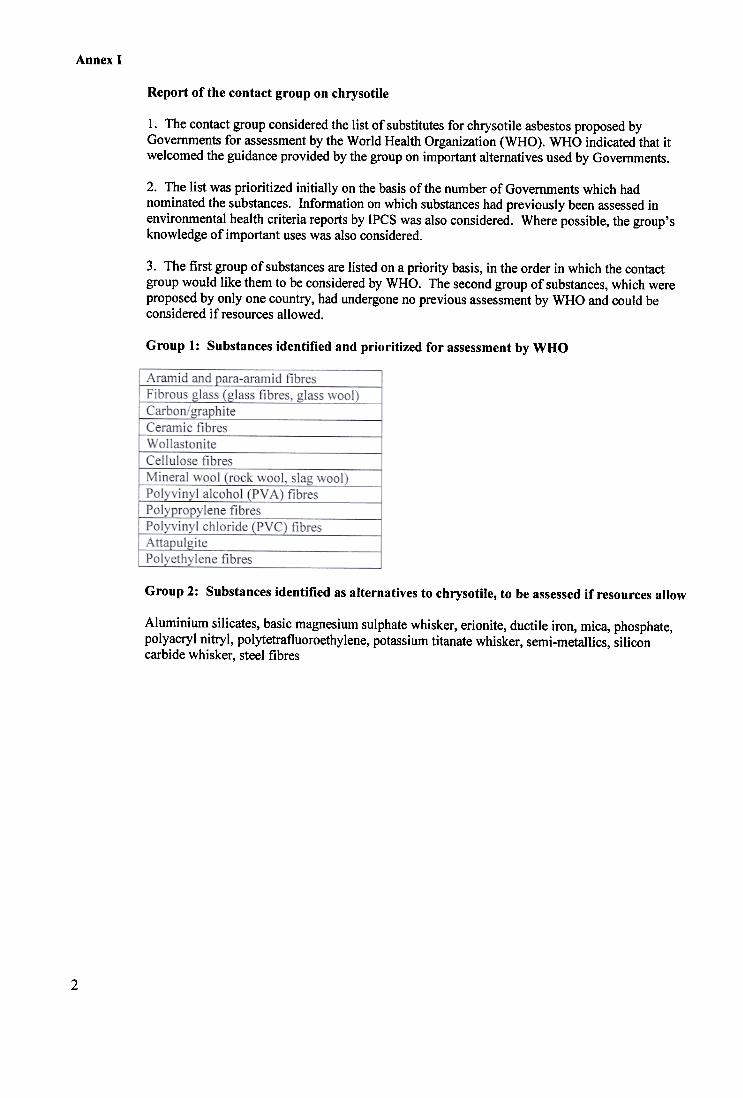
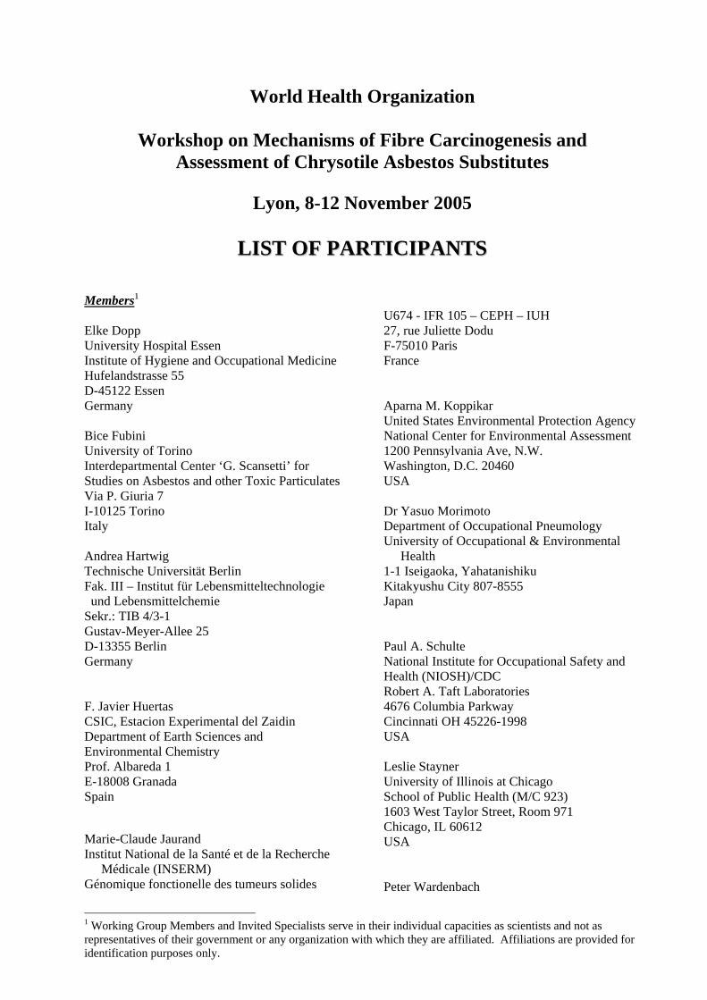
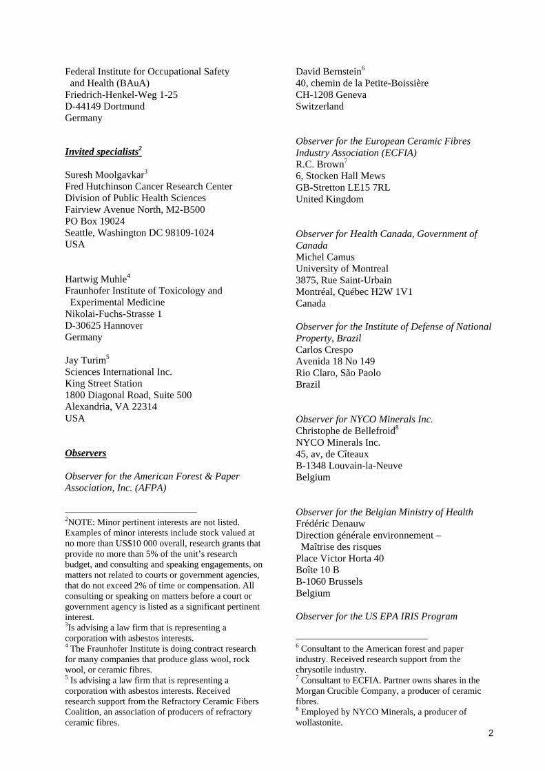