Embed Size (px)
Citation preview

Accuracy of faecal occult blood testand Helicobacter pylori stool antigentest for detection of uppergastrointestinal lesions
Yi-Chia Lee,1,2 Han-Mo Chiu,1,2 Tsung-Hsien Chiang,1,3 Amy Ming-Fang Yen,4
Sherry Yueh-Hsia Chiu,5 Sam Li-Sheng Chen,4 Jean Ching-Yuan Fann,6
Yen-Po Yeh,7 Chao-Sheng Liao,8 Tsung-Hui Hu,9 Chia-Hung Tu,1 Ping-Huei Tseng,1
Chien-Chuan Chen,1 Mei-Jyh Chen,1,3 Jyh-Ming Liou,1,2 Wei-Chih Liao,1,2
Yo-Ping Lai,1 Chen-Ping Wang,10 Jenq-Yuh Ko,10 Hsiu-Po Wang,1 Hung Chiang,11
Jaw-Town Lin,1,8,12,13 Hsiu-Hsi Chen,2 Ming-Shiang Wu1
To cite: Lee Y-C, Chiu H-M,Chiang T-H, et al. Accuracy offaecal occult blood testand Helicobacter pylori stoolantigen test for detection ofupper gastrointestinal lesions.BMJ Open 2013;3:e003989.doi:10.1136/bmjopen-2013-003989
▸ Prepublication history andadditional material for thispaper is available online. Toview these files please visitthe journal online(http://dx.doi.org/10.1136/bmjopen-2013-003989).
H-HC and M-SW contributedequally.
Received 10 September 2013Revised 27 September 2013Accepted 3 October 2013
For numbered affiliations seeend of article.
Correspondence toDr Ming-Shiang Wu;[email protected]
ABSTRACTObjective: Highly sensitive guaiac-based faecal occultblood (Hemoccult SENSA) and Helicobacter pylori stoolantigen testing might help detect upper gastrointestinallesions when appended to a colorectal cancer screeningprogramme with faecal immunochemical testing. Weevaluated the diagnostic accuracies of two stool tests indetecting upper gastrointestinal lesions.Design: Cross-sectional design.Setting: Hospital-based and community-basedscreening settings.Participants: A hospital-based deviation cohort of 3172participants to evaluate test performance and acommunity-based validation cohort of 3621 to verify thefindings.Interventions: Three types of stool tests withbidirectional endoscopy as the reference standard.Outcomes: Sensitivity, specificity and positive andnegative likelihood ratios.Results: For detecting upper gastrointestinal lesions incases with negative immunochemical tests, thesensitivity, specificity, and positive and negativelikelihood ratios of the guaiac-based and H pylori antigentests were 16.3% (95% CI 13.3% to 19.8%), 90.1%(88.9% to 91.2%), 1.64 (1.31 to 2.07), and 0.93 (0.89 to0.97), respectively, and 52.5% (48.1% to 56.9%), 80.6%(79.0% to 82.1%), 2.71 (2.41 to 3.04) and 0.59 (0.54 to0.65), respectively. For detecting upper gastrointestinallesions in cases with normal colonoscopy, the results ofthe guaiac-based and H pylori antigen tests were 17.9%(14.8% to 21.5%), 90.1% (88.9% to 91.2%), 1.81 (1.45to 2.26) and 0.91 (0.87 to 0.95), respectively, and 53.1%(48.6% to 57.4%), 80.7% (79.1% to 82.2%), 2.75 (2.45to 3.08) and 0.58 (0.53 to 0.64), respectively. Within thecommunity, positive predictive values of theimmunochemical and H pylori antigen tests were 36.0%(26.0% to 46.0%) and 31.9% (28.3% to 35.5%),respectively, for detecting lower and uppergastrointestinal lesions, which were similar to expectedvalues.
Strengths and limitations of this study
▪ Faecal occult blood tests include guaiac-basedtests and immunochemical tests; the former testsfor haeme while the latter tests for globin. Becauseglobin can be digested by enzymes in the uppergastrointestinal tract, the immunochemical test ismore specific to colorectal diseases. To differenti-ate lesions in the upper gastrointestinal tract fromlesions in the lower gastrointestinal tract, thehighly sensitive guaiac-based test (HemoccultSENSA) combined with the faecal immunochem-ical test may be helpful. In addition to the highlysensitive guaiac-based test, the Helicobacter pyloristool antigen test may be an alternative choice formass screening because H pylori is well known asa major cause of peptic ulcers and gastric cancer.
▪ In participants with normal colonoscopies, thehighly sensitive guaiac-based test can detect occultblood from the upper gastrointestinal tract inapproximately one-fourth of cancerous lesions andthe detectability increases with cancer stage. Inparticipants with negative immunochemical testsor normal colonoscopies, the H pylori stoolantigen test is more accurate than the highly sensi-tive guaiac-based test in the detection of uppergastrointestinal lesions.
▪ In community setting, it is applicable to add the Hpylori stool antigen test into the colorectal cancerscreening with the faecal immunochemical test.
▪ This is the first study to determine the diagnosticaccuracy of commercially available stool tests in thedetection of upper gastrointestinal lesions and alsothe first one to evaluate the feasibility of a two-in-onestool test for the simultaneous screening of upperand lower gastrointestinal lesions in the community.
▪ Further randomised controlled trials are neededto confirm the long-term efficacy and cost-effectiveness of using our screening strategy.
Lee Y-C, Chiu H-M, Chiang T-H, et al. BMJ Open 2013;3:e003989. doi:10.1136/bmjopen-2013-003989 1
Open Access Research
on February 6, 2022 by guest. P
rotected by copyright.http://bm
jopen.bmj.com
/B
MJ O
pen: first published as 10.1136/bmjopen-2013-003989 on 30 O
ctober 2013. Dow
nloaded from

Conclusions: The H pylori stool antigen test is more accurate than theguaiac-based test in the screening of upper gastrointestinal lesions in apopulation with high prevalence of H pylori infection and uppergastrointestinal lesions. It is applicable to add the H pylori antigen testto the immunochemical test for pan detection.Trial registration: NCT01341197 (ClinicalTrial.gov).
INTRODUCTIONUpper and lower gastrointestinal diseases have animpact on global health and economics.1 2 Althoughmass screening can detect early lesions,3 coordination ofdifferent methods to simultaneously screen lesions at dif-ferent depths is required.4 Thus, an efficient alternativescreening method is needed.A campaign against colorectal cancer has suggested
that the use of faecal occult blood tests can efficientlyreach the population and decrease mortality.5 Faecaloccult blood tests include guaiac-based test and faecalimmunochemical test (FIT); the former test measureshaeme, while the latter test measures globin. Becauseglobin can be digested by enzymes in the upper gastro-intestinal tract,6 the guaiac-based test combined with theimmunochemical test may help differentiate lesions inthe upper gastrointestinal tract from lesions in the lowergastrointestinal tract.7 8
Other than occult blood, stool tests detecting molecularmarkers are under continuous development; however,most stool tests are not widely available commercially.9 Oneexception is the Helicobacter pylori stool antigen test (HPSA)because H pylori is well-known as a major cause of pepticulcers and gastric cancer.10 As the absolute number ofpatients with gastric cancer is increasing due to advancingage of the global population,11 it makes sense to combinetwo specific tests (HPSA and FIT) into one panel for thedetection of upper as well as lower gastrointestinal lesions.Beginning in 2004, the Taiwanese Government
initiated a nationwide colorectal cancer screening pro-gramme using biennial FIT.12 Thus, a unique opportunityexists to evaluate the diagnostic accuracies of the guaiac-based and HPSA tests for detecting upper gastrointestinallesions, which was the primary aim of our study. Our sec-ondary aim was to evaluate the feasibility of combiningtwo stool tests for pan detection in a real world setting.
METHODSStudy designWe recruited consecutive participants from hospital-basedand community-based screening sources; the former(deviation cohort) was used to evaluate the test perform-ance and the latter (validation cohort) was to confirm thereproducibility and applicability of the screening strategyin the community. All participants provided signedinformed consent before enrolment. Both studies wereapproved by the Hospital Institutional Review Board(numbers IRB201101016RC and 201205030RIB).
DEVIATION COHORTRecruitment of study participantsFirst, beginning on 1 March 2011, we recruited partici-pants at the National Taiwan University Hospital (HealthManagement Center; Taipei, Northern Taiwan) throughadvertising messages for cancer screening. Participants>18 years of age who had completed the FIT, guaiac-based test, HPSA and bidirectional endoscopy wereincluded. Cancer detection was the main purpose of thestool test; a small sample of cancers was obtained from asingle site. To enrich the deviation cohort with thesample of gastrointestinal tract cancers, we also recruitedparticipants in whom gastrointestinal tract cancers weresuspected by the non-invasive screening test, such as radi-ology and oral inspection, at the Gastroenterologic orOtolaryngologic Clinics at National Taiwan UniversityHospital. Research staff recruited participants by explain-ing the purpose and eligibility requirements of the study.The participants were requested to complete three typesof stool tests and undergo bidirectional endoscopy;however, those participants who underwent only oneendoscopy for detection of a cancerous lesion were stilleligible because it was rare (approximately 0.9%) for onesubject to have important lesions in the upper as well aslower gastrointestinal tracts in the cancer screeninggroup. All of the stool samples were collected before con-firmatory endoscopic diagnoses were available.In the deviation cohort, we ensured that bleeding was
occult by excluding those participants who had overtgastrointestinal bleeding, including haematemesis, tarrystools, melena or hematochezia. We also excluded parti-cipants with histories of a gastrectomy and/or colectomyand pregnant or lactating women.
Stool testsTen days before cancer screening, we mailed one collec-tion card for the highly sensitive guaiac-based test(Hemoccult SENSA Single Slides; Beckman Coulter, Inc,USA) and two sampling tubes for the HPSA (Easy OneStep Test; Firstep Bioresearch, Inc, Taiwan) and the FIT(OC-SENSOR; Eiken Chemical Co, Ltd, Tokyo, Japan)to eligible participants. We used the 1-day method for allstool tests and advised participants to start diet and drugrestrictions 3 days before they obtained the stool samplesand to obtain stool samples within 2 days before startingthe bowel preparation. Stool samples were brought tothe hospital on the screening day and tested immedi-ately. Two technicians executed the guaiac-based andH pylori stool antigen tests and read the results inde-pendently; disagreements were resolved by consensus.For the cancer group, they were also requested to followthe same rule for stool sample collection.The sensitivity and specificity of the HPSA for the deter-
mination of H pylori infection status were 88% and 99%,respectively.13 The FIT was processed at the hospital’scentral laboratory using an automated reader and thecut-off concentration was set at 100 ng of haemoglobin/mLof buffer (equivalent to 20 μg haemoglobin/g of faeces).14
2 Lee Y-C, Chiu H-M, Chiang T-H, et al. BMJ Open 2013;3:e003989. doi:10.1136/bmjopen-2013-003989
Open Access
on February 6, 2022 by guest. P
rotected by copyright.http://bm
jopen.bmj.com
/B
MJ O
pen: first published as 10.1136/bmjopen-2013-003989 on 30 O
ctober 2013. Dow
nloaded from

Bidirectional endoscopyEndoscopic findings and histological results served asthe reference standard. Participants were given polyethyl-ene glycol for bowel preparation, which they drank at least4 h before endoscopic examinations. This bowel prepar-ation scheme had been shown with less than 5% poor orinadequate preparation in our population.15 The partici-pants were advised to stop anticoagulant or antiplatelettherapy for 7 days. Oesophagogastroduodenoscopy andcolonoscopy were performed by nine experiencedendoscopists, each with a minimum experience of 5000colonoscopies. Endoscopic findings were recorded elec-tronically with information on quality of preparation,completeness of colonoscopy, endoscopic findings andwhether or not a biopsy was performed. Participantswho declined endoscopy or had an incomplete colonos-copy, including failed caecal intubation and poor bowelpreparation, were excluded.
Definition of important lesionsLesions consistent with occult bleeding were defined asimportant, and were classified into three major categor-ies, as follows in flow diagrams: neoplastic, inflammatoryand vascular.7 16 Important upper gastrointestinal lesionsincluded cancer, an ulcer >0.5 cm in diameter, refluxoesophagitis with a severity of Los Angeles grade C orD,17 Barrett’s oesophagus, oesophageal varix with aseverity of form II or III,18 and gastric antral vascularectasia. Gastritis and ulcer scars were not included.Important lower gastrointestinal lesions included colo-rectal cancer, advanced adenomas, ulcers or colitis andangiodysplasia. An advanced adenoma was defined as>10 mm in diameter or having a villous component orhigh-grade dysplasia.11 Adenomas <10 mm in diameter,hyperplastic polyps and haemorrhoids were notincluded. All endoscopists were blinded to stool testresults before endoscopic examination.
VALIDATION COHORTRecruitment of study subjectsAccording to the results of the deviation cohort, wefound that the H pylori stool antigen test had a betterperformance in the detection of upper gastrointestinallesions. We then evaluated the applicability and reprodu-cibility of this strategy in a community population aged50–69 years (Changhua County, central Taiwan) in theChanghua Community-based Integrated Screening(CHCIS). This integrated screening programme has pro-vided oral inspection for oral/throat cancer, mammog-raphy for breast cancer, Pap smear for cervical cancerand FIT for colorectal cancer since 2005.4 In 2012, weadded the HPSA into this screening programme andused standard screening indicators, including participa-tion rate, positive rate, referral rate, endoscopic findings,positive predictive value and detection rate, to confirmthe applicability of this new strategy in the communityand compared the observed and estimated positive
predictive values to confirm the reproducibility of thestool test performance.19 The endoscopic diagnoses ofgastrointestinal neoplasia were confirmed by histologicalresults under routine medical practice.Under the auspices of the Changhua County Public
Health Bureau and the Health Promotion Administration,Ministry of Health and Welfare, a series of consensusmeetings and educational programmes were held forprimary care physicians and first-line healthcare workersbefore implementation. Beginning on 21 April 2012, eli-gible participants were invited by telephone or postcardfrom 27 public health units covering a total of 26 town-ships. Participants with positive results were referred to 15local gastrointestinal clinics and 9 hospitals in ChanghuaCounty for antibiotic treatment and/or endoscopic diag-noses. The sample size was planned to be similar to thehospital-based deviation cohort.
STATISTICAL ANALYSISStool test performance and sample size estimationWe began our analyses from evaluations of the guaiac-based and FIT for the screening of lower gastrointestinallesions. We used test results and colonoscopic findingsto construct a 2×2 table and expressed test performanceas sensitivity, specificity, positive and negative likelihoodratios and corresponding 95% CIs.For the detection of upper gastrointestinal lesions, our
sample size estimation was based on the fact that thedetectability of the guaiac-based test for the uppergastrointestinal lesions should be interpreted togetherwith a negative FIT.7 We anticipated that the prevalenceof the upper gastrointestinal lesions would be higher incases with positive guaiac-based tests, but negative FIT.We were aware that approximately 12% and 4% of ageneral population would test positive with the guaiac-based and FIT, respectively, so at least 8% of the partici-pants would have positive results for the guaiac-basedtest, but negative results for the FIT.20 21 We assumedthat the prevalence of upper gastrointestinal lesionswould be at least 30%, which was 10% higher than thatof the general population,6 thus the guaiac-based testwould yield a diagnostic OR of at least 1.7. By setting apower of 80% and a 0.05 one-side type 1 error, we deter-mined that an overall sample size of 3138 would be suffi-cient. To account for 10% participants who might returnan inadequate sample, we planned to test 3500participants.We also evaluated whether or not the HPSA would be
effective under the same testing conditions. We assumedthat the prevalence of upper gastrointestinal lesions withoccult bleeding would be 10% higher in participantswith H pylori infection than the prevalence in thegeneral population. We knew that approximately one-fourth of our population were infected with H pylori andthat 88% would test positive.13 Based on the predefinedtype 1 error and predetermined sample size, the powerto reject our null hypothesis was 99%.
Lee Y-C, Chiu H-M, Chiang T-H, et al. BMJ Open 2013;3:e003989. doi:10.1136/bmjopen-2013-003989 3
Open Access
on February 6, 2022 by guest. P
rotected by copyright.http://bm
jopen.bmj.com
/B
MJ O
pen: first published as 10.1136/bmjopen-2013-003989 on 30 O
ctober 2013. Dow
nloaded from

In the previous scenario, performance of the guaiac-based test in detecting upper gastrointestinal lesionscould be underestimated because some colorectallesions would test negative by the FIT. Therefore, we alsoevaluated whether or not we could use the guaiac-basedtest to guide the use of oesophagogastroduodenoscopyin cases with normal colonoscopy results.16
For different clinical scenarios, we calculated the testperformance of the guaiac-based and HPSA in detectingupper gastrointestinal lesions and used the two-sampleproportional test to make comparisons between thetests. A two-sided p value <0.05 indicated a significantdifference. Statistical analyses were performed using SASV.9.2. Knowing that the guaiac-based and HPSA testsmay detect different lesions of the gastrointestinal tract,we did subgroup analyses according to the anatomicsites and tumour stages22 to determine whether or nottest sensitivity would change.
Community validationUsing sensitivity and specificity derived from ourhospital-based study and reported prevalence of gastro-intestinal lesions in the community, we estimated thepositive predictive values of stool tests in the communitywith the Bayes’ rule23
Positive predictive value ¼ðSensitivityÞðprevalenceÞ
ðSensitivityÞðprevalenceÞþð1�specificityÞð1�prevalenceÞ
We compared the estimated values with observedvalues using WinBUGS (V.1.4; MRC Biostatistics Unit,Cambridge, UK (see online supplementary appendixfigure S1)).
SENSITIVITY ANALYSISTo solidify the generalisability of our findings, we con-structed a decision model using TreeAge Pro 2009(TreeAge Software, Inc, USA (see online supplementaryappendix figure S2)) based on the choice of stool tests,the prevalence of important lesions in the upper gastro-intestinal tract, and the prevalence of H pylori infectionin participants with or without upper gastrointestinallesions to determine how changes in population charac-teristics might affect positive predictive values.23 24
Base-case estimates were derived from our hospital-basedstudy and ranges of sensitivity analyses are shown inonline supplementary appendix table S1.13 25 We set apositive predictive value of >10% as a minimal require-ment to support screen tests in the community.26
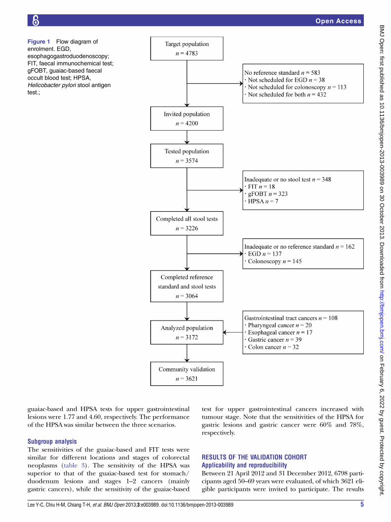
RESULTSResults of the deviation cohortParticipation rateBetween 1 March 2011 and 15 February 2012, 4783participants who underwent cancer screening were
evaluated, of which 4200 were invited to participate inthe study and 3574 (85.1%) ultimately participated(figure 1). The completion rates for the guaiac-based,HPSA and FIT tests were 91%, 99.8% and 99.5%,respectively. Between 19 July 2011 and 21 November2012, we also enriched the hospital-based cohort with108 screen-detected cancers from other sites and all par-ticipants completed the stool tests. Our deviation cohortof 3172 participants was comprised of two groups.
Results of stool tests and endoscopyDescriptive results are shown in table 1 and the flow dia-grams are shown in figures 2 and 3. Among the cancerscreening group (n=3064), the positive rates of theguaiac-based, HPSA and immunochemical tests were11.3%, 24.2% and 3.1%, respectively, and the prevalenceof upper and lower gastrointestinal lesions was 14.5%and 4.6%, respectively. Among the screen-detectedcancer group (n=108), 33 participants with upper gastro-intestinal cancers did not undergo colonoscopy, whileone participant with colon cancer did not undergooesophagogastroduodenoscopy.Overall, the prevalence of upper gastrointestinal
lesions was similar between participants with positiveand negative results on the FIT (26/133 (19.6%) vs497/3039 (16.4%), p=0.31). Also, the prevalence of colo-rectal lesions was similar between participants with posi-tive and negative results on the HPSA (44/789 (5.6%) vs130/2383 (5.5%), p=0.90). However, the prevalence ofupper gastrointestinal lesions was higher in participantswith positive results on the guaiac-based test than thosewith negative results (98/397 (24.7%) vs 425/2775(15.3%), p<0.001). No serious adverse events occurredrelated to the endoscopic screening.In participants with a negative FIT or normal colonos-
copy, the prevalence of upper gastrointestinal lesionswas higher in participants with a positive guaiac-basedtest (81/333 (24.3%) and 80/328 (24.4%), respectively)and in participants with a positive HPSA (261/754(34.6%) and 249/733 (34.0%), respectively) than theoverall population (523/3172 (16.5%); all p<0.001).
Stool test performanceTo detect colorectal lesions or cancers, the sensitivityand negative likelihood ratio were similar between theguaiac-based and immunochemical tests, while the speci-ficity and positive likelihood ratio of the FIT were signifi-cantly higher (table 2, scenario 1, p<0.001).In participants with a negative FIT (scenario 2) or
normal colonoscopy (scenario 3), the sensitivity and posi-tive and negative likelihood ratios of the HPSA were sig-nificantly better than those of the guaiac-based test indetecting upper gastrointestinal lesions (p<0.001), whilethe specificity was lower. For upper gastrointestinalcancers, the results were similar, except that there was nosignificant difference between the two tests with respectto the positive and negative likelihood ratios. In partici-pants with a negative FIT, the diagnostic ORs of the
4 Lee Y-C, Chiu H-M, Chiang T-H, et al. BMJ Open 2013;3:e003989. doi:10.1136/bmjopen-2013-003989
Open Access
on February 6, 2022 by guest. P
rotected by copyright.http://bm
jopen.bmj.com
/B
MJ O
pen: first published as 10.1136/bmjopen-2013-003989 on 30 O
ctober 2013. Dow
nloaded from
guaiac-based and HPSA tests for upper gastrointestinallesions were 1.77 and 4.60, respectively. The performanceof the HPSA was similar between the three scenarios.
Subgroup analysisThe sensitivities of the guaiac-based and FIT tests weresimilar for different locations and stages of colorectalneoplasms (table 3). The sensitivity of the HPSA wassuperior to that of the guaiac-based test for stomach/duodenum lesions and stages 1–2 cancers (mainlygastric cancers), while the sensitivity of the guaiac-based
test for upper gastrointestinal cancers increased withtumour stage. Note that the sensitivities of the HPSA forgastric lesions and gastric cancer were 60% and 78%,respectively.
RESULTS OF THE VALIDATION COHORTApplicability and reproducibilityBetween 21 April 2012 and 31 December 2012, 6798 parti-cipants aged 50–69 years were evaluated, of which 3621 eli-gible participants were invited to participate. The results
Figure 1 Flow diagram of
enrolment. EGD,
esophagogastroduodenoscopy;
FIT, faecal immunochemical test;
gFOBT, guaiac-based faecal
occult blood test; HPSA,
Helicobacter pylori stool antigen
test.;
Lee Y-C, Chiu H-M, Chiang T-H, et al. BMJ Open 2013;3:e003989. doi:10.1136/bmjopen-2013-003989 5
Open Access
on February 6, 2022 by guest. P
rotected by copyright.http://bm
jopen.bmj.com
/B
MJ O
pen: first published as 10.1136/bmjopen-2013-003989 on 30 O
ctober 2013. Dow
nloaded from

are shown in table 4. We estimated that the positive pre-dictive values were 34.5% (95% CI 31.6% to 37.5%) and35.5% (28.0% to 43.8%), respectively, for the screening ofupper and lower gastrointestinal lesions in the commu-nity27 28; the results were similar to the observed values of31.9% (28.3% to 35.5%) and 36.0% (26.0% to 46.0%),respectively. The number of endoscopies needed to findone upper and one lower gastrointestinal lesion was 3.1(2.8–3.5) and 2.8 (2.1–3.7), respectively.For cancerous lesions, the annual rates were 50.4 and
103.8/100 000 participants aged 50–69 years, respect-ively, for upper (pharyngeal, oesophageal and gastriccancers) and lower gastrointestinal cancers, which wereestimated by searching the databases of the TaiwanCancer Registry29 and weighting the age and gender dis-tributions of our tested population in the community.The estimated positive predictive values were 0.11%(95% CI 0.08% to 0.12%) and 2.57% (1.99 to 3.20%)for upper and lower gastrointestinal cancers, respectively,and the number of endoscopies needed to find oneupper and one lower gastrointestinal cancer was 890(705–1200) and 39 (31–50), respectively. We expectedthat approximately 1 of 643 and 2 of 89 participants whounderwent endoscopy had upper and lower gastrointes-tinal cancers, respectively. In the real community-basedscreening, we detected 2 gastric cancers and 2 colorectalcancers.
SENSITIVITY ANALYSISIn our study under the scenario of a negative FIT, thepositive predictive value of the HPSA was significantlyhigher than that of the guaiac-based test (see onlinesupplementary appendix figure S3). When the preva-lence of upper gastrointestinal lesions was >5%, the posi-tive predictive values of the guaiac-based and HPSAwould be >10% (see online supplementary appendixtable S2). The positive predictive value of the HPSAwould be higher than that of the guaiac-based test whenthe prevalence of latent H pylori infections in partici-pants with no upper gastrointestinal lesions was <30%(see online supplementary appendix figure S4). Withinthe reported range of sensitivity and specificity of theHPSA, the above findings were not substantiallychanged.
DISCUSSIONHigh cure rates can be achieved for gastrointestinal tractlesions identified in the presymptomatic stage.Colorectal neoplasms are currently screened at thepopulation level. In the present study, we first supportedthe use of FIT for the screening of colorectal lesions.Second, we evaluated the accuracies of the guaiac-basedand HPSA tests for the screening of upper gastrointes-tinal lesions in cases with negative FIT or normal
Table 1 Demographic data, stool test results and endoscopic findings in the hospital-based deviation cohort
Characteristics Population, n=3172
Male gender (%) 1919 (60.5)
Age in years (SD; range) 53.0 (11.7; 19.0–91.8)
Positive stool test results (%)
Guaiac-based faecal occult blood test 397 (12.5)
Helicobacter pylori stool antigen test 789 (24.9)
Faecal immunochemical test 133 (4.2)
Important lesions in the upper gastrointestinal tract (%) 523 (16.5)
Categories
Pharyngeal or oesophageal carcinoma 37 (1.2)
Erosive oesophagitis, grade C or D 46 (1.5)
Barrett’s oesophagus 8 (0.3)
Oesophageal varices, form II or III 5 (0.2)
Gastric carcinoma 41 (1.2)
Peptic ulcers 365 (11.5)
Gastric antral vascular ectasia 9 (0.3)
Anatomic sites
Pharynx or oesophagus 96 (3.0)
Stomach or duodenum 439 (13.8)
Important lesions in the lower gastrointestinal tract (%) 174 (5.5)
Categories
Colorectal carcinoma 39 (1.2)
Advanced adenoma 106 (3.3)
Colitis or ulcer 25 (0.8)
Angiodysplasia 4 (0.1)
Anatomic sites*
Proximal colon 98 (3.1)
Distal colon 76 (2.4)
*Proximal colon was defined as the level above splenic flexure (including splenic flexure). When synchronous colon lesions were found, theanatomic site of the most important one was used to define the location.
6 Lee Y-C, Chiu H-M, Chiang T-H, et al. BMJ Open 2013;3:e003989. doi:10.1136/bmjopen-2013-003989
Open Access
on February 6, 2022 by guest. P
rotected by copyright.http://bm
jopen.bmj.com
/B
MJ O
pen: first published as 10.1136/bmjopen-2013-003989 on 30 O
ctober 2013. Dow
nloaded from

colonoscopy. Third, we verified our findings by addingthe HPSA to the colorectal screening programme in acommunity population. Taken together, the results ofour work contribute new knowledge to the pan detec-tion of gastrointestinal tract diseases.A recent review suggested that current evidence
remained insufficient to recommend for or againstroutine upper endoscopy for patients with positive stooloccult blood tests, but normal colonoscopy, because pre-vious studies have found that upper endoscopic exami-nations can detect lesions in a wide range 13–45%.30
Such a heterogeneity is related to the fact that the yieldrate of oesophagogastroduodenoscopy is subject to thedifferences in the types of faecal occult blood tests and
the population characteristics across different studies.Using the reference standard of bidirectional endosco-pies for participants with either positive or negative testresults or with guaiac-based or immunochemical tests,our study quantified the accuracy of this finding byshowing that in patients with normal colonoscopies, thehighly sensitive guaiac-based test can detect occult bloodfrom the upper gastrointestinal tract in approximatelyone-fourth of cancerous lesions and the detectabilityincreased with cancer stage, thus providing guidance forthe use of oesophagogastroduodenoscopy, especiallywhen clinical symptoms/signs, such as iron-deficiencyanaemia, are present.31 However, in participants with anegative FIT alone, a non-significant positive likelihood
Figure 2 Flow diagram for the screening using the guaiac-based occult blood test and the faecal immunochemical test
according to the Standards for Reporting of Diagnostic Accuracy statement in the hospital-based deviation cohort.
Lee Y-C, Chiu H-M, Chiang T-H, et al. BMJ Open 2013;3:e003989. doi:10.1136/bmjopen-2013-003989 7
Open Access
on February 6, 2022 by guest. P
rotected by copyright.http://bm
jopen.bmj.com
/B
MJ O
pen: first published as 10.1136/bmjopen-2013-003989 on 30 O
ctober 2013. Dow
nloaded from

ratio did not support this interpretation. The positivityof FIT was irrelevant to the presence of upper gastro-intestinal lesions, similar to the results we previouslyreported.6
Regarding the use of HPSA, we found that the sensitiv-ities of 60% and 78% for gastric lesions and gastriccancer were non-inferior to those of 35% and 85% forFIT to detect colorectal lesions and colorectal cancers.Our results were consistent with the estimated fractionof 75% for gastric cancers attributable to H pylori infec-tion.32 Screening for H pylori infection is unique becauseof its once-in-a-lifetime design,33 34 and effective treat-ments are available.35 36 We invited individuals ataverage risk for colorectal neoplasms to have the HPSA
because in this age range the prevalence of latent Hpylori infection is decreased.37 Without previous oppor-tunistic treatment, peptic ulcers and irreversible molecu-lar changes are prevalent after long-term infection.38
Furthermore, the use of non-steroidal anti-inflammatorydrugs/aspirin increased (8% in our community popula-tion), which would have a synergistic effect with H pyloriinfection on the risk of peptic ulcers.39 Therefore, itwould be appropriate to undergo endoscopy for screen-ing early gastric neoplasms, in addition to the obviousbenefit from antibiotic treatment for the peptic ulcer,chronic gastritis and chemoprevention.34
In areas where the prevalence of upper gastrointes-tinal lesions is high and the prevalence of latent H pylori
Figure 3 Flow diagram for the screening using the Helicobacter pylori stool antigen test and the faecal immunochemical test
according to the Standards for Reporting of Diagnostic Accuracy statement in the hospital-based deviation cohort.
8 Lee Y-C, Chiu H-M, Chiang T-H, et al. BMJ Open 2013;3:e003989. doi:10.1136/bmjopen-2013-003989
Open Access
on February 6, 2022 by guest. P
rotected by copyright.http://bm
jopen.bmj.com
/B
MJ O
pen: first published as 10.1136/bmjopen-2013-003989 on 30 O
ctober 2013. Dow
nloaded from

Table 2 Performance and the corresponding 95% CIs of three stool tests in screening important lesions in the lower or upper gastrointestinal tract under three different
scenarios in the hospital-based deviation cohort
Outcome variables Sensitivity (%) Specificity (%) Positive likelihood ratio Negative likelihood ratio
Scenario 1: screen for lesions in the lower or upper gastrointestinal tract in all participants
Guaiac-based faecal occult blood test
Important lesions in the lower gastrointestinal tract 61/174 (35.1; 28.4–42.4) 2637/2965 (88.9; 87.8–90.0) 3.17 (2.53–3.98) 0.73 (0.65–0.82)
Colorectal cancers 34/39 (87.2; 73.3–94.4) 2770/3133 (88.4; 87.3–89.5) 7.52 (6.45–8.78) 0.15 (0.06–0.33)
Important lesions in the upper gastrointestinal tract 98/523 (18.7; 15.6–22.3) 2350/2648 (88.8; 87.5–89.9) 1.67 (1.35–2.05) 0.92 (0.88–0.96)
Cancers in the upper gastrointestinal tract 21/78 (26.9; 18.3–37.7) 2718/3093 (87.9; 86.7–89.0) 2.22 (1.52–3.24) 0.83 (0.73–0.95)
Faecal immunochemical test
Important lesions in the lower gastrointestinal tract 65/174 (37.4; 30.5–44.7) 2900/2965 (97.8; 97.2–98.3) 17.04 (12.52–23.19) 0.64 (0.57–0.72)
Colorectal cancers 32/39 (82.1; 67.3–91.0) 3032/3133 (96.8; 96.1–97.3) 25.45 (19.99–32.41) 0.19 (0.10–0.36)
Helicobacter pylori stool antigen test
Important lesions in the upper gastrointestinal tract 277/523 (53.0; 48.7–57.2) 2137/2648 (80.7; 79.2–82.2) 2.75 (2.45–3.07) 0.58 (0.53–0.64)
Cancers in the upper gastrointestinal tract 42/78 (53.9; 42.9–64.5) 2347/3093 (75.9; 74.3–77.4) 2.23 (1.80–2.77) 0.61 (0.48–0.77)
Scenario 2: screen for lesions in the upper gastrointestinal tract in participants with negative results on the faecal immunochemical test
Guaiac-based faecal occult blood test
Important lesions in the upper gastrointestinal tract 81/497 (16.3; 13.3–19.8) 2290/2542 (90.1; 88.9–91.2) 1.64 (1.31–2.07) 0.93 (0.89–0.97)
Cancers in the upper gastrointestinal tract 12/69 (17.4; 10.2–28.0) 2649/2970 (89.2; 88.1–90.3) 1.61 (0.96–2.73) 0.93 (0.83–1.03)
H pylori stool antigen test
Important lesions in the upper gastrointestinal tract 261/497 (52.5; 48.1–56.9) 2049/2542 (80.6; 79.0–82.1) 2.71 (2.41–3.04) 0.59 (0.54–0.65)
Cancers in the upper gastrointestinal tract 36/69 (52.2; 40.6–63.5) 2252/2970 (75.8; 74.2–77.3) 2.16 (1.71–2.73) 0.63 (0.49–0.81)
Scenario 3: screen for lesions in the upper gastrointestinal tract in participants with normal results on the colonoscopy
Guaiac-based faecal occult blood test
Important lesions in the upper gastrointestinal tract 88/492 (17.9; 14.8–21.5) 2258/2506 (90.1; 88.9–91.2) 1.81 (1.45–2.26) 0.91 (0.87–0.95)
Cancers in the upper gastrointestinal tract 21/77 (27.3; 18.6–38.1) 2606/2921 (89.2; 88.0–90.3) 2.53 (1.73–3.70) 0.82 (0.71–0.94)
H pylori stool antigen test
Important lesions in the upper gastrointestinal tract 261/492 (53.1; 48.6–57.4) 2022/2506 (80.7; 79.1–82.2) 2.75 (2.45–3.08) 0.58 (0.53–0.64)
Cancers in the upper gastrointestinal tract 41/77 (53.3; 42.2–64.0) 2217/2921 (75.9; 74.3–77.4) 2.21 (1.78–2.75) 0.62 (0.49–0.78)
LeeY-C,Chiu
H-M,Chiang
T-H,etal.BMJOpen
2013;3:e003989.doi:10.1136/bmjopen-2013-003989
9
OpenAccess
on February 6, 2022 by guest. Protected by copyright. http://bmjopen.bmj.com/ BMJ Open: first published as 10.1136/bmjopen-2013-003989 on 30 October 2013. Downloaded from

infection is modest, our sensitivity analyses showed thatmass screening with HPSA may be worthwhile.Representative areas may include East Asia, LatinAmerica and East Europe.40–46 The results also may beapplied to select populations with high risk, includingthose with a history of upper gastrointestinal bleeding,those with previous history of peptic ulcer, users of non-steroidal anti-inflammatory drugs, aspirin or clopidogrel,seriously ill patients, elderly participants and immigrantsfrom high-risk areas.10 However, the positive predictivevalue of the HPSA was lower than that of the guaiac-based test when the prevalence of latent H pylori infec-tion was >30%. Such a circumstance may be observed inareas where the prevalence of upper gastrointestinallesions was moderate, but the prevalence of latentH pylori infection was high. The representative areas mayinclude India and Africa.47 48
The strengths of this study include large sample size,use of bidirectional endoscopy as the reference standard,evaluation of three tests with high commercial availability,and assessment of applicability and reproducibility in the
community. A sensitivity analysis according to the preva-lence of upper gastrointestinal lesions and H pylori infec-tion further solidified the generalisability of thesefindings. Nevertheless, our study had limitations. First, weused the 1-day method for the highly sensitive guaiac-based test. We found that its sensitivity was similar to thatof the FIT for detecting colorectal lesions, indicating thatsuch a 1-day test was also an option. However, we mighthave underestimated the sensitivity of the guaiac-basedtest for detecting upper gastrointestinal lesions as occultblood was more likely intermittent and unevenly distribu-ted in the stool, requiring multiple samples. However, inour population, this approach must be weighed againstthe lower completion rate (91%) of using the woodenspatula collection of stool samples for the guaiac-basedtests. Second, there is still room for improvement in testperformance. Both tests were not able to predict lesionsin the upper aerodigestive tract. Distal small bowel lesionsmight be a possible source for the guaiac or immuno-chemical positivity.49 Other choices, including the hae-meporphyrin test7 50 and the stool-based DNA test,51 52
Table 3 The number of positive cases and the corresponding sensitivity of stool tests in the screening of important lesions in
the upper or lower gastrointestinal tract, stratified by the anatomic sites and cancer stages in the hospital-based deviation
cohort
Lesions in the lower gastrointestinal tract Faecal immunochemical test (%)
Guaiac-based fecal
occult blood test (%) All
Location of the lesions
Overall 65 (37.4) 61 (35.1) 174
Distal colon lesions 28 (36.8) 28 (36.8) 76
Proximal colon lesions 37 (37.8) 33 (33.7) 98
Location of the cancers
Overall 32 (82.1) 34 (87.2) 39
Distal colon cancers 17 (94.4) 17 (94.4) 18
Proximal colon cancers 15 (71.4) 17 (81) 21
Stage of the cancers
Stages 1–2 colon cancers 17 (81) 17 (81) 21
Stages 3–4 colon cancers 15 (83.3) 17 (94.4) 18
Lesions in the upper gastrointestinal tract Helicobacter pylori stool antigen test (%)
Guaiac-based faecal
occult blood test (%)
Location of the lesions
Overall 261 (53.1) 88 (17.9) 492
Stomach and duodenum lesions 247 (59.4) 72 (17.3) 416
Pharynx and oesophagus lesions 14 (18.4) 16 (21.1) 76
Location of the cancers
Overall 41 (53.3) 21 (27.3) 77
Gastric carcinoma 31 (77.5) 10 (25) 40
Pharynx and oesophagus cancers 10 (27) 11 (29.7) 37
Stage of upper gastrointestinal cancers
Stages 1–2 upper gastrointestinal cancers 21 (75) 5 (17.9) 28
Stages 3–4 upper gastrointestinal cancers 20 (40.8) 16 (32.7) 49
The tumour stage was defined by the American Joint Committee on Cancer seventh edition.22
The colon above the level of the splenic flexure (including splenic flexure) was defined as the proximal colon. Synchronous lesions denotedconcurrent proximal and distal lesions of colorectal neoplasms. When multiple lesions were present, participants were categorised accordingto the highest severity of lesions in the proximal and distal colon.Note that those who had important lower gastrointestinal lesions have been excluded from the evaluation of test positivity for the uppergastrointestinal lesions. Also note that the majority (25/28, 89.3%) of stages 1–2 cancers were stomach cancers while the majority (34/49,69.4%) of stages 3–4 cancers were pharynx or oesophagus cancers.
10 Lee Y-C, Chiu H-M, Chiang T-H, et al. BMJ Open 2013;3:e003989. doi:10.1136/bmjopen-2013-003989
Open Access
on February 6, 2022 by guest. P
rotected by copyright.http://bm
jopen.bmj.com
/B
MJ O
pen: first published as 10.1136/bmjopen-2013-003989 on 30 O
ctober 2013. Dow
nloaded from

may be evaluated in future studies. Third, although wehave proven the applicability of our two-in-one strategy ina real world setting with a high uptake rate and a fairreferral rate, its feasibility would be limited in areaswhere the resource for endoscopists is constrained or theexpense for upper endoscopy is high. Further risk-scorebased approach with targeted endoscopy may be onepotential solution.53 Finally, a low proportion of maleswas seen in the community-based population because theinvitation list was also designed for female cancer screen-ing. Although we have adjusted the age and gender distri-butions of our community population in the evaluationof test reproducibility, self-selection bias could not beexcluded. Also, the present study was not designed in theway to give a definite answer with respect to the massH pylori screening/eradication strategy. Further rando-mised trials and cost-effectiveness analyses are warrantedto confirm the long-term benefit in adding screening forgastric lesions to the colorectal cancer screening.
In summary, we have demonstrated that, in our popu-lation with negative FIT or normal colonoscopy, theHPSA is more accurate than the guaiac-based test inscreening for upper gastrointestinal lesions. In a popula-tion with prevalent upper and lower gastrointestinallesions, the HPSA may be appended to the colorectalcancer screening with the FIT.
Author affiliations1Department of Internal Medicine, College of Medicine, National TaiwanUniversity, Taipei, Taiwan2Graduate Institute of Epidemiology and Preventive Medicine, College ofPublic Health, National Taiwan University, Taipei, Taiwan3Department of Integrated Diagnostic and Therapeutics, National TaiwanUniversity Hospital, Taipei, Taiwan4School of Oral Hygiene, College of Oral Medicine, Taipei Medical University,Taipei, Taiwan5Department and Graduate Institute of Health Care Management, Chang GungUniversity, Tao-Yuan, Taiwan6Department and Graduate Institute of Health Care Management, KainanUniversity, Tao-Yuan, Taiwan
Table 4 Demographic data, participation rate, positive rate, referral rate, endoscopic findings and stool test performance in
the community-based validation cohort
Characteristics Invited population, n=3621
Male gender (%) 941 (26)
Age in years (SD; range) 57.9 (5.3; 50–69)
Participation rate (%)*
Helicobacter pylori stool antigen test 3454/3621 (95.4)
Faecal immunochemical test 3432/3621 (94.8)
Positive rate for stool test results (%)
H pylori stool antigen test 1251/3454 (36.2)
Faecal immunochemical test 152/3432 (4.4)
Referral rate (%)
Positive H pylori stool antigen test 817/1251 (65.3)
Antibiotic treatment 755/817 (92.4)
Oesophagogastroduodenoscopy 643/817 (78.7)
Colonoscopy for positive faecal immunochemical test 89/152 (58.6)
Endoscopic findings (%)
Important lesions in the upper gastrointestinal tract 205/643 (31.9)
Categories
Erosive oesophagitis, grade C or D 7 (1.1)
Barrett’s oesophagus 1 (0.2)
Oesophageal varices, form II or III 4 (0.6)
Gastric carcinoma 2 (0.3)
Peptic ulcers 189 (29.4)
Gastric antral vascular ectasia 2 (0.3)
Important lesions in the lower gastrointestinal tract (%) 32/89 (36.0)
Categories
Colorectal carcinoma 2 (2.2)
Advanced adenoma 27 (30.3)
Colitis or ulcer 3 (3.4)
Stool test performance (%)
Positive predictive value for gastrointestinal tract lesions
H pylori stool antigen test 205/643 (31.9)
Faecal immunochemical test 32/89 (36.0)
Detection rate for gastrointestinal tract lesions
H pylori stool antigen test 205/3454 (5.9)
Faecal immunochemical test 32/3432 (0.9)
*Total number of participants who have returned an adequate stool test sample.
Lee Y-C, Chiu H-M, Chiang T-H, et al. BMJ Open 2013;3:e003989. doi:10.1136/bmjopen-2013-003989 11
Open Access
on February 6, 2022 by guest. P
rotected by copyright.http://bm
jopen.bmj.com
/B
MJ O
pen: first published as 10.1136/bmjopen-2013-003989 on 30 O
ctober 2013. Dow
nloaded from

7Changhua County Public Health Bureau, Changhua County, Taiwan8Division of Gastroenterology, Department of Internal Medicine, Shin-KongWu Ho-Su Memorial Hospital, Taipei, Taiwan9Division of Hepatogastroenterology, Department of Internal Medicine,Kaohsiung Chang Gung Memorial Hospital and Chang Gung UniversityCollege of Medicine, Kaohsiung, Taiwan10Department of Otolaryngology, National Taiwan University Hospital, Taipei,Taiwan11Taipei Institute of Pathology, Taipei, Taiwan12School of Medicine, Fu Jen Catholic University, New Taipei City, Taiwan13Department of Internal Medicine, E-Da Hospital, Kaohsiung County, Taiwan
Acknowledgements The authors would like to thank the nurses at the HealthManagement Center of National Taiwan University Hospital for their assistancein the hospital-based study and the staff of the Eighth Core Laboratory,Department of Medical Research of National Taiwan University Hospital fortechnical support for stool tests. The authors also express special thanks tothe following clinics and hospitals for their assistance and providing care tothe participants of the community-based study in Changhua County: ChangHong Medicine Clinic, Chang Yu Medicine Clinic, Chen Yen Hsueh MedicineClinic, Chi Hsu Lien Clinic, Chia Ho Medicine Clinic, Chia Lin Clinic, ChingHung Clinic, Huang Gung Chin Medicine and Pediatrics Clinic, Feng-An GroupClinic, Lu Tian En Clinic, Shih-Ren Clinic, Wang Chang Hao Medicine Clinic,Wang Jian Lung Medicine Clinic, Wu Shun Yu Medicine Clinic, Ye Teng HsinMedicine Clinic, Chang-Hua Hospital, Department of Health, Executive Yuan,ROC, Chang Bing Show Chwan Memorial Hospital, Changhua Show ChwanMemorial Hospital, Changhua Christian Hospital, Changhua ChristianHospital-Erlin Branch, Changhua Christian Hospital-Lukang Branch, Han MingHospital, Yuan Rung Hospital, and Yuan Sheng Hospital.
Contributors Y-CL and M-SW have full access to the data in thehospital-based study, take responsibility for the integrity of the data, and theaccuracy of the data analysis. Y-CL and H-HC have full access to thecommunity-based study, take responsibility for the integrity of the data andthe accuracy of the data analysis. Y-CL, H-MC, Y-PY, C-SL, T-HH, H-HC andM-SW contributed to the study concept and design; Y-CL, H-MC, T-HC,AM-FY, SY-HC, SL-SC, JC-YF, Y-PY, C-SL, T-HH, C-HT, P-HT, C-CC, M-JC,J-ML, W-CL, Y-PL, C-PW, J-YK, H-PW, H-HC and M-SW contributed toacquisition of the data; Y-CL, H-MC, AM-FY, H-HC and M-SW contributed tothe drafting of the manuscript; Y-CL, H-MC, HC, J-TL, H-HC and M-SWcontributed to the critical revision of the manuscript for important intellectualcontent; Y-CL, SY-HC, AM-FY and H-HC conducted the statistical analysis;Y-CL, H-MC, AM-FY, SY-HC, SL-SC, JC-YF, Y-PY, H-HC and M-SW wereinvolved in administrative, technical or material support; H-HC and M-SWwere involved in study supervision and M-SW had the final responsibility forthe decision to submit for publication.
Funding This study was supported by research grants from the NationalCenter of Excellence for Clinical Trial and Research in the National TaiwanUniversity Hospital, the National Science Council of Taiwan, the TaipeiInstitute of Pathology, and the Health Promotion Administration, Ministry ofHealth and Welfare. The funding source had no role in the study design, datacollection, analysis, or interpretation, report writing or the decision to submitthis paper for publication.
Competing interests None.
Patient consent Signed informed consent was obtained from each studyparticipant prior to participation in the study.
Ethics approval The study protocol was approved by the institutional reviewboard of National Taiwan University Hospital.
Provenance and peer review Not commissioned; externally peer reviewed.
Data sharing statement No additional data are available.
Open Access This is an Open Access article distributed in accordance withthe Creative Commons Attribution Non Commercial (CC BY-NC 3.0) license,which permits others to distribute, remix, adapt, build upon this work non-commercially, and license their derivative works on different terms, providedthe original work is properly cited and the use is non-commercial. See: http://creativecommons.org/licenses/by-nc/3.0/
REFERENCES1. Ferlay J, Shin HR, Bray F, et al. Globocan 2008 v2.0, Cancer
Incidence and Mortality Worldwide: IARC CancerBase No. 10[Internet]. Lyon, France: International Agency for Research onCancer, 2010. http://globocan.iarc.fr (accessed 10 Oct 2012).
2. Bloom DE, Cafiero ET, Jané-Llopis E, et al. The Global EconomicBurden of noncommunicable diseases. Geneva: World EconomicForum, 2011.
3. Smith RA, Cokkinides V, Brawley OW. Cancer screening in theUnited States, 2009: a review of current American Cancer Societyguidelines and issues in cancer screening. CA Cancer J Clin2009;59:27–41. Review.
4. Chen TH, Chiu YH, Luh DL, et al. Taiwan Community-BasedIntegrated Screening Group. Community-based multiple screeningmodel: design, implementation, and analysis of 42,387 participants.Cancer 2004;100:1734–43.
5. Shaukat A, Mongin SJ, Geisser MS, et al. Long-term mortalityafter screening for colorectal cancer. N Engl J Med 2013;369:1106–14.
6. Chiang TH, Lee YC, Tu CH, et al. Performance of theimmunochemical fecal occult blood test in predicting lesions in thelower gastrointestinal tract. CMAJ 2011;183:1474–81.
7. Rockey DC. Occult gastrointestinal bleeding. N Engl J Med1999;341:38–46. Review.
8. Rockey DC, Auslander A, Greenberg PD. Detection of uppergastrointestinal blood with fecal occult blood tests. Am JGastroenterol 1999;94:344–50.
9. Young GP, Bosch LJ. Fecal tests: from blood to molecular markers.Curr Colorectal Cancer Rep 2011;7:62–70.
10. Malfertheiner P, Megraud F, O’Morain CA, et al. EuropeanHelicobacter Study Group. Management of Helicobacter pyloriinfection—the Maastricht IV/ Florence Consensus Report. Gut2012;61:646–64.
11. Bosman FT, Carneiro F, Hruban RH, et al. eds. WHO classificationof tumours of the digestive system. Lyon: IARC, 2010.
12. Cancer Control and Prevention. Health Promotion Administration,Ministry of Health and Welfare. Taiwan, ROC. http://www.hpa.gov.tw/BHPNet/English/Index.aspx (accessed 25 Jul 2013).
13. Lee YC, Tseng PH, Liou JM, et al. Performance of a one-step fecalsample-based test for diagnosis of Helicobacter pylori infection inprimary care and mass screening settings. J Formos Med Assocdoi:10.1016/j.jfma.2012.05.014. [Epub ahead of print].
14. Chen LS, Liao CS, Chang SH, et al. Cost-effectiveness analysis fordetermining optimal cut-off of immunochemical faecal occult bloodtest for population-based colorectal cancer screening (kcis 16).J Med Screen 2007;14:191–9.
15. Chiu HM, Lin JT, Lee YC, et al. Different bowel preparation scheduleleads to different diagnostic yield of proximal and nonpolypoidcolorectal neoplasm at screening colonoscopy in average-riskpopulation. Dis Colon Rectum 2011;54:1570–7.
16. Rockey DC, Koch J, Cello JP, et al. Relative frequency of uppergastrointestinal and colonic lesions in patients with positive fecaloccult-blood tests. N Engl J Med 1998;339:153–9.
17. Lundell LR, Dent J, Bennett JR, et al. Endoscopic assessment ofoesophagitis: clinical and functional correlates and further validationof the Los Angeles classification. Gut 1999;45:172–80.
18. Tajiri T, Yoshida H, Obara K, et al. General rules for recordingendoscopic findings of esophagogastric varices (2nd edition). DigEndosc 2010;22:1–9.
19. Benson VS, Atkin WS, Green J, et al. International colorectal cancerscreening network. Toward standardizing and reporting colorectalcancer screening indicators on an international level: theinternational colorectal cancer screening network. Int J Cancer2012;130:2961–73.
20. Allison JE, Tekawa IS, Ransom LJ, et al. A comparison of fecaloccult-blood tests for colorectal-cancer screening. N Engl J Med1996;334:155–9.
21. Greenberg PD, Bertario L, Gnauck R, et al. A prospectivemulticenter evaluation of new fecal occult blood tests in patientsundergoing colonoscopy. Am J Gastroenterol 2000;95:1331–8.
22. Edge SB, Byrd DR, Carducci MA, et al. eds. American JointCommittee on Cancer (AJCC) Cancer Staging Manual. 7th edn.New York: Springer, 2009.
23. Altman DG, Bland JM. Diagnostic tests 2: predictive values. BMJ1994;309:102.
24. Vecchio TJ. Predictive value of a single diagnostic test in unselectedpopulations. N Engl J Med 1966;274:1171–3.
25. Gisbert JP, de la Morena F, Abraira V. Accuracy of monoclonalstool antigen test for the diagnosis of H. pylori infection: asystematic review and meta-analysis. Am J Gastroenterol2006;101:1921–30.
12 Lee Y-C, Chiu H-M, Chiang T-H, et al. BMJ Open 2013;3:e003989. doi:10.1136/bmjopen-2013-003989
Open Access
on February 6, 2022 by guest. P
rotected by copyright.http://bm
jopen.bmj.com
/B
MJ O
pen: first published as 10.1136/bmjopen-2013-003989 on 30 O
ctober 2013. Dow
nloaded from
26. Clarke-Pearson DL. Clinical practice. Screening for ovarian cancer.N Engl J Med 2009;361:170–7. Review.
27. Tseng PH, Lee YC, Chiu HM, et al. Association of diabetes andHbA1c levels with gastrointestinal manifestations. Diabetes Care2012;35:1053–60.
28. Chiu HM, Lee YC, Tu CH, et al. Association between early-stagecolon neoplasms and false-negative results from the fecalimmunochemical test. Clin Gastroenterol Hepatol 2013;11:832–8.
29. Cancer Statistics. Taiwan Cancer Registry. http://crs.cph.ntu.edu.tw/main.php (accessed 21 Nov 2012).
30. Allard J, Cosby R, Del Giudice ME, et al. Gastroscopy following apositive fecal occult blood test and negative colonoscopy: systematicreview and guideline. Can J Gastroenterol 2010;24:113–20.
31. Thomas WM, Hardcastle JD. Role of upper gastrointestinalinvestigation in a screening study for colorectal neoplasia. Gut1990;31:1294–7.
32. de Martel C, Ferlay J, Franceschi S, et al. Global burden of cancersattributable to infections in 2008: a review and synthetic analysis.Lancet Oncol 2012;13:607–15.
33. Lee YC, Lin JT, Wu HM, et al. Cost-effectiveness analysis betweenprimary and secondary preventive strategies for gastric cancer.Cancer Epidemiol Biomarkers Prev 2007;16:875–85.
34. Lee YC, Chen TH, Chiu HM, et al. The benefit of mass eradicationof Helicobacter pylori infection: a community-based study of gastriccancer prevention. Gut 2013;62:676–82.
35. Greenberg ER, Anderson GL, Morgan DR, et al. 14-day triple, 5-dayconcomitant, and 10-day sequential therapies for Helicobacter pyloriinfection in seven Latin American sites: a randomised trial. Lancet2011;378:507–14.
36. Liou JM, Chen CC, Chen MJ, et al. Taiwan HelicobacterConsortium. Sequential versus triple therapy for the first-linetreatment of Helicobacter pylori: a multicentre, open-label,randomised trial. Lancet 2013;381:205–13.
37. Shiotani A, Cen P, Graham DY. Eradication of gastric cancer is nowboth possible and practical. Semin Cancer Biol 2013. doi:pii:S1044-579X(13)00066-7. 10.1016/j.semcancer.2013.07.004. [Epubahead of print].
38. Wong BC, Lam SK, Wong WM, et al.; China Gastric Cancer StudyGroup. Helicobacter pylori eradication to prevent gastric cancer in ahigh-risk region of China: a randomized controlled trial. JAMA2004;291:187–94.
39. Chan FK, Chung SC, Suen BY, et al. Preventing recurrent uppergastrointestinal bleeding in patients with Helicobacter pylori infectionwho are taking low-dose aspirin or naproxen. N Engl J Med2001;344:967–73.
40. Aro P, Storskrubb T, Ronkainen J, et al. Peptic ulcer disease in ageneral adult population: the Kalixanda study: a randompopulation-based study. Am J Epidemiol 2006;163:1025–34.
41. Sung JJ, Kuipers EJ, El-Serag HB. Systematic review: the globalincidence and prevalence of peptic ulcer disease. AlimentPharmacol Ther 2009;29:938–46.
42. Wang AY, Peura DA. The prevalence and incidence of Helicobacterpylori-associated peptic ulcer disease and upper gastrointestinalbleeding throughout the world. Gastrointest Endosc Clin N Am2011;21:613–35.
43. World Gastroenterology Organisation. World GastroenterologyOrganisation Global Guideline: Helicobacter pylori in developingcountries. J Clin Gastroenterol 2011;45:383–8.
44. Musumba C, Jorgensen A, Sutton L, et al. The relative contributionof NSAIDs and Helicobacter pylori to the aetiology ofendoscopically-diagnosed peptic ulcer disease: observations from atertiary referral hospital in the UK between 2005 and 2010. AlimentPharmacol Ther 2012;36:48–56.
45. Sánchez-Delgado J, Gené E, Suárez D, et al. Has H. pyloriprevalence in bleeding peptic ulcer been underestimated? Ameta-regression. Am J Gastroenterol 2011;106:398–405.
46. Goss PE, Lee BL, Badovinac-Crnjevic T, et al. Planning cancercontrol in Latin America and the Caribbean. Lancet Oncol2013;14:391–436.
47. Dutta AK, Chacko A, Balekuduru A, et al. Time trends inepidemiology of peptic ulcer disease in India over two decades.Indian J Gastroenterol 2012;31:111–15.
48. Agha A, Graham DY. Evidence-based examination of the Africanenigma in relation to Helicobacter pylori infection. Scand JGastroenterol 2005;40:523–9.
49. Annibale B, Capurso G, Baccini F, et al. Role of small bowelinvestigation in iron deficiency anaemia after negative endoscopic/histologic evaluation of the upper and lower gastrointestinal tract.Dig Liver Dis 2003;35:784–7.
50. Harewood GC, McConnell JP, Harrington JJ, et al. Detection ofoccult upper gastrointestinal tract bleeding: performancedifferences in fecal occult blood tests. Mayo Clin Proc 2002;77:23–8.
51. Nagasaka T, Tanaka N, Cullings HM, et al. Analysis of fecal DNAmethylation to detect gastrointestinal neoplasia. J Natl Cancer Inst2009;101:1244–58.
52. Ahlquist DA. Next-generation stool DNA testing: expanding thescope. Gastroenterology 2009;136:2068–73.
53. Asaka M, Kato M, Graham DY. Strategy for eliminating gastriccancer in Japan. Helicobacter 2010;15:486–90.
Lee Y-C, Chiu H-M, Chiang T-H, et al. BMJ Open 2013;3:e003989. doi:10.1136/bmjopen-2013-003989 13
Open Access
on February 6, 2022 by guest. P
rotected by copyright.http://bm
jopen.bmj.com
/B
MJ O
pen: first published as 10.1136/bmjopen-2013-003989 on 30 O
ctober 2013. Dow
nloaded from