Embed Size (px)
Citation preview
Materials 2013, 6, 4096-4108; doi:10.3390/ma6094096OPEN ACCESS
materialsISSN 1996-1944
www.mdpi.com/journal/materials
Article
Complete Permittivity Tensor in Sputtered CuFe2O4 Thin Filmsat Photon Energies between 2 and 5 eVMartin Veis 1,*, Roman Antos 1, Stefan Visnovsky 1, Prasanna D. Kulkarni 2, NarayananVenkataramani 3, Shiva Prasad 2, Jan Mistrik 4 and Ramanathan Krishnan 5
1 Faculty of Mathematics and Physics, Charles University in Prague, Prague 12116, Czech Republic;E-Mails: [email protected] (R.A.); [email protected] (S.V.)
2 Department of Physics, Indian Institute of Technology, Bombay 400076, India;E-Mails: [email protected] (P.D.K.); [email protected](S.P.)
3 Department of Metallurgical Engineering & Material Science, Indian Institute of Technology,Bombay 400076, India; E-Mail: [email protected] (N.V.)
4 Faculty of Chemical Technology, Univerzity of Pardubice, Pardubice 53210, Czech Republic;E-Mail: [email protected] (J.M.)
5 Groupe d’Etude de la Matiere Condensee, CNRS, Universite Versailles St. Quentin, F-78035Versailles, France; E-Mail: [email protected] (R.K.)
* Author to whom correspondence should be addressed; E-Mail: [email protected];Tel.: +420-221-911-328.
Received: 26 July 2013; in revised form: 19 August 2013 / Accepted: 26 August 2013 /Published: 16 September 2013
Abstract: This work is devoted to the systematic study of the optical and magneto-opticalproperties of sputter deposited CuFe2O4 thin films in the photon energy region between 2and 5 eV using spectroscopic ellipsometry and magneto-optical Kerr spectroscopy. Thespectral dependence of both the diagonal and off-diagonal elements of the permittivity tensoris determined. A complete picture about the electron transitions in CuFe2O4 is suggested inthe frame of intervalence charge transfer and intersublattice charge transfer transitions. Theeffect of deposition conditions and post-deposition treatment in CuFe2O4 films upon theoptical and magneto-optical properties is discussed.
Keywords: spectroscopic ellipsometry; magneto-optics; spinel ferrite; CuFe2O4;permittivity tensor
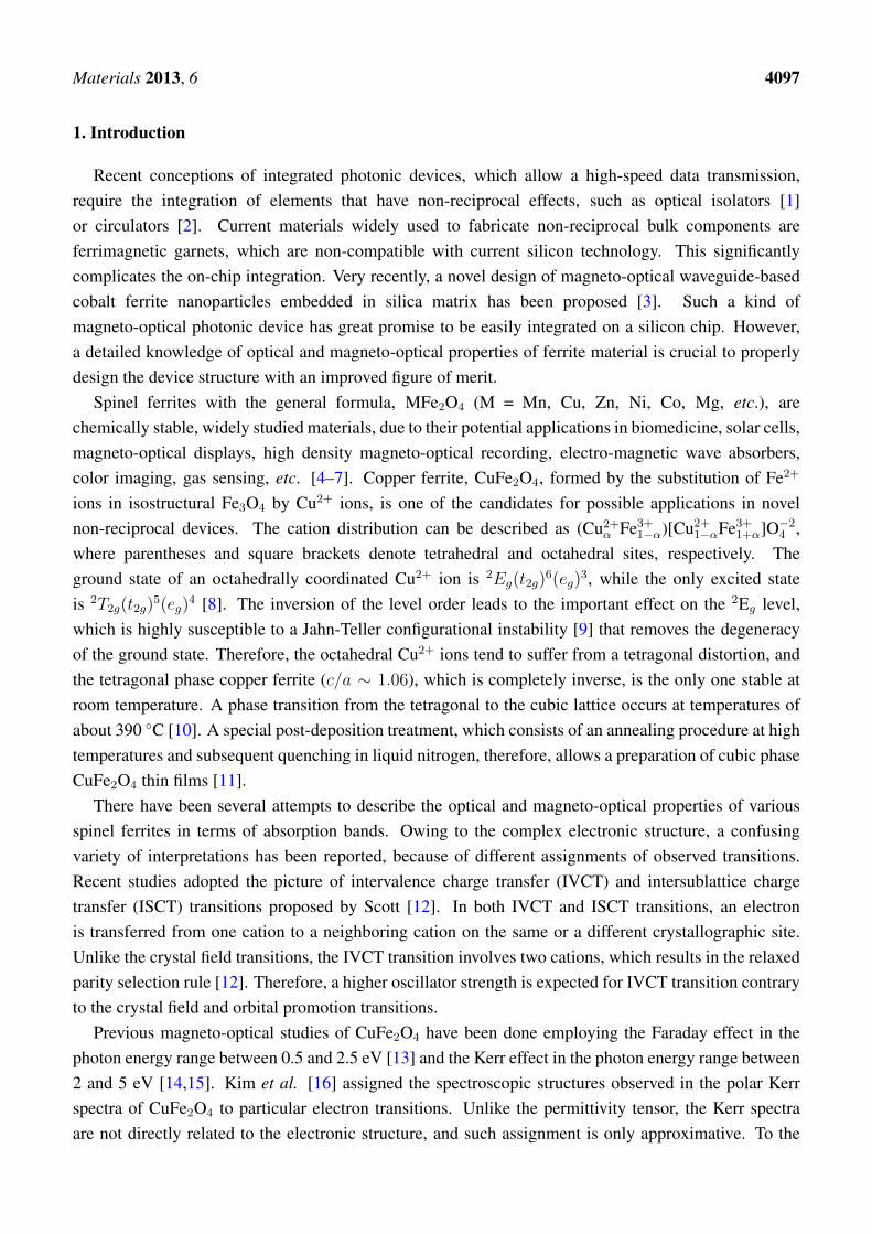
Materials 2013, 6 4097
1. Introduction
Recent conceptions of integrated photonic devices, which allow a high-speed data transmission,require the integration of elements that have non-reciprocal effects, such as optical isolators [1]or circulators [2]. Current materials widely used to fabricate non-reciprocal bulk components areferrimagnetic garnets, which are non-compatible with current silicon technology. This significantlycomplicates the on-chip integration. Very recently, a novel design of magneto-optical waveguide-basedcobalt ferrite nanoparticles embedded in silica matrix has been proposed [3]. Such a kind ofmagneto-optical photonic device has great promise to be easily integrated on a silicon chip. However,a detailed knowledge of optical and magneto-optical properties of ferrite material is crucial to properlydesign the device structure with an improved figure of merit.
Spinel ferrites with the general formula, MFe2O4 (M = Mn, Cu, Zn, Ni, Co, Mg, etc.), arechemically stable, widely studied materials, due to their potential applications in biomedicine, solar cells,magneto-optical displays, high density magneto-optical recording, electro-magnetic wave absorbers,color imaging, gas sensing, etc. [4–7]. Copper ferrite, CuFe2O4, formed by the substitution of Fe2+
ions in isostructural Fe3O4 by Cu2+ ions, is one of the candidates for possible applications in novelnon-reciprocal devices. The cation distribution can be described as (Cu2+
α Fe3+1−α)[Cu2+1−αFe3+1+α]O−24 ,
where parentheses and square brackets denote tetrahedral and octahedral sites, respectively. Theground state of an octahedrally coordinated Cu2+ ion is 2Eg(t2g)
6(eg)3, while the only excited state
is 2T2g(t2g)5(eg)
4 [8]. The inversion of the level order leads to the important effect on the 2Eg level,which is highly susceptible to a Jahn-Teller configurational instability [9] that removes the degeneracyof the ground state. Therefore, the octahedral Cu2+ ions tend to suffer from a tetragonal distortion, andthe tetragonal phase copper ferrite (c/a ∼ 1.06), which is completely inverse, is the only one stable atroom temperature. A phase transition from the tetragonal to the cubic lattice occurs at temperatures ofabout 390 ◦C [10]. A special post-deposition treatment, which consists of an annealing procedure at hightemperatures and subsequent quenching in liquid nitrogen, therefore, allows a preparation of cubic phaseCuFe2O4 thin films [11].
There have been several attempts to describe the optical and magneto-optical properties of variousspinel ferrites in terms of absorption bands. Owing to the complex electronic structure, a confusingvariety of interpretations has been reported, because of different assignments of observed transitions.Recent studies adopted the picture of intervalence charge transfer (IVCT) and intersublattice chargetransfer (ISCT) transitions proposed by Scott [12]. In both IVCT and ISCT transitions, an electronis transferred from one cation to a neighboring cation on the same or a different crystallographic site.Unlike the crystal field transitions, the IVCT transition involves two cations, which results in the relaxedparity selection rule [12]. Therefore, a higher oscillator strength is expected for IVCT transition contraryto the crystal field and orbital promotion transitions.
Previous magneto-optical studies of CuFe2O4 have been done employing the Faraday effect in thephoton energy range between 0.5 and 2.5 eV [13] and the Kerr effect in the photon energy range between2 and 5 eV [14,15]. Kim et al. [16] assigned the spectroscopic structures observed in the polar Kerrspectra of CuFe2O4 to particular electron transitions. Unlike the permittivity tensor, the Kerr spectraare not directly related to the electronic structure, and such assignment is only approximative. To the
Materials 2013, 6 4098
best of the authors knowledge, there is no systematic study on the spectral dependence of the completepermittivity tensor of CuFe2O4. However, information about the spectral dependence of the completepermittivity tensor provides important information about the electronic structure of the material and isnecessary for the design of novel photonic devices based on CuFe2O4.
In this paper, we present a systematic attempt to investigate the electronic structure of copper ferritesusing the experimental techniques of spectroscopic ellipsometry and magneto-optical spectroscopy.Coherently with the results presented on various types of ferrite compounds, we carefully describe alloptical transitions revealed by experiment. Moreover, we discuss the influence of the structural changeof CuFe2O4 on magneto-optical properties.
2. Experimental Section
CuFe2O4 thin films were RF sputtered on 10 × 10 mm polished fused quartz substrates using atetragonal copper ferrite target, which was prepared by the conventional ceramic technique. The basepressure in a vacuum chamber was 6 × 10⁻⁷ mbar. The film deposition was carried out in Ar + O2 gasmixture at working pressure 6× 10⁻³ mbar, and the oxygen to argon ratio was maintained at 15%. TheRF power was 50 and 200 W at 13.6 MHz. The substrates were neither heated nor cooled during thesputtering. After the deposition, the samples were annealed at 850 ◦C for two hours and, then, slowlyfurnace-cooled (SC samples) or quenched (Q samples) in liquid nitrogen.
The crystallographic structure and magnetic properties of deposited films were studied by a PhilipsPW 1729 X-ray diffractometer (XRD) and a vibrating sample magnetometer (VSM) [10]. XRD studiesrevealed peaks typical for cubic spinel structure (c/a = 1), in the case of the quenched sample, and peakstypical for tetragonal structure (c/a ∼ 1.05), in the case of slowly cooled samples. This is consistent withthe knowledge that the copper ferrite can be transformed from the low temperature tetragonal to the hightemperature cubic phase only at temperatures higher than 390 ◦C [17]. VSM measurements showedthe highest saturation magnetization, MS , in the quenched sample and the lowest in the as depositedfilm [10]. The quenched sample exhibits 35% higher MS than the bulk value (MS bulk = 1700 G [18]),indicating the Cu2+ cation redistribution. The parameters of investigated samples are summarized inTable 1.
Table 1. Basic parameters of the set of CuFe2O4 samples. TA denotes the annealingtemperature and MS denotes the saturation magnetization.
Sample Thickness [nm] RF power [W] 4πMS [G] TA [◦C] Structure
1—Quenched 112 50 2300 850 Cubic2—As deposited 90 50 750 – As deposited
3—Slowly cooled 280 50 1500 850 Tetragonal4—Slowly cooled 230 200 1600 850 Tetragonal
The magneto-optical spectroscopy was carried out using an azimuth modulation technique withsynchronic detection in polar and longitudinal configuration. The experiment has been done in thephoton energy range between 1.2 and 4.6 eV. The experimental optical set up included a 450 W high
Materials 2013, 6 4099
power Xe arc lamp, quartz prism monochromator, polarizer, DC compensating Faraday rotator, ACmodulating Faraday rotator, phase plate (for Kerr ellipticity measurements), sample in magnetic field,analyzer and photomultiplier [19]. In the small angle approximation, the complex polar magneto-opticalKerr effect was measured at nearly normal light incidence, as a ratio of θK + iεK ≈ (ryx/rxx) of Jonesreflection matrix elements, where θK and εK are the Kerr rotation and ellipticity. The longitudinalmagneto-optical Kerr effect was measured similarly at the angle of incidence, adjusted to 72 degrees forp-polarized incident light as a ratio of θK + iεK ≈ (rps/rpp) [20]. The applied magnetic field was 470mT and 100 mT in the polar and longitudinal configuration, respectively. In both configurations, themagnetic field was sufficient for the film saturation (as was checked by the measurement of the magneticfield dependence of the magneto-optical Kerr effect). During the polar configuration measurements, thesamples were placed on a water-cooled pole piece of electromagnet, and their temperature was stabilizedat 285 K. In the longitudinal configuration measurements, the samples were kept at the stabilized roomtemperature of 295 K. The effect of stray magnetic field on the optics was accounted for using an Alreflector.
Theoretical models of the magneto-optical Kerr effect have been calculated employing transfer matrixformalism [21]. In polar magnetization and normal light incidence, a layer of CuFe2O4 was characterizedby the permittivity tensor:
ε ≈
ε1 jε2 0
−jε2 ε1 0
0 0 ε1
(1)
where all elements have real and imaginary part: εj = ε′j − iε′′j . The diagonal element, ε1, is relatedto the normal refractive index, n, and the normal extinction coefficient, k. The off-diagonal element isrelated to the refractive index and the extinction coefficient of right and left polarized light.
A four-zone null ellipsometer was employed to obtain the spectral dependences of ellipsometricparameters in the spectral range from 1.5 to 5.4 eV. To increase the accuracy of measured data processing,the spectra were recorded for three angles of incident light at 65◦, 70◦ and 75◦. Because the fused quartzsubstrate was side polished, incoherent back-reflections from the backside of the substrate contributedto the measured signal and complicated the optical characterization. Therefore, a liquid solutionprocedure (LSP) [22], in which a small amount of wadding paper infused by a mixture of glycerinand water, optically matched to the quartz, was attached to the bottom of the substrate to avoid theback-reflections.
3. Results and Discussion
3.1. Spectroscopic Ellipsometry
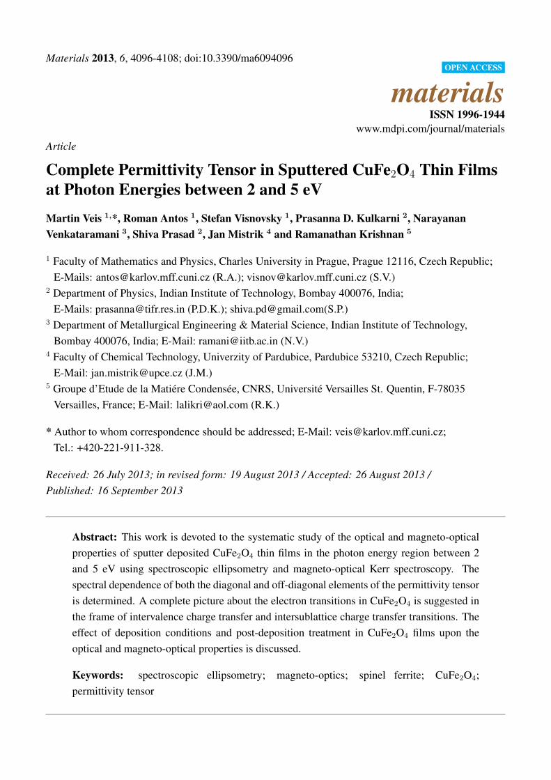
The spectral dependence of ε1 was parametrized by the sum of four damped Lorentz oscillators, andthe least square method was employed to adjust the film thickness, the transition energy, strength andbroadening for each oscillator. The spectral dependence of ε1 obtained for the quenched CuFe2O4 sampleis displayed in Figure 1. It is similar in shape to dependences reported on Fe3O4 [23], CoFe2O4 [24] andMgFe2O4 [25], indicating a similar electronic structure.

Materials 2013, 6 4100
Spectroscopic ellipsometry revealed four optically active transitions centered around 2.4, 3.1, 4.8 and13.2 eV. The first three transitions were also observed by magneto-optical experiments. The differencesin energies are small and are within the experimental data errors. Therefore, we postpone the discussionof these transitions to the next section and focus here only on the transition centered near 13.2 eV.
Figure 1. The diagonal element, ε1, of the permittivity tensor of quenched CuFe2O4 thinfilm (Sample 1).
5
4
3
2
ε 1
4.54.03.53.02.5E [eV]
Real part Imaginary part
The separation energy of about 5–10 eV between the valence band of oxygen 2p orbitalsand the 4s orbital of the transition metal ions has been reported in various transition metaloxides [26,27]. Strong absorption above 8 eV has been reported by Zhang et al., [25] in opticalreflection measurements on Mg and Li ferrites. Alvarado et al. [28] reported the same spectral behaviorin photoelectron-spin-polarization measurements, with photon energies up to 11 eV on Fe3O4. Thissuggests that the electric-dipole allowed transition between the O2− 2p valence band and the Fe3+ 4s
conduction band is responsible for the spectral structure near 13.2 eV.The obtained spectra of ε1 were subsequently used in the calculations of the off-diagonal element of
the permittivity tensor, ε2, from the magneto-optical Kerr measurements.
3.2. Magneto-Optical Spectroscopy
3.2.1. Polar Geometry
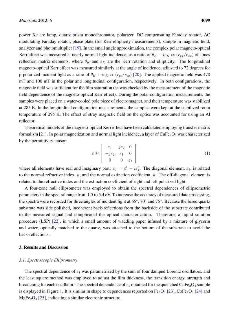
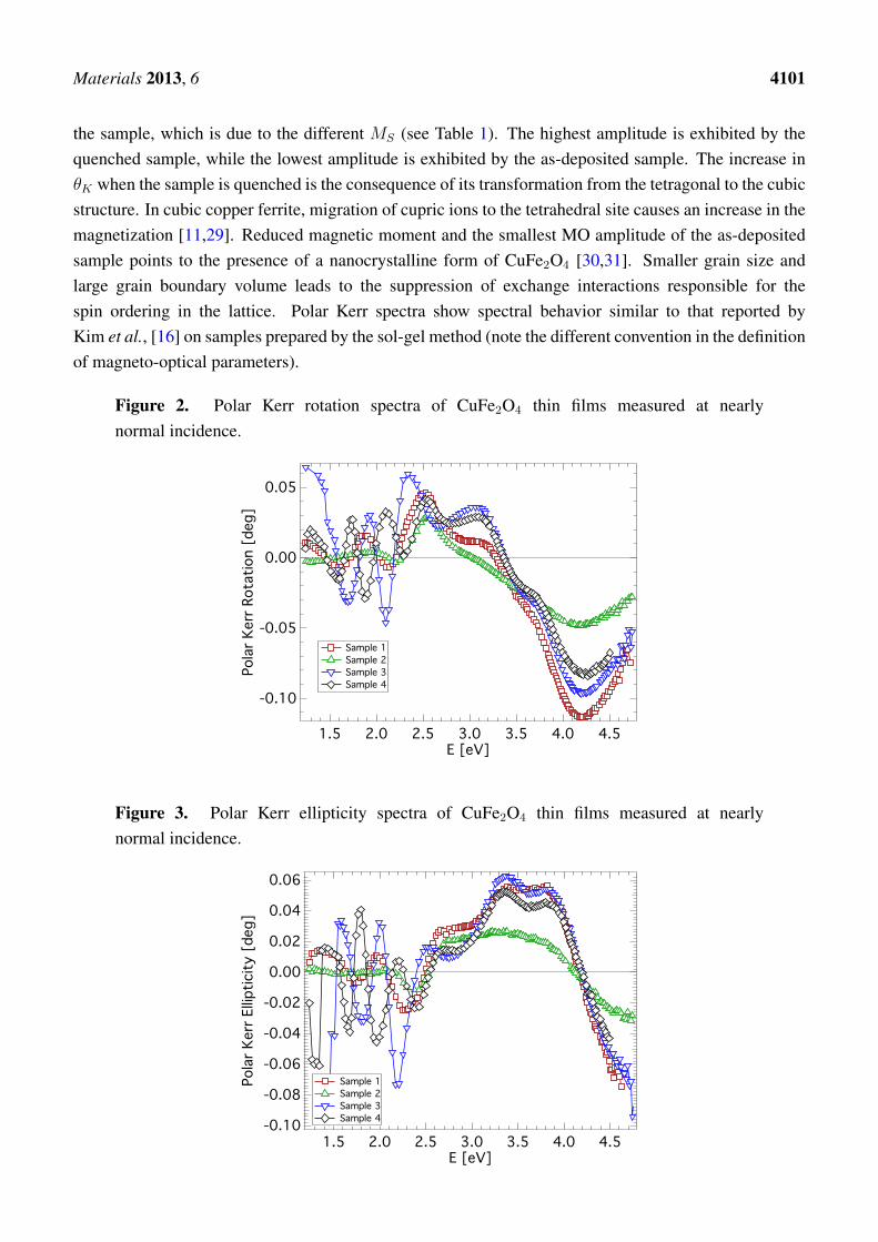
Experimental spectra of polar Kerr rotation, θK , and ellipticity, εK , of all investigated samplesare shown in Figures 2 and 3. A low level of noise in the spectra reflects the very good quality ofCuFe2O4 films. All samples exhibit similar spectral behavior of the polar Kerr effect with only minordifferences. A contribution of the propagation across the film resulting in the interference is clearlyvisible in the photon energy range below 2.4 eV. Besides, the polar Kerr rotation spectra are dominatedby two visible peaks with opposite signs near 3.1 and 4.2 eV. On the other hand, polar Kerr ellipticityspectra show positive peaks near 3.5 and 3.8 eV. The amplitudes of the polar Kerr effect differ with
Materials 2013, 6 4101
the sample, which is due to the different MS (see Table 1). The highest amplitude is exhibited by thequenched sample, while the lowest amplitude is exhibited by the as-deposited sample. The increase inθK when the sample is quenched is the consequence of its transformation from the tetragonal to the cubicstructure. In cubic copper ferrite, migration of cupric ions to the tetrahedral site causes an increase in themagnetization [11,29]. Reduced magnetic moment and the smallest MO amplitude of the as-depositedsample points to the presence of a nanocrystalline form of CuFe2O4 [30,31]. Smaller grain size andlarge grain boundary volume leads to the suppression of exchange interactions responsible for thespin ordering in the lattice. Polar Kerr spectra show spectral behavior similar to that reported byKim et al., [16] on samples prepared by the sol-gel method (note the different convention in the definitionof magneto-optical parameters).
Figure 2. Polar Kerr rotation spectra of CuFe2O4 thin films measured at nearlynormal incidence.
-0.10
-0.05
0.00
0.05
Pola
r Ker
r Rot
atio
n [d
eg]
4.54.03.53.02.52.01.5E [eV]
Sample 1 Sample 2 Sample 3 Sample 4
Figure 3. Polar Kerr ellipticity spectra of CuFe2O4 thin films measured at nearlynormal incidence.
-0.10
-0.08
-0.06
-0.04
-0.02
0.00
0.02
0.04
0.06
Pola
r Ker
r Ellip
ticity
[de
g]
4.54.03.53.02.52.01.5E [eV]
Sample 1 Sample 2 Sample 3 Sample 4

Materials 2013, 6 4102
To get a deeper insight into the magneto-optical properties of CuFe2O4 thin films, a spectraldependence of ε2 has been deduced from the polar Kerr measurements, considering a model structure of athin CuFe2O4 layer on a semi-infinite quartz substrate. The spectral dependence of ε2 for all investigatedsamples in the photon energy range from 2 to 4.8 eV is displayed in Figure 4. All samples exhibit similarspectral behavior of ε2, with only minor differences. The most departing spectrum appears to be that ofthe as-deposited sample (Sample 2). This is, however, acceptable with respect to the similar differencesin XRD and magnetic measurements. Nevertheless, all spectra of the real part of ε2 exhibit negative peaksnear 2.6 and 3.1 eV and a broad positive peak near 4.2 eV. On the other hand, spectra of the imaginarypart of ε2 are dominated by two positive peaks near 2.5 and 4.7 eV and a negative spectroscopic structurecomposed of two peaks near 3.3 and 3.9 eV. Such spectral dependences are similar to those reported onMgFe2O4 bulk samples [32], as well as to those reported on Li0.5Fe2.5O4 single crystals [25,33,34].Martens et al. [24] reported experimental results on Co ferrite, but those results differ from the resultspresented in this paper.
Figure 4. The off-diagonal elements, ε2, of CuFe2O4 thin films deduced from the polar Kerrmeasurements along with the fitted theoretical dependence.
0.010
0.005
0.000
-0.005
ε 2
4.54.03.53.02.5E [eV]
Real part Imaginary part Fit
Sample 1a)
-0.004
-0.002
0.000
0.002
0.004
ε 2
4.54.03.53.02.5E [eV]
Real part Imaginary part Fit
Sample 2
0.015
0.010
0.005
0.000
-0.005
-0.010
ε 2
4.54.03.53.02.5E [eV]
Real part Imaginary part Fit
Sample 30.010
0.005
0.000
-0.005
ε 2
4.03.53.02.5E [eV]
Real part Imaginary part Fit
Sample 4b)
The spectral dependences of ε2 derived from the magneto-optical measurements were parametrizedby a summation of five paramagnetic line shapes [35], and the least square method was employed toadjust the energy, strength and broadening for each transition. The resulting fits are included in Figure 4,and the fitting parameters are summarized in Table 2.
Materials 2013, 6 4103
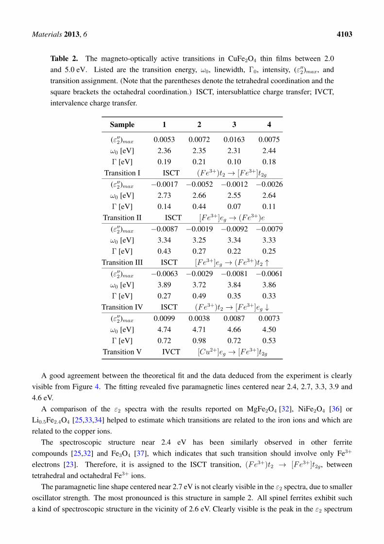
Table 2. The magneto-optically active transitions in CuFe2O4 thin films between 2.0and 5.0 eV. Listed are the transition energy, ω0, linewidth, Γ0, intensity, (ε′′2)max, andtransition assignment. (Note that the parentheses denote the tetrahedral coordination and thesquare brackets the octahedral coordination.) ISCT, intersublattice charge transfer; IVCT,intervalence charge transfer.
Sample 1 2 3 4
(ε′′2)max 0.0053 0.0072 0.0163 0.0075ω0 [eV] 2.36 2.35 2.31 2.44Γ [eV] 0.19 0.21 0.10 0.18
Transition I ISCT (Fe3+)t2 → [Fe3+]t2g
(ε′′2)max −0.0017 −0.0052 −0.0012 −0.0026ω0 [eV] 2.73 2.66 2.55 2.64Γ [eV] 0.14 0.44 0.07 0.11
Transition II ISCT [Fe3+]eg → (Fe3+)e
(ε′′2)max −0.0087 −0.0019 −0.0092 −0.0079ω0 [eV] 3.34 3.25 3.34 3.33Γ [eV] 0.43 0.27 0.22 0.25
Transition III ISCT [Fe3+]eg → (Fe3+)t2 ↑(ε′′2)max −0.0063 −0.0029 −0.0081 −0.0061ω0 [eV] 3.89 3.72 3.84 3.86Γ [eV] 0.27 0.49 0.35 0.33
Transition IV ISCT (Fe3+)t2 → [Fe3+]eg ↓(ε′′2)max 0.0099 0.0038 0.0087 0.0073ω0 [eV] 4.74 4.71 4.66 4.50Γ [eV] 0.72 0.98 0.72 0.53
Transition V IVCT [Cu2+]eg → [Fe3+]t2g
A good agreement between the theoretical fit and the data deduced from the experiment is clearlyvisible from Figure 4. The fitting revealed five paramagnetic lines centered near 2.4, 2.7, 3.3, 3.9 and4.6 eV.
A comparison of the ε2 spectra with the results reported on MgFe2O4 [32], NiFe2O4 [36] orLi0.5Fe2.4O4 [25,33,34] helped to estimate which transitions are related to the iron ions and which arerelated to the copper ions.
The spectroscopic structure near 2.4 eV has been similarly observed in other ferritecompounds [25,32] and Fe3O4 [37], which indicates that such transition should involve only Fe3+
electrons [23]. Therefore, it is assigned to the ISCT transition, (Fe3+)t2 → [Fe3+]t2g, betweentetrahedral and octahedral Fe3+ ions.
The paramagnetic line shape centered near 2.7 eV is not clearly visible in the ε2 spectra, due to smalleroscillator strength. The most pronounced is this structure in sample 2. All spinel ferrites exhibit sucha kind of spectroscopic structure in the vicinity of 2.6 eV. Clearly visible is the peak in the ε2 spectrum

Materials 2013, 6 4104
of Li0.5Fe2.4O4 reported by Zhang et al. [25]. Since there is no change in energy with different ionsubstitution for this transition, only Fe3+ ions should be involved. Fontijn et al. predicted an ISCTtransition in Fe3O4 near 2.64 eV [23]. Therefore, this structure is assigned to the ISCT transition,[Fe3+]eg → (Fe3+)e, between octahedral and tetrahedral Fe3+ ions.
The spectroscopic structures in the ε2 spectra centered near 3.3 and 3.9 eV were observedacross all ferrite compounds [23,25,37], including CuFe2O4 [16]. Consistently with previousreports, these structures were assigned to ISCT transitions between octahedral and tetrahedral sites,[Fe3+]eg → (Fe3+)t2 ↑ and (Fe3+)t2 → [Fe3+]eg ↓, respectively. The 3.3 eV structure is attributed toan ISCT transition in the majority spin bands, while the 3.9 eV one, to an ISCT transition in the minorityspin bands.
These two transitions are responsible for the magneto-optical properties of CuFe2O4 in the spectralrange between 2.8 and 3.5 eV. In this region, the slowly cooled (tetragonal) samples exhibit higher Kerramplitudes than the quenched (cubic) sample. This is related to considerably higher amplitude (ε′′2)maxand broadening of transition IV in these samples. Since this transition involves tetrahedral (Fe3+)t2 ions,the migration of Cu2+ ions to tetrahedral sites in the cubic sample causes the decrease of (Fe3+)t2 ionsper unit volume, resulting in the smaller oscillator strength and, consequently, the Kerr effect amplitudes.
Unlike in the previous cases, the energy of the spectroscopic structure centered near 4.4∼4.7 eVnoticeable varies with the sample. This is a consequence of the decreased accuracy of the fit procedure,due to the end of the measured spectral region. Moreover, in the UV region, the magneto-optical Kerrmeasurement suffers from a higher level of noise, due to the increased role of light scattering.
There is no comparable transition reported in Li0.5Fe2.4O4 [25], NiFe2O4 [36] and CoFe2O4
[24], which points to the contribution of Cu2+ ions. IVCT transitions between divalent substitutedion and trivalent iron ion, both situated at octahedral sites, were observed in Co and Ni ferrites,[Co2+]t2g → [Fe3+]t2g, at about 2.2 eV, and [Ni2+]t2g → [Fe3+]t2g, at about 3.1 eV. Owing to thelarger binding energies of more localized 3d electrons of Cu2+ compared to Co2+ and Ni2+, a similartransition is expected at higher energies. Considering the inversion of the electron level order for Cu2+
ions, the last spectroscopic structure was assigned to an IVCT [Cu2+]eg → [Fe3+]t2g transition.In the presented results, we did not observe crystal-field transitions of Cu2+ ions, which are
expected to be around 2.5 eV [38]. Such transitions are spin-allowed, but owing to the inversionsymmetry at octahedral sites (which have a strong preference for Cu2+ ions in CuFe2O4), they are parityforbidden, resulting in their small oscillator strength. Therefore, these transitions are not visible in thepresented spectra.
As follows from Table 2, the slowly cooled sample sputtered at 50 W RF power exhibits aconsiderably higher amplitude (ε′′2)max of transitions (except Transition II) than the sample sputteredat 200 W RF power. This might indicate a decomposition of the target material at higher sputteringpowers, which results in the decrease of exchange interactions and a lower number of active absorbingcenters per unit volume. This is expected to cause the decrease of the transition strength.
On the other hand, the transitions in the quenched sample are broader compared to those in slowlycooled samples. It seems that this has a connection with the migration of cupric ions to the tetrahedralsite. Because the center of the symmetry is missing at the tetrahedral sites, electron orbitals are more
Materials 2013, 6 4105
opened, and covalent bonding is increased, which results in the broadening of the transition line shapes.However, more detailed structural and optical studies are necessary to confirm this hypothesis.
Finally, we make a comment on the crystal field (CF) splitting energy, ∆CF , of tetrahedral andoctahedral Fe3+ ions in CuFe2O4 thin films. As follows from Table 2, the crystal field energy splittingfor the octahedral Fe3+ iron, ∆O
CF , is about 1.5 eV, while in the case of tetrahedral Fe3+ iron, ∆TCF ,
it is about 0.7 eV. These values have been obtained as differences in transition energies. Camphausenet al. [26] reported that the octahedrally coordinated Fe3+ ions give ∆O
CF = 1.7 − 2.0 eV, while thetetrahedrally coordinated Fe3+ ions give ∆T
CF = 0.86 − 1.17 eV. Kim et al. [37] reported the valueof ∆O
CF = 1.4 eV. A reasonable agreement between presented ∆CF values and previously publishedstudies has been found. This confirmed the correctness of the assignment of the spectroscopic structuresobserved in ε2 spectra to the particular transitions.
3.3. Longitudinal Geometry
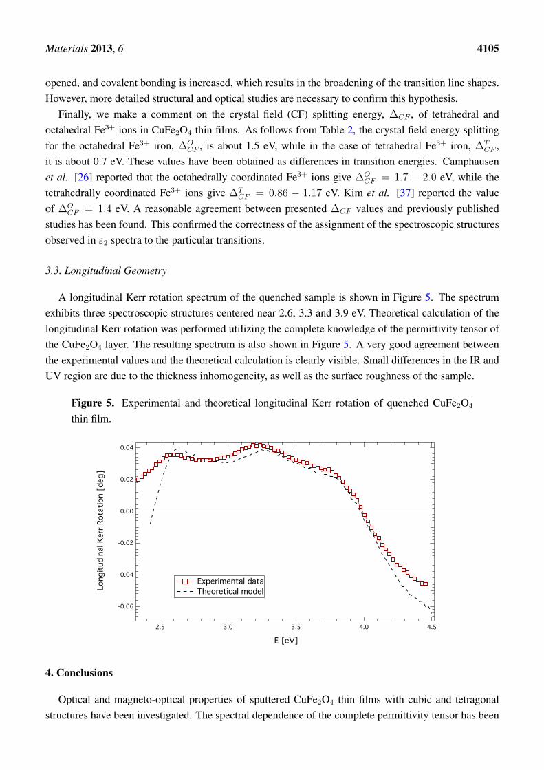
A longitudinal Kerr rotation spectrum of the quenched sample is shown in Figure 5. The spectrumexhibits three spectroscopic structures centered near 2.6, 3.3 and 3.9 eV. Theoretical calculation of thelongitudinal Kerr rotation was performed utilizing the complete knowledge of the permittivity tensor ofthe CuFe2O4 layer. The resulting spectrum is also shown in Figure 5. A very good agreement betweenthe experimental values and the theoretical calculation is clearly visible. Small differences in the IR andUV region are due to the thickness inhomogeneity, as well as the surface roughness of the sample.
Figure 5. Experimental and theoretical longitudinal Kerr rotation of quenched CuFe2O4
thin film.
-0.06
-0.04
-0.02
0.00
0.02
0.04
Lon
gitu
dina
l Ker
r Rot
atio
n [d
eg]
4.54.03.53.02.5
E [eV]
Experimental data Theoretical model
4. Conclusions
Optical and magneto-optical properties of sputtered CuFe2O4 thin films with cubic and tetragonalstructures have been investigated. The spectral dependence of the complete permittivity tensor has been
Materials 2013, 6 4106
derived in the photon energy range between 2 and 5 eV, and the influence of the post-deposition treatmenton the magneto-optical properties of studied films was discussed. The combination of spectroscopicellipsometry and magneto-optical spectroscopy revealed six spectroscopic structures in a broad spectralregion near energies of 2.4, 2.7, 3.3, 3.9, 4.6 and 13.2 eV. The first five structures were described in theframe of ISCT and IVCT transitions between Fe3+ and Cu2+ ions. The last structure was discussed as anelectron transfer between the O 2p valence band and the Fe 4s conduction band. Such assignment wasconfirmed by the derivation of the crystal field splitting energy for both octahedral and tetrahedral ironions, respectively. The obtained energies reasonably agree with theoretically predicted values, as well aswith experimental results obtained on similar compounds.
Acknowledgments
This work was supported by Czech Grant Agency grant No. P204/10/P346.
Conflicts of Interest
The authors declare no conflict of interest.
References
1. Zamani, M.; Ghanaatshoar, M. Adjustable magneto-optical isolators with flat-top responses. Opt.Express 2012, 20, 24524–24535.
2. Saib, A.; Darques, M.; Piraux, L.; vanhoenacker-Janvier, D.; Huynen, I. Unbiased microwavecirculator based on ferromagnetic nanowires arrays of tunable magnetization state. J. Phys. DAppl. Phys. 2005, 38, doi:10.1088/0022-3727/38/16/003.
3. Choueikani, F.; Royer, F.; Jamon, D.; Siblini, A.; Rousseau, J.J.; Neveu, S.; Charara, J.Magneto-optical waveguides made of cobalt ferrite nanoparticles embedded in silica/zirconiaorganic-inorganic matrix. Appl. Phys. Lett. 2009, 94, 051113:1–051113:3.
4. Liu, T.Y.; Hu, S.H.; Hu, S.H.; Tsai, S.P.; Chen, S.Y. Preparation and characterization ofthermal-sensitive ferrofluids for drug delivery application. J. Magn. Magn. Mater. 2007, 310,2850–2852.
5. Nixon, L.; Koval, C.A.; Noble, R.D.; Slaff, G.S. Preparation and characterization of novelmagnetite-coated ion-exchange particles. Chem. Mater. 1992, 4, 117–121.
6. Hankare, P.; Sanadi, K.; Pandav, R.; Patil, N.; Garadkar, K.; Mulla, I. Structural, electrical andmagnetic properties of cadmium substituted copper ferrite by solgel method. J. Alloys Compd.2012, 540, 290–296.
7. Chen, N.S.; Yang, X.J.; Liu, E.S.; Huang, J.L. Reducing gas-sensing properties of ferritecompounds MFe2O4 (M = Cu, Zn, Cd and Mg). Sens. Actuators B Chem. 2000, 66, 178–180.
8. Ballhausen, C.J. Introduction to Ligand Field Theory; McGraw-Hill: New York, NY, USA, 1962.9. Jahn, H.A.; Teller, E. Stability of polyatomic molecules in degenerate electronic states. I. Orbital
degeneracy. Proc. R. Soc. Lond. 1937, 161, 220–235.

Materials 2013, 6 4107
10. Desai, M.; Prasad, S.; Venkataramani, N.; Samajdar, I.; Nigam, A.K.; Krishnan, R. Enhancedmagnetization in sputter-deposited copper ferrite thin films. J. Magn. Magn. Mater. 2002,246, 266–269.
11. Desai, M.; Prasad, S.; Venkataramani, N.; Samajdar, I.; Nigam, A.K.; Krishnan, R. Annealinginduced structural change in sputter deposited copper ferrite thin films and its impact on magneticproperties. J. Appl. Phys. 2002, 91, 2220–2227.
12. Scott, G.B.; Lacklison, D.E.; Ralph, H.I.; Page, J.L. Magnetic circular dichroism and Faradayrotation spectra of Y3Fe5O12. Phys. Rev. B 1975, 12, 2562–2571.
13. Kucera, M.; Kolinsky, V.; Visnovsky, S.; Chvostova, D.; Venkataramani, N.; Prasad, S.;Kulkarni, P.; Krishnan, R. Faraday effect in cubic and tetragonal copper ferrite CuFe2O4 films:Comparative studies. J. Magn. Magn. Mater. 2007, 316, e688–e691.
14. Veis, M.; Kolinsky, V.; Visnovsky, S.; Kulkarni, P.D.; Desai, M.; Venkataramani, N.; Prasad, S.;Krishnan, R. Moke spectroscopy of sputter deposited Cu-ferrite films. J. Magn. Magn. Mater.2004, 272–276, E885–E886.
15. Visnovsky, S.; Veis, M.; Liskova, E.; Kolinsky, V.; Kulkarni, P.D.; Venkataramani, N.; Prasad, S.;Krishnan, R. MOKE spectroscopy of sputter-deposited Cu-ferrite films. J. Magn. Magn. Mater.2005, 290–291, 195–197.
16. Kim, K.J.; Lee, J.H.; Lee, S.H. Magneto-optical investigation of spinel ferrite CuFe2O4:Observation of Jahn–Teller effect in Cu2+ ion. J. Magn. Magn. Mater. 2004, 279, 173–177.
17. Tang, X.X.; Manthiram, A.; Goodenough, J. Copper ferrite revisited. J. Solid State Chem. 1989,79, 250–262.
18. Yang, A.; Zuo, X.; Chen, L.; Chen, Z.; Vittoria, C.; Harris, V.G. Magnetic and structural propertiesof pulsed laser deposited CuFe2O4 films. J. Appl. Phys. 2005, 97, 10G107:1–10G107:3.
19. The magneto-optical spectrometer operating at the Institute of Physics of Charles University atPrague since 1975 was built by one of us (S.V.).
20. Veis, M.; Visnovsky, S.; Lecoeur, P.; Haghiri-Gosnet, A.M.; Renard, J.P.; Beauvillain, P.;Prellier, W.; Mercey, B.; Mistrik, J.; Yamaguchi, T. Magneto-optic spectroscopy ofLa2/3Sr1/3MnO3 films on SrTiO3 (100) and (110) substrates. J. Phys. D Appl. Phys. 2009,42, doi:10.1088/0022-3727/42/19/195002.
21. Yeh, P. Optics of anisotropic layered media: A new 4 × 4 matrix algebra. Surf. Sci. 1980,96, 41–53.
22. Antos, R.; Pistora, J.; Ohlidal, I.; Postava, K.; Mistrik, J.; Yamaguchi, T.; Visnovsky, S.; Horie, M.Specular spectroscopic ellipsometry for the critical dimension monitoring of gratings fabricated ona thick transparent plate. J. Appl. Phys. 2005, 97, 053107:1–053107:7.
23. Fontijn, W.F.J.; van der Zaag, P.J.; Devillers, M.A.C.; Brabers, V.A.M.; Metselaar, R. Optical andmagneto-optical polar Kerr spectra of Fe3O4 and Mg2+- or Al3+-substituted Fe3O4. Phys. Rev. B1997, 56, 5432–5442.
24. Martens, J.W.D.; Peeters, W.L.; van Noort, H.M.; Erman, M. Optical, magneto-optical andmossbauer spectroscopy on Co3+ substituted cobalt ferrite Co2+Fe2−xCo3+
x O4 (0 < x < 2).J. Phys. Chem. Solids 1985, 46, 411–416.
Materials 2013, 6 4108
25. Zhang, X.X.; Schoenes, J.; Reim, W.; Wachter, P. Evidence for 3dn to 3dn−14s transitions inmagnetite and in lithium and magnesium ferrites. J. Phys. C Solid State Phys. 1983, 16,6055–6072.
26. Camphausen, D.L.; Coey, J.M.D.; Chakraverty, B.K. One-electron energy levels in Fe3O4. Phys.Rev. Lett. 1972, 29, 657–660.
27. Wettling, W. Magneto-optics of ferrites. J. Magn. Magn. Mater. 1976, 3, 147–160.28. Alvarado, S.F.; Erbudak, M.; Munz, P. Final-state effects in the 3d photoelectron spectrum of Fe3O4
and comparison with FexO. Phys. Rev. B 1976, 14, 2740–2745.29. Sultan, M.; Singh, R. Magnetization and crystal structure of RF-sputtered nanocrystalline CuFe2O4
thin films. Mater. Lett. 2009, 63, 1764–1766.30. Baubet, C.; Tailhades, P.; Bonningue, C.; Rousset, A.; Simsa, Z. Influence of tetragonal distortion
on magnetic and magneto-optical properties of copper ferrite films. J. Phys. Chem. Solids 2000,61, 863–867.
31. Srinivasan, G.; Rao, B.U.M.; Zhao, J.; Seehra, M.S. Magnetically ordered amorphous copperferrite. Appl. Phys. Lett. 1991, 59, 372–374.
32. Visnovsky, S.; Prosser, V.; Krishnan, R.; Parizek, V.; Nitsch, K.; Svobodova, L. Magnetoopticalpolar kerr effect in ferrimagnetic garnets and spinels. IEEE Trans. Magn. 1981, 17, 3205–3210.
33. Visnovsky, S.; Thuy, N.P.; Stepanek, J.; Prosser, V.; Krishnan, R. Magnetooptical spectra ofY3Fe5O12 and Li0.5Fe2.5O4 between 2.0 and 5.8 eV. J. Appl. Phys. 1979, 50, 7466–7469.
34. Visnovsky, S.; Krishnan, R.; Thuy, N.; Stepanek, J.; Parizek, V.; Prosser, V. UV magnetoopticalKerr effect and reflectivity spectra of Y3Fe5O12 and Li0.5Fe2.5O4. J. Magn. Magn. Mater. 1980,15–18, 831–832.
35. Kahn, F.J.; Pershan, P.S.; Remeika, J.P. Ultraviolet magneto-optical properties of single-crystalorthoferrites, garnets, and other ferric oxide compounds. Phys. Rev. 1969, 186, 891–918.
36. Fontijn, W.F.J.; van der Zaag, P.J.; Metselaar, R. On the origin of the magneto-optical effects in Li,Mg, Ni, and Co ferrite. J. Appl. Phys. 1998, 83, 6765–6767.
37. Kim, K.J.; Lee, H.S.; Lee, M.H.; Lee, S.H. Comparative magneto-optical investigation of d–dcharge–transfer transitions in Fe3O4, CoFe2O4, and NiFe2O4. J. Appl. Phys. 2002, 91, 9974–9977.
38. Balhausen, C.J. Ligand Field Theory; McGraw-Hill: New York, NY, USA, 1962.
c© 2013 by the authors; licensee MDPI, Basel, Switzerland. This article is an open access articledistributed under the terms and conditions of the Creative Commons Attribution license(http://creativecommons.org/licenses/by/3.0/).