Embed Size (px)
Citation preview
© 2011 Li and Bluth, publisher and licensee Dove Medical Press Ltd. This is an Open Access article which permits unrestricted noncommercial use, provided the original work is properly cited.
Pharmacogenomics and Personalized Medicine 2011:4 11–33
Pharmacogenomics and Personalized Medicine Dovepress
submit your manuscript | www.dovepress.com
Dovepress 11
R e v i e w
open access to scientific and medical research
Open Access Full Text Article
DOI: 10.2147/PGPM.S18861
Pharmacogenomics of drug metabolizing enzymes and transporters: implications for cancer therapy
Jing LiMartin H BluthKarmanos Cancer institute, wayne State University School of Medicine, Detroit, Mi, USA
Correspondence: Jing Li Karmanos Cancer institute, wayne State University, 4100 John R HwCRC, Room 523 Detroit, Mi 48201, USA Tel +1 313 576 8258 email [email protected]
Abstract: The new era of personalized medicine, which integrates the uniqueness of an
individual with respect to the pharmacokinetics and pharmacodynamics of a drug, holds promise
as a means to provide greater safety and efficacy in drug design and development. Personalized
medicine is particularly important in oncology, whereby most clinically used anticancer drugs
have a narrow therapeutic window and exhibit a large interindividual pharmacokinetic and
pharmacodynamic variability. This variability can be explained, at least in part, by genetic
variations in the genes encoding drug metabolizing enzymes, transporters, or drug targets.
Understanding of how genetic variations influence drug disposition and action could help in
tailoring cancer therapy based on individual’s genetic makeup. This review focuses on the
pharmacogenomics of drug metabolizing enzymes and drug transporters, with a particular
highlight of examples whereby genetic variations in the metabolizing enzymes and transporters
influence the pharmacokinetics and/or response of chemotherapeutic agents.
Keywords: polymorphisms, personalized medicine, oncology, enzymes, transporters, drug
IntroductionThe new era of personalized medicine, which integrates the uniqueness of an individual
with respect to the pharmacokinetics and pharmacodynamics of a drug, holds promise
as a means to provide greater safety and efficacy in drug design and development.
Personalized medicine is particularly important in oncology whereby most clinically
used anticancer drugs have a narrow therapeutic window and exhibit a large
interindividual pharmacokinetic and pharmacodynamic variability. This variability can
lead to therapeutic failure or severe toxicity. Understanding of how genetic variations
influence drug disposition and action could help in tailoring cancer therapy based on
individual’s genetic makeup. Pharmacogenomics is the study of how variations in the
human genome affect the response to medications. Each drug, after it enters the body,
interacts with numerous proteins, such as carrier proteins, transporters, metabolizing
enzymes, and multiple types of receptors. These protein interactions determine drug
pharmacokinetics (ie, drug absorption, distribution, metabolism, and excretion) and
pharmacodynamics (ie, target site of action, pharmacological and toxicological effects).
Moreover, drugs trigger downstream secondary events which may impact additional
gene or protein expression responses and can also vary among patients. As a result,
the overall response to a drug is determined by the interplay of multiple genes that are
involved in the pharmacokinetic and pharmacodynamic pathways of a drug. In general,
important genetic variation in drug effect can be envisioned at the level of drug metabo-
lizing enzymes, drug transporters, and drug targets. This review provides an overview
Pharmacogenomics and Personalized Medicine 2011:4submit your manuscript | www.dovepress.com
Dovepress
Dovepress
12
Li and Bluth
on the commonly occurring, functionally and/or clinically
relevant genetic polymorphisms within the genes encoding
important drug metabolizing enzymes and drug transporters,
with a particular highlight of examples whereby genetic varia-
tions in these genes influence the pharmacokinetics and/or
response of chemotherapeutic agents.
Drug metabolizing-enzyme pharmacogenomicsDrug metabolizing enzymes are proteins that catalyze the
biochemical modifications of xenobiotics (eg, drugs) and
endogenous chemicals (eg, hormones, neurotransmitters).
Broadly, drug metabolizing enzymes are divided into two
categories: Phase I (functionalizing) enzymes that introduce
or remove functional groups in a substrate through oxida-
tion, reduction, or hydrolysis; and Phase II (conjugating)
enzymes that transfer moieties from a cofactor to a substrate.
Essentially all of the major human metabolizing enzymes
exhibit genetic polymorphisms at the genomic level, and
many of these enzymes have clinically relevant genetic
polymorphisms.1 A gene is considered to be polymorphic
when the frequency of a variant allele in a specific population
is at least 1%.
Phase i enzymesPhase I metabolizing enzymes include those involved in:
• Oxidation – cytochrome P450, alcohol dehydrogenase,
aldehyde dehydrogenase, dihydropyrimidine dehy-
drogenase, monoamine oxidase, and flavin-containing
monooxygenase;
• Reduction – nicotinamide adenine dinucleotide phosphate
(NADPH)-cytochrome P450 reductase and reduced cyto-
chrome P450;
• Hydrolysis – epoxide hydrolase, esterases, and amidases.
The most important Phase I enzymes that exhibit clinical
relevant genetic polymorphisms are the cytochrome P450
(CYP) superfamily. The human CYP superfamily represents
the most important system responsible for catalyzing the
oxidation of a large number of endogenous and exogenous
compounds including drugs, toxins, and carcinogens. In this
superfamily, 57 genes and 58 pseudogenes have been identi-
fied, which are divided into 18 families and 43 subfamilies
(http://drnelson.utmem.edu/cytochromeP450.html). Among
them, three subfamilies of CYPs, including CYP1, CYP2,
and CYP3, contribute to the oxidative metabolism of more
than 90% of clinically used drugs.
The human CYP genes are highly polymorphic. The
polymorphisms within the CYP genes, which include
gene deletions, missense mutations, deleterious mutations
creating splicing defects or premature stop codon, and gene
duplications, could produce abolished, reduced, normal,
or enhanced enzyme activity. As a result, patients can be
classified into four phenotypes based on the level of a CYP
enzyme activity: poor metabolizer (abolished activity),
intermediate metabolizer (reduced activity), extensive
metabolizer (normal activity), and ultrarapid metabolizer
(enhanced activity). It is expected that poor metabolizers
would have higher concentrations of a drug that is inactivated
by that enzyme pathway and therefore require a lower dose
to avoid adverse reactions, whereas ultrarapid metabolizers
would require a higher dose to achieve therapeutic effective
drug concentrations. The opposite pattern of reactions is
expected for a prodrug that undergoes metabolic activation.
A prodrug may have little therapeutic effect in poor
metabolizers, while producing a toxic level of active form
in ultrarapid metabolizers. Substantial evidence suggests
that genetic polymorphisms within the CYP genes have
significant impact on drug disposition and/or response. The
common functional polymorphisms in the major human CYP
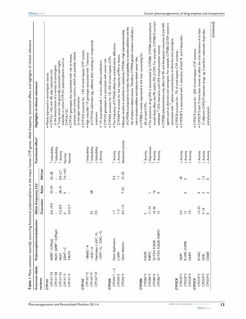
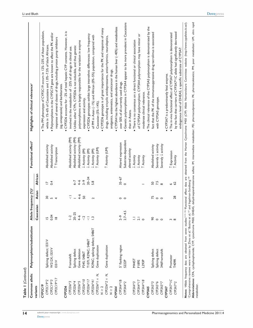
genes and their clinical relevance are summarized in Table 1.
Notably, the most pharmacologically and clinically relevant
CYP polymorphisms are found in CYP2D6, CYP2C9, and
CYP2C19. Of the Food and Drug Administration (FDA)-
approved drug labels referring human genomic biomarkers,
62% are pertained to polymorphisms in the CYP enzymes,
with CYP2D6 (35%), CYP2C19 (17%), and CYP2C9 (7%)
being the most common.2
CYP2D6 is not inducible and therefore, the variations in
the enzyme expression and activity are largely attributable
to genetic polymorphisms. The CYP2D6 gene is highly
polymorphic with more than 63 functional variants identified
to date (http://www.cypalleles.ki.se). These alleles result in
abolished, decreased, normal, or ultrarapid CYP2D6 enzyme
activity. The most important null alleles are CYP2D6*4
(splicing defect) and CYP2D6*5 (gene deletion); the common
alleles with severely reduced enzyme activity are represented
by CYP2D6*10, *17, and *41; duplication or multidu-
plications of active CYP2D6 genes (eg, CYP2D6*1 × N
(N $ 2)) result in ultrarapid enzyme activity (Table 1). The
distributions of CYP2D6 alleles exhibit notable interethnic
differences. The nonfunctional allele CYP2D6*4 is prevalent
in Caucasians (allelic frequency, ∼25%), while the reduced
function allele CYP2D6*10 and CYP2D6*17 is common
in Asians (allelic frequency, ∼40%) and Africans (allelic
frequency, ∼34%).3 As a result, poor metabolizers of
CYP2D6, mainly resulted from null allele CYP2D6*4, has
Pharmacogenomics and Personalized Medicine 2011:4 submit your manuscript | www.dovepress.com
Dovepress
Dovepress
13
Cancer pharmacogenomics of drug enzymes and transporters
Tab
le 1
Mos
t co
mm
on n
atur
ally
occ
urri
ng fu
nctio
nal p
olym
orph
ism
s in
the
maj
or h
uman
CY
P ge
nes:
alle
le fr
eque
ncy,
func
tiona
l effe
ct, a
nd h
ighl
ight
s of
clin
ical
rel
evan
ce
Com
mon
alle
lic
vari
ants
Pol
ymor
phis
m/s
ubst
itut
ion
Alle
le fr
eque
ncy
(%)a
Func
tion
al e
ffect
bH
ighl
ight
s of
clin
ical
rel
evan
cec
Cau
casi
anA
sian
Afr
ican
CYP
1A1
CY
P1A1
*2A
CY
P1A1
*2B
CY
P1A1
*2C
CY
P1A1
*3
CYP1
A1*4
3698
T.
C(M
spi)
i462
v; 3
698T
.C
(Msp
i)i4
62v
3204
T.
CT
461N
6.6–
19.0
– 2.2–
8.9
0 2.0–
5.7
33–5
4– 28
–31
0 –
22–2
8– 0.
0–2.
77.
6–14
.0–
↑in
duci
bilit
y↑
indu
cibi
lity
↑A
ctiv
ityN
orm
alN
orm
al
• M
ainl
y ex
pres
sed
in e
xtra
hepa
tic t
issu
es.
• C
YP1
A1,
1A
2, a
nd 1
B1 p
lay
impo
rtan
t ro
le
in t
he b
ioac
tivat
ion
of a
var
iety
of c
arci
noge
ns.
• ↑L
ung
canc
er r
isk
gene
rally
ass
ocia
ted
with
hig
hly
in
duci
ble
or a
ctiv
e C
YP1
A1
poly
mor
phis
ms
such
as
C
YP1
A1*
2C.
• C
YP1
A1
geno
type
s al
so a
ssoc
iate
d w
ith r
isk
to b
reas
t,
pros
tate
, and
ova
rian
can
cers
, whi
ch a
re p
ossi
bly
rela
ted
to
est
roge
n ac
tivat
ion.
CYP
1A2
CY
P1A2
*1C
CY
P1A2
*1F
CY
P1A2
*1K
−386
0G.
A−1
63C
.A
Hap
loty
pe (
−63C
.A
, −7
39T
.G
, −72
9C.
T)
33 0.5
68↓
indu
cibi
lity
↑in
duci
bilit
y↓
indu
cibi
lity
↓A
ctiv
ity
• C
YP1
A2
acco
unts
for
∼13%
of t
otal
hep
atic
CY
P co
nten
t.•
Hig
h in
duci
ble
*1F
geno
type
ass
ocia
ted
with
↑cl
eara
nce
of
CY
P1A
2 su
bstr
ates
(eg
, caf
fein
e) a
fter
smok
ing
or o
mep
razo
le
trea
tmen
t.•
*1K
ass
ocia
ted
with
↓in
-viv
o ca
ffein
e m
etab
olis
m.
• C
YP1
A2
geno
type
s ar
e as
soci
ated
with
can
cer
risk
.C
YP2A
6
CYP2
A6*1
×2
CY
P2A6
*2
CYP2
A6*4
Gen
e du
plic
atio
nL1
60H
Gen
e de
letio
n
1.7
1–3
0.5–
1.0
7–22
0.4
,1
15–2
0
↑A
ctiv
ity↓
Act
ivity
Abo
lishe
d ac
tivity
• C
YP2
A6
acco
unts
for
1%–1
0% o
f tot
al h
epat
ic C
YPs
.•
The
freq
uenc
y of
CY
P2A
6 al
lele
s ha
s m
arke
d et
hnic
diff
eren
ce.
CY
P2A
6*4
acco
unts
for
the
maj
ority
of P
Ms
in A
sian
s.•
Beca
use
nico
tine
is co
nver
ted
to c
otin
ine
by C
YP2A
6, a
high
exp
ress
ion/
activ
ity
of C
YP2A
6 is
prop
osed
to in
crea
se th
e su
scep
tibilit
y to
nico
tine
addi
ctio
n an
d th
e
risk
of to
bacc
o-re
lated
can
cers
. The
refo
re, C
YP2A
6 ge
netic
var
iatio
n co
uld
play
a
role
in n
icotin
e ad
ditio
n an
d to
bacc
o-re
lated
can
cer
risks
.C
YP2B
6
CYP2
B6*4
CY
P2B6
*5
CYP2
B6*6
CY
P2B6
*7
K26
2RR
487C
Q17
2H; K
262R
Q17
2H; K
262R
; R48
7C
5 11–1
416
–26
13
1 16 0
↑A
ctiv
ity↓
expr
essi
on↑
Act
ivity
↑A
ctiv
ity
• C
YP2
B6 is
mai
nly
expr
esse
d in
the
live
r, a
ccou
ntin
g fo
r
6% o
f tot
al C
YPs
.•
The
ant
ican
cer
drug
CPA
is b
ioac
tivat
ed b
y C
YP2
B6. C
YP2
B6 p
olym
orph
ism
s w
ould
like
ly a
ffect
the
PK
and
/or
PD o
f CPA
. For
exa
mpl
e, C
YP2
B6*6
car
rier
s ex
hibi
ted
↑C
PA c
lear
ance
and
CPA
4-h
ydro
xyla
tion
activ
ity.
• C
YP2B
6 po
lym
orph
isms
may
affe
ct th
e PK
and
ther
apeu
tic o
utco
me
of a
nti-H
iv
agen
ts s
uch
as e
favi
renz
and
nev
irapi
ne. F
or e
xam
ple,
CYP
2B6
Q17
2H v
aria
nt is
as
soci
ated
with
↑p
lasm
a co
ncen
trat
ions
of e
favi
renz
and
nev
irapi
ne.
CYP
2C8
CY
P2C8
*2
CYP2
C8*3
CY
P2C8
*4
i269
FR
139K
; K39
9Ri2
64M
0.4
13 7.5
018 2
↓A
ctiv
ity↓
Act
ivity
↓A
ctiv
ity
• C
YP2
C8
acco
unts
for
∼7%
of t
otal
hep
atic
CY
P co
nten
ts.
• C
YP2
C8*
3 is
ass
ocia
ted
with
↓c
lear
ance
of b
oth
R-
and
S-ib
upro
fen.
CYP
2C9
CY
P2C9
*2
CYP2
C9*3
CY
P2C9
*5
R14
4Ci3
59L
D36
0e
13–2
23–
160
0 3 2
3 1.3
0
↓A
ctiv
ity↓
Act
ivity
↓A
ctiv
ity
• C
YP2
C9
acco
unts
for
∼20%
of t
otal
hep
atic
CY
P co
nten
ts.
• C
YP2
C9*
2 an
d *3
hav
e be
en s
how
n to
affe
ct t
he o
ral c
lear
ance
of a
t le
ast
17
diff
eren
t C
YP2
C9
subs
trat
e dr
ugs,
eg,
S-w
arfa
rin,
cel
ecox
ib, i
bupr
ofen
, an
d ph
enyt
oin.
(Con
tinue
d)
Pharmacogenomics and Personalized Medicine 2011:4submit your manuscript | www.dovepress.com
Dovepress
Dovepress
14
Li and Bluth
Tab
le 1
(Co
ntin
ued)
Com
mon
alle
lic
vari
ants
Poly
mor
phis
m/s
ubst
itut
ion
Alle
le fr
eque
ncy
(%)a
Func
tion
al e
ffect
bH
ighl
ight
s of
clin
ical
rel
evan
cec
Cau
casi
anA
sian
Afr
ican
CYP
2C19
CY
P2C1
9*2
CY
P2C1
9*3
CY
P2C1
9*17
Splic
ing
defe
ct; i
331v
w21
2X; i
331v
i331
v
15 0.04
18
30 5 4
17 0.4
Abo
lishe
d ac
tivity
Abo
lishe
d ac
tivity
↑T
rans
crip
tion
• T
he P
M p
heno
type
of C
YP2
C19
occ
urs
in 1
2%–2
3% o
f Asi
an p
opul
atio
n,
whi
le in
1%
–6%
of C
auca
sian
s an
d 1.
0%–7
.5%
of b
lack
Afr
ican
s.•
Poly
mor
phism
s in
the
CYP
2C19
gen
e ar
e kn
own
to a
ffect
the
PK
and
/or
re
spon
se o
f sev
eral
cla
sses
of d
rugs
, inc
ludi
ng p
roto
n pu
mp
inhi
bito
rs
(eg,
omep
razo
le) a
nd b
arbi
tura
tes.
CYP
2D6
CY
P2D
6*3
CY
P2D
6*4
CY
P2D
6*5
CY
P2D
6*10
CY
P2D
6*17
CY
P2D
6*41
CY
P2D
6*1
× N
, N
$ 2
CY
P2D
6*2
× N
, N
$ 2
Fram
eshi
ftSp
licin
g de
fect
Gen
e de
letio
nP3
4S; S
486T
T10
7i; R
296C
; S48
6TR
296C
; spl
icin
g de
fect
; S48
6TG
ene
dupl
icat
ion
Gen
e du
plic
atio
n
1–2
20–2
54–
6,
2,
11.
3
,1
1 4–6
50 2
6–7
4–6
3–9
20–3
45.
8
Abo
lishe
d ac
tivity
(PM
)A
bolis
hed
activ
ity (
PM)
Abo
lishe
d ac
tivity
(PM
)↓
Act
ivity
(iM
)↓
Act
ivity
(iM
)↓
Act
ivity
(iM
)↑
Act
ivity
(U
M)
↑A
ctiv
ity (
UM
)
• C
YP2
D6
acco
unts
for
∼2%
of t
otal
hep
atic
CY
P co
nten
ts. H
owev
er, i
t is
in
volv
ed in
the
met
abol
ism
of ∼
25%
of a
ll dr
ugs
in c
linic
al u
se.
• U
nlik
e ot
her
CY
Ps, C
YP2
D6
is n
ot in
duci
ble,
and
thu
s ge
netic
po
lym
orph
ism
s ar
e la
rgel
y re
spon
sibl
e fo
r th
e va
riat
ion
in e
nzym
e
expr
essi
on a
nd a
ctiv
ity.
• C
YP2
D6
geno
type
s ex
hibi
t la
rge
inte
reth
nic
diffe
renc
es: l
ow fr
eque
ncy
of
PM
in A
sian
(∼1%
) an
d A
fric
an (
0%–5
%)
popu
latio
n, c
ompa
red
with
C
auca
sian
(5%
–14%
).•
CY
P2D
6 ge
noty
pe is
of g
reat
impo
rtan
ce fo
r th
e PK
and
res
pons
e of
man
y dr
ugs,
incl
udin
g tr
icyc
lic a
ntid
epre
ssan
ts, a
ntia
rrhy
tmic
s, n
euro
lept
ics,
an
alge
sics
, ant
iem
etic
s, a
nd a
ntic
ance
r dr
ugs.
CYP
3A4
CY
P3A4
*1B
CY
P3A4
*2
CY
P3A4
*3
CYP3
A4*1
7
CYP3
A4*1
8
5′ fl
anki
ng r
egio
nS2
22P
M44
5TF1
89S
L293
P
2–9
2.7–
4.5
1.1
2.1
0
0 035
–67
0 1
Alte
red
expr
essi
onSu
bstr
ate-
depe
nden
t
alte
red
activ
ity↓
Act
ivity
↓A
ctiv
ity↑
Act
ivity
• C
YP3A
4 ha
s th
e hi
ghes
t abu
ndan
ce in
the
hum
an li
ver
(∼40
%) a
nd m
etab
oliz
es
over
50%
of a
ll cu
rren
tly u
sed
drug
s.•
Gen
etic
pol
ymor
phis
ms
in C
YP3
A4
appe
ar t
o be
mor
e pr
eval
ent
in C
auca
sian
s th
an in
Asi
ans.
• T
here
is n
o co
nsen
sus
on a
dir
ect
func
tiona
l or
clin
ical
ass
ocia
tion
of
CY
P3A
4 po
lym
orph
ism
. CY
P3A
4 po
lym
orph
ism
may
hav
e m
inor
or
mod
erat
e cl
inic
al r
elev
ance
.C
YP3A
5
CYP3
A5*3
CY
P3A5
*6
CYP3
A5*7
Splic
ing
defe
ctSp
licin
g de
fect
346F
ram
eshi
ft
90 0 0
75 0 0
50 7.5
8
Abo
lishe
d ac
tivity
Seve
rely
↓a
ctiv
itySe
vere
ly ↓
act
ivity
• T
he c
linic
al r
elev
ance
of t
he C
YP3
A5
poly
mor
phis
m is
dem
onst
rate
d by
the
fa
ct t
hat
the
PK o
f the
imm
unos
uppr
essi
ve d
rug
tacr
olim
us is
ass
ocia
ted
w
ith C
YP3
A5
geno
type
.
CYP
3A7
CY
P3A7
*1C
CY
P3A7
*2Pr
omot
erT
409R
3 828
6 62↑
expr
essi
on↑
Act
ivity
• C
YP3
A7
is a
pre
dom
inan
tly fe
tal e
nzym
e.•
The
in-v
ivo
func
tiona
l effe
ct o
f CY
P3A
7 po
lym
orph
ism
is d
emon
stra
ted
by
the
fact
tha
t ca
rrie
rs o
f CY
P3A
7*1C
alle
le h
ad s
igni
fican
tly d
ecre
ased
en
doge
nous
leve
l of D
HEA
S, a
spe
cific
sub
stra
te o
f CY
P3A
7.
Not
es:
a Alle
le f
requ
ency
dat
a ar
e ob
tain
ed f
rom
sev
en s
tudi
es;3,
131–
136
b Fun
ctio
nal
effe
ct d
ata
are
obta
ined
fro
m t
he H
uman
Cyt
ochr
ome
P450
(CY
P) A
llele
Nom
encl
atur
e C
omm
ittee
web
site
(ht
tp://
ww
w.c
ypal
lele
s.ki
.se/
); c C
ompr
ehen
sive
rev
iew
s w
ere
wri
tten
by
Zho
u et
al,4
Bozi
na e
t al
,137
and
inge
lman
-Sun
dber
g.13
8
Abb
revi
atio
ns:
CPA
, cy
clop
hosp
ham
ide;
CY
P, c
ytoc
hrom
e P4
50;
DH
eAS,
deh
ydro
epia
ndro
ster
one
sulfa
te;
iM,
inte
rmed
iate
met
abol
izer
; PD
, ph
arm
acod
ynam
ics;
PK
, ph
arm
acok
inet
ics;
PM
, po
or m
etab
oliz
er;
UM
, ul
tra
rapi
d m
etab
oliz
er.
Pharmacogenomics and Personalized Medicine 2011:4 submit your manuscript | www.dovepress.com
Dovepress
Dovepress
15
Cancer pharmacogenomics of drug enzymes and transporters
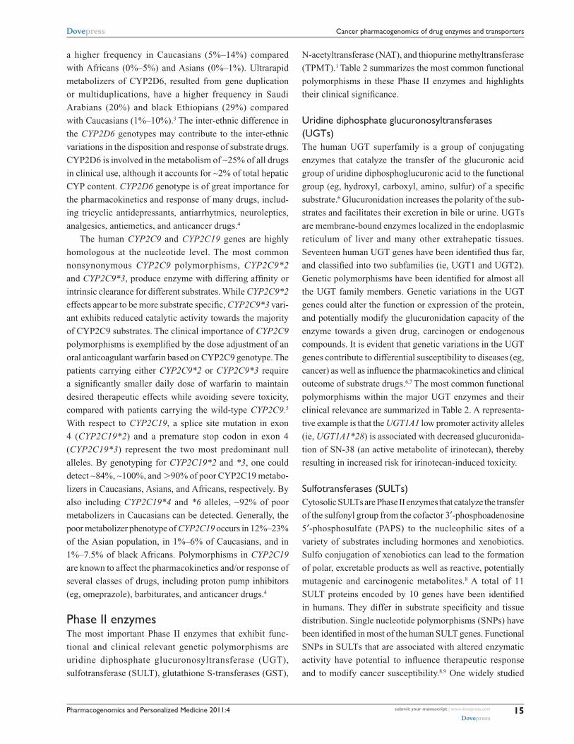
a higher frequency in Caucasians (5%–14%) compared
with Africans (0%–5%) and Asians (0%–1%). Ultrarapid
metabolizers of CYP2D6, resulted from gene duplication
or multiduplications, have a higher frequency in Saudi
Arabians (20%) and black Ethiopians (29%) compared
with Caucasians (1%–10%).3 The inter-ethnic difference in
the CYP2D6 genotypes may contribute to the inter-ethnic
variations in the disposition and response of substrate drugs.
CYP2D6 is involved in the metabolism of ∼25% of all drugs
in clinical use, although it accounts for ∼2% of total hepatic
CYP content. CYP2D6 genotype is of great importance for
the pharmacokinetics and response of many drugs, includ-
ing tricyclic antidepressants, antiarrhytmics, neuroleptics,
analgesics, antiemetics, and anticancer drugs.4
The human CYP2C9 and CYP2C19 genes are highly
homologous at the nucleotide level. The most common
nonsynonymous CYP2C9 polymorphisms, CYP2C9*2
and CYP2C9*3, produce enzyme with differing affinity or
intrinsic clearance for different substrates. While CYP2C9*2
effects appear to be more substrate specific, CYP2C9*3 vari-
ant exhibits reduced catalytic activity towards the majority
of CYP2C9 substrates. The clinical importance of CYP2C9
polymorphisms is exemplified by the dose adjustment of an
oral anticoagulant warfarin based on CYP2C9 genotype. The
patients carrying either CYP2C9*2 or CYP2C9*3 require
a significantly smaller daily dose of warfarin to maintain
desired therapeutic effects while avoiding severe toxicity,
compared with patients carrying the wild-type CYP2C9.5
With respect to CYP2C19, a splice site mutation in exon
4 (CYP2C19*2) and a premature stop codon in exon 4
(CYP2C19*3) represent the two most predominant null
alleles. By genotyping for CYP2C19*2 and *3, one could
detect ∼84%, ∼100%, and .90% of poor CYP2C19 metabo-
lizers in Caucasians, Asians, and Africans, respectively. By
also including CYP2C19*4 and *6 alleles, ∼92% of poor
metabolizers in Caucasians can be detected. Generally, the
poor metabolizer phenotype of CYP2C19 occurs in 12%–23%
of the Asian population, in 1%–6% of Caucasians, and in
1%–7.5% of black Africans. Polymorphisms in CYP2C19
are known to affect the pharmacokinetics and/or response of
several classes of drugs, including proton pump inhibitors
(eg, omeprazole), barbiturates, and anticancer drugs.4
Phase ii enzymesThe most important Phase II enzymes that exhibit func-
tional and clinical relevant genetic polymorphisms are
uridine diphosphate glucuronosyltransferase (UGT),
sulfotransferase (SULT), glutathione S-transferases (GST),
N-acetyltransferase (NAT), and thiopurine methyltransferase
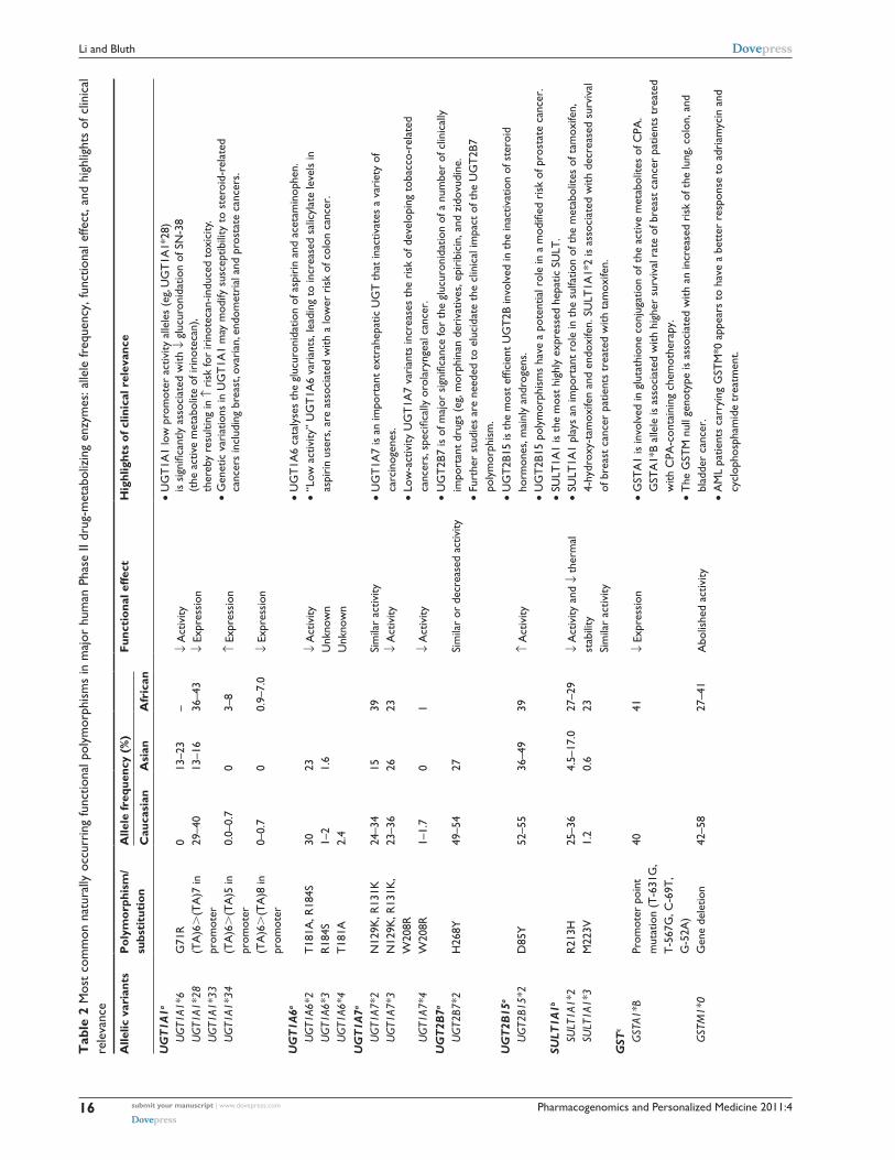
(TPMT).1 Table 2 summarizes the most common functional
polymorphisms in these Phase II enzymes and highlights
their clinical significance.
Uridine diphosphate glucuronosyltransferases (UGTs)The human UGT superfamily is a group of conjugating
enzymes that catalyze the transfer of the glucuronic acid
group of uridine diphosphoglucuronic acid to the functional
group (eg, hydroxyl, carboxyl, amino, sulfur) of a specific
substrate.6 Glucuronidation increases the polarity of the sub-
strates and facilitates their excretion in bile or urine. UGTs
are membrane-bound enzymes localized in the endoplasmic
reticulum of liver and many other extrahepatic tissues.
Seventeen human UGT genes have been identified thus far,
and classified into two subfamilies (ie, UGT1 and UGT2).
Genetic polymorphisms have been identified for almost all
the UGT family members. Genetic variations in the UGT
genes could alter the function or expression of the protein,
and potentially modify the glucuronidation capacity of the
enzyme towards a given drug, carcinogen or endogenous
compounds. It is evident that genetic variations in the UGT
genes contribute to differential susceptibility to diseases (eg,
cancer) as well as influence the pharmacokinetics and clinical
outcome of substrate drugs.6,7 The most common functional
polymorphisms within the major UGT enzymes and their
clinical relevance are summarized in Table 2. A representa-
tive example is that the UGT1A1 low promoter activity alleles
(ie, UGT1A1*28) is associated with decreased glucuronida-
tion of SN-38 (an active metabolite of irinotecan), thereby
resulting in increased risk for irinotecan-induced toxicity.
Sulfotransferases (SULTs)Cytosolic SULTs are Phase II enzymes that catalyze the transfer
of the sulfonyl group from the cofactor 3′-phosphoadenosine
5′-phosphosulfate (PAPS) to the nucleophilic sites of a
variety of substrates including hormones and xenobiotics.
Sulfo conjugation of xenobiotics can lead to the formation
of polar, excretable products as well as reactive, potentially
mutagenic and carcinogenic metabolites.8 A total of 11
SULT proteins encoded by 10 genes have been identified
in humans. They differ in substrate specificity and tissue
distribution. Single nucleotide polymorphisms (SNPs) have
been identified in most of the human SULT genes. Functional
SNPs in SULTs that are associated with altered enzymatic
activity have potential to influence therapeutic response
and to modify cancer susceptibility.8,9 One widely studied
Pharmacogenomics and Personalized Medicine 2011:4submit your manuscript | www.dovepress.com
Dovepress
Dovepress
16
Li and Bluth
Tab
le 2
Mos
t co
mm
on n
atur
ally
occ
urri
ng fu
nctio
nal p
olym
orph
ism
s in
maj
or h
uman
Pha
se ii
dru
g-m
etab
oliz
ing
enzy
mes
: alle
le fr
eque
ncy,
func
tiona
l effe
ct, a
nd h
ighl
ight
s of
clin
ical
re
leva
nce
Alle
lic v
aria
nts
Pol
ymor
phis
m/
subs
titu
tion
Alle
le fr
eque
ncy
(%)
Func
tion
al e
ffect
Hig
hlig
hts
of c
linic
al r
elev
ance
Cau
casi
anA
sian
Afr
ican
UG
T1A
1a•
UG
T1A
1 lo
w p
rom
oter
act
ivity
alle
les
(eg,
UG
T1A
1*28
)
is s
igni
fican
tly a
ssoc
iate
d w
ith ↓
glu
curo
nida
tion
of S
N-3
8
(the
act
ive
met
abol
ite o
f iri
note
can)
, th
ereb
y re
sulti
ng in
↑ri
sk fo
r ir
inot
ecan
-indu
ced
toxi
city
.•
Gen
etic
var
iatio
ns in
UG
T1A
1 m
ay m
odify
sus
cept
ibili
ty t
o st
eroi
d-re
late
d
canc
ers
incl
udin
g br
east
, ova
rian
, end
omet
rial
and
pro
stat
e ca
ncer
s.
U
GT1
A1*6
U
GT1
A1*2
8
UG
T1A1
*33
U
GT1
A1*3
4
G71
R(T
A)6
.(T
A)7
in
prom
oter
(TA
)6.
(TA
)5 in
pr
omot
er(T
A)6
.(T
A)8
in
prom
oter
0 29–4
0
0.0–
0.7
0–0.
7
13–2
313
–16
0 0
– 36–4
3
3–8
0.9–
7.0
↓A
ctiv
ity↓
expr
essi
on
↑ex
pres
sion
↓ex
pres
sion
UG
T1A
6a•
UG
T1A
6 ca
taly
ses
the
gluc
uron
idat
ion
of a
spir
in a
nd a
ceta
min
ophe
n.•
“Low
act
ivity
” U
GT
1A6
vari
ants
, lea
ding
to
incr
ease
d sa
licyl
ate
leve
ls in
as
piri
n us
ers,
are
ass
ocia
ted
with
a lo
wer
ris
k of
col
on c
ance
r.
UG
T1A6
*2
UG
T1A6
*3
UG
T1A6
*4
T18
1A, R
184S
R18
4ST
181A
30 1–2
2.4
23 1.6
↓A
ctiv
ityU
nkno
wn
Unk
now
nU
GT
1A7a
U
GT1
A7*2
U
GT1
A7*3
U
GT1
A7*4
N12
9K, R
131K
N12
9K, R
131K
, w
208R
w20
8R
24–3
423
–36
1–1.
7
15 26 0
39 23 1
Sim
ilar
activ
ity↓
Act
ivity
↓A
ctiv
ity
• U
GT
1A7
is a
n im
port
ant
extr
ahep
atic
UG
T t
hat
inac
tivat
es a
var
iety
of
carc
inog
enes
.•
Low
-act
ivity
UG
T1A
7 va
rian
ts in
crea
ses
the
risk
of d
evel
opin
g to
bacc
o-re
late
d
canc
ers,
spe
cific
ally
oro
lary
ngea
l can
cer.
UG
T2B
7a•
UG
T2B
7 is
of m
ajor
sig
nific
ance
for
the
gluc
uron
idat
ion
of a
num
ber
of c
linic
ally
im
port
ant
drug
s (e
g, m
orph
inan
der
ivat
ives
, epi
ribi
cin,
and
zid
ovud
ine.
• Fu
rthe
r st
udie
s ar
e ne
eded
to
eluc
idat
e th
e cl
inic
al im
pact
of t
he U
GT
2B7
po
lym
orph
ism
.
U
GT2
B7*2
H26
8Y49
–54
27Si
mila
r or
dec
reas
ed a
ctiv
ity
UG
T2B
15a
• U
GT
2B15
is t
he m
ost
effic
ient
UG
T2B
invo
lved
in t
he in
activ
atio
n of
ste
roid
ho
rmon
es, m
ainl
y an
drog
ens.
• U
GT
2B15
pol
ymor
phis
ms
have
a p
oten
tial r
ole
in a
mod
ified
ris
k of
pro
stat
e ca
ncer
.
UG
T2B1
5*2
D85
Y52
–55
36–4
939
↑A
ctiv
ity
SULT
1A1b
• SU
LT1A
1 is
the
mos
t hi
ghly
exp
ress
ed h
epat
ic S
ULT
.•
SULT
1A1
play
s an
impo
rtan
t ro
le in
the
sul
fatio
n of
the
met
abol
ites
of t
amox
ifen,
4-
hydr
oxy-
tam
oxife
n an
d en
doxi
fen.
SU
LT1A
1*2
is a
ssoc
iate
d w
ith d
ecre
ased
sur
viva
l of
bre
ast
canc
er p
atie
nts
trea
ted
with
tam
oxife
n.
SU
LT1A
1*2
SU
LT1A
1*3
R21
3HM
223v
25–3
61.
24.
5–17
.00.
627
–29
23↓
Act
ivity
and
↓th
erm
al
stab
ility
Sim
ilar
activ
ityG
STc
G
STA1
*B
G
STM
1*0
Prom
oter
poi
nt
mut
atio
n (T
-631
G,
T-5
67G
, C-6
9T,
G-5
2A)
Gen
e de
letio
n
40 42–5
8
41 27–4
1
↓ex
pres
sion
Abo
lishe
d ac
tivity
• G
STA
1 is
invo
lved
in g
luta
thio
ne c
onju
gatio
n of
the
act
ive
met
abol
ites
of C
PA.
GST
A1*
B al
lele
is a
ssoc
iate
d w
ith h
ighe
r su
rviv
al r
ate
of b
reas
t ca
ncer
pat
ient
s tr
eate
d w
ith C
PA-c
onta
inin
g ch
emot
hera
py.
• T
he G
STM
nul
l gen
otyp
e is
ass
ocia
ted
with
an
incr
ease
d ri
sk o
f the
lung
, col
on, a
nd
blad
der
canc
er.
• A
ML
patie
nts
carr
ying
GST
M*0
app
ears
to
have
a b
ette
r re
spon
se t
o ad
riam
ycin
and
cy
clop
hosp
ham
ide
trea
tmen
t.
Pharmacogenomics and Personalized Medicine 2011:4 submit your manuscript | www.dovepress.com
Dovepress
Dovepress
17
Cancer pharmacogenomics of drug enzymes and transporters
G
STP1
*B
GST
T1*0
i105
vG
ene
dele
tion
6–40
2–42
54↓
Act
ivity
Abo
lishe
d ac
tivity
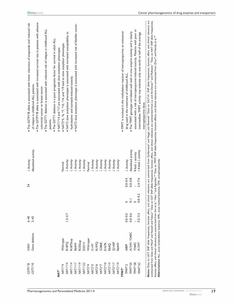
• T
he G
STP1
*B a
llele
is a
ssoc
iate
d w
ith lo
wer
cle
aran
ce o
f eto
posi
de a
nd r
educ
ed r
isk
of
rel
apse
in c
hild
hood
ALL
pat
ient
s.•
The
GST
P1*B
alle
le is
ass
ocia
ted
with
incr
ease
d su
rviv
al r
ate
in p
atie
nts
with
adv
ance
co
lore
ctal
can
cer
or b
reas
t ca
ncer
.•
The
GST
T1
dele
tion
is a
ssoc
iate
d w
ith r
educ
ed r
isk
of r
elap
se in
chi
ldho
od A
LL
patie
nts.
• T
he G
STT
1 de
letio
n is
a p
oor
prog
nost
ic fa
ctor
for
surv
ival
in a
dult
ALL
.N
AT
d•
NA
T1*
14 a
nd *
17 a
re a
ssoc
iate
d w
ith s
low
ace
tyla
tor
phen
otyp
e.•
NA
T2*
5, *
6, *
7, *
10, *
14, a
nd *
19 le
ad t
o sl
ow a
cety
lato
r ph
enot
ype.
• N
AT
2 sl
ow a
cety
lato
r ph
enot
ype
is a
ssoc
iate
d w
ith in
crea
sed
susc
eptib
ility
to
hy
drol
azin
e- a
nd is
onia
zid-
indu
ced
toxi
city
.•
NA
T2
slow
ace
tyla
tor
phen
otyp
e is
ass
ocia
ted
with
incr
ease
d ri
sk o
f bla
dder
can
cer.
N
AT1*
4
NAT
1*14
N
AT1*
14
NAT
1*17
N
AT1*
19
NAT
1*22
N
AT2*
4
NAT
2*5
N
AT2*
6
NAT
2*7
N
AT2*
10
NAT
2*14
N
AT2*
17
NAT
2*19
wild
-typ
eR
187Q
R18
7Sto
pR
64w
R33
Stop
D25
1vw
ild-t
ype
i114
TR
197Q
G28
6ee1
67K
R64
145P
R64
w
1.3–
3.7
Nor
mal
↓A
ctiv
ity↓
Act
ivity
↓A
ctiv
ity↓
Act
ivity
↓A
ctiv
ityN
orm
al↓
Act
ivity
↓A
ctiv
ity↓
Act
ivity
↓A
ctiv
ity↓
Act
ivity
↓A
ctiv
ity↓
Act
ivity
TPM
Te
• T
PMT
is in
volv
ed in
the
met
hyla
tion
reac
tion
of m
erca
ptop
urin
e, a
n an
tican
cer
dr
ug u
sed
in t
he t
reat
men
t of
chi
ldho
od A
LL.
• T
he T
PMT
gen
otyp
e co
rrel
ated
wel
l with
in-v
ivo
enzy
me
activ
ity a
nd is
cle
arly
as
soci
ated
with
a r
isk
of m
erca
ptop
urin
e-in
duce
d to
xici
ty. P
atie
nts
with
poo
r or
in
term
edia
te T
MPT
act
ivity
may
tol
erat
e on
ly o
ne-t
enth
to
half
of t
he a
vera
ge
mer
capt
opur
ine
dose
.
TP
MT*
2
TPM
T*3A
TP
MT*
3B
TPM
T*3C
A80
PA
154Y
, Y24
0CY
240C
A15
4Y
0.0–
0.5
0.0–
0.6
– 0.2–
3.3
0 0–1
0 0.0–
0.2
0.0–
0.4
0.0–
0.8
– 2.4–
7.6
↓A
ctiv
ityA
bolis
hed
activ
ity9-
fold
↓a
ctiv
ity1.
4-fo
ld ↓
act
ivity
Not
es: a
Dat
a on
UG
T S
NP
alle
le f
requ
enci
es, f
unct
ion
effe
ct, a
nd c
linic
al r
elev
ance
are
sum
mar
ized
fro
m G
uille
met
te6
and
Nag
ar a
nd R
emm
el;7
b Dat
a on
SU
LT1A
1 SN
P al
lele
fre
quen
cies
, fun
ctio
n ef
fect
, and
clin
ical
rel
evan
ce a
re
sum
mar
ized
from
Gla
tt a
nd M
einl
8 an
d N
owel
l and
Fal
any;
9 c D
ata
on G
ST S
NP
alle
le fr
eque
ncie
s, fu
nctio
n ef
fect
, and
clin
ical
rel
evan
ce a
re s
umm
ariz
ed fr
om L
o an
d A
li-O
sman
10 a
nd M
cilw
ain;
11 d D
ata
on N
AT
SN
P al
lele
freq
uenc
ies,
fu
nctio
n ef
fect
, and
clin
ical
rel
evan
ce a
re s
umm
ariz
ed fr
om S
im e
t al
,13 H
ein,
15 a
nd A
gund
ez;13
9 e Dat
a on
TPM
T S
NP
alle
le fr
eque
ncie
s, fu
nctio
n ef
fect
s, a
nd c
linic
al r
elev
ance
are
sum
mar
ized
from
Zho
u18 a
nd H
amdy
et
al.14
0
Abb
revi
atio
ns: A
LL, a
cute
lym
phob
last
ic le
ukem
ia; A
ML,
acu
te m
yelo
id le
ukem
ia; C
PA, c
yclo
phos
pham
ide.
Pharmacogenomics and Personalized Medicine 2011:4submit your manuscript | www.dovepress.com
Dovepress
Dovepress
18
Li and Bluth
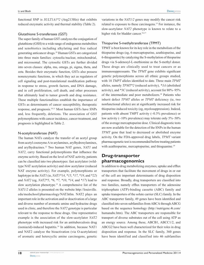
functional SNP is SULT1A1*2 (Arg213His) that exhibits
reduced enzymatic activity and thermal stability (Table 2).
Glutathione S-transferases (GST)The super family of human GST catalyzes the conjugation of
glutathione (GSH) to a wide range of endogenous metabolites
and xenobiotics including alkylating and free radical
generating anticancer drugs.10 Human GSTs are categorized
into three main families: cytosolic/nuclear, mitochondrial,
and microsomal. The cytosolic GSTs are further divided
into seven classes: alpha, mu, omega, pi, sigma, theta, and
zeta. Besides their enzymatic function, GSTs also possess
nonenzymatic functions, in which they act as regulators of
cell signaling and post-translational modification pathway
in response to stress, growth factors, and DNA damage,
and in cell proliferation, cell death, and other processes
that ultimately lead to tumor growth and drug resistance.
These multiple functionalities establish the importance of
GSTs as determinants of cancer susceptibility, therapeutic
response, and prognosis.10,11 Most human GSTs have SNPs
and, less frequently, deletions. The association of GST
polymorphisms with cancer incidence, cancer treatment, and
prognosis is highlighted in Table 2.
N-acetyltransferase (NAT)The human NATs catalyze the transfer of an acetyl group
from acetyl coenzyme A to arylamines, arylhydroxylamines,
and arylhydrazines.12 Two human NAT genes, NAT1 and
NAT2, carry functional polymorphisms that influence the
enzyme activity. Based on the level of NAT activity, patients
can be classified into two phenotypes: fast acetylator (wild-
type NAT acetylation activity) and slow acetylator (reduced
NAT enzyme activity). For example, polymorphisms or
haplotype in the NAT1 (ie, NAT1*14, *15, *17, *19, and *22)
and NAT2 (eg, NAT2*5, *6, *7, *10, *14, and *17) lead to
slow acetylation phenotype.13 A comprehensive list of the
NAT1/2 alleles is presented on the website http://louisville.
edu/medschool/pharmacology/NAT.htlm. NAT2 plays an
important role in the activation and/or deactivation of a large
and diverse number of aromatic amine and hydrazine drugs
used in clinic, and therefore the NAT2 genotype is particular
relevant to the response to these drugs. One representative
example is the association of the slow-acetylator NAT2
phenotype with increased risk for an antituberculosis drug
(isoniazid)-induced hepatitis.14 In addition, because NAT1
and NAT2 catalyze the bioactivation (via O-acetylation)
of aromatic and hetercyclic amine carcinogens, genetic
variations in the NAT1/2 genes may modify the cancer risk
related to exposure to these carcinogens.15 For instance, the
slow-acetylator NAT2 phenotype is known to relate to a
higher risk for bladder cancer.16,17
Thiopurine S-methyltransferase (TPMT)TPMT is best known for its key role in the metabolism of the
thiopurine drugs (eg, 6-mercaptopurine, azathiopurine, and
6-thioguanine) by catalyzing the S-methylation of thiopurine
drugs via S-adenosyl-L-methionine as the S-methyl donor.
These drugs are clinically used to treat cancers or as
immunosuppressants. The TPMT gene exhibits significant
genetic polymorphisms across all ethnic groups studied,
with 18 TMPT alleles identified to date. Three main TPMT
alleles, namely TPMT*2 (reduced activity), *3A (abolished
activity), and *3C (reduced activity), account for 80%–95%
of the intermediate and poor metabolizers.18 Patients who
inherit defect TPMT alleles or TPMT deficiency (ie, two
nonfunctional alleles) are at significantly increased risk for
thiopurine-induced toxicity (eg, myelosuppression). Indeed,
patients with absent TMPT activity (∼0.3% prevalence) or
low activity (∼10% prevalence) may tolerate only 5%–50%
of the average mercaptopurine dose. Clinical diagnostic tests
are now available for the detection of the SNPs in the human
TPMT gene that lead to decreased or abolished enzyme
activity. On the FDA-approved drug labels, TPMT variant
pharmacogenetic test is recommended before treating patients
with azathiopurine, mercaptopurine, and thioguanine.19
Drug-transporter pharmacogenomicsIn addition to drug metabolizing enzymes, uptake and efflux
transporters that facilitate the movement of drugs in or out
of the cell are important determinants of drug disposition
and response. Broadly, drug transporters are classified into
two families, namely efflux transporters of the adenosine
triphosphate (ATP)-binding cassette (ABC) family and
uptake transporters of the solute carrier (SLC) family. In the
ABC transporter family, 49 genes have been identified and
classified into seven subfamilies from ABCA through ABCG
based on the sequence homology (http://nutrigene.4t.com/
humanabc.htm). The ABC transporters are responsible for
transport of diverse substrates out of the cell using ATP as
an energy source. Among these, ABCB1, ABCC1/2, and
ABCG2 have been well characterized for their roles in drug
disposition and response. In the SLC family, 360 genes
have been identified and classified into 46 subfamilies
Pharmacogenomics and Personalized Medicine 2011:4 submit your manuscript | www.dovepress.com
Dovepress
Dovepress
19
Cancer pharmacogenomics of drug enzymes and transporters
(http://www.bioparadigms.org/slc/menu.asp). Of particular
relevance to drug disposition are members of the organic
anion transporting polypeptides (OATP), organic cation
transporter (OCT), and organic anion transporter (OAT)
subfamilies.
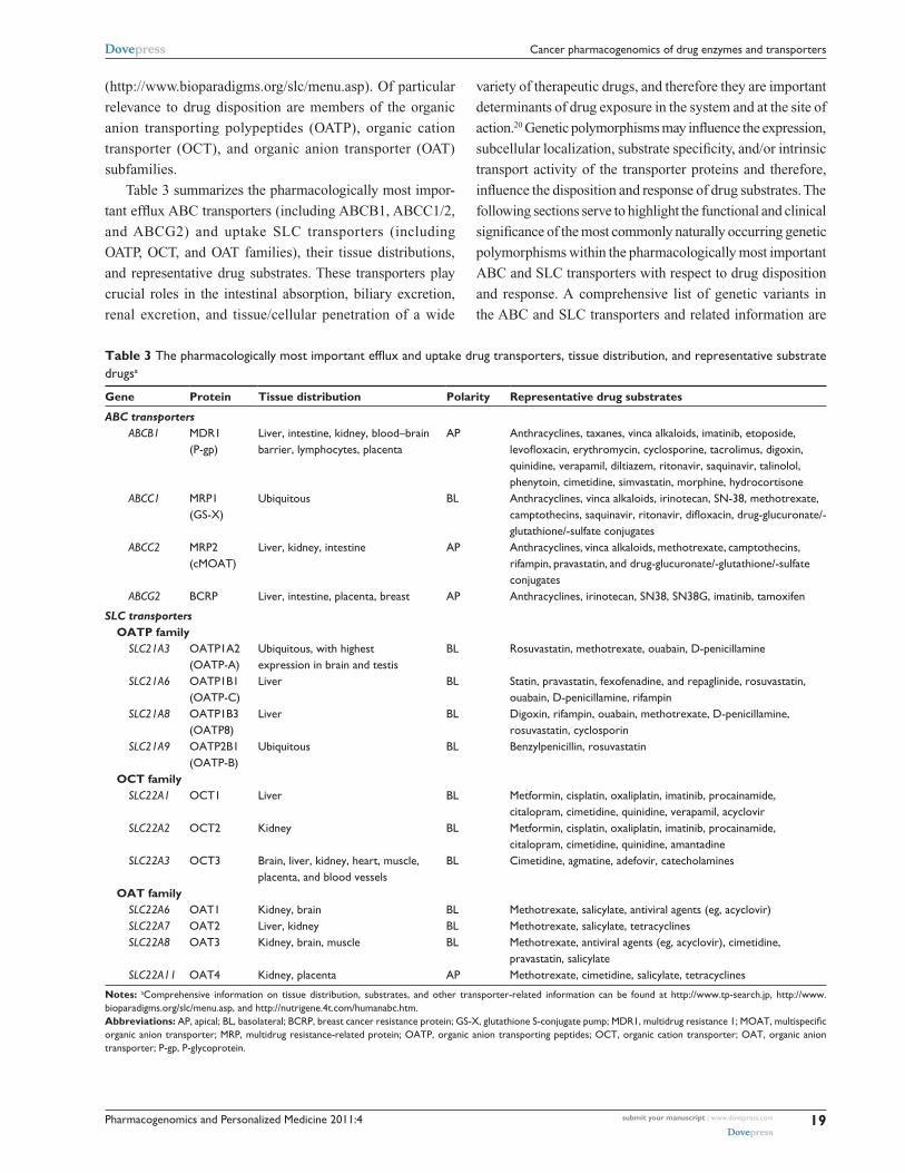
Table 3 summarizes the pharmacologically most impor-
tant efflux ABC transporters (including ABCB1, ABCC1/2,
and ABCG2) and uptake SLC transporters (including
OATP, OCT, and OAT families), their tissue distributions,
and representative drug substrates. These transporters play
crucial roles in the intestinal absorption, biliary excretion,
renal excretion, and tissue/cellular penetration of a wide
variety of therapeutic drugs, and therefore they are important
determinants of drug exposure in the system and at the site of
action.20 Genetic polymorphisms may influence the expression,
subcellular localization, substrate specificity, and/or intrinsic
transport activity of the transporter proteins and therefore,
influence the disposition and response of drug substrates. The
following sections serve to highlight the functional and clinical
significance of the most commonly naturally occurring genetic
polymorphisms within the pharmacologically most important
ABC and SLC transporters with respect to drug disposition
and response. A comprehensive list of genetic variants in
the ABC and SLC transporters and related information are
Table 3 The pharmacologically most important efflux and uptake drug transporters, tissue distribution, and representative substrate drugsa
Gene Protein Tissue distribution Polarity Representative drug substrates
ABC transporters ABCB1 MDR1
(P-gp)Liver, intestine, kidney, blood–brain barrier, lymphocytes, placenta
AP Anthracyclines, taxanes, vinca alkaloids, imatinib, etoposide, levofloxacin, erythromycin, cyclosporine, tacrolimus, digoxin, quinidine, verapamil, diltiazem, ritonavir, saquinavir, talinolol, phenytoin, cimetidine, simvastatin, morphine, hydrocortisone
ABCC1 MRP1 (GS-X)
Ubiquitous BL Anthracyclines, vinca alkaloids, irinotecan, SN-38, methotrexate, camptothecins, saquinavir, ritonavir, difloxacin, drug-glucuronate/-glutathione/-sulfate conjugates
ABCC2 MRP2 (cMOAT)
Liver, kidney, intestine AP Anthracyclines, vinca alkaloids, methotrexate, camptothecins, rifampin, pravastatin, and drug-glucuronate/-glutathione/-sulfate conjugates
ABCG2 BCRP Liver, intestine, placenta, breast AP Anthracyclines, irinotecan, SN38, SN38G, imatinib, tamoxifen
SLC transporters OATP family SLC21A3 OATP1A2
(OATP-A)Ubiquitous, with highest expression in brain and testis
BL Rosuvastatin, methotrexate, ouabain, D-penicillamine
SLC21A6 OATP1B1 (OATP-C)
Liver BL Statin, pravastatin, fexofenadine, and repaglinide, rosuvastatin, ouabain, D-penicillamine, rifampin
SLC21A8 OATP1B3 (OATP8)
Liver BL Digoxin, rifampin, ouabain, methotrexate, D-penicillamine, rosuvastatin, cyclosporin
SLC21A9 OATP2B1 (OATP-B)
Ubiquitous BL Benzylpenicillin, rosuvastatin
OCT family SLC22A1 OCT1 Liver BL Metformin, cisplatin, oxaliplatin, imatinib, procainamide,
citalopram, cimetidine, quinidine, verapamil, acyclovir SLC22A2 OCT2 Kidney BL Metformin, cisplatin, oxaliplatin, imatinib, procainamide,
citalopram, cimetidine, quinidine, amantadine SLC22A3 OCT3 Brain, liver, kidney, heart, muscle,
placenta, and blood vesselsBL Cimetidine, agmatine, adefovir, catecholamines
OAT family SLC22A6 OAT1 Kidney, brain BL Methotrexate, salicylate, antiviral agents (eg, acyclovir) SLC22A7 OAT2 Liver, kidney BL Methotrexate, salicylate, tetracyclines SLC22A8 OAT3 Kidney, brain, muscle BL Methotrexate, antiviral agents (eg, acyclovir), cimetidine,
pravastatin, salicylate SLC22A11 OAT4 Kidney, placenta AP Methotrexate, cimetidine, salicylate, tetracyclines
Notes: aComprehensive information on tissue distribution, substrates, and other transporter-related information can be found at http://www.tp-search.jp, http://www.bioparadigms.org/slc/menu.asp, and http://nutrigene.4t.com/humanabc.htm.Abbreviations: AP, apical; BL, basolateral; BCRP, breast cancer resistance protein; GS-X, glutathione S-conjugate pump; MDR1, multidrug resistance 1; MOAT, multispecific organic anion transporter; MRP, multidrug resistance-related protein; OATP, organic anion transporting peptides; OCT, organic cation transporter; OAT, organic anion transporter; P-gp, P-glycoprotein.
Pharmacogenomics and Personalized Medicine 2011:4submit your manuscript | www.dovepress.com
Dovepress
Dovepress
20
Li and Bluth
available in Pharmacogenetics Research Network databases
at http://www.pharmGKB.org.
ABCB1, ABCC1/2, and ABCG2 efflux transportersABCB1 geneThe ABCB1 gene, also named as the multidrug resistance 1
(MDR1) gene, encodes a polypeptide (P-glycoprotein) that has
two halves, each containing six hydrophobic transmembrane
domains and an ATP-binding domain. ABCB1, located on
the apical or luminal surface of the epithelial cells, functions
as an efflux transporter in restricting intestinal absorption,
facilitating hepatobiliary excretion and renal excretion, and
protecting the brain and fetus from xenobiotics. In addition,
ABCB1 overexpression in cancer cells is implicated in
multidrug resistance to chemotherapeutic agents.21 ABCB1
transports a broad spectrum of structurally and functionally
diverse drugs, including anticancer agents, antibiotics, immu-
nosuppresants, cardiac drugs, calcium channel antagonists,
and HIV protease inhibitors (Table 3). Of note, there is a
strong overlap in substrate specificity and tissue distribution
for ABCB1 and CYP3A4/5.22
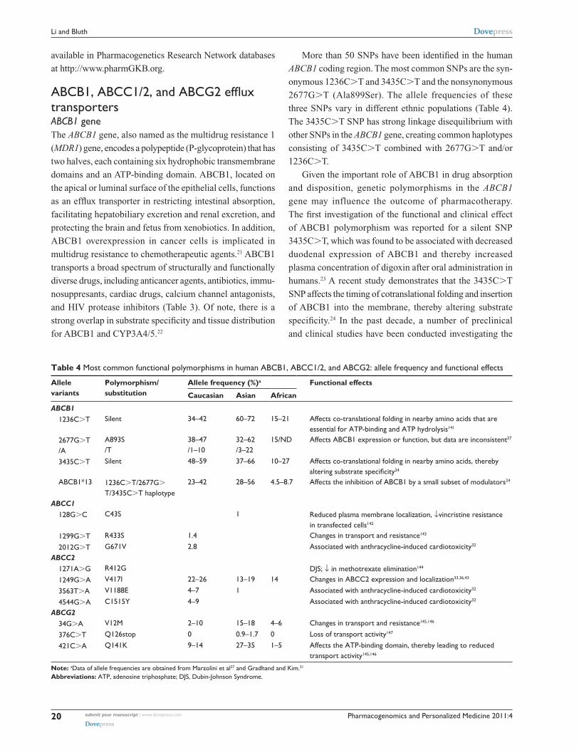
More than 50 SNPs have been identified in the human
ABCB1 coding region. The most common SNPs are the syn-
onymous 1236C.T and 3435C.T and the nonsynonymous
2677G.T (Ala899Ser). The allele frequencies of these
three SNPs vary in different ethnic populations (Table 4).
The 3435C.T SNP has strong linkage disequilibrium with
other SNPs in the ABCB1 gene, creating common haplotypes
consisting of 3435C.T combined with 2677G.T and/or
1236C.T.
Given the important role of ABCB1 in drug absorption
and disposition, genetic polymorphisms in the ABCB1
gene may influence the outcome of pharmacotherapy.
The first investigation of the functional and clinical effect
of ABCB1 polymorphism was reported for a silent SNP
3435C.T, which was found to be associated with decreased
duodenal expression of ABCB1 and thereby increased
plasma concentration of digoxin after oral administration in
humans.23 A recent study demonstrates that the 3435C.T
SNP affects the timing of cotranslational folding and insertion
of ABCB1 into the membrane, thereby altering substrate
specificity.24 In the past decade, a number of preclinical
and clinical studies have been conducted investigating the
Table 4 Most common functional polymorphisms in human ABCB1, ABCC1/2, and ABCG2: allele frequency and functional effects
Allele variants
Polymorphism/ substitution
Allele frequency (%)a Functional effects
Caucasian Asian African
ABCB1 1236C.T Silent 34–42 60–72 15–21 Affects co-translational folding in nearby amino acids that are
essential for ATP-binding and ATP hydrolysis141
2677G.T /A
A893S /T
38–47 /1–10
32–62 /3–22
15/ND Affects ABCB1 expression or function, but data are inconsistent27
3435C.T Silent 48–59 37–66 10–27 Affects co-translational folding in nearby amino acids, thereby altering substrate specificity24
ABCB1*13 1236C.T/2677G.
T/3435C.T haplotype23–42 28–56 4.5–8.7 Affects the inhibition of ABCB1 by a small subset of modulators24
ABCC1 128G.C C43S 1 Reduced plasma membrane localization, ↓vincristine resistance
in transfected cells142
1299G.T R433S 1.4 Changes in transport and resistance143
2012G.T G671v 2.8 Associated with anthracycline-induced cardiotoxicity32
ABCC2 1271A.G R412G DJS; ↓ in methotrexate elimination144
1249G.A v417i 22–26 13–19 14 Changes in ABCC2 expression and localization33,36,43
3563T.A v1188e 4–7 1 Associated with anthracycline-induced cardiotoxicity32
4544G.A C1515Y 4–9 Associated with anthracycline-induced cardiotoxicity32
ABCG2 34G.A v12M 2–10 15–18 4–6 Changes in transport and resistance145,146
376C.T Q126stop 0 0.9–1.7 0 Loss of transport activity147
421C.A Q141K 9–14 27–35 1–5 Affects the ATP-binding domain, thereby leading to reduced transport activity145,146
Note: aData of allele frequencies are obtained from Marzolini et al27 and Gradhand and Kim.31
Abbreviations: ATP, adenosine triphosphate; DJS, Dubin-Johnson Syndrome.
Pharmacogenomics and Personalized Medicine 2011:4 submit your manuscript | www.dovepress.com
Dovepress
Dovepress
21
Cancer pharmacogenomics of drug enzymes and transporters
association of ABCB1 genotype with its tissue expression and
function, and with pharmacokinetics and pharmacodynamics
of substrates drugs (Table 4).25 However, data reported on
the functional and clinical impacts of ABCB1 polymorphisms
are often inconsistent.20,25–28 The desrepancies may be
partly explained by lack of standardized methodology
and assays among different studies. In addition, SNPs of
ABCB1 may often result in very subtle functional outcomes.
For example, a recent study demostrates that the haplo-
type 1236C.T/2677G.T/3435C.T does not change the
substrate transport per se but instead affects the inhibition
of transport by a small subset of modulators.24 Conflicting
results on the clinical impact of ABCB1 polymorphisms
may reflect the complex disposition pathway of the substrate
drugs. For example, the commonly used in-vivo ABCB1
probe drugs such as digoxin, fexofenadine, and talinolol
were found to be the dual substrates for both ABCB1 and
OATP transporters; cyclosporine is not only transported by
ABCB1 but also metabolized by CYP3A4. This means that
potential ABCB1 effect may be marked by the activity of
OATP transporters or CYP3A4. Hence, a systemic analysis
of polymorphisms in multiple genes known or suspected to
contribute to drug disposition and response would provide
better insights on the genetic impact on pharmacotherapy.
In addition, the ABCB1 has multiple polymorphisms, some
of which are in linkage disequilibrium, and therefore a
haplotype approach would allow a more accurate prediction
of clinical phenotypes.
ABCC1 and ABCC2ABCC1/2, also called multidrug resistance-related proteins
(MRP1/2), plays an essential role in transport and excretion
of organic anions including physiological metabolites,
carcinogens, and drugs. They are also believed to confer
multidrug resistance to chemotherapeutic agents.29 ABCC1
and ABCC2 have overlapping substrate specif icities,
typically glutathione, glucuronate, or sulfate conjugated
and unconjugated drugs, including many anticancer
agents (eg, vincristine and doxorubicin), HIV protease
inhibitors (eg, ritonavir and saquinavir), and antibiotics (eg,
difloxacin and grepafloxacin) (Table 3). Both ABCC1 and
ABCC2 require co-transport of reduced glutathione (GSH)
to transport some of their substrates.30 ABCC1 is located
in basolateral membranes of polarized cells, whereas
ABCC2 is located to the apical domain. While ABCC1
is ubiquitously expressed, ABCC2 is mainly expressed
in hepatocytes, renal proximal tubule cells, intestine, and
brain (Table 3).
The human ABCC1 appears to be a conserved gene
because many of the naturally occurring genetic vari-
ants in ABCC1 are relatively rare. Of the identified SNPs
in the non-oding and coding region of ABCC1, 16 are
known to result in amino acid changes, and some of them
exhibit functional effects on either expression or function
of the protein (Table 4).31 Data on the role of ABCC1
polymorphisms in terms of in-vivo physiology and clinical
drug resistance or toxicity are rather limited. Interestingly,
one study has identified significant associations of ABCC1
2012G.T (Gly671Val) and a haplotype of ABCC2 with
anthracycline-induced cardiotoxicity among non-Hodgkin
lymphoma patients treated with doxorubicin.32
Mutations in the ABCC2 gene have been initially
identified in Dubin–Johnson Syndrome (DJS), a relatively
rare recessive disorder characterized by conjugated
hyperbilirubinemia resulting from loss of expression and
function of ABCC2 in the liver. However, the impact of
this loss of hepatic ABCC2-medicated transport on the
pharmacokinetics of drug substrates in humans is unknown.
Of the more commonly occurring ABCC2 SNPs, 1249G.A
(Val417Ile) has been extensively studied. The effect of this
SNP on ABCC2 expression varies depending on the tissue
examined. For example, 1249G.A SNP was associated with
lower ABCC2 mRNA and protein levels in preterm placenta,
but not in duodenum and liver.33,34 One study demonstrated
a possible association of 1249G.A variant with tenofovir-
induced renal proximal tubulopathy, suggesting this SNP may
influence renal excretion of some ABCC2 substrates.35 In
addition, 1249G.A SNP has been associated with changes
in the ABCC2 localization in neuroepithelial tumors.36
A number of other nonsynonymous and synonymous SNPs
have been studied for their potential functional influence
on the ABCC2 expression and transport activity (Table 4).
It appears that ABCC2 SNPs have varied functional influence
on different organs, or different substrates, or between in vitro
and in vivo studies.31
ABCG2The ABCG2 (also known as BCRP, ABCP, or MXR) protein
is an ABC half-transporter that bears six transmembrane
domains and one ATP-binding domain. The protein actively
extrudes a wide variety of chemically unrelated hydrophobic
or partially hydrophobic compounds from the cells, including
cytotoxic compounds (eg, mitoxantrone, topotecan, SN-38,
flavopiridol, and methotrexate), fluorescent dyes (eg, Hoechst
33342), and toxic compounds found in normal food (eg, pheo-
phorbide A) (Table 3). ABCG2 is expressed in the canalicular
Pharmacogenomics and Personalized Medicine 2011:4submit your manuscript | www.dovepress.com
Dovepress
Dovepress
22
Li and Bluth
membrane of hepatocytes, in the epithelia of small intestine,
colon, placenta, lung, kidney, adrenal and sweat glands, as well
as in the endothelia of the central nerve system vasculature.
It is responsible for host detoxification and protection against
potentially toxic xenobiotics.37–39 ABCG2 transporter-
mediated efflux has been increasingly recognized to not only
confer drug resistance but also significantly modulate drug
absorption, distribution, metabolism, and excretion.40–44
More than 80 polymorphisms in the ABCG2 gene have
been identified in different ethnic populations.45–48 Several
naturally occurring SNPs in ABCG2 have been found to affect
the function and/or expression of its encoded protein.46,49–51
In particular, a functional SNP in exon 5 of the ABCG2 gene,
in which a C→A nucleotide transition at position 421 (ABCG2
421C.A), results in a nonsynonymous variant protein with
a glutamine to lysine amino acid substitution in codon 141
(Q141K).46 The ABCG2 421C.A variant has been associated
with low ABCG2 expression levels and altered substrate
specificity,46 and has been found to alter the pharmacokinetics
of diflomotecan and topotecan.40,52 In addition, recent studies
have demonstrated that the epidermal growth factor receptor
(EGFR) tyrosine kinase inhibitors such as gefitinib and
erlotinib are ABCG2 substrates, and the ABCG2 421C.A
variant is associated with greater gefitinib accumulation at
steady-state and related to higher incidence of gefitinib-
induced grade 1 or 2 diarrhea in cancer patients compared
with the wild-type ABCG2.53,54
OATP, OCT, and OAT uptake transportersOrganic anion transporting polypeptides (OATPs)OATPs are membrane influx transporters that facilitate
cellular uptake of a wide range of endogenous compounds (eg,
bile salts, hormones, and steroid conjugates) and clinically
important drugs (eg, HMG-CoA-reductase inhibitors, cardiac
glycosides, anticancer agents, and antibiotics) (Table 3). Of
the 11 human OATP transporters, OATP1A2, OATP1B1,
OATP1B3, and OATP2B1 are best characterized for their
roles in drug pharmacokinetics. OATP1A2 is expressed on
the luminal membrane of small intestinal enterocytes and
at the blood–brain barrier and may facilitate the intestinal
absorption and brain penetration of its substrates. OATP1B1,
OATP1B3, and OATP2B1 are mainly expressed on the
sinusoidal membrane of hepatocytes and can facilitate the
hepatic uptake of their substrate drugs for further metabolism
or biliary excretion.55
A number of SNPs and other genetic variations have
been identified in the SLCO1B1 gene (encoding OATP1B1),
and their allele frequencies vary markedly across different
populations (Table 5).56 Some of SLCO1B1 SNPs and
haplotypes have been associated with impaired transport
activity in vitro towards different substrates.57–59 These
functional impaired OATP1B1 variants may limit the uptake
of the substrate drugs into the hepatocytes, thereby resulting
in decreased biliary excretion or hepatic metabolism and
thus increased systemic exposure. For example, a common
variant allele, 521T.C, is associated with increased systemic
exposure (eg, AUC) of several OATP1B1 drug substrates,
including repaglinide and statins such as pravastatin.60,61
OATP1B1*15 (a haplotype of 388A.G and 512T.C)
is associated with increased plasma concentrations of
pravastatin62 and increased concentrations of SN-38.63,64
OATP1B1*17 (a haplotype of -11187G.A, 388A.G and
512T.C) is associated with increased effect of pravastatin
on rate of cholesterol synthesis. A recent genome-wide asso-
ciation study has demonstrated that a noncoding rs4363657
SNP, which is in nearly complete linkage disequilibrium
with the SLCO1B1 521T.C SNP, is the only strong marker
associated with simvastatin-induced myopathy.65
With respect to the SLCO1A2 gene (encoding OATP1A2),
several nonsynonymous polymorphisms have been identified,
some of which demonstrate decreased in-vitro transport
activity towards OATP1A2 substrates (Table 5).66 The
impacts of these functional SNPs on the pharmacokinetics
and clinical outcome of clinical used drugs need further
studies. With respect to OATP1B1 and OATP2B1, there are
few data on the clinical relevance of SLCO1B3 and SLCO2B1
polymorphisms, although some genetic variations within
these two genes have been associated with altered in-vitro
transport activity of the protein (Table 5).67,68
Organic cation transporter (OCT)The OCTs belong to the solute carrier SLC22A family that
mediate intracellular uptake of a broad range of structurally
diverse small organic cations (molecular weight , 400).
Three isoforms, OCT1, OCT2, and OCT3, with partially
overlapping substrate spectrum, are identified in humans
(Table 3). OCT1 is primarily expressed in the sinusoidal
membrane of hepatocytes, whereas OCT2 is predominantly
expressed in the basolateral membrane of the kidney proximal
tubules; OCT3 is expressed in many tissues including
placenta, heart, liver, and skeletal muscle (Table 3). The
expression of OCTs was also detected in several cancer cell
lines and tumor tissue samples.69,70
A number of nonsynonymous SNPs have been identified
in the SLC22A1 (encoding OCT1) and SLC22A2 (encoding
Pharmacogenomics and Personalized Medicine 2011:4 submit your manuscript | www.dovepress.com
Dovepress
Dovepress
23
Cancer pharmacogenomics of drug enzymes and transporters
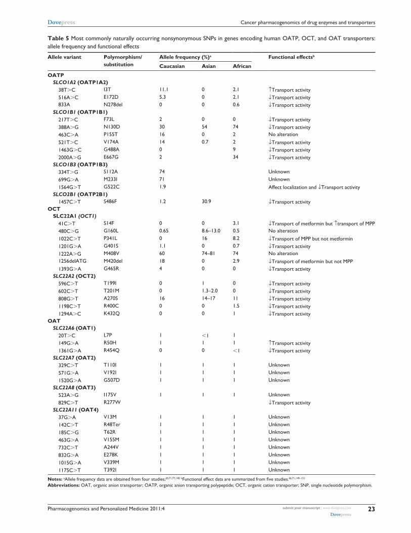
Table 5 Most commonly naturally occurring nonsynonymous SNPs in genes encoding human OATP, OCT, and OAT transporters: allele frequency and functional effects
Allele variant Polymorphism/substitution
Allele frequency (%)a Functional effectsb
Caucasian Asian African
OATP SLCO1A2 (OATP1A2) 38T.C i3T 11.1 0 2.1 ↑Transport activity 516A.C e172D 5.3 0 2.1 ↓Transport activity 833A N278del 0 0 0.6 ↓Transport activity SLCO1B1 (OATP1B1) 217T.C F73L 2 0 0 ↓Transport activity 388A.G N130D 30 54 74 ↓Transport activity 463C.A P155T 16 0 2 No alteration 521T.C v174A 14 0.7 2 ↓Transport activity 1463G.C G488A 0 9 ↓Transport activity 2000A.G e667G 2 34 ↓Transport activity SLCO1B3 (OATP1B3) 334T.G S112A 74 Unknown 699G.A M233i 71 Unknown 1564G.T G522C 1.9 Affect localization and ↓Transport activity SLCO2B1 (OATP2B1) 1457C.T S486F 1.2 30.9 ↓Transport activityOCT SLC22A1 (OCT1) 41C.T S14F 0 0 3.1 ↓Transport of metformin but ↑transport of MPP 480C.G G160L 0.65 8.6–13.0 0.5 No alteration 1022C.T P341L 0 16 8.2 ↓Transport of MPP but not metformin 1201G.A G401S 1.1 0 0.7 ↓Transport activity 1222A.G M408v 60 74–81 74 No alteration 1256delATG M420del 18 0 2.9 ↓Transport of metformin but not MPP 1393G.A G465R 4 0 0 ↓Transport activity SLC22A2 (OCT2) 596C.T T199i 0 1 0 ↓Transport activity 602C.T T201M 0 1.3–2.0 0 ↓Transport activity 808G.T A270S 16 14–17 11 ↓Transport activity 1198C.T R400C 0 0 1.5 ↓Transport activity 1294A.C K432Q 0 0 1 ↓Transport activityOAT SLC22A6 (OAT1) 20T.C L7P 1 ,1 1 149G.A R50H 1 1 1 ↑Transport activity 1361G.A R454Q 0 0 ,1 ↓Transport activity SLC22A7 (OAT2) 329C.T T110i 1 1 1 Unknown 571G.A v192i 1 1 1 Unknown 1520G.A G507D 1 1 1 Unknown SLC22A8 (OAT3) 523A.G i175v 1 1 1 Unknown 829C.T R277w ↓Transport activity SLC22A11 (OAT4) 37G.A v13M 1 1 1 Unknown 142C.T R48Ter 1 1 1 Unknown 185C.G T62R 1 1 1 Unknown 463G.A v155M 1 1 1 Unknown 732C.T A244v 1 1 1 Unknown 832G.A e278K 1 1 1 Unknown 1015G.A v339M 1 1 1 Unknown 1175C.T T392i 1 1 1 Unknown
Notes: aAllele frequency data are obtained from four studies;20,71,77,148 bFunctional effect data are summarized from five studies.66,71,149–151
Abbreviations: OAT, organic anion transporter; OATP, organic anion transporting polypeptide; OCT, organic cation transporter; SNP, single nucleotide polymorphism.
Pharmacogenomics and Personalized Medicine 2011:4submit your manuscript | www.dovepress.com
Dovepress
Dovepress
24
Li and Bluth
OCT2) from different ethnic groups; some of them have
demonstrated altered (mostly impaired) transport function
in vitro (Table 5).71 With respect to the SLC22A3 (encoding
OCT3), several synonymous SNPs have been identified, but
their functional consequence is unknown. The functional
polymorphisms in the OCT genes may influence the clini-
cal pharmacokinetics and response of drug substrates. For
example, functional polymorphisms of OCT1 and OCT2
impact the clinical effects and pharmacokinetics of meta-
formin, a drug used as a primary therapy for type 2 diabetes
mellitus. Metaformin is eliminated predominantly by renal
excretion in humans.72 Because of the high expression
of OCT2 in the kidney and the active renal secretion of
metaformin via OCT2, defect function in OCT2 transport
would result in decreased renal clearance of this drug. There
is evidence that carriers of homozygous for low activity
OCT2 variant 270S have a significant lower renal clearance
and higher plasma concentration of metformin than those
homozygous for the active variant (270A).73,74 On the other
hand, low-function OCT1 variants including R61C, G401S,
M420del, and G465R have been associated with significantly
higher renal clearance of metaformin.75 In addition to the
effect on metformin pharmacokinetics, low-function OCT1
variants (R61C, G401S, M420del, and G465R) have been
also associated with significantly decreased glucose-lowering
response of metaformin in healthy volunteers probably by
reducing the metaformin uptake in hepatocytes, which is the
major target site of metformin’s action.76
Organic anion transporter (OAT)The OATs belong to the SLC22 family of solute carriers
that mediate cellular uptake of a broad range of structur-
ally diverse small hydrophilic organic anions. OAT sub-
strates include many clinically important anionic drugs,
such as β-lactam antibiotics, diuretics, nonsterioidal
anti-inflammatory drugs, nucleoside/nucleotide antiviral
drugs, and anticancer agents (Table 3). There are at least six
OAT members (OAT1–6). OAT1–3 localize to the basolateral
membrane of the renal proximal tubule mediating the uptake
of drug substrates from blood into the proximal tubule cells,
whereas OAT4 localizes to the apical side of the renal proxi-
mal tubule functioning in the secretion of drug substrates into
urine. Collectively, these transporters are responsible for the
movement of drug substrates from the blood to the urine.
Thus, genetic variations in the genes encoding OATs may
contribute to interindividual variability in the renal clearance
of drug substrates. To date, a number of polymorphisms have
been reported in the coding region and 5′ regulatory region
of human SLC22A6 (encoding OAT1), SLC22A7 (encoding
OAT2), SLC22A8 (encoding OAT3), and SLC22A11 gene
(encoding OAT4) (Table 5); some of them resulted in altered
in vitro transport activity of the protein.77,78 Nevertheless, the
coding region polymorphisms in these genes are infrequent
(∼1%). The regulatory region polymorphisms of these genes,
particularly SLC22A8 (encoding OAT3), may be especially
important in accounting for variation in the renal clearance
of drug substrates.78 The functional and clinical relevance
of these coding and regulatory region polymorphisms need
further study.
Pharmacogenomics in cancer therapy5-fluorouracil (5-FU)Since its introduction more than 50 years ago,79 5-FU has
remained the most frequently prescribed anticancer drug
for the treatment of malignancies of the gastrointestinal
tract including colorectal and gastric cancer. 5-FU, a
fluoropyrimidine analog, is a prodrug that is converted to the
active metabolite, 5-fluoro-2-deoxyuridine monophosphate
(FdUMP) that leads to inhibition of thymidylate synthase
(TS) and subsequently inhibition of DNA synthesis.
5-FU is converted to FdUMP through three pathways:
oratate phosphoribosyltransferase (OPRT) (pathway 1),
uridine phosphorylase (UP) (pathway 2), and thymidine
phosphorylase (TP) (pathway 3) (Figure 1).80 The vast major-
ity (∼80%–85%) of administered 5-FU is metabolized by
the enzyme dihydropyrimidine dehydrogenase (DPD) in the
liver into the inactive form, dihydrofluorouracil (FDHU), and
excreted as a fluoro-β-alanine.
The Phase I metabolizing enzyme DPD plays the
most important role in detoxification of 5-FU. Expression
of DPD has been related to tolerance and response to
5-FU-based chemotherapy. Specifically, low expression or
absence of DPD has been associated with accumulation of
5-FU, thereby exposing patients to increased risk of severe
toxicities, while high expression of DPD has been associated
with poor response to 5-FU.81,82 Patients lacking the DPD
enzyme may experience severe to lethal toxicities when
receiving 5-FU-based chemotherapy. Genetic aberration in
the dihydropyrimidine dehydrogenase (DPYD) gene, such as
exon skipping, deletion, and missense mutation, contributes
to a DPD-deficiency phenotype. Approximately 3%–5% of
the population is partially or completely deficient in DPD
enzyme activity.83 The most known DPYD SNPs associated
with grade 3 and 4 toxicities are IVS14 + 1G.A, 2846A.T,
1679T.G, and 85T.C.84 In particular, the exon 14-skipping
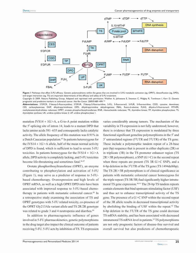
Pharmacogenomics and Personalized Medicine 2011:4 submit your manuscript | www.dovepress.com
Dovepress
Dovepress
25
Cancer pharmacogenomics of drug enzymes and transporters
mutation IVS14 + 1G.A, a G-to-A point mutation within
the 5′-splicing site of intron 14, leads to a mutant DPD that
lacks amino acids 581–635 and consequently lacks catalytic
activity. The allele frequency of this mutation was 0.91% in
a Dutch Caucasian population.85 In patients heterozygous for
the IVS14 + 1G.A allele, half of the mean normal activity
of DPD is found, which is sufficient to lead to severe 5-FU
toxicities. In patients homozygous for the IVS14 + 1G.A
allele, DPD activity is completely lacking, and 5-FU toxicities
become life-threatening and sometimes fatal.86,87
Orotate phosphoribosyltransferase (OPRT), an enzyme
contributing to phosphorylation and activation of 5-FU
(Figure 1), may serve as a predictor of response to 5-FU-
based chemotherapy. Overexpression and high levels of
OPRT mRNA, as well as a high OPRT/DPD ratio have been
associated with improved response to 5-FU-based chemo-
therapy in patients with metastatic colorectal cancer.88 In
a retrospective study examining the association of TS and
OPRT genotypes with 5-FU related toxicity, co-presence of
the OPRT Gly213Ala variant allele and TS 2R/2R genotype
was related to grade 3 and 4 neutropenia and diarrhea.89
In addition to pharmacogenetic influence of genes
involved in 5-FU pharmacokinetics, genetic polymorphisms
in the drug target also impact the clinical outcome of patients
receiving 5-FU. 5-FU acts by inhibition of TS. TS expression
varies considerably among tumors. The mechanism of the
variability in TS expression is not fully understood; however,
there is evidence that TS expression is modulated by three
functional significant gemeline polymorphisms in the 5′ and
3′ untranslated regions (5′UTR and 3′UTR) of the TS gene.
These include a polymorphic tandem repeat of a 28-base
pair (bp) sequence that is present in either duplicate (2R) or
in triplicate (3R) in the TS promoter enhancer region (TS
2R.3R polymorphism), a SNP (G.C) in the second repeat
when three repeats are present (TS 3R G.C SNP), and a
6-bp deletion in the 3′UTR of the TS gene (TS 1494del6bp).
The TS 2R.3R polymorphism is of clinical significance as
patients with metastatic colorectal cancer homozygous for
the triple repeat (TS 3R/3R) had significantly higher intratu-
moral TS gene expression.90,91 The 28-bp TS tandem repeats
contain elements that bind upstream stimulating factor (USF)
and thus act to enhance transcriptional activity of the TS
gene. The presence of a G.C SNP within the second repeat
of the 3R allele results in decreased transcriptional activity
by abolishing the binding of USF within the repeat.92 The
6-bp deletion in the 3′UTR of the TS gene could decrease
TS mRNA stability, and has been associated with decreased
intratumoral TS mRNA level in patients.93 TS polymorphisms
are not only prognostic factors of disease-free survival and
overall survival but also predictors of chemotherapeutic
TS Folate pool MTHFR
FdUDP
FUDP FUTP
FdUTP
dTMP
dUMP
FUMP
FdUMP
FUdR
5-FU
5-FURFBAL
5’DFUR
5’DFCR
dTDP dTTP
5-MTHF
5,10-MTHF
TK
UK
UP
DPD
DHP
CDDTP
CES
Capecitabine
FUH2
OPRT
RNR
DNA disruption
RNA disruption
DNA synthesis
Figure 1 Pathways that affect 5-FU efficacy. Genetic polymorphisms within the genes that are involved in 5-FU metabolic activation (eg, OPRT), detoxification (eg, DPD), and target interaction (eg, TS) are important determinants of the efficacy and safety of 5-FU treatment. Copyright © 2009. Nature Publishing Group. Adapted and reprinted with permission: walther A, Johnstone e, Swanton C, Midgley R, Tomlinson i, Kerr D. Genetic prognostic and predictive markers in colorectal cancer. Nat Rev Cancer. 2009;9:489–499.152
Abbreviations: 5′DFCR, 5′deoxy-5-fluorocytidine; 5′DFUR, 3′deoxy-5-fluorouridine; 5-FU, 5-fluorouracil; 5-FUR, 5-fluorouridine; CDD, cytosine deaminase; CES, carboxylesterase; DHP, dihydropyrimidinase; DPD, dihydropyrimidine dehydrogenase; FBAL, fluoro-b-alanine; FUH2, dihydro-5-fluorouracil; MTHFR, methylenetetrahydrofolate reductase; OPRT, orotate phosphoribosyltransferase; RNR, ribonucleotide reductase; TK, thymidine kinase; TP, thymidine phosphorylase; TS, thymidylate synthase; UK, uridine-cytidine kinase 2; UP, uridine phosphorylase 1.
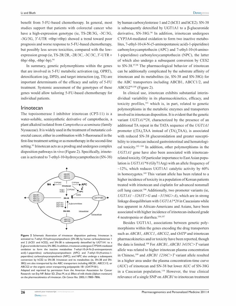
Pharmacogenomics and Personalized Medicine 2011:4submit your manuscript | www.dovepress.com
Dovepress
Dovepress
26
Li and Bluth
benefit from 5-FU-based chemotherapy. In general, most
studies support that patients with colorectal cancer who
have a high-expression genotype (ie, TS-2R/3G, -3C/3G,
-3G/3G, 3′-UTR +6bp/+6bp) showed a trend toward poor
prognosis and worse response to 5-FU-based chemotherapy,
but possibly less severe toxicities, compared with the low-
expression group (ie, TS-2R/2R, -2R/3C, -3C/3C, 3′-UTR +6bp/-6bp, -6bp/-bp).94
In summary, genetic polymorphisms within the genes
that are involved in 5-FU metabolic activation (eg, OPRT),
detoxification (eg, DPD), and target interaction (eg, TS) are
important determinants of the efficacy and safety of 5-FU
treatment. Systemic assessment of the genotypes of these
genes would allow tailoring 5-FU-based chemotherapy for
individual patients.
irinotecanThe topoisomerase I inhibitor irinotecan (CPT-11) is a
water-soluble, semisynthetic derivative of camptothecin, a
plant alkaloid isolated from Camptotheca acuminata (family
Nyssaceae). It is widely used in the treatment of metastatic col-
orectal cancer, either in combination with 5-fluorouracil in the
first-line treatment setting or as monotherapy in the second-line
setting.95 Irinotecan acts as a prodrug and undergoes complex
disposition pathways in vivo (Figure 2). Specifically, irinote-
can is activated to 7-ethyl-10-hydroxycamptothecin (SN-38)
by human carboxylesterase 1 and 2 (hCE1 and hCE2). SN-38
is subsequently detoxified by UGT1A1 to a β-glucuronide
derivative, SN-38G.96 In addition, irinotecan undergoes
CYP3A4-mediated oxidation to form two inactive metabo-
lites, 7-ethyl-10-(4-N-(5- aminopentanoic acid)-1-piperidino)
carbonyloxycamptothecin (APC) and 7-ethyl-10-(4-amino-
1-piperidino) carbonyloxycamptothecin (NPC), the latter
of which also undergo a subsequent conversion by CES2
to SN-38.97,98 The pharmacological behavior of irinotecan
can be additionally complicated by the substrate affinity of
irinotecan and its metabolites (ie, SN-38 and SN-38G) for
the ABC transporters including ABCB1, ABCC1/2, and
ABCG299,100 (Figure 2).
In clinical use, irinotecan exhibits substantial interin-
dividual variability in its pharmacokinetics, efficacy, and
toxicity profiles,101 which is, in part, related to genetic
polymorphisms in the metabolic enzymes and transporters
involved in irinotecan disposition. It is evident that the genetic
variant UGT1A1*28, characterized by the presence of an
additional TA repeat in the TATA sequence of the UGT1A1
promoter ((TA)7TAA instead of (TA)
6TAA), is associated
with reduced SN-38 glucuronidation and greater suscepti-
bility to irinotecan induced gastrointestinal and hematologi-
cal toxicity.102–104 In addition, other polymorphisms in the
UGT1A1 gene have also been associated with irinotecan-
related toxicity. Of particular importance to East Asian popu-
lation is UGT1A1*6 (Gly71Arg) with an allele frequency of
∼12%, which reduces UGT1A1 catalytic activity by 60%
in homozygotes.105 This variant allele has been related to a
higher incidence of toxicity in a population of Korean patients
treated with irinotecan and cisplatin for advanced nonsmall
cell lung cancer.106 Additionally, two promoter variants (ie,
UGT1A1 −3263T.G and −3156G.A), which are in strong
linkage disequilibrium with UGT1A1*28 in Caucasians while
less apparent in African-Americans and Asians, have been
associated with higher incidence of irinotecan-induced grade
4 neutropenia or diarrhea.104,107
Besides UGT1A1, associations between genetic poly-
morphisms within the genes encoding the drug transporters
such as ABCB1, ABCC1, ABCG2, and OATP and irinotecan
pharmacokinetics and/or toxicity have been reported, though
the data is limited.108 For ABCB1, ABCB1 3435C.T variant
allele was related to higher irinotecan plasma concentration
in Chinese,109 and ABCB1 1236C.T variant allele resulted
in a higher area under the plasma concentration-time curve
(AUC) of irinotecan and SN-38 but lower AUC of SN-38G
in a Caucasian population.110 However, the true clinical
relevance of a single SNP on ABCB1 to irinotecan treatment
M4 APC
NPC
SN-38
SN-38G
CYP3A5 CYP3A4
Irinotecan
CYP3A4hCE1hCE2
hCE2
UGT1A1 β-glucuronidaseUGT1A7
Intracellular
Extracellular
UGT1A9
ABCB1ABCC1ABCC2
ABCB1ABCC1
ABCC2
OATP1B1ABCC2ABCG2
ABCG2
Figure 2 Schematic illustration of irinotecan disposition pathway. irinotecan is activated to 7-ethyl-10-hydroxycamptothecin (SN-38) by human carboxylesterase 1 and 2 (hCE1 and hCE2), and SN-38 is subsequently detoxified by UGT1A1 to a β-glucuronide derivative, SN-38G. in addition, irinotecan undergoes CYP3A4-mediated oxidation to form the inactive metabolites 7-ethyl-10-(4-N-(5-aminopentanoic acid)-1-peperidino) carbonyloxycamptothecin (APC) and 7-ethyl-10-(4-amino-1-peperidino) carbonyloxycamptothecin (NPC), and NPC also undergo a subsequent conversion by hCe2 to SN-38. irinotecan and its metabolites (ie, SN-38 and SN-38G) are also transported by the ABC transporters including ABCB1, ABCC1/2, or ABCG2 or the organic anion transporting polypeptide 1B1 (OATP1B1). Adapted and reprinted by permission from the American Association for Cancer Research: van erp NP, Baker SD, Zhao M, et al. effect of milk thistle (Silybum marianum) on the pharmacokinetics of irinotecan. Clin Cancer Res. 2005;11:7800–7806.
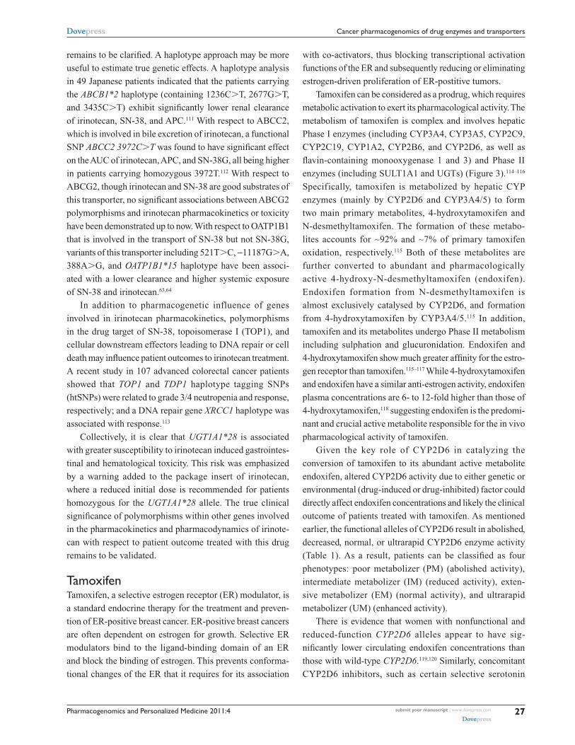
Pharmacogenomics and Personalized Medicine 2011:4 submit your manuscript | www.dovepress.com
Dovepress
Dovepress
27
Cancer pharmacogenomics of drug enzymes and transporters
remains to be clarified. A haplotype approach may be more
useful to estimate true genetic effects. A haplotype analysis
in 49 Japanese patients indicated that the patients carrying
the ABCB1*2 haplotype (containing 1236C.T, 2677G.T,
and 3435C.T) exhibit significantly lower renal clearance
of irinotecan, SN-38, and APC.111 With respect to ABCC2,
which is involved in bile excretion of irinotecan, a functional
SNP ABCC2 3972C.T was found to have significant effect
on the AUC of irinotecan, APC, and SN-38G, all being higher
in patients carrying homozygous 3972T.112 With respect to
ABCG2, though irinotecan and SN-38 are good substrates of
this transporter, no significant associations between ABCG2
polymorphisms and irinotecan pharmacokinetics or toxicity
have been demonstrated up to now. With respect to OATP1B1
that is involved in the transport of SN-38 but not SN-38G,
variants of this transporter including 521T.C, −11187G.A,
388A.G, and OATP1B1*15 haplotype have been associ-
ated with a lower clearance and higher systemic exposure
of SN-38 and irinotecan.63,64
In addition to pharmacogenetic influence of genes
involved in irinotecan pharmacokinetics, polymorphisms
in the drug target of SN-38, topoisomerase I (TOP1), and
cellular downstream effectors leading to DNA repair or cell
death may influence patient outcomes to irinotecan treatment.
A recent study in 107 advanced colorectal cancer patients
showed that TOP1 and TDP1 haplotype tagging SNPs
(htSNPs) were related to grade 3/4 neutropenia and response,
respectively; and a DNA repair gene XRCC1 haplotype was
associated with response.113
Collectively, it is clear that UGT1A1*28 is associated
with greater susceptibility to irinotecan induced gastrointes-
tinal and hematological toxicity. This risk was emphasized
by a warning added to the package insert of irinotecan,
where a reduced initial dose is recommended for patients
homozygous for the UGT1A1*28 allele. The true clinical
significance of polymorphisms within other genes involved
in the pharmacokinetics and pharmacodynamics of irinote-
can with respect to patient outcome treated with this drug
remains to be validated.
TamoxifenTamoxifen, a selective estrogen receptor (ER) modulator, is
a standard endocrine therapy for the treatment and preven-
tion of ER-positive breast cancer. ER-positive breast cancers
are often dependent on estrogen for growth. Selective ER
modulators bind to the ligand-binding domain of an ER
and block the binding of estrogen. This prevents conforma-
tional changes of the ER that it requires for its association
with co-activators, thus blocking transcriptional activation
functions of the ER and subsequently reducing or eliminating
estrogen-driven proliferation of ER-postitive tumors.
Tamoxifen can be considered as a prodrug, which requires
metabolic activation to exert its pharmacological activity. The
metabolism of tamoxifen is complex and involves hepatic
Phase I enzymes (including CYP3A4, CYP3A5, CYP2C9,
CYP2C19, CYP1A2, CYP2B6, and CYP2D6, as well as
flavin-containing monooxygenase 1 and 3) and Phase II
enzymes (including SULT1A1 and UGTs) (Figure 3).114–116
Specifically, tamoxifen is metabolized by hepatic CYP
enzymes (mainly by CYP2D6 and CYP3A4/5) to form
two main primary metabolites, 4-hydroxytamoxifen and
N-desmethyltamoxifen. The formation of these metabo-
lites accounts for ∼92% and ∼7% of primary tamoxifen
oxidation, respectively.115 Both of these metabolites are
further converted to abundant and pharmacologically
active 4-hydroxy-N-desmethyltamoxifen (endoxifen).
Endoxifen formation from N-desmethyltamoxifen is
almost exclusively catalysed by CYP2D6, and formation
from 4-hydroxytamoxifen by CYP3A4/5.115 In addition,
tamoxifen and its metabolites undergo Phase II metabolism
including sulphation and glucuronidation. Endoxifen and
4- hydroxytamoxifen show much greater affinity for the estro-
gen receptor than tamoxifen.115–117 While 4- hydroxytamoxifen
and endoxifen have a similar anti-estrogen activity, endoxifen
plasma concentrations are 6- to 12-fold higher than those of
4-hydroxytamoxifen,118 suggesting endoxifen is the predomi-
nant and crucial active metabolite responsible for the in vivo
pharmacological activity of tamoxifen.
Given the key role of CYP2D6 in catalyzing the
conversion of tamoxifen to its abundant active metabolite
endoxifen, altered CYP2D6 activity due to either genetic or
environmental (drug-induced or drug-inhibited) factor could
directly affect endoxifen concentrations and likely the clinical
outcome of patients treated with tamoxifen. As mentioned
earlier, the functional alleles of CYP2D6 result in abolished,
decreased, normal, or ultrarapid CYP2D6 enzyme activity
(Table 1). As a result, patients can be classified as four
phenotypes: poor metabolizer (PM) (abolished activity),
intermediate metabolizer (IM) (reduced activity), exten-
sive metabolizer (EM) (normal activity), and ultrarapid
metabolizer (UM) (enhanced activity).
There is evidence that women with nonfunctional and
reduced-function CYP2D6 alleles appear to have sig-
nificantly lower circulating endoxifen concentrations than
those with wild-type CYP2D6.119,120 Similarly, concomitant
CYP2D6 inhibitors, such as certain selective serotonin
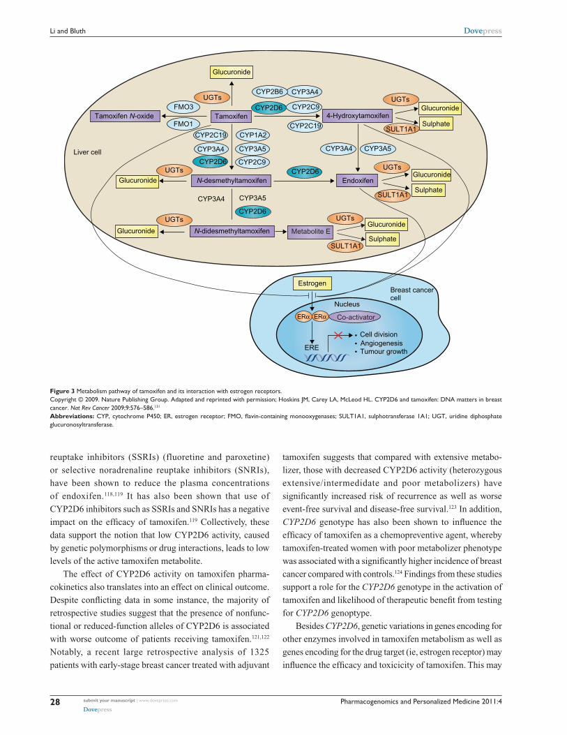
Pharmacogenomics and Personalized Medicine 2011:4submit your manuscript | www.dovepress.com
Dovepress
Dovepress
28
Li and Bluth
reuptake inhibitors (SSRIs) (fluoretine and paroxetine)
or selective noradrenaline reuptake inhibitors (SNRIs),
have been shown to reduce the plasma concentrations
of endoxifen.118,119 It has also been shown that use of
CYP2D6 inhibitors such as SSRIs and SNRIs has a negative
impact on the efficacy of tamoxifen.119 Collectively, these
data support the notion that low CYP2D6 activity, caused
by genetic polymorphisms or drug interactions, leads to low
levels of the active tamoxifen metabolite.
The effect of CYP2D6 activity on tamoxifen pharma-
cokinetics also translates into an effect on clinical outcome.
Despite conflicting data in some instance, the majority of
retrospective studies suggest that the presence of nonfunc-
tional or reduced-function alleles of CYP2D6 is associated
with worse outcome of patients receiving tamoxifen.121,122
Notably, a recent large retrospective analysis of 1325
patients with early-stage breast cancer treated with adjuvant
tamoxifen suggests that compared with extensive metabo-
lizer, those with decreased CYP2D6 activity (heterozygous
extensive/intermedidate and poor metabolizers) have
significantly increased risk of recurrence as well as worse
event-free survival and disease-free survival.123 In addition,
CYP2D6 genotype has also been shown to influence the
efficacy of tamoxifen as a chemopreventive agent, whereby
tamoxifen-treated women with poor metabolizer phenotype
was associated with a significantly higher incidence of breast
cancer compared with controls.124 Findings from these studies
support a role for the CYP2D6 genotype in the activation of
tamoxifen and likelihood of therapeutic benefit from testing
for CYP2D6 genoptype.
Besides CYP2D6, genetic variations in genes encoding for
other enzymes involved in tamoxifen metabolism as well as
genes encoding for the drug target (ie, estrogen receptor) may
influence the efficacy and toxicicity of tamoxifen. This may
Glucuronide
Tamoxifen N-oxide Tamoxifen
Liver cell
N-desmethyltamoxifen
N-didesmethyltamoxifen Metabolite E
Glucuronide
Glucuronide
Estrogen
ERα ERα
ERE
Cell divisionAngiogenesisTumour growth
Nucleus
•••
Breast cancercell
Glucuronide
Glucuronide
Glucuronide
UGTs
UGTs
UGTs
UGTs
UGTs
UGTs
CYP2B6 CYP3A4
CYP3A5
CYP3A4
CYP3A4
CYP2C9CYP2D6
CYP2D6
CYP2D6CYP2D6
CYP1A2
CYP3A5
CYP2C9
CYP2C19CYP2C19
CYP3A4 CYP3A5
Endoxifen
4-Hydroxytamoxifen
Sulphate
Sulphate
SULT1A1
SULT1A1
SULT1A1Sulphate
FMO3
FMO1
Co-activator
Figure 3 Metabolism pathway of tamoxifen and its interaction with estrogen receptors.Copyright © 2009. Nature Publishing Group. Adapted and reprinted with permission; Hoskins JM, Carey LA, McLeod HL. CYP2D6 and tamoxifen: DNA matters in breast cancer. Nat Rev Cancer 2009;9:576–586.131
Abbreviations: CYP, cytochrome P450; ER, estrogen receptor; FMO, flavin-containing monooxygenases; SULT1A1, sulphotransferase 1A1; UGT, uridine diphosphate glucuronosyltransferase.
Pharmacogenomics and Personalized Medicine 2011:4 submit your manuscript | www.dovepress.com
Dovepress
Dovepress
29
Cancer pharmacogenomics of drug enzymes and transporters
explain, at least in part, the descrepancies of results from
different studies with respect to the role of CYP2D6 geno-
type in the clinical outcome of tamoxifen. Tamoxifen and
its metabolites undergo Phase II metabolism by SULT1A1
and UGTs. Interindividual variation in the activity of
Phase II enzymes may contribute to variability in circulating
endoxifen levels and patient response to tamoxifen. It has
been reported that women with high activity UGT2B15
genotype (UGT2B15*2/*2) had a worse recurrence-free
survival than those with wild-type alleles.125 Interestingly, a
retrospective analysis of 337 tamoxifen-treated women with
breast cancer found that those with low-activity SULT1A1
genotype (SULT1A1*2/*2) had approximately three times
the risk of death as controls.126 One possible explanation for
this observation is that sulfation of tamoxifen metabolites
(4-hydroxy-tamoxifen and endoxifen) by SULT1A1 forms
highly reactive products leading to DNA adducts,127,128 and
therefore, individuals with low-activity SULT1A1 genotype
have poor clinical outcomes. In addition, genetic polymor-
phisms within the target genes (ESR1 and ESR2) have been
associated with altered susceptibility to tamoxifen-induced
hot flashes and hormone-resistance.129,130
In conclusion, tamoxifen can be considered as a prodrug.
Its metabolism is complex and involves multiple Phase I
and II enzymes. Genetic variations in these metabolizing
enzymes likely contribute to the variability in tamoxifen
active metabolite concentrations and patient outcome.
It is generally agreed that women with reduced CYP2D6
activity genotype appear to have lower circulating endoxifen
concentration and are less likely to derive therapeutic benefit
from tamoxifen compared with those with normal CYP2D6
activity. However, because of the lack of concordant data,
mandatory CYP2D6 genotyping test to guide the selection
and dose of tamoxifen is premature. Idealy, large, prospective
clinical studies should be conducted to systematically assess
the impact of multiple genetic polymorphisms within multiple
genes involved in the disposition and action of tamoxifen on
the clinical outcome of patients receiving tamoxifen.
ConclusionPharmacogenomics provides a unique approach toward
investigating, appreciating, and therapeutically serving
the individual cancer patient. Continued investigation and
adaptation of pharmacogenomics with respect to malignancy
should likely provide improved risk versus benefit ratios with
respect to therapeutic efficacy versus side-effect profiles.
Further, effective re-evaluation of drug design toward
the generation of novel and specific therapies focused on
enzyme and transporter biology pertaining to malignancy
may eventually be personalized and individualized to the
patient for maximum efficacy.
DisclosureThe authors report no conflicts of interest in this work.
References 1. Evans WE, Relling MV. Pharmacogenomics: translating functional
genomics into rational therapeutics. Science. 1999;286:487–491. 2. Frueh FW, Amur S, Mummaneni P, et al. Pharmacogenomic biomarker
information in drug labels approved by the United States food and drug administration: prevalence of related drug use. Pharmacotherapy. 2008;28:992–998.
3. Bradford LD. CYP2D6 allele frequency in European Caucasians, Asians, Africans and their descendants. Pharmacogenomics. 2002; 3:229–243.
4. Zhou SF, Liu JP, Chowbay B. Polymorphism of human cytochrome P450 enzymes and its clinical impact. Drug Metab Rev. 2009;41: 89–295.
5. Higashi MK, Veenstra DL, Kondo LM, et al. Association between CYP2C9 genetic variants and anticoagulation-related outcomes during warfarin therapy. JAMA. 2002;287:1690–1698.
6. Guillemette C. Pharmacogenomics of human UDP-glucuronosyltrans-ferase enzymes. Pharmacogenomics J. 2003;3(3):136–158.
7. Nagar S, Remmel RP. Uridine diphosphoglucuronosyltransferase pharmacogenetics and cancer. Oncogene. 2006;25:1659–1672.
8. Glatt H, Meinl W. Pharmacogenetics of soluble sulfotransferases (SULTs). Naunyn Schmiedebergs Arch Pharmacol. 2004;369: 55–68.
9. Nowell S, Falany CN. Pharmacogenetics of human cytosolic sulfo-transferases. Oncogene. 2006;25:1673–1678.
10. Lo HW, Ali-Osman F. Genetic polymorphism and function of gluta-thione S-transferases in tumor drug resistance. Curr Opin Pharmacol. 2007;7:367–374.
11. McIlwain CC, Townsend DM, Tew KD. Glutathione S-transferase polymorphisms: cancer incidence and therapy. Oncogene. 2006;25: 1639–1648.
12. Blum M, Grant DM, McBride W, et al. Human arylamine N-acetyl-transferase genes: isolation, chromosomal localization, and functional expression. DNA Cell Biol. 1990;9:193–203.
13. Sim E, Lack N, Wang CJ, et al. Arylamine N-acetyltransferases: structural and functional implications of polymorphisms. Toxicology. 2008;254:170–183.
14. Huang YS, Chern HD, Su WJ, et al. Polymorphism of the N-acetyl-transferase 2 gene as a susceptibility risk factor for antituberculosis drug-induced hepatitis. Hepatology. 2002;35:883–889.
15. Hein DW. Molecular genetics and function of NAT1 and NAT2: role in aromatic amine metabolism and carcinogenesis. Mutat Res. 2002; 506–507:65–77.
16. Cartwright RA, Glashan RW, Rogers HJ, et al. Role of N-acetyl-transferase phenotypes in bladder carcinogenesis: a pharmacogenetic epidemiological approach to bladder cancer. Lancet. 1982;2: 842–845.
17. Garcia-Closas M, Malats N, Silverman D, et al. NAT2 slow acetylation, GSTM1 null genotype, and risk of bladder cancer: results from the Spanish Bladder Cancer Study and meta-analyses. Lancet. 2005;366: 649–659.
18. Zhou S. Clinical pharmacogenomics of thiopurine S-methyltransferase. Curr Clin Pharmacol. 2006;1:119–128.
19. Relling MV, Gardner EE, Sandborn WJ, et al. Clinical pharmacogenetics implementation consortium guidelines for thiopurine methyltransferase genotype and thiopurine dosing. Clin Pharmacol Ther. 2011;89: 387–391.
Pharmacogenomics and Personalized Medicine 2011:4submit your manuscript | www.dovepress.com
Dovepress
Dovepress
30
Li and Bluth
20. Kerb R. Implications of genetic polymorphisms in drug transporters for pharmacotherapy. Cancer Lett. 2006;234:4–33.
21. Gottesman MM, Fojo T, Bates SE. Multidrug resistance in cancer: role of ATP-dependent transporters. Nat Rev Cancer. 2002;2:48–58.
22. Wacher VJ, Wu CY, Benet LZ. Overlapping substrate specificities and tissue distribution of cytochrome P450 3A and P-glycoprotein: implications for drug delivery and activity in cancer chemotherapy. Mol Carcinog. 1995;13:129–134.
23. Hoffmeyer S, Burk O, von Richter O, et al. Functional polymorphisms of the human multidrug-resistance gene: multiple sequence variations and correlation of one allele with P-glycoprotein expression and activity in vivo. Proc Natl Acad Sci U S A. 2000;97:3473–3478.
24. Kimchi-Sarfaty C, Oh JM, Kim IW, et al. A “silent” polymorphism in the MDR1 gene changes substrate specificity. Science. 2007;315: 525–528.
25. Ieiri I, Takane H, Otsubo K. The MDR1 (ABCB1) gene polymorphism and its clinical implications. Clin Pharmacokinet. 2004;43:553–576.
26. Pauli-Magnus C, Kroetz DL. Functional implications of genetic polymorphisms in the multidrug resistance gene MDR1 (ABCB1). Pharm Res. 2004;21:904–913.
27. Marzolini C, Paus E, Buclin T, et al. Polymorphisms in human MDR1 (P-glycoprotein): recent advances and clinical relevance. Clin Pharmacol Ther. 2004;75:13–33.
28. Sakaeda T. MDR1 genotype-related pharmacokinetics: fact or fiction? Drug Metab Pharmacokinet. 2005;20:391–414.
29. Hinoshita E, Uchiumi T, Taguchi K, et al. Increased expression of an ATP-binding cassette superfamily transporter, multidrug resistance protein 2, in human colorectal carcinomas. Clin Cancer Res. 2000;6: 2401–2407.
30. Rothnie A, Callaghan R, Deeley RG, et al. Role of GSH in estrone sulfate binding and translocation by the multidrug resistance protein 1 (MRP1/ABCC1). J Biol Chem. 2006;281:13906–13914.
31. Gradhand U, Kim RB. Pharmacogenomics of MRP transport-ers (ABCC1-5) and BCRP (ABCG2). Drug Metab Rev. 2008;40: 317–354.
32. Wojnowski L, Kulle B, Schirmer M, et al. NAD(P)H oxidase and multidrug resistance protein genetic polymorphisms are associated with doxorubicin-induced cardiotoxicity. Circulation. 2005;112: 3754–3762.
33. Meyer zu Schwabedissen HE, Jedlitschky G, Gratz M, et al. Variable expression of MRP2 (ABCC2) in human placenta: influence of gestational age and cellular differentiation. Drug Metab Dispos. 2005; 33:896–904.
34. Meier Y, Pauli-Magnus C, Zanger UM, et al. Interindividual variability of canalicular ATP-binding-cassette (ABC)-transporter expression in human liver. Hepatology. 2006;44:62–74.
35. Izzedine H, Hulot JS, Villard E, et al. Association between ABCC2 gene haplotypes and tenofovir-induced proximal tubulopathy. J Infect Dis. 2006;194:1481–1491.
36. Vogelgesang S, Kunert-Keil C, Cascorbi I, et al. Expression of multi-drug transporters in dysembryoplastic neuroepithelial tumors causing intractable epilepsy. Clin Neuropathol. 2004;23:223–231.
37. Krishnamurthy P, Schuetz JD. Role of ABCG2/BCRP in biology and medicine. Annu Rev Pharmacol Toxicol. 2006;46:381–410.
38. Jonker JW, Buitelaar M, Wagenaar E, et al. The breast cancer resistance protein protects against a major chlorophyll-derived dietary phototoxin and protoporphyria. Proc Natl Acad Sci U S A. 2002;99:15649–15654.
39. Jonker JW, Smit JW, Brinkhuis RF, et al. Role of breast cancer resistance protein in the bioavailability and fetal penetration of topotecan. J Natl Cancer Inst. 2000;92:1651–1656.
40. Sparreboom A, Loos WJ, Burger H, et al. Effect of ABCG2 genotype on the oral bioavailability of topotecan. Cancer Biol Ther. 2005;4: 650–658.
41. Burger H, Nooter K. Pharmacokinetic resistance to imatinib mesylate: role of the ABC drug pumps ABCG2 (BCRP) and ABCB1 (MDR1) in the oral bioavailability of imatinib. Cell Cycle. 2004;3:1502–1505.
42. Kondo C, Onuki R, Kusuhara H, et al. Lack of improvement of oral absorption of ME3277 by prodrug formation is ascribed to the intestinal efflux mediated by breast cancer resistant protein (BCRP/ABCG2). Pharm Res. 2005;22:613–618.
43. Merino G, Jonker JW, Wagenaar E, et al. The breast cancer resistance protein (BCRP/ABCG2) affects pharmacokinetics, hepatobiliary excre-tion, and milk secretion of the antibiotic nitrofurantoin. Mol Pharmacol. 2005;67:1758–1764.
44. Hirano M, Maeda K, Matsushima S, et al. Involvement of BCRP (ABCG2) in the biliary excretion of pitavastatin. Mol Pharmacol. 2005; 68:800–807.
45. Iida A, Saito S, Sekine A, et al. Catalog of 605 single-nucleotide polymorphisms (SNPs) among 13 genes encoding human ATP-binding cassette transporters: ABCA4, ABCA7, ABCA8, ABCD1, ABCD3, ABCD4, ABCE1, ABCF1, ABCG1, ABCG2, ABCG4, ABCG5, and ABCG8. J Hum Genet. 2002;47:285–310.
46. Imai Y, Nakane M, Kage K, et al. C421A polymorphism in the human breast cancer resistance protein gene is associated with low expression of Q141K protein and low-level drug resistance. Mol Cancer Ther. 2002;1:611–616.
47. Zamber CP, Lamba JK, Yasuda K, et al. Natural allelic variants of breast cancer resistance protein (BCRP) and their relationship to BCRP expression in human intestine. Pharmacogenetics. 2003;13:19–28.
48. Ishikawa T, Tamura A, Saito H, et al. Pharmacogenomics of the human ABC transporter ABCG2: from functional evaluation to drug molecular design. Naturwissenschaften. 2005;92:451–463.
49. Mitomo H, Kato R, Ito A, et al. A functional study on polymorphism of the ATP-binding cassette transporter ABCG2: critical role of arginine-482 in methotrexate transport. Biochem J. 2003;373:767–774.
50. Kobayashi D, Ieiri I, Hirota T, et al. Functional assessment of ABCG2 (BCRP) gene polymorphisms to protein expression in human placenta. Drug Metab Dispos. 2005;33:94–101.
51. Lepper ER, Nooter K, Verweij J, et al. Mechanisms of resistance to anticancer drugs: the role of the polymorphic ABC transporters ABCB1 and ABCG2. Pharmacogenomics. 2005;6:115–138.
52. Sparreboom A, Gelderblom H, Marsh S, et al. Diflomotecan pharma-cokinetics in relation to ABCG2 421C.A genotype. Clin Pharmacol Ther. 2004;76:38–44.
53. Li J, Cusatis G, Brahmer J, et al. Association of variant ABCG2 and the pharmacokinetics of epidermal growth factor receptor tyrosine kinase inhibitors in cancer patients. Cancer Biol Ther. 2007;6:432–438.
54. Cusatis G, Gregorc V, Li J, et al. Pharmacogenetics of ABCG2 and adverse reactions to gefitinib. J Natl Cancer Inst. 2006;98:1739–1742.
55. Niemi M. Role of OATP transporters in the disposition of drugs. Pharmacogenomics. 2007;8:787–802.
56. Meyer zu Schwabedissen HE, Kim RB. Hepatic OATP1B transport-ers and nuclear receptors PXR and CAR: interplay, regulation of drug disposition genes, and single nucleotide polymorphisms. Mol Pharm. 2009;6:1644–1661.
57. Kameyama Y, Yamashita K, Kobayashi K, et al. Functional characterization of SLCO1B1 (OATP-C) variants, SLCO1B1*5, SLCO1B1*15 and SLCO1B1*15+C1007G, by using transient expres-sion systems of HeLa and HEK293 cells. Pharmacogenet Genomics. 2005;15:513–522.
58. Nozawa T, Minami H, Sugiura S, et al. Role of organic anion transporter OATP1B1 (OATP-C) in hepatic uptake of irinotecan and its active metabolite, 7-ethyl-10-hydroxycamptothecin: in vitro evidence and effect of single nucleotide polymorphisms. Drug Metab Dispos. 2005; 33:434–439.
59. Tirona RG, Leake BF, Merino G, et al. Polymorphisms in OATP-C: identification of multiple allelic variants associated with altered trans-port activity among European- and African-Americans. J Biol Chem. 2001;276:35669–35675.
60. Niemi M, Pasanen MK, Neuvonen PJ. SLCO1B1 polymorphism and sex affect the pharmacokinetics of pravastatin but not fluvastatin. Clin Pharmacol Ther. 2006;80:356–366.
Pharmacogenomics and Personalized Medicine 2011:4 submit your manuscript | www.dovepress.com
Dovepress
Dovepress
31
Cancer pharmacogenomics of drug enzymes and transporters
61. Niemi M, Backman JT, Kajosaari LI, et al. Polymorphic organic anion transporting polypeptide 1B1 is a major determinant of repaglinide pharmacokinetics. Clin Pharmacol Ther. 2005;77:468–478.
62. Nishizato Y, Ieiri I, Suzuki H, et al. Polymorphisms of OATP-C (SLC21A6) and OAT3 (SLC22A8) genes: consequences for pravastatin pharmacokinetics. Clin Pharmacol Ther. 2003;73:554–565.
63. Xiang X, Jada SR, Li HH, et al. Pharmacogenetics of SLCO1B1 gene and the impact of *1b and *15 haplotypes on irinotecan disposition in Asian cancer patients. Pharmacogenet Genomics. 2006;16: 683–691.
64. Han JY, Lim HS, Shin ES, et al. Influence of the organic anion- transporting polypeptide 1B1 (OATP1B1) polymorphisms on irinotecan-pharmacokinetics and clinical outcome of patients with advanced non-small cell lung cancer. Lung Cancer. 2008;59:69–75.
65. Link E, Parish S, Armitage J, et al. SLCO1B1 variants and statin-induced myopathym – a genomewide study. N Engl J Med. 2008;359: 789–799.
66. Franke RM, Scherkenbach LA, Sparreboom A. Pharmacogenetics of the organic anion transporting polypeptide 1A2. Pharmacogenomics. 2009;10:339–344.
67. Letschert K, Keppler D, Konig J. Mutations in the SLCO1B3 gene affecting the substrate specificity of the hepatocellular uptake transporter OATP1B3 (OATP8). Pharmacogenetics. 2004;14:441–452.
68. Nozawa T, Nakajima M, Tamai I, et al. Genetic polymorphisms of human organic anion transporters OATP-C (SLC21A6) and OATP-B (SLC21A9): allele frequencies in the Japanese population and functional analysis. J Pharmacol Exp Ther. 2002;302:804–813.
69. Hayer-Zillgen M, Bruss M, Bonisch H. Expression and pharmacological profile of the human organic cation transporters hOCT1, hOCT2 and hOCT3. Br J Pharmacol. 2002;136:829–836.
70. Zhang S, Lovejoy KS, Shima JE, et al. Organic cation transporters are determinants of oxaliplatin cytotoxicity. Cancer Res. 2006;66: 8847–8857.
71. Choi MK, Song IS. Organic cation transporters and their pharmacokinetic and pharmacodynamic consequences. Drug Metab Pharmacokinet. 2008;23:243–253.
72. Pentikainen PJ, Neuvonen PJ, Penttila A. Pharmacokinetics of metformin after intravenous and oral administration to man. Eur J Clin Pharmacol. 1979;16:195–202.
73. Song IS, Shin HJ, Shim EJ, et al. Genetic variants of the organic cation transporter 2 influence the disposition of metformin. Clin Pharmacol Ther. 2008;84:559–562.
74. Wang ZJ, Yin OQ, Tomlinson B, et al. OCT2 polymorphisms and in-vivo renal functional consequence: studies with metformin and cimetidine. Pharmacogenet Genomics. 2008;18:637–645.
75. Tzvetkov MV, Vormfelde SV, Balen D, et al. The effects of genetic polymorphisms in the organic cation transporters OCT1, OCT2, and OCT3 on the renal clearance of metformin. Clin Pharmacol Ther. 2009; 86:299–306.
76. Shu Y, Sheardown SA, Brown C, et al. Effect of genetic variation in the organic cation transporter 1 (OCT1) on metformin action. J Clin Invest. 2007;117:1422–1431.
77. Xu G, Bhatnagar V, Wen G, et al. Analyses of coding region polymorphisms in apical and basolateral human organic anion trans-porter (OAT) genes [OAT1 (NKT), OAT2, OAT3, OAT4, URAT (RST)]. Kidney Int. 2005;68:1491–1499.
78. Bhatnagar V, Xu G, Hamilton BA, et al. Analyses of 5’ regulatory region polymorphisms in human SLC22A6 (OAT1) and SLC22A8 (OAT3). J Hum Genet. 2006;51:575–580.
79. Heidelberger C, Chaudhuri NK, Danneberg P, et al. Fluorinated pyrimidines, a new class of tumour-inhibitory compounds. Nature. 1957;179:663–666.
80. Omura K. Clinical implications of dihydropyrimidine dehydrogenase (DPD) activity in 5-FU-based chemotherapy: mutations in the DPD gene, and DPD inhibitory fluoropyrimidines. Int J Clin Oncol. 2003;8: 132–138.
81. Soong R, Shah N, Salto-Tellez M, et al. Prognostic significance of thymidylate synthase, dihydropyrimidine dehydrogenase and thymidine phosphorylase protein expression in colorectal cancer patients treated with or without 5-fluorouracil-based chemotherapy. Ann Oncol. 2008; 19:915–919.
82. Salonga D, Danenberg KD, Johnson M, et al. Colorectal tumors responding to 5-fluorouracil have low gene expression levels of dihydropyrimidine dehydrogenase, thymidylate synthase, and thymidine phosphorylase. Clin Cancer Res. 2000;6:1322–1327.
83. Lu Z, Zhang R, Diasio RB. Dihydropyrimidine dehydrogenase activity in human peripheral blood mononuclear cells and liver: population char-acteristics, newly identified deficient patients, and clinical implication in 5-fluorouracil chemotherapy. Cancer Res. 1993;53:5433–5438.
84. Morel A, Boisdron-Celle M, Fey L, et al. Clinical relevance of different dihydropyrimidine dehydrogenase gene single nucleotide polymorphisms on 5-fluorouracil tolerance. Mol Cancer Ther. 2006;5: 2895–2904.
85. Vreken P, Van Kuilenburg AB, Meinsma R, et al. A point mutation in an invariant splice donor site leads to exon skipping in two unrelated Dutch patients with dihydropyrimidine dehydrogenase deficiency. J Inherit Metab Dis. 1996;19:645–654.
86. Wei X, McLeod HL, McMurrough J, et al. Molecular basis of the human dihydropyrimidine dehydrogenase deficiency and 5-fluorouracil toxicity. J Clin Invest. 1996;98:610–615.
87. Van Kuilenburg AB, Vreken P, Beex LV, et al. Heterozygosity for a point mutation in an invariant splice donor site of dihydropyrimidine dehydrogenase and severe 5-fluorouracil related toxicity. Eur J Cancer. 1997;33:2258–2264.
88. Ichikawa W, Uetake H, Shirota Y, et al. Both gene expression for orotate phosphoribosyltransferase and its ratio to dihydropyrimidine dehydrogenase influence outcome following fluoropyrimidine-based chemotherapy for metastatic colorectal cancer. Br J Cancer. 2003;89: 1486–1492.
89. Ichikawa W, Takahashi T, Suto K, et al. Orotate phosphoribosyltrans-ferase gene polymorphism predicts toxicity in patients treated with bolus 5-fluorouracil regimen. Clin Cancer Res. 2006;12:3928–3934.
90. Pullarkat ST, Stoehlmacher J, Ghaderi V, et al. Thymidylate synthase gene polymorphism determines response and toxicity of 5-FU chemotherapy. Pharmacogenomics J. 2001;1:65–70.
91. Kawakami K, Omura K, Kanehira E, et al. Polymorphic tandem repeats in the thymidylate synthase gene is associated with its protein expression in human gastrointestinal cancers. Anticancer Res. 1999;19:3249–3252.
92. Mandola MV, Stoehlmacher J, Muller-Weeks S, et al. A novel single nucleotide polymorphism within the 5’ tandem repeat polymorphism of the thymidylate synthase gene abolishes USF-1 binding and alters transcriptional activity. Cancer Res. 2003;63:2898–2904.
93. Mandola MV, Stoehlmacher J, Zhang W, et al. A 6 bp polymorphism in the thymidylate synthase gene causes message instability and is associ-ated with decreased intratumoral TS mRNA levels. Pharmacogenetics. 2004;14:319–327.
94. Lurje G, Manegold PC, Ning Y, Pohl A, Zhang W, Lenz HJ. Thymidylate synthase gene variations: predictive and prognostic markers. Mol Cancer Ther. 2009;8(5):1000–1007.
95. Saltz LB, Cox JV, Blanke C, et al. Irinotecan plus fluorouracil and leucovorin for metastatic colorectal cancer. Irinotecan Study Group. N Engl J Med. 2000;343:905–914.
96. Slatter JG, Su P, Sams JP, et al. Bioactivation of the anticancer agent CPT-11 to SN-38 by human hepatic microsomal carboxylesterases and the in vitro assessment of potential drug interactions. Drug Metab Dispos. 1997;25:1157–1164.
97. Rivory LP, Riou JF, Haaz MC, et al. Identification and properties of a major plasma metabolite of irinotecan (CPT-11) isolated from the plasma of patients. Cancer Res. 1996;56:3689–3694.
98. Dodds HM, Haaz MC, Riou JF, et al. Identification of a new metabolite of CPT-11 (irinotecan): pharmacological properties and activation to SN-38. J Pharmacol Exp Ther. 1998;286:578–583.
Pharmacogenomics and Personalized Medicine 2011:4submit your manuscript | www.dovepress.com
Dovepress
Dovepress
32
Li and Bluth
99. Luo FR, Paranjpe PV, Guo A, et al. Intestinal transport of irinotecan in Caco-2 cells and MDCK II cells overexpressing efflux transporters Pgp, cMOAT, and MRP1. Drug Metab Dispos. 2002;30:763–770.
100. Schellens JH, Maliepaard M, Scheper RJ, et al. Transport of topoi-somerase I inhibitors by the breast cancer resistance protein. Potential clinical implications. Ann NY Acad Sci. 2000;922:188–194.
101. Mathijssen RH, van Alphen RJ, Verweij J, et al. Clinical pharma-cokinetics and metabolism of irinotecan (CPT-11). Clin Cancer Res. 2001;7:2182–2194.
102. Tukey RH, Strassburg CP, Mackenzie PI. Pharmacogenomics of human UDP-glucuronosyltransferases and irinotecan toxicity. Mol Pharmacol. 2002;62:446–450.
103. Iyer L, Das S, Janisch L, et al. UGT1A1*28 polymorphism as a determinant of irinotecan disposition and toxicity. Pharmacogenomics J. 2002;2:43–47.
104. Innocenti F, Undevia SD, Iyer L, et al. Genetic variants in the UDP-glucuronosyltransferase 1A1 gene predict the risk of severe neutropenia of irinotecan. J Clin Oncol. 2004;22:1382–1388.
105. Premawardhena A, Fisher CA, Liu YT, et al. The global distribution of length polymorphisms of the promoters of the glucuronosyltransferase 1 gene (UGT1A1): hematologic and evolutionary implications. Blood Cells Mol Dis. 2003;31:98–101.
106. Han JY, Lim HS, Shin ES, et al. Comprehensive analysis of UGT1A polymorphisms predictive for pharmacokinetics and treatment outcome in patients with non-small-cell lung cancer treated with irinotecan and cisplatin. J Clin Oncol. 2006;24:2237–2244.
107. Kitagawa C, Ando M, Ando Y, et al. Genetic polymorphism in the phenobarbital-responsive enhancer module of the UDP- glucuronosyltransferase 1A1 gene and irinotecan toxicity. Pharmacogenet Genomics. 2005;15:35–41.
108. De Jong FA, de Jonge MJ, Verweij J, et al. Role of pharmacogenetics in irinotecan therapy. Cancer Lett. 2006;234:90–106.
109. Zhou Q, Sparreboom A, Tan EH, et al. Pharmacogenetic profiling across the irinotecan pathway in Asian patients with cancer. Br J Clin Pharmacol. 2005;59:415–424.
110. Mathijssen RH, Marsh S, Karlsson MO, et al. Irinotecan pathway genotype analysis to predict pharmacokinetics. Clin Cancer Res. 2003;9:3246–3253.
111. Sai K, Kaniwa N, Itoda M, et al. Haplotype analysis of ABCB1/MDR1 blocks in a Japanese population reveals genotype-dependent renal clearance of irinotecan. Pharmacogenetics. 2003;13:741–757.
112. Kim RB. MDR1 single nucleotide polymorphisms: multiplicity of haplotypes and functional consequences. Pharmacogenetics. 2002; 12:425–427.
113. Hoskins JM, Marcuello E, Altes A, et al. Irinotecan pharmacogenetics: influence of pharmacodynamic genes. Clin Cancer Res. 2008; 14:1788–1796.
114. Dehal SS, Kupfer D. CYP2D6 catalyzes tamoxifen 4-hydroxylation in human liver. Cancer Res. 1997;57:3402–3406.
115. Desta Z, Ward BA, Soukhova NV, et al. Comprehensive evaluation of tamoxifen sequential biotransformation by the human cytochrome P450 system in vitro: prominent roles for CYP3A and CYP2D6. J Pharmacol Exp Ther. 2004;310:1062–1075.
116. Parte P, Kupfer D. Oxidation of tamoxifen by human flavin-containing monooxygenase (FMO) 1 and FMO3 to tamoxifen-N-oxide and its novel reduction back to tamoxifen by human cytochromes P450 and hemoglobin. Drug Metab Dispos. 2005;33:1446–1452.
117. Crewe HK, Notley LM, Wunsch RM, et al. Metabolism of tamoxifen by recombinant human cytochrome P450 enzymes: formation of the 4-hydroxy, 4′-hydroxy and N-desmethyl metabolites and isomerization of trans-4-hydroxytamoxifen. Drug Metab Dispos. 2002;30: 869–874.
118. Stearns V, Johnson MD, Rae JM, et al. Active tamoxifen metabolite plasma concentrations after coadministration of tamoxifen and the selective serotonin reuptake inhibitor paroxetine. J Natl Cancer Inst. 2003;95:1758–1764.
119. Jin Y, Desta Z, Stearns V, et al. CYP2D6 genotype, antidepressant use, and tamoxifen metabolism during adjuvant breast cancer treatment. J Natl Cancer Inst. 2005;97:30–39.
120. Lim HS, Ju Lee H, Seok Lee K, et al. Clinical implications of CYP2D6 genotypes predictive of tamoxifen pharmacokinetics in metastatic breast cancer. J Clin Oncol. 2007;25:3837–3845.
121. Hoskins JM, Carey LA, McLeod HL. CYP2D6 and tamoxifen: DNA matters in breast cancer. Nat Rev Cancer. 2009;9:576–586.
122. Higgins MJ, Stearns V. CYP2D6 polymorphisms and tamoxifen metabolism: clinical relevance. Curr Oncol Rep. 2010;12:7–15.
123. Schroth W, Goetz MP, Hamann U, et al. Association between CYP2D6 polymorphisms and outcomes among women with early stage breast cancer treated with tamoxifen. JAMA. 2009;302:1429–1436.
124. Bonanni B, Macis D, Maisonneuve P, et al. Polymorphism in the CYP2D6 tamoxifen-metabolizing gene influences clinical effect but not hot flashes: data from the Italian Tamoxifen Trial. J Clin Oncol. 2006;24:3708–3709; author reply 3709.
125. Nowell SA, Ahn J, Rae JM, et al. Association of genetic variation in tamoxifen-metabolizing enzymes with overall survival and recurrence of disease in breast cancer patients. Breast Cancer Res Treat. 2005; 91:249–258.
126. Nowell S, Sweeney C, Winters M, et al. Association between sulfotransferase 1A1 genotype and survival of breast cancer patients receiving tamoxifen therapy. J Natl Cancer Inst. 2002;94: 1635–1640.
127. Davis W, Venitt S, Phillips DH. The metabolic activation of tamoxifen and alpha-hydroxytamoxifen to DNA-binding species in rat hepato-cytes proceeds via sulphation. Carcinogenesis. 1998;19:861–866.
128. Dasaradhi L, Shibutani S. Identification of tamoxifen-DNA adducts formed by alpha-sulfate tamoxifen and alpha-acetoxytamoxifen. Chem Res Toxicol. 1997;10:189–196.
129. Jin Y, Hayes DF, Li L, et al. Estrogen receptor genotypes influence hot flash prevalence and composite score before and after tamoxifen therapy. J Clin Oncol. 2008;26:5849–5854.
130. Zhang QX, Borg A, Wolf DM, et al. An estrogen receptor mutant with strong hormone-independent activity from a metastatic breast cancer. Cancer Res. 1997;57:1244–1249.
131. Solus JF, Arietta BJ, Harris JR, et al. Genetic variation in eleven phase I drug metabolism genes in an ethnically diverse population. Pharmacogenomics. 2004;5:895–931.
132. Mizutani T. PM frequencies of major CYPs in Asians and Caucasians. Drug Metab Rev. 2003;35:99–106.
133. Xie HG, Kim RB, Wood AJ, et al. Molecular basis of ethnic differ-ences in drug disposition and response. Annu Rev Pharmacol Toxicol. 2001;41:815–850.
134. Suarez-Kurtz G. Pharmacogenomics in admixed populations. Trends Pharmacol Sci. 2005;26:196–201.
135. Sistonen J, Sajantila A, Lao O, et al. CYP2D6 worldwide genetic variation shows high frequency of altered activity variants and no continental structure. Pharmacogenet Genomics. 2007;17:93–101.
136. Roy JN, Lajoie J, Zijenah LS, et al. CYP3A5 genetic polymorphisms in different ethnic populations. Drug Metab Dispos. 2005;33:884–887.
137. Bozina N, Bradamante V, Lovric M. Genetic polymorphism of meta-bolic enzymes P450 (CYP) as a susceptibility factor for drug response, toxicity, and cancer risk. Arh Hig Rada Toksikol. 2009;60:217–242.
138. Ingelman-Sundberg M. Pharmacogenetics of cytochrome P450 and its applications in drug therapy: the past, present and future. Trends Pharmacol Sci. 2004;25:193–200.
139. Agundez JA. Polymorphisms of human N-acetyltransferases and cancer risk. Curr Drug Metab. 2008;9:520–531.
140. Hamdy SI, Hiratsuka M, Narahara K, et al. Genotype and allele frequencies of TPMT, NAT2, GST, SULT1A1 and MDR-1 in the Egyptian population. Br J Clin Pharmacol. 2003;55:560–569.
141. Fung KL, Gottesman MM. A synonymous polymorphism in a common MDR1 (ABCB1) haplotype shapes protein function. Biochim Biophys Acta. 2009;1794:860–871.
Pharmacogenomics and Personalized Medicine
Publish your work in this journal
Submit your manuscript here: http://www.dovepress.com/pharmacogenomics-and-personalized-medicine-journal
Pharmacogenomics and Personalized Medicine is an international, peer-reviewed, open access journal characterizing the influence of genotype on pharmacology leading to the development of personalized treatment programs and individualized drug selection for improved safety, efficacy and sustainability. This journal is indexed on the American Chemical
Society’s Chemical Abstracts Service (CAS). The manuscript manage-ment system is completely online and includes a very quick and fair peer-review system, which is all easy to use. Visit http://www.dovepress.com/testimonials.php to read real quotes from published authors.
Pharmacogenomics and Personalized Medicine 2011:4 submit your manuscript | www.dovepress.com
Dovepress
Dovepress
Dovepress
33
Cancer pharmacogenomics of drug enzymes and transporters
142. Leslie EM, Letourneau IJ, Deeley RG, et al. Functional and structural consequences of cysteine substitutions in the NH2 proximal region of the human multidrug resistance protein 1 (MRP1/ABCC1). Biochemistry. 2003;42:5214–5224.
143. Conrad S, Kauffmann HM, Ito K, et al. A naturally occurring mutation in MRP1 results in a selective decrease in organic anion transport and in increased doxorubicin resistance. Pharmacogenetics. 2002;12: 321–330.
144. Hulot JS, Villard E, Maguy A, et al. A mutation in the drug transporter gene ABCC2 associated with impaired methotrexate elimination. Pharmacogenet Genomics. 2005;15:277–285.
145. Mizuarai S, Aozasa N, Kotani H. Single nucleotide polymorphisms result in impaired membrane localization and reduced atpase activity in multidrug transporter ABCG2. Int J Cancer. 2004;109:238–246.
146. Tamura A, Wakabayashi K, Onishi Y, et al. Re-evaluation and func-tional classification of non-synonymous single nucleotide polymor-phisms of the human ATP-binding cassette transporter ABCG2. Cancer Sci. 2007;98:231–239.
147. Tamura A, Watanabe M, Saito H, et al. Functional validation of the genetic polymorphisms of human ATP-binding cassette (ABC) trans-porter ABCG2: identification of alleles that are defective in porphyrin transport. Mol Pharmacol. 2006;70:287–296.
148. Konig J, Seithel A, Gradhand U, et al. Pharmacogenomics of human OATP transporters. Naunyn Schmiedebergs Arch Pharmacol. 2006; 372:432–443.
149. Zair ZM, Eloranta JJ, Stieger B, et al. Pharmacogenetics of OATP (SLC21/SLCO), OAT and OCT (SLC22) and PEPT (SLC15) transporters in the intestine, liver and kidney. Pharmacogenomics. 2008;9:597–624.
150. Marzolini C, Tirona RG, Kim RB. Pharmacogenomics of the OATP and OAT families. Pharmacogenomics. 2004;5:273–282.
151. Zhou F, You G. Molecular insights into the structure-function relationship of organic anion transporters OATs. Pharm Res. 2007;24: 28–36.
152. Walther A, Johnstone E, Swanton C, Midgley R, Tomlinson I, Kerr D. Genetic prognostic and predictive markers in colorectal cancer. Nat Rev Cancer. 2009;9:489–499.
153. van Erp NP, Baker SD, Zhao M, et al. Effect of milk thistle (Silybum marianum) on the pharmacokinetics of irinotecan. Clin Cancer Res. 2005;11:7800–7806.


![Open Access Austin Biomolecules: Open Access Austin Biomolecules: Open Access. Open Access [6]. Biodegradable porous aerogels composed of pectin and clay with overall mechanical properties](https://img.pdfslide.us/doc/110x75/5f676fd601b841051326d882/open-access-austin-biomolecules-open-access-austin-biomolecules-open-access-open.jpg)