Embed Size (px)
Citation preview
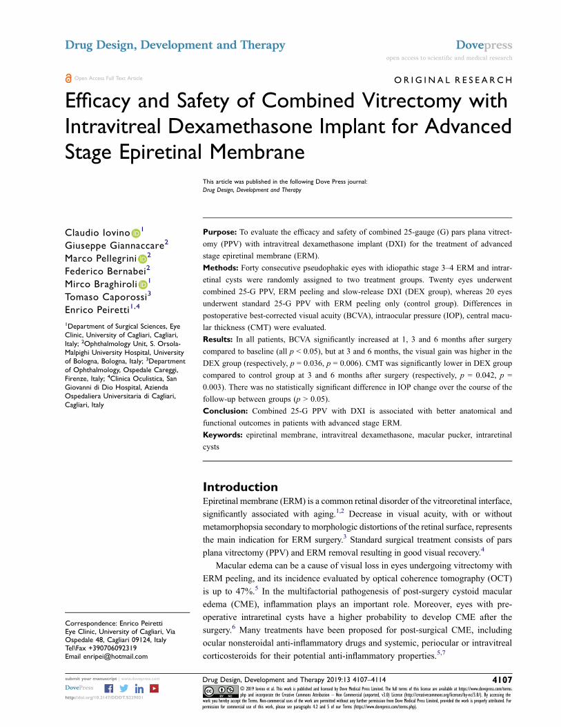
OR I G I N A L R E S E A R C H
Efficacy and Safety of Combined Vitrectomy with
Intravitreal Dexamethasone Implant for Advanced
Stage Epiretinal MembraneThis article was published in the following Dove Press journal:
Drug Design, Development and Therapy
Claudio Iovino 1
Giuseppe Giannaccare2
Marco Pellegrini 2
Federico Bernabei2
Mirco Braghiroli 1
Tomaso Caporossi3
Enrico Peiretti1,4
1Department of Surgical Sciences, Eye
Clinic, University of Cagliari, Cagliari,
Italy; 2Ophthalmology Unit, S. Orsola-
Malpighi University Hospital, University
of Bologna, Bologna, Italy; 3Department
of Ophthalmology, Ospedale Careggi,
Firenze, Italy; 4Clinica Oculistica, San
Giovanni di Dio Hospital, Azienda
Ospedaliera Universitaria di Cagliari,
Cagliari, Italy
Purpose: To evaluate the efficacy and safety of combined 25-gauge (G) pars plana vitrect-
omy (PPV) with intravitreal dexamethasone implant (DXI) for the treatment of advanced
stage epiretinal membrane (ERM).
Methods: Forty consecutive pseudophakic eyes with idiopathic stage 3–4 ERM and intrar-
etinal cysts were randomly assigned to two treatment groups. Twenty eyes underwent
combined 25-G PPV, ERM peeling and slow-release DXI (DEX group), whereas 20 eyes
underwent standard 25-G PPV with ERM peeling only (control group). Differences in
postoperative best-corrected visual acuity (BCVA), intraocular pressure (IOP), central macu-
lar thickness (CMT) were evaluated.
Results: In all patients, BCVA significantly increased at 1, 3 and 6 months after surgery
compared to baseline (all p < 0.05), but at 3 and 6 months, the visual gain was higher in the
DEX group (respectively, p = 0.036, p = 0.006). CMT was significantly lower in DEX group
compared to control group at 3 and 6 months after surgery (respectively, p = 0.042, p =
0.003). There was no statistically significant difference in IOP change over the course of the
follow-up between groups (p > 0.05).
Conclusion: Combined 25-G PPV with DXI is associated with better anatomical and
functional outcomes in patients with advanced stage ERM.
Keywords: epiretinal membrane, intravitreal dexamethasone, macular pucker, intraretinal
cysts
IntroductionEpiretinal membrane (ERM) is a common retinal disorder of the vitreoretinal interface,
significantly associated with aging.1,2 Decrease in visual acuity, with or without
metamorphopsia secondary to morphologic distortions of the retinal surface, represents
the main indication for ERM surgery.3 Standard surgical treatment consists of pars
plana vitrectomy (PPV) and ERM removal resulting in good visual recovery.4
Macular edema can be a cause of visual loss in eyes undergoing vitrectomy with
ERM peeling, and its incidence evaluated by optical coherence tomography (OCT)
is up to 47%.5 In the multifactorial pathogenesis of post-surgery cystoid macular
edema (CME), inflammation plays an important role. Moreover, eyes with pre-
operative intraretinal cysts have a higher probability to develop CME after the
surgery.6 Many treatments have been proposed for post-surgical CME, including
ocular nonsteroidal anti-inflammatory drugs and systemic, periocular or intravitreal
corticosteroids for their potential anti-inflammatory properties.5,7
Correspondence: Enrico PeirettiEye Clinic, University of Cagliari, ViaOspedale 48, Cagliari 09124, ItalyTel\Fax +390706092319Email [email protected]
Drug Design, Development and Therapy Dovepressopen access to scientific and medical research
Open Access Full Text Article
submit your manuscript | www.dovepress.com Drug Design, Development and Therapy 2019:13 4107–4114 4107
http://doi.org/10.2147/DDDT.S229031
DovePress © 2019 Iovino et al. This work is published and licensed by Dove Medical Press Limited. The full terms of this license are available at https://www.dovepress.com/terms.php and incorporate the Creative Commons Attribution – Non Commercial (unported, v3.0) License (http://creativecommons.org/licenses/by-nc/3.0/). By accessing the
work you hereby accept the Terms. Non-commercial uses of the work are permitted without any further permission from Dove Medical Press Limited, provided the work is properly attributed. Forpermission for commercial use of this work, please see paragraphs 4.2 and 5 of our Terms (https://www.dovepress.com/terms.php).
Recently, some authors reported the efficacy of intravi-
treal dexamethasone implant (DXI) for the treatment of post-
operative CME after macular pucker removal alone8 or
combined with cataract surgery.9 Supporting the hypothesis
of a possible inflammatory etiology, Suzuki et al10 postulated
that ERM traction stimulates a leukocyte response in the
macular region causing persistent macular edema even after
its surgical removal.
Based on this concept, the aim of the present study was to
prospectively investigate the efficacy and safety of combined
25-gauge (G) PPV with intraoperative DXI for the treatment
of advanced stage ERM with intraretinal cysts.
MethodsThis was a prospective randomized controlled study con-
ducted at the Retina Center of the Eye Clinic, University
of Cagliari, Italy. The investigation was approved by the
Office of Research Ethics, University of Cagliari and
performed according to the guidelines of the Declaration
of Helsinki and to the recommendation described in the
Consolidated Standard of Reporting Trials (CONSORT)
Statement.
Patients and Clinical ExaminationPseudophakic patients (operated at least 6 months before the
enrollment) with an idiopathic stage 3–4 ERM defined on the
basis of a recent spectral-domain optical coherence tomogra-
phy (SD-OCT) classification and intraretinal cysts were
included in the study.11 Phakic eyes were excluded to reduce
any possible bias for postoperative analysis. ERMwas visua-
lised on OCT, as irregular and hyperreflective bands above
the inner retinal surface. A stage 3 ERM was defined in case
of absence of the foveal pit, presence of ectopic inner foveal
layer (EIFL) and well-defined retinal layers. Stage 4 ERM
was assigned to all eyes with the absence of foveal pit,
presence of EIFL and disrupted retinal layers. Exclusion
criteria are summarized in Table 1.
Patients were randomly assigned into two groups: one
receiving the combined 25-G PPV with ERM peeling plus
DXI (DEX group), and one undergoing 25-G PPV with
ERM peeling alone (control group). All patients who
received the DEX implant were aware of its off-label use
and provided written informed consent.
The randomization process was computer generated
with a 1:1 ratio. Follow-up visits were scheduled at 1
and 7 days and 1, 3 and 6 months after the surgery and
included: best-corrected visual acuity (BCVA) reported in
Snellen fraction with minimal angle of resolution
(logMAR) conversion for statistical analysis, intraocular
pressure (IOP) measurement with Goldmann applanation
tonometry, slit-lamp and fundus examination and SD-OCT
analysis (SD-OCT, Heidelberg Spectralis HRA + OCT,
Heidelberg Engineering, Germany). The “follow-up” func-
tion of the Heidelberg OCT was used to ensure the same
macular area was evaluated at each visit.
The OCT recording protocol consisted of a sequence of
49 horizontal sections covering an area of 20 degree. Central
macular thickness (CMT) was obtained with the automatic
“thickness map” function of the Heidelberg Eye Explorer.
Outer nuclear layer (ONL) and EIFL thickness were manu-
ally measured with the caliper function of the Heidelberg
instrument. The presence of intraretinal cystoid spaces along
with the integrity of the ellipsoid zone and external limiting
membrane in the macular zone were recorded. Any discon-
tinuity or interruption of these latter was considered as an
alteration. All imaging data were collected and analyzed by
one examiner (CI), then selectively reviewed by the senior
author (EP) to ascertain all retinal findings.
Surgical ProcedureAll patients were operated by the same surgeon (EP) under
local anesthesia with retrobulbar lidocaine injection. They
underwent a standard three-port sutureless 25-G PPV
using the Constellation vitrectomy system (Alcon
Laboratories). After core vitrectomy, a peripheral vitreous
Table 1 Study of Intravitreal Dexamethasone Implant for the
Treatment of Advanced Stage Epiretinal Membrane: Exclusion
Criteria
● Age-related macular degeneration
● History of choroidal neovascularization of any etiology
● Nonproliferative and proliferative diabetic retinopathy
● Severe myopia (>6 diopters or axial length >25 mm)
● Macular telangiectasia
● Retinal dystrophy
● Central serous chorioretinopathy
● History of uveitis or inflammatory eye disorders (including Irvine-
Gass Syndrome)
● History of any intraocular infection
● History of corneal herpetic infection
● History of central or branch retinal vein and/or artery occlusion
● Glaucoma or optic neuropathy of any kind
● Associated lamellar macular holes
● History of retinal detachment and any previous intraocular surgery
with the exclusion of uncomplicated phacoemulsification
● Amblyopia or any other potential cause of vision loss other than
epiretinal membrane in the study eye
Iovino et al Dovepress
submit your manuscript | www.dovepress.com
DovePressDrug Design, Development and Therapy 2019:134108
shaving with a scleral indentation in every clock hour was
also performed. Whenever required, a posterior vitreous
detachment was inducted using the vitrectomy probe in the
suction mode around the optic nerve disc. The ERM was
peeled with intraocular forceps until the vascular arcades
after 0.1 mL of Membraneblue-dual (TrypanBlue 0.15% +
Brilliant Blue 0.025% + PEG 4%, DORC, Zuidland, The
Netherlands) coloration for approximately 1 min after
stopping the infusion.
The ILM was removed routinely in both groups, either
at the same time or after the ERM removal up to two-three
disk diameters centered on the fovea. Second staining was
performed to ascertain the complete removal of the ILM in
the intended area only in case it was not clearly evident.
A detailed examination of the periphery was conducted
during surgery to ascertain the absence of any retinal holes
or tears. At the end of the surgery, a fluid–air exchange
procedure was done for all study eyes followed by trocars
removal. In the DEX group patients, a 0.7 mg sustained-
release DXI (Ozurdex®, Allergan, Inc., Irvine, CA, USA
and Allergan Pharmaceuticals, Ireland) was cautiously
injected under air through the superotemporal scleral port
with a gentle force applied on the injector gradually to
avoid any projectile damage to the retina. The implant
position was confirmed at the end of the procedure by
direct visualization through the indirect operating micro-
scope. Postoperative medication consisted of topical anti-
biotic-corticosteroid association tapered and completely
withdrawn at 4 weeks after surgery.
Statistical AnalysisThe SPSS statistical software (SPSS Inc, Chicago, Illinois,
USA) was used for data analysis. Values are expressed as
mean ± standard deviation (SD). An independent-samples
t-test was used to compare continuous variables between
groups. A χ2 test was used to compare dichotomous vari-
ables between groups. A repeated-measures ANOVA was
used to determine whether there was a statistically signifi-
cant change in clinical variables over the course of the
6-month follow-up after surgery. A mixed ANOVA was
used to determine whether any change in clinical variables
was the result of the interaction between the type of inter-
vention and time. The Shapiro-Wilk’s test was used to
assess the normality of data.
The Levene’s test and the Box’s M test were used to
assess the homogeneity of variances and covariances,
respectively. For the interaction between the type of inter-
vention and time on BCVA, CMT, ONL thickness, EIFL
thickness and IOP, the assumption of sphericity was vio-
lated, as assessed by Mauchly’s test (all p < 0.05).
Therefore, a Greenhouse-Geisser correction was applied.
A p value <0.05 was considered statistically significant.
A power analysis indicated that a minimum sample of
24 patients was required to detect a 0.017 logMAR differ-
ence in terms of BCVA between groups, with a power of
80% and a significance level of 0.05.12
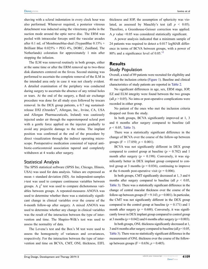
ResultsStudy PopulationOverall, a total of 89 patients were recruited for eligibility and
40 met the inclusion criteria (Figure 1). Baseline and clinical
characteristics of study patients are reported in Table 2.
No significant differences in age, sex, ERM stage, IOP,
EZ and ELM integrity were found between the two groups
(all p > 0.05). No intra or post-operative complications were
recorded in either group.
No patient of the ones who met the inclusion criteria
dropped out from the study.
In both groups, BCVA significantly improved at 1, 3
and 6 months after surgery compared to baseline (all
p < 0.05, Table 3).
There was a statistically significant difference in the
change of BCVA over the course of the follow-up between
groups (F = 17.050; p < 0.001).
BCVA was not significantly different in DEX group
compared to control group at baseline (p = 0.702) and 1
month after surgery (p = 0.198). Conversely, it was sig-
nificantly better in DEX implant group compared to con-
trol group at 3 months (p = 0.036), continuing to improve
at the 6-month post-operative visit (p = 0.006).
In both groups, CMT significantly decreased at 1, 3 and 6
months after surgery compared to baseline (all p < 0.05,
Table 3). There was a statistically significant difference in the
change of central macular thickness over the course of the
follow-up between groups (F = 9.143; p = 0.001). In particular,
the CMT was not significantly different in the DEX group
compared to the control group at baseline (p = 0.171) and 1
month after surgery (p = 0.688). Conversely, it was signifi-
cantly lower in DEX implant group compared to control group
at 3months (p = 0.042) and 6months after surgery (p = 0.003).
In both groups, ONL thickness significantly decreased at 1,
3 and 6months after surgery compared to baseline (all p<0.05,
Table 3). There was no statistically significant difference in the
measurement of ONL thickness over the course of the follow-
up between groups (F = 0.656; p = 0.485).
Dovepress Iovino et al
Drug Design, Development and Therapy 2019:13 submit your manuscript | www.dovepress.com
DovePress4109
In the DEX group, EIFL thickness significantly
decreased at 1, 3 and 6 months after surgery compared to
baseline (all p < 0.05, Table 3), while in the control group,
EIFL thickness did not significantly change (all p > 0.05,
Table 3). There were no statistically significant differences
in the change of EIFL over the course of the follow-up
between groups (F = 1.710, p = 0.195).
In the DEX group, IOP significantly increased at 1
month after surgery compared to baseline (from 14.7 ±
2.1 to 18.0 ± 1.5 mmHg, p < 0.001). However, it returned
to values not significantly different from baseline at 3 and
6 months after surgery (respectively, 16.0 ± 2.6 mmHg,
p = 0.809; 15.4 ± 2.4 mmHg, p = 1.000). In the control
group, IOP did not significantly change at 1, 3 and 6
months after surgery (all p > 0.05). There were no statis-
tically significant differences in the change of IOP over the
course of the follow-up between groups (F = 1.970; p =
0.095).
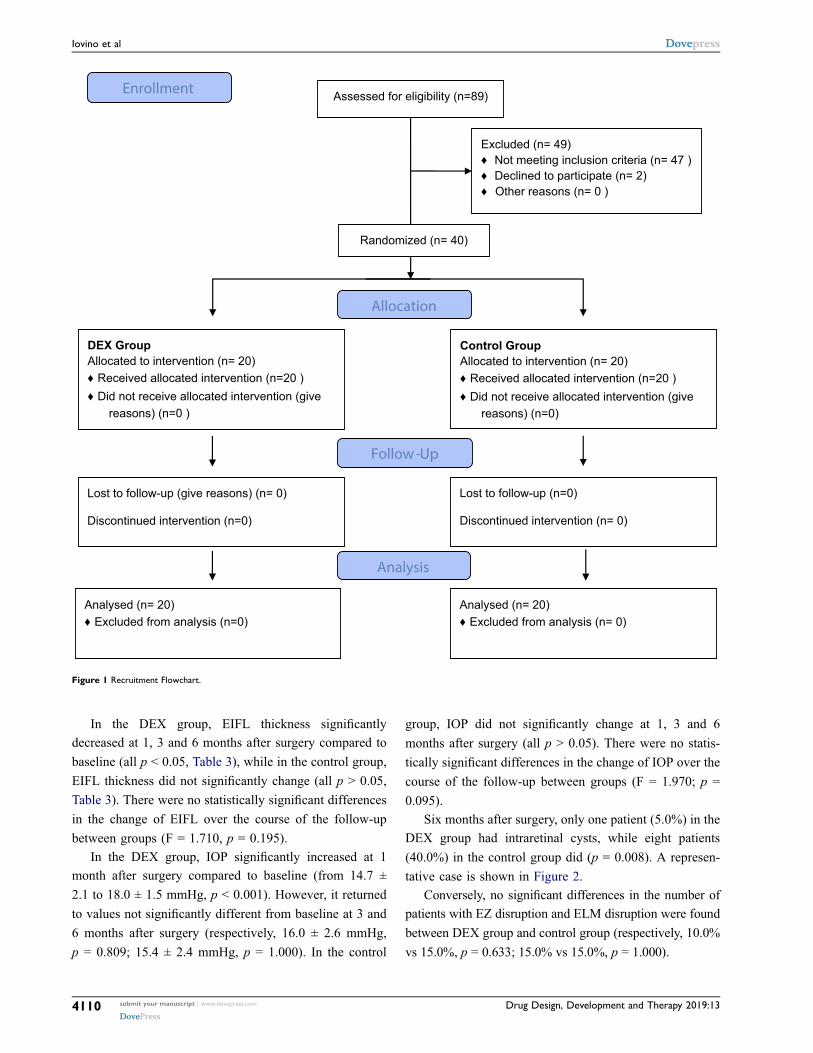
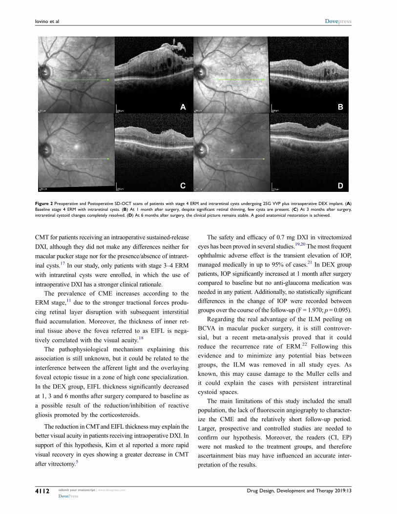
Six months after surgery, only one patient (5.0%) in the
DEX group had intraretinal cysts, while eight patients
(40.0%) in the control group did (p = 0.008). A represen-
tative case is shown in Figure 2.
Conversely, no significant differences in the number of
patients with EZ disruption and ELM disruption were found
between DEX group and control group (respectively, 10.0%
vs 15.0%, p = 0.633; 15.0% vs 15.0%, p = 1.000).
Assessed for eligibility (n=89)
Excluded (n= 49)♦ Not meeting inclusion criteria (n= 47 )♦ Declined to participate (n= 2)♦ Other reasons (n= 0 )
Analysed (n= 20)♦ Excluded from analysis (n=0)
Lost to follow-up (give reasons) (n= 0)
Discontinued intervention (n=0)
DEX Group Allocated to intervention (n= 20)♦ Received allocated intervention (n=20 )♦ Did not receive allocated intervention (give
reasons) (n=0 )
Lost to follow-up (n=0)
Discontinued intervention (n= 0)
Control GroupAllocated to intervention (n= 20)♦ Received allocated intervention (n=20 )♦ Did not receive allocated intervention (give
reasons) (n=0)
Analysed (n= 20)♦ Excluded from analysis (n= 0)
Allocation
Analysis
Follow-Up
Randomized (n= 40)
Enrollment
Figure 1 Recruitment Flowchart.
Iovino et al Dovepress
submit your manuscript | www.dovepress.com
DovePressDrug Design, Development and Therapy 2019:134110
DiscussionEpiretinal membrane is a fibrocellular preretinal tissue com-
posed by fibroblasts, glial cells and hyalocytes over the
ILM.13 Pars plana vitrectomy with pucker removal remains
the standard treatment, albeit CME is a common source of
postoperative visual limitation.5 In the present randomized
controlled trial, we investigated the safety and efficacy of
intravitreal DXI combined with 25G vitrectomy for
advanced stage ERM removal. The use of DXI was asso-
ciated with a better visual outcome and a lower CMTat 3 and
6 months after surgery.
An inflammation component may co-exist and be
a contributory cause of intraretinal fluid accumulation
either before or after the surgery. Indeed, the mechanical
distortion of the retinal surface induced by ERM traction
stimulates an inflammation pathway involving different
growth factors and cytokines, with leukocyte migration
in the macular region.10 This biochemical process contri-
butes to a diffuse retinal thickening associated in some
cases to the formation of CME.
Azzolini and associate analyzed the composition of the
ERM by means of scanning electron microscopy, reporting
four types of structures from the ILM to the vitreous side.14
The layer facing towards the vitreous was defined as
a lacunar structure with inflammatory material. On the
other hand, inflammation plays an important role also in
CMEoccurring after ERM surgical removal. Its pathogenesis
is attributed to the breakdown of the blood–aqueous barrier
caused by the inflammatory surgery-related reaction.15 Based
on these concepts, corticosteroids may be able to reduce the
inflammatory events related to the mechanical distortion
caused by ERM and to the surgical insult. Chatziralli and co-
authors investigated the long-term outcomes of DXI for the
treatment of macular edema following ERM surgical
removal demonstrating its safety and efficacy.12 Likewise,
Furino et al demonstrated the efficacy of a single DXI in
refractory macular edema secondary to combined cataract
extraction and vitrectomy for ERM removal.9
The data about the intraoperative use of DXI are contro-
versial. In a case–control study, intraoperative intravitreal cor-
ticosteroids, both DXI and triamcinolone acetonide, showed to
accelerate the normalization of macular morphology and were
effective in improving visual and anatomic outcomes in
patients with macular pucker.16 More recently, Guidi and
associate reported no advantages in terms of BCVA and
Table 2 Demographical and Clinical Characteristics of Patients in Dexamethasone Group and Control Group Before Surgery
DEX Group (n = 20) Control Group (n = 20) P
Age (years) 74.2 ± 6.0 70.1 ± 9.2 0.136
Sex (m/f) 8/12 7/13 0.744
ERM stage (stage 3/4) 9/11 7/13 0.519
IOP (mmHg) 14.7 ± 2.1 15.1 ± 2.0 0.550
Intraretinal cysts 20 (100.0%) 20 (100.0%) 1.000
EZ disruption (%) 8 (40.0%) 5 (25.0%) 0.311
ELM disruption (%) 7 (35.0%) 3 (15.0%) 0.144
Abbreviations: ERM, epiretinal membrane; IOP, intraocular pressure; EZ, ellipsoid zone; ELM, external limiting membrane.
Table 3 Best Corrected Visual Acuity, Central Macular
Thickness, Outer Nuclear Layer Thickness and Ectopic Inner
Foveal layer Thickness in Dexamethasone Group and Control
Group Over the Course of the 6-Month Follow-Up
DEX Group Control Group P #
Best corrected visual acuity (logMAR, Snellen)
Baseline 0.69 ± 0.32 (20/100) 0.65 ± 0.26 (20/90) 0.702
1 month 0.43 ± 0.27* (20/55) 0.55 ± 0.24* (20/70) 0.198
3 months 0.32 ± 0.23* (20/40) 0.46 ± 0.23* (20/60) 0.036
6 months 0.26 ± 0.23* (20/35) 0.41 ± 0.22* (20.50) 0.006
Central macular thickness (µm)
Baseline 567.3 ± 118.4 522.7 ± 71.9 0.171
1 month 446.2 ± 99.1* 450.6 ± 65.4* 0.688
3 months 374.8 ± 64.5* 420.6 ± 64.4* 0.042
6 months 343.1 ± 70.6* 414.6 ± 62.0* 0.003
Outer nuclear layer thickness (µm)
Baseline 270.9 ± 101.5 254.1 ± 70.8 0.561
1 month 217.8 ± 50.1* 222.4 ± 50.0* 0.788
3 months 194.9 ± 36.2* 198.6 ± 54.0* 0.817
6 months 191.8 ± 35.5* 197.3 ± 55.2* 0.730
Ectopic inner foveal layer thickness (µm)
Baseline 223.4 ± 123.8 183.0 ± 103.8 0.294
1 month 189.4 ± 93.1* 181.8 ± 78.1 0.960
3 months 138.8 ± 84.1* 138.8 ± 59.5 0.998
6 months 130.6 ± 86.6* 124.0 ± 46.9 0.773
Notes: *p < 0.05 compared to baseline, Tukey post hoc test for repeated-measures
ANOVA. #DEX group compared to control group, one-way ANOVA.
Dovepress Iovino et al
Drug Design, Development and Therapy 2019:13 submit your manuscript | www.dovepress.com
DovePress4111
CMT for patients receiving an intraoperative sustained-release
DXI, although they did not make any differences neither for
macular pucker stage nor for the presence/absence of intraret-
inal cysts.17 In our study, only patients with stage 3–4 ERM
with intraretinal cysts were enrolled, in which the use of
intraoperative DXI has a stronger clinical rationale.
The prevalence of CME increases according to the
ERM stage,11 due to the stronger tractional forces produ-
cing retinal layer disruption with subsequent interstitial
fluid accumulation. Moreover, the thickness of inner ret-
inal tissue above the fovea referred to as EIFL is nega-
tively correlated with the visual acuity.18
The pathophysiological mechanism explaining this
association is still unknown, but it could be related to the
interference between the afferent light and the overlaying
foveal ectopic tissue in a zone of high cone specialization.
In the DEX group, EIFL thickness significantly decreased
at 1, 3 and 6 months after surgery compared to baseline as
a possible result of the reduction/inhibition of reactive
gliosis promoted by the corticosteroids.
The reduction in CMTand EIFL thickness may explain the
better visual acuity in patients receiving intraoperative DXI. In
support of this hypothesis, Kim et al reported a more rapid
visual recovery in eyes showing a greater decrease in CMT
after vitrectomy.5
The safety and efficacy of 0.7 mg DXI in vitrectomized
eyes has been proved in several studies.19,20 Themost frequent
ophthalmic adverse effect is the transient elevation of IOP,
managed medically in up to 95% of cases.21 In DEX group
patients, IOP significantly increased at 1 month after surgery
compared to baseline but no anti-glaucoma medication was
needed in any patient. Additionally, no statistically significant
differences in the change of IOP were recorded between
groups over the course of the follow-up (F = 1.970; p = 0.095).
Regarding the real advantage of the ILM peeling on
BCVA in macular pucker surgery, it is still controver-
sial, but a recent meta-analysis proved that it could
reduce the recurrence rate of ERM.22 Following this
evidence and to minimize any potential bias between
groups, the ILM was removed in all study eyes. As
known, this may cause damage to the Muller cells and
it could explain the cases with persistent intraretinal
cystoid spaces.
The main limitations of this study included the small
population, the lack of fluorescein angiography to character-
ize the CME and the relatively short follow-up period.
Larger, prospective and controlled studies are needed to
confirm our hypothesis. Moreover, the readers (CI, EP)
were not masked to the treatment groups, and therefore
ascertainment bias may have influenced an accurate inter-
pretation of the results.
Figure 2 Preoperative and Postoperative SD-OCT scans of patients with stage 4 ERM and intraretinal cysts undergoing 25G VVP plus intraoperative DEX implant. (A)
Baseline stage 4 ERM with intraretinal cysts. (B) At 1 month after surgery, despite significant retinal thinning, few cysts are present. (C) At 3 months after surgery,
intraretinal cystoid changes completely resolved. (D) At 6 months after surgery, the clinical picture remains stable. A good anatomical restoration is achieved.
Iovino et al Dovepress
submit your manuscript | www.dovepress.com
DovePressDrug Design, Development and Therapy 2019:134112
An additional study limitation is the absence of a third
arm receiving triamcinolone acetonide to ascertain any simi-
lar effect at a lower cost. However, the clearance of the
intravitreal triamcinolone is relatively fast in vitrectomized
eyes,23 and we believe DXI may, therefore, be advantageous
providing a slow-release and steady dose of potent steroid.
In conclusion, this report provides evidence of the efficacy
and safety of combined 25GvitrectomywithDXI procedure in
patients with advanced stage ERM and intraretinal cysts. The
anti-inflammatory properties of the intraocular dexamethasone
in addition to vitrectomy and ERM peeling can be very useful
in stage 3 and 4 ERM where an increased traction component
exists.
AcknowledgmentsThis work was supported by the Open Access Publishing
Fund of the University of Cagliari, with the funding of the
Regione Autonoma della Sardegna - L.R. n. 7/2007.
DisclosureThe authors report no conflicts of interest in this work.
References1. Ng CH, Cheung N,Wang JJ, et al. Prevalence and risk factors for epiretinal
membranes in a multi-ethnic United States population. Ophthalmology.2011;118(4):694–699. doi:10.1016/j.ophtha.2010.08.009
2. Mitchell P, Smith W, Chey T, Wang JJ, Chang A. Prevalence andassociations of epiretinal membranes: the blue mountains eye study,Australia. Ophthalmology. 1997;104(6):1033–1040. doi:10.1016/S0161-6420(97)30190-0
3. Smiddy WE, Michels RG, Green WR. Morphology, pathology, andsurgery of idiopathic vitreoretinal macular disorders. A review. Retina.1990;10(4):288–296. doi:10.1097/00006982-199010000-00012.
4. Miliatos I, Lindgren G. Epiretinal membrane surgery evaluated by subjec-tive outcome. Acta Ophthalmol. 2017;95(1):52–59. doi:10.1111/aos.13001
5. Kim SJ, Martin DF, Hubbard GB, et al. Incidence of postvitrectomymacular edema using optical coherence tomography. Ophthalmology.2009;116(8):1531–1537. doi:10.1016/j.ophtha.2009.02.008
6. Frisina R, Pinackatt SJ, Sartore M, et al. Cystoid macular edema after parsplana vitrectomy for idiopathic epiretinal membrane. Graefe’s Arch ClinExp Ophthalmol. 2014;253(1):47–56. doi:10.1007/s00417-014-2655-x
7. Johnson MW. Etiology and treatment of macular edema. AmJ Ophthalmol. 2009;147(1):11–21.e1. doi:10.1016/j.ajo.2008.07.024
8. Hattenbach LO, Springer-Wanner C, Hoerauf H, et al. Intravitrealsustained-release steroid implants for the treatment of macular edemafollowing surgical removal of epiretinal membranes. Ophthalmologica.2017;237(4):232–237. doi:10.1159/000464259
9. Furino C, Boscia F, Recchimurzo N, Sborgia C, Alessio G.Intravitreal dexamethasone implant for refractory macular edemasecondary to vitrectomy for macular pucker. Retina. 2014;34(8):1612–1616. doi:10.1097/IAE.0000000000000105
10. Suzuki T, Hayakawa K, Nakagawa Y, Onouchi H, Ogata M,Kawai K. Topical dorzolamide for macular edema in the earlyphase after vitrectomy and epiretinal membrane removal. ClinOphthalmol. 2013;7:549–553. doi:10.2147/OPTH.S42188
11. Govetto A, Lalane RA, Sarraf D, Figueroa MS, Hubschman JP.Insights into epiretinal membranes: presence of ectopic inner foveallayers and a new optical coherence tomography staging scheme. AmJ Ophthalmol. 2017;175:99–113. doi:10.1016/j.ajo.2016.12.006
12. Chatziralli I, Dimitriou E, Theodossiadis G, Chatzirallis A, Kazantzis D,Theodossiadis P. Treatment of macular edema after pars plana vitrectomyfor idiopathic epiretinal membrane using intravitreal dexamethasoneimplant: long-term outcomes. Ophthalmologica. 2019;1–6. doi:10.1159/000496705
13. Compera D, Entchev E, Haritoglou C, et al. Lamellar hole–associatedepiretinal proliferation in comparison to epiretinal membranes ofmacular pseudoholes. Am J Ophthalmol. 2015;160(2):373–384.e1.doi:10.1016/j.ajo.2015.05.010
14. Azzolini C, Congiu T, Donati S, et al. Multilayer microstructure ofidiopathic epiretinal macular membranes. Eur J Ophthalmol. 2017;27(6):762–768. doi:10.5301/ejo.5000982
15. Tso MO. Pathology of cystoid macular edema. Ophthalmology.1982;89(8):902–915. doi:10.1016/S0161-6420(82)34698-9.
16. Yonekawa Y, Mammo DA, Thomas BJ, Wolfe JD, Hassan TS.A comparison of intraoperative dexamethasone intravitreal implant andtriamcinolone acetonide used during vitrectomy and epiretinal membranepeeling: a case control study. Ophthalmic Surgery, Lasers Imaging Retin.2016;47(3):232–237. doi:10.3928/23258160-20160229-05
17. Guidi G, Casini G, Ripandelli G, et al. Residual intraretinal edemaafter 25-gauge vitrectomy and macular pucker removal: is intraopera-tive sustained-release dexamethasone a real treatment option? Retina.2017:993–999. doi:10.1097/IAE.0000000000001627.
18. Song SJ, Lee MY, Smiddy WE. Ganglion cell layer thickness andvisual improvement after epiretinal membrane surgery. Retina.2016;36(2):305–310. doi:10.1097/IAE.0000000000000705
19. Boyer DS, Faber D, Gupta S, et al. Dexamethasone intravitreal implantfor treatment of diabetic macular edema in vitrectomized patients. Retina.2011;31(5):915–923. doi:10.1097/IAE.0b013e318206d18c
20. Dutra Medeiros M, Alkabes M, Navarro R, Garcia-Arumí J,Mateo C, Corcóstegui B. Dexamethasone intravitreal implant invitrectomized versus nonvitrectomized eyes for treatment of patientswith persistent diabetic macular edema. J Ocul Pharmacol Ther.2014;30(9):709–716. doi:10.1089/jop.2014.0010
21. Rajesh B, Zarranz-Ventura J, Fung AT, et al. Safety of 6000 intravi-treal dexamethasone implants. Br J Ophthalmol. 2019:bjophthalmol-2019-313991. doi:10.1136/bjophthalmol-2019-313991.
22. Azuma K, Ueta T, Eguchi S, Aihara M. Effects of internal limitingmembrane peeling combined with removal of idiopathic epiretinalmembrane a systematic review of literature and meta-analysis.Retina. 2017:1–7. doi:10.1097/IAE.0000000000001537
23. Chin HS, Park TS, Moon YS, Oh JH. Difference in clearance ofintravitreal triamcinolone acetonide between vitrectomized and non-vitrectomized eyes. Retina. 2005;25(5):556–560. doi:10.1097/00006982-200507000-00002
Dovepress Iovino et al
Drug Design, Development and Therapy 2019:13 submit your manuscript | www.dovepress.com
DovePress4113
Drug Design, Development and Therapy DovepressPublish your work in this journalDrug Design, Development and Therapy is an international, peer-reviewed open-access journal that spans the spectrum of drug designand development through to clinical applications. Clinical outcomes,patient safety, and programs for the development and effective, safe,and sustained use of medicines are a feature of the journal, which has also
been accepted for indexing on PubMed Central. The manuscriptmanagement system is completely online and includes a very quickand fair peer-review system, which is all easy to use. Visit http://www.dovepress.com/testimonials.php to read real quotes from publishedauthors.
Submit your manuscript here: https://www.dovepress.com/drug-design-development-and-therapy-journal
Iovino et al Dovepress
submit your manuscript | www.dovepress.com
DovePressDrug Design, Development and Therapy 2019:134114