Embed Size (px)
Citation preview
© 2012 Ragab et al, publisher and licensee Dove Medical Press Ltd. This is an Open Access article which permits unrestricted noncommercial use, provided the original work is properly cited.
International Journal of Nanomedicine 2012:7 3167–3189
International Journal of Nanomedicine
Controlled release of 5-fluorouracil and progesterone from magnetic nanoaggregates
Doaa M Ragab1
Sohrab Rohani1
Styliani Consta2
1Department of Chemical and Biochemical Engineering, 2Department of Chemistry, The University of Western Ontario, London, Ontario, Canada
Correspondence: Sohrab Rohani Department of Chemical and Biochemical Engineering, The University of Western Ontario, London, Ontario N6A 5B9, Canada Tel +1 519 661 4116 Fax +1 519 661 3498 Email [email protected]
Background: The potential use of magnetic nanoparticles in biomedical applications has
witnessed an exponential growth in recent years.
Methods: In this study, we used nanoaggregates of magnetic nanoparticles as carriers for
controlled drug delivery. The nanoaggregates are formed due to the presence of the block
copolymer of polyethylene oxide-polypropylene oxide (Pluronic F-68) and beta-cyclodextrin
that surround the magnetic core of the nanoparticles. The administration of the drug carriers
occurs by inhalation, and the drug is delivered systemically via the pulmonary route. We tested
the delivery of 5-fluorouracil and progesterone, which are used as models of hydrophilic and
hydrophobic drugs, respectively.
Results: The estimated nanoaggregates’ diameters are between 293 nm ± 14.65 nm and
90.2 nm ± 4.51 nm, respectively. In-situ and post-synthesis techniques are two approaches for
drug loading. The polymer composition of nanoaggregates and initial drug concentration showed
a significant effect on both the drug entrapment efficiency and release kinetics. Average drug
entrapment efficiencies ranged between 16.11% and 83.25%. In-situ loaded samples showed
significantly slower release rates. The drug release mechanism is investigated by mathemati-
cal curve fitting to different drug release kinetics models. In most cases, the Peppas model has
shown good correlations (coefficients of correlation, R2, between 0.85 and 0.99) with the exam-
ined release profiles. The estimated release indices are below 0.5, which indicates the Fickian
diffusion mechanism. For samples with an initial burst effect, the modified Peppas model can
provide a better understanding of the drug release mechanism, both in the samples loaded with
progesterone, or those high polymer concentrations.
Conclusion: Our work showed prolonged delivery of drugs (5-fluorouracil and progesterone)
by diffusion from nanoaggregates, with the potential to reduce dose-related adverse effects.
Keywords: Nanoaggregates, 5-fluorouracil, progesterone, release kinetics, Fickian diffusion
IntroductionPulmonary drug delivery of therapeutic agents is currently an active field of research,
which has been progressing rapidly after the approval of insulin dry powder inhaler.
The pulmonary delivery of drugs has several advantages over the oral and injection
delivery routes.1–4 The ability to circumvent the hepatic first-pass metabolism makes this
route very promising for reducing the dose and side effects. Additionally, the delivery
of drugs through the pulmonary tract can be modulated to target the drug locally in
the respiratory region; it can therefore be applied for the treatment of diseases such as
asthma, chronic obstructive pulmonary diseases, and cystic fibrosis.1–3 However, the
pulmonary delivery route has been recognized as attractive for systemic delivery, due
to the contribution of the large surface area (more than 100 m2) and thin epithelium
Dovepress
submit your manuscript | www.dovepress.com
Dovepress 3167
O R I g I N A L R E S E A R C h
open access to scientific and medical research
Open Access Full Text Article
http://dx.doi.org/10.2147/IJN.S30190

International Journal of Nanomedicine 2012:7
layer (0.2 µm–1 µm thickness).3 Additionally, the pulmonary
route has been associated with an improved bioavailability,
which can be attributed to the relatively low enzymatic activ-
ity for this region.4
In pulmonary drug delivery, the inhaled particles are
subject to an alveolar phagocytosis clearance mechanism
by the alveolar macrophage cells. Notably, the alveolar
phagocytosis is a more effective mechanism for particles
having a geometric diameter of between 1 µm and 2 µm.
Inhaled particles with a geometric diameter below 1 µm
or larger than 2 µm are subject to reduced macrophage
phagocytosis clearance;5,6 thus, nanoparticles offer many
advantages to the systemic pulmonary drug delivery field.
Hoet et al reported that the optimum particle size of inhalable
particles should be below 100 nm in order to maximize the
alveolar deposition, and minimize the phagocytic clearance
mechanisms.7 In spite of the great advantages offered by
nanoparticles in the field of drug development and delivery,
their pulmonary application is not straightforward. Significant
formulation challenges arise from their high Gibbs free
energy, due to the huge surface area. Therefore, prevention
of particle agglomeration creates a significant challenge
during manufacture.8 To meet this challenge, two strategies
have been proposed in the literature: the first approach is to
manufacture large, hollow carriers of agglomerated particles
which encapsulate the active therapeutic agent. This approach
was first introduced by Tsapis et al9 and was later modified
by Hadinoto et al.10
The second strategy to meet the challenge of nano-
particles’ high Gibbs free energy is through the addition
of surfactants. These can elevate the activation energy of
interparticulate agglomeration, through the formation of
electrostatic or steric barriers between the particles.8–11
In the present study, we focus on the development of nano-
carriers which can be magnetically targeted for the treatment
of lung cancer and hormone replacement. Emphasis will be
placed on polymers and surfactant self-assembly as a novel
approach for the synthesis of well-defined nanoaggregates.
Our approach follows directly from the second strategy;
however, our method differs from other magnetic nanocarri-
ers, namely by the coating surrounding the magnetic core.12
These nanoaggregates are formed spontaneously by the self-
assembly of block copolymers and beta-cyclodextrins, without
the application of a crosslinker. They have the advantages of
facile preparation without the use of any organic solvents,
and of the lack of toxic polymer degradation products.
These nanocarriers have the capability of encapsulating
both hydrophilic and hydrophobic drugs. Controlled drug
delivery can be achieved by variation in the concentration
of polymers and/or surfactant. To our knowledge, we are the
first group to propose Pluronic F-68 and beta-cyclodextrin for
preparation of diffusion-controlled super-paramagnetic nano-
aggregates for both hormonal and anticancer drug delivery.
The magnetization features (coercivity and retentivity) of our
prepared nanoaggregates can also be modulated by varying
the surfactant/ polymer relative concentrations.13
Drug release by diffusion of biologically active molecules
from nanocarriers is an important and commonly-used
approach to achieving controlled release. The release from
porous systems has been previously reported in several pub-
lications.4–16 Depending on the composition of the porous
system (both the type of the polymer, and the type of drug),
the topology of the nanocarrier (size and shape) and the drug
loading technique, one or more of the following physical
phenomena affect the drug release kinetics: (1) saturation of
the nanocarrier drug delivery system with release medium,
(2) penetration of the release medium into the porous structure
of the nanoparticles, (3) creation of pores filled with water
within the nanoparticles’ structure, (4) drug diffusion through
the pores, and (5) drug diffusion to the release medium.
To enable mathematical modeling of drug release kinetics
through a porous system, it is crucial to take into account only
the dominant process for drug transport. From the transport
processes’ point of view, the mechanism of drug release for
porous systems consists mainly of exterior and interior dif-
fusion phenomena.15,16
Additionally, the hollow nature of the structure of nano-
particulate carriers is specifically designed to enhance both
the aerosolization efficiency, and the therapeutic efficacy.17
Despite the advantages of introducing hollow structure to
nanoparticles, many challenges related to drug release profile
and drug release kinetics are yet to be addressed. Dailey et al
have introduced the possibility of developing surfactant-free
nanoparticulate carriers using variable physico-chemical
properties for the pulmonary application via nebulization.18
Slow drug release rates have been observed for their nanoag-
glomerates, especially for hydrophobic drugs. They attributed
these results to the large geometric diameter of agglomerated
nanoparticles.
The synergistic effect of combining both the hollow nature
of nanoaggregates’ structure, and their magnetic properties,
appears to be promising for controlled and targeted drug
delivery technology.19 For instance, magnetic liposomes and
hollow capsules entrapped with magnetic nanoparticles have
shown great potential for drug loading and encapsulation
efficiency.19–21 In order to produce magnetic nanoaggregates
submit your manuscript | www.dovepress.com
Dovepress
Dovepress
3168
Ragab et al

International Journal of Nanomedicine 2012:7
with both controlled drug release kinetics and magnetic
retention characteristics, it is necessary to control the collec-
tive properties of magnetic nanoaggregates.22 In our study,
the magnetic properties of nanoaggregates are dependent on
the interparticulate interactions between the block copolymers
and the beta-cyclodextrins; these interactions also have a
significant effect on the nanoparticles’ aggregation.23
Drugs can be loaded into the nanoparticles’ porous struc-
tures using two techniques: (1) postsynthesis, and (2) in-situ
loading.15 In the postsynthesis method, the nanoaggregates
are synthesized and the drug is subsequently absorbed to the
porous structures. In this case, drug diffusion is the major
mechanism for drug uptake. In the in-situ loading technique,
the drug is mixed with nanoaggregates, polymer precursors.
The drug release will be determined by the diffusion or
degradation of labile covalent bonds. In this study, both
drug loading methods will be analyzed, based on the two
techniques’ different drug loading percentages, and different
formulation parameters. The drug release kinetics will also
be analyzed in terms of experiments and modeling, in order
to understand how the release is sustained.
Materials and methodsMaterialsThe iron precursor ferrous sulphate heptahydrate
(FeSO4 ⋅ 7H
2O) from VWR International, PLC (VWR,
Mississauga, Canada), and magnetic nanostructures
consisting of polyethylene oxide – lypropylene oxide
(Pluronic F-68, Molecular weight 8400) from Sigma-
Aldrich, and beta-cyclodextrin (molecular weight 1135), also
from Sigma-Aldrich formed the basis of this experiment. The
two modeled drugs, progesterone and 5-fluorouracil were
purchased from Sigma-Aldrich. The ammonium hydroxide,
methylene chloride, acetonitrile and phosphate buffer saline
tablets are also all products of VWR.
Preparation of magnetic nanoaggregatesThe magnetic nanoaggregates were synthesized accord-
ing to a modified method reported by Xia et al.24 The
reaction involved two iron precursors (ferrous and fer-
ric chloride salts) under nitrogen gas, with vigorous
stirring. We used a simple and easy synthetic method.
Our experiments were all conducted under an aerobic
environment, and by the use of a single iron precursor
(ferrous sulphate).
Block copolymer polyethylene oxide-poly propylene oxide
(Pluronic F-68, molecular weight 8400, 2.5 mmol, 21 g) was
dissolved in 50 mL of distilled water with vigorous magnetic
stirring using a VWR 230 magnetic stirrer/hot plate for
30 minutes. The iron precursor (ferrous sulphate heptahydrate
FeSO4 ⋅ 7H
2O, molecular weight 278.01, 5.895 mmol, and
1.396 g) was then added to the reaction mixture. The reaction
was mixed again for a further 30 minutes under an aerobic
environment. Then beta-cyclodextrin (molecular weight 1135,
5 mmol, 5.675 g) was dissolved in ammonium hydroxide and
added, drop-wise, to the reaction mixture. The reaction mixing
was then continued for more than 90 minutes until homoge-
neous stable aqueous suspension occurred. The experiment
was conducted without the need for purging nitrogen gas. The
concentration of Pluronic F-68 was varied between 0.5 and
3 mmol under fixed experimental conditions.
To investigate the magnetic properties and drug release
kinetics of the prepared nanoaggregates, the amount of
beta-cyclodextrin was varied from 0% to 25% as a fraction
of total solids in the formulations. The prepared magnetic
nanoaggregates were then thoroughly washed with ethanol,
and were then freeze-dried (using a FreeZone Plus 6-Liter
freeze dry system, Labconco Corporation, Kansas city, MO).
The chemical structure of the drugs and polymers used is
presented in Scheme 1.
Drug LoadingIn-situ drug loading of 5-fluorouracilAn amount of 50 mg of 5-fluorouracil and a predetermined
amount of beta-cyclodextrin were dissolved in 28% ammo-
nium hydroxide solution. In addition, the block copolymer
and the iron precursor (FeSO4 ⋅ 7H
2O) were dissolved
in 50 mL of distilled water. The two mixtures were then
combined for at least 90 minutes at room temperature. The
resulting 5-fluorouracil loaded aggregates were washed, and
then oven dried for further characterization.
Drug loading by freeze-drying 5-fluorouracilThe freeze-drying technique for drug loading can be
considered as a postsynthetic step. In this method, 10 mg
of 5-fluorouracil was mixed with 20 mg of magnetic nano-
aggregates in distilled water. The mixture was then stirred
mechanically overnight at room temperature. The resulting
mixture was then freeze-dried for twenty four hours.
Progesterone loading through inclusion complex formation with beta-cyclodextrinThe progesterone-loaded magnetic nanoaggregates were
prepared according to the method reported by Lemos-Senna
submit your manuscript | www.dovepress.com
Dovepress
Dovepress
3169
Controlled release of 5-fluorouracil and progesterone from magnetic nanoaggregates

International Journal of Nanomedicine 2012:7
and colleagues.26 Briefly, progesterone (5 mg) and beta-
cyclodextrin (25 mg) were dissolved in methylene chloride
(3 mL). The mixture was then added to 28% ammonium
hydroxide aqueous solution (50 mL). In a separate beaker,
Pluronic F-68 was mixed with an aqueous solution of
the iron precursor (1.396 g of FeSO4 ⋅ 7H
2O in 50 mL
of distilled water). The ammonium hydroxide solution
containing progesterone and beta-cyclodextrin was added
to the iron/block copolymer solution, and vigorously mixed
for 90 minutes at room temperature. The precipitated
magnetic nanostructures were washed and then oven-
dried.
CH2OH
CH2OH
CH2OHCH2OH
CH2
CH2
CH3
CH2
CH3
CH
O
H
OO
m
n n
HO
O
NHF
O
N
H
CH2
CH2
HOH2
H3C
H3C
O
O
H
H
H
H
HO
HO
HO
HO
HO
HOHOHO
OH
OH
OH
OH
OHO
O
O
C
HOH2 C
HOH2 C
O
OO
O
O
O
O
OO
OO
OH
Beta-cyclodextrin
Pluronic F-68
5-Fluorouracil Progesterone
Scheme 1 Chemical structures of beta-cyclodextrin, polypropylene oxide/polypropylene oxide block copolymer (Pluronic F-68, hO (C2h4O)n(C3h6O)m(C2h4O)n Oh, m = 80 and n = 27) and the two encapsulated drugs: progesterone and 5-fluorouracil.
submit your manuscript | www.dovepress.com
Dovepress
Dovepress
3170
Ragab et al

International Journal of Nanomedicine 2012:7
Characterization of 5-fluorouracil- and progesterone-loaded magnetic nanoaggregatesParticle size measurementThe mean diameter of the nanoaggregates and the poly-
dispersity index were determined using a dynamic light
scattering technique, using a Zetasizer 3000 Has particle
sizer, (Malvern Instruments Ltd, Malvern, UK). The size
analysis was performed at a scattering angle of 90° and at a
temperature of 20°C.
Particle morphologyThe morphology of the magnetic nanoaggregates was exam-
ined using scanning electron microscopy, with a scanning
electron 600F model microscope (Jeol Ltd, Tokyo, Japan).
The samples were prepared on aluminum stabs and coated
with gold prior to the examination.
X-ray diffractometryAn X-ray diffractometer (Rigaku-Miniflex, The Woodlands,
TX) was used to examine the crystal profile of both the loaded
and unloaded samples. The samples were exposed to X-ray
radiation (CuKα, 40 KV, 20 mA) at a wavelength of 1.54 Å.
The samples were scanned over a 2-theta range of between
15° to 70°, and at a step size of 0.02°.
Fourier transform infra-red spectroscopy (FTIR)FTIR spectra were viewed in the solid state by Bruker-
Vector 22 FTIR spectrophotometer (Bruker-Vector, Milton,
Canada).
Powder magnetizationThe magnetic properties were measured using a model 74035
vibrating sample magnetometer (Lake Shore Cryotronics
Inc, Westerville, OH) at 300 K. The magnetic properties
of nanoaggregate samples were studied at f ield range
of ±10,000 gauss.
Investigation of drug release profile and kinetics of 5-fluorouracil-loaded magnetic nanoaggregatesDrug loading and entrapment efficiencyThe drug loading can be defined as the amount of drug
encapsulated per unit mass of nanoparticles. The respective
concentrations of 5-fluorouracil and progesterone were deter-
mined for all formulations after dissolving a known amount
of sample nanoaggregates in acetonitrile. The amount of the
supernatants obtained after centrifugation at 15,000 rpm was
determined quantitatively for the amount of drug loaded. The
ratio of the mass of drug recovered in the nanoparticles to
the mass of drug initially loaded can be defined as the drug
entrapment efficiency (EE). A summary of experimental data
and their effects on drug encapsulation is listed in Table 1.
In-vitro release testA dialysis bag (cellulose membrane, MW cut-off 12400,
Sigma-Aldrich Ltd) was used to conduct the in-vitro release
experiments. The dialysis in-vitro experiments confirmed
that only the free drug molecules diffuse into the release
medium without the passage of nanocarriers. The release
study was conducted in phosphate buffer saline (pH 7.4),
and the samples were withdrawn at predetermined time
intervals for 14 days.
Analysis of 5-fluorouracil and progesterone release kineticsBoth the drug release profile and kinetics can be controlled
through modulation of the drug delivery architecture.26,27 The
5-fluorouracil and progesterone release data as functions of
different formulation parameters were examined, for fitting to
the following release kinetics models: zero order, first order,
Table 1 Entrapment efficiencies of 5-fluorouracil as a function of different initial drug concentrations and nanoaggregates’ average diameters
Concentration of pluronic F-68
Particle average diameter (nm ± RSD)
Initial drug concentration (mg drug/mg nanoparticles)
Drug loading (%)
Drug entrapment efficiency (%)
In-situ loading Freeze-drying loading
0.5 293 ± 14.65 30 10 82.65 83.251.0 207 ± 10.35 25 9.5 50.66 63.141.5 146 ± 7.30 20 9 48.25 56.902 124 ± 6.20 10 5 23.69 31.562.5 98.70 ± 4.93 20 3 16.11 19.123.0 90.20 ± 4.51 20 2 32.01 46.22
Abbreviation: RSD, relative standard deviation.
submit your manuscript | www.dovepress.com
Dovepress
Dovepress
3171
Controlled release of 5-fluorouracil and progesterone from magnetic nanoaggregates

International Journal of Nanomedicine 2012:7
Hixson-Crowell, Higuchi, Peppas, Weibull and Lonsdale
models, respectively. Table 1 (Supplementary section) sum-
marizes these functions, together with the corresponding
equations.14–16 The data were fitted using SigmaPlot software
(v11.0; Systat Software Inc, San Jose, CA), and regression
analysis was used for calculation of both the correlation and
release parameter coefficients for each function. The student
t-test was then applied to calculate which mathematical param-
eters best fitted each kinetic model. The results were found to
be significantly different, based on 95% probability values.
In vitro cytotoxicity studyAn in-vitro cytotoxicity study was performed for the purpose of
investigating the effect of 5-fluorouracil-loaded magnetic nano-
aggregates on lung adenocarcinoma. The human tumorigenic
lung epithelial cell line A549 was obtained from the Ameri-
can Type Culture Collection (CCL-185TM; Sigma-Aldrich,
St Louis, MO). For these experiments, lung cancer cells were
plated in 100 µL of tissue culture medium at a density of
2.5 x 10⁴ cells. Examination of the effect of the nanoaggregate
formulations on the growth of A549 cell line was carried out
by adding serial dilutions of 5-fluorouracil-loaded magnetic
nanoaggregates to each well containing cells that had been
grown for 1 day. The studied concentrations of 5-fluorouracil
were 10−4, 10−3, 10−2, 0.1, 1, 10, 100 and 1000 µL of anti-cancer
drug, respectively. The cells were incubated for 4 days at 37°C
under a 5% CO2 atmosphere, and then the cell viability was
determined using Alamar Blue assay (VWR International,
Mississauga, Canada).28 The cytotoxicity data were expressed
as percentages of the residual viability, using the plates cultured
in absence of 5-fluorouracil as 100% viability samples.
Test of statistical significanceThe correlations between our experimental data and the
release kinetics models were calculated using SigmaPlot
software. The statistical significance of the results was
evaluated using a student t-test. The aim of the statistical
study was to examine the inter-relationship between the drug
release profiles and the studied parameters, ie, the impact of
such variables as particle size, drug loading and drug loading
technique on the overall correlation results.
Results and discussionParticle size and morphologyScanning electron micrograph (SEM) images of magnetic
nanoaggregates with loaded drugs are presented in Figure 1.
Figure 1 Scanning electron micrograph (SEM) images for different magnetic nanoaggregate formulations. Effect of polymeric composition on morphology of nanoaggregates: (A) 0.5 mmol block copolymer and 0 wt% beta-cyclodextrin; (B) 3 mmol block copolymer and 0 wt% beta-cyclodextrin; (C) 3 mmol block copolymer and 5 wt% beta-cyclodextrin; and (D) 3 mmol block copolymer and 25 wt% beta-cyclodextrin.
submit your manuscript | www.dovepress.com
Dovepress
Dovepress
3172
Ragab et al

International Journal of Nanomedicine 2012:7
The presented images reveal that the powders consist of
uniform, almost spherical primary particles, which are
arranged in aggregated nanostructures. In the present work,
the produced magnetic nanostructures composed primarily
from magnetic nanoparticles (Figure 2). These primary nano-
particles were further aggregated, to yield well-organized
nanostructures having an aggregate diameter ranging from
90.20 nm ± 4.51 nm to a maximum of 293 nm ± 14.65 nm.
As shown in Figure 1, increasing the concentration of block
copolymer does not significantly affect the morphology of
nanoaggregates. On the contrary, beta-cyclodextrin does
not show a significant effect on the particle diameters; how-
ever, it has more influence on the spherical organization of
nanoaggregates.
An increase in the concentration of beta-cyclodextrin
was associated with an increase in the degree of the hollow
nature of nanoaggregate structure. The X-ray diffraction pat-
terns of magnetic nanoaggregates prepared with 0.5 mmol
and 3 mmol of Pluronic F-68 is presented in Figure 3. The
addition of Pluronic F-68 is usually associated with particle
size reduction, which is indicated by the peak broadening.
The five characteristic peaks for iron oxide magnetic nano-
particles were at 2-theta angles of 30.95, 35.89, 44.34, 55.06
and 64.51, respectively. These five peaks correspond to the
diffraction from each of the 111, 200, 211, 221, 310, 222
planes of face-centered cubic iron oxide crystals.
FTIR analysis was performed to confirm the presence
of beta-cyclodextrin and Pluronic F-68 on the magnetic
nanoaggregates. In the case of uncoated magnetic nanoaggre-
gates, the presence of strong peaks in the region of between
600 cm−1 and 660 cm−1 was attributed to the Fe−O bond of
the iron oxide skeleton (Figure 4). Magnetic nanoaggregates
were coated with beta-cyclodextrin, and exhibited an intense
band at 1010 cm−1, due to the glycoside vibrations (C−O−C).
The same peak was observed for nanoaggregate samples
coated with Pluronic F-68; this is a result of the C−O−C
stretch vibrations of Pluronic F-68. The broad peak in the
region of between 3000 cm−1 and 3500 cm−1 corresponded to
the multiple hydroxyl groups of both beta-cyclodextrin and
Pluronic F-68. A characteristic C−O stretch vibration band
85
80
75
70
65
60
55
50
45
Pri
mar
y p
arti
cle
dia
met
er (
nm
)
Block copolymer concentration (mmol) Block copolymer concentration (mmol)
0.5 1.0 1.5 2.0 2.5 3.0 3.02.52.01.51.00.5
50
100
150
200
250
300
Ag
gre
gat
e d
iam
eter
(n
m)
Figure 2 Effect of block copolymer concentrations on the average primary and aggregated particle diameters.
submit your manuscript | www.dovepress.com
Dovepress
Dovepress
3173
Controlled release of 5-fluorouracil and progesterone from magnetic nanoaggregates

International Journal of Nanomedicine 2012:7
was clearly observed at 1750 cm−1 in Pluronic F-68-coated
magnetic nanoaggregates, which indicates the presence of the
polymer. The magnetic samples coated with both polymers
did not show any C−O stretch vibration band at 1750 cm−1,
due to the formation of a hydrogen bond between the beta-
cyclodextrin and the ether oxygen of the Pluronic F-68
polymeric chain (Figure 5).
Figure 6 represents the magnetization data for the
293 nm ± 20.51 nm and the primary magnetic core
(15 nm ± 0.56 nm), prepared by the conventional ther-
mal decomposition method of iron precursor. The mag-
netic properties of all samples were studied at a field
range of ± 10,000 gauss. The data obtained indicates the
super-paramagnetic characteristics for both samples. The
mass saturation magnetization values were 26.5 emu/g for
the nanoaggregates prepared with 0.5 mmol of Pluronic
F-68, compared to 16 emu/g for the as-prepared magnetic
core. The significant reduction observed in the coerciv-
ity of the nanoaggregate (1.65 gauss for nanoaggregates
compared to 168.19 gauss for the as-prepared magnetic
core), together with the absence of hysteresis indicates the
super-paramagnetic nature of the samples. Additionally,
the retentivity data obtained for the primary magnetic nano-
particles prepared by the conventional thermal deposition
method was significantly higher than their corresponding
nanoaggregated samples (0.59 emu versus 0.0016 emu).
0
1000
2000
3000
4000
5000
6000
Inte
nsi
ty (
cps)
Angle (2-theta)
A
A
B
3020 40 50 60 70
3020 40 50 60 70
400
300
500
600
700
800
900
1000
1100B
Inte
nsi
ty (
cps)
Angle (2-theta)
Figure 3 X-ray diffraction profiles of magnetic nanoaggregates as a function of block copolymer concentration. (A) 0.5 mmol; and (B) 3 mmol of block copolymer (Pluronic F-68).Abbreviation: CPS, counts per second.
submit your manuscript | www.dovepress.com
Dovepress
Dovepress
3174
Ragab et al

International Journal of Nanomedicine 2012:7
This finding also confirmed the super-paramagnetic nature of
our nanoaggregated structures. Similar super-paramagnetic
behavior was observed for the samples prepared at different
concentrations of beta-cyclodextrin.
Interestingly, increasing the concentration of beta-
cyclodextrin from 5 wt% to 25 wt% showed significant
improvement in the mass saturation magnetization values
of nanoaggregates. The saturation magnetization values for
the 5 wt% and 25 wt% beta-cyclodextrin were 38.71 emu/g
and 81.22 emu/g, respectively. This value is comparable to
the published saturation magnetization value reported by
Xia and colleagues,25 and is also comparable to the com-
mercially available magnetite nanoparticles supplied from
Sigma-Aldrich. The increased magnetization observed upon
increasing the concentration of polymer can be explained by
the reduction in the shell thickness. This will consequently
lead to an increase in the size of the magnetic core. In other
words, the decreased fraction of block copolymer (Pluronic
F-68) is associated with an enhancement in the saturation
magnetization. The presence of Pluronic F-68 on the surface
of magnetic nanoparticles can be considered as a magnetic
dead layer, thus affecting the saturation magnetization as a
result of quenching of the surface moment.29
Drug release profile through polymeric nanoaggregates as a function of different formulation parametersThe release profiles of 5-fluorouracil- and progesterone-
loaded nanoaggregates were investigated. The amount of
drug loaded, the loading technique, and the nanoaggregates’
morphology, significantly affected the release patterns. The
drug encapsulation efficiency showed significant depen-
dence on the loading technique for all samples smaller
than 293 nm ± 14.65 nm. For the samples prepared with
3.0 mmol of block copolymer (nanoaggregate particle
size = 90.2 nm ± 4.51 nm), significant reduction in the drug
entrapment efficiency was observed upon switching of the
drug loading technique from the freeze-drying to the in-situ
drug loading method (Table 1).
Influence of drug loading and nanocluster size on 5-fluorouracil releaseThe results of 5-fluorouracil-loaded magnetic nanoaggre-
gates’ release profile as a function of the drug loading and
particle diameter are presented in Figure 7. The samples
examined in this Figure were prepared at high 5-fluorouracil-
loading percentages (9%, 9.5% and 10%, respectively).
Increasing the particle diameter from 146 nm ± 3.0 nm
to 293 nm ± 14.65 nm resulted in a significant reduction
in the drug release rate (Table 2, Supplementary section).
In addition, samples prepared at lower percentages of drug
loading (2%, 3% and 5%, respectively) showed a higher
dependence on the drug loading technique. A comparison
between the calculated release rates as a function of the drug
loading technique is presented in Table 2. The release rate is
significantly higher for all samples loaded using the freeze-
drying technique, especially for the sample loaded with 5%
5-fluorouracil. This high release rate could be attributed to
0
20
40
60
80
100
400
500 1000 1500 2000 2500 3000 3500 4000
450 500 550 600
% T
cm−1
Uncoated Fe3O4 magnetic nanoaggregates
Fe-O
Fe-O
Figure 4 FTIR spectrum of uncoated magnetic nanoaggregates.Abbreviation: FTIR, fourier transform infrared spectroscopy.
submit your manuscript | www.dovepress.com
Dovepress
Dovepress
3175
Controlled release of 5-fluorouracil and progesterone from magnetic nanoaggregates

International Journal of Nanomedicine 2012:7
the additional drug lost during the decomposition reaction
of iron precursor for the in-situ loaded samples.
Upon analysis of the release kinetics of these three
samples, the three release profiles showed perfect correla-
tions to the Peppas model of release kinetics. The release
calculated indices in all cases are below 0.5, which indicates
the presence of the Fickian diffusion mechanism. It should
be noted that increasing the drug loading from 2% to 5% was
associated with a significant enhancement in the drug release
rate. Further increase in the drug loading (from 5% to 9%)
results in a drop in the release rate, and consequently in the
release index. For example, the calculated release rate based
on the Peppas model is 40.43 per day−1; for the sample loaded
with 9% 5-fluorouracil compared to 64.08 day−1 at the 5%
drug loading capacity. Interestingly, the sample loaded with
2% of 5-fluorouracil showed a good correlation with both the
Peppas and first order release models for the two drug loading
techniques. The drug release rate for the samples loaded by
freeze-drying is significantly higher than the corresponding
samples loaded by the in-situ technique.
The influence of drug loading on progesterone release
was also investigated. Figure 1 (Supplementary section)
shows the release patterns of different progesterone-
loaded nanoaggregates produced using the freeze-drying
1750 cm−1
1010 cm−1
500 1000 1500 2000 2500 3000 3500 4000
500 1000 1500 2000 2500 3000 3500 4000cm−1
Pluronic F-68
1010 cm−1 Beta-cyclodextrin
1010 cm−1
% T
Beta-cyclodextrin coated nanoaggregates
1010 cm−1
Beta-cyclodextrin/pluronic F-68 coated nanoaggregates
1750 cm−1
Pluronic F-68 coated nanoaggregates1010 cm−1
Figure 5 FTIR spectra of different polymer coated magnetic nanoaggregates.Abbreviation: %T, % Transmittance.
submit your manuscript | www.dovepress.com
Dovepress
Dovepress
3176
Ragab et al

International Journal of Nanomedicine 2012:7
0.6
0.4
0.2
0.0
Mo
men
t (e
mu
)
−10000 −8000 −6000 −4000 −2000 0
Magnetic field (Gauss)
2000 4000 6000 8000 10000
−0.2
−0.4
−0.6
Hollow magnetic nanoaggregates
Conventionally prepared magnetic nanoparticles
Figure 6 Room temperature (300 K) magnetization curves of magnetic nanoaggregates prepared with 3 mmol of block copolymer and 5 wt% beta-cyclodextrin compared to magnetic nanoparticles prepared by conventional method.Abbreviation: emu, electro-magnetic unit.
0 2 4 6 8 10 12 140
10
20
30
40
50
60
70
80
90
100
0.00 0.02 0.04 0.06 0.08 0.10 0.12
05
10152025303540
% C
um
ula
tive
dru
g r
elea
se
Time (days)
9% 5-fluorouracil loading, 146 nm
10% 5-fluorouracil loading, 293 nm
9.5% 5-fluorouracil loading, 207 nm
% C
um
ula
tive
dru
g r
elea
se
Time (days)
Figure 7 Drug release profiles of 5-fluorouracil nanoaggregates prepared by in situ loading method.
submit your manuscript | www.dovepress.com
Dovepress
Dovepress
3177
Controlled release of 5-fluorouracil and progesterone from magnetic nanoaggregates

International Journal of Nanomedicine 2012:7
technique. Because of the limited aqueous solubility of
progesterone, the drug loading and entrapment efficiency
were low for the samples with an average diameter below
293 nm ± 14.65 nm. The release profile of samples prepared
with 2% to 10% progesterone is demonstrated in Figure 1
(Supplementary section). The variation in the drug release
rate can be attributed to the differences in the average par-
ticle diameter, particle morphology, and physico-chemical
properties of the drugs. For example, the nanoaggregates
loaded with 5% of progesterone demonstrated an aver-
age particle diameter of 124 nm ± 6.2 nm, compared to
89.5 nm ± 4.95 nm for the corresponding sample loaded
with 5-fluorouracil.
The dependence of the drug release kinetics on the par-
ticle diameter can be directly correlated to the variation on the
drug loading. Figure S2 (Supplementary material) focuses on
the effect of the average particle diameter on 5-fluorouracil
release rate through nanoaggregates with approximately
equal drug loading. The selected drug loading in all samples
was adjusted to be 10%. It should be noted that there is little
effect of the particle size on the drug release rate for the
samples loaded by the in-situ method. A comparison between
the release profiles of both the in-situ loaded and freeze-dried
samples prepared at 10% drug loading is shown in Figure 2
(Supplementary section). Based on our previously discussed
data, the drug loading, particle size and loading techniques
all contribute to the release profile of examined samples. The
drug loading techniques exhibited more significant roles in
controlling the release rate.
Influence of beta-cyclodextrin mass fraction on the drug release rate and profileThe incorporation of beta-cyclodextrin in the nanoag-
gregate formulations was found to significantly influence
the produced nanoaggregates’ size and morphology.
Most crucially, variations in the beta-cyclodextrin con-
centration added to the nanoaggregate formulations
affected the particle aggregation index and the degree of
nanocluster hollowness. However, the incorporation of
beta-cyclodextrin in the nanocluster formulation had no
significant effect on primary particle size distribution. The
experiments were conducted for both drugs (progesterone
and 5-fluorouracil). The samples with an average diameter
of 146 nm ± 7.3 nm and 293 nm ± 14.65 nm were loaded
with progesterone (the drug loading was 9% and 10%,
respectively). The 5-fluorouracil was then loaded onto the
samples with an average diameter of 207 nm ± 10.35 nm.
Beta-cyclodextrin was dissolved in 5 mL to 10 mL of 28%
ammonium hydroxide solution, and then added to 50 mL of
iron precursor–block copolymer solution. The addition of
beta-cyclodextrin significantly affected the initial percent-
age progesterone release for the sample prepared with an
average diameter of 293 nm ± 14.65 nm (the initial percent-
age release increased to 50% within the first hour).
Furthermore, the cumulative amount of progesterone
released remained constant for all the samples examined
at variable concentrations of added beta-cyclodextrin
(ranging from 5% to 25% of the total solid added). After
72 hours of release experiments, the change in the release
profiles for these nanoaggregate samples as a function of
increasing beta-cyclodextrin concentration became more
significant. The cumulative percentage of drug released
after 7 days decreased from 83.35% for the nanoaggregate
samples prepared in the absence of beta-cyclodextrin to
about 72.36% for the samples prepared using a 15% beta-
cyclodextrin concentration. Increasing the concentration of
beta-cyclodextrin to 25% was associated with the reduction
in the cumulative drug release of progesterone to 65.95%
after 7 days (Figure 8).
Additionally, the 5-fluorouracil release profile was also
investigated as a function of beta-cyclodextrin concentration.
The 124 nm ± 6.2 nm nanoaggregate formulation loaded with
2% of the anti-cancer drug was examined for the release rate
and profile as a function of increasing the concentration of
beta-cyclodextrin. The effect of beta-cyclodextrin concentra-
tion on the initial burst was more significant in the case of
5-fluorouracil (Table S3, Supplementary section).
Table 2 Estimated Peppas parameters as a function of drug loading percentages and loading techniques
Drug loading In-situ drug loading Freeze-drying drug loading
Release rate constant (K, day-1)
Regression coefficient (R2)
Release index (n)
Release rate constant (K, day-1)
Regression coefficient (R2)
Release index (n)
2% 5-FU 58.75 ± 4.32 0.87 0.23 ± 0.03 66.22 ± 2.94 0.91 0.18 ± 0.023% 5-FU 56.78 ± 3.80 0.88 0.22 ± 0.03 67.39 ± 1.55 0.95 0.14 ± 0.015% 5-FU 64.08 ± 1.93 0.92 0.14 ± 0.01 94.73 ± 1.55 0.96 0.15 ± 0.02
Abbreviation: 5-FU, 5- fluorouracil.
submit your manuscript | www.dovepress.com
Dovepress
Dovepress
3178
Ragab et al

International Journal of Nanomedicine 2012:7
10
20
30
40
50
60
70
80
90
100
10
20
30
40
50
60
70
% C
um
ula
tive
dru
g r
elea
se
Time (days)
Progesterone loaded nanoaggregates, 0% beta-cyclodextrin
Progesterone loaded nanoaggregates, 5% beta-cyclodextrin
Progesterone loaded nanoaggregates, 15% beta-cyclodextrin
Progesterone loaded nanoaggregates, 25% beta-cyclodextrin
% C
um
ula
tive
dru
g r
elea
se
Time (days)
0 2 4 6 8 10 12 14 16
0.00 0.02 0.04 0.06 0.08 0.10 0.12
Figure 8 Effect of beta-cyclodextrin mass fraction on the release of progesterone samples loaded by freeze-drying.
Analysis of release mechanism and mathematical model fittingSuccessful curve fittings were obtained when the Peppas
model equation was fitted to the total release curves.
A typical example of excellent curve fitting is shown in
Figure 9. An overview of the derived estimates for the
release index (n) is listed in Table 2. The calculated values
of standard error, and the correlation coefficients of the
nonlinear regression analysis, are indicative of the optimal
fitting of the Peppas model equation to the experimental data
(Table 4, Supplementary section). For all examined samples,
the estimates for the release index (n) is below 0.5, which
indicates pure diffusion controlled release from a sphere.30
Interestingly, good coefficients of correlation were
obtained when the Weibull empirical equation was fitted to
the release curves for the samples loaded with 5-fluorouracil.
The computed parameter, b, for the Weibull curve fitting was
0.74 for the in-situ sample loaded with 9% 5-fluorouracil.
This is consistent with the parameter values for the Fickian
diffusion reported by Costa et al30 (Figure 9). The mathemati-
cal and physical interrelationship between the Peppas and
Weibull models have been previously reported; thus, the
Weibull function can be considered as further corroboration
of the drug release mechanism.
The drug loading technique appears to affect both the
release rate and the drug release mechanisms. The Peppas
model fitting to the release data of 5-fluorouracil nanoaggre-
gates loaded by freeze-drying showed a significant reduction
in the coefficient of correlations for the 293 nm and 146 nm
samples. However, good coefficients of correlation were also
obtained with the Weibull function. The estimates for the b
values were 1.12 and 1.14 (close to 1), which is consistent
submit your manuscript | www.dovepress.com
Dovepress
Dovepress
3179
Controlled release of 5-fluorouracil and progesterone from magnetic nanoaggregates

International Journal of Nanomedicine 2012:7
with the first-order release. In this case, the concentration
gradient in the release medium was believed to control the
release rate. A typical example of Weibull curve fitting is
shown in Figure 8.
The dissimilarity in the fitting parameters between
5-fluorouracil and progesterone could be attributed to the
difference in their aqueous solubility. The differences in
the estimates of release index (n) were more significant
with increasing the mass fraction of beta-cyclodextrin.
The Peppas model equation failed to fit the release data for
progesterone-loaded samples prepared with either 15% or
25% mass fraction of beta-cyclodextrin. The calculated coef-
ficients of correlations were 0.78 and 0.70, respectively. The
correlations for the Weibull model equation showed perfect
fitting for these samples. The estimates for the b parameter
are 0.09 and 0.1 for samples prepared with 15% and 25%
beta-cyclodextrin, respectively. The predicted mechanism in
this case is diffusion through highly disordered spaces.
In addition, an increase in the initial burst effect for the
progesterone-loaded nanoaggregates prepared with a high
percentage of beta-cyclodextrin could be attributed to the
enhancement of the drug’s aqueous solubility. Therefore, a
modified form of the Peppas model equation was developed
to accommodate the initial burst effect. Figures S3 and S4
(Supplementary section) are typical examples for the curve
fitting of our experimental release data to the modified
Peppas model. A mathematical presentation for the modified
Peppas function is presented in the following equation:
Q = atn + b (1)
where Q is the amount of drug released at time t, a is a con-
stant incorporating structural and geometrical characteristics
Time (days)
0 2 4 6 8 10 12 14
% C
um
ula
tive
dru
g r
elea
se
0
20
40
60
80
100
Experimental data (9% 5-fluorouracil sample, 146 nm)
Peppas model data
Weibull model data
Figure 9 Mathematical modeling of 5-fluorouracil release from 146 nm magnetic nanoaggregates.
Table 3 Release parameters for mathematical modeling of progesterone-loaded magnetic nanoaggregates
Mass fraction of Beta-cyclodextrin
Peppas model Modified Peppas model with initial burst effectRegression
coefficient (R2)
Release index (n)
Regression coefficient (R2)
Initial release parameter (b)
5% Beta-cyclodextrin 0.83 0.07 ± 0.02 0.86 19.99 ± 0.6325% Beta-cyclodextrin 0.69 0.03 ± 0.01 0.91 22.53 ± 0.31
submit your manuscript | www.dovepress.com
Dovepress
Dovepress
3180
Ragab et al

International Journal of Nanomedicine 2012:7
of the drug dosage form, n is the release exponent and b is
a parameter which is indicative of the initial burst effect.
In the absence of such initial burst effect, b value would be
zero and only the term, at n, is used. Table 3 summarizes the
results for the curve fitting parameters for the progesterone-
loaded nanoaggregates prepared at two different concen-
trations of beta-cyclodextrin: 5% and 25%, respectively.
The samples prepared with 25% beta-cyclodextrin showed
a higher coefficient of correlation to the modified Peppas
model. The estimated initial burst parameter is higher for
the 25% beta-cyclodextrin sample. This could be attributed
to the relatively small fraction of hydrophobic drug that
enters the cyclodextrin cavity. At a high concentration of
beta-cyclodextrin, it is highly probable that a larger frac-
tion of progesterone interacts with the long aliphatic chain
of the polymer. This is consistent with the results obtained
by Memisoglu et al.31 In addition, the presence of block
co-polymer as the solubilizer influences the rapid initial
release of progesterone.
In the present work, we analyzed the effect of differ-
ent formulation parameters on the viability of A549 lung
cancerous cells. First, we examined the effect of different
percentages of 5-fluorouracil-loading on the living cells.
Thereafter, the effect of different drug loading techniques
was also investigated.
In vitro cytotoxicity studyEffect of drug loading percentages on the viability of lung cancer cellsNanoaggregates loaded with different percentages of
5-fluorouracil were found to exert a cytotoxic effect on
the A549 cancerous cell line, in a dose-dependent manner
(Figure S5, Supplementary section). The samples exhibited
different sensitivity based on their loading percentages.
The higher percentage of cytotoxicity was observed for
the sample loaded with 5% 5-fluorouracil. Increasing the
percentage of drug loaded up to 10% did not result in
an enhanced cytotoxicity. The lowest inactivation rate of
the cells was observed for the samples loaded with 10%
5-fluorouracil, which could be attributed to the decreased
release rate of drug.
Effect of drug loading technique on viability of lung cancer cellsLung cancer cells were treated with magnetic nanoaggregate
formulations, loaded using both the in-situ and freeze-drying
techniques. Similarly, all samples induced cytotoxiciy in a
dose-dependent manner. The higher cell inactivation rate was
recorded for the samples loaded by the freeze-drying technique.
In addition, the effect of freeze-dried samples seemed to be
Time (days)
% C
um
ula
tive d
rug
rele
ase
0
20
40
60
80
100
Experimental data (5-fluorouracil loaded nanoaggregates,293 nm, freeze dried)
Weibull model data
Peppas model data
0 2 4 6 8 10 12 14
Figure 10 Mathematical modeling of 5-fluorouracil release from 293 nm magnetic nanoaggregates.
submit your manuscript | www.dovepress.com
Dovepress
Dovepress
3181
Controlled release of 5-fluorouracil and progesterone from magnetic nanoaggregates
International Journal of Nanomedicine 2012:7
extremely strong, with the residual cells’ viability found to
be 6.25% at a drug concentration of 100 nM.
ConclusionMagnetic nanoaggregates based on block copolymer and
beta-cyclodextrin can be described as good candidates for
controlled drug delivery of both hydrophilic and hydrophobic
drugs. The rates of drug loading and encapsulation efficiency
can be modulated by controlling different formulation
parameters. Significant enhancement in drug encapsulation
was observed for the postsynthesis drug-loaded samples.
However, by using the in-situ loading method at 0.5% of
Pluronic F-68, it was possible to obtain nanoaggregates with
high amounts of drug (up to 10% drug loading). Additionally,
the drug release mechanism was investigated by mathemati-
cal curve fitting to different drug release kinetics models.
In most cases, the Peppas model showed good correlations
with the examined release profiles, with estimated release
indices of below 0.5. In case of drugs with limited aqueous
solubility or at high polymer concentrations, the Peppas
model failed to fit the entire release curve. Therefore, an
estimation of Weibull parameters can provide a hint about
the drug release mechanism.
Our designed polymeric magnetic nanoaggregates
were verified to be more efficient for encapsulation of both
hydrophilic and hydrophobic drugs relative to polyalkylcy-
anoacrylates, which are the most investigated polymers in
the development of 5-fluorouracil magnetic nanocarriers.32
The nanoaggregates that we proposed are advantageous by
their facile and organic solvent free method of preparation,
and lack of toxic degradation products which comes from
polymeric degradation. In addition, these nanoaggregates
exhibited superparamagnetic properties at approximately
body temperature. In terms of the development of drug carrier
systems, the postsynthesis drug loading of both progester-
one and 5-fluorouracil showed better drug loading results
than the in-situ loading method. Our nanoaggregates were
easily loaded with progesterone and 5-fluorouracil. Both
the polymer composition and the initial drug concentration
were found to play the most effective role in drug loading
and release kinetics.
The drug release mechanism was found to be controlled
by diffusion. The release profile was sustained for 14 days
with considerable release of both drugs within the first few
hours. The kinetics of drug release (release rate and indices)
was mainly controlled by the type of drug incorporation and
the amount of drug loaded. A modified Peppas model with
an initial burst effect can provide a better understanding of
the drug release mechanism for the samples loaded with
progesterone or at high polymer concentrations. The burst
effect could be advantageous for drugs that show consider-
able lag time between the dose administration and therapeutic
effect. In addition, we studied the cytotoxic effect of 5-fluo-
rouracil-loaded magnetic nanoaggregates on the viability of
A549 lung cancer cells. The cell viability was determined in
relation to different percentages of drug loadings. Both in-situ
loaded and freeze-dried magnetic nanoaggregates inhibited
the proliferation of lung cancer cells. The cytotoxic effect was
significantly affected by different polymer concentrations in a
dose-dependent manner. These results highlight the biological
applicability of our synthesized magnetic nanoaggregates
as carriers for anticancer drugs. Further examination of the
pulmonary deposition component of our proposed nanoag-
gregates is being performed.
AcknowledgmentsThis work was supported by the Pharmaceutical Crystallization
and Control of Drug Laboratory, Department of Chemical
and Biochemical Engineering, Faculty of Engineering,
University of Western Ontario, London, Ontario, Canada.
DisclosureThe authors report no conflicts of interest in this work.
References 1. Mansour HM, Rhee YS, Wu X. Nanomedicine in pulmonary delivery.
Int J Nanomedicine. 2009;4:299–319. 2. Labiris NR, Dolovich MB. Pulmonary drug delivery. Part I:
physiological factors affecting therapeutic effectiveness of aerosolized medications. Br J Clin Pharmacol. 2003;56(6):588–599.
3. Patton JS, Byron PR. Inhaling medicines: delivering drugs to the body through the lungs. Nat Rev Drug Discov. 2007;6(1):67–74.
4. Yang W, Johnston KP, Williams RO III. Comparison of bioavailability of amorphous versus crystalline itraconazole nanoparticles via pulmonary administration in rats. Eur J Pharm Biopharm. 2010;75(1):33–41.
5. Ahsan F, Rivas IP, Khan MA, Torres Suarez AI. Targeting to macrophages: role of physicochemical properties of particulate carriers – liposomes and microspheres – on the phagocytosis by macrophages. J Control Release. 2002;79(1–3):29–40.
6. Makino K, Yamamoto N, Higuchi K, Harada N, Ohshima H, Terada H. Phagocytic uptake of polystyrene microspheres by alveolar macrophages: effects of the size and surface properties of the microspheres. Colloids and Surfaces B: Biointerfaces. 2003;27(1):33–39.
7. Hoet PH, Bruske-Hohlfeld I, Salata OV. Nanoparticles – known and unknown health risks. J Nanobiotechnology. 2004;2(1):12.
8. Rabinow BE. Nanosuspensions in drug delivery. Nat Rev Drug Discov. 2004;3(9):785–796.
9. Tsapis N, Bennett D, Jackson B, Weitz DA, Edwards DA. Trojan particles: large porous carriers of nanoparticles for drug delivery. Proc Natl Acad Sci U S A. 2002;99(19):12001–12005.
10. Hadinoto K, Phanapavudhikul P, Kewu Z, Tan RB. Dry powder aerosol delivery of large hollow nanoparticulate aggregates as prospective carriers of nanoparticulate drugs: effects of phospholipids. Int J Pharm. 2007;333(1–2):187–198.
submit your manuscript | www.dovepress.com
Dovepress
Dovepress
3182
Ragab et al

International Journal of Nanomedicine 2012:7
11. Van Eerdenbrugh B, Van den Mooter G, Augustijns P. Top-down production of drug nanocrystals: nanosuspension stabilization, miniaturization and transformation into solid products. Int J Pharm. 2008;364(1):64–75.
12. Kim BS, Qiu JM, Wang JP, Taton TA. Magnetomicelles: compos-ite nanostructures from magnetic nanoparticles and cross-linked amphiphilic block copolymers. Nano Lett. 2005;5(10):1987–1991.
13. Ge J, Hu Y, Biasini M, Beyermann WP, Yin Y. Superparamagnetic magnetite colloidal nanocrystal clusters. Angew Chem Int Ed Engl. 2007;46(23):4342–4345.
14. Tongwen X, Binglin H. Mechanism of sustained drug release in diffusion-controlled polymer matrix-application of percolation theory. Int J Pharm. 1998;170(2):139–149.
15. Maderuelo C, Zarzuelo A, Lanao JM. Critical factors in the release of drugs from sustained release hydrophilic matrices. J Controlled Release. 2011;154(1):2–19.
16. Peppas NA, Bures P, Leobandung W, Ichikawa H. Hydrogels in phar-maceutical formulations. European Journal of Pharmaceutics and Biopharmaceutics. 2000;50(1):27–46.
17. Edwards DA, Hanes J, Caponetti G, et al. Large porous particles for pulmonary drug delivery. Science. 1997;276(5320):1868–1871.
18. Dailey LA, Kleemann E, Wittmar M, et al. Surfactant-free, biodegrad-able nanoparticles for aerosol therapy based on the branched polyesters, DEAPA-PVAL-g-PLGA. Pharm Res. 2003;20(12):2011–2020.
19. Zhang C, Zhang H, Du B, Hou R, Guo S. Facile organic solvent-free synthesis of size-controlled hierarchically structured magnetic hollow spheres and potential application in adsorption for bovine serum album. J Colloid Interface Sci. 2012;368(1):97–106.
20. Kuznetsov AA, Filippov VI, Alyautdin RN, Torshina NL, Kuznetsov OA. Application of magnetic liposomes for magnetically guided transport of muscle relaxants and anti-cancer photodynamic drugs. J Magn Magn Mater. 2001;225(1–2):95–100.
21. Voigt A, Buske N, Sukhorukov GB, et al. Novel polyelectrolyte multilayer micro- and nanocapsules as magnetic carriers. J Magn Magn Mater. 2001;225(1–2):59–66.
22. Krahne R, Morello G, Figuerola A, George C, Deka S, Manna L. Physical properties of elongated inorganic nanoparticles. Physics Reports. 2011;501(3–5):75–221.
23. Jin J, Iyoda T, Cao C, et al. Self-Assembly of Uniform Spherical Aggre-gates of Magnetic Nanoparticles through pi-pi Interactions. Angew Chem Int Ed Engl. 2001;40(11):2135–2138.
24. Siepmann J, Siepmann F. Mathematical modeling of drug delivery. Int J Pharm. 2008;364(2):328–343.
25. Xia H, Foo P, Yi J. Water-Dispersible Spherically Hollow Clusters of Magnetic Nanoparticles. Chem Mater. 2009;21(12):2442–2451. http://dx.doi.org/10.1021/cm900268z.
26. Lemos-Senna E, Wouessidjewe D, Lesieur S, Duchêne D. Preparation of amphiphilic cyclodextrin nanospheres using the emulsification solvent evaporation method. Influence of the surfactant on preparation and hydrophobic drug loading. Int J Pharm. 1998;170(1):119–128.
2 7. Shuguang Z. Emerging biological materials through molecular self-assembly. Biotechnol Adv. 2002;20(5–6):321–339.
28. Al-Nasiry S, Geusens N, Hanssens M, Luyten C, Pijnenborg R. The use of Alamar Blue assay for quantitative analysis of viability, migra-tion and invasion of choriocarcinoma cells. Human Reproduction. 2007;22(5):1304–1309.
29. Rana S, Gallo A, Srivastava RS, Misra RDK. On the suitability of nanocrystalline ferrites as a magnetic carrier for drug delivery: Func-tionalization, conjugation and drug release kinetics. Acta Biomaterialia. 2007;3(2):233–242.
30. Costa P, Sousa Lobo JM. Modeling and comparison of dissolution profiles. European Journal of Pharmaceutical Sciences. 2001;13(2): 123–133.
31. Memişoğlu E, Bochot A, şen M, Duchêne D, Hıncal AA. Non-surfactant nanospheres of progesterone inclusion complexes with amphiphilic β-cyclodextrins. Int J Pharm. 2003;251(1–2):143–153.
32. Arias JL, Gallardo V, Ruiz MA, Delgado ÁV. Magnetite/poly(alkylcyanoacrylate) (core/shell) nanoparticles as 5-Fluorouracil delivery systems for active targeting. European Journal of Pharma-ceutics and Biopharmaceutics. 2008;69(1):54–63.
submit your manuscript | www.dovepress.com
Dovepress
Dovepress
3183
Controlled release of 5-fluorouracil and progesterone from magnetic nanoaggregates

International Journal of Nanomedicine 2012:7
Supplementary materials
0 2 4 6 8 10 12 14 16
10
20
30
40
50
60
70
80
90
100
0.00 0.02 0.04 0.06 0.08 0.10 0.1210
20
30
40
50
60
70
80
90
100
10% Progesterone loading
5% Progesterone loading
3% Progesterone loading
2% Progesterone loading
% C
um
ula
tive
dru
g r
elas
e
Time (days)
% C
um
ula
tive
dru
g r
elea
se
Time (days)
Drug loading Release rate constant
(K, day−1)
Regression coefficient
(R (n)2)
Release index
2% Progesterone 87.87 ± 3.09 0.60 0.05 ± 0.003
3% Progesterone 77.17 ± 2.67 0.84 0.11 ± 0.01
5% Progesterone 66.55 ± 2.07 0.89 0.13 ± 0.01
64.08 ± 1.93 0.91 0.15 ± 0.0210% Progesterone
Figure S1 Drug release profiles of 5-fluorouracil-loaded nanoaggregates prepared by in-situ loading method.
submit your manuscript | www.dovepress.com
Dovepress
Dovepress
3184
Ragab et al

International Journal of Nanomedicine 2012:7
Drug loading Release rate constant
(K, day )−1
Regression coefficient Release index
(R2) (n)
Freeze dried, 293 nm
Freeze dried, 207 nm
Freeze dried, 146 nm
In-situ, 293 nm
0 2 4 6 8 10 12 140
20
40
60
80
100
0.00 0.02 0.04 0.06 0.08 0.10 0.120
5
10
15
20
25
30
35
40
% C
um
ula
tive
dru
g r
elea
se
Time (days)
Freeze dried loaded 5-fluorouracil nanoaggregates, 207 nm, 10% drug loading
Freeze dried loaded 5-fluorouracil nanoaggregates, 146 nm, 10% drug loading
In situ loaded 5-fluorouracil nanoaggregates, 293 nm, 10% drug loading
Freeze dried loaded 5-fluorouracil nanoaggregates, 293 nm, 10% drug loading
% C
um
ula
tive
dru
g r
elea
se
Time (days)
58.75 ± 4.32 0.87 0.23 ± 0.03
60.09 ± 2.14 0.96 0.20 ± 0.02
56.78 ± 3.87 0.88 0.22 ± 0.03
32.76 ± 0.33 0.95 0.07 ± 0.004
Figure S2 Effect of particle size and loading procedures on the release profile of 5-fluorouracil-loaded nanoaggregates at constant percentage of drug loading.
Time (days)0 2 4 6 8 10 12 14
% C
um
ula
tive
dru
g r
elea
se
0
20
40
60
80
100
Experimental data for progesterone loadednanoaggregates (0% beta-cyclodextrin,freeze dried) Modified Peppas model dataPeppas model data
Figure S3 Modified Peppas model equation for prediction of initial burst effect of progesterone-loaded nanoaggregates prepared at 0% beta-cyclodextrin.
submit your manuscript | www.dovepress.com
Dovepress
Dovepress
3185
Controlled release of 5-fluorouracil and progesterone from magnetic nanoaggregates

International Journal of Nanomedicine 2012:7
Time (days)
% C
um
ula
tive
dru
g r
elea
se
0
20
40
60
80
100A
B
Experimental dataPeppas model data for progesterone loaded nanoaggregates, 5% beta-cyclodextrinModified Peppas data
Time (days)
0 2 4 6 8 10 12 14
0 2 4 6 8 10 12 14
% C
um
ula
tive
dru
g r
elea
se
0
20
40
60
80
100
Modified Peppas model dataExperimental data for progesterone loadednanoaggregates, 25% beta-cyclodextrin Peppas model data
Figure S4 Modified Peppas model equation for prediction of initial burst effect of progesterone-loaded nanoaggregates sample prepared at (A) 0%; and (B) 25% beta-cyclodextrin.
submit your manuscript | www.dovepress.com
Dovepress
Dovepress
3186
Ragab et al

International Journal of Nanomedicine 2012:7
5-FU In-situ loaded nano-aggregates Freeeze dried loaded nano-aggregates
100
80
60
40
20
0
% V
iab
ility
0.1 1 10 100 1000 10000 100000 1000000
Concentration of 5-FU (nM)
Figure S6 Effect of drug loading technique on viability of lung cancer cells.Abbreviation: 5-FU, 5- fluorouracil.
100
5-FU 10% Loading 5% Loading 2% Loading
80
60
40
20
00.1 1 10 100
Concentration of 5-FU (nM)
% V
iab
ility
1000 10000 100000 1000000
Figure S5 Effect of drug loading percentages on the viability of cancerous cells.
Table S1 Mathematical models describing release rates of 5-fluorouracil and progesterone from the hollow nanoaggregates14–16
Model Equation
Zero order C = K0tFirst order Log C = Log C0 − Kt/2.303hixson-Crowell Q Q Kt
t0
1/3 1/3− =higuchi Q = Kt0.5
Peppas Q = ktn
Modified Peppas Q = atn + bWeibull Q = 1 − exp(−atb)Lonsdale and Baker
2
31 1
100 100
23
− − − =
{ }Q Q
Kt
Notes: where C is the concentration of drug released, C0 is the initial concentration of drug, K is the release rate constant in units of concentration/time, Q is the amount of drug released in time t, Q0 is the initial amount of drug loaded into the nanoaggregates, and the release index is symbolized as n.
Table S2 Effect of drug loading on the estimated release rates and release indices according to Peppas model equation
Drug loading Release rate constant (K, day-1)
Regression coefficient (R2)
Release index (n)
9% 5-fluorouracil 40.43 ± 0.47 0.97 0.075 ± 0.0059.5% 5-fluorouracil 35.29 ± 0.39 0.92 0.069 ± 0.00210% 5-fluorouracil 32.76 ± 0.32 0.98 0.071 ± 0.004
Note: The samples are all loaded by the in-situ method.
submit your manuscript | www.dovepress.com
Dovepress
Dovepress
3187
Controlled release of 5-fluorouracil and progesterone from magnetic nanoaggregates

International Journal of Nanomedicine 2012:7
Table S3 Effect of beta-cyclodextrin mass fraction on the release parameters of progesterone and 5-fluorouracil freeze-dried loaded samples
Drug loading Release rate constant (K, day-1)
Regression coefficient (R2)
Release index (n)
Progesterone, 0% beta-cyclodextrin 64.08 ± 0.14 0.92 0.14 ± 0.01Progesterone, 5% beta-cyclodextrin 67.67 ± 1.72 0.83 0.07 ± 0.01Progesterone, 15% beta-cyclodextrin 65.94 ± 0.05 0.78 0.05 ± 0.01Progesterone, 25% beta-cyclodextrin 61.76 ± 1.42 0.70 0.03 ± 0.015-Fluorouracil, 0% beta-cyclodextrin 66.22 ± 2.94 0.91 0.18 ± 0.025-Fluorouracil, 5% beta-cyclodextrin 44.29 ± 1.81 0.93 0.19 ± 0.025-Fluorouracil, 15% beta-cyclodextrin 39.13 ± 1.34 0.95 0.21 ± 0.015-Fluorouracil, 25% beta-cyclodextrin 33.13 ± 1.18 0.94 0.17 ± 0.02
Table S4 Results for the curve fitting parameters of different model functions for 5-fluorouracil release profiles
Model function % 5-FU loading Release rate constant (K, day-1)
Regression coefficient (R2)
Release index (n)
Probability (P)
Lonsdale model 9% 5-FU 0.01 ± 0.002 0.01 – 0.003Peppas model 9% 5-FU 40.43 ± 0.47 0.97 0.075 ± 0.005 ,0.001hixson model 9% 5-FU 0.02 ± 0.004 0.03 – ,0.001higuchi model 9% 5-FU 16.82 ± 1.71 0.01 – ,0.001First order model 9% 5-FU 0.08 ± 0.02 0.02 – ,0.001Lonsdale model 9.5% 5-FU 0.004 ± 0.0001 0.79 – ,0.001Peppas model 9.5% 5-FU 35.29 ± 0.39 0.92 0.069 ± 0.002 ,0.001hixson model 9.5% 5-FU 0.02 ± 0.003 0.03 – ,0.001higuchi model 9.5% 5-FU 14.67 ± 1.42 0.09 – ,0.001First order model 9.5% 5-FU 0.06 ± 0.01 0.02 – ,0.001Lonsdale model 10% 5-FU 0.04 ± 0.01 0.01 – 0.004Peppas model 10% 5-FU 32.76 ± 0.32 0.98 0.071 ± 0.004 ,0.001hixson model 10% 5-FU 0.02 ± 0.002 0.03 – ,0.001higuchi model 10% 5-FU 13.48 ± 1.41 0.01 – ,0.001First order model 10% 5-FU 0.06 ± 0.01 0.02 – ,0.001
Abbreviation: 5-FU, 5-Fluorouracil.
submit your manuscript | www.dovepress.com
Dovepress
Dovepress
3188
Ragab et al
International Journal of Nanomedicine
Publish your work in this journal
Submit your manuscript here: http://www.dovepress.com/international-journal-of-nanomedicine-journal
The International Journal of Nanomedicine is an international, peer-reviewed journal focusing on the application of nanotechnology in diagnostics, therapeutics, and drug delivery systems throughout the biomedical field. This journal is indexed on PubMed Central, MedLine, CAS, SciSearch®, Current Contents®/Clinical Medicine,
Journal Citation Reports/Science Edition, EMBase, Scopus and the Elsevier Bibliographic databases. The manuscript management system is completely online and includes a very quick and fair peer-review system, which is all easy to use. Visit http://www.dovepress.com/ testimonials.php to read real quotes from published authors.
International Journal of Nanomedicine 2012:7
Supplementary materials
Methodology of quantitative drug measurement1. 5-Fluorouracil HPLC assay: 5-Fluorouracil was quantified
by high performance liquid chromatography (HPLC). A Var-
ian HPLC system (Varian ProStar, Santa Clara, CA) with
RP C-18 column (250 mm × 4.6 mm, particle size 5 µm) and
variable wavelength UV/Vis detector was used. A mixture of
methanol and sodium acetate buffer (pH = 4.0) was used as
a mobile phase. The eluent was detected by UV detector at
260 nm.
2. Progesterone UV spectrophotometric assay:
Quantitative determination of progesterone was
performed using UV spectrophotomeric method at
wavelength 254 nm (Varian ProStar).
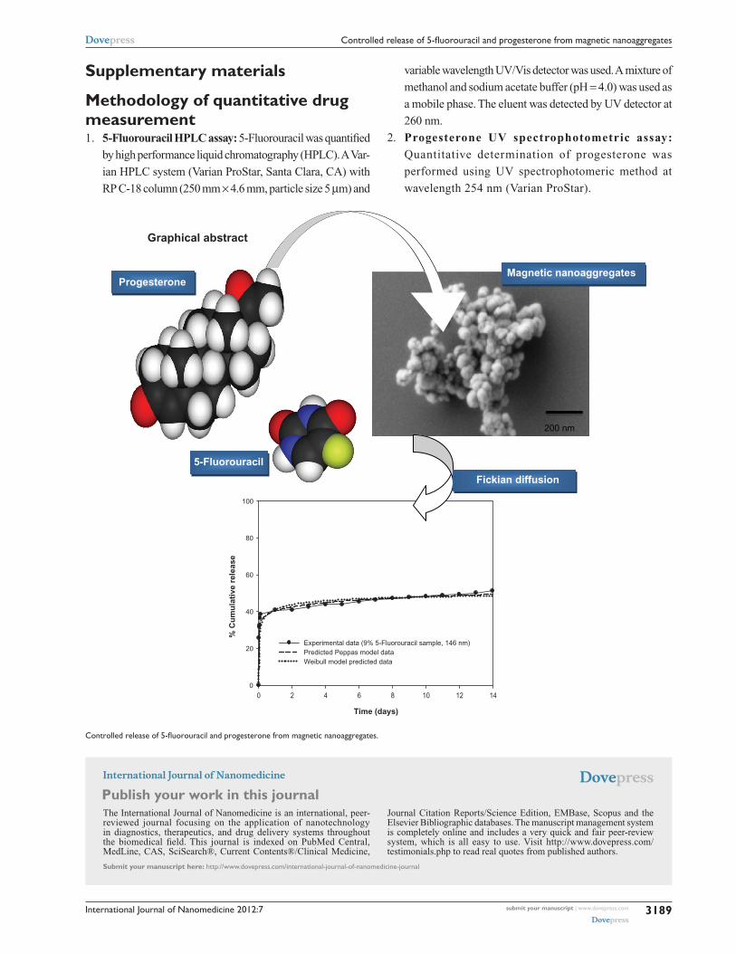
Graphical abstract
Time (days)
0 2 4 6 8 10 12 14
% C
um
ula
tive
rel
ease
0
20
40
60
80
100
Experimental data (9% 5-Fluorouracil sample, 146 nm)Predicted Peppas model dataWeibull model predicted data
Magnetic nanoaggregates
Fickian diffusion
200 nm
Progesterone
5-Fluorouracil
Controlled release of 5-fluorouracil and progesterone from magnetic nanoaggregates.
submit your manuscript | www.dovepress.com
Dovepress
Dovepress
Dovepress
3189
Controlled release of 5-fluorouracil and progesterone from magnetic nanoaggregates