Embed Size (px)
Citation preview
© 2017 Charbgoo et al. This work is published and licensed by Dove Medical Press Limited. The full terms of this license are available at https://www.dovepress.com/terms.php and incorporate the Creative Commons Attribution – Non Commercial (unported, v3.0) License (http://creativecommons.org/licenses/by-nc/3.0/). By accessing the work you
hereby accept the Terms. Non-commercial uses of the work are permitted without any further permission from Dove Medical Press Limited, provided the work is properly attributed. For permission for commercial use of this work, please see paragraphs 4.2 and 5 of our Terms (https://www.dovepress.com/terms.php).
International Journal of Nanomedicine 2017:12 1401–1413
International Journal of Nanomedicine Dovepress
submit your manuscript | www.dovepress.com
Dovepress 1401
R e v I e w
open access to scientific and medical research
Open Access Full Text Article
http://dx.doi.org/10.2147/IJN.S124855
Cerium oxide nanoparticles: green synthesis and biological applications
Fahimeh Charbgoo1
Mansor Bin Ahmad2,*Majid Darroudi3,*1Department of Pharmaceutical Biotechnology, School of Pharmacy, Mashhad University of Medical Sciences, Mashhad, Iran; 2Department of Chemistry, Faculty of Science, Universiti Putra Malaysia, Serdang, Selangor, Malaysia; 3Nuclear Medicine Research Center, Mashhad University of Medical Sciences, Mashhad, Iran
*These authors contributed equally to this work
Abstract: CeO2 nanoparticles (NPs) have shown promising approaches as therapeutic agents
in biology and medical sciences. The physicochemical properties of CeO2-NPs, such as size,
agglomeration status in liquid, and surface charge, play important roles in the ultimate interac-
tions of the NP with target cells. Recently, CeO2-NPs have been synthesized through several
bio-directed methods applying natural and organic matrices as stabilizing agents in order to
prepare biocompatible CeO2-NPs, thereby solving the challenges regarding safety, and provid-
ing the appropriate situation for their effective use in biomedicine. This review discusses the
different green strategies for CeO2-NPs synthesis, their advantages and challenges that are to
be overcome. In addition, this review focuses on recent progress in the potential application of
CeO2-NPs in biological and medical fields. Exploiting biocompatible CeO
2-NPs may improve
outcomes profoundly with the promise of effective neurodegenerative therapy and multiple
applications in nanobiotechnology.
Keywords: cerium oxide nanoparticles, green synthesis, biocompatibility, surface Ce3+, size,
morphology
IntroductionCeO
2 nanoparticles (NPs) have received much attention in nanotechnology due to their
useful applications as catalysts, fuel cells and antioxidants in biological systems.1–5
In general, cerium can exist in two oxidation states: Ce3+ and Ce4+. Therefore, cerium
dioxide can have two different oxide forms, CeO2 (Ce4+) or Ce
2O
3 (Ce3+), in bulk
material.4,6 On the nanoscale, the cerium oxide lattice has a cubic fluorite structure,
and both Ce3+ and Ce4+ can coexist on its surface. Charge deficiency due to the
presence of Ce3+ is compensated by oxygen vacancy in the lattice; thus, CeO2-NPs
contain intrinsic oxygen defects.7 These oxygen defects are actually sites of catalytic
reactions. The concentration of oxygen defects increases with reduction in particle
size.8 Therefore, CeO2-NPs have improved redox properties with respect to the bulk
materials. Moreover, the presence of a mixed valance state plays an important role
in scavenging reactive oxygen and nitrogen species. CeO2-NPs are found to be effec-
tive against pathologies associated with chronic oxidative stress and inflammation.
Recently, CeO2-NPs have also been reported to have multienzyme, including super-
oxide oxidase, catalase and oxidase, and mimetic properties, and have emerged as a
fascinating material in biological fields, such as in bioanalysis,9–14 biomedicine15 and
drug delivery.16,17 These applications are derived from quick transition of the oxida-
tion state between Ce3+ and Ce4+.6 The surface Ce3+:Ce4+ ratio is influenced by the
microenvironment. Therefore, the microenvironment and synthesis method adopted
also plays an important role in determining the biological activity and toxicity of
Correspondence: Majid DarroudiNuclear Medicine Research Center, Mashhad University of Medical Sciences, Mashhad, IranTel +98 513 800 2286Fax +98 513 800 2287email [email protected]
Mansor Bin AhmadDepartment of Chemistry, Faculty of Science, Universiti Putra Malaysia, 43400 Serdang, Selangor, MalaysiaTel +60 3 8946 6793Fax +60 3 8943 5380email [email protected]
Journal name: International Journal of NanomedicineArticle Designation: ReviewYear: 2017Volume: 12Running head verso: Charbgoo et alRunning head recto: Cerium oxide biosynthesisDOI: http://dx.doi.org/10.2147/IJN.S124855

International Journal of Nanomedicine 2017:12submit your manuscript | www.dovepress.com
Dovepress
Dovepress
1402
Charbgoo et al
CeO2-NPs. The CeO
2-NPs have been prepared through the
means of several routes and synthesis methods including
solution precipitation,18 sonochemical,19 hydrothermal,20
solvothermal,21 ball milling,22 thermal decomposition,23 spray
pyrolysis,24 thermal hydrolysis25 and sol–gel methods.26–28
However, applying the mentioned methods deals with sev-
eral drawbacks, such as toxic solvents and reagents usage,
high temperature and pressure, and the requirement of
external additives as stabilizing or capping agents during the
reaction. As the physiochemical properties of NPs mostly
depend on the synthesis procedure, the synthesis method of
NPs for biological applications is very important. The physi-
cal properties (size, surface charge, agglomeration status in
liquid and coating or residual contamination of the surfactant
on the surface) of NPs mainly influence interactions at the
nano–bio interface.29 Moreover, the surface Ce3+:Ce4+ ratio
(chemical property) also influences the biocatalysis and the
biological interactions. Manipulation of the surface Ce3+:Ce4+
ratio can be achieved by controlling their synthesis method.30
However, coating the NPs with biocompatible/organic poly-
mers increases dispersion/stability, decreases nonspecific
interactions with cells and proteins, increases blood circula-
tion time and reduces the toxicity of the NPs.31
Biomaterials possess functional groups such as –COOH,
–OH and –NH2, and have the potential to stabilize and/or cap
metal ions for preparation of various NPs via green chemistry
methods. Recently, CeO2-NPs have been synthesized through
several bio-directed methods applying natural and organic
matrices as stabilizing agents in order to prepare biocompat-
ible CeO2-NPs and solve the challenges to safely and effec-
tively use this metal oxide for biomedicinal purposes.27,28,32
In the first part of the review, we discuss the literature on
different green synthesis methods of CeO2-NPs (Table 1).
Next, we discuss the effect of these CeO2-NPs on reducing
their cytotoxicity in the biological environment. Finally, a
brief review on the updates of the potential biological appli-
cation of CeO2-NPs is presented.
Green approaches for CeO2-NP synthesisPlant-mediated synthesis of CeO2-NPsPhytosynthesis of metal and metal oxide NPs is a new emerg-
ing issue in nanoscience and technology.33 Recently, phyto-
synthesis of CeO2-NPs was reported using different plants,
such as Gloriosa superba, Acalypha indica and even Aloe
vera plant leaf extract (Figure 1).33–35 The plant extracts acted
as stabilizing and capping agents in the CeO2-NPs synthesis
process. Investigating biological effects of the phytosynthe-
sized NPs, antibacterial activity of them was examined. The
results showed that smaller crystal sizes with a higher surface
area led to higher antibacterial activity. These reports applied
bio-directed methods of CeO2-NP synthesis. However, the
synthesized nanoparticles were generally so large in size
Table 1 Green synthesis methods of CeO2-NPs
Method of green synthesis Applied material/ organism
Particle size (nm)
Morphology of NPs
Critical point of view Reference
Plant-mediated synthesis Gloriosa superba 5 Spherical Different kinds of alkaloids acted as stabilizing agents
33
Plant-mediated synthesis Acalypha indica 36 Spherical Agglomeration of particles were observed due to covalent bonding of the individual particles
34
Plant-mediated synthesis Aloe vera 63.6 Spherical 35Fungus-mediated synthesis Curvularia lunata 5–20 Spherical enzymes, proteins and heterocyclic
derivatives could act as reducing and capping agent
37
Nutrient-mediated synthesis ew protein 8.2, 11.7 and 17.3
Spherical Being soluble and foam-like in water, ew has several proteins acting as stabilizing agents
47
Nutrient-mediated synthesis Honey 23 Spherical Follow-up the sol–gel method 48Biopolymer-mediated synthesis Agarose 10.5 Spherical Follow-up the sol–gel method 51Biopolymer-mediated synthesis Starch 6 Spherical Providing ultrafine product 27Biopolymer-mediated synthesis Gum 10 Spherical 27Biopolymer-mediated synthesis Dextran 5 Spherical pH-dependent response 57Biopolymer-mediated synthesis Polyethylene glycol ~2 Spherical Providing a framework for designing
a hybrid metal oxide sol58
Biopolymer-mediated synthesis Chitosan ~10 Spherical Applicable in food borne mycoplasma detection
61
Abbreviations: ew, egg white; NPs, nanoparticles.

International Journal of Nanomedicine 2017:12 submit your manuscript | www.dovepress.com
Dovepress
Dovepress
1403
Cerium oxide biosynthesis
that, according to literature, they were not appropriate for
biomedical applications.1,36 Recently, biosynthesis of NPs
using yeast and fungi has also been noted. Munusamy et al
had explained rapid and extracellular synthesis of cerium
oxide NPs using fungus Curvularia lunata culture media.37
The synthesized NPs had a cubic structure and exhibited
antibacterial effects against different kinds of bacteria.37 It
is known that CeO2-NPs cannot enter bacterial and algal
cells. Noninternalized CeO2-NPs seem to show toxic effects
by direct attachment of CeO2-NPs to cell walls of algae and
bacteria.38–41 Several mechanisms have been suggested to
demonstrate how CeO2-NPs in contact with the membrane
may exert cytotoxicity. CeO2-NPs could interfere with
the nutrient transport functions of the membrane,39 cause
mechanical damage and membrane disruption42,43 or gener-
ate reactive oxygen species (ROS) and induce oxidative
stress.38–40 The generation of ROS, most probably hydrogen
peroxide, by CeO2-NPs is in agreement with observations
noted by Xia et al44 and Zhao et al.45 Hydrogen peroxide is
capable of freely diffusing across cell walls and membranes,
inducing cell damage.
Consequently, myco-synthesis of CeO2-NPs showed
advantages including manageability, cost-effectiveness, and
used techniques that were less time-consuming and required
less energy,46 and therefore can be used as an economic
and valuable alternative for the large-scale production of
CeO2-NPs. Moreover, myco-synthesized CeO
2-NPs had
more stability, water dispersibility and high fluorescent
properties. The fungal extracellular compounds, such as
proteins (especially enzymes), and heterocyclic derivatives
could act as reducing and capping agents. Other methods of
plant-based CeO2-NPs synthesis were also easy, rapid and
cost-effective, but the size of obtained NPs exhibited a wide
distribution range, which demonstrates that the necessity of
optimizing the biosynthesis methods mentioned earlier in
order for application in biological systems.
Nutrient-mediated synthesis of CeO2-NPsAs mentioned, synthetic methods determine the size, charge,
surface properties, solubility and morphology of NPs, there-
fore affecting response of CeO2-NPs in biological systems.
That is why green synthesis of CeO2-NPs has received much
attention recently. Several studies widely reported different
nutrients and natural materials, such as egg white (EW)
protein and honey for CeO2-NPs green synthesis.47,48 Kargar
et al47 proposed that the two major proteins of EW, ovalbumin
and lysozyme, acted as a green binders/stabilizing agents for
the preparation of CeO2-NPs. The general mechanism for
synthesizing CeO2-NPs in EW media includes formation of
the electrostatic interaction between cerium cations (Ce3+)
and oppositely charged proteins which leads to controllable
growth and subsequent isotropic formation of small and
stable CeO2-NPs.47,49 Some of the green methods of CeO
2-NP
preparation mimic the common traditional approaches in
NP synthesis in a safe and eco-friendly way.48 For example,
honey-based synthesis of CeO2-NPs mimics the sol–gel
method. The extensive number of carbohydrates, enzymes
and vitamins containing hydroxyl and amine groups in the
honey matrix structure can facilitate the complexation of
°°
Figure 1 Schematic representation of Gloriosa superba-based method of cerium oxide nanoparticle synthesis.
International Journal of Nanomedicine 2017:12submit your manuscript | www.dovepress.com
Dovepress
Dovepress
1404
Charbgoo et al
cerium cations (Ce3+) to an initial molecular matrix. There-
fore, honey was capable of coating and stabilizing cerium
species and CeO2-NPs while inhibiting their excessive
aggregation or crystal growth.48 However, advancement of
the EW-based method for CeO2-NP green synthesis is obvi-
ous due to nontoxic effects of CeO2-NPs at concentrations
up to 800 μg/mL, compared with the safe concentration
of ~25 μg/mL for honey-based CeO2-NPs. Therefore, the
synthesis of CeO2-NPs in EW was found to be an excellent
alternative for the preparation of CeO2-NPs, using food and
bio-derived materials.
Biopolymer-mediated synthesis of CeO2-NPsNatural polymers in the form of macromolecules can
also be used as templates for bio-directed synthesis of
CeO2-NPs. As the surface of the NPs could be covered by
hydroxyl groups, biopolymers that intrinsically possess
hydroxyl moieties are capable of stabilizing CeO2-NPs.
Applying the polymers as capping/stabilizing agents, the
diameter of NPs can be logically controlled.50 Kargar et al
reported the green synthesis of small cerium oxide NPs,
stabilized with agarose polymers via a sol–gel method.51
While heating to 90°C, the agarose powder is normally
dissolved in water, and when the temperature is reduced
to 35°C–40°C, semisolid gel is formed that is stable over a
wide pH range of (from 3 to 9). Interpenetrating H-binding
between sugar moieties resulted in production of this
sol–gel network and nanochannel containing pore sizes of
200 nm. CeO2-NPs were synthesized in these nanochan-
nels. Similarly, Darroudi et al had synthesized CeO2-NPs
using starch as a capping biopolymer.27 The proposed
mechanism, for starch-based synthesis of CeO2-NPs was
that after dissolving starch in water, metal cations were
attracted by oxygen of the OH branches. In vitro studies
on Neuro2A cells demonstrated a dose-dependent toxicity
with a nontoxic concentration of 175 μg/mL. Applying
starch as a template for CeO2-NP synthesis by Darroudi
et al27 resulted in the formation of ultrafine CeO2-NP par-
ticles that were small in size and uniform in shape. There-
fore, this method seems to be more appropriate for CeO2-NP
synthesis for medical purposes. Furthermore, in line with
the required characteristics, this method was found to be
easy, economical and green for large-scale preparation of
cerium oxide in nanoscale.
Regarding unique potential of biopolymers in the
development of bio-directed methods of CeO2-NP syn-
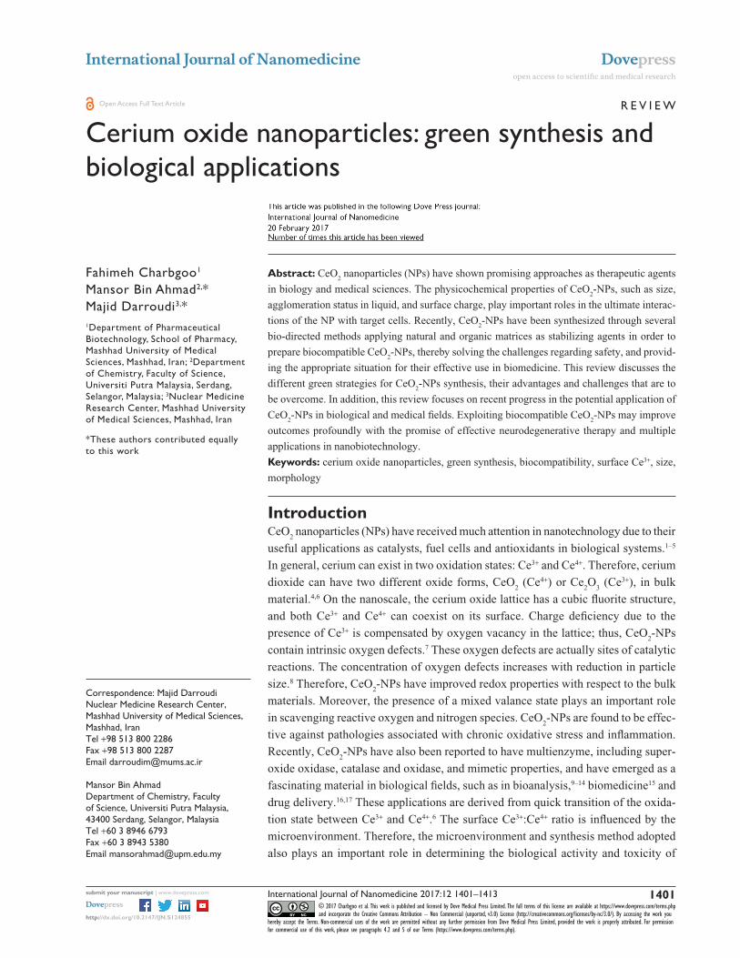
thesis, Darroudi et al27 also used Gum tragacanth (GT)
for the production of CeO2-NPs by both chemical and
biological methods.28 The soluble fraction (tragacanthin
or tragacanthic acid) of GT gives a sol form in distilled
water, whereas the insoluble fraction (bassorin) swells
to a gel form (Figure 2).52,53 While heating the sol–gel
solution up to 40°C, the GT became soluble in water and
the semicrystalline structures were lost. After adding
the cerium nitrate to the solution, the metal cations were
attracted by the oxygen of OH branches of GT polysac-
charides. During the heating process, the amount of water
was decreased and the nitrate decomposed to nitrogen
dioxide and oxygen molecules, which were then removed
from the compounds. Ce(OH)4 nuclei were converted into
CeO2 nuclei via dehydration and, subsequently, highly
crystallized CeO2-NPs particles grew. The required
energy for the above reactions was provided by the
subsequent sol–gel procedure and heat. The stabilizing
effect of GT could be attributed to the steric repulsion
force arising as the gum formed a layer around the cerium
hydroxides and cerium oxide NPs. However, the ability
of GT to stabilize CeO2-NPs might also be due to elec-
trostatic interactions in addition to the enhancement of
suspension viscosity.54,55 Although the formation of CeO2-
NPs particles involved several complicated reactions,56
controlling the nucleation of initial precipitate Ce(OH)3
would mainly determine the properties of the final CeO2-
NPs. Furthermore, the CeO2-NPs exhibited very low
cytotoxic effects on Neuro2A cell lines, making them
suitable candidates for various biological applications.
Dextran was also used for CeO2-NP stabilizing and coat-
ing, as it is a biocompatible, complex and highly water-
soluble polysaccharide.57 Accordingly, NPs as small as
5 nm were produced which were toxic to cancer cells
at pH 6 and much less toxic to normal cells at the same
pH value.57 Moreover, the importance and versatility of
polyethylene glycol (PEG) for the functionalization of
rare earth cerium oxide NPs were also investigated.58–60
The suggested mechanism for PEG-mediated ceria syn-
thesis was the presence of an electrostatic driving force
for the complexation.59 The branched structure of PEG
is sufficient to solubilize the CeO2-NPs and create true
dispersible nanopowders in aqueous solution and in cer-
tain organic solvents, providing a framework for design-
ing a versatile hybrid metal oxide sol.58 Furthermore,
chitosan-based synthesis of CeO2-NPs was also reported
due to specific properties, such as good film-forming
ability, biocompatibility, nontoxicity, biodegradability
and antibacterial activity (Table 2).61,62

International Journal of Nanomedicine 2017:12 submit your manuscript | www.dovepress.com
Dovepress
Dovepress
1405
Cerium oxide biosynthesis
The toxicologic effect of green synthesized CeO2-NPsAll cerium oxide NPs contain the same core elements,
however, do not display similar biological effects. There
are some studies that reported prooxidant toxicity of NPs
in some cases and antioxidant protective effects in others
that could be attributed to different physiochemical param-
eters of the various NPs that were used. Method of NP
synthesis, type of stabilizing agent used, and the Ce3+/Ce4+
surface ratio have been demonstrated to play major roles
in producing CeO2-NPs with different physicochemical
properties.63,64 The most important parameters are discussed
below (Figure 3).
Particle sizeSeveral green methods of CeO
2-NPs synthesis have provided
NPs as small as 10 nm. Previous results demonstrated that
among different strategies reported for bio-directed synthe-
sis of CeO2-NPs, biopolymer and nutrient-based methods
provided the smallest NPs compared with plant-based pro-
cesses. Reports indicated that plant-based CeO2-NP synthesis
provided larger NP with antibacterial properties that exhib-
ited high levels of cytotoxicity to bacterial cells.35,37 However,
biopolymer- and nutrient-based methods have provided small
NPs which show no cytotoxic effects to human cell lines at
high concentrations of CeO2-NPs.27,28,47,48,51
MorphologyMorphology is another physical property that is also required
to be considered for biological applications. For example,
NPs in polygonal, cube or rod shapes have sharp edges and
could cause mechanical damage to cells.7,65,66 Therefore, the
effect of NP shape cannot be ignored for biological applica-
tions. As mentioned earlier, almost all the green methods of
ceria synthesis that are mentioned herein have produced NPs
with spherical morphology. However, starch-based synthesis
of CeO2-NPs seems to be the most appropriate method to
provide CeO2-NPs for biomedical purposes.27
Figure 2 Schematic representation of the Gum base method of CeO2-NP synthesis.Abbreviation: CeO2-NPs, cerium oxide nanoparticles.

International Journal of Nanomedicine 2017:12submit your manuscript | www.dovepress.com
Dovepress
Dovepress
1406
Charbgoo et al
Figure 3 Major parameters affect the cytotoxicity of CeO2-NPs.Abbreviation: CeO2-NPs, cerium oxide nanoparticles.
Table 2 Advantages and challenges of different methods of CeO2-NPs green synthesis
Type of green method Advantages Disadvantages/challenges
Plant-mediated synthesis of CeO2-NPs
Capable of generating spherical shaped NPs that possessed reduced cytotoxicity
Possibility of providing nonuniform morphology in some case which could be attributed to agglomeration of the individual NPs
easy process, cost-effectiveness, energy and time-consuming technique
Size of obtained NPs exhibited wide distribution range from 5 to 63.6 nm using different bio-organisms for synthesis
Capable of producing stable, water dispersible and highly fluorescent NPs
Nutrient-mediated synthesis of CeO2-NPs
Controllable growth and subsequent isotropic formation of small and stable CeO2-NPs
Significant difference at the maximum concentration, which was safe for the cells using ew (800 μg/mL) or honey (100 μg/mL) as a stabilizing agents
Capable of providing spherical shaped CeO2-NPsNarrow distribution range of particle sizeNontoxic effects of synthesized CeO2-NPs toward human cell lines at physiological concentrations of NPs
Biopolymer-mediated synthesis of CeO2-NPs
Generating NP with spherical morphologyProviding NPs with no significant cytotoxic effect in human cell line at physiological concentrationsCapable of controlling diameter of CeO2-NPsProviding NPs with high final purityProducing small CeO2-NPs
Abbreviations: CeO2-NPs, cerium oxide nanoparticles; ew, egg white.
Percentage of surface Ce3+
In 2015, Pulido-Reyes et al67 presented a report that differed
from previous reports about CeO2-NPs synthesis. They
demonstrated that neither concentration, surface charge nor
size of CeO2-NPs plays any important role in their observed
toxic properties. The report demonstrated that percentage of
surface Ce3+ correlated with toxicity and was the main driver
of CeO2-NPs toxic effects.67 They proposed that CeO
2-NPs
International Journal of Nanomedicine 2017:12 submit your manuscript | www.dovepress.com
Dovepress
Dovepress
1407
Cerium oxide biosynthesis
with the highest percentage of surface Ce3+ (58%) exhibited
the most toxic effect, and CeO2-NPs with lower percentage
of surface Ce3+ values (between 26% and 36%) were evi-
dently nontoxic for the model organism. In fact, CeO2-NPs
with lower Ce3+ and, therefore, higher Ce4+ on their surface
showed catalase mimetic activity,68 which broke down H2O
2
to molecular oxygen, protecting the cells against this toxic
ROS. CeO2-NPs with higher Ce3+ on their surface could
efficiently scavenge radicals of superoxide (superoxide dis-
mutase [SOD] mimetic activity) and produce H2O
2, which is
toxic to the cells. They suggested that in a narrow range of
surface Ce3+, there seemed to be a shift from SOD activity
to catalase mimetic activity; however, the mechanisms and
whether the observed biological effect reported at their study
may also occur in other cellular systems, requires further
investigation.67 However, there is no report on the effect
of applying green methods of CeO2-NPs synthesis on the
percentage of surface Ce3+ of NPs and this should be inves-
tigated to clearly demonstrate the effect of green synthesis
of CeO2-NPs on their cytotoxicity.
A CeO2-NP enters cells by energy-dependent, clathrin-
mediated and caveolae-mediated endocytic pathways. Its
localization in mitochondria, lysosomes and endoplasmic
reticulum, as well as the cytoplasm and nucleus, were
demonstrated by Singh et al.69 Considering radical scaveng-
ing properties of cerium oxide and its widespread cellular
disposition, a CeO2-NP likely acts as a cellular antioxidant
in multiple compartments of the cell, presenting protection
against a variety of oxidant injuries.69
Biological applications of CeO2-NPsAntibacterial effectThere are different studies that have reported antibacterial
activity of CeO2-NPs and demonstrated their significant
inhibition toward both gram-negative and gram-positive
bacteria.34–37 It is suggested that CeO2-NPs with a particle
size of over 20 nm possess antibacterial properties. Moreover,
the most antibacterial effects due to the highest percentage
of surface Ce3+ of NP are in agreement with Pulido-Reyes
et al’s observations.67
Neurodegenerative effectThe brain and central nervous system are the most active organ
systems in the body; therefore, they are particularly sensitive to
oxidative stress because of high oxygen utilization, high levels
of polyunsaturated fatty acid peroxidation and low levels of
endogenous antioxidant systems. Increased oxidative stress
and free radical production could be attributed to several neu-
rodegenerative diseases, such as Parkinson’s disease, trauma,
ischemic stroke, Alzheimer’s disease (AD) and aging.70 A
beneficial therapy for neurodegenerative diseases is CeO2-NP
utilization, which removes ROS or prevents their formation
and affects different key points in the brain cells or central
nervous tissue. Reducing ROS production, CeO2-NPs were
demonstrated to affect (directly or indirectly) signal transduc-
tion pathways involved in neuronal death and neuroprotection.
For example, it is reported that cerium oxide NPs could trigger
neuronal survival in a human AD model through modulating
the brain-derived neurotrophic factor (BDNF) pathway. BDNF
is a factor involved in the signal transduction pathways of
neuronal survival.71 In a similar approach, Guo et al reported
that ceria NPs protect neurons against oxidative stress induced
injury by modulating transforming growth factor beta (TGF-β)
signaling.72 There are so many reports on the neuroprotective
effect of engineered CeO2-NPs. Recently, Arya et al3 reported
that CeO2-NPs promoted neurogenesis and modulated hypox-
ia-induced memory impairment through the AMPK–PKC–
CBP signaling cascade. Using PEG-coated 3 nm CeO2-NPs,
they demonstrated that NPs were efficiently localized in the
brain and significantly decreased oxidative stress. Therefore,
associated damage during hypoxia exposure was also reduced
by applying PEG/CeO2-NPs. They also provided evidence that
PEG/CeO2-NPs enhanced hippocampus neuronal survival and
promoted neurogenesis.3
Regarding the reductive effect of CeO2-NPs on oxidative
stress, which is known to play an important role in neurode-
generation, Fiorani et al73 had investigated the role of CeO2-
NPs on microglial activation and neurodegenerative processes
in light damaged retina. They demonstrated the ability of
CeO2-NPs to reduce microglial activation and their migration
toward the outer nuclear layer,73 raising the possibility of their
use as therapeutic agents for neurodegenerative diseases.
enzyme mimetic applicationsCeO
2-NPs are forms of powerful artificial oxidase enzymes
capable of mimicking catalase and SOD and peroxidase-like
activities (Table 3).
Oxidase-like activity of these NPs originated from surface
Ce3+ atoms as the catalytic center.74 CeO2-NPs with lower
Ce3+ on their surface showed catalase or peroxidase mimetic
activity,68 which could break down H2O
2 into water and
oxygen. CeO2-NPs with higher Ce3+ on their surface could
efficiently scavenge radicals of superoxide (SOD mimetic
activity) and produce H2O
2.
SOD mimicking activityComparing with natural enzymes, CeO
2-NPs showed several
advantages, such as high sensitivity, low cost, easy storage

International Journal of Nanomedicine 2017:12submit your manuscript | www.dovepress.com
Dovepress
Dovepress
1408
Charbgoo et al
and catalytic stability under harsh conditions. Construction
of efficient artificial enzymes, as a strong and cost-effective
alternative to natural enzymes, has been an interesting subject
in the field of biomimetic chemistry. In a new report on SOD-
like activity of ceria, Bhushan and Gopinath75 developed a
stable and biocompatible artificial enzymatic system based on
CeO2-NPs that possessed high ROS scavenging activity over
a period of time. They synthesized a CeO2-NP encapsulated
biocompatible ceria-albumin nanoparticle (BCNP) capable
of reducing intracellular ROS. The BCNPs preserved the
antioxidant defense system of the cells and protected them
from oxidant-mediated apoptosis.75 Importantly, the enzyme
mimicking activity of CeO2-NPs remained almost constant
and stable over a wide range of pH and temperature. There-
fore, the as-prepared BCNPs were promising as potential
candidates against ROS-induced diseases and disorders uti-
lizing SOD-like activity of ceria.75 Moreover, the SOD ability
of CeO2-NPs with sizes 5 nm and diversity in shape and a
negligible Ce3+/Ce4+ ratio were also investigated by Li et al.76
So far, inherent superoxide-scavenging ability has only
been found in the CeO2-NPs with sizes of 5 nm, and these
bioactive CeO2-NPs showed very limited diversity with
respect to shape. Li et al76 believed that without the coating of
surface ligands to stabilize the oxygen vacancies, CeO2-NPs
of 3 nm could not maintain a substantially higher Ce3+/Ce4+
ratio under ambient conditions when compared to their bulk
counterpart.77 Therefore, even CeO2-NPs of 5 nm would
lose their inherent SOD mimetic activity because of Ce3+
oxidation, and the time required to regenerate that activity
would usually take days and weeks.78,79 Li et al76 proposed a
strategy to significantly improve the superoxide-scavenging
activity of CeO2-NPs of 5 nm. However, they activated the
SOD mimetic activity of different sized CeO2-NPs within
minutes by incubation with native CuZn-SOD in phosphate-
buffered saline (Figure 4).76
Catalase mimicking activityThe first report on catalase mimicking activity of CeO
2-NPs
was presented by Pirmohamed et al.68 Recently, the catalytic
activity of CeO2-NPs was applied in different biomedical
approaches.80–82 For example, Akhtar et al have demonstrated
that the catalase activity of CeO2-NPs could increase the
intracellular glutathione (GSH) in cells challenged with H2O
2,
protecting cells from oxidative damage.80 Considering major
roles of GSH in the regulation of cell growth and division,
metabolism of carcinogens and protecting DNA from oxida-
tive damage, the effect of CeO2-NPs on increasing the amount
of intracellular GSH marks a revolution in medical biology.
Moreover, Nicolini et al had introduced a kind of bioactive
glass based on catalytic activity of CeO2-NPs, which was
used for bone tissue engineering.82 The design of bioactive
glasses capable of preventing oxidative stress after implan-
tation would reduce the convalescence and decrease the
amount of anti-inflammatory responses in patients. Applying
biomedical properties of CeO2-NPs requires more investiga-
tion of the NPs’ fate in vivo. For example, cerium atoms of
CeO2-NPs have the potential to interact with peptides, sugar
and small anion molecules, such as phosphate in vitro and in
vivo. Singh et al investigated the role of phosphate on stabil-
ity and catalase mimetic activity of cerium oxide NPs.81,83
Given the abundance of inorganic phosphate in biological
systems, they demonstrated that catalase mimetic activity
of CeO2-NPs (Ce4+) is resistant to the phosphate anions, pH
changes and composition of cell culture media. Thus, Singh
et al provided a promising approach to more practical and
attractive biomedical applications for cerium oxide NPs.
Table 3 Different types of enzyme mimicking activities of cerium oxide nanoparticles
Enzyme mimicking activities Mechanism References
SOD M(n+1)+-SOD + O2− → Mn+-SOD + O2
Mn+-SOD + O2− + 2H+ → M(n+1)+-SOD + H2O2
75, 76
Catalase H2O2 + H2R → 2H2O + R 68, 80, 81Peroxidase ROOR’ + 2e− + 2H+ → ROH + R’OH 2
Abbreviation: SOD, superoxide dismutase.
Figure 4 Superoxide dismutase mimetic activity of CeO2-nanoparticles.
International Journal of Nanomedicine 2017:12 submit your manuscript | www.dovepress.com
Dovepress
Dovepress
1409
Cerium oxide biosynthesis
In other work, Sardesai et al developed a biosensor based
on oxygen-rich platinum doped CeO2-NPs (Pt-ceria) and
lactate oxidase for in vitro and in vivo monitoring of lactate
during hypoxia.86 Integration of the oxygen-rich CeO2-NPs
in the enzyme-containing layer ensured operation of the
biosensor in hypoxic conditions, and provided continuous,
sensitive lactate monitoring. Measurements of lactate levels
in blood and tissues are important indications of the state
and progress of a variety of diseases. In vitro evaluation of
the biosensor demonstrated a detection limit of 100 pM and
high selectivity against physiological levels of coexisting
interference species, as well as a quick response time of 6
seconds. In vivo studies have been performed by placing the
designed biosensor in the hippocampus of anesthetized rats.
The results provided the possibility of continuous lactate
monitoring under 2 hours ischemia and reperfusion.86 More-
over, all the mentioned reports have documented the ability
of cerium oxide NPs to provide third-generation biosensors
with high sensitivity and specificity of detection.
Angiogenesis inductionA unique property of CeO
2-NPs could also induce angiogene-
sis in vivo. Angiogenesis is the physiological process through
which new blood vessels form from pre-existing ones.
In particular, CeO2-NPs trigger angiogenesis by modulat-
ing the intracellular oxygen environment and endogenously
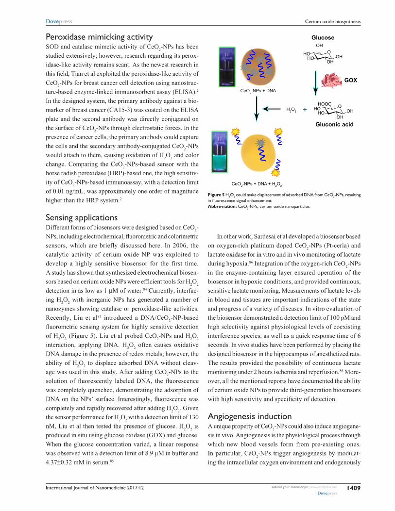
Figure 5 H2O2 could make displacement of adsorbed DNA from CeO2-NPs, resulting in fluorescence signal enhancement.Abbreviation: CeO2-NPs, cerium oxide nanoparticles.
Peroxidase mimicking activitySOD and catalase mimetic activity of CeO
2-NPs has been
studied extensively; however, research regarding its perox-
idase-like activity remains scant. As the newest research in
this field, Tian et al exploited the peroxidase-like activity of
CeO2-NPs for breast cancer cell detection using nanostruc-
ture-based enzyme-linked immunosorbent assay (ELISA).2
In the designed system, the primary antibody against a bio-
marker of breast cancer (CA15-3) was coated on the ELISA
plate and the second antibody was directly conjugated on
the surface of CeO2-NPs through electrostatic forces. In the
presence of cancer cells, the primary antibody could capture
the cells and the secondary antibody-conjugated CeO2-NPs
would attach to them, causing oxidation of H2O
2 and color
change. Comparing the CeO2-NPs-based sensor with the
horse radish peroxidase (HRP)-based one, the high sensitiv-
ity of CeO2-NPs-based immunoassay, with a detection limit
of 0.01 ng/mL, was approximately one order of magnitude
higher than the HRP system.2
Sensing applicationsDifferent forms of biosensors were designed based on CeO
2-
NPs, including electrochemical, fluorometric and colorimetric
sensors, which are briefly discussed here. In 2006, the
catalytic activity of cerium oxide NP was exploited to
develop a highly sensitive biosensor for the first time.
A study has shown that synthesized electrochemical biosen-
sors based on cerium oxide NPs were efficient tools for H2O
2
detection in as low as 1 μM of water.84 Currently, interfac-
ing H2O
2 with inorganic NPs has generated a number of
nanozymes showing catalase or peroxidase-like activities.
Recently, Liu et al85 introduced a DNA/CeO2-NP-based
fluorometric sensing system for highly sensitive detection
of H2O
2 (Figure 5). Liu et al probed CeO
2-NPs and H
2O
2
interaction, applying DNA. H2O
2 often causes oxidative
DNA damage in the presence of redox metals; however, the
ability of H2O
2 to displace adsorbed DNA without cleav-
age was used in this study. After adding CeO2-NPs to the
solution of fluorescently labeled DNA, the fluorescence
was completely quenched, demonstrating the adsorption of
DNA on the NPs’ surface. Interestingly, fluorescence was
completely and rapidly recovered after adding H2O
2. Given
the sensor performance for H2O
2 with a detection limit of 130
nM, Liu et al then tested the presence of glucose. H2O
2 is
produced in situ using glucose oxidase (GOX) and glucose.
When the glucose concentration varied, a linear response
was observed with a detection limit of 8.9 μM in buffer and
4.37±0.32 mM in serum.85
International Journal of Nanomedicine 2017:12submit your manuscript | www.dovepress.com
Dovepress
Dovepress
1410
Charbgoo et al
stabilizing hypoxia inducing factor 1α, which alters gene
regulation. Furthermore, the high surface area, increased
Ce3+/Ce4+ ratio and small size make CeO2-NPs more catalyti-
cally active toward regulating intracellular oxygen, which in
turn leads to more robust induction of angiogenesis.87
ConclusionThe unique property of CeO
2-NPs that makes them distinct
from other antioxidants is their ability to self-regenerate their
surface. Thus, one small dose can work for a long time before
being cleared from the body.7 Accordingly, various kinds
of CeO2-NPs have been synthesized in order to target the
Achilles’ heel of any oxidative stress-associated diseases.88,89
Investigating previous literature on ceria NPs demonstrated
that different synthesis methods could provide cerium oxide
NPs with various catalytic and physiochemical properties that
could contribute to antioxidant or prooxidant properties.29
Considering CeO2-NPs as potential therapeutic agents, it is
important to pay attention to their synthesis method. Among
different strategies reported for the synthesis of CeO2-NPs,
green synthesis methods have shown to be promising for
CeO2-NP production and in their application in biological sys-
tems. Another consideration of CeO2-NPs is that the in vitro
measured properties of the NP (eg, zeta potential, size and
redox activity) could change under physiological conditions.90
For example, Kumari et al has shown that the hydrodynamic
diameter of CeO2-NPs increased dramatically in cell culture
media due to the tendency of NPs to agglomerate in physi-
ological conditions.91 Furthermore, adsorption of proteins
in biological fluids, such as blood, could also affect the size
and distribution of metal oxide NPs. Generally, smaller sized
particles that are free of contamination are suitable for bio-
applications. Using bio-directed methods, synthesis of small
CeO2-NPs is possible. For example, as mentioned earlier,
applying starch-based methods resulted in the production of
CeO2-NPs as small as 6 nm. Since bio-directed methods of
CeO2-NP synthesis used biocompatible stabilizers and pro-
duced nontoxic NPs, of all the different methods of CeO2-NP
synthesis, green synthesis is proposed to be applied for the
production of CeO2-NPs for therapeutic purposes. Moreover,
green synthesis of CeO2-NPs suggests several advantages,
such as cost-effectiveness, large-scale commercial production
and the potential for pharmaceutical applications.
Future perspectivesCeO
2-NPs were recently shown to have regenerative antioxi-
dant activity. Therefore, low levels of CeO2-NPs can work
for extended time periods. However, these NPs provided
some toxicologic concerns. Currently, the green synthesis of
CeO2-NPs gets more attention in order to solve the challenges
regarding safety and use of this metal oxide for biomedicine,
but there are still some considerations. Previous reports sug-
gested that the protein corona provides NPs with particular
biological identity which subsequently play important roles in
the ultimate interactions of NPs with target cells. Therefore,
physiochemical characteristics of NPs after interaction with
biological fluids should be investigated in order to achieve
correct interpretations of the biocompatibility of green meth-
ods of CeO2-NPs synthesis. Moreover, regarding the effect
of percentage of surface Ce3+ on the properties of CeO2-NPs
in biological systems, the green synthesized CeO2-NPs
should be investigated from this point of view. In addition,
an important consideration in clinical usage of CeO2-NPs
is how cerium oxide NPs behave in biological systems.
Addressing this is not a simple endeavor and requires some
in vivo-based research of the effect of CeO2-NPs produced
by bio-directed methods.
DisclosureThe authors report no conflicts of interest in this work.
References 1. Gagnon J, Fromm KM. Toxicity and protective effects of cerium oxide
nanoparticles (Nanoceria) depending on their preparation method, par-ticle size, cell type, and exposure route. Eur J Inorg Chem. 2015;27: 4510–4517.
2. Tian Z, Li J, Zhang Z, Gao W, Zhou X, Qu Y. Highly sensitive and robust peroxidase-like activity of porous nanorods of ceria and their application for breast cancer detection. Biomaterials. 2015;59:116–124.
3. Arya A, Gangwar A, Singh SK, et al. Cerium oxide nanoparticles pro-mote neurogenesis and abrogate hypoxia-induced memory impairment through AMPK–PKC–CBP signaling cascade. Int J Nanomedicine. 2016; 11:1159–1173.
4. Beaudoux X, Virot M, Chave T, Durand G, Leturcq G, Nikitenko SI. Vitamin C boosts ceria-based catalyst recycling. Green Chem. 2016;18: 3656–3668.
5. Gawande MB, Bonifacio VDB, Varma RS, et al. Magnetically recyclable magnetite-ceria (Nanocat-Fe-Ce) nanocatalyst – applications in multi-component reactions under benign conditions. Green Chem. 2013;15(5): 1226–1231.
6. Xu C, Qu X. Cerium oxide nanoparticle: a remarkably versatile rare earth nanomaterial for biological applications. NPG Asia Mater. 2014;6:e90.
7. Das S, Dowding JM, Klump KE, McGinnis JF, Self W, Seal S. Cerium oxide nanoparticles: applications and prospects in nanomedicine. Nanomedicine (Lond). 2013;8(9):1483–1508.
8. Deshpande S, Patil S, Kuchibhatla SV, Seal S. Size dependency varia-tion in lattice parameter and valency states in nanocrystalline cerium oxide. Appl Phys Lett. 2005;87(13):133113.
9. Asati A, Santra S, Kaittanis C, Nath S, Perez JM. Oxidase-like activity of polymer-coated cerium oxide nanoparticles. Angew Chem Int Ed Engl. 2009;48(13):2308–2312.
10. Asati A, Kaittanis C, Santra S, Perez JM. The pH-tunable oxidase-like activity of cerium oxide nanoparticles achieves sensitive fluorigenic detection of cancer biomarkers at neutral pH. Anal Chem. 2011;83(7): 2547–2553.

International Journal of Nanomedicine 2017:12 submit your manuscript | www.dovepress.com
Dovepress
Dovepress
1411
Cerium oxide biosynthesis
11. Li X, Sun L, Ge A, Guo Y. Enhanced chemiluminescence detection of thrombin based on cerium oxide nanoparticles. Chem Commun. 2011; 47(3):947–949.
12. Kaittanis C, Santra S, Asati A, Perez JM. A cerium oxide nanoparticle-based device for the detection of chronic inflammation via optical and magnetic resonance imaging. Nanoscale. 2012;4(6):2117–2123.
13. Ornatska M, Sharpe E, Andreescu D, Andreescu S. Paper bioassay based on ceria nanoparticles as colorimetric probes. Anal Chem. 2011;83(11): 4273–4280.
14. Lin Y, Xu C, Ren J, Qu X. Using thermally regenerable cerium oxide nanoparticles in biocomputing to perform label-free, resettable, and colorimetric logic operations. Angew Chem Int Ed Engl. 2012;51(50): 12579–12583.
15. Celardo I, Pedersen JZ, Traversa E, Ghibelli L. Pharmacological potential of cerium oxide nanoparticles. Nanoscale. 2011;3(4): 1411–1420.
16. Li M, Shi P, Xu C, Ren J, Qu X. Cerium oxide caged metal chelator: anti-aggregation and anti-oxidation integrated H
2O
2-responsive con-
trolled drug release for potential Alzheimer’s disease treatment. Chem Sci. 2013;4(6):2536–2542.
17. Xu C, Lin Y, Wang J, et al. Nanoceria-triggered synergetic drug release based on CeO2-capped mesoporous silica host–guest interactions and switchable enzymatic activity and cellular effects of CeO2. Adv Healthc Mater. 2013;2(12):1591–1599.
18. Chen HI, Chang HY. Synthesis of nanocrystalline cerium oxide particles by the precipitation method. Ceramics Int. 2005;31(6):795–802.
19. Yu JC, Zhang L, Lin J. Direct sonochemical preparation of high-surface-area nanoporous ceria and ceria–zirconia solid solutions. J Colloid Interface Sci. 2003;260(1):240–243.
20. Yan Z, Wang J, Zou R, Liu L, Zhang Z, Wang X. Hydrothermal synthesis of CeO2 nanoparticles on activated carbon with enhanced desulfuriza-tion activity. Energy Fuels. 2012;26(9):5879–5886.
21. Chunwen S, Hong L, Huairuo Z, Zhaoxiang W, Liquan C. Controlled synthesis of CeO2 nanorods by a solvothermal method. Nanotechnology. 2005;16(9):1454.
22. Yadav TP, Srivastava ON. Synthesis of nanocrystalline cerium oxide by high energy ball milling. Ceramics Int. 2012;38(7):5783–5789.
23. Wang Y, Mori T, Li JG, Ikegami T. Low-temperature synthesis of praseodymium-doped ceria nanopowders. J Am Ceramic Soc. 2002; 85(12):3105–3107.
24. Feng X, Sayle DC, Wang ZL, et al. Converting ceria polyhedral nano-particles into single-crystal nanospheres. Science. 2006;312(5779): 1504–1508.
25. Hirano M, Fukuda Y, Iwata H, Hotta Y, Inagaki M. Preparation and spherical agglomeration of crystalline cerium(IV) oxide nanoparticles by thermal hydrolysis. J Am Ceramic Soc. 2000;83(5):1287–1289.
26. He HW, Wu XQ, Ren W, Shi P, Yao X, Song ZT. Synthesis of crystal-line cerium dioxide hydrosol by a sol–gel method. Ceramics Int. 2012; 38(Suppl 1):S501–S504.
27. Darroudi M, Sarani M, Kazemi Oskuee R, Khorsand Zak A, Hosseini HA, Gholami L. Green synthesis and evaluation of metabolic activity of starch mediated nanoceria. Ceramics Int. 2014;40(1, Part B): 2041–2045.
28. Darroudi M, Sarani M, Kazemi Oskuee R, Khorsand Zak A, Amiri MS. Nanoceria: gum mediated synthesis and in vitro viability assay. Ceramics Int. 2014;40(2):2863–2868.
29. Dowding JM, Seal S, Self WT. Cerium oxide nanoparticles accelerate the decay of peroxynitrite (ONOO−). Drug Deliv Transl Res. 2013;3(4): 375–379.
30. Dowding JM, Das S, Kumar A, et al. Cellular interaction and toxicity depend on physicochemical properties and surface modification of redox-active nanomaterials. ACS Nano. 2013;7(6):4855–4868.
31. Adschiri T, Lee YW, Goto M, Takami S. Green materials synthesis with supercritical water. Green Chem. 2011;13(6):1380–1390.
32. Ko JW, Lee BI, Chung YJ, Park CB. Carboxymethyl cellulose-templated synthesis of hierarchically structured metal oxides. Green Chem. 2015;17(8):4167–4172.
33. Arumugam A, Karthikeyan C, Haja Hameed AS, Gopinath K, Gowri S, Karthika V. Synthesis of cerium oxide nanoparticles using Gloriosa superba L. leaf extract and their structural, optical and antibac-terial properties. Mater Sci Eng C Mater Biol Appl. 2015;49:408–415.
34. Kannan SK, Sundrarajan M. A green approach for the synthesis of a cerium oxide nanoparticle: characterization and antibacterial activity. Int J Nanosci. 2014;13(03):1450018.
35. Priya GS, Kanneganti A, Kumar KA, Rao KV, Bykkam S. Bio synthesis of cerium oxide nanoparticles using Aloe arbadensis Miller Gel. Int J Sci Res Publications. 2014;4(6):1–4.
36. Kumar A, Das S, Munusamy P, et al. Behavior of nanoceria in biologically-relevant environments. Environ Sci Nano. 2014;1(6):516–532.
37. Munusamy S, Bhakyaraj K, Vijayalakshmi L, Stephen A, Narayanan V. Synthesis and characterization of cerium oxide nanoparticles using Curvularia lunata and their antibacterial properties. Int J Innovative Res Sci Eng. 2014;2(1):318–323.
38. Thill A, Zeyons O, Spalla O, et al. Cytotoxicity of CeO2 nanoparticles for Escherichia coli. Physico-chemical insight of the cytotoxicity mechanism. Environ Sci Technol. 2006;40(19):6151–6156.
39. Zeyons O, Thill A, Chauvat F, et al. Direct and indirect CeO2 nanopar-ticles toxicity for Escherichia coli and Synechocystis. Nanotoxicology. 2009;3(4):284–295.
40. Rodea-Palomares I, Gonzalo S, Santiago-Morales J, et al. An insight into the mechanisms of nanoceria toxicity in aquatic photosynthetic organisms. Aquat Toxicol. 2012;122–123:133–143.
41. Hoecke KV, Quik JTK, Mankiewicz-Boczek J, et al. Fate and effects of CeO2 nanoparticles in aquatic ecotoxicity tests. Environ Sci Technol. 2009;43(12):4537–4546.
42. Rogers NJ, Franklin NM, Apte SC, et al. Physico-chemical behaviour and algal toxicity of nanoparticulate CeO2 in freshwater. Environ Chem. 2010;7(1):50–60.
43. Rodea-Palomares I, Boltes K, Fernández-Piñas F, et al. Physico-chemical characterization and ecotoxicological assessment of CeO2 nanoparticles using two aquatic microorganisms. Toxicol Sci. 2011; 119(1):135–145.
44. Xia T, Kovochich M, Liong M, et al. Comparison of the mechanism of toxicity of zinc oxide and cerium oxide nanoparticles based on dissolution and oxidative stress properties. ACS Nano. 2008;2(10): 2121–2134.
45. Zhao L, Peng B, Hernandez-Viezcas JA, et al. Stress response and tolerance of Zea mays to CeO2 nanoparticles: cross talk among H
2O
2, heat shock protein, and lipid peroxidation. ACS Nano. 2012;
6(11):9615–9622.46. Mohanpuria P, Rana NK, Yadav SK. Biosynthesis of nanoparticles:
technological concepts and future applications. J Nanopart Res. 2007;10(3):507–517.
47. Kargar H, Ghazavi H, Darroudi M. Size-controlled and bio-directed synthesis of ceria nanopowders and their in vitro cytotoxicity effects. Ceramics Int. 2015;41(3, Part A):4123–4128.
48. Darroudi M, Hoseini SJ, Kazemi Oskuee R, Hosseini HA, Gholami L, Gerayli S. Food-directed synthesis of cerium oxide nanoparticles and their neurotoxicity effects. Ceramics Int. 2014;40(5):7425–7430.
49. Singh AV, Bandgar BM, Kasture M, Prasad BLV, Sastry M. Synthesis of gold, silver and their alloy nanoparticles using bovine serum albu-min as foaming and stabilizing agent. J Mater Chem. 2005;15(48): 5115–5121.
50. Darroudi M, Ahmad MB, Abdullah AH, Ibrahim NA. Green synthesis and characterization of gelatin-based and sugar-reduced silver nano-particles. Int J Nanomedicine. 2011;6:569–574.
51. Kargar H, Ghasemi F, Darroudi M. Bioorganic polymer-based synthesis of cerium oxide nanoparticles and their cell viability assays. Ceramics Int. 2015;41(1, Part B):1589–1594.
52. Loth F. Industrial Gums: Polysaccharides and Their Derivatives. 3rd edition. Edited by Roy L. Whistler and James N. BeMiller. ISBN 0-12-746253-8. Academic Press, Inc., San Diego/New York/Boston/London/Sidney/Tokyo/Toronto 1993.642P. Acta Polymerica. 1993;44(3): 172–173.
International Journal of Nanomedicine 2017:12submit your manuscript | www.dovepress.com
Dovepress
Dovepress
1412
Charbgoo et al
53. Davidson RL. Handbook of Water-Soluble Gums and Resins/Robert L. Davidson, editor in chief. New York, NY: McGraw-Hill; 1980.
54. Remani KC, Ghosh S. Nanocrystalline ceria through homogeneous precipitation in alcohol-water mixed solvent. Trans Indian Ceramic Soc. 2009;68(4):185–188.
55. Yokoyama A, Srinivasan KR, Fogler HS. Stabilization mechanism of colloidal suspensions by gum tragacanth: the influence of pH on stabil-ity. J Colloid Interface Sci. 1988;126(1):141–149.
56. Khorsand Zak A, Abd Majid WH, Mahmoudian MR, Darroudi M, Yousefi R. Starch-stabilized synthesis of ZnO nanopowders at low temperature and optical properties study. Adv Powder Technol. 2013; 24(3):618–624.
57. Alpaslan E, Yazici H, Golshan NH, Ziemer KS, Webster TJ. pH-dependent activity of dextran-coated cerium oxide nanoparticles on prohibiting osteosarcoma cell proliferation. ACS Biomater Sci Eng. 2015;1(11):1096–1103.
58. Qi L, Fresnais J, Mullera P, Theodoly O, Berretb F, Chapel P. Interfacial activity of phosphonated-polyethylene glycol functionalized cerium oxide nanoparticles. Langmuir. 2012;28(31):11448–11456.
59. Qi L, Sehgal A, Castaing JC, et al. Redispersible hybrid nanopowders: cerium oxide nanoparticle complexes with phosphonated-PEG oligom-ers. ACS Nano. 2008;2(5):879–888.
60. Satapathy S. PEG-Assisted Synthesis and Characterization of Ceria Nanoparticles. Rourkela, India: National Institute of Technology; 2011.
61. Kaushik A, Solanki PR, Pandey MK, Ahmad S, Malhotra BD. Cerium oxide-chitosan based nanobiocomposite for food borne mycotoxin detection. Appl Phys Lett. 2009;95(17):173703.
62. Hassannejad H, Nouri A. Synthesis and evaluation of self-healing cerium-doped chitosan nanocomposite coatings on AA5083-H321. Int J Electrochem Sci. 2016;11:2106–2118.
63. Karakoti A, Singh S, Dowding JM, Seal S, Self WT. Redox-active radical scavenging nanomaterials. Chem Soc Rev. 2010;39(11):4422–4432.
64. Alili L, Sack M, von Montfort C, et al. Downregulation of tumor growth and invasion by redox-active nanoparticles. Antioxid Redox Signal. 2013; 19(8):765–778.
65. Dahle J, Arai Y. Environmental geochemistry of cerium: applications and toxicology of cerium oxide nanoparticles. Int J Environ Res Public Health. 2015;12(2):1253–1278.
66. Prabaharan DMDM, Sadaiyandi K, Mahendran M, Sagadevan S. Structural, optical, morphological and dielectric properties of cerium oxide nanoparticles. Mater Res. 2016;19(2):478–482.
67. Pulido-Reyes G, Rodea-Palomares I, Das S, et al. Untangling the biological effects of cerium oxide nanoparticles: the role of surface valence states. Sci Rep. 2015;5:15613.
68. Pirmohamed T, Dowding JM, Singh S, et al. Nanoceria exhibit redox state-dependent catalase mimetic activity. Chem Commun. 2010;46(16): 2736–2738.
69. Singh S, Kumar A, Karakoti A, Seal S, Self WT. Unveiling the mechanism of uptake and sub-cellular distribution of cerium oxide nanoparticles. Mol Biosyst. 2010;6(10):1813–1820.
70. Uttara B, Singh AV, Zamboni P, Mahajan RT. Oxidative stress and neu-rodegenerative diseases: a review of upstream and downstream antioxi-dant therapeutic options. Curr Neuropharmacol. 2009;7(1):65–74.
71. D’Angelo B, Santucci S, Benedetti E, et al. Cerium oxide nanoparticles trigger neuronal survival in a human Alzheimer disease model by modulating BDNF pathway. Curr Nanosci. 2009;5(2):p167.
72. Guo C, Smith R, Gant TW, Leonard MO. Cerium dioxide nanoparticles protect against oxidative stress induced injury through modulation of TGF-β signalling. Toxicol Res. 2015;4(2):464–475.
73. Fiorani L, Passacantando M, Santucci S, Di Marco S, Bisti S, Maccarone R. Cerium oxide nanoparticles reduce microglial activation and neurodegener-ative events in light damaged retina. PLoS One. 2015;10(10):e0140387.
74. Juarez R, Corma A, Garcia H. Gold nanoparticles promote the cata-lytic activity of ceria for the transalkylation of propylene carbonate to dimethyl carbonate. Green Chem. 2009;11(7):949–952.
75. Bhushan B, Gopinath P. Antioxidant nanozyme: a facile synthesis and evaluation of the reactive oxygen species scavenging potential of nanoceria encapsulated albumin nanoparticles. J Mater Chem B. 2015; 3(24):4843–4852.
76. Li Y, He X, Yin JJ, et al. Acquired superoxide-scavenging ability of ceria nanoparticles. Angew Chem Int Ed Engl. 2015;54(6):1832–1835.
77. Zhang D, Wen X, Shi L, Yan T, Zhang J. Enhanced capacitive deioniza-tion of graphene/mesoporous carbon composites. Nanoscale. 2012;4(17): 5440–5446.
78. Heckert EG, Karakoti AS, Seal S, Self WT. The role of cerium redox state in the SOD mimetic activity of nanoceria. Biomaterials. 2008;29(18): 2705–2709.
79. Karakoti AS, Singh S, Kumar A, et al. PEGylated nanoceria as radical scavenger with tunable redox chemistry. J Am Chem Soc. 2009;131(40): 14144–14145.
80. Akhtar MJ, Ahamed M, Alhadlaq HA, Khan MA, Alrokayan SA. Glu-tathione replenishing potential of CeO2 nanoparticles in human breast and fibrosarcoma cells. J Colloid Interface Sci. 2015;453:21–27.
81. Singh R, Singh S. Role of phosphate on stability and catalase mimetic activity of cerium oxide nanoparticles. Colloids Surf B Biointerfaces. 2015;132:78–84.
82. Nicolini V, Gambuzzi E, Malavasi G, et al. Evidence of catalase mimetic activity in Ce3+/Ce4+ doped bioactive glasses. J Phys Chem B. 2015;119(10):4009–4019.
83. Singh S, Dosani T, Karakoti AS, Kumar A, Seal S, Self WT. A phosphate-dependent shift in redox state of cerium oxide nanopar-ticles and its effects on catalytic properties. Biomaterials. 2011;32(28): 6745–6753.
84. Patil SD. Fundamental Aspects of Regenerative Cerium Oxide Nano-particles and Their Applications in Nanobiotechnology [dissertation]. Florida, USA: Department of Mechanical, Materials and Aerospace Engineering University of Central Florida; 2006.
85. Liu B, Sun Z, Huang PJJ, Liu J. Hydrogen peroxide displacing DNA from nanoceria: mechanism and detection of glucose in serum. J Am Chem Soc. 2015;137(3):1290–1295.
86. Sardesai NP, Ganesana M, Karimi A, Leiter JC, Andreescu S. Platinum-doped ceria based biosensor for in vitro and in vivo monitoring of lactate during hypoxia. Anal Chem. 2015;87(5):2996–3003.
87. Das S, Singh S, Dowding JM, et al. The induction of angiogenesis by cerium oxide nanoparticles through the modulation of oxygen in intracellular environments. Biomaterials. 2012;33(31):7746–7755.
88. Estevez AY, Erlichman JS. Cerium oxide nanoparticles for the treatment of neurological oxidative stress diseases. Oxidative Stress: Diagnostics, Prevention, and Therapy. Vol 1083: New York: American Chemical Society; 2011:255–288.
89. Andreescu S, Hepel M. Oxidative Stress: Diagnostics, Prevention, and Therapy. Vol. 1083. ACS Symposium Series. New York: American Chemical Society; 2011.
90. Estevez AY, Erlichman JS. The potential of cerium oxide nanoparticles (nanoceria) for neurodegenerative disease therapy. Nanomedicine (Lond). 2014;9(10):1437–1440.
91. Kumari M, Singh SP, Chinde S, Rahman MF, Mahboob M, Grover P. Toxicity study of cerium oxide nanoparticles in human neuroblastoma cells. Int J Toxicol. 2014;33(2):86–97.
International Journal of Nanomedicine
Publish your work in this journal
Submit your manuscript here: http://www.dovepress.com/international-journal-of-nanomedicine-journal
The International Journal of Nanomedicine is an international, peer-reviewed journal focusing on the application of nanotechnology in diagnostics, therapeutics, and drug delivery systems throughout the biomedical field. This journal is indexed on PubMed Central, MedLine, CAS, SciSearch®, Current Contents®/Clinical Medicine,
Journal Citation Reports/Science Edition, EMBase, Scopus and the Elsevier Bibliographic databases. The manuscript management system is completely online and includes a very quick and fair peer-review system, which is all easy to use. Visit http://www.dovepress.com/testimonials.php to read real quotes from published authors.
International Journal of Nanomedicine 2017:12 submit your manuscript | www.dovepress.com
Dovepress
Dovepress
Dovepress
1413
Cerium oxide biosynthesis

![Electropositive Metal N-heterocyclic Carbene …...myriad applications in both organic[8, 9] and inorganic [10] oxidation reactions. 1.1.1 Tetravalent cerium complexes Well authenticated](https://img.pdfslide.us/doc/110x75/5f0b31cc7e708231d42f502a/electropositive-metal-n-heterocyclic-carbene-myriad-applications-in-both-organic8.jpg)


![Antioxidant Cerium Oxide Nanoparticles in Biology and … · Antioxidant Cerium Oxide Nanoparticles in Biology ... dermal burn cream (Flammacerium) [5] ... Antioxidant Cerium Oxide](https://img.pdfslide.us/doc/110x75/5ade477c7f8b9ae1408e286b/antioxidant-cerium-oxide-nanoparticles-in-biology-and-cerium-oxide-nanoparticles.jpg)