Embed Size (px)
Citation preview
Abstract. – OBJECTIVE: Eosinophilicesophagitis (EoE) is diagnosed with the pres-ence of characteristic esophageal symptomsand eosinophilic infiltration of the esophagealmucosa after other causes of eosinophilia areexcluded. EoE has been reported to co-occurwith some allergic diseases. In this study, weevaluated the co-existence of EoE in Ear-Nose-Throat (ENT) outpatient clinic patients with aller-gic rhinitis (AR).
PATIENTS AND METHODS: The study groupconsists of 67 AR patients (AR group) and thecontrol group (CG) was formed with 53 caseswith dyspepsia symptoms. Symptoms of AR andCG groups were compared in terms of endo-scopic and histological findings. Moreover, inAR group, accompanying symptoms, im-munoglobulin E (IgE), skin prick test (SPT) posi-tivity, Helicobacter pylori (H. pylori) presence,endoscopic findings and biopsy results werecompared between patients with EoE and thosewithout.
RESULTS: Seven of the cases with AR werediagnosed with EoE. Reflux symptoms weremore common in patients with EoE (71.4%). Thepresence of H. pylori was similar betweengroups. Blood IgE levels were significantlyhigher among EoE patients compared to thosewithout EoE (p = 0.003). SPT positivity was pre-sent in the 85.7% of patients with EoE and 50%of the patients without EoE (p = 0.113). Aller-gens were more likely to be mites Der-matophagoides farinae and Dermatophagoidespteronyssinus in patients with EoE (p = 0.042and p = 0.034 respectively).
CONCLUSIONS: The most common symptomamong patients with EoE is reflux. In AR pa-tients with EoE, serum IgE levels were highercompared to those without EoE. In AR patientswith reflux symptoms, high serum IgE levels,and especially in patients whose tests are posi-tive for allergy to mites, referral to a gastroen-terologist for EoE evaluation may be recom-mended.
European Review for Medical and Pharmacological Sciences
The coexistence of eosinophilic esophagitiswith allergic rhinitis
A. SOYLU1, A. ALTINTAS2, S. CAKMAK1, S. POTUROGLU3, H. KAYA2,I. SEVINDIR1, Y. OKUTURLAR4, N. SEVER5
1Department of Gastroenterology, 2Department of Otolaryngology, 4Department of InternalMedicine, 5Department of Pathology; Bakirkoy Dr. Sadi Konuk Education and Research Hospital,Istanbul, Turkey3Department of Gastroenterology, Haseki Education and Research Hospital, Istanbul, Turkey
Corresponding Author: Aliye Soylu, MD; e-mail: [email protected] 2315
Key Words:Allergic rhinitis, Eosinophilic esophagitis, Helicobac-
ter pylori, Immunoglobulin E, Reflux.
Abbreviations
EoE = Eosinophilic esophagitis; AD = allergic disease; SPT= skin prick test; AR = allergic rhinitis; PPI = proton pumpinhibitors = CG = control group; IgE = immunoglobulin E;SNOT = sinonasal outcome test; H. pylori = Helicobacterpylori; NCSS = Number Cruncher Statistical System;GERD = gastroesophageal reflux disease.
Introduction
Diagnosed clinicopathologically, eosinophilicesophagitis (EoE) is a notable disease with a re-cent increase in frequency1-4. Eosinophilic infil-tration localized to the esophageal mucosa andsymptoms vary according to age groups. Whilerefractory reflux treatment, dysphagia and foodimpaction are the most common symptoms inadults; heartburn, regurgitation, dysphagia, vom-iting and abdominal pain are more frequentlyseen in children2,4-7.Clinically, patients with EoE may show chron-
ic esophageal reflux symptoms refractory totreatment, dysphagia, and food impaction as wellas allergic manifestations, such as allergic rhini-tis (AR), atopic dermatitis and asthma. Similar toother atopic pathologies, its diagnosis has be-come more common recently due to increasedsuspicion by clinicians2,4,8-11. Allergic diseases(AD), positive skin prick test (SPT), food aller-gies or aero-allergies is seen in approximately70% of patients with EoE3. Similarly, EoE andAR symptoms may be exacerbated by aero orfood allergens2,12,13.
2016; 20: 2315-2323
A. Soylu, A. Altintas, S. Cakmak, S. Poturoglu, H. Kaya, I. Sevindir, Y. Okuturlar, N. Sever
patients with chronic sinusitis through questionsrelating to their symptoms16,18. SNOT results andpresence of major symptoms were noted andSNOT test scores were compared between EoEand non-EoE sub groups of AR.Symptoms, endoscopic and histological find-
ings between AR and CG were compared. Also,in the AR group, symptoms, IgE levels, SPT pos-itivity, allergy SNOT test results, presence of H.pylori, endoscopic and biopsy findings werecompared between those with positive for EoEand those negative.
Statistical AnalysisStatistical analysis was performed using the
NCSS (Number Cruncher Statistical System)2007 (Kaysville, UT, USA) program. Apart fromdescriptive statistics (mean, standard deviation,median, frequency, rate, minimum, maximum),quantitative data was compared using indepen-dent groups t-test for data with normal distribu-tion and Mann-Whitney U test for data with non-normal distribution; qualitative data was com-pared using the Pearson Chi-square test and Fish-er’s exact chi-square test. Statistical significanceevaluated for p < 0.01 and p < 0.05.
Results
Patients’ ages ranged from 18 to 67 years. Pa-tients in the AR group were younger than thosein CG (33.93 ± 11.29 vs. 44.74 ± 13.34, respec-tively p < 0.001). There was no difference forgender between the two groups (females: AR 50,CG 39, p = 0.897).Reflux symptoms, epigastric pain, fullness and
bloating symptoms were similar between the twogroups (p > 0.05). More patients in the AR groupwere diagnosed with EoE after histopathologicalevaluation when compared to CG (p = 0.017), al-though there was no difference in the presence ofH. pylori (AR 61.2% vs. CG 49.1%, p > 0.05).The results are summarized in Table I.
Evaluation of EoE Cases in AR GroupThere was no difference between the average
age of male and female patients in the AR group(p = 0.129). EoE was diagnosed in 7 patients(10.4%) in this group.When patients diagnosed with EoE were com-
pared to those without EoE, there was no differ-ence between age, gender, epigastric pain, food
In this study, we aimed to determine if thereare any foreseeable factors for EoE presence inpatients with AR and to evaluate the presence ofany predictive factors in these patients. To thisend, we compared the symptoms, laboratoryfindings and presence of allergic conditions inpatients with EoE and patients without EoE.
Patients and Methods
PatientsA total of 120 patients were included in the
study. The AR group consisted of sixty-sevenconsecutive patients referred to our OutpatientClinic from the Ear-Nose-Throat (ENT) Depart-ment, with a diagnosis of AR and ate least twoyears of treatment. The control group (CG) con-sisted of 53 patients indicated for gastroscopy,which had been evaluated in our Outpatient Clin-ic due to dyspepsia symptoms. The presence ofany malign disease, use of steroid during the lastthree months and irregular use of PPI for the lasttwo months were exclusion criteria. All the pa-tients had used PPI twice a day regularly duringthe last two months. Patients with pathologiesthat would lead to increase in eosinophilic cells2,14were not included in either group.
Study ProcedureThis non-randomized, single-arm, open-la-
beled prospective study has been approved by thelocal research ethics committee. All patients wereevaluated for esophageal and gastric symptoms(epigastric pain, dyspepsia, regurgitation, heart-burn, food impaction and dysphagia).Patients’ blood immunoglobulin E (IgE) levels
were measured and upper gastroscopy was per-formed. For diagnosis of EoE, biopsies were ob-tained from the proximal and distal esophagus(3-4 from different locations) as well as the gas-tric corpus/antrum and duodenum (multiple biop-sies). Eosinophilic infiltration was accepted asbeing positive if, under high magnification, therewere≥ 15 eosinophils for patients taking PPI(twice a day/two months) in the esophageal squa-mous epithelium, and no eosinophils in the gas-tric or duodenal biopsies under the same magnifi-cation2,15. After modified Giemsa stain, the pres-ence of H. pylori was investigated in the biopsymaterials taken from antrum and corpus. In ARgroup, allergy sino-nasal outcome test (SNOT)16-18 was performed. SNOT is a questionnaire thatmeasures the health status and quality of life in
2316
Control group (n = 53) AR group (n = 67)Mean ± SD Mean ± SD p
Age 44.74 ± 13.34 33.93 ± 11.29 a< 0.001**
n (%) n (%)
Gender Male 14 (26.4) 17 (25.4) b0.897Female 39 (73.6) 50 (74.6)
EoE Yok 53 (100.0) 60 (89.6) c0.017*Var 0 (0.0) 7 (10.4)
Biopsy distal and proximal Eosinophils ≥ 15hpf 0 (0.0) 7 (10.4) c0.017*H. pylori Negative 27 (50.9) 26 (38.8) b0.184
Positive 26 (49.1) 41 (61.2)Symptoms Dyspepsia 16 (30.2) 13 (19.4) b0.171
Epigastric pain 16 (30.2) 23 (34.3) b0.631Reflux 17 (32.1) 23 (34.3) b0.795Dysphagia 0 (0.0) 3 (4.5) c0.254No 4 (7.5) 7 (10.4) c0.753
Table I. The overall clinical and demographic profile.
aIndependent samples t-test, bPearson chi-square test; cFisher’s exact chi-square test, *p < 0.05, **p < 0.01.
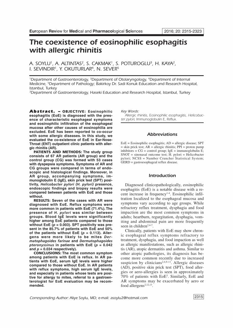
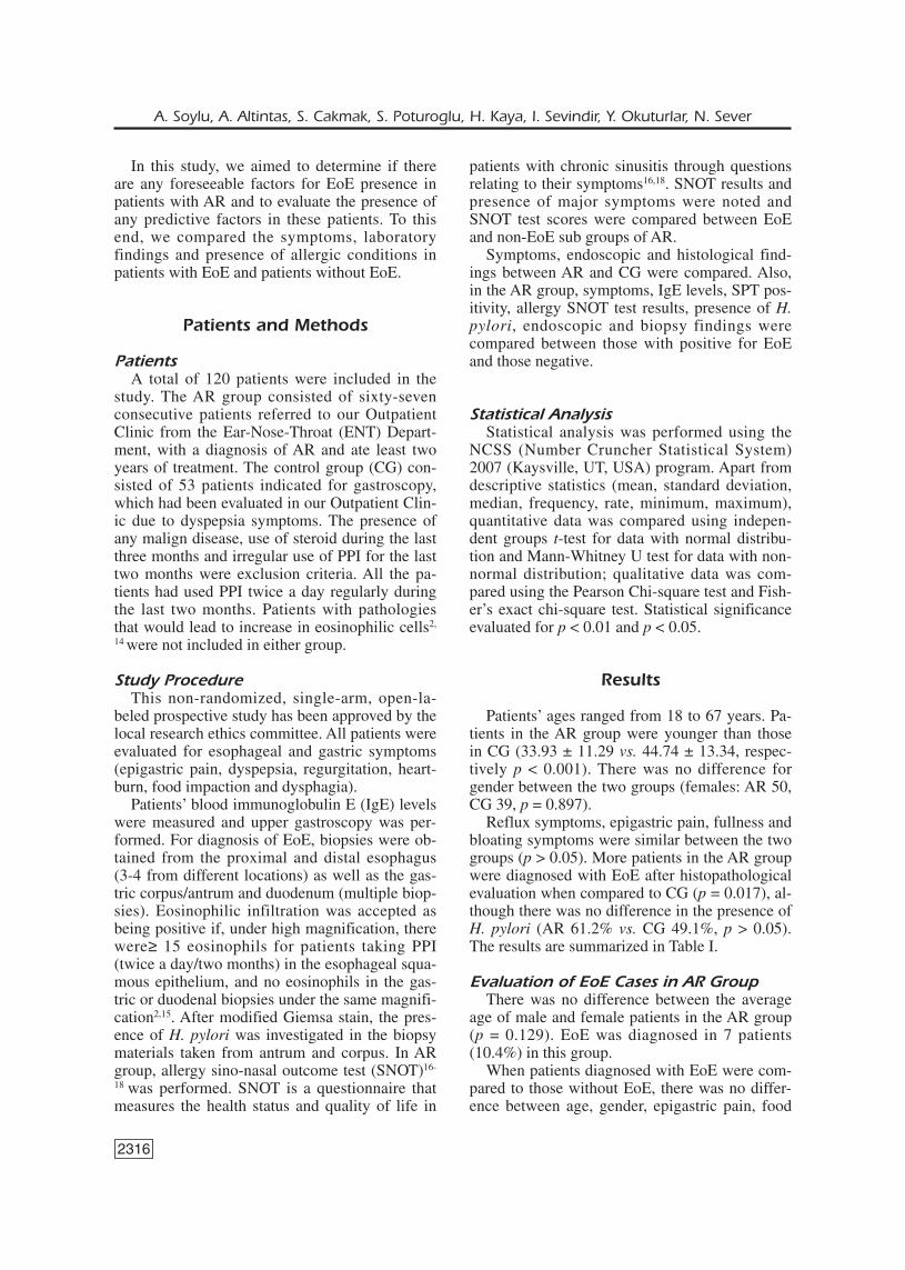
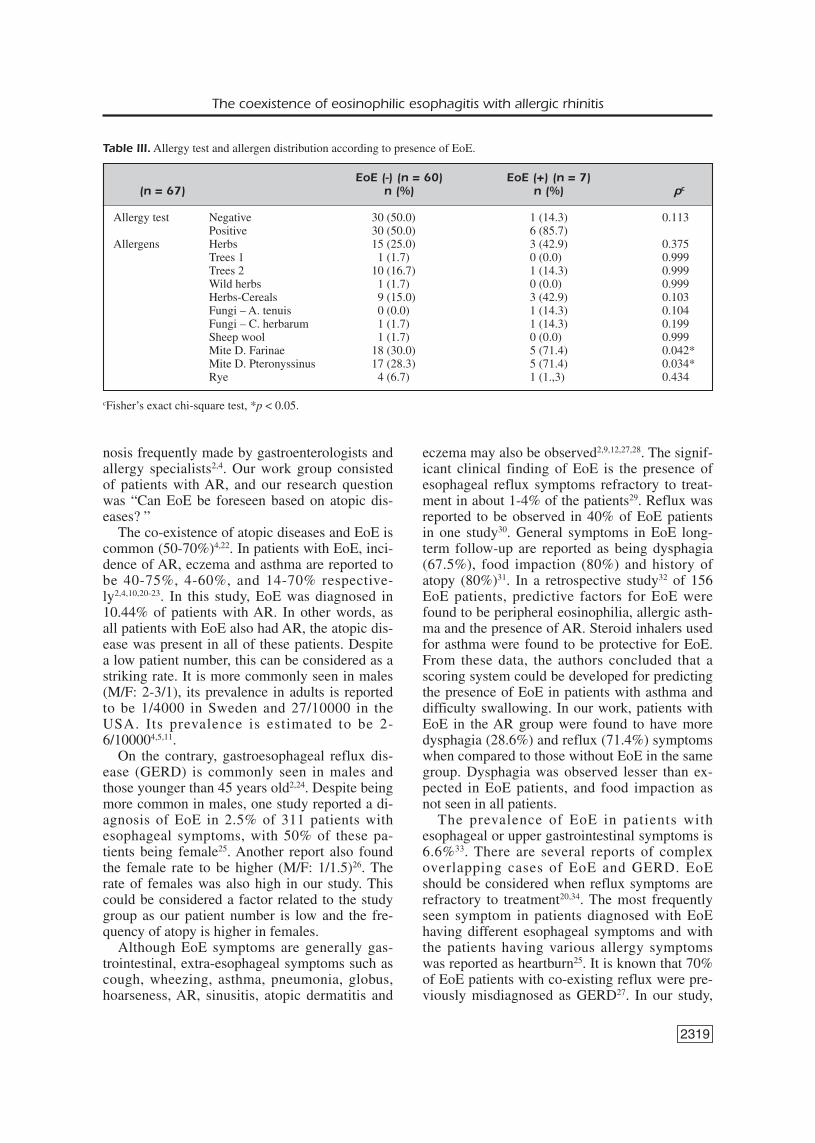
Figure 1. Endoscopic appearance of esophageal mucosawith longitudinal furrowing, mucosal white plaques, loss ofcapillary markings along the entire length of the esophagus,and mucosal white plaques in the esophagus. Figure 2.Mucosal rings.
impaction or dyspepsia symptoms (p > 0.05). Re-flux symptoms and dysphagia were more fre-quent in patients diagnosed with EoE (71.4% vs.28.3%, p = 0.042 and 28.57% vs. 1.67%, p =0.027 respectively).In patients with EoE, four (57%) had specific
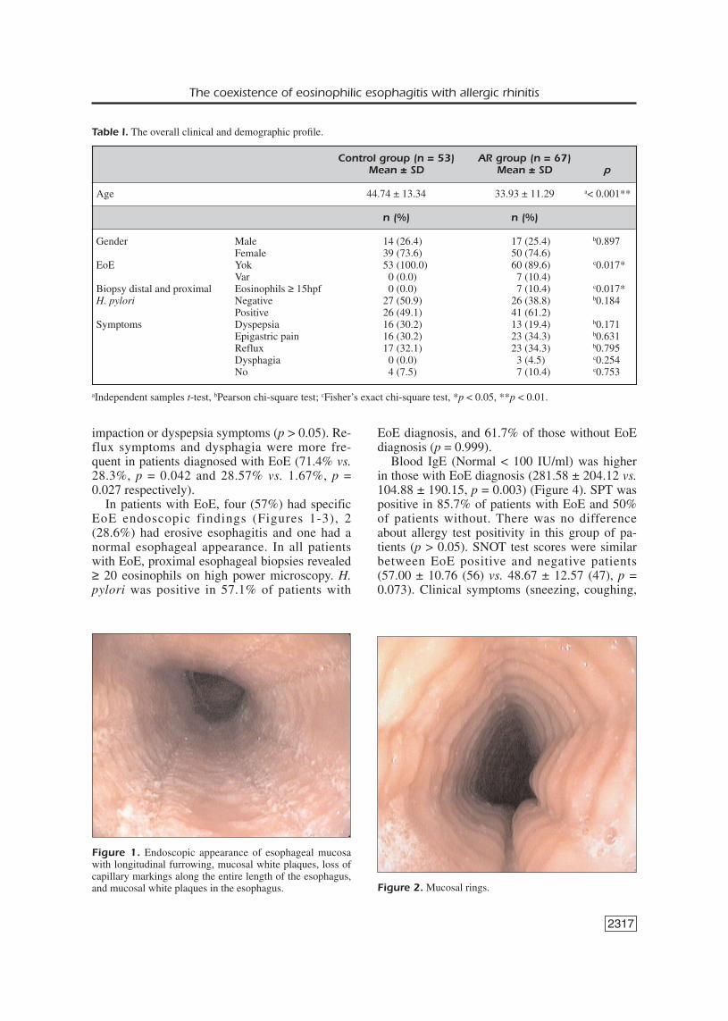
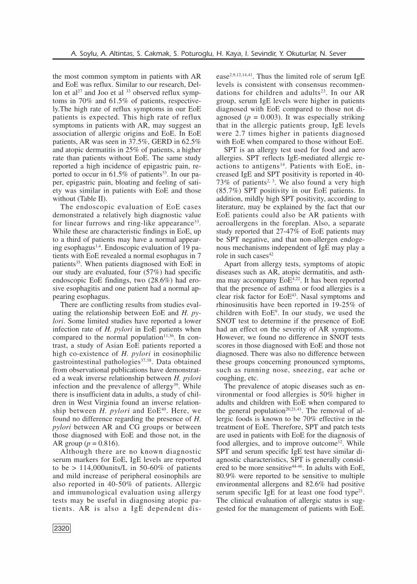
EoE endoscopic findings (Figures 1-3), 2(28.6%) had erosive esophagitis and one had anormal esophageal appearance. In all patientswith EoE, proximal esophageal biopsies revealed≥ 20 eosinophils on high power microscopy. H.pylori was positive in 57.1% of patients with
EoE diagnosis, and 61.7% of those without EoEdiagnosis (p = 0.999).Blood IgE (Normal < 100 IU/ml) was higher
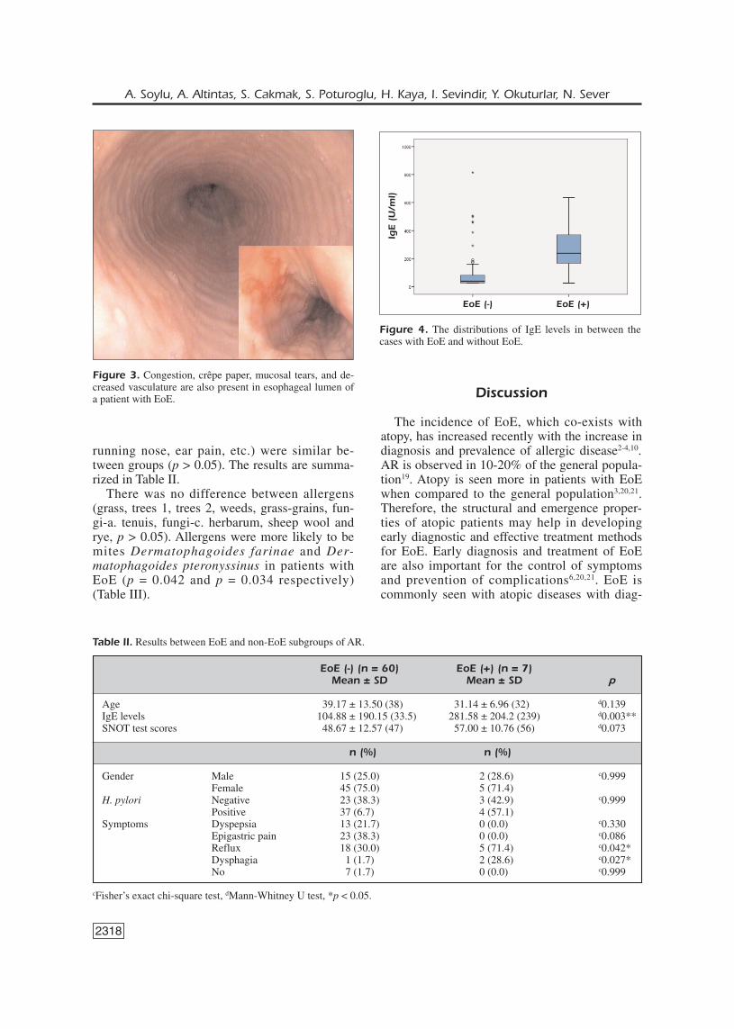
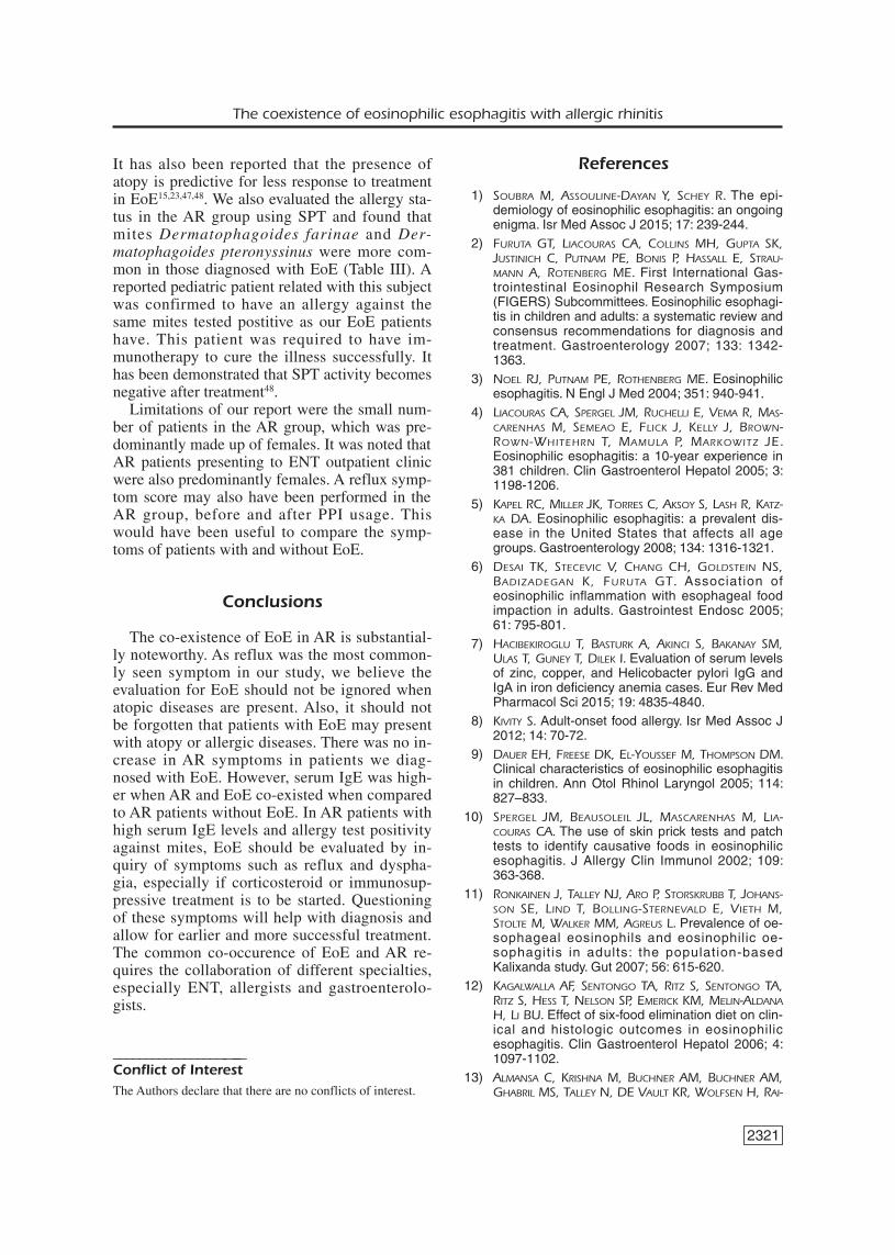
in those with EoE diagnosis (281.58 ± 204.12 vs.104.88 ± 190.15, p = 0.003) (Figure 4). SPT waspositive in 85.7% of patients with EoE and 50%of patients without. There was no differenceabout allergy test positivity in this group of pa-tients (p > 0.05). SNOT test scores were similarbetween EoE positive and negative patients(57.00 ± 10.76 (56) vs. 48.67 ± 12.57 (47), p =0.073). Clinical symptoms (sneezing, coughing,
2317
The coexistence of eosinophilic esophagitis with allergic rhinitis
2318
running nose, ear pain, etc.) were similar be-tween groups (p > 0.05). The results are summa-rized in Table II.There was no difference between allergens
(grass, trees 1, trees 2, weeds, grass-grains, fun-gi-a. tenuis, fungi-c. herbarum, sheep wool andrye, p > 0.05). Allergens were more likely to bemites Dermatophagoides farinae and Der-matophagoides pteronyssinus in patients withEoE (p = 0.042 and p = 0.034 respectively)(Table III).
Discussion
The incidence of EoE, which co-exists withatopy, has increased recently with the increase indiagnosis and prevalence of allergic disease2-4,10.AR is observed in 10-20% of the general popula-tion19. Atopy is seen more in patients with EoEwhen compared to the general population3,20,21.Therefore, the structural and emergence proper-ties of atopic patients may help in developingearly diagnostic and effective treatment methodsfor EoE. Early diagnosis and treatment of EoEare also important for the control of symptomsand prevention of complications6,20,21. EoE iscommonly seen with atopic diseases with diag-
A. Soylu, A. Altintas, S. Cakmak, S. Poturoglu, H. Kaya, I. Sevindir, Y. Okuturlar, N. Sever
Figure 3. Congestion, crêpe paper, mucosal tears, and de-creased vasculature are also present in esophageal lumen ofa patient with EoE.
Figure 4. The distributions of IgE levels in between thecases with EoE and without EoE.
IgE
(U/m
l)
EoE (-) EoE (+)
EoE (-) (n = 60) EoE (+) (n = 7)Mean ± SD Mean ± SD p
Age 39.17 ± 13.50 (38) 31.14 ± 6.96 (32) d0.139IgE levels 104.88 ± 190.15 (33.5) 281.58 ± 204.2 (239) d0.003**SNOT test scores 48.67 ± 12.57 (47) 57.00 ± 10.76 (56) d0.073
n (%) n (%)
Gender Male 15 (25.0) 2 (28.6) c0.999Female 45 (75.0) 5 (71.4)
H. pylori Negative 23 (38.3) 3 (42.9) c0.999Positive 37 (6.7) 4 (57.1)
Symptoms Dyspepsia 13 (21.7) 0 (0.0) c0.330Epigastric pain 23 (38.3) 0 (0.0) c0.086Reflux 18 (30.0) 5 (71.4) c0.042*Dysphagia 1 (1.7) 2 (28.6) c0.027*No 7 (1.7) 0 (0.0) c0.999
Table II. Results between EoE and non-EoE subgroups of AR.
cFisher’s exact chi-square test, dMann-Whitney U test, *p < 0.05.
nosis frequently made by gastroenterologists andallergy specialists2,4. Our work group consistedof patients with AR, and our research questionwas “Can EoE be foreseen based on atopic dis-eases? ”The co-existence of atopic diseases and EoE is
common (50-70%)4,22. In patients with EoE, inci-dence of AR, eczema and asthma are reported tobe 40-75%, 4-60%, and 14-70% respective-ly2,4,10,20-23. In this study, EoE was diagnosed in10.44% of patients with AR. In other words, asall patients with EoE also had AR, the atopic dis-ease was present in all of these patients. Despitea low patient number, this can be considered as astriking rate. It is more commonly seen in males(M/F: 2-3/1), its prevalence in adults is reportedto be 1/4000 in Sweden and 27/10000 in theUSA. Its prevalence is estimated to be 2-6/100004,5,11.On the contrary, gastroesophageal reflux dis-
ease (GERD) is commonly seen in males andthose younger than 45 years old2,24. Despite beingmore common in males, one study reported a di-agnosis of EoE in 2.5% of 311 patients withesophageal symptoms, with 50% of these pa-tients being female25. Another report also foundthe female rate to be higher (M/F: 1/1.5)26. Therate of females was also high in our study. Thiscould be considered a factor related to the studygroup as our patient number is low and the fre-quency of atopy is higher in females.Although EoE symptoms are generally gas-
trointestinal, extra-esophageal symptoms such ascough, wheezing, asthma, pneumonia, globus,hoarseness, AR, sinusitis, atopic dermatitis and
eczema may also be observed2,9,12,27,28. The signif-icant clinical finding of EoE is the presence ofesophageal reflux symptoms refractory to treat-ment in about 1-4% of the patients29. Reflux wasreported to be observed in 40% of EoE patientsin one study30. General symptoms in EoE long-term follow-up are reported as being dysphagia(67.5%), food impaction (80%) and history ofatopy (80%)31. In a retrospective study32 of 156EoE patients, predictive factors for EoE werefound to be peripheral eosinophilia, allergic asth-ma and the presence of AR. Steroid inhalers usedfor asthma were found to be protective for EoE.From these data, the authors concluded that ascoring system could be developed for predictingthe presence of EoE in patients with asthma anddifficulty swallowing. In our work, patients withEoE in the AR group were found to have moredysphagia (28.6%) and reflux (71.4%) symptomswhen compared to those without EoE in the samegroup. Dysphagia was observed lesser than ex-pected in EoE patients, and food impaction asnot seen in all patients.The prevalence of EoE in patients with
esophageal or upper gastrointestinal symptoms is6.6%33. There are several reports of complexoverlapping cases of EoE and GERD. EoEshould be considered when reflux symptoms arerefractory to treatment20,34. The most frequentlyseen symptom in patients diagnosed with EoEhaving different esophageal symptoms and withthe patients having various allergy symptomswas reported as heartburn25. It is known that 70%of EoE patients with co-existing reflux were pre-viously misdiagnosed as GERD27. In our study,
2319
The coexistence of eosinophilic esophagitis with allergic rhinitis
EoE (-) (n = 60) EoE (+) (n = 7)(n = 67) n (%) n (%) pc
Allergy test Negative 30 (50.0) 1 (14.3) 0.113Positive 30 (50.0) 6 (85.7)
Allergens Herbs 15 (25.0) 3 (42.9) 0.375Trees 1 1 (1.7) 0 (0.0) 0.999Trees 2 10 (16.7) 1 (14.3) 0.999Wild herbs 1 (1.7) 0 (0.0) 0.999Herbs-Cereals 9 (15.0) 3 (42.9) 0.103Fungi – A. tenuis 0 (0.0) 1 (14.3) 0.104Fungi – C. herbarum 1 (1.7) 1 (14.3) 0.199Sheep wool 1 (1.7) 0 (0.0) 0.999Mite D. Farinae 18 (30.0) 5 (71.4) 0.042*Mite D. Pteronyssinus 17 (28.3) 5 (71.4) 0.034*Rye 4 (6.7) 1 (1.,3) 0.434
Table III.Allergy test and allergen distribution according to presence of EoE.
cFisher’s exact chi-square test, *p < 0.05.
2320
the most common symptom in patients with ARand EoE was reflux. Similar to our research, Del-lon et al27 and Joo et al 33 observed reflux symp-toms in 70% and 61.5% of patients, respective-ly.The high rate of reflux symptoms in our EoEpatients is expected. This high rate of refluxsymptoms in patients with AR, may suggest anassociation of allergic origins and EoE. In EoEpatients, AR was seen in 37.5%, GERD in 62.5%and atopic dermatitis in 25% of patients, a higherrate than patients without EoE. The same studyreported a high incidence of epigastric pain, re-ported to occur in 61.5% of patients33. In our pa-per, epigastric pain, bloating and feeling of sati-ety was similar in patients with EoE and thosewithout (Table II).The endoscopic evaluation of EoE cases
demonstrated a relatively high diagnostic valuefor linear furrows and ring-like appearance33.While these are characteristic findings in EoE, upto a third of patients may have a normal appear-ing esophagus1,4. Endoscopic evaluation of 19 pa-tients with EoE revealed a normal esophagus in 7patients35. When patients diagnosed with EoE inour study are evaluated, four (57%) had specificendoscopic EoE findings, two (28.6%) had ero-sive esophagitis and one patient had a normal ap-pearing esophagus.There are conflicting results from studies eval-
uating the relationship between EoE and H. py-lori. Some limited studies have reported a lowerinfection rate of H. pylori in EoE patients whencompared to the normal population11,36. In con-trast, a study of Asian EoE patients reported ahigh co-existence of H. pylori in eosinophilicgastrointestinal pathologies37,38. Data obtainedfrom observational publications have demonstrat-ed a weak inverse relationship between H. pyloriinfection and the prevalence of allergy39. Whilethere is insufficient data in adults, a study of chil-dren in West Virginia found an inverse relation-ship between H. pylori and EoE40. Here, wefound no difference regarding the presence of H.pylori between AR and CG groups or betweenthose diagnosed with EoE and those not, in theAR group (p = 0.816).Although there are no known diagnostic
serum markers for EoE, IgE levels are reportedto be > 114,000units/L in 50-60% of patientsand mild increase of peripheral eosinophils arealso reported in 40-50% of patients. Allergicand immunological evaluation using allergytests may be useful in diagnosing atopic pa-tients. AR is also a IgE dependent dis-
ease2,9,12,14,41. Thus the limited role of serum IgElevels is consistent with consensus recommen-dations for children and adults23. In our ARgroup, serum IgE levels were higher in patientsdiagnosed with EoE compared to those not di-agnosed (p = 0.003). It was especially strikingthat in the allergic patients group, IgE levelswere 2.7 times higher in patients diagnosedwith EoE when compared to those without EoE.SPT is an allergy test used for food and aero
allergies. SPT reflects IgE-mediated allergic re-actions to antigens14. Patients with EoE, in-creased IgE and SPT positivity is reported in 40-73% of patients2, 3. We also found a very high(85.7%) SPT positivity in our EoE patients. Inaddition, mildly high SPT positivity, according toliterature, may be explained by the fact that ourEoE patients could also be AR patients withaeroallergens in the foreplan. Also, a separatestudy reported that 27-47% of EoE patients maybe SPT negative, and that non-allergen endoge-nous mechanisms independent of IgE may play arole in such cases42.Apart from allergy tests, symptoms of atopic
diseases such as AR, atopic dermatitis, and asth-ma may accompany EoE4,22. It has been reportedthat the presence of asthma or food allergies is aclear risk factor for EoE43. Nasal symptoms andrhinosinusitis have been reported in 19-25% ofchildren with EoE9. In our study, we used theSNOT test to determine if the presence of EoEhad an effect on the severity of AR symptoms.However, we found no difference in SNOT testsscores in those diagnosed with EoE and those notdiagnosed. There was also no difference betweenthese groups concerning pronounced symptoms,such as running nose, sneezing, ear ache orcoughing, etc.The prevalence of atopic diseases such as en-
vironmental or food allergies is 50% higher inadults and children with EoE when compared tothe general population20,21,41. The removal of al-lergic foods is known to be 70% effective in thetreatment of EoE. Therefore, SPT and patch testsare used in patients with EoE for the diagnosis offood allergies, and to improve outcome12. WhileSPT and serum specific IgE test have similar di-agnostic characteristics, SPT is generally consid-ered to be more sensitive44-46. In adults with EoE,80.9% were reported to be sensitive to multipleenvironmental allergens and 82.6% had positiveserum specific IgE for at least one food type21.The clinical evaluation of allergic status is sug-gested for the management of patients with EoE.
A. Soylu, A. Altintas, S. Cakmak, S. Poturoglu, H. Kaya, I. Sevindir, Y. Okuturlar, N. Sever
It has also been reported that the presence ofatopy is predictive for less response to treatmentin EoE15,23,47,48. We also evaluated the allergy sta-tus in the AR group using SPT and found thatmites Dermatophagoides farinae and Der-matophagoides pteronyssinus were more com-mon in those diagnosed with EoE (Table III). Areported pediatric patient related with this subjectwas confirmed to have an allergy against thesame mites tested postitive as our EoE patientshave. This patient was required to have im-munotherapy to cure the illness successfully. Ithas been demonstrated that SPT activity becomesnegative after treatment48.Limitations of our report were the small num-
ber of patients in the AR group, which was pre-dominantly made up of females. It was noted thatAR patients presenting to ENT outpatient clinicwere also predominantly females. A reflux symp-tom score may also have been performed in theAR group, before and after PPI usage. Thiswould have been useful to compare the symp-toms of patients with and without EoE.
Conclusions
The co-existence of EoE in AR is substantial-ly noteworthy. As reflux was the most common-ly seen symptom in our study, we believe theevaluation for EoE should not be ignored whenatopic diseases are present. Also, it should notbe forgotten that patients with EoE may presentwith atopy or allergic diseases. There was no in-crease in AR symptoms in patients we diag-nosed with EoE. However, serum IgE was high-er when AR and EoE co-existed when comparedto AR patients without EoE. In AR patients withhigh serum IgE levels and allergy test positivityagainst mites, EoE should be evaluated by in-quiry of symptoms such as reflux and dyspha-gia, especially if corticosteroid or immunosup-pressive treatment is to be started. Questioningof these symptoms will help with diagnosis andallow for earlier and more successful treatment.The common co-occurence of EoE and AR re-quires the collaboration of different specialties,especially ENT, allergists and gastroenterolo-gists.
–––––––––––––––––-––––Conflict of InterestThe Authors declare that there are no conflicts of interest.
References
1) SOUBRA M, ASSOULINE-DAYAN Y, SCHEY R. The epi-demiology of eosinophilic esophagitis: an ongoingenigma. Isr Med Assoc J 2015; 17: 239-244.
2) FURUTA GT, LIACOURAS CA, COLLINS MH, GUPTA SK,JUSTINICH C, PUTNAM PE, BONIS P, HASSALL E, STRAU-MANN A, ROTENBERG ME. First International Gas-trointestinal Eosinophil Research Symposium(FIGERS) Subcommittees. Eosinophilic esophagi-tis in children and adults: a systematic review andconsensus recommendations for diagnosis andtreatment. Gastroenterology 2007; 133: 1342-1363.
3) NOEL RJ, PUTNAM PE, ROTHENBERG ME. Eosinophilicesophagitis. N Engl J Med 2004; 351: 940-941.
4) LIACOURAS CA, SPERGEL JM, RUCHELLI E, VEMA R, MAS-CARENHAS M, SEMEAO E, FLICK J, KELLY J, BROWN-ROWN-WHITEHRN T, MAMULA P, MARKOWITZ JE .Eosinophilic esophagitis: a 10-year experience in381 children. Clin Gastroenterol Hepatol 2005; 3:1198-1206.
5) KAPEL RC, MILLER JK, TORRES C, AKSOY S, LASH R, KATZ-KA DA. Eosinophilic esophagitis: a prevalent dis-ease in the United States that affects all agegroups. Gastroenterology 2008; 134: 1316-1321.
6) DESAI TK, STECEVIC V, CHANG CH, GOLDSTEIN NS,BADIZADEGAN K, FURUTA GT . Association ofeosinophilic inflammation with esophageal foodimpaction in adults. Gastrointest Endosc 2005;61: 795-801.
7) HACIBEKIROGLU T, BASTURK A, AKINCI S, BAKANAY SM,ULAS T, GUNEY T, DILEK I. Evaluation of serum levelsof zinc, copper, and Helicobacter pylori IgG andIgA in iron deficiency anemia cases. Eur Rev MedPharmacol Sci 2015; 19: 4835-4840.
8) KIVITY S. Adult-onset food allergy. Isr Med Assoc J2012; 14: 70-72.
9) DAUER EH, FREESE DK, EL-YOUSSEF M, THOMPSON DM.Clinical characteristics of eosinophilic esophagitisin children. Ann Otol Rhinol Laryngol 2005; 114:827–833.
10) SPERGEL JM, BEAUSOLEIL JL, MASCARENHAS M, LIA-COURAS CA. The use of skin prick tests and patchtests to identify causative foods in eosinophilicesophagitis. J Allergy Clin Immunol 2002; 109:363-368.
11) RONKAINEN J, TALLEY NJ, ARO P, STORSKRUBB T, JOHANS-SON SE, LIND T, BOLLING-STERNEVALD E, VIETH M,STOLTE M, WALKER MM, AGREUS L. Prevalence of oe-sophageal eosinophils and eosinophilic oe-sophagitis in adults: the population-basedKalixanda study. Gut 2007; 56: 615-620.
12) KAGALWALLA AF, SENTONGO TA, RITZ S, SENTONGO TA,RITZ S, HESS T, NELSON SP, EMERICK KM, MELIN-ALDANA
H, LI BU. Effect of six-food elimination diet on clin-ical and histologic outcomes in eosinophilicesophagitis. Clin Gastroenterol Hepatol 2006; 4:1097-1102.
13) ALMANSA C, KRISHNA M, BUCHNER AM, BUCHNER AM,GHABRIL MS, TALLEY N, DE VAULT KR, WOLFSEN H, RAI-
2321
The coexistence of eosinophilic esophagitis with allergic rhinitis
2322
MONDO M, GUARDERAS JC, ACHEM SR. Seasonal dis-tribution in newly diagnosed cases of eosinophilicesophagitis in adults. Am J Gastroenterol 2009;104: 828-833.
14) DELLON ES. Eosinophilic esophagitis: diagnostictests and criteria. Curr Opin Gastroenterol 2012;28: 382-388.
15) DELLON ES, GONSALVES N, HIRANO I, FURUTA GT, LIA-COURAS CA, KATZKA DA. ACG clinical guideline: Evi-denced based approach to the diagnosis andmanagement of esophageal eosinophilia andeosinophilic esophagitis (EoE). Am J Gastroen-terol 2013; 108: 679-692.
16) PICCIRILLO JF, MERRITT MG JR, RICHARDS ML. Psycho-metric and clinimetric validity of the 20-Item Sino-Nasal Outcome Test (SNOT-20). OtolaryngolHead Neck Surg 2002; 126: 41-47.
17) PYNNONEN MA, KIM HM, TERRELL JE. Validation ofthe Sino-Nasal Outcome Test 20 (SNOT-20) do-mains in nonsurgical patients. Am J Rhinol Aller-gy 2009; 23: 40-45.
18) VAN OENE CM, VAN REIJ EJ, SPRANGERS MA, SPRANGERS
MA, FOKKENS WJ. Quality-assessment of disease-specific quality of life questionnaires for rhinitisand rhinosinusitis: A systematic review. Allergy2007; 62: 1359-1371.
19) DYKEWICZ MS, HAMILOS DL. Rhinitis and sinusitis. JAllergy Clin Immunol 2010; 125(Suppl 2): 103-115.
20) BROWN-WHITEHORN TF, SPERGEL JM. The link betweenallergies and eosinophilic esophagitis: implica-tions for management strategies. Expert Rev ClinImmunol 2010; 6: 101-109.
21) ROY-GHANTA S, LAROSA DF, KATZKA DA. Atopic char-acteristics of adult patients with eosinophilicesophagitis. Clin Gastroenterol Hepatol 2008; 6:531-535.
22) SIMON D, MARTI H, HEER P, SIMON HU, BRAATHEN LR,STRAUMANN A. Eosinophilic esophagitis is frequent-ly associated with IgE-mediated allergic airwaydiseases. J Allergy Clin Immunol 2005; 115:1090-1092.
23) LIACOURAS CA, FURUTA GT, HIRANO I, HIRANO I,ATKINS D, ATTWOOD SE, BONIS PA, BURKS AW,CHEHADE M, COLLINS MH, DELLON ES, DOHIL R,FALK GW, GONSALVES N, GUPTA SK, KATZKA DA, LU-CENDO AJ, MARKOWITZ JE, NOEL RJ, LUCENDO AJ,MARKOWITZ JE, NOEL RJ, ODZE RD, PUTNAM PE,RICHTER JE, ROMERO Y, RUCHELLI E, SAMPSON HA,SCHOEPFER A, SHAHEEN NJ, SICHERER SH, SPECHLER S,SPERGEL JM, STRAUMANN A, WERSHIL BK, ROTHEN-BERG ME, ACEVES SS. Eosinophilic esophagitis:updated consensus recommendations for chil-dren and adults. J Allergy Clin Immunol 2011;128: 3-20.
24) MULDER DJ, HURLBUT DJ, NOBLE AJ, JUSTINICH CJ.Clinical features distinguish eosinophilic and re-flux-induced esophagitis. J Pediatr GastroenterolNutr 2013; 56: 263-270.
25) ALTUN R, AKBAS E, YILDIRIM AE, OCAL S, KORKMAZ M,SELCUK H. Frequency of eosinophilic esophagitis in
patients with esophageal symptoms: a single cen-ter Turkish experience. Dis Esophagus 2013; 26:776-781.
26) FOROUTAN M, NOROUZI A, MOLAEI M, MIRBAGHERI SA,IRVANI S, SADEGHI A, DERAKHSHAN F, TAVASSOLI S, BE-SHARAT S, ZALI M. Eosinophilic esophagitis in pa-tients with refractory gastroesophageal reflux dis-ease. Dig Dis Sci 2010; 55: 28-31.
27) DELLON ES, GIBBS WB, FRITCHIE KJ, FRITCHIE KJ, RUBI-NAS TC, WILSON LA, WOOSLEY JT, SHAHEEN NJ. Clini-cal, endoscopic, and histologic findings distin-guish eosinophilic esophagitis from gastroe-sophageal reflux disease. Clin Gastroenterol He-patol 2009; 7: 1305-1313.
28) THOMPSON DM, ARORA AS, ROMERO Y, DAUER EH.Eosinophilic esophagitis: its role in aerodigestivetract disorders. Otolaryngol Clin North Am 2006;39: 205-221.
29) GARCÍA-COMPEÁN D, GONZÁLEZ GONZÁLEZ JA, MARRU-FO GARCÍA CA, FLORES GUTIERREZ JP, BARBOZA QUIN-TANA O, GALINDO RODRIGUEZ G, MAR RUIZ MA, DE
LEON VALDEZ D, JAQUEZ QUINTANA JO, MALDONADO
GARZA HJ. Prevalence of eosinophilic esophagitisin patients with refractory gastroesophageal refluxdisease symptoms: a prospective study. Dig LiverDis 2011; 43: 204-208.
30) REMEDIOS M, CAMPBELL C, JONES DM, KERLIN P.Eosinophilic esophagitis in adults: clinical, endo-scopic, histologic findings, and response to treat-ment with fluticasone propionate. Gastrointest En-dosc 2006; 63: 3-12.
31) ARIAS Á, LUCENDO AJ. Prevalence of eosinophilicoesophagitis in adult patients in a central regionof Spain. Eur J Gastroenterol Hepatol 2013; 25:208-212.
32) HARER KN, ENDERS FT, LIM KG, ALEXANDER JA, KATZKADA. An allergic phenotype and the use of steroidinhalers predict eosinophilic oesophagitis in pa-tients with asthma. Aliment Pharmacol Ther 2013;37: 107-113.
33) JOO MK, PARK JJ, KIM SH, KIM KH, JUNG W, YUN JW,LEE BJ, KIM JH, YEON JE, KIM JS, BYUN KS, LEE SW,BAK YT. Prevalence and endoscopic features ofeosinophil ic esophagit is in patients withesophageal or upper gastrointestinal symptoms. JDig Dis 2012; 13: 296-303.
34) SPECHLER SJ, GENTA RM, SOUZA RF. Thoughts on thecomplex relationship between gastroesophagealreflux disease and eosinophilic esophagitis. Am JGastroenterol 2007; 102: 1301-1306.
35) STRAUMANN A, BEGLINGER C. Eosinophilic esophagi-tis: the endoscopist's enigma. Gastrointest En-dosc 2006; 63: 13-15.
36) DELLON ES, PEERY AF, SHAHEEN NJ, MORGAN DR,HURRELL JM, LASH RH, GENTA RM. Inverse associa-tion of esophageal eosinophilia with Helicobac-ter pylori based on analysis of a US pathologydatabase. Gastroenterology 2011; 141: 1586-1592.
37) FURUTA K, ADACHI K, AIMI M, ISHIMURA N, SATO S, ISHI-HARA S, KINOSHITA Y. Case-control study of associa-
A. Soylu, A. Altintas, S. Cakmak, S. Poturoglu, H. Kaya, I. Sevindir, Y. Okuturlar, N. Sever
tion of eosinophilic gastrointestinal disorders withHelicobacter pylori infection in Japan. J ClinBiochem Nutr 2013; 53: 60-62.
38) ZHANG L, DUAN L, DING S, LU J, JIN Z, CUI R, MCNUTT
M, WANG A. Eosinophilic gastroenteritis: clinicalmanifestations and morphological characteristics,a retrospective study of 42 patients. Scand J Gas-troenterol 2011; 46: 1074-1080.
39) DAUGULE I, ZAVORONKOVA J, SANTARE D. Helicobacterpylori and allergy: Update of research. World JMethodol 2015; 5: 203-211.
40) ELITSUR Y, ALRAZZAK BA, PRESTON D. Does Heli-cobacter pylori protect against eosinophilicesophagitis in children? Helicobacter 2014; 19:367-371.
41) ERWIN EA, JAMES HR, GUTEKUNST HM, RUSSO JM,KELLEHER KJ, PLATTS-MILLS TA. Serum IgE measure-ment and detection of food allergy in pediatric pa-tients with eosinophilic esophagitis. Ann AllergyAsthma Immunol 2010; 104: 496-502.
42) SGOUROS SN, BERGELE C, MANTIDES A. Eosinophilicesophagitis in adults: what is the clinical signifi-cance? Endoscopy 2006; 38: 515-520.
43) MACKENZIE SH, GO M, CHADWICK B, THOMAS K, FANG
J, KUWADA S, LAMPHIER S, HILDEN K, PETERSON K.Eosinophilic oesophagitis in patients presentingwith dysphagia--a prospective analysis. AlimentPharmacol Ther 2008; 28: 1140-1146.
44) SEIDMAN MD, GURGEL RK, LIN SY, SCHWARTZ SR, BA-ROODY FM, BONNER JR, DAWSON DE, DYKEWICZ MS,HACKELL JM, HAN JK, ISHMAN SL, KROUSE HJ,MALEKZADEH S, MIMS JW, OMOLE FS, REDDY WD, WAL-
LACE DV, WALSH SA, WARREN BE, WILSON MN, NA-CHETA LC; GUIDELINE OTOLARYNGOLOGY DEVELOPMENT
GROUP. AAO-HNSF. Guideline Otolaryngology De-velopment Group. AAO-HNSF. Clinical practiceguideline: Allergic rhinitis. Otolaryngol Head NeckSurg 2015; 152(Supp l): S1-43.
45) BERNSTEIN IL, LI JT, BERNSTEIN DI, HAMILTON R, SPECTOR
SL, TAN R, SICHERER S, GOLDEN DB, KHAN DA, NICKLAS
RA, PORTNOY JM, BLESSING-MOORE J, COX L, LANG
DM, OPPENHEIMER J, RANDOLPH CC, SCHULLER DE,TILLES SA, WALLACE DV, LEVETIN E, WEBER R; AMERICAN
ACADEMY OF ALLERGY, ASTHMA AND IMMUNOLOGY; AMERI-CAN COLLEGE OF ALLERGY, ASTHMA AND IMMUNOLOGY.Allergy diagnostic testing: an updated practice pa-rameter. Ann Allergy Asthma Immunol 2008;100(Suppl 3): S1-148.
46) TSCHOPP J, SISTEK D, SCHINDLER C, SCHINDLER C, LEUEN-BERGER P, PERRUCHOUD AP, WUTHRICH B, BRUTSCHE M,ZELLWEGER JP, KARRERV W, BRANDLI O. Current aller-gic asthma and rhinitis: diagnostic efficiency ofthree commonly used atopic markers (IgE, skinprick tests, and Phadiatop). Results from 8329randomized adults from the SAPALDIA Study.Swiss Study on Air Pollution and Lung Diseasesin Adults. Allergy 1998; 53: 608-613.
47) NOEL RJ, PUTNAM PE, COLLINS MH, ASSA’AD AH, GUA-JARDO JR, JAMESON SC, ROTHENBERG ME. Clinical andimmunopathologic effects of swallowed fluticas-one for eosinophilic esophagitis. Clin Gastroen-terol Hepatol 2004; 2: 568-575.
48) RAMIREZ RM, JACOBS RL. Eosinophilic esophagitistreated with immunotherapy to dust mites. J Aller-gy Clin Immunol 2013; 132: 503-504.
2323
The coexistence of eosinophilic esophagitis with allergic rhinitis
![ARTICLE IN PRESS - UPCommons · 2 R. Jerez-Mesa et al. / Mechatronics 000 (2016) 1–8 ARTICLE IN PRESS JID: MECH [m5G;April 28, 2016;20:12] Fig. 1. Schematic representation of a](https://img.pdfslide.us/doc/110x75/5c77db8409d3f23a068c341e/article-in-press-upcommons-2-r-jerez-mesa-et-al-mechatronics-000-2016.jpg)


![ARTICLE IN PRESS - COnnecting REpositories · ARTICLE IN PRESS JID: NME [m5G;June 20, 2016;20:53] ... HHF tests performed at GLADIS indicated that the W-10Cr-2Ti alloy is able to](https://img.pdfslide.us/doc/110x75/5e927f061b0bc068346f4a9e/article-in-press-connecting-repositories-article-in-press-jid-nme-m5gjune-20.jpg)


![ARTICLE IN PRESS · V. Jalili et al. / Information Sciences 000 (2016) 1–20 3 ARTICLE IN PRESS JID: INS [m3Gsc;September 1, 2016;20:57] Fig. 2. Two applications’ design of Di3](https://img.pdfslide.us/doc/110x75/608181be86d33a08d65a25d3/article-in-v-jalili-et-al-information-sciences-000-2016-1a20-3-article-in.jpg)