Embed Size (px)
Citation preview

Park et al. Appl Biol Chem (2021) 64:14 https://doi.org/10.1186/s13765-020-00587-x
ARTICLE
Optimal bioconversion for compound K production from red ginseng root (C.A. Mayer) by sequential enzymatic hydrolysis and its characteristicsYeong‑Ju Park1, Unsik Hwang1, Suyeon Park1, Sol Sim1, Soyeon Jeong1, Misun Park1, Minji Kang1, Youngsoo Lee1, Youngju Song2, Hoon Park1 and Hee‑Jae Suh1*
Abstract
Compound K (CK; 20‑O‑β‑(d‑glucopyranosyl)‑20(S)‑protopanaxadiol) is one of the metabolites of ginsenosides contained in red ginseng (RG) and is known to have high bioavailability. This study aimed to establish the optimal conditions for enzyme treatment to convert ginsenosides from RG extract to CK, and to prove the characteristics of bioconverted red ginseng (BRG) extract. CK was not detected in unenzyme‑treated RG extract, and in the single‑step enzyme treatment, it was produced at less than 4.58 mg/g only in treatment group with Pyr‑flo or Sumizyme AC (at 50 °C for 48 h). The highest yield of CK (14.32 mg/g) was obtained by Ultimase MFC treatment at 50 °C for 48 h after treatment with a mixture of Pyr‑flo and Rapidase at 50 °C for 24 h. Total polyphenol, 2,2‑diphenyl‑1‑picrylhydrazyl (DPPH), and 2,2‑azinobis‑(3‑ethylbenzothiazoline‑6‑sulfonic acid)) (ABTS) radical scavenging activity were higher in BRG than in RG (p < 0.5). High‑fat diet (HD) rat fed 1% BRG had significantly lower body weight, heart weight, fat pads (periosteal fat, epididymal fat), serum glucose levels, and hepatic triglyceride levels than those HD rat fed 1% RG (p < 0.05). In conclusion, the sequential enzymatic bioconversion was produces higher CK in RG root extract than single‑step enzyme treatment.
Keywords: Compound K, Enzymatic bioconversion, Ginsenoside, Red ginseng root
© The Author(s) 2021. This article is licensed under a Creative Commons Attribution 4.0 International License, which permits use, sharing, adaptation, distribution and reproduction in any medium or format, as long as you give appropriate credit to the original author(s) and the source, provide a link to the Creative Commons licence, and indicate if changes were made. The images or other third party material in this article are included in the article’s Creative Commons licence, unless indicated otherwise in a credit line to the material. If material is not included in the article’s Creative Commons licence and your intended use is not permitted by statutory regulation or exceeds the permitted use, you will need to obtain permission directly from the copyright holder. To view a copy of this licence, visit http://creat iveco mmons .org/licen ses/by/4.0/.
IntroductionKorean ginseng, Panax ginseng C. A. Meyer, has been used as herbal medicine in East Asian countries for thou-sands of years. Red ginseng (RG) is produced by steam-ing and drying raw ginseng, and it is edible. RG has been reported to have various pharmacological effects such as anti-influenza, antitumor, immunomodulation, anti-dia-betes, and antioxidant activity; these beneficial effects are due to the composition of ginsenosides in RG [1–3]. The
dammarane-type triterpenoid saponins of RG (including raw ginseng) can be classified into two major types pro-topanaxadiol (PPD) and protopanaxatriol (PPT) based on the position and number of sugar moieties attached to an aglycone [4]. PPD-type ginsenosides include Rb1, Rb2, Rc, Rd, Rg2, Rg3, Ph2, and compound K [CK; 20-O-β-(d-glucopyranosyl)-20(S)-protopanaxadiol], while PPT-type ginsenosides include Re, Rf, Rg1, Rg2, Rh1, and F1.
Rb1, Rb2, and Rc of PPD series, and Re, Rf, and Rg1 of PPT series account for > 90% of the total ginsenoside content in RG, but it has been reported to have low bioavailability due to high polarity [3, 5, 6]. However, Kim and Lee et al. reported that although major gin-senosides are not directly absorbed by the body, they can be pharmacologically active when metabolized by
Open Access
*Correspondence: [email protected] Research Center for Food and Bio Convergence, Department of Food Science, Sun Moon University, Asan, Chungchengnam‑do, 31460, Republic of KoreaFull list of author information is available at the end of the article
Page 2 of 11Park et al. Appl Biol Chem (2021) 64:14
intestinal microbes and absorbed into the blood [7, 8]. Among deglycosylated ginsenosides, CK has been reported to be a major pharmacologically active metab-olite found in the human bloodstream after oral admin-istration of RG [8–10]. Recent studies show that CK exhibits multiple pharmacological properties includ-ing anti-inflammatory, anticarcinogenic, antiallergic, antidiabetic, antiaging, and hepatoprotective effects [3]. As this material has been used safely for thousands of years, CK has recently gained interest as a potential therapeutic agent for many diseases. The PPD-type ginsenosides Rb1, Rb2, and Rc are major ginsenosides that can be converted to CK after ingestion of RG. The degree of CK conversion depends on the action of enzymes secreted by intestinal microbes [7]. However, the capacity for deglycosylation of ginsenosides in the intestine varies depending on the number of intestinal microorganisms, and, therefore, CK conversion ability varies from individual to individual [8, 9].
CK is not contained in RG itself and is produced only when PPD series ginsenosides are deglycosylated; CK may be produced by processing RG before it is ingested. Various methods have been used for deglycosylation of major ginsenosides for CK generation. Some require harsh chemical treatments such as acid or alkaline hydrolysis. These methods are accompanied by inevi-table and undesirable side reactions including epimeri-zation, hydration, and hydroxylation [10]. Ginsenoside hydrolysis using edible enzymes has been recognized as a promising method for CK production because it exhib-its characteristics such as a short reaction cycle, high specificity, higher purity, and minimal pollution. Several studies have reported enzymatic biotransformation of ginseng with the application of commercial enzymatic preparations or microbial enzymes [3, 9, 11–13]. How-ever, most studies used refined ginsenosides; only a few have used RG extract, especially RG root extract. In addi-tion, in previous studies, crude microbial enzymes or a single commercial enzyme were used, resulting in a low yield of CK. However, it is expected that the use of mul-tiple and sequential enzymatic bioconversion based on the aglycon specificity of each enzyme could improve CK production. In this study, in order to increase the conver-sion from ginsenosides of red ginseng extract to CK, the optimal enzymatic conditions for conversion to CK in RG root extract were investigated using multiple and sequen-tial treatment of commercially available edible enzymes. We also confirmed the antioxidant and biological prop-erties of extract of CK-rich bioconverted RG (BRG) root. The commercially available enzyme mixture used in this study has been approved for food processing, so the enzymatic method for producing ginsenoside CK can be safely applied to food production.
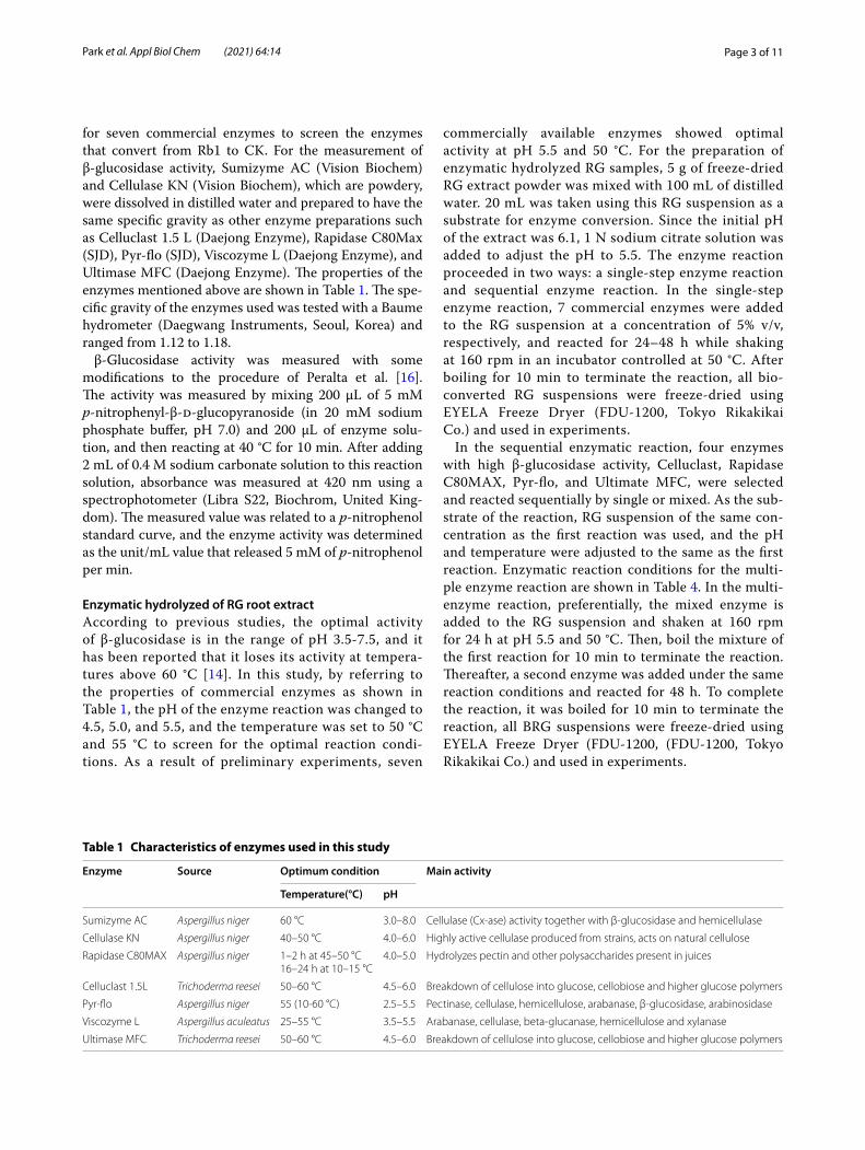
Materials and methodsMaterialsFour-year-old or 6-year-old RG was purchased from a ginseng market in Geumsan, Korea. Standard ginseno-sides (Rb1, Rd, Rg3, Rh2, F2, and CK) were purchased from Dongmyung Scientific, Inc. (Seoul, Korea). Water and acetonitrile (high-performance liquid chromatog-raphy [HPLC] grade) were purchased by J. T. Baker (PA, USA). Sodium citrate, sodium carbonate, and sulfuric acid were obtained from Sigma-Aldrich (St. Louis, MO, USA) and Daejung Chemicals Co. (Siheung, Korea). 60F264 silica gel plates for thin-layer chromatography (TLC) were purchased from Merck (Darmstadt, Ger-many). 2,2-Azino-bis (3-ethylbenzothiazoline-6-sulfonic acid) (ABTS), 2,2-di-phenyl-1-picrylhydrazyl (DPPH), Folin-Ciocalteu phenol reagent were purchased from Sigma-Aldrich. We obtained Sumizyme AC (Vision Biochem, Seongnam, Korea), Cellulase KN (Vision Bio-chem), Rapidase C80 MAX (SJD, Seongnam, Korea), Celluclast 1.5 L (Daejong Enzyme, Seoul, Korea), Pyr-flo (SJD), Viscozyme L (Daejong Enzyme), and Ultimase MFC (Daejong Enzyme). Commercial kits for animal tests were purchased from Asan Pharmaceutical Co. (Asan, Korea).
Extraction of ginsenosides from RG rootThe ginsenoside Rb1 is a major ginsenoside of the PPD type. Recent studies have shown that Rb1 is hydrolyzed to ginsenoside Rd, then deglycosylated to ginsenoside F2, and further converted to CK [3, 13]. Therefore, gin-senosides Rb1 and Rd are important candidates for preparation of CK. To determine the optimal extrac-tion conditions for obtaining high concentrations of Rb1 and Rd, 20 g of dried RG roots was added to 200 mL of hot water or 10%, 30%, 50%, and 70% ethanol solution, respectively, and was extracted at 80 °C for 24 h and 48 h. The Rb1 content of each extract was in the range 1.19 to 1.63 mg/g; it was 1.63 and 1.62 mg/g, respectively, in the water extract and 70% ethanol extract for 48 h. The Rd content extracted for 48 h was also highest in the water extract and the 70% ethanol extract (0.31 mg/g in each). Considering food safety and industrial economy, the experiment was conducted by extracting ginseno-side with water at 80 °C for 48 h. The RG root extract was freeze-dried, stored in a refrigerator, and used for experiments.
Measurement of β‑glucosidase activity of commercial enzyme preparationsSince various glycosidases participate in the bioconver-sion of ginsenoside Rb1, enzymes with glycosidase activ-ity that can hydrolyze Rb1 must be selected first. [14, 15]. This study measured the activity of β-glucosidase
Page 3 of 11Park et al. Appl Biol Chem (2021) 64:14
for seven commercial enzymes to screen the enzymes that convert from Rb1 to CK. For the measurement of β-glucosidase activity, Sumizyme AC (Vision Biochem) and Cellulase KN (Vision Biochem), which are powdery, were dissolved in distilled water and prepared to have the same specific gravity as other enzyme preparations such as Celluclast 1.5 L (Daejong Enzyme), Rapidase C80Max (SJD), Pyr-flo (SJD), Viscozyme L (Daejong Enzyme), and Ultimase MFC (Daejong Enzyme). The properties of the enzymes mentioned above are shown in Table 1. The spe-cific gravity of the enzymes used was tested with a Baume hydrometer (Daegwang Instruments, Seoul, Korea) and ranged from 1.12 to 1.18.
β-Glucosidase activity was measured with some modifications to the procedure of Peralta et al. [16]. The activity was measured by mixing 200 μL of 5 mM p-nitrophenyl-β-d-glucopyranoside (in 20 mM sodium phosphate buffer, pH 7.0) and 200 μL of enzyme solu-tion, and then reacting at 40 °C for 10 min. After adding 2 mL of 0.4 M sodium carbonate solution to this reaction solution, absorbance was measured at 420 nm using a spectrophotometer (Libra S22, Biochrom, United King-dom). The measured value was related to a p-nitrophenol standard curve, and the enzyme activity was determined as the unit/mL value that released 5 mM of p-nitrophenol per min.
Enzymatic hydrolyzed of RG root extractAccording to previous studies, the optimal activity of β-glucosidase is in the range of pH 3.5-7.5, and it has been reported that it loses its activity at tempera-tures above 60 °C [14]. In this study, by referring to the properties of commercial enzymes as shown in Table 1, the pH of the enzyme reaction was changed to 4.5, 5.0, and 5.5, and the temperature was set to 50 °C and 55 °C to screen for the optimal reaction condi-tions. As a result of preliminary experiments, seven
commercially available enzymes showed optimal activity at pH 5.5 and 50 °C. For the preparation of enzymatic hydrolyzed RG samples, 5 g of freeze-dried RG extract powder was mixed with 100 mL of distilled water. 20 mL was taken using this RG suspension as a substrate for enzyme conversion. Since the initial pH of the extract was 6.1, 1 N sodium citrate solution was added to adjust the pH to 5.5. The enzyme reaction proceeded in two ways: a single-step enzyme reaction and sequential enzyme reaction. In the single-step enzyme reaction, 7 commercial enzymes were added to the RG suspension at a concentration of 5% v/v, respectively, and reacted for 24–48 h while shaking at 160 rpm in an incubator controlled at 50 °C. After boiling for 10 min to terminate the reaction, all bio-converted RG suspensions were freeze-dried using EYELA Freeze Dryer (FDU-1200, Tokyo Rikakikai Co.) and used in experiments.
In the sequential enzymatic reaction, four enzymes with high β-glucosidase activity, Celluclast, Rapidase C80MAX, Pyr-flo, and Ultimate MFC, were selected and reacted sequentially by single or mixed. As the sub-strate of the reaction, RG suspension of the same con-centration as the first reaction was used, and the pH and temperature were adjusted to the same as the first reaction. Enzymatic reaction conditions for the multi-ple enzyme reaction are shown in Table 4. In the multi-enzyme reaction, preferentially, the mixed enzyme is added to the RG suspension and shaken at 160 rpm for 24 h at pH 5.5 and 50 °C. Then, boil the mixture of the first reaction for 10 min to terminate the reaction. Thereafter, a second enzyme was added under the same reaction conditions and reacted for 48 h. To complete the reaction, it was boiled for 10 min to terminate the reaction, all BRG suspensions were freeze-dried using EYELA Freeze Dryer (FDU-1200, (FDU-1200, Tokyo Rikakikai Co.) and used in experiments.
Table 1 Characteristics of enzymes used in this study
Enzyme Source Optimum condition Main activity
Temperature(°C) pH
Sumizyme AC Aspergillus niger 60 °C 3.0–8.0 Cellulase (Cx‑ase) activity together with β‑glucosidase and hemicellulase
Cellulase KN Aspergillus niger 40–50 °C 4.0–6.0 Highly active cellulase produced from strains, acts on natural cellulose
Rapidase C80MAX Aspergillus niger 1–2 h at 45–50 °C16–24 h at 10–15 °C
4.0–5.0 Hydrolyzes pectin and other polysaccharides present in juices
Celluclast 1.5L Trichoderma reesei 50–60 °C 4.5–6.0 Breakdown of cellulose into glucose, cellobiose and higher glucose polymers
Pyr‑flo Aspergillus niger 55 (10‑60 °C) 2.5–5.5 Pectinase, cellulase, hemicellulose, arabanase, β‑glucosidase, arabinosidase
Viscozyme L Aspergillus aculeatus 25–55 °C 3.5–5.5 Arabanase, cellulase, beta‑glucanase, hemicellulose and xylanase
Ultimase MFC Trichoderma reesei 50–60 °C 4.5–6.0 Breakdown of cellulose into glucose, cellobiose and higher glucose polymers

Page 4 of 11Park et al. Appl Biol Chem (2021) 64:14
Analysis of ginsenosidesThe freeze-dried RG and BRG extracts were extracted twice with n-butanol saturated with water, and remove the solvent [17]. Filtered through a 0.45 μm membrane (Agilent Technologies, CA, USA) prior to TLC and HPLC analysis. The extracts (50 μL) were spotted on TLC plates with CHCl3–CH3OH–H2O (65:35:5 v/v/v) as the developing solvent. The spot was visualized by spray-ing with 10% (v/v) H2SO4, followed by heating at 105 °C for 10 min. HPLC analysis was performed according to the method of Zhou et al. with modifications using an Agilent HPLC system (1260 Series) with an ultravio-let detector (at 203 nm) and a Zorbax SB-C18 column (4.6 × 150 mm, 5 µm; Agilent Technologies) [18]. The operating temperature was 30 °C, and the flow rate was 1.0 mL/min. Mobile phases A and B consisted of water and acetonitrile, respectively. Samples were eluted with the following gradient: 0 min, 30% B; 20 min, 60% B; 30 min, 90% B; 31 min, 30% B.
Measurement of total phenolic content and antioxidant activityThe freeze-dried RG root extract and BRG extract were diluted with distilled water to a concentration of 1%, respectively, and determined the total phenol contents and antioxidant activity. The total phenol content of each extract was determined by modifications of the previously published procedure [19]. Briefly, 300 μL of each extract and gallic acid (as a standard phenolic compound) were dispensed into a test tube, and added 300 μL of 50% diluted Folin-Ciocalteu (Sigma-Aldrich Co.) with distilled water into the test tube. After incubat-ing for 3 min at room temperature, 1 mL of sodium car-bonate (6% w/v) was mixed in the same tube and reacted at room temperature for 30 min. The absorbance of the mixture was measured at 720 nm and the total phenol content was determined using a standard curve for gallic acid and expressed as gallic acid equivalent (GAE). The antioxidant activity of the samples was evaluated accord-ing to DPPH radical scavenging activity and ABTS radi-cal scavenging activity. DPPH radical scavenging activity was performed by preparing a 100 μM DPPH solution according to Liang et al. [20]. Two millimeters of each sample extract dispensed into a test tube and mixed with 2 mL of 100 μM DPPH solution. After reacting for 30 min in the dark, absorbance was measured at 517 nm in glass cuvettes with a diameter of 1 cm using a spec-trophotometer (Libra S22, biochrom Co.). Distilled water (1 mL) and DPPH solution (1 mL) were collected for the control sample.
The ABTS radical scavenging ability was measured by modifying the method of Kim et al. [21]. After preparing
7 mM ABTS and 2.45 mM potassium persulfate and mixed two stock solutions at a ratio of 1:1. The mixed ABTS solution reacted in the dark for 24 h at room tem-perature. After adjusting the absorbance value of the mixed ABTS solution to 0.71 to 0.70 with ethanol, 1 mL of the mixed ABTS solution was added, 1 mL of the sam-ple extract, respectively. After reacting in the dark for 40 min at room temperature and the test solution was measured for absorbance at 750 nm in glass cuvettes with a diameter of 1 cm using a spectrophotometer (Libra S22, biochrom Co.). Distilled water (1 mL) and ABTS solution (1 mL) were collected for the control sample. All samples were measured in triplicate. DPPH and ABTS radical scavenging activity were calculated as follows.
where antioxidant activity-DPPH or ABTS radical scav-enging activity; As, Ab and Ac represent the absorb-ance of DPPH or ABTS with specific samples, blank, and DPPH or ABTS solutions, respectively.
Experimental animals and dietsThe experimental protocol was reviewed and approved by the Animal Study Committee of Sunmoon Univer-sity (Asan, Korea) (approval code SM-2019-01-02). Forty male Sprague–Dawley rats weighing 200–220 g were purchased from Samtaco Bio Korea (Hwaseong, Korea), following a 1-week acclimation period, the rats were used at 6 weeks-of-age. The rats were randomly divided into four groups: the normal diet group (CD, n = 10); the high-fat diet group (HD, n = 10); the 1% RG pow-der-supplemented HD group (RGD, n = 10); and the 1% bio-transformed RG powder-supplemented HD group (BRGD, n = 10). The RGD and BRGD group diets were prepared by adding 1% freeze-dried RG extract or 1% freeze-dried enzyme-treated RG extract, respectively, to a high-fat diet in which some of the sucrose components were replaced with HD (Table 2). The rats were housed in individual cages at controlled temperature (23 ± 1 °C) and humidity (50 ± 5%) with a 12-h light–dark cycle. They were provided free access to tap water and food. The CD group was composed of 20% protein, 5% fat, and 65% carbohydrates based on the AIN-76G diet. The HD group diet consisted of 20% protein, 35% carbohydrate, and 35% fat (shown in Table 2). During the experimental period, dietary intake was measured daily, and the change in the body weight of each animal was noted weekly.
Collection of serum and tissue samplesAfter 12 h of overnight fasting, the rats were sacrificed by exsanguination, and blood was drawn from the left ventricle under light diethyl ether anesthesia. Serum was obtained by centrifuging the blood at 700 rpm for 20 min
Antioxidant activity (%) = 1− [(As− Ab)/Ac]× 100%,

Page 5 of 11Park et al. Appl Biol Chem (2021) 64:14
at 4 °C. The tissue samples such as liver and perirenal fat pad were dissected and immediately snap-frozen in liq-uid nitrogen. The serum and tissue samples were stored at − 80 °C until needed for analysis.
Measurement of serum components and triglycerides in liverThe serum glucose, total cholesterol (TC), high-density lipoprotein cholesterol (HDLC) level was analyzed using commercial kits (Asan Pharmaceutical Co., Yongin, S. Korea).
Hepatic TG was extracted using the following method. Briefly, liver and gastrocnemius muscle were homog-enized in 5% Triton X-100 solution and heated in an 80-100 °C water bath for 2-5 min to solubilize the TG. The samples were then centrifuged at 10,000 rpm for 10 min, and the resulting supernatant was used to deter-mine the TG level following the manufacturer’s protocol (Cayman chemical, MI, USA).
Statistical analysisData are expressed as the mean ± standard error of the mean determined using the SPSS/PC program (version 18.0 for Windows; SPSS Inc., USA). All statistical analy-ses were conducted using one-way analysis of variance followed by the least significant difference post hoc test. Statistical significance was set at p < 0.05.
Results and discussionThe β‑glucosidase activities of commercial enzyme preparationsTo select enzyme capable of bioconverting the ginseno-sides present in RG suspension, the β-glucosidase activ-ity of commercially available enzyme preparations was determined at 40 °C for 10 min. According to Kim et al. β-glucosidase activity was highest in the early stages of the reaction and decreased rapidly over time [12]. There-fore, in this study, the β-glucosidase activity of a com-mercial enzyme was compared with data obtained by reacting for 10 min. The range of β-glucosidase activi-ties of the commercial enzymes was 68.2–180.2 U/mL. Pyr-flo showed the highest β-glucosidase activity (180.2 U/mL), followed by Rapidse (164.6 U/mL), Ultimase MFC (160.6 U/mL), and Celluclast 1.5 L (156.3 U/mL). The β-glucosidase activities of Sumizyme AC, Viscoz-yme L, and Cellulase KN were significantly lower than those of the other products (p < 0.05). The previous study of Kim et al. also reported that Viscozyme had the low-est β-glucosidase activity, and the value was 10.6 U/mL, which was lower than that of this study [12].
β-Glucosidase can remove glucose residues from the nonreducing end of a β-glucoside by catalyzing the hydrolysis of glycosidic bonds [22]. Previous studies have used commercial enzyme preparations such as Sumizyme AC, Rapidase, Viscozyme L, and Celluclast 1.5 L for bio-transformation of ginsenosides, but this did not produce high levels of CK because of the limited substrate speci-ficity of the β-glucosidases [9, 12, 13, 23]. However, since the function of β-glucosidases is broad, if an appropriate enzyme is used according to the characteristics of the substrate to be degraded, the desired degradation prod-uct can be obtained. Therefore, it is assumed that single treatment, mixing and sequential treatment of enzymes with adequate β-glucosidase activity in RG suspension will be able to produce high levels of CK.
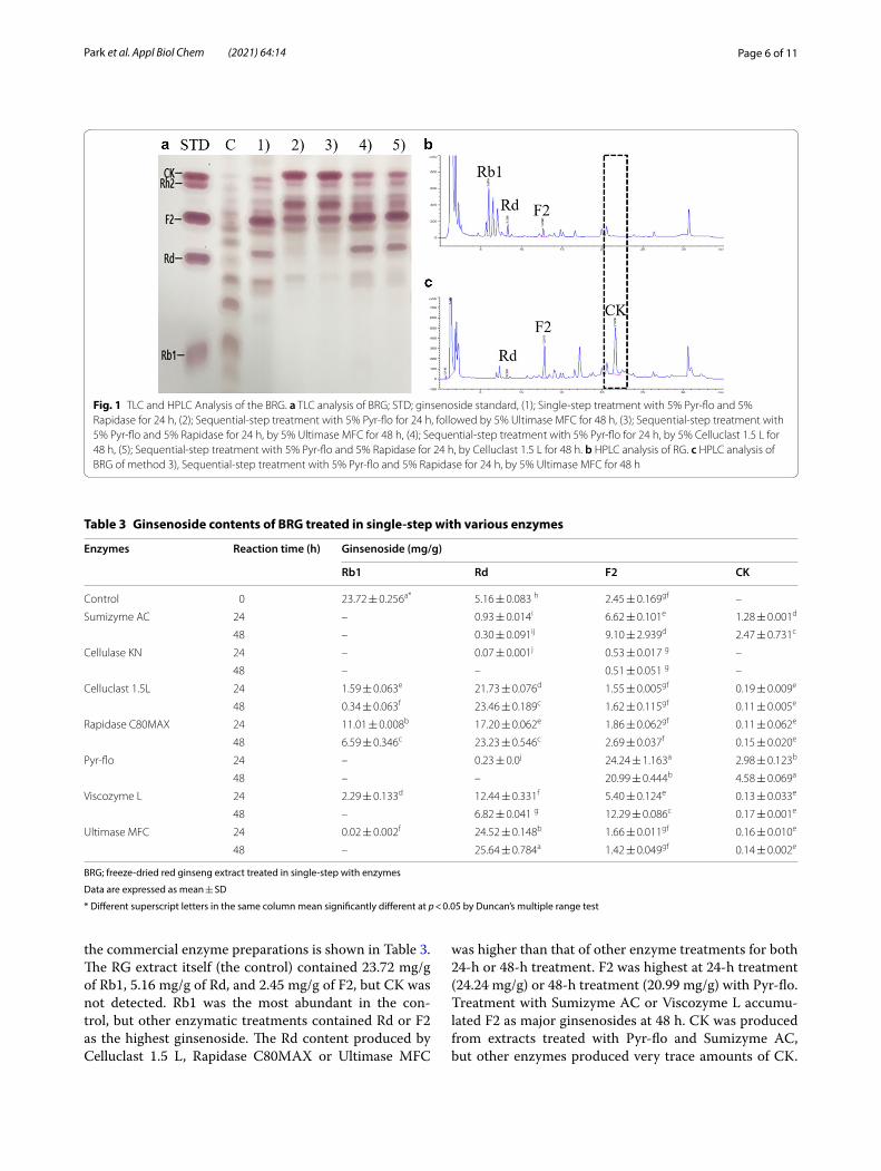
Ginsenoside contents of BRG suspension treated in single‑step with various enzymesEnzymatic treatment of RG extract was carried out by applying commercial enzyme preparations including Sumizyme AC, Cellulase KN, Celluclast 1.5 L, Rapi-dase C80MAX, Pyr-flo, Viscozyme L, and Ultimase MFC, reacted for 24 or 48 h. As a result of TLC analy-sis of the ginsenoside derivatives of each enzyme-treated sample, spots corresponding to CK appeared in sam-ples treated with Sumizyme AC, Rapidase C80MAX, and Pyr-flo, but the TLC spots of Rh2 and CK partially overlapped (Fig. 1a). Therefore, the exact ginsenoside content was determined using HPLC system. The ginse-noside concentration in RG extract treated with each of
Table 2 Dietary composition of experimental animals
RG extract; freeze-dried red ginseng extract containing 23.7, 5.2, and 2.5 mg/g of Rb1, Rd, and F2, respectively
BRG extract; freeze-dried biotransformed red ginseng extract containing 0.2, 8.0, and 14.3 mg/g of Rd, F2, and CK, respectively
CD; normal diet group, HD; high-fat diet group, RGD; 1% red ginseng powder supplemented HD group, BRGD; 1% biotransformed red ginseng powder supplemented HD group
CD HD RGD BRGD
Casein 200 200 200 200
Starch 500 200 200 200
Sucrose 150 150 140 140
Lard ∙ 350 350 350
Vit. mix 10 10 10 10
Mineral mix 35 35 35 35
Cellulose 50 50 50 50
d, l‑Methionine 3 3 3 3
Choline bitartrate 2 2 2 2
Corn oil 50 – – –
tert‑Butylhydroquinone 0.01 – – –
d, l‑Alpha‑tocopherol – 1.2 1.2 1.2
RG extract – – 10 –
BRG extract – – – 10
Page 6 of 11Park et al. Appl Biol Chem (2021) 64:14
the commercial enzyme preparations is shown in Table 3. The RG extract itself (the control) contained 23.72 mg/g of Rb1, 5.16 mg/g of Rd, and 2.45 mg/g of F2, but CK was not detected. Rb1 was the most abundant in the con-trol, but other enzymatic treatments contained Rd or F2 as the highest ginsenoside. The Rd content produced by Celluclast 1.5 L, Rapidase C80MAX or Ultimase MFC
was higher than that of other enzyme treatments for both 24-h or 48-h treatment. F2 was highest at 24-h treatment (24.24 mg/g) or 48-h treatment (20.99 mg/g) with Pyr-flo. Treatment with Sumizyme AC or Viscozyme L accumu-lated F2 as major ginsenosides at 48 h. CK was produced from extracts treated with Pyr-flo and Sumizyme AC, but other enzymes produced very trace amounts of CK.
Fig. 1 TLC and HPLC Analysis of the BRG. a TLC analysis of BRG; STD; ginsenoside standard, (1); Single‑step treatment with 5% Pyr‑flo and 5% Rapidase for 24 h, (2); Sequential‑step treatment with 5% Pyr‑flo for 24 h, followed by 5% Ultimase MFC for 48 h, (3); Sequential‑step treatment with 5% Pyr‑flo and 5% Rapidase for 24 h, by 5% Ultimase MFC for 48 h, (4); Sequential‑step treatment with 5% Pyr‑flo for 24 h, by 5% Celluclast 1.5 L for 48 h, (5); Sequential‑step treatment with 5% Pyr‑flo and 5% Rapidase for 24 h, by Celluclast 1.5 L for 48 h. b HPLC analysis of RG. c HPLC analysis of BRG of method 3), Sequential‑step treatment with 5% Pyr‑flo and 5% Rapidase for 24 h, by 5% Ultimase MFC for 48 h
Table 3 Ginsenoside contents of BRG treated in single-step with various enzymes
BRG; freeze-dried red ginseng extract treated in single-step with enzymes
Data are expressed as mean ± SD
* Different superscript letters in the same column mean significantly different at p < 0.05 by Duncan’s multiple range test
Enzymes Reaction time (h) Ginsenoside (mg/g)
Rb1 Rd F2 CK
Control 0 23.72 ± 0.256a* 5.16 ± 0.083 h 2.45 ± 0.169gf –
Sumizyme AC 24 – 0.93 ± 0.014i 6.62 ± 0.101e 1.28 ± 0.001d
48 – 0.30 ± 0.091ij 9.10 ± 2.939d 2.47 ± 0.731c
Cellulase KN 24 – 0.07 ± 0.001j 0.53 ± 0.017 g –
48 – – 0.51 ± 0.051 g –
Celluclast 1.5L 24 1.59 ± 0.063e 21.73 ± 0.076d 1.55 ± 0.005gf 0.19 ± 0.009e
48 0.34 ± 0.063f 23.46 ± 0.189c 1.62 ± 0.115gf 0.11 ± 0.005e
Rapidase C80MAX 24 11.01 ± 0.008b 17.20 ± 0.062e 1.86 ± 0.062gf 0.11 ± 0.062e
48 6.59 ± 0.346c 23.23 ± 0.546c 2.69 ± 0.037f 0.15 ± 0.020e
Pyr‑flo 24 – 0.23 ± 0.0j 24.24 ± 1.163a 2.98 ± 0.123b
48 – – 20.99 ± 0.444b 4.58 ± 0.069a
Viscozyme L 24 2.29 ± 0.133d 12.44 ± 0.331f 5.40 ± 0.124e 0.13 ± 0.033e
48 – 6.82 ± 0.041 g 12.29 ± 0.086c 0.17 ± 0.001e
Ultimase MFC 24 0.02 ± 0.002f 24.52 ± 0.148b 1.66 ± 0.011gf 0.16 ± 0.010e
48 – 25.64 ± 0.784a 1.42 ± 0.049gf 0.14 ± 0.002e
Page 7 of 11Park et al. Appl Biol Chem (2021) 64:14
Among the seven commercially available enzymes, the highest CK production was found to be 4.58 mg/g at 48 h treatment with Pyr-flo. In particular, it is estimated that Pyr-flo has the best activity in the conversion of Rb1 to F2 and CK.
Ginsenoside Rb1, which is the main PPD-type ginse-noside in RG, is mainly deglycosylated via the pathway Rb1 → Rd → F2 → CK [3, 13]. Rb1, which contains two C-20 glucoses and two C-3 glucoses, loses one C-20 glu-cose to generate Rd; Rd loses one C-3 glucose to generate F2; and F2 loses the other C-3 glucose to generate CK. As shown in Fig. 2 a), our data suggest that Celluclast 1.5 L, Rapidase C80MAX, and Ultimase MFC specifically cleave the β-(1 → 6)-glucosidic linkage at the C-20 posi-tion of Rb1, but do not hydrolyze the β-(1 → 2)-gluco-sidic linkage at the C-3 position of Rd. Especially, when Rb1 was reacted with Celluclast 1.5 L or Ultimase MFC, the Rb1 almost completely disappeared, and was con-verted into Rd as the main product after 48 h (Table 3). However, Sumizyme AC, Pyr-flo, and Viscozyme L can further hydrolyze the glucose moiety attached to the C-3 position of Rd to generate F2. Additionally, Sumizyme AC and Pyr-flo can hydrolyze other glucose attached to the C-3 position of F2 to produce CK. Of these enzymes, Pyr-flo was found to be the most efficient for the conver-sion of Rb1 from RG extract into F2 and CK (Table 3). However, if the subsequent enzyme treatment that regio-specificity hydrolyzes the glucose portion at the C-3 posi-tion of ginsenoside F2 is added, effective production of CK will be possible. Hence, we optimized the conditions for the production of CK-enriched ginseng concentrate by applying sequential enzymatic biotransformation.
Optimization of CK production of BRG suspensions sequentially treated with mixed enzymesTable 4 shows the ginsenoside content of RG extracts treated with mixed enzymes or sequentially treated. As described earlier, Pyr-flo in RG extract was found to be the most efficient in converting Rb1 to F2, so subsequent experiments were performed by mixing or sequentially treating Pyr-flo with Rapidase, Celluclast 1.5L, and Ulti-mase MFC, respectively. As a result, F2 was detected highest (18.65 mg/g) when RG extract was treated with a mixture of Pyr-flo and Rapidase for 24 h. The highest yield of CK (14.32 mg/g) was obtained after treating the RG extract with Pyr-flo and Rapidase for 24 h, then treat-ing the product with Ultimase MFC for 48 h. However, the conversion of F2 into CK was not promoted by Cel-luclast 1.5L. We found that Ultimase MFC significantly promoted the bioconversion of F2 to CK. Ultimase MFC treatment following Pyr-flo resulted in about five-fold increase in CK compared with Pyr-flo treatment alone (2.98 mg/g as shown in Table 3). However, Celluclast 1.5L
has not been shown to promote the conversion of F2 to CK. These results suggest that Ultimase MFC has high β-glucosidase activity involved in the conversion of F2 to CK, specifically hydrolyzing the glucose moiety at the C-3 position of ginsenoside F2. Choi et al. reported that Rd was produced but CK was not produced when RG extract was treated with enzymes such as Rapidase and Viscoz-yme in a single-step [11]. Another study found that treat-ing Celluclast to RG extract, Rd was effectively generated, but CK was not produced [23]. Kim et al. reported that CK was produced when the enzyme was reacted with RG extract at 50 °C (pH 4.5) for 60 h, especially when Sumizyme AC was treated, the highest CK (1.16 mg/g) was obtained [9]. In comparison, Pectinex converted a 10% ginseng root extract to 1.3 mg/g CK within 24 h [24]. However, current studies with sequential enzymatic treatment were able to produce large amounts of CK much faster than the methods of previous studies.
From our results, the rapid hydrolytic pathways of PPD-type ginsenosides to CK by commercial enzyme preparations is suggested as follows (Fig. 2b): Pyr-flo or a mixture of Pyr-flo and Rapidase can convert Rb1 into F2 as the main product, and some F2 into CK; Ultimase MFC has a high hydrolytic activity capable of converting F2 into CK. This study suggests that sequential enzymatic treatment may act on the optimized production of high-concentration CK in RG extract.
Total polyphenol content and antioxidant activityPolyphenolic compounds are secondary metabolites synthesized in plants, which have various biological activities such as antitumor, antioxidant, antiobesity, and anti-inflammatory effects [25, 26]. The total polyphe-nol content of RG extract and RG extract treated with enzymes is shown in Table 5. Total polyphenol content was significantly increased by enzymatic biotransforma-tion (p < 0.05). The total phenol content of the RG and BRG extracts was 117.13 µg/g and 153.02 µg/g, respec-tively. This indicates that the phenol content of RG extract increases by about 35% by enzyme treatment. Pyr-flo, Rapidase, and Ultimase MFC contain plant cell-wall degrading enzymes such as cellulase, pectinase, and hemicellulase. Kim et al. reported that Rapidase treat-ment under hydrostatic pressure increased the polyphe-nol content of RG [12]. However, the results of Choi et al. did not show a significant increase of polyphenol con-tent after Rapidase treatment [11]. In the present study, sequential enzyme treatment including Pyr-flo, Rapidase, and Ultimase MFC could efficiently disintegrate the plant cell wall matrix to enhance the extraction of polyphenols.
Antioxidant activities, including the DPPH radical scavenging activity, and ABTS radical scavenging activity were determined (Table 5). The ABTS radical scavenging

Page 8 of 11Park et al. Appl Biol Chem (2021) 64:14
Fig. 2 Theoretical hydrolysis pathway of ginsenoside Rb1 to Compound K (CK) by enzymatic hydrolysis. a Hydrolytic pathways by single‑step enzymatic treatment. b Optimal hydrolytic pathways by sequential enzymatic treatment for CK production

Page 9 of 11Park et al. Appl Biol Chem (2021) 64:14
activity and the DPPH radical scavenging activity of RG extract were slightly increased by enzymatic bioconver-sion (p < 0.05). Lee et al. reported that both DPPH and ABTS radical scavenging activities were improved when RG extract was fermented with Lactobacillus brevis, which was similar to this study [26] The improvement in the overall radical scavenging activity of RG extracts in bio-converted RG could be explained by the increased amounts of polyphenolic compounds [27].
Experimental animal studyIn an animal study, rats were randomly divided into four groups: a CD group, a HD group, a RGD group, and a BRGD group. Changes in their body weight during the 4-week experimental period are presented in Table 6. During the obesity-inducing period, the body weights of rats in the HD group significantly increased com-pared with those in the CD group (p < 0.05). As shown in Table 6, the body weight, food intake, and food efficiency
ratio (FER; obtained by dividing the increase in body mass during the experimental period by the food intake during the same period) of rats in the BRGD group were significantly lower than that of the HD group rats (p < 0.05). Tissue weights (heart, kidney, and spleen) were also significantly lower in the BRGD group than in the HD group (p < 0.05).
The weights of the perirenal and epididymal fat pads of rats in the HD group were significantly higher than those of animals in the CD group (p < 0.01), while the fat weights in the BRGD group were significantly lower than those in the HD and RGD groups (p < 0.01 and p < 0.05, respectively). The hepatic TG levels in BRGD group rats were significantly lower than those in the HD and RGD groups (p < 0.05 and p < 0.01, respectively). Increased fat accumulation in the body is a major characteris-tic of obesity, which has classically been characterized by expansion of intra-adipose tissue [28]. The perirenal and epididymal fat pads of HD rats were significantly accumulated compared with those in rats in the CD group. However, BRG extract consumption of rats effec-tively inhibited the fat accumulation with accompanying downregulation of body weight. These results suggest that BRG extract consumption may be useful for inhibit-ing the development of fat accumulation and obesity. In addition, a HD increased fat accumulation in the liver, whereas BRG extract consumption decreased accumu-lation of fat deposition in the liver (Table 6). Kho et al. reported that consumption of BRG extract reduced plasma levels of leptin, which supports the findings of this study [29].
There was no significant difference in serum glucose levels in the HD group and the RGD group but those
Table 4 Ginsenoside content of BRG treated with mixed and sequential enzymes
BRG; freeze-dried red ginseng extract treated with mixed and sequential enzymes
Pyr; Pyr-flo, Rap; Rapidase C80MAX, MFC; Ultimase MFC, Cel; Celluclast 1.5 L1 Single-step treatment with 5% Pyr-flo and 5% Rapidase for 24 h2 Sequential-step treatment with 5% Pyr-flo for 24 h, followed by 5% Ultimase MFC for 48 h3 Sequential-step treatment with 5% Pyr-flo and 5% Rapidase for 24 h, by 5% Ultimase MFC for 48 h4 Sequential-step treatment with 5% Pyr-flo for 24 h, by 5% Celluclast 1.5 L for 48 h5 Sequential-step treatment with 5% Pyr-flo and 5% Rapidase for 24 h, by Celluclast 1.5 L for 48 h
Data are expressed as mean ± SD
* Different superscript letters in the same column mean significantly different at p < 0.05 by Duncan’s multiple range test
Enzyme treatment Ginsenoside (mg/g)
1st (24 h) 2nd (48 h) Rb1 Rd F2 CK
Control 20.88 ± 0.01a* 4.60 ± 0.02a 2.01 ± 0.01f –
Pyr + Rap1 – – 0.17 ± 0.00e 18.65 ± 0.01a 3.32 ± 0.12d
Pyr‑flo2 MFC – 0.30 ± 0.005b 10.17 ± 0.018d 13.93 ± 0.041a
Pyr + Rap3 MFC – 0.23 ± 0.001d 7.99 ± 0.011e 14.32 ± 0.028a
Pyr‑flo4 Cel – 0.26 ± 0.002c 17.47 ± 0.019b 4.13 ± 0.139b
Pyr + Rap5 Cel – 0.18 ± 0.010e 15.81 ± 0.093c 3.70 ± 0.065c
Table 5 Total polyphenol contents and antioxidant effects of RG extract and BRG extract
RG; freeze-dried red ginseng extract
BRG; freeze-dried red ginseng extract treated with multiple enzymes by optimal method
Data are expressed as mean ± SD
* Different superscript letters in the same column mean significantly different at p < 0.05 by Duncan’s multiple range test
Sample Total polyphenol (µg/g) DPPH (%) ABTS (%)
RG 117.13 ± 1.00b* 81.43 ± 0.53b 34.53 ± 0.85b
BRG 153.02 ± 0.67a 87.67 ± 1.11a 37.72 ± 0.93a
Page 10 of 11Park et al. Appl Biol Chem (2021) 64:14
level of BRGD group was significantly lower than that of the HD group (p < 0.05). The level of HDLC was signifi-cantly higher in both the RGD and BRGD group com-pared to the HD group, and BRGD group was found to be the highest (Table 7).
A number of natural substances have been clinically evaluated for the treatment of metabolic disorders such as obesity, dyslipidemia, diabetes, and hyperten-sion and in particular, the effects of ginseng extract on prevention of diseases in animal or human studies has been investigated [30–33]. In the present study, we investigated the weight loss effect of BRG extract in HD rats. HD-induced obesity is a well-known rodent model
of induced hypertriglyceridemia, obesity, impaired glu-cose tolerance, and fatty liver [34, 35]. As expected, HD rats were obese (increased body weight and fat weight), and had a fatty liver (high hepatic TG). In this study, 1% BRG diet group reduced body weight compared with that of the animals in the HD and RGD groups. Moreover, the FER in the BRGD group was significantly lower than that in the HD group, similar to the results of a previous study [36]. These results suggest that HD-induced obesity was suppressed by BRG consumption by decreasing food efficiency.
Authors’ contributionsConceptualization and funding acquisition: HJS supervision: HJS, H Park, YJS designed the experiments; YJP, USH, SS, SYP performed the experimental work and data analyses: YJP, USH, SS, SYP, SYJ, MSP, MJK; original draft preparation YJP; editing: YSL; All authors read and approved the final manuscript.
FundingThis research was supported by the Ministry of Trade, Industry and Energy in 2019 [Grant Number P0010311].
Availability of data and materialsThe datasets used and/or analyzed during the current study are available from the corresponding author on reasonable request.
Competing interestsThe authors declare that there is no conflict of interest.
Author details1 Research Center for Food and Bio Convergence, Department of Food Science, Sun Moon University, Asan, Chungchengnam‑do, 31460, Republic of Korea. 2 Institute of Sports Health Science, Sun Moon University, Asan, Chungcheongnam‑do 31460, Republic of Korea.
Received: 29 October 2020 Accepted: 28 December 2020
Table 6 Effects of BRG on body weight, FER, tissue weight and hepatic TG level
CD; normal diet group, HD; high-fat diet group, 1% RG in HD; 1% red ginseng powder supplemented HD group, 1% BRG in HD; 1% biotransformed red ginseng powder supplemented HD group. BW: body weight, FER: food efficiency ratio = (body mass gain for experimental period (g/day)/food intake for experimental period (g/day). PFT; perirenal fat tissue, EFT; epididymal fat tissue, TG; triglyceride
Data are the mean ± SD
NS not significant
* Different superscript letters in the same raw mean significantly different at p < 0.05 by Duncan’s multiple range test
CD HD 1% RG in HD 1% BRG in HD
Initial BW (g) 226.7 ± 10.98NS 226.6 ± 9.90NS 227.0 ± 8.42NS 227.6 ± 1.38NS
Final BW (g) 351.8 ± 1.91a* 431.1 ± 14.29b 407.6 ± 6.19b 376.6 ± 11.15a
Food intake (g/day) 16.9 ± 0.18a 16.4 ± 0.36ac 16.4 ± 0.23ac 15.8 ± 0.35bc
FER 0.23 ± 0.013a 0.37 ± 0.014b 0.36 ± 0.017bc 0.32 ± 0.018c
Tissue weights and hepatic TG level
Heart (g) 1.37 ± 0.021ac 1.51 ± 0.014b 1.40 ± 0.036a 1.28 ± 0.040dc
Liver (g) 9.98 ± 0.075a 12.73 ± 0.43b 12.77 ± 0.317b 13.27 ± 0.42bc
Kidney (g) 1.25 ± 0.034a 1.43 ± 0.042b 1.26 ± 0.030a 1.30 ± 0.037ac
Spleen (g) 0.85 ± 0.034acd 0.88 ± 0.040a 0.8 ± 0.026acd 0.78 ± 0.03d
PFT (g) 5.10 ± 0.317a 12.91 ± 1.650b 10.67 ± 0.592bc 7.82 ± 1.179a
EFT (g) 5.40 ± 0.306a 11.33 ± 1.440b 10.45 ± 0.694b 6.55 ± 0.804a
Hepatic TG (mg/g tissue) 15.75 ± 0.220a 16.71 ± 0.210b 17.09 ± 0.422b 15.46 ± 0.228a
Table 7 Serum components
CD; normal diet group, HD; high-fat diet group, 1% RG in HD; 1% red ginseng powder supplemented HD group, 1% BRG in HD; 1% biotransformed red ginseng powder supplemented HD group
Data are the mean ± SD
* Different superscript letters in the same raw mean significantly different at p < 0.05 by Duncan’s multiple range test
CD HD 1% RG in HD 1% BRG in HD
Glucose (mg/dL)
121.9 ± 8.87a* 144.8 ± 4.64b 137.2 ± 7.21bc 125.8 ± 2.69a
TC (mg/dL)
59.9 ± 3.95a 79.5 ± 2.67b 91.4 ± 3.71c 90.4 ± 4.78c
HDLC (mg/dL)
27.1 ± 1.29a 36.0 ± 1.34b 41.5 ± 1.32c 48.7 ± 1.81d
Page 11 of 11Park et al. Appl Biol Chem (2021) 64:14
References 1. Park SY, Park JH, Kim HS, Lee CY, Lee HJ, Kang KS, Kim CE (2018) Systems‑
level mechanisms of action of Panax ginseng: a network pharmacological approach. J Ginseng Res 42:98–106
2. Park YC, Lim JD, Kim JB, Lee S (2012) Review of red ginseng in terms of mechanisms for pharmacodynamics and toxicity. J Korean Med 33:200–230
3. Yang XD, Yang YY, Ouyang DS, Yang (2015) GP A review of biotransfor‑mation and pharmacology of ginsenoside compound K. Fitoterapia 100:208–220
4. Lee SM, Bae BS, Park HW, Ahn NG, Cho BG, Cho YL, Kwak YS (2015) Char‑acterization of Korean Red Ginseng (Panax ginseng Meyer): history, prepa‑ration method, and chemical composition. J Ginseng Res 39:384–391
5. Christensen LP (2009) Ginsenosides chemistry, biosynthesis, analysis, and potential health effects. Adv Food Nutr Res 55:1–99
6. Santangelo R, Silvestrini A, Mancuso C (2019) Ginsenosides, catechins, quercetin and gut microbiota: current evidence of challenging interac‑tions. Food Chem Toxicol 123:42–49
7. Kim DH (2018) Gut microbiota‑mediated pharmacokinetics of ginseng saponins. J Ginseng Res 42:255–263
8. Lee JY, Lee EJ, Kim DH, Lee JH, Yoo JH, Koh BH (2009) Studies on absorp‑tion, distribution and metabolism of ginseng in humans after oral administration. J Ethnopharmacol 122:143–148
9. Kim EH, Lim S, Kim SO, Ahn SH, Choi YJ (2013) Optimization of enzy‑matic treatment for compound K production from white ginseng extract by response surface methodology. Biosci Biotechnol Biochem 77(5):1138–1140
10. Zheng MM, Xu FX, Li YJ, Xi XZ, Cui XW, Han CC, Zhang XL (2017) Study on Transformation of Ginsenosides in Different Methods. BioMed Research Int 1‑10
11. Choi HS, Kim SY, Park Y, Jung EY, Suh HJ (2014) Enzymatic transformation of ginsenosides in Korean Red Ginseng (Panax ginseng Meyer) extract prepared by Spezyme and Optidex. J Ginseng Res 38(4):264–269
12. Kim HW, Han SH, Lee SW, Choi HS, Suh HJ, Hong KB (2019) Enzymatic hydrolysis increases ginsenoside content in Korean red ginseng (Panax ginseng CA Meyer) and its biotransformation under hydrostatic pressure. J Sci Food Agric 99(15):6806–6813
13. Bae EA, Park SY, Kim DH (2000) Constitutive β‑glucosidases hydrolyzing ginsenoside Rb1 and Rb2 from human intestinal bacteria. Biol Pharm Bull 23(12):1481–1485
14. Wang WN, Yan BX, Xu WD, Qiu Y, Guo YL, Qiu ZD (2015) Highly selective bioconversion of ginsenoside Rb1 to compound K by the myce‑lium of Cordyceps sinensis under optimized conditions. Molecules 20(10):19291–19309
15. Choi HJ, Kim EA, Kim DH, Shin KS (2014) The bioconversion of red ginseng ethanol extract into compound K by Saccharomyces cerevisiae HJ‑014. Mycobiology 42(3):256–261
16. Peralta RM, Kadowaki MK, Terenzi HF, Jorge JA (1997) A highly thermosta‑ble β‑glucosidase activity from the thermophilic fungus Humicola grisea var. thermoidea: purification and biochemical characterization. FEMS Microbiol Lett 146(2):291–295
17. Quan LH, Piao JY, Min JW, Kim HB, Kim SR, Yang DU, Yang DC (2011) Biotransformation of Ginsenoside Rb1 to Prosapogenins, Gypenoside XVII, Ginsenoside Rd, Ginsenoside F2, and compound K by Leuconostoc mesenteroides. J Ginseng Res 35(3):344–351
18. Zhou W, Li J, Li X, Yan Q, Zhou P (2008) Development and validation of a reversed‑phase HPLC method for quantitative determination of ginseno‑sides Rb1, Rd, F2, and compound K during the process of biotransforma‑tion of ginsenoside Rb1. J Sep Sci 31:921–925
19. Negi PS, Jayaprakasha GK, Jena BS (2003) Antioxidant and antimutagenic activities of pomegranate peel extracts. Food Chem 80(3):393–397
20. Liang XL, Wang XL, Li Z, Hao QH, Wang SY (2010) Improved in vitro assays of superoxide anion and 1, 1‑diphenyl‑2‑picrylhydrazyl (DPPH) radical‑scavenging activity of isoflavones and isoflavone metabolites. J Agric Food Chem 58(22):11548–11552
21. Kim J, Soh SY, Bae H, Nam SY (2019) Antioxidant and phenolic contents in potatoes (Solanum tuberosum L.) and micropropagated potatoes. Appl Biol Chem 62(1):17
22. Singh G, Verma AK, Kumar V (2016) Catalytic properties, functional attrib‑utes and industrial applications of β‑glucosidases. 3 Biotech 6(3):1–14
23. Shin BK, Park HY, Han JH (2010) Enzymatic biotransformation of red gin‑seng and the compositional change of ginsenosides. J Korean Soc Appl Biol Chem 53:553–558
24. Kim BH, Lee SY, Cho HJ, You SN, Kim YJ, Park YM, Ahn SC (2006) Bio‑transformation of Korean Panax ginseng by pectinex. Biol Pharm Bull 29(12):2472–2478
25. Kong YH, Lee YC, Choi SY (2009) Neuroprotective and anti‑inflammatory effects of phenolic compounds in Panax ginseng C.A. Meyer. J Ginseng Res 33:111–114
26. Lee HS, Kim MR, Park Y, Park HJ, Chang UJ, Kim SY, Suh HJ (2012) Ferment‑ing red ginseng enhances its safety and efficacy as a novel skin care anti‑aging ingredient: in vitro and animal study. J Med Food 15(11):1015–1023
27. Lee LS, Cho CW, Hong HD, Lee YC, Choi UK, Kim YC (2013) Hypolipidemic and antioxidant properties of phenolic compound‑rich extracts from white ginseng (Panax ginseng) in cholesterol‑fed rabbits. Molecules 18:12548–12560
28. Friedman JM, Halass JL (1998) Leptin and the regulation of body weight in mammals. Nature 395:763–770
29. Kho MC, Lee YJ, Park JH, Kim HY, Yoon JJ, Ahn YM, Tan R, Park MC, Cha JD, Choi KM, Kang DG, Lee HS (2016) Fermented red ginseng potenti‑ates improvement of metabolic dysfunction in metabolic syndrome rat models. Nutrients 8(6):369
30. Kang JY, Lee JH, Kwon DJ, Song YJ (2013) Effect of Opuntia humifusa supplementation and acute exercise on insulin sensitivity and associate with PPAR‑ γ and PGC‑ 1α protein expression in skeletal muscle of rats. Int J Mol Sci 14(4):7140–7154
31. Kim HY, Kim K (2012) Regulation of signaling molecules associated with insulin action, insulin secretion and pancreatic β‑cell mass in the hypo‑glycemic effect of Korean red ginseng in Goto‑Kakizaki rat. J Ethnophar‑macol 142:53–58
32. Kwak YS, Kyung JS, Kim JS, Cho JY, Rhee MH (2010) Antihyperlipidemic effects of red ginseng acidic polysaccharide from Korean red ginseng. Biol Pharm Bull 33:468–472
33. Kim SH, Park KS (2003) Effects of Panax ginseng extract on lipid metabo‑lism in humans. Pharmacol Res 48:511–513
34. Miatello R, Vázquez M, Renna N, Cruzado M, Zumino AP, Risler N (2005) Chronic administration of resveratrol prevents biochemical cardiovascu‑lar changes in fructose‑fed rats. Am J Hypertens 18:864–870
35. Renna NF, Vazquez MA, Lama MC, González ES, Miatello RM (2009) Effect of chronic aspirin administration on an experimental model of metabolic syndrome. Clin Exp Pharmacol Physiol 36(2):162–168
36. Kim MH, Lee EJ, Cheon JM, Nam KJ, Oh TH, Kim KS (2016) Antioxidant and hepatoprotective effects of fermented red ginseng against high fat diet‑induced hyperlipidemia in rats. Lab Anim Res 32:217–223
Publisher’s NoteSpringer Nature remains neutral with regard to jurisdictional claims in pub‑lished maps and institutional affiliations.