Embed Size (px)
Citation preview

The Rockefeller University Press, 0021-9525/99/09/1289/14 $5.00The Journal of Cell Biology, Volume 146, Number 6, September 20, 1999 1289–1302http://www.jcb.org 1289
Op18/stathmin Mediates Multiple Region-specific Tubulin and Microtubule-regulating Activities
Niklas Larsson,* Bo Segerman,* Bonnie Howell,
‡
Kajsa Fridell,* Lynne Cassimeris,
‡
and Martin Gullberg*
*Department of Cell and Molecular Biology, University of Umeå, Sweden; and
‡
Department of Biological Sciences, Lehigh University, Bethlehem, Pennsylvania
Abstract.
Oncoprotein18/stathmin (Op18) is a regula-tor of microtubule (MT) dynamics that binds tubulin heterodimers and destabilizes MTs by promoting catas-trophes (i.e., transitions from growing to shrinking MTs). Here, we have performed a deletion analysis to mechanistically dissect Op18 with respect to (a) modu-lation of tubulin GTP hydrolysis and exchange, (b) tu-bulin binding in vitro, and (c) tubulin association and MT-regulating activities in intact cells. The data reveal distinct types of region-specific Op18 modulation of tu-bulin GTP metabolism, namely inhibition of nucleotide exchange and stimulation or inhibition of GTP hydroly-sis. These regulatory activities are mediated via two-site cooperative binding to tubulin by multiple nonessential physically separated regions of Op18. In vitro analysis
revealed that NH
2
- and COOH-terminal truncations of Op18 have opposite effects on the rates of tubulin GTP hydrolysis. Transfection of human leukemia cells with these two types of mutants result in similar decrease of MT content, which in both cases appeared independent of a simple tubulin sequestering mechanism. However, the NH
2
- and COOH-terminal–truncated Op18 mu-tants regulate MTs by distinct mechanisms as evi-denced by morphological analysis of microinjected newt lung cells. Hence, mutant analysis shows that Op18 has the potential to regulate tubulin/MTs by more than one specific mechanism.
Key words: microtubules • phosphoproteins • pheno-type • signal transduction • GTP phosphohydrolase
M
ICROTUBULES
(MTs)
1
are ever changing dynamicstructures that switch abruptly between growthand shortening by events called catastrophes,
and vice versa by rescue events, a behavior called dynamicinstability. MTs utilize polymerization-induced GTP hy-drolysis to generate dynamic instability, and the tip of apolymerizing MT is thought to contain a stabilizing cap ofGTP tubulin, the loss of which results in depolymerization,i.e., a catastrophe. Massive MT reorganization occurs dur-ing cell division, cell differentiation, and upon cytotoxic Tcell recognition of its target cell (for review see Desai andMitchison, 1997). MTs also act as an intracellular scaffoldfor a multitude of protein kinases and other signaling pro-teins, suggesting that regulation of dynamic instability inresponse to specific stimulation could be important for lo-calizing signal transducing proteins (for review see Gun-
dersen and Cook, 1999). Classically, regulation of MT dy-namics has been ascribed to a class of nonmotor proteinscollectively termed MT-associated proteins but more re-cently, oncoprotein 18/stathmin (Op18) has been identi-fied as a regulator of MT dynamics both in vitro and in in-tact cells (for review see Cassimeris, 1999).
The MT destabilizing activity of Op18 is turned off in re-sponse to phosphorylation of four serine residues by bothcell cycle– and cell surface receptor–regulated kinase sys-tems (for review see Deacon et al., 1999). The phenotypeof kinase target sites–deficient Op18 mutants in humancell lines show that phosphorylation downregulation is es-sential to allow microtubules to segregate condensed chro-mosomes (Marklund et al., 1994, 1996; Larsson et al.,1995). Functional inactivation by extensive mitotic phos-phorylation argues against a functional role of Op18during mitosis (Larsson et al., 1997). Rather, the high sto-ichiometry of Op18 phosphorylation by several receptor-regulated kinase systems suggests that the primary role forOp18 may be to regulate the MT system in response to ex-ternal signals in interphase cells. We have recently pro-vided direct evidence for such a role by demonstrating thatthe MT-destabilizing activity of ectopic Op18 is switchedoff in intact cells by cotransfection of the two distinct cog-
nate kinases, Ca
2
1
/calmodulin–dependent protein IV/Gr
Address correspondence to Martin Gullberg, Department of Cell andMolecular Biology, University of Umeå, S-901 87 Umeå, Sweden. Tel.: 46-90-7852532. Fax: 46-90-771420. E-mail: [email protected]
The present address for Bonnie Howell is Department of Biology, Uni-versity of North Carolina, Chapel Hill, NC 27599.
1.
Abbreviations used in this paper:
AMP-PNP, adenyl-5
9
-yl imidodiphos-phate; GST, glutathione-S-transferase; MT, microtubule; Op18, oncopro-tein 18/stathmin.
The Journal of Cell Biology, Volume 146, 1999 1290
and cAMP-dependent protein kinase (Gradin et al., 1997,1998).
Op18 is a prototype member of a new family of proteinswith microtubule regulatory function (Ozon et al., 1997;Riederer et al., 1997; Gavet et al., 1998), but the primarysequence does not give any clues to the mechanism in-volved. Microinjection of neutralizing anti–Op18 antibod-ies into newt lung cells results in a 2.5-fold increase in MTpolymer and an associated decrease in catastrophe fre-quency, which suggests a physiological importance ofOp18 for regulation of the MT system (Howell et al.,1999a). Evidence has been presented that support two dif-ferent mechanisms for Op18-mediated MT destabilization:(1) simple tubulin sequestration (Curmi et al., 1997; Jour-dain et al., 1997) or (2) specific promotion of microtubulecatastrophes (Belmont and Mitchison, 1996). In a more re-cent study (Howell et al., 1999b), which involved in vitroMT assembly assays, we showed that a specific catastrophepromoting activity of Op18 is predominant at physiologi-cal pH (
z
7.4) and that the sequestering activity is predom-inant at pH 6.8. This was consistent with the reported pHdependence of Op18–tubulin binding (Curmi et al., 1997),since lowering the pH appears to increase the binding af-finity. The first indication that Op18 has the potential todestabilize MTs by more than one mechanism came froma mutational analysis in vitro, where it was found that theNH
2
-terminal region of Op18 possesses determinantsessential for catastrophe promotion, whereas the COOH-terminal region possesses determinants essential for re-duced polymerization rate (Howell et al., 1999b). To mech-anistically understand Op18 action, we addressed theimportance of the stability of the Op18–tubulin complexby analyzing the phenotypes of two classes of mutations(Larsson et al., 1999). One class was mutated in heptad re-peats of hydrophobic residues and the other involved sub-stitution of phosphorylation sites to negatively chargedglutamic acid residues. Both classes of Op18 mutants werepartially compromised in their tubulin binding but theMT-destabilizing activities of the class of mutants with ex-change of hydrophobic residues were more severely re-duced in intact cells than the glutamic acid–substitutedOp18 derivative. Analysis of Op18 activities, such as stim-ulation of tubulin GTP hydrolysis and inhibition of tubulinpolymerization, revealed that the class of mutants with ex-change of hydrophobic residues was essentially inactive,whereas the glutamic acid–substituted Op18 derivativewas active. This suggested that Op18 transmits tubulin-directed signals that are independent of the stability of theOp18–tubulin complex, and that tubulin sequestering isnot of major functional importance.
As outlined above, Op18 lacks any cues or signatures inits primary sequence indicative for a specific mechanism ofaction. Here, using truncated derivatives of Op18, we havedefined distinct region-specific tubulin GTPase-modulat-ing activities and determined how these in vitro activitiessegregate from tubulin binding properties and specificMT-regulating activities in intact cells. The results providea clear demonstration that Op18 regulation of MTs in in-tact cells is independent of tubulin sequestering, and thatthis regulatory protein is versatile in its tubulin/MT–directed activities and has the potential to regulate MT dy-namics by distinct mechanisms.
Materials and Methods
DNA Constructs, Expression and Purification of Recombinant Proteins
Human Op18 derivatives, with or without an 8–amino acid COOH-termi-nal Flag epitope, were expressed using the pET-3d expression vector andpurified from
Escherichia coli
as described (Marklund et al., 1994). TheCOOH-terminal–truncated Op18, with the sequence encoding amino ac-ids 100–147 deleted (Op18-
D
100-147), and the NH
2
-terminal–truncatedOp18, with the sequence encoding amino acids 5–55 deleted (Op18-
D
5-55), were constructed using PCR strategies (Marklund et al., 1994). Thesame strategies were used to construct the Op18-
D
5-9, Op18-
D
5-16, Op18-
D
5-25, Op18-
D
5-38, Op18-
D
5-46, and Op18-
D
90-147 derivatives andprimer sequences are available from the authors on request. Constructsfor expression of glutathione-S-transferase (GST)–Op18 fusion proteinsin
E
.
coli
were generated by inserting an NcoI-NotI fragment of wild-typeand deleted Op18 derivatives into NcoI-EcoRI–digested pGEX 4T-1(Pharmacia) together with a double stranded adapter of the two oligonu-cleotides: 5
9
-AATTCGCGGC-3
9
and 5
9
-CATGGCCGCG-3
9
. The GST-Op18 fusion proteins were expressed and purified on glutathione-Sepharose 4B beads as recommended by the manufacturer (Pharmacia).For expression of Op18 deletion mutants in human cell lines, Op18 deriv-atives were excised from pBluescript SK
1
using HindIII and BamHI, andOp18 fragments were cloned into the corresponding sites of the EBV-based shuttle vector pMEP4 (Invitrogen Corp.; Marklund et al., 1994).
Assays of Tubulin GTPase Activity
Analysis of tubulin GTPase activity was performed in PEM buffer (80 mMpiperazine-
N,N
9
-bis[2-ethanesulfonic acid], 1 mM EGTA, 4 mM Mg
2
1
,pH 6.8) containing 17% glycerol and 5 mM adenyl-5
9
-yl imidodiphosphate(AMP-PNP; to inhibit nonspecific ATPase activity).
GTPase activity in the presence of free GTP, which allows multiplesubstrate turnover, was monitored by incubating tubulin (5–10
m
M) with
a
-[
32
P]GTP (100
m
M; 2
3
10
6
dpm per 25-
m
l reaction) at 37
8
C in the ab-sence or presence of specific Op18 derivatives. To analyze exchange inde-pendent GTP hydrolysis in a single turnover reaction, tubulin (100
m
M)was preloaded with
a
-[
32
P]GTP (100
m
M; 50
3
10
6
dpm per 25-
m
l reac-tion) on ice for 30 min. Tubulin in complex with
a
-[
32
P]GTP was recov-ered by centrifugation through a desalting column (P-30 Micro Bio-Spin;Bio-Rad Laboratories) and GTP hydrolysis was followed at 37
8
C. In theabsence of tubulin, but in the presence of BSA or Op18 (15
m
M), thesecolumns retained
.
99.99% of all
a
-[
32
P]GTP. Control experimentsshowed that the Op18 preparations used neither bind nor hydrolyze
a
-[
32
P]GTP.Nucleotide hydrolysis was quantitated as described (Austin and Dixon,
1992). In brief, aliquots were removed at the times indicated, adjusted tocontain 0.1% SDS, and heated for 2 min at 80
8
C. Aliquots (0.6
m
l) werespotted onto polyethyleneimine cellulose plates (Merck) and GDP-sepa-rated from GTP by ascending chromatography in 1.2 M NH
4
COOH acidi-fied with 1.2 M HCl. PhosphorImager (Molecular Dynamics, Inc.) analysisof radioactive spots was used for quantification.
Analysis of Op18–Tubulin Binding
Analyses of Op18–tubulin association in crude extracts from transfectedK562 cells were performed after cell lysis (50
3
10
6
per ml, on ice) in PEMbuffer, pH 6.8, containing glycerol (5%), Triton X-100 (0.5%),
b
-glycero-phosphate (10 mM), leupeptin (20
m
M), pefablock (1 mM), and benz-amide (1 mM). Cell extracts were clarified by centrifugation and, thereafterused for pull-down assays as described below. For equilibrium binding ex-periments, COOH terminally Flag-tagged wild-type and truncated Op18derivatives (2–10
m
M) and tubulin (0.8–36
m
M) were mixed and allowedto associate on ice for
z
15 min to ensure equilibrium. Independent of theOp18 derivative tested, association of Op18–F-tubulin complexes is rapidand equilibrium reached within a few minutes (data not shown and How-ell et al., 1999b). Op18–Flag-tubulin mixes (48
m
l) were added to agarosebeads (12
m
l) coupled with the Flag-epitope specific M2 antibody (SigmaChemical Co.), and incubated for 30 min at 8
8
C to capture Op18-Flag-tubulin complexes. Alternatively, glutathione-Sepharose beads (Pharma-cia) were used to analyze tubulin binding to NH
2
terminally GST-taggedOp18 derivatives.
To allow rapid separation of Op18–tubulin complexes bound to M2 or

Larsson et al.
Tubulin/microtubule–directed Activities of Op18
1291
glutathione beads, the bead suspension was applied into the cap of a1.5-ml Eppendorf tube containing a bottom layer of 0.4 ml of PEM com-plemented with 27%:17% sucrose/glycerol, pH 6.8, and a top layer of 0.2 mlof PEM with 17% glycerol, pH 6.8. The caps were closed with care, tokeep the bead suspension hanging in the cap, and the samples were centri-fuged (for 1 min at 21,000
g
) to separate bead-bound and free material.The supernatants and pellets were boiled in SDS–sample buffer and re-solved by 10–20% gradient SDS-PAGE as described (Brattsand et al.,1993). Tubulin and Op18 content were quantified by Coomassie bluestaining of protein bands followed by scanning using a personal densitom-eter (Molecular Dynamics). Bovine tubulin and a standard recombinantOp18 preparation, in which the protein mass had been determined byamino acid analysis, were used as internal standards. In a more sensi-tive binding assay, binding was analyzed after labeling of tubulin with
a
-[
32
P]GTP as described above under Assays of Tubulin GTPase Activ-ity. Control experiments confirmed that in the absence of free GTP,
a
-[
32
P]GTP remained stably associated with tubulin over the time-courseof the assay, both in the presence and absence of Op18. A trace of
125
I–Op18-F was also included to allow simultaneous quantification of Op18and tubulin in the same sample. There are two major benefits with thisstrategy. First, the amount of Op18-F (
z
30% of total Op18-F) present inthe fraction of free tubulin after separation of M2-coupled beads can becompensated for in each data point and, second, only biologically active(i.e., GTP bound) tubulin was detected in the fraction of free tubulin. Thecontribution of nonspecific tubulin binding was generally
z
3% of theamount of free tubulin and was subtracted from presented data. Sincethe position of the epitope tag used for pull-down of Op18–tubulin com-plexes may influence binding, in particular if large deletions are intro-duced close to the epitope tag, tubulin binding of Op18 derivatives wasanalyzed using either the NH
2
-terminal GST-tag or COOH-terminal Flag-tag. The data showed that the position of the epitope tag does not signifi-cantly alter Op18–tubulin binding characteristics (data not shown).
To calculate equilibrium dissociation constants, data points from equi-librium binding experiments were fitted either to a hyperbola or to amodel assuming two-site positive cooperativity in binding (Koshland et al.,1966). Written in the form of a binding curve, a two-site positive cooperat-ivity model has the form,
where
K
d
1 and
K
d
2 are the equilibrium dissociation constants for Op18binding of the first and second tubulin heterodimer respectively, B is theOp18/tubulin molar ratio of complex-bound proteins, and F is the free tu-bulin concentration. Comparison of fits was performed using the F-testprovided by GraphPad Prism (GraphPad Software, Inc.).
To determine dissociation rates of Op18–tubulin complexes, M2 beadscoated with Flag-tagged Op18 derivatives were incubated with tubulin(20
m
M) for 15 min at 8
8
C to attain equilibrium binding. Before centrifu-gation through a sucrose/glycerol cushion, bead suspension was diluted100-fold in PEM
6
glycerol (17%) and incubated for 20–900 s. Op18–tubulin complexes were quantitated by the dual isotope labeling strategydescribed above. Dissociation rates were calculated assuming one phaseexponential decay using GraphPad Prism software.
SDS-PAGE and Quantitative Western Blotting
Cells were washed in PBS and disrupted by boiling in SDS–sample buffer.The cell extract was clarified by centrifugation and, thereafter, separatedby 10–20% gradient SDS-PAGE, followed by transfer to nitrocellulose fil-ters. Cellular Op18 was detected by probing filters with affinity-purifiedanti-Op18 (Brattsand et al., 1993). For detection of Flag epitope-taggedOp18, the anti–Flag-M2 antibody (Sigma Chemical Co.) was used. Cel-lular tubulin was detected by probing filters with anti–
a
-tubulin (cloneB-5-1-2; Sigma Chemical Co.). Probing of filters, detection of specific anti-bodies by
125
I–protein A, and PhosphorImager analysis of radioactivebands was performed as described (Brattsand et al., 1993). Quantificationof Op18 and tubulin in cell lysates was obtained by comparing signals withstandard curve generated by separating graded amounts of bovine tubulinand recombinant Op18 on the same SDS-PAGE. The errors between in-dependent determinations were routinely
,
20% and the protein mass ofbovine tubulin and recombinant Op18 was determined by amino acidanalysis.
BB max
2--------------- 1 1 2F
Kd1--------- F2
Kd1 Kd× 2( )------------------------------+ +⁄
× 2FKd1--------- 1 F
Kd2----------+
,×=
Transfection of Human K562 Erythroleukemia Cells, Analysis of MT-polymerization Status, and Flow Cytometric Analysis
The conditions used for transfection studies and the pMEP4 shuttle vectorsystem have been described previously (Marklund et al., 1994). Condi-tional expression of various Op18 derivatives was achieved by using thehMTIIa promoter, which can be suppressed by low concentrations ofEDTA (50
m
M) and induced by Cd
2
1
(0.5
m
M; Marklund et al., 1994). Theamount of pMEP-Op18 constructs per electroporation was adjusted to ob-tain comparable expression levels of all Op18 derivatives. 5
m
g DNA wasused of pMEP-Op18-wt-F and pMEP-Op18-
D
5-9-F, whereas 12.5
m
g wasused of pMEP-Op18-
D
5-25-F and Op18-
D
100-147-F.The cellular content of MT polymers was determined by extracting sol-
uble tubulin in an MT-stabilizing buffer followed by quantification of tu-bulin in the particulate and soluble fraction as described (Minotti et al.,1991; Marklund et al., 1996). For flow cytometric analysis, cells were ex-tracted, fixed, and stained with anti–
a
-tubulin (clone B-5-1-2) as described(Gradin et al., 1998). Analysis of 150,000 cells per transfected Op18 deriv-ative was performed using a FACs-calibur together with the Cell Questsoftware (Becton Dickinson & Co.).
Culture and Microinjection Studies on Primary Newt Lung Epithelial Cells
Primary cultures of newt lung cells were grown on coverslips in chambersat room temperature (23
8
C) as previously described (Rieder and Hard,1990; Howell et al., 1997). For microinjection studies, cells were typicallyused 4–6 d after each culture had begun. Newt lung cells at the peripheralregions of the cell sheet were microinjected with various Op18 protein de-rivatives in a 2-mM phosphate buffer. Injected volumes are likely consis-tent with previous estimates of 5–10% of cell volume (Graessmann et al.,1980; Saxton et al., 1984), resulting in final intracellular concentrations of8–16
m
M of various Op18 derivatives. Cells were injected on an OlympusCK2 inverted microscope with attached Narishige MN-151 manipulator,using a capillary holder and prepulled glass microtips (ID 0.5
m
m; WorldPrecision Instruments, Inc.). Cells were microinjected, incubated for 3 h atroom temperature, and then lysed and fixed for antitubulin immunofluo-rescence as described (Howell et al., 1997). Microinjected cells were iden-tified based on their position relative to the tissue explant. Cells were ex-amined using a 60
3
1.4 NA Plan Apo objective on an Olympus BH-2microscope. Images were projected to an MTI CCD-72 camera, integrated(32 frames), and stored using NIH Image software (version 1.57). The fi-nal figure was composed using Canvas 6.0 software (Deneba Software).
Results
Molecular Dissection of Distinct TubulinGTPase-modulating Activities of Op18
Complex formation with tubulin is a likely minimal re-quirement for tubulin-directed signals of Op18. To analyzetubulin complex formation of the series of NH
2
- andCOOH-terminal truncations of Op18 depicted in Fig. 1 A,Flag epitope-tagged Op18 (Op18-F) and tubulin weremixed at an equimolar ratio (10
m
M each). Op18-F-tubulincomplexes were recovered using anti–Flag antibody cou-pled beads together with a rapid one-step gradient proto-col. The bead-bound Op18-F-tubulin complexes wereresolved and quantified by SDS-PAGE followed by densi-tometric scanning of stained gels. The result shows that ata 1:1 molar ratio, essentially all tubulin was in complexwith Op18-wt (Fig. 1 B). A partial loss of tubulin bindingwas evident after truncations of the NH
2
-terminal regioncovering amino acids 5–9 to amino acids 5–46. Furthertruncation into the functionally important heptad repeats(Op18-
D
5-55; Larsson et al., 1999) results in a major loss oftubulin binding. It is also shown that large COOH-termi-nal truncations, represented by the Op18-
D
100-147 and

The Journal of Cell Biology, Volume 146, 1999 1292
D
90-147 derivatives, result in a major loss of tubulinbinding.
The next step in the analysis was to determine the effectof Op18 truncations on stimulation of tubulin GTPase ac-tivity. This analysis was performed under identical bufferconditions and tubulin concentrations as in Fig 1 B. Underthese conditions, Op18-wt causes a modest 1.7-fold in-crease in the basal tubulin GTPase activity (Fig. 1 C). In-terestingly, despite decreased tubulin binding, truncationsin the NH
2
-terminal region up to amino acid 46 result inenhanced GTPase activity. Decreased tubulin binding ofthe COOH-terminal–truncated derivatives was associatedwith the opposite activity, namely significant suppressionof the basal tubulin GTPase activity. Consistent with itslack of tubulin binding, Op18-
D
5-55 showed no detectablemodulation of GTPase activity.
Fig. 1 D shows Op18-mediated stimulation of tubulinGTPase activity in the presence of nocodazole, an MT-dis-rupting agent that by itself stimulates tubulin GTPase ac-tivity. Nocodazole, under these conditions, mediates afivefold enhancement of both basal and Op18-wt–stimu-lated GTPase activity. Interestingly, albeit that there is ageneral fivefold increase, we observed the same pattern ofGTPase stimulatory/suppressive activities of the truncatedOp18 derivatives as in the absence of nocodazole. Sincetruncations of an NH
2
-terminal region of Op18 results inenhanced stimulation of tubulin GTPase activity, andtruncation of a COOH-terminal region results in suppres-sion of both the basal- and nocodazole-induced GTPaseactivity, it appears that the native Op18 contains tubulinbinding determinants with multiple regulatory effects ontubulin GTP metabolism.
In the experiments shown in Fig. 1, the tubulin concen-tration is too low to support detectable polymerization(data not shown). In the assays performed in the presenceof the antipolymerizing drug nocodazole, it is even lesslikely that the observed Op18-specific effects are causedby a mechanism involving tubulin polymerization. Fur-thermore, all the Op18 derivatives tested, except Op18-
D
5-55, mediate varying degrees of antipolymerizing activ-ity under the present conditions (data not shown). Hence,we conclude that the activity profiles of the truncatedOp18 derivatives reflect modulation of the tubulin GTP-ase activity under nonassembly conditions.
Op18 may modulate tubulin GTP metabolism by alter-ing the rate of nucleotide exchange and/or hydrolysis. Tospecifically analyze Op18 regulation of the GTP hydrolysisrate, selected Op18 derivatives were mixed in the presenceof nocodazole with tubulin prebound to
a
-[
32
P]GTP (notethat a threefold molar excess of Op18 was used to increasethe fraction of tubulin in complex with mutated Op18).The assay was performed in the absence of free GTP, and
Figure 1
. Tubulin binding and modulation of tubulin GTPase ac-tivity by Op18 deletion mutants reveal region specific tubulin-directed activities. (A) Schematic representation of Flag-taggeddeletion derivatives of Op18. (B) Tubulin binding to the indicatedOp18 Flag-epitope–tagged derivatives was analyzed at equimolarconcentration (10
m
M) in PEM, pH 6.8, containing 17% glycerol.The Op18–tubulin mixture was incubated at 37
8
C together withanti-Flag coated beads for 30 min and bead-bound material waspelleted through a sucrose/glycerol cushion. The percent of thetubulin recovered in complex with Op18 was determined by den-sitometric scanning of Coomassie blue–stained SDS-PAGE gels.
(C and D) Tubulin (10
mM) GTPase activity was determined inthe presence of the indicated Op18 derivatives (15 mM). Sampleswere incubated at 378C for 90 min with a-[32P]GTP (100 mM) inPEM, pH 6.8, containing 17% glycerol in the absence (C) or pres-ence (D) of nocodazole (33 mM). To facilitate comparison, GTP-ase activity in the absence (Co) or presence of Op18-wt-F (wt)are indicated by dashed lines. The means of duplicate determina-tions of hydrolyzed GTP are shown and data are representativefor at least three independent experiments.

Larsson et al. Tubulin/microtubule–directed Activities of Op18 1293
hydrolysis of tubulin-bound a-[32P]GTP (i.e., a single turn-over event) was followed over time. As shown in Fig. 2 A,in contrast to the result obtained in the presence of freea-[32P]GTP in the reaction mix (Fig. 1 D), the data showthat Op18-wt and NH2-terminal–truncated derivatives wereindistinguishable in their stimulation of GTP hydrolysis ina single turnover assay. It is also shown that the COOH-terminal–truncated derivative (D100-147) has a potentsuppressive effect on the rate of GTP hydrolysis. Since no-codazole was included in the reaction mix, which results ina fivefold increase in both the basal- and Op18-stimulatedlevel of GTP hydrolysis (Fig. 1), it appears that this deriva-tive exerts a general suppression of tubulin GTP hydrolysis.
In the presence of free a-[32P]GTP in the reaction mix,which allows multiple turnovers of GTP, truncation of theNH2-terminal region of Op18 enhances stimulation of tu-bulin GTPase activity both in the absence and presence ofnocodazole (Fig. 1, C and D). However, in a single turn-over reaction, these same derivatives were indistinguish-able from Op18-wt (Fig. 2 A). This suggests that Op18-wtmay contain an NH2-terminal region that inhibits GTP ex-change. To address this, selected Op18 derivatives andprebound a-[32P]GTP-tubulin were first mixed, and GTPhydrolysis in the absence of nocodazole was followed for20 min at 378C. As shown in Fig. 2 B, Op18-wt and NH2-terminal–truncated derivatives show the same stimulationof tubulin a-[32P]GTP hydrolysis, as measured in a single
turnover assay, whereas the COOH-terminal–truncatedderivative suppresses the basal hydrolysis obtained withtubulin alone, which agrees with the data obtained in thepresence of nocodazole (Fig. 2 A). To assess if Op18blocks GTP exchange, the reaction was chased with coldGTP after 21 min and subsequent alterations in the rate ofa-[32P]GTP hydrolysis were monitored (Fig. 2 B). As ex-pected from the rapid GTP exchange rate of free tubulin(Brylawski and Caplow, 1983), the data show that a chasewith cold GTP results in an immediate attenuation of thebasal a-[32P]GTP hydrolysis. In the presence of Op18-wt,however, the result of a chase with cold GTP was modestand the rate of a-[32P]GTP hydrolysis shows only a slowdecline with time, which is contrasted by a more profoundeffect of the chase in the presence of NH2-terminal–trun-cated derivatives. This indicates that Op18 exerts a potenttubulin GTP exchange inhibitory activity that involves theNH2-terminal region.
To further address the importance of the NH2-terminalregion for regulating GTP exchange of tubulin, tubulinand cold GTP were mixed and allowed to reach steadystate GTP hydrolysis at 378C. Thereafter, a-[32P]GTP wasadded and the generation of a-[32P]GDP was monitored.This protocol provides selective detection of GTP hydro-lysis after exchange to a labeled nucleotide. Given the in-distinguishable GTP hydrolysis rates in the presence of wtand NH2-terminal–truncated derivatives (Fig. 2, A and B),
Figure 2. Independent regions of Op18 regulate the rate of tubulin GTP hydrolysis and nucleotide exchange. (A) Modulation of tubulin(5 mM, in PEM with 17% glycerol, pH 6.8) GTP hydrolysis rates were determined in the presence of the indicated Op18 derivatives (15mM). Hydrolysis of a-[32P]GTP preloaded onto tubulin (i.e., a single turnover event) was followed over time in the presence of nocoda-zole (33 mM). (B) Modulation of tubulin GTP hydrolysis rates in the absence of nocodazole was followed for 20 min with different Op18derivatives (conditions and symbols as in A). After 21 min, the reaction was chased by addition of 200 mM of cold GTP and the effect onhydrolysis of a-[32P]GTP was followed over time. (C) GTP exchange dependent a-[32P]GTP hydrolysis was analyzed by mixing tubulin(5 mM, buffer as in A and B and 33 mM nocodazole) with the indicated Op18 derivatives (15 mM, same symbols as in A) and unlabeledGTP (100 mM). After 10 min at 378C, a-[32P]GTP was added to the reaction mixtures and GTP hydrolysis was followed over time. Themeans of duplicate determinations are shown. (D) A schematic representation of the importance of distinct Op18 regions for modula-tion of tubulin GTP metabolism.

The Journal of Cell Biology, Volume 146, 1999 1294
inhibition of nucleotide exchange would be manifested asdecreased generation of a-[32P]GDP in the presence ofOp18-wt. As predicted, the data in Fig. 2 C shows thatNH2-terminal truncation of Op18 results in a twofold in-crease of a-[32P]GTP hydrolysis as compared with the na-tive Op18 protein. Thus, Op18-mediated inhibition ofGTP exchange via its NH2-terminal region provides amechanism that explains the observation that NH2-termi-nal truncation enhances stimulation of tubulin GTPase ac-tivity under conditions allowing multiple turnovers ofGTP (Fig. 1, C and D).
Inhibition of GTP exchange together with the demon-strated positive and negative modulation of the rate ofGTP hydrolysis, as schematically outlined in Fig. 2 D, indi-cates that Op18 binding to tubulin results in multiple tubu-lin-directed activities that can be genetically dissected andassigned to specific regions of the protein. Since theCOOH-terminal is essential for stimulating hydrolysis,whereas deletion of this region results in a derivative withthe opposite activity, namely suppression of the GTP hy-drolysis rate, it appears that there exist regions in the re-maining polypeptide that must counteract the stimulatoryactivity of the COOH-terminal. Since Op18-D5-46 stimu-lates GTP hydrolysis to the same extent as Op18-wt, it fol-lows that the region with suppressive potential would belocated between amino acids 46 and 100 as depicted in Fig.2 D. However, it is not possible to test this prediction bydeletion analysis since Op18-D5-46 represent the largestpossible NH2-terminal truncation that does not cause ter-mination of tubulin binding and, thereby, modulation ofGTPase activity (Fig. 1, see Op18-D5-55).
Op18 and Tubulin Form Dynamic Complexes
It has been proposed that the Op18–tubulin complex isrelatively stable since it resists gel filtration and analyticalultracentrifugation (Curmi et al., 1997; Jourdain et al.,1997). To evaluate if the tubulin-directed activities de-scribed above is the result of a stable complex betweenOp18–tubulin or, alternatively, if Op18 mediates tubulin-directed activities in a dynamic complex, we determinedthe dissociation rates of selected Op18 derivatives in thepresence or absence of glycerol as described in Materialsand Methods. The data in Table I show that Op18-wt-tubulin complexes have a rapid dissociation rate that de-creases about fourfold in the presence of glycerol. The dis-sociation rate constant in the absence of glycerol (0.010s21) is similar to what has been reported using plasmonresonance measurement (0.005 s21; Curmi et al., 1997).NH2-terminal truncations result in faster dissociation thatis slowed down z10-fold by glycerol. The Op18-D100-147derivative formed the most unstable complex with tubulinand glycerol has only a minor stabilizing effect. In conclu-sion, Op18 forms dynamic complexes with tubulin and, inthe case of Op18-D100-147, the complex has a half-life ofz35 s in the buffer conditions used throughout this study(i.e., in the presence of glycerol).This indicates that Op18modulates GTP hydrolysis by multiple transient tubulininteractions during the time of the assay and not by form-ing stable complexes with tubulin.
The dynamic nature of the Op18–tubulin complex mayappear contradictory to a previous report by us in which
we employed a similar experimental approach (Larsson et al.,1999). Thus, we noted that the estimated Op18/tubulin ra-tio was z1:3 at equilibrium, and upon a 20-fold dilutionthe ratio rapidly changed to an apparently stable 1:2 ratio.Therefore, we suggested that one Op18 complexed withthree tubulins rapidly changed upon dilution to a stablecomplex containing two tubulins. However, the apparentstability was due to formation of a new equilibrium afterthe dilution step. This misinterpretation, together with a33% error in our previous estimation of Op18/tubulin mo-lar ratio, was the cause of this misconception.
Op18 Binds Two Tubulin Heterodimers Via Multiple Regions by a Mechanism Involving Two-sitePositive Cooperativity
As shown above, Op18 deletion mutants are useful analyt-ical tools to dissect modulation of tubulin GTP metab-olism, and the results suggest that physically separatetubulin binding motifs of Op18 transmit distinct tubulin-directed signals. Assuming that separate tubulin bindingmotifs are all involved in the overall binding affinity be-tween Op18 and tubulin, specific truncations would beanticipated to result in a step-wise loss of affinity. To ap-proach these questions, we performed detailed analyses ofOp18–tubulin interactions. Op18–tubulin association pre-viously has been shown to be rapid, and equilibrium isreached in ,5 min using either wild-type (Curmi et al.,1997) or truncated derivatives (Howell et al., 1999b). Se-lected COOH terminally Flag-tagged Op18 derivatives,and their NH2 terminally GST-tagged counterparts (seeMaterials and Methods) were analyzed for their ability tobind tubulin over a range of concentrations by the strategydescribed under Fig. 1. Scatchard analysis of these datashowed that all Op18 derivatives tested, bind close to 2mol tubulin per mol (Op18-wt, 2.3 6 0.07; Op18-D5-9, 2.1 60.05; Op18-D5-25, 2.1 6 0.08; and Op18-D100-147, 2.0 61.3; three independent determinations in PEM, pH 6.8).However, because of difficulties to quantify low tubulinconcentrations by scanning Coomassie blue–stained gels,the independence or cooperativity in Op18–tubulin bind-ing was difficult to assess. Therefore, we adopted a strat-egy involving labeling of tubulin with a-[32P]GTP. Controlexperiments showed that in the absence of free GTP,a-[32P]GTP remained stably associated with tubulin over
Table I. Op 18–Tubulin Complexes Are Dynamic
Glycerol 6 SE T1/2 6 SE
(s21) (s)
Op18-wt-F 0% 0.010 6 0.0007 70 6 5.1Op18-D5-25-F 0% 0.022 6 0.0009 32 6 1.4Op18-D5-46-F 0% 0.022 6 0.0014 31 6 1.9Op18-D100-147-F 0% 0.036 6 0.0024 19 6 1.2Op18-wt-F 17% 0.0025 6 0.0009 274 6 98Op18-D5-25-F 17% 0.0021 6 0.0004 324 6 67Op18-D5-46-F 17% 0.0022 6 0.0003 311 6 41Op18-D100-147-F 17% 0.020 6 0.0019 35 6 3.3
Determination of dissociation rates, expressed as either k (s21) or half-lives (T1/2, s) ofOp18–tubulin complexes was determined in the absence and presence of 17% glyc-erol, assuming one phase exponential decay as described in Materials and Methods.

Larsson et al. Tubulin/microtubule–directed Activities of Op18 1295
the time-course of the assay both in the presence and ab-sence of Op18 (for 30 min at 88C). To allow simultaneousquantification of Op18 and tubulin in the same sample, atrace of 125I-Op18-F was added. Using this dual labelingapproach, we obtained bindings curves of sufficient qualityfor detailed analysis of affinities (Fig. 3). It is clear fromthe binding curves for Op18-wt that half saturation of tu-bulin binding to Op18-wt is reached at 1.2 mM at pH 6.8(A) and 4.3 mM at pH 7.4 (B). A previous study has alsonoted pH regulation of affinity (Curmi et al., 1997), and, inthis study, an equilibrium dissociation constant (Kd) of 0.5mM at pH 6.5 was determined by plasmon resonance mea-surement, which is in reasonable agreement with our dataobtained at pH 6.8. However, Scatchard analysis of bothpHs (C and D), shows that tubulin binding to Op18 is a
complex process with observed nonlinear data point distri-bution typical for positive cooperativity in binding (Kosh-land et al., 1966). Analysis of tubulin binding curves ofselected Op18 mutants showed that half saturation ofNH2-terminal truncation derivatives requires two to four-fold higher tubulin concentrations, as compared withOp18-wt, and that the COOH terminally truncated pro-tein is most severely affected in its tubulin affinity. Sincethe position of the epitope tag used for pull-down may in-fluence the effect of specific truncations, we also analyzedtubulin binding of selected Op18 derivatives tagged in theNH2 terminus with GST (see Materials and Methods). Theresults obtained using GST-tagged derivatives were essen-tially the same as using the COOH-terminal Flag-tag (datanot shown). Hence, the binding appears independent of
Figure 3. Complexes between Op18 and two tubulin heterodimers are generated via two-site positive cooperativity. Op18–tubulin equi-librium binding curves at pH 6.8 (A) and 7.4 (B) were determined for the indicated Op18 derivatives (2 mM) at 8 8C as described in Ma-terials and Methods. The contribution of nonspecific binding (3% of free tubulin) is subtracted. Curves assuming two-site positive coop-erative binding were fitted to the data points. Scatchard conversion of the binding curves are shown below (note difference in scales inC and D). Data are representative for two independent experiments.

The Journal of Cell Biology, Volume 146, 1999 1296
the position of the epitope tag. It is notable that tubulinbinding to all Op18 derivatives tested is a cooperative pro-cess as indicated by the sigmoidal binding curves and non-linear Scatchard plots.
Glycerol, which is commonly used in tubulin buffers,slows down Op18–tubulin dissociation (Table I). In agree-ment with this result, it is shown in Fig. 4 A that glycerolenhances tubulin binding of Op18-wt and NH2 terminallytruncated derivatives. The binding of Op18-D100-147 wasessentially unaltered, consistent with the minor effect ofglycerol observed on the dissociation rate of this deriva-tive. The experiment in Fig. 4 was performed with 17%glycerol, but even 6% glycerol was sufficient for a majorenhancement in tubulin binding (data not shown). Thepresence of glycerol shifts the curves of the nonlinear Scat-chard plots but the data are still typical for positive coop-erativity in the interaction.
Two-site positive cooperativity implies that Op18 firstbinds one tubulin with a given affinity (termed Kd1), andthis generates a second site with a higher affinity (termedKd2). A two-site positive cooperativity model gives thebest fit with experimental data, obtained using differentbuffer conditions in Figs. 3 and 4, and was used to calcu-late Kd1 and Kd2. The results are summarized in Table II.Deletion analysis shows that both the NH2- and COOH-terminal regions are important for binding of the first tu-bulin (defined by Kd1), and this is particularly evident inthe presence of glycerol, which primarily enhances Kd1. Itis also clear that COOH-terminal truncation, as defined byOp18-D100-147, results in a major decrease in binding ofthe second tubulin (defined by Kd2), whereas NH2-termi-nal truncations have only a minor effect. Finally, compari-son of Op18-wt with deletion mutants reveals a stepwisedecrease in affinity as reflected by Kd1 and Kd2 as well as
the estimated free tubulin concentration required for halfsaturation of Op18. This indicates that Op18 binds tubulinvia multiple nonessential contact points distributed over amajor part of the Op18 sequence. This is consistent withthe above dissection of tubulin-directed activities of Op18,which reveals multiple tubulin-directed activities that canbe separated by deletion analysis and assigned to distinctregions spanning a major part of the Op18 peptide. Hence,the data support a model where physically separate tu-bulin binding motifs of Op18 transmit distinct tubulin-directed signals.
Figure 4. Glycerol increases Op18–tubulin binding affinity in accordance with a two-site positive cooperativity model. (A) Op18–tubulin equilibrium binding curves using the indicated Op18 derivatives (2 mM) was determined in the presence of 17% glycerol as inFig. 3. Scatchard conversions of binding curves are shown in (B).
Table II. Calculations According to a Two-site Positive Cooperativity Model Reveal Dependency of Tubulin Binding Affinities on Specific Op18 Regions
pH Glycerol Kd1 6 SE Kd2 6 SE
Free [tubulin]at half
saturation ofOp18
mM mM mM
Op18-wt-F 6.8 0% 7.5 6 2.9 0.20 6 0.09 1.2Op18-D5-25-F 6.8 0% 36 6 17 0.68 6 0.40 5.0Op18-wt-F 7.4 0% 19 6 5.4 1.0 6 0.38 4.3Op18-D5-9-F 7.4 0% 74 6 37 0.98 6 0.60 7.1Op18-D5-25-F 7.4 0% 110 6 55 1.7 6 1.1 14.0Op18-D100-147-F 7.4 0% 41 6 11 16 6 6.4 25.5Op18-wt-F 7.4 17% 2.1 6 0.7 0.19 6 0.08 0.6Op18-D5-9-F 7.4 17% 6.5 6 1.4 0.91 6 0.27 2.4Op18-D5-25-F 7.4 17% 24 6 7.7 0.82 6 0.33 4.5Op18-D100-147-F 7.4 17% 24 6 1.9 15 6 1.8 19.4
Calculation of equilibrium dissociation constants based on the data shown in Figs. 3and 4. The free tubulin concentration at half saturation was estimated from curves fit-ted according to a two-site cooperativity model.
Larsson et al. Tubulin/microtubule–directed Activities of Op18 1297
Op18 Deletion Mutants Fail to Form Complexes with Tubulin in Crude Extracts of Transfected K562 Cells
The significance of Op18 mutant phenotypes observed invitro was evaluated by transfection experiments using ashuttle vector directing inducible expression of Op18-wtand the deletion derivatives (Marklund et al., 1994). Theamount of transfected DNA was adjusted to obtain com-parable expression levels of the ectopic proteins in the hu-man K562 cell line. Estimated levels of endogenous tubu-lin and Op18, together with the induced level of each ofthe ectopic Op18 derivatives, are shown in Table III. Theestimated tubulin concentration (23 mM) is similar to thatreported for frog extracts (20 mM; Gard and Kirschner,
1987), and assuming that each Op18 binds two tubulin het-erodimers, it is notable that the molar concentration of en-dogenous Op18 (10 mM) is sufficient for complex for-mation with essentially all cellular tubulin, even underconditions of complete tubulin depolymerization.
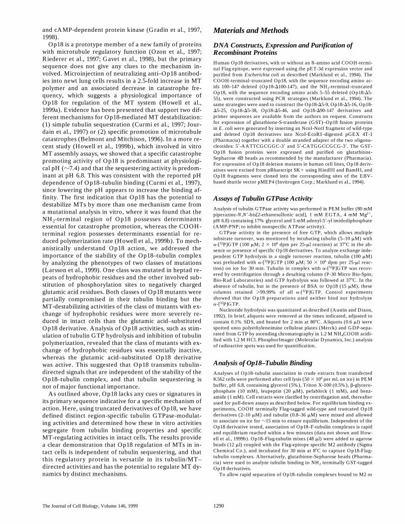
Since Op18-wt has higher affinity for tubulin than thetruncated protein, it seems likely that endogenous Op18levels are sufficient to outcompete tubulin binding of over-expressed deletion derivatives. To test this prediction, weperformed an in vitro experiment using components at asimilar molar ratio as observed in intact cells. As shown inFig. 5 A, pull-down assays using GST-fused Op18-wt inthe presence of a fivefold molar excess of the indicatedOp18 derivatives show the expected 80% competition oftubulin binding by Op18-wt, whereas Op18-D5-25 com-petes with only 20% of the binding and competition byOp18-D100-147 is essentially undetectable. In light of thisfinding, it seems likely that endogenous Op18 will inhibitmost of the complex formation of ectopic Op18 deletionmutants with cellular tubulin in transfected cells. This wasevaluated in crude cell extracts of K562 cells transfectedwith Flag-epitope–tagged Op18 (treated exactly as thecells analyzed in Table III). Complex formation was evalu-ated and as shown in Fig. 5 B, about half of all cellular tu-bulin is in complex with Op18-wt-F. As predicted, tubulincomplex formation by truncated derivatives was muchlower. This data predict that the modest five to eightfoldoverexpression of Op18 deletion mutants would not resultin significant tubulin sequestering, and that potential MTdestabilization is likely to be attributable to more specificmechanisms.
Table III. Intracellular Concentrations of Endogenous/ectopic Op18 and Tubulin in Transfected K562 Cells
Percentage of totalprotein* 6 SD
Cellularconcentration‡ 6 SD
mm
Endogenous tubulin 2.9 6 0.5% 23 6 4.2Endogenous Op18 0.22 6 0.03% 10 6 1.4Op18-wt-F 1.0% 47Op18-D5-9-F 1.4% 65Op18-D5-25-F 1.5% 79Op18-D100-147-F 1.2% 75
*Determination of Op18 and tubulin concentrations after 6 h of induced expressionwas performed by quantitative Western blot analysis as described in Materials andMethods. ‡Total cytosolic protein concentration was calculated to 80 mg/ml. This is aminimal estimate since the calculation was based on volumes of packed cell pellets.
Figure 5. Truncated Op18proteins fail to compete withOp18-wt for tubulin complexformation. (A) The indi-cated Op18 derivatives (20mM) were mixed with bovinetubulin (10 mM) and, thereaf-ter, transferred to pelletedglutathione beads coatedwith GST-Op18-wt (amountcorresponding to 4 mM in thefinal mixture). After 20 minat 378C, beads were sepa-rated and the GST-Op18/tu-bulin ratio in complexes wasdetermined as in Fig. 1. Themeans of three independentexperiments are shown. (B)Complex formation betweenendogenous tubulin andFlag-tagged Op18 was ana-lyzed in extracts of K562 cellsinduced for 6 h to express theindicated Op18 derivatives.Cell extracts (8 mg of pro-tein/ml) were added to anti-Flag–coupled beads within 10
min of cell lysis (PEM, pH 6.8, 5% glycerol and 0.5% Triton X-100) and incubated for 15 min at 88C. Bead-bound and soluble materialwas separated as in Fig. 1 and Op18–tubulin was quantified by Western blot as in Table III. The means of duplicate determinations areshown.

The Journal of Cell Biology, Volume 146, 1999 1298
Op18 Mutants Destabilize MTs by a Nontubulin Sequestering Mechanism in Transfected K562 Cells
To search for a phenotype of Op18 deletion mutants,transfected K562 cells were induced to express wild-typeand truncated Op18 derivatives for 4 and 6 h. As shown inFig. 6 A, all the Op18 derivatives analyzed are expressedat comparable levels, and expression of all derivatives re-sults in dramatic MT destabilization (Fig. 6, B–D). Thus,as shown by both biochemical determination (B) and flowcytometric analysis (C and D, note the log scale), all thetruncated Op18 derivatives retain at least 50% of wild-type MT destabilizing activity. The specificity of destabili-zation by the deletion mutants analyzed in Fig. 6 is indi-cated by previous studies of Op18-D5-55, which by severalcriteria is functionally inactive in vivo (Larsson et al., 1995;
Marklund et al., 1996). Since truncated Op18 proteins inthe presence of endogenous Op18 have only a minor po-tential to complex with cellular tubulin (Fig. 5), formationof stable tubulin sequestering complexes cannot explainthe phenotype. Hence, Op18 mutants destabilize MTs bymore specific mechanisms, possibly involving tubulin/MT–directed signaling.
Fig. 6 shows that both the NH2-and COOH-terminal–truncated derivatives have a similar gross phenotype withrespect to the MT-destabilizing activities, whereas the invitro analysis shows that these two classes of deletion de-rivatives have opposite effects on the rates of tubulin GTPhydrolysis. Assuming that the tubulin-directed activitiesare linked to the phenotype mediated by Op18 derivativesin intact cells, it follows that the NH2- and COOH-termi-
Figure 6. Expression of truncated Op18 proteins destabilize MTs in K562 cells. K562 cells were transfected with the pMEP4 shuttle vec-tor without (Vec-Co) or with insertion of the indicated Op18 derivatives. Transfected cell lines were selected during 5 d in hygromycinand expression from the hMTIIa promoter was induced as described in Materials and Methods. (A) Op18 expression levels, expressedas fold induction over the endogenous Op18 level determined as in Table III. (B) The level of polymerized tubulin, as determined byWestern blot analysis of the soluble and particulate fraction of lysed cells. (C and D) Flow cytometric analysis of MTs in the same cellpopulations shown in A and B (induced to express Op18 for 4.5 h). Open graphs show a-tubulin–specific fluorescence and filled graphsshow control staining in the absence of anti–a-tubulin but in the presence of fluorescein-conjugated rabbit anti–mouse immunoglobulin.Histograms depicting control staining and cells expressing vector control are shown in both panels. Median fluorescence signals: controlstaining, 9; vector control, 698; Op18-wt-F, 168; Op18-D5-9-F, 286; Op18-D5-25-F, 382; and Op18-D100-147-F, 385. Data are representa-tive for at least two independent experiments.

Larsson et al. Tubulin/microtubule–directed Activities of Op18 1299
nal–truncated mutants cause the same gross phenotype bydistinct mechanisms.
Evidence for Distinct MT-directed Activities of Op18 in Intact Cells
A prediction from the proposition above, namely thatNH2- and COOH-terminal–truncated mutants cause MTdestabilization by distinct mechanisms, is that the remain-ing cellular MT networks would exhibit morphologicalcues, reflecting the different modes of action. The spheri-cal shape of K562 leukemia cells make them unsuitable formorphological analysis. Therefore, we analyzed MTs innewt lung cells 3 h after microinjection of purified Op18protein derivatives. At high intracellular concentrations(40–100 mM), Op18-wt, Op18-D5-9, Op18-D5-25, andOp18-D100-147 all mediated a massive loss of MT poly-mer, whereas the inactive variant Op18-D5-55 (Marklundet al., 1996) did not alter MT polymers, demonstratingthe specificity of Op18-mediated polymer loss (data notshown). To analyze the phenotypes at concentrations simi-lar to that of endogenous Op18 in K562 cells (Table III),Op18 derivatives were microinjected to an estimated in-tracellular concentration of 8–16 mM. Under these condi-
tions, we could discern clear differences in MT-directedactivities of Op18 derivatives. As shown in Fig. 7, injectionof Op18-wt and the COOH-terminal–truncated derivative(Op18-D100-147) resulted in loss of MTs from the lamella.While a few long MTs continued to extend out towards thecell surface, many MTs appeared shorter and did not enterthe lamella region. In contrast, the NH2-terminal trunca-tions (Op18-D5-9 and Op18-D5-25) had less effects, butsome loss of MT polymer was observed.
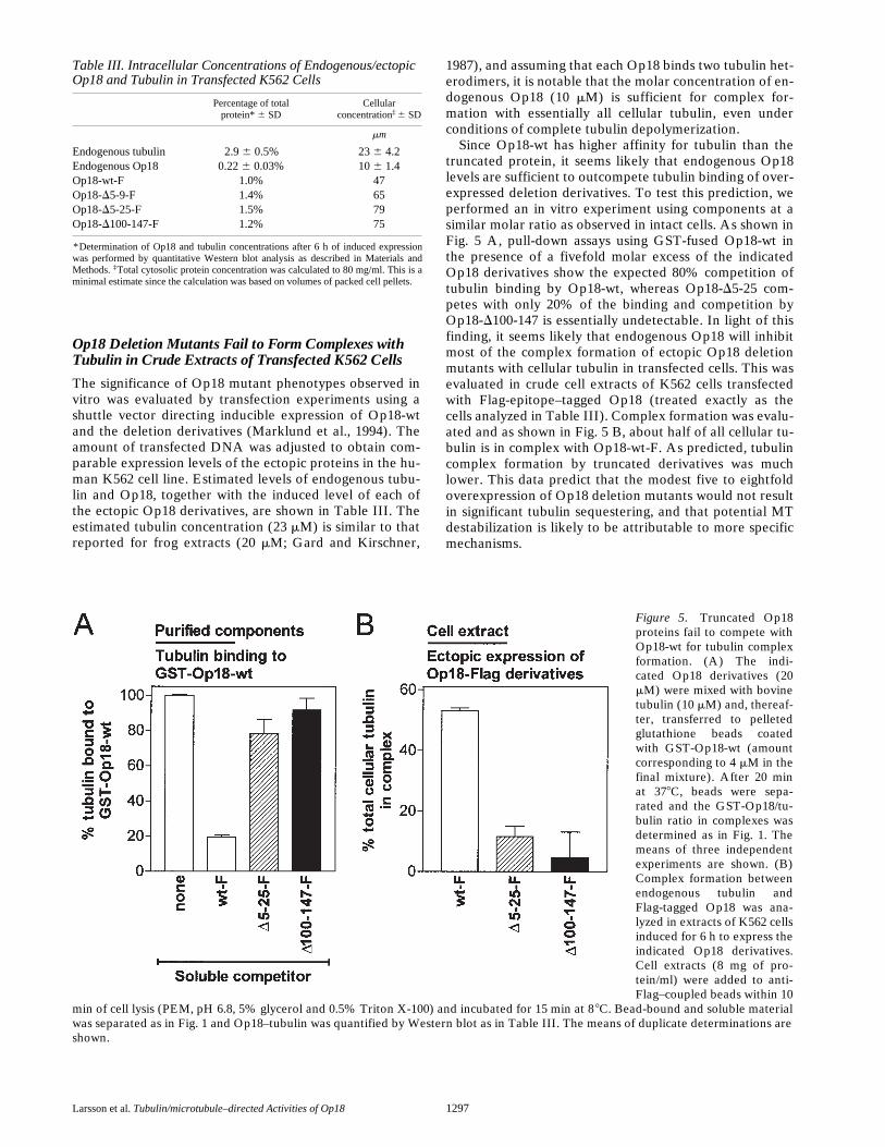
The loss of MTs from the lamellar region, which mostlikely reflect shortened MTs, provided a clear morphologi-cal assay to compare the phenotype of Op18 derivatives.We measured the number of cells that exhibited 25% ormore of the lamella region cleared of MTs. The results aresummarized in Fig. 8 and show that microinjection ofOp18-wt resulted in a 12-fold increase in the fraction ofcells with cleared lamella. The NH2 terminally truncatedproteins showed low activity, whereas the Op18-D100-147protein was almost as active as the wild type. Hence, anal-ysis of lamellar clearing indicates an overlapping pheno-type of Op18-wt and the Op18-D100-147 derivative that isdissociated from that of the NH2-terminal–truncated de-rivatives. It is notable that while the Op18-D5-9 derivativeis significantly more efficient than Op18-D100-147 in de-
Figure 7. MT appearance in newt lung cells microinjectedwith low concentrations of Op18 reveal a lamellar clearingphenotype that requires the NH2-terminal region. Cellswere microinjected with the indicated Op18 protein deriv-ative (160 mM needle concentration, 8–16 mM estimatedintracellular concentration), fixed, and stained with antitu-bulin after 3 h, and the MT-network was observed by epi-immunofluorescence. Microinjection of all Op18 deriva-tives resulted in some loss of MT polymer, but the resultsfrom Op18-wt or Op18-D100-147 were most pronouncedwhere the lamella regions often lacked MTs, most likelybecause of MT shortening. Two independent preparationsof the recombinant Op18 proteins, expressed with or with-out the Flag-tag, were used with similar results in microin-jection experiments. In the data shown, Op18 proteinswithout the Flag-tag were used.
The Journal of Cell Biology, Volume 146, 1999 1300
creasing the total MT content of K562 cells (Fig. 6), thesame 4–amino acid NH2-terminal truncation attenuatesmost of the lamellar clearing phenotype in newt cells (Fig.8). These results dissociate the lamellar clearing pheno-type observed in newt cells from the decrease in total MTcontent of K562 cells and support the idea that the NH2-and COOH-terminal–truncated proteins in part regulatesMTs by distinct mechanisms. This in turn suggests thatOp18-wt has the potential to regulate the MT system bymore than one specific mechanism.
DiscussionHere, we have performed a deletion analysis of Op18, theprototype member of a novel class of MT regulators, andidentified regions involved in differential modulation oftubulin GTP metabolism. How these in vitro activities seg-regate from tubulin binding properties and specific MT-regulating activities in intact cells was determined to ad-dress the mechanism by which Op18 exerts its regulatoryrole. These analyses were facilitated by the maintenanceof protein stability by all the truncations tested, which in-dicated that Op18 lacks distinct domain structures. Usingthese truncated Op18 proteins, physically separated tubu-lin binding motifs of both the NH2- and COOH-terminalregions were found to transmit distinct tubulin-directedsignals. These signals were manifested on the level of mod-ulation of both exchange and hydrolysis of tubulin-boundGTP (Fig. 2). By analyzing the activities of deleted Op18derivatives in K562 cells, we obtained data most consistentwith MT destabilization by a mechanism other than tubu-lin sequestration. Hence, K562 cells contain sufficient en-dogenous Op18 to sequester all unpolymerized tubulin(Fig. 6 and Table III) and NH2- and COOH-terminal dele-
tion mutants, with decreased tubulin binding affinity, wereunable to compete for tubulin binding (Fig. 5). However,despite a low level of association with cellular tubulin,these mutants still cause a drastic phenotype in intact cells(Fig. 6). Moreover, a detailed analysis of tubulin bindingshows that Op18-D100-147 has even less potential thanOp18-D5-25 to regulate MT polymerization by a simple se-questering mechanism (Tables I and II). Nevertheless,these two derivatives still caused a similar decrease of MTcontent in transfected K562 cells (Fig. 6).
Given the demonstration that tubulin sequestering isnot a principal mechanism, the result in this study raisestwo major questions: (1) are the demonstrated region-spe-cific in vitro activities of Op18 also manifested as distinctMT-directed activities in intact cells? (2) And if so, what isthe specific role of modulation of tubulin GTP metabo-lism? By comparing the phenotypes mediated by Op18 de-rivatives in transfected K562 cells and analysis of MT mor-phology in microinjected newt cells (Figs. 6 and 8), it isevident that NH2- and COOH-terminal–truncated Op18do exert different MT-directed activities consistent with invitro data. Moreover, given the demonstrated in vitro tu-bulin GTP modulatory activities of Op18 and the well es-tablished functional importance of GTP hydrolysis by tu-bulin (Desai and Mitchison, 1997), it seems reasonable toassume that Op18 has the potential to regulate MT assem-bly by its modulation of tubulin GTP status. Analysis oftruncated Op18 derivatives showed distinct levels of mod-ulation, namely inhibition of GTP exchange together witheither inhibition or stimulation of GTP hydrolysis. If mod-ulation of tubulin GTP status is central to Op18 function,it should be possible in the future to link these in vitro ac-tivities to specific MT-directed activities. However, giventhe observed complexity, evaluation of this possibility bysegregation analysis requires design of additional mutantswith a more defined spectrum of activities together with amore refined phenotypic analysis in intact cells.
A central mechanistic question is whether Op18 regu-lates MT assembly by modulation of the GTP status of thefree tubulin pool or possibly by local regulation at the tipof the MT. Op18-mediated accumulation of free GDP–tubulin would be a potential mechanism for catastrophe pro-motion by Op18-wt (Caplow and Shanks, 1995). However,the Op18-D100-147 derivative also promotes catastrophes(Howell et al., 1999b), but has the opposite overall activityof Op18-wt, namely inhibition both of the basal- and no-codazole-stimulated GTP hydrolysis of free tubulin (Figs.1 and 2). This excludes accumulation of free GDP–tubulinas the mechanism behind catastrophe promotion. Analysisof Op18–tubulin association, in crude cell extracts fromtransfected cells, provides independent evidence againstregulation of MT dynamics via modulation of the free tu-bulin pool. The data show that endogenous native Op18successfully competes with truncated Op18 mutants forbinding to cellular tubulin (Fig. 5), which implies thatthese mutants exert their effect in intact cells by an alter-native mechanism (Fig. 6). Hence, some of the availabledata are clearly incompatible with a mechanism inferringOp18 modulation of the GTP status of the free tubulinpool. While this does not exclude a potential role of Op18modulation of the GTP-status of free tubulin, it does sug-gest that Op18 exerts at least part of its regulatory activity
Figure 8. The lamellar clearing phenotype of microinjected newtlung cells is dependent on the extreme NH2-terminal region ofOp18. Cells were microinjected with the indicated Op18 proteinderivative as in Fig. 7. The data presented were generated from 2to 5 coverslips per Op18 derivative. The percentage of cells with25% or more of the lamella region empty of MTs was determinedfrom 67–245 cells per Op18 derivative. Data are means 6 SD.

Larsson et al. Tubulin/microtubule–directed Activities of Op18 1301
by an alternative mechanism, such as interaction with thetips of MTs. A finding in favor to such a mechanism is thatOp18 promotes catastrophes only from the plus end ofMTs (Howell et al., 1999), the end where the E site GTP ofb-tubulin is exposed (Nogales et al., 1999), which hints toan MT end–specific activity that may involve GTP hydro-lysis at the tip. As would be predicted from a requirementfor GTP hydrolysis, it has been shown that Op18 does notdepolymerize MTs capped with tubulin containing thenonhydrolyzable GTP analogue guanylyl (a, b)-methylenediphosphonate (Howell et al., 1999). Given the evidenceof plus end specificity and involvement of GTP hydrolysis,it is clear that Op18 acts by a different mechanism fromthat demonstrated for another class of catastrophe pro-moters, exemplified by XKCM1 and XKIF2, which act atboth ends of MTs by inducing a conformational changethat does not involve GTP hydrolysis (Desai et al., 1999;McNally, 1999).
Here, we show that Op18 retains significant tubulinbinding after extensive deletions and the data demonstratethat Op18 binds tubulin heterodimers via multiple nones-sential contacts that span residue 9 through the major partof the Op18 polypeptide (Tables I and II). It is notablethat all tubulin binding Op18 derivatives (i.e., all deriva-tives except Op18-D5-55) also manifest one or more levelsof tubulin-directed modulation of GTP metabolism (Fig.1). Our data show that Op18 binds tubulin according to atwo-site cooperative binding model, which implies thatbinding of the first tubulin creates a high affinity bindingsite for the second tubulin (Figs. 3 and 4). Hence, tubulin–tubulin interactions may be important for generation ofthe second binding site. This raises the possibility thatsome of the demonstrated tubulin-directed activities ofOp18 are generated by Op18-promoted tubulin dimeriza-tion. However, two lines of evidence indicate that this isnot sufficient for stimulation of GTPase activity. First,such a mechanism predicts that increasing Op18 concen-trations (i.e., well above Kd1; Table II), which favors Op18binding to a single tubulin, would eventually lead to inhibi-tion of tubulin GTPase activity. However, Op18 dose re-sponses up to 60 mM, using 5 mM tubulin both in the pres-ence or absence of glycerol, fail to reveal high doseinhibition of tubulin GTPase activity (data not shown).Second, we have recently reported that two classes ofOp18 mutants dissociate tubulin binding from stimulationof GTPase activity (Larsson et al., 1999). One class wasmutated in heptad repeats of hydrophobic residues andthe other involved substitution of phosphorylation sites tonegatively charged glutamic acid residues. The resultingmutants showed a comparable decrease in their tubulinbinding, but only the class of mutants with exchange of hy-drophobic residues were defective in stimulation of tubu-lin GTPase activity. Hence, although Op18-promoted tu-bulin–tubulin interactions may be functionally important,it is evidently not sufficient for Op18-mediated stimulationof GTPase activity.
In a previous in vitro study of MT assembly (Howell et al.,1999b), the Op18-D5-25 and Op18-D100-147 derivativeswere used to show that the catastrophe promoting activityof Op18 requires the NH2-terminal region of Op18,whereas a polymerization rate inhibiting activity requiresthe COOH-terminal region. We proposed that a tight tu-
bulin complex formed by either Op18-wt or Op18-D5-25inhibited the polymerization rate by tubulin sequestering.Under the conditions used for both derivatives, inhibitionof the tubulin polymerization rate was only observed atpH 6.8 and not at 7.4. At the time, this was readily ex-plained by the reported increase of Op18–tubulin bindingat the lower pH. In this study, we determined binding af-finities of Op18-wt and Op18-D5-25 at pH 6.8 and 7.4, us-ing the same conditions as during MT assembly (i.e., PEMbuffer in the absence of glycerol; Table II). It is evidentthat tubulin binding of Op18-D5-25 at pH 6.8 is essentiallythe same as Op18-wt binding at pH 7.4. Hence, assuminga simple sequestering mechanism, the present bindingdata predict that the polymerization inhibitory activity ofOp18-wt at pH 7.4 should be similar to the activity ofOp18-D5-25 at pH 6.8. This was clearly not the case sincethese two derivatives showed indistinguishable polymer-ization inhibitory activity at pH 6.8 and none showed inhi-bition at pH 7.4 (Howell et al., 1999b). These results showthat the pH sensitive Op18 inhibition of polymerizationrates cannot be explained by a sequestering mechanism asproposed by us and others (Curmi et al., 1997; Howell etal., 1999b) and that a more specific pH-regulated mecha-nism must be involved.
The in vitro study discussed above (Howell et al., 1999b)shows that the Op18-D100-147 derivative promotes catas-trophes at both pH 6.8 and 7.4 without a detectable inhibi-tion of the polymerization rate. It is notable that this deriv-ative was found here to be almost as efficient as Op18-wtin causing the observed lamellar clearing phenotype innewt cells (Figs. 5 and 6). This phenotype is diagnostic forshortening of MTs and is the predicted phenotype of aspecific catastrophe promoting factor. This supports thephysiological significance of the catastrophe activity ofspecific Op18 derivatives observed in vitro. However, al-though Op18 is versatile with respect to modulation ofGTP metabolism of free tubulin, more work is required todeduce the mechanism by which Op18 regulates MT dy-namics.
We thank V. Shingler (University of Umeå) for helpful discussions.N. Larsson, B. Segerman, K. Fridell, and M. Gullberg were supported
by Swedish Natural Science Research Council and the Foundation forMedical Research at the University of Umeå. B. Howell and L. Cassimeriswere supported by a National Institutes of Health grant.
Submitted: 18 June 1999Revised: 10 August 1999Accepted: 12 August 1999
References
Austin, S., and R. Dixon. 1992. The prokaryotic enhancer binding proteinNTRC has an ATPase activity which is phosphorylation and DNA depen-dent. EMBO (Eur. Mol. Biol. Organ.) J. 11:2219–2228.
Belmont, L.D, and T.J. Mitchison. 1996. Identification of a protein that inter-acts with tubulin dimers and increases the catastrophe rate of microtubules.Cell. 84:623–631.
Brattsand, G., G. Roos, U. Marklund, H. Ueda, G. Landberg, E. Nanberg, P.Sideras, and M. Gullberg. 1993. Quantitative analysis of the expression andregulation of an activation-regulated phosphoprotein (oncoprotein 18) innormal and neoplastic cells. Leukemia. 7:569–579.
Brylawski, B.P., and M. Caplow. 1983. Rate for nucleotide release from tubulin.J. Biol. Chem. 258:760–763.
Caplow, M., and J. Shanks. 1995. Induction of microtubule catastrophe by for-mation of tubulin-GDP and apotubulin subunits at microtubule ends. Bio-chemistry. 34:15732–15741.
Cassimeris, L. 1999. Accessory protein regulation of microtubule dynamics

The Journal of Cell Biology, Volume 146, 1999 1302
throughout the cell cycle. Curr. Opin. Cell Biol. 11:134–141.Curmi, P.A., S.S. Andersen, S. Lachkar, O. Gavet, E. Karsenti, M. Knossow,
and A. Sobel. 1997. The stathmin/tubulin interaction in vitro. J. Biol. Chem.272:25029–25036.
Deacon, H.W., T.J. Mitchison, and M. Gullberg. 1999. Op18/stathmin. InGuidebook to the Cytoskeletal and Motor Proteins. T. Kreis and R. Vale,editors. Oxford University Press, Oxford. In press.
Desai, A., and T.J. Mitchison. 1997. Microtubule polymerization dynamics.Annu. Rev. Cell Dev. Biol. 13:83–117.
Desai, A., S. Verma, T.J. Mitchison, and C.E. Walczak. 1999. Kin I kinesins aremicrotubule-destabilizing enzymes. Cell. 96:69–78.
Gard, D.L., and M.W. Kirschner. 1987. Microtubule assembly in cytoplasmicextracts of Xenopus oocytes and eggs. J. Cell Biol. 105:2191–2201.
Gavet, O., S. Ozon, V. Manceau, S. Lawle, P. Curmi, and A. Sobel. 1998. Thestathmin phosphoprotein family: intracellular localization and effects on themicrotubule network. J. Cell Sci. 22:3333–3334.
Gradin, H.M., U. Marklund, N. Larsson, T.A. Chatila, and M. Gullberg. 1997.Regulation of microtubule dynamics by Ca21/calmodulin-dependent kinaseIV/Gr-dependent phosphorylation of oncoprotein 18. Mol. Cell Biol. 17:3459–3467.
Gradin, H.M., N. Larsson, U. Marklund, and M. Gullberg. 1998. Regulation ofmicrotubule dynamics by extracellular signals: cAMP-dependent protein ki-nase switches off the activity of oncoprotein 18 in intact cells. J. Cell Biol.140:131–141.
Graessmann, A., M. Graessmann, and C. Mueller. 1980. Microinjection of earlySV40 DNA fragments and T antigen. Methods Enzymol. 65:816–825.
Gundersen, G.G., and T.A. Cook. 1999. Microtubules and signal transduction.Curr. Opin. Cell Biol. 11:81–94.
Howell, B., D.J. Odde, and L. Cassimeris. 1997. Kinase and phosphatase inhibi-tors cause rapid alterations in microtubule dynamic instability in living cells.Cell. Motil. Cytoskeleton. 38:202–214.
Howell, B.J., H. Deacon, and L. Cassimeris. 1999a. Decreasing oncoprotein 18levels reduce microtubule catastrophes and increase microtubule polymer invivo. J. Cell Sci. In press.
Howell, B., N. Larsson, M. Gullberg, and L. Cassimeris. 1999b. Dissociation ofthe tubulin-sequestering and microtubule catastrophe-promoting activitiesof oncoprotein 18/stathmin. Mol. Biol. Cell. 10:105–118.
Jourdain, L., P. Curmi, A. Sobel, D. Pantaloni, and M.F. Carlier. 1997. Stath-min: a tubulin-sequestering protein which forms a ternary T2S complex withtwo tubulin molecules. Biochemistry. 36:10817–10821.
Koshland, D.E., Jr., G. Nemethy, and D. Filmer. 1966. Comparison of experi-mental binding data and theoretical models in proteins containing subunits.Biochemistry. 5:365–385.
Larsson, N., H. Melander, U. Marklund, O. Osterman, and M. Gullberg. 1995.G2/M transition requires multisite phosphorylation of oncoprotein 18 bytwo distinct protein kinase systems. J. Biol. Chem. 270:14175–14183.
Larsson, N., U. Marklund, H.M. Gradin, G. Brattsand, and M. Gullberg. 1997.Control of microtubule dynamics by oncoprotein 18: dissection of the regu-latory role of multisite phosphorylation during mitosis. Mol. Cell Biol. 17:5530–5539.
Larsson, N., B. Segerman, H.M. Gradin, E. Wandzioch, L. Cassimeris, and M.Gullberg. 1999. Mutations of oncoprotein 18/stathmin identify tubulin-directed regulatory activities distinct from tubulin association. Mol. CellBiol. 19:2242–2250.
Marklund, U., O. Osterman, H. Melander, A. Bergh, and M. Gullberg. 1994.The phenotype of a “Cdc2 kinase target site-deficient” mutant of oncopro-tein 18 reveals a role of this protein in cell cycle control. J. Biol. Chem. 269:30626–30635.
Marklund, U., N. Larsson, H.M. Gradin, G. Brattsand, and M. Gullberg. 1996.Oncoprotein 18 is a phosphorylation-responsive regulator of microtubuledynamics. EMBO (Eur. Mol. Biol. Organ.) J. 15:5290–5298.
McNally, F.J. 1999. Microtubule dynamics: controlling split ends. Curr. Biol.9:R274–R276.
Minotti, A.M., S.B. Barlow, and F. Cabral. 1991. Resistance to antimitoticdrugs in Chinese hamster ovary cells correlates with changes in the level ofpolymerized tubulin. J. Biol. Chem. 266:3987–3994.
Nogales, E., M. Whittaker, R.A. Milligan, and K.H. Downing. 1999. High-reso-lution model of the microtubule. Cell. 96:79–88.
Ozon, S., A. Maucuer, and A. Sobel. 1997. The stathmin family—molecular andbiological characterization of novel mammalian proteins expressed in thenervous system. Eur. J. Biochem. 248:794–806.
Rieder, C.L., and R. Hard. 1990. Newt lung epithelial cells: cultivation, use, andadvantages for biomedical research. Int. Rev. Cytol. 122:153–220.
Riederer, B.M., V. Pellier, B. Antonsson, G. Di Paolo, S.A. Stimpson, R.Lutjens, S. Catsicas, and G. Grenningloh. 1997. Regulation of microtubuledynamics by the neural growth-associated protein SCG10. Proc. Natl. Acad.Sci. USA. 94:741–745.
Saxton, W.M., D.L. Stemple, R.J. Leslie, E.D. Salmon, M. Zavortink, and J.R.McIntosh. 1984. Tubulin dynamics in cultured mammalian cells. J. Cell Biol.99:2175–2186.