Embed Size (px)
Citation preview

Supplemental Methods
Immunoblotting
Protein lysates (30 µg protein) were and separated by SDS gel
electrophoresis and transferred to nitrocellulose membranes as described previously 1.
Membranes were exposed to a rabbit antibody against rabbit-15-LO-1 (Caymen
chemicals) (dilution 1:5000 in TBS) for 1 h at room temperature, rinsed with TBS buffer
containing 0.1% Tween-20 and incubated with a 1:5000 diluted goat anti-rabbit IgG
(horseradish peroxidase conjugated, Zymed). Membranes were re-probed with mouse
anti-β-actin as a loading control. Immunoreactive bands were identified and
densitometric analysis was performed.
Quantitative real time polymerase chain reaction (qRTPCR)
Total nucleotides were isolated from rabbit arteries using Trizol reagent (Life
Technologies). The DNA was digested with amplification grade DNase-1 (Invitrogen).
cDNA was synthesized from total RNA by using SuperScript III first strand synthesis
system for qRTPCR (Invitrogen). For control, one reaction mixture without reverse
transcriptase and one without RNA was made. cDNAs for 15-LO-1 and GAPDH in the
reaction mixtures was amplified using BIO-Rad iCycler in a 25 µL mixture containing; 2
X 10-7 g cDNA, 2 X 10⁻⁷ M primers and RT² SYBR Green / Fluorescein qPCR Master
Mix (Super Array Bioscience Technologies). The primers for 15-LO-1, (reverse) 5’- CCT
GGC GCG GAC GTT GAT CTC-3’ and (forward) 5'- TGG CTG CCC CGC TGG TCA
TGC-3', were designed from the cDNA sequence of rabbit 15-LO-1, and were

synthesized by the Operon Co (Huntsville, AL). The primers for GAPDH were supplied
by Super Array Bioscience Technologies. The program for the iCycler was 94°C for 30 s,
58°C for 1 min, and 72°C for 1.5 min, repeated 40 times followed by final extension at
72°C for 7 min. The PCR products obtained after the iCycler amplification were
separated by 1% agarose gel electrophoresis and visualized by ethidium bromide staining
to confirm the presence of a single amplified product.
Immunohistochemistry
Rabbit aortic or mesenteric arterial segments were fixed in 4% paraformaldehyde
and cut into sections as described previously 1, 2. Sections were incubated with mouse
anti-human platelet endothelial cell adhesion molecule (PECAM) (kindly provided by Dr.
Peter Newman, Blood Center for Southeastern Wisconsin, Milwaukee, WI) and rabbit
anti-human 15-LO-1 diluted 1:500 in 0.2% Triton X-100 containing 1% normal goat
serum. Sections were incubated for 1 h at room temperature, rinsed, and incubated
with 1:500 anti-mouse Alexa-Fluor 594 and anti-rabbit FITC labeled secondary
antibodies (Molecular Probes) for 1 h at 25°C. Sections were mounted with media
containing 1% 4,6-diamidino-2-phenylindole (DAPI) and protected by a glass coverslip.
Fluorescent images were captured (200X magnification) using Nikon Eclipse E600
microscope and Spot Advanced software.
Metabolism of 14C-AA
Arteries were dissected, cleaned and cut into 2-3 mm rings in N-2-
hydroxyethylpiperazine-N'-2-ethanesulfonic acid (HEPES) buffer (mM): 10 HEPES,

150 NaCl, 5 KCl, 2 CaCl2, 1 MgCl2, 6 glucose, pH 7.4. Rings were incubated at 37°C
with indomethacin (INDO) (10-5 M) (Sigma, MO) or with INDO and BW755C (5 X
10-5 M) (Burroughs Wellcome, Sandwich, England) in 5 ml HEPES for 10 min and
then [14C]-AA (0.5 µCi, 10-7 M) was added. After 5 min, A23187 (10-5 M)
(Sigma, MO) was added. After 15 min, the reaction was stopped with ethanol (15% final
concentration), and the samples were extracted using Bond Elute octadecylsilyl columns3.
The extracts were analyzed by reverse phase high-pressure liquid chromatography
(HPLC) using a Nucleosil C-18 (5µ, 4.6 x 250 mm) column. The solvent system
consisted of a 40 min linear gradient (flow rate=1 ml/min) from 50% solvent B
(acetonitrile with 0.1% glacial acetic acid) in solvent A (deionized water) to 100%
solvent B. Column effluent was collected in 0.2 ml fractions, and the radioactivity was
determined.
Isometric tension recording
Aortic rings were suspended in a 6 ml tissue bath with Krebs buffer of
composition: (mM) 119 NaCl, 4.7 KCl, 1.8 KH2PO4, 1.17 MgSO4, 25 NaHCO3, 2.5
CaCl2, 0.026 EDTA, and 5.5 glucose bubbled with 95% O2 and 5% CO2 at 37°C.
Isometric tension was measured with force-displacement transducers using glass muscle
baths and recorded with a Macintosh computer and MacLab software. The aortic rings
were gradually adjusted to the resting tension of 2gm and allowed to equilibrate for 1 h.
The rings were then tested for the maximum response with 44 mM KCl. The rings were
treated with INDO (10-5 M) and LNA (3x10⁻⁵ or 3x10⁻⁴ M) for 20 min. In some studies,
the aortic rings were pretreated with CDC (20x10-5 M), BW755C (5x10⁻⁵ M), ebselen

(20x10-5 M), APA (10-7 M), CTX (10-7 M), or TRAM-34 (10-5 M) either alone or in
combination with each other. These inhibitors were dissolved in a proper solvent and
added to the muscle bath along with INDO and LNA. After 20 mins of incubation with
the inhibitors, arteries were contracted by PHE (approx. 10-7 M) to 50-60% of the
maximal KCl contraction and cumulative concentrations of AA (10-7-10-4 M) or ACH
(10-9-10-5 M) were added to the bath and changes in isomeric tension were measured.
Relaxations to EBIO, P1075 and DPTA: Arteries were prepared in the Krebs
buffer as described above. Contractions to KCl (44mM) or PHE were determined,
precontracted with PHE to half maximal contractions and relaxations to cumulative doses
of IKCa-channel opener 1-ethyl-2-benzimidazolinone (1-EBIO; 10-8-10-3 M), KATP-
channel opener P1075 (10-8-10-3 M) or NO-donor dipropylenetriamine NONOate
(DPTA;10-9-10-5 M) were measured. In some experiments the arteries were treated with
INDO, LNA, CTX, ebselen or BW755C either alone or in combination with each other
for 20 mins and then the relaxations were determined.
Measurement of membrane potential
Membrane potential (Em) was measured in the SMCs of the rabbit aortas as
published previously 5. Briefly, aortic rings were cut open laterally, pinned to a silastic
layer in a perfusion chamber, with the endothelium exposed and perfused with warmed
(37 0C) Krebs buffer bubbled with 95% O2 and 5% CO2. After 30 min of equilibration, Em
of SMCs was measured either in presence of vehicle, INDO (10-5 M), INDO and PNE (l0-
7 M), INDO and PNE plus BW755C (10-4 M) or BW755C alone. Impalements were done
with a glass electrode only in a small section of the aortas where the endothelium was
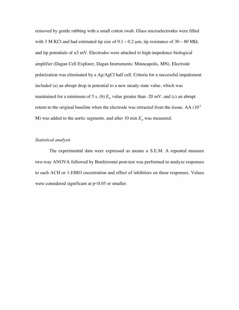
removed by gentle rubbing with a small cotton swab. Glass microelectrodes were filled
with 3 M KCl and had estimated tip size of 0.1 – 0.2 µm, tip resistance of 30 – 80 MΩ,
and tip potentials of ≤3 mV. Electrodes were attached to high-impedence biological
amplifier (Dagan Cell Explorer, Dagan Instruments; Minneapolis, MN). Electrode
polarization was eliminated by a Ag/AgCl half cell. Criteria for a successful impalement
included (a) an abrupt drop in potential to a new steady-state value, which was
maintained for a minimum of 5 s, (b) Em value greater than -20 mV, and (c) an abrupt
return to the original baseline when the electrode was retracted from the tissue. AA (10-5
M) was added to the aortic segments, and after 10 min Em was measured.
Statistical analysis
The experimental data were expressed as means ± S.E.M. A repeated measure
two-way ANOVA followed by Bonferronni post-test was performed to analyze responses
to each ACH or 1-EBIO cncentration and effect of inhibitors on these responses. Values
were considered significant at p<0.05 or smaller.

References to Online Supplement
1. Aggarwal NT, Holmes BB, Cui L, Viita H, Yla-Herttuala S, Campbell WB. Adenoviral expression of 15-lipoxygenase-1 in rabbit aortic endothelium: role in arachidonic acid-induced relaxation. Am J Physiol Heart Circ Physiol 2007;292(2):H1033-41.
2. Tang X, Spitzbarth N, Kuhn H, Chaitidis P, Campbell WB. Interleukin-13 upregulates vasodilatory 15-lipoxygenase eicosanoids in rabbit aorta. Arterioscler Thromb Vasc Biol 2003;23(10):1768-74.
3. Pfister SL, Spitzbarth N, Nithipatikom K, Edgemond WS, Falck JR, Campbell WB. Identification of 11,14,15- and 11,12,15-trihydroxyeicosatrienoic acids as endothelium-derived relaxing factors of rabbit aorta. J. Biol. Chem. 1998;273:30879-30887.
4. Campbell WB, Spitzbarth N, Gauthier KM, Pfister SL. 11,12,15-Trihydroxyeicosatrienoic acid mediates acetylcholine-induced relaxations in the rabbit aorta. Am J Physiol Heart Circ Physiol. 2003;285(6):H2648-2456.
5. Chawengsub Y, Aggarwal NT, Nithipatikom K, Gauthier KM, Anjaiah S, Hammock BD, et al. Identification of 15-hydroxy-11,12-epoxyeicosatrienoic acid as a vasoactive 15-lipoxygenase metabolite in rabbit aorta. Am J Physiol Heart Circ Physiol 2008;294(3):H1348-56.

Legends to Supplement Figures Figure S1. Immunohistochemical localization of 15-LO-1 in arteries from
hypercholesterolemic or normal rabbits. Arterial sections were co-stained for platelet
endothelial cell adhesion molecule (PECAM), 15-LO-1, and nucleus. For controls
sections were either not stained (panel E & M) or stained with only secondary antibodies
(panels A & I). 15-LO-1 expression is localized to the endothelium (white arrow).
Figure S2. Agonist-induced relaxations in aortas from normal or hypercholesterolemic
rabbits. (A) Aortas were precontracted with phenylephrine (PNE) and relaxations to
cumulative doses of acetylcholine (ACH) were determined. (B) Aortas from normal
rabbits were incubated with indomethacin (INDO; 10-5 M) and L-nitroarginine (LNA; 3
X 10-5 M or 3 X 10⁻⁴ M), precontracted with PNE and relaxations to ACH were
determined. (C and D) Aortic rings were incubated with INDO (10-5 M), precontracted
with PNE and relaxations to ( C) a nitric oxide-donor, dipropylenetriamine NONOate
(DPTA) , and (D) a KATP-channel opener, P1075, were determined. Values are expressed
as percentage mean �± SEM. *** = p<0.001, ** = p < 0.01, * = p<0.05.
Figure S3: Relaxations in aortic rings from normal rabbits. Aortic rings were pretreated
with indomethacin (INDO, 10-5 M), L-nitroarginine (LNA, 3 X 10⁻⁵ M) and either
vehicle, BW755C (5 X 10⁻⁵ M), ebselen (2 X 10⁻⁵ M), or CDC (2 X 10⁻⁵ M). Rings were
than precontracted with phenylephrine (PNE, 10-7 to 10-6 M) and relaxations to (A)
acetylcholine (ACH) or (B) arachidonic acid (AA) determined. Values represent mean
±SEM. (*** = p<0.001, * or # = p<0.05). * BW755C treatment, # ebselen treatment.
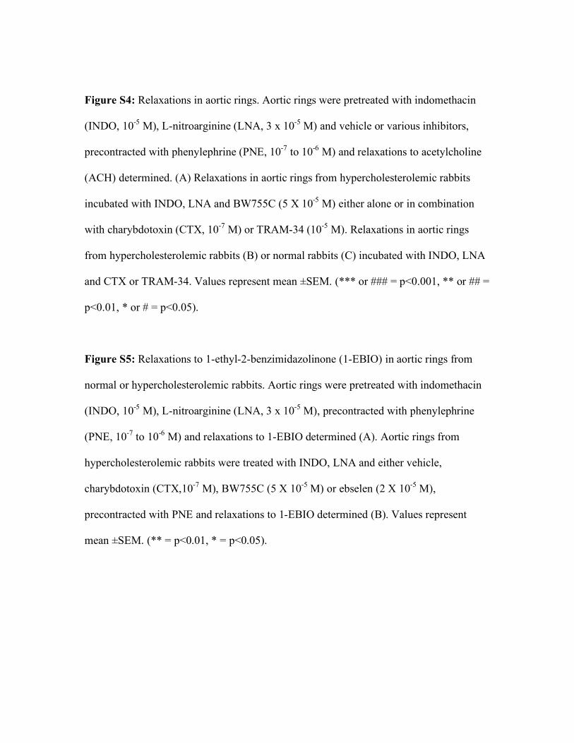
Figure S4: Relaxations in aortic rings. Aortic rings were pretreated with indomethacin
(INDO, 10-5 M), L-nitroarginine (LNA, 3 x 10⁻⁵ M) and vehicle or various inhibitors,
precontracted with phenylephrine (PNE, 10-7 to 10-6 M) and relaxations to acetylcholine
(ACH) determined. (A) Relaxations in aortic rings from hypercholesterolemic rabbits
incubated with INDO, LNA and BW755C (5 X 10⁻⁵ M) either alone or in combination
with charybdotoxin (CTX, 10-7 M) or TRAM-34 (10-5 M). Relaxations in aortic rings
from hypercholesterolemic rabbits (B) or normal rabbits (C) incubated with INDO, LNA
and CTX or TRAM-34. Values represent mean ±SEM. (*** or ### = p<0.001, ** or ## =
p<0.01, * or # = p<0.05).
Figure S5: Relaxations to 1-ethyl-2-benzimidazolinone (1-EBIO) in aortic rings from
normal or hypercholesterolemic rabbits. Aortic rings were pretreated with indomethacin
(INDO, 10-5 M), L-nitroarginine (LNA, 3 x 10⁻⁵ M), precontracted with phenylephrine
(PNE, 10-7 to 10-6 M) and relaxations to 1-EBIO determined (A). Aortic rings from
hypercholesterolemic rabbits were treated with INDO, LNA and either vehicle,
charybdotoxin (CTX,10-7 M), BW755C (5 X 10⁻⁵ M) or ebselen (2 X 10⁻⁵ M),
precontracted with PNE and relaxations to 1-EBIO determined (B). Values represent
mean ±SEM. (** = p<0.01, * = p<0.05).
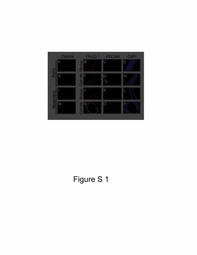
Figure S 1

Figure S 2
10-10 10-9 10-8 10-7 10-6 10-5
0
20
40
60
80
100
120Cholesterol-fedNormal
**
*
ACH (M)
10-10 10-9 10-8 10-7 10-6 10-5
0
20
40
60
80
100
120
INDOINDO+30µM LNAINDO+300µM LNA
******
ACH (M)
10 -9 10 -8 10 -7 10 -6 10 -5
0
20
40
60
80
100
120
NormalCholesterol-fed
DPTA (M)10-9 10-8 10-7 10-6 10 -5 10-4
0
20
40
60
80NormalCholesterol-fed
P1075 (M)
A B
D C

10-8 10 -7 10-6 10 -50
25
50
75
100 INDO + LNA
INDO + LNA + CDCINDO + LNA + EbselenINDO + LNA + BW
ACH (M)
10-7 10-6 10 -5 10 -4 10-30
25
50
75
100
******
INDO + BWINDO
AA (M)
A
B
Figure S 3
* #

Figure S 4
10-9 10-8 10-7 10-6 10-5
0
25
50
75
100
INDO + LNA + TRAM + BW
INDO + LNA
INDO + LNA + CTX + BW
INDO + LNA+ BW
ACH (M)
10-9 10-8 10-7 10-6 10-5
0
25
50
75
100 INDO +LNAINDO + LNA + CTXINDO + LNA + TRAM
ACH (M)
******
###
###
*
#
10-9 10-8 10-7 10-6 10-50
25
50
75
100 INDO + LNAINDO + LNA + CTXINDO + LNA + TRAM
**#
#
ACH (M)
### *** ***
A
B
C

A
B
Figure S 3
10-8 10-7 10-6 10-5 10-4 10-3
0
25
50
75
100 Cholesterol-fedNormal
1-EBIO
10-8 10-7 10 -6 10-5 10-4 10 -3
0
25
50
75
100 INDO + LNA
INDO + LNA + CTXINDO + LNA + BW
INDO + LNA + Ebselen
**
***
1-EBIO (M)
Figure S 5