Embed Size (px)
Citation preview
One- and Two-Dimensional ESEEM Spectroscopy of Flavoproteins†
Jesu´s I. Martınez,‡ Pablo J. Alonso,‡ Carlos Go´mez-Moreno,§ and Milagros Medina*,§
Instituto de Ciencia de Materiales de Arago´n, Consejo Superior de InVestigaciones Cientı´ficassUniVersidad de Zaragoza, andDepartamento de Bioquı´mica y Biologı´a Molecular y Celular, Facultad de Ciencias, UniVersidad de Zaragoza,
50009 Zaragoza, Spain
ReceiVed June 23, 1997; ReVised Manuscript ReceiVed September 15, 1997X
ABSTRACT: One- and two-dimensional (1D and 2D) electron spin echo envelope modulation (ESEEM)spectroscopy was applied to study the flavin cofactors in the neutral semiquinone states of flavodoxinand ferredoxin-NADP+ reductase (FNR) from the cyanobacteriumAnabaenaPCC 7119, and the anionicsemiquinone state of cholesterol oxidase fromBreVibacterium sterolicum. High-resolution crystal structuresare available for all these proteins. Three- and 4-pulse ESEEM and hyperfine sublevel correlationspectroscopy (HYSCORE) techniques at X-band were used. HYSCORE spectra showed correlationsbetween transitions caused by interaction of the isoalloxazine unpaired electronic spin present in thesemiquinone state with several nitrogen and hydrogen nuclei. Measurements of isotopic labeled samples([15N]FMN flavodoxin and [2H]flavodoxin) allowed the assignment of all the detected transitions to nucleibelonging to the FMN cofactor group. Interactions of nitrogens in positions 1 and 3 of the isoalloxazinering were determined to have isotropic hyperfine coupling constants in the 1-2 and 0.5-1 MHz rangesfor all the different flavoprotein semiquinones studied. Information about the quadrupolar term of thesenuclei was also obtained. An intense correlation in the negative quadrant was detected. It has beenassociated to the strongly interacting N(10) nucleus. The complete hyperfine term parameters (includingthe sign) were obtained from detailed analysis of this signal, being the quadrupolar parameter,K, alsoestimated. Another correlation in the HYSCORE spectra, corresponding to hydrogen bound to the N(5)position in neutral flavin semiquinones, was detected. Its interaction parameters were also determined.This study demonstrates that ESEEM spectroscopy, and in particular the HYSCORE technique, are ofparticular utility for detecting and assigning nuclear transition frequencies in flavoprotein semiquinones.Moreover, the results reported here are complementary to ENDOR studies, and both techniques togetherprovide an important tool for obtaining information about spin distribution in the flavin ring of flavoproteinsin the semiquinone state.
A large number of flavin-containing proteins have beenuncovered to date, many of them being enzymes. Flavopro-teins participate in a large number of oxidation/reductionreactions that function either in energy transduction or inbiosynthesis and degradation of metabolic intermediates(Edmondson & McCormick, 1987; Yagi, 1993). This meansthat the same cofactor (the flavin) is able to take part incatalytic processes which must vary widely from themechanistic point of view. The activation of a particularactivity of the flavin results from its interaction with theprotein polypeptidic chain at the active center, and, for mostof the enzymes, a transfer of electrons must take placebetween the substrate and the flavin during the catalyticevent. Due to their unique ability to transfer either one ortwo electrons, flavoproteins are used for electron transferbetween pyridine (two-electron donor/acceptor) nucleotidesand metal-containing heme or iron-sulfur clusters (one-electron donor/acceptor) present in proteins. They alsoparticipate in the transfer of electrons to other flavin-containing proteins, which raises interesting questions relatedto the thermodynamic and structural requirements for ef-
ficient flavin-flavin electron transfer. The involvement offlavin semiquinone intermediates in flavoenzyme-catalyzedreaction has been the focus of a large number of studies. Insome enzymes it has already been shown that the semi-quinone state is an important intermediate in the overallcatalytic pathway (Janot et al., 1990; White et al., 1993).Nevertheless, a number of uncertainties about their participa-tion in electron-transfer reactions in biological systems stillremain (Edmondson & Tollin, 1983; Mu¨ller, 1983). In thepresent study we have deepened our knowledge of thesemiquinone state of three flavoproteins, flavodoxin andferredoxin-NADP+ reductase (FNR) fromAnabaenaPCC7119, both of them exhibiting a neutral semiquinone radical,and cholesterol oxidase fromBreVibacterium sterolicum,which presents an anionic semiquinone.
The photochemical reduction of NADP+ to NADPH hasbeen shown to proceed via ferredoxin and the flavoenzymeFNR (Rogers, 1987; Knaff & Hirasawa, 1991). Whencultures of cyanobacteria are deprived of iron, they synthesizea low-Mr FMN-containing protein, flavodoxin, which re-places ferredoxin in most reactions (Fillat et al., 1988).AnabaenaPCC 7119 FNR has been extensively characterized(Pueyo & Gomez-Moreno, 1991; Pueyo et al., 1991). It hasa molecular mass of 36 kDa and contains one mole of non-covalently bound FAD cofactor/mole of enzyme. The three-dimensional structure ofAnabaenaPCC 7119 FNR and thatof a complex with NADP+ have been recently solved at 1.8
† This work was supported by Grant BIO-94-0621-C02-01 from theComision Interministerial de Ciencia y Tecnologı´a to C.G.-M.* To whom correspondence should be addressed. Phone:+34-976-
761279. Fax:+34-976-762123. E-mail: [email protected].‡ Instituto de Ciencia de Materiales de Arago´n.§ Departamento de Bioquı´mica y Biologıa Molecular y Celular.X Abstract published inAdVance ACS Abstracts,November 1, 1997.
15526 Biochemistry1997,36, 15526-15537
S0006-2960(97)01495-5 CCC: $14.00 © 1997 American Chemical Society

and 2.25 Å, respectively (Serre et al., 1996). Redox titrationsshowed that the maximum proportion ofAnabaenaFNRsemiquinone at equilibrium, in the pH range 6-8, was 29-12% (Pueyo et al., 1991). Nevertheless, in FNR, the FADsemiquinone state certainly plays a role in the enzyme-catalyzed reduction of NADP+. Although the mechanismof this reaction has not been revealed, it is generally acceptedthat ferredoxin is a one-electron carrier and there is only asingle ferredoxin-binding site on FNR. In this case, it isdifficult to imagine mechanisms for electron transfer fromreduced ferredoxin to the FAD of FNR that do not involveat least the transient formation of the FAD semiquinone state(Knaff & Hirasawa, 1991). Moreover, the structure of thisprotein is the prototype of a large family of flavin-dependentoxidoreductases that function as transducers between nico-tinamide dinucleotides (two-electron carriers) and one-electron carriers (Karplus et al., 1991; Correll et al., 1993;Bruns & Karplus, 1995). Flavodoxin is isolated fromAnabaenaPCC 7119 vegetative cells when iron is limitedin the culture medium (Fillat et al., 1988). It has a molecularmass of 20 kDa, contains 1 mol of FMN/mol of protein,and forms a 1:1 complex with FNR (Fillat et al., 1988, 1990).The semiquinone of FMN in flavodoxin is highly stable, sothat close to 100% of the flavin is in this form after additionof one electron. This is a consequence of the relativemidpoint potential for the oxidized/semiquinone couple(-212 mV) and the semiquinone/hydroquinone couple(-436 mV) (Pueyo et al., 1991).Cholesterol oxidase is a flavin-dependent enzyme that
catalyzes the oxidation and isomerization of 3â-hydroxys-teroids having a double bond at∆5-∆6 of the steroid ringbackbone (Smith & Brooks, 1975; Kamei et al., 1978; Inouyeet al., 1982). The enzyme from the soil bacteriumB.sterolicumis a monomeric oxidase containing one moleculeFAD/molecule protein (Uwajima et al., 1974). The crystalstructure of the enzyme fromB. sterolicum has beendetermined at 1.8 Å resolution, both in the presence and inthe absence of a bound steroid (Vrielink et al., 1991; Li etal., 1993). A significant structural homology has beenobserved between cholesterol oxidase and the members ofthe glucose-methanol-choline (GMC) oxidoreductasesfamily (Caverner, 1992). This family of enzymes all undergosimilar oxidative chemistry, namely, the flavin-assistedoxidation of an alcohol to an aldehyde or ketone function.EPR spectroscopy has played an important role in the
detection of flavin semiquinones, being particularly usefulfor distinguishing the anionic and neutral radicals. However,this technique provides little insight into the structure ofprotein-bound semiquinones because of the large number ofunresolved anisotropic hyperfine couplings. Electron nucleardouble resonance (ENDOR) spectroscopy offered improvedspectral resolution and has provided information on the
molecular structure and electronic distribution on modelflavin and flavoprotein radicals (Kurreck et al., 1984, 1987;Edmondson, 1985; Bretz et al., 1987). Structural data onparamagnetic centers in biological systems have also beenobtained by analyzing electron spin-echo envelope modula-tions (ESEEM) arising from the interaction with variousnuclei (Tsvetkov & Dikanov, 1987). This technique isparticularly sensitive to weak hyperfine interactions (Mims& Peisach, 1981). ENDOR and ESEEM spectroscopiccharacterizations of the semiquinone forms from flavodoxinand FNR fromAnabaenaPCC 7119, as well as fromcholesterol oxidase fromB. sterolicumhave already beenreported (Medina et al., 1994, 1995, 1997; Medina &Cammack, 1996). ENDOR studies of these flavoproteinsemiquinones allowed to detect strong hyperfine couplingsdue to the 8-CH3 and 6-CH protons in anionic (cholesteroloxidase) and neutral (flavodoxin and FNR) semiquinoneforms. ENDOR spectroscopy has also shown that uponNADP+ binding to FNR, a change in the electron densitydistribution on the flavin ring takes place, consistent withelectron withdrawal by interaction with the nicotinamide ring(Medina et al., 1995). The same effect was observed whencholesterol oxidase was studied in the presence of its steroidsubstrate (Medina et al., 1994). Recent three-pulse 1D-ESEEM studies on the frozen solution of cholesterol oxidase,flavodoxin, and FNR semiquinones at X-band showedprominent nuclear modulation frequencies. These frequen-cies seem to be consistent with the presence of at least one14N magnetically coupled to the paramagnet and could beinterpreted as arising from nitrogens at positions 1 and/or 3of the flavin ring system (Medina & Cammack, 1996;Medina et al., 1997; Edmondson et al., 1990).Difficulties in associating 1D-ESEEM features to each
nucleus, as well as those arising from working with orien-tationally disordered samples, can be overcome by using 2Dtechniques. Four pulse 2D-HYSCORE spectroscopy pro-vides correlations between nuclear transition frequenciesbelonging to different manifolds of a single electronic spin(Hofer et al., 1986). It has been proved to be very usefulfor assignment of ESEEM signals and for the examinationof broad signals which are too weak to be detected in 1D-ESEEM experiments, as occurs for orientationally disorderedsamples in the case of anisotropic interactions (Shane et al.,1994; Kofman et al., 1995). Recently, the HYSCOREtechnique has already been successfully applied to the studyof the metal ligand environment in different proteins (Shergillet al., 1995; Kofman et al., 1996; Dikanov et al., 1996).In the present study we have characterized the neutral
semiquinone states of flavodoxin and FNR fromAnabaenaPCC 7119, and the anionic semiquinone state of cholesteroloxidase fromB. sterolicum, using three-pulse (3P) and four-pulse (4P) 1D-ESEEM and 2D-HYSCORE techniques. Toour knowledge this is the first report on a flavoproteinsemiquinone HYSCORE study. We show that 1D-ESEEMsignals assigned to weakly interacting nitrogen in cholesteroloxidase, flavodoxin, and FNR semiquinones (Medina et al.,1997; Medina & Cammack, 1996) are readily detected byHYSCORE spectroscopy. Detailed information about hy-perfine and quadrupolar interaction terms is also reported.From our 1D-ESEEM and HYSCORE experiments we canget new, valuable data about spin density in the pyrimidineand pyrazine rings of the flavin. The characterization offlavoprotein semiquinones by these techniques opens more
1 Abbreviations: EPR, electron paramagnetic resonance; ENDOR,electron nuclear double resonance; ESEEM, electron spin-echo envelopemodulation; FFT, fast Fourier transform; HYSCORE, hyperfine sublevelcorrelation spectroscopy; [15N]FMN flavodoxin, flavodoxin sample inwhich the FMN cofactor has been replaced by a FMN molecule inwhich all nitrogens are15N; [2H]flavodoxin; flavodoxin sample in whichexchangeable1H have been replaced by2H; a, isotropic hyperfinecoupling constant;T, anisotropic hyperfine coupling constant;TM, shortphase memory time; dq, double quantum; sq, single quantum;νL,nuclear Zeeman frequency;e2qQ, quadrupole coupling constant;q,quadrupolar interaction;K, quadrupolar parameter;η, asymmetryparameter; hf, hyperfine.
HYSCORE Spectroscopy of Flavoproteins Biochemistry, Vol. 36, No. 49, 199715527
possibilities in the study of the environment of the flavincofactor in proteins and in the modulation of its propertiesby the apoprotein.
MATERIALS AND METHODS
Biological Material
AnabaenaPCC 7119 flavodoxin had been previouslycloned in the pTrc99a plasmid and expressed inEscherichiacoli PC 2495 strain. Cell culture, protein induction, isolationand reconstitution with FMN was as previously described(Fillat et al., 1991; Genzor et al., 1996). FNR was purifiedfrom E. coliPC 0225 strain transformed with the FNR geneas previously described (Go´mez-Moreno et al., 1995).Cholesterol oxidase fromB. sterolicumwas isolated andpurified using the methods described by Uwajima et al.(1973). To prepare15N-labeled FMN a culture ofE. colicontaining theAnabaenaflavodoxin gene was grown in SVminimal medium using15NH4Cl (enrichment in15N >99.5%)as the only nitrogen source.15N-labeled flavodoxin waspurified as described for unlabeled protein (Fillat et al., 1991),and its15N-labeled FMN was extracted by precipitation ofthe apoprotein component with trichloroacetic acid (Genzoret al., 1996). The supernatant, containing the15N-labeledFMN, was neutralized and used as described below. Sinceunlabeled antibiotic was also used during the cultures, theenrichment on15N of the 15N-labeled FMN prepared forapoflavodoxin reconstitution was estimated uniform and over92%. Unlabeled apoflavodoxin was prepared by removalof the FMN group by treatment of the holoprotein withtrichloroacetic acid (Genzor et al., 1996). Apoflavodoxinwas solved in buffer and dialyzed to remove the acid.Reconstitution with15N-labeled FMN was followed spec-trophotometrically by titration of the apoflavodoxin with theobtained [15N]FMN.Samples were transferred into the desired buffer (usually
10 mM HEPES, pH 7, or HEPES-D2O, pD 7) by dilutionand ultrafiltration through Centricon 10 microconcentrators(Amicon, Witten-Herdecke), at 4°C. The cycle was repeatedthree times, to give a final buffer enrichment of 95-99%.
HYSCORE Sample Preparation
Flavodoxin, FNR and cholesterol oxidase samples werereduced anaerobically to the semiquinone state at 4°C bylight irradiation with a 150 W Barr & Stroud light source,approximately 7.5 cm from the sample, in the presence of20 mM EDTA and 2.5µM 5-deazariboflavin. Maximalproduction of semiquinone was obtained by taking samplesand recording EPR spectra during the illumination process.Samples (400-800 µM protein) were prepared in a sealedglass vessel under argon and transferred anaerobically usinga gas-tight microsyringe into the EPR tubes, which wereimmediately frozen in liquid nitrogen. To avoid oxygenintroduction during sample transfer, the EPR tubes wereconnected to the vessel through a lateral arm and flushedwith argon prior to sample withdrawal. The samplescontaining the highest proportion of semiquinone state wereused for ESEEM measurements. The samples were storedin liquid nitrogen, or at-70 °C, until use.
Spectroscopic Measurements
A Bruker ESP380E spectrometer operating in X-band (9-10 GHz) was used for pulsed-EPR measurements. Spectra
were taken at a temperature of 15 K. The static magneticfield was set at about 347 mT, and the microwavefrequency was 9.75 GHz. Field position was selected inthe center of the EPR signal to give a maximum echointensity. Orientation selection effects are not expectedprovided that EPR signals are narrow (peak-to-peak widthless than 2 mT) and the main contribution to their widthcomes from unresolved hyperfine interactions. Micro-wave pulse sequences were (π/2-τ-π/2-t1-π/2) for 3PESEEM, (π/2-τ-π/2-t1-π-t1-π/2) for 4P ESEEM, and(π/2-τ-π/2-t1-π-t2-π/2) for HYSCORE experiments. Ineach case, appropriate phase cycling was applied to removeunwanted echoes. For 3P experiments microwave pulseswere set 16 ns long, whereas for 4P experiments bothπ/2and π pulses were set 24 ns long. In 1D experiments,τwas fixed in a typical value of 144 ns and 1024 points werecollected, varyingt1 with a step of 8 ns.τ was selected tobe 96 ns in HYSCORE experiments, and spectra varyingt1and t2 independently had (256, 256) points. Typical stepsfor t1 and t2 were 16 ns. HYSCORE spectra with steps of32 ns were also recorded to improve resolution in the low-frequency region. ESEEM signal of our samples at 15 Kwas quite intense but shot repetition time should be large(about 25 ms) in order to avoid saturation effects.
Data Handling/Analysis
Frequency domain spectra were obtained using the WIN-EPR program from Bruker in the following way: the baseline was subtracted in the time domain spectrum, windowingwith a square sine bell function was applied for enlargingthe signal:noise ratio, and a fast Fourier transform (FFT)algorithm was then applied, being the modulus of the resultthe frequency spectrum. To analyze our results the 3P 1D-ESEEM signal of I ) 1 nuclei was simulated using acomputer program developed at the University of Nijmegenby E. J. Reijerse. Nuclear transition frequencies andHYSCORE signal intensities forI ) 1/2 nuclei signals werecalculated using the expressions given by Dikanov andTsvetkov (1992a).
EXPERIMENTAL RESULTS
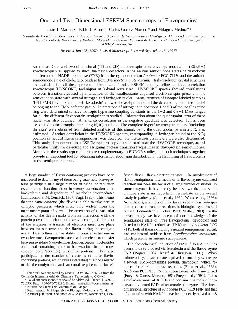
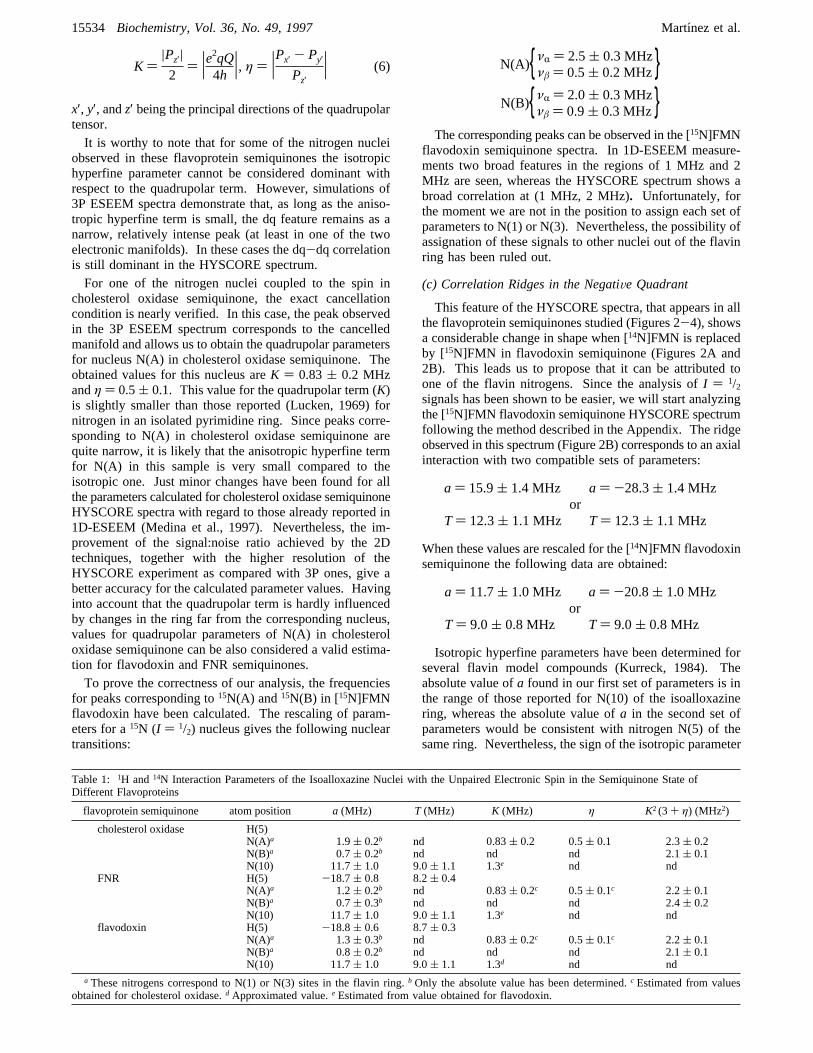
Different ESEEM experiments have been applied to thestudy of the semiquinone states of flavodoxin, FNR, andcholesterol oxidase. Two-pulse (π/2-τ-π) 1D-ESEEMspectra are poorly resolved due to the short phase memorytime, TM, of the systems. Nevertheless, well-resolved 3Pand 4P 1D-ESEEM spectra can be obtained. 3P spectra shownuclear transition main frequencies, whereas 4P spectra alsoproduce peaks corresponding to “combination frequencies”(Schweiger, 1990). Figure 1A shows the 3P 1D-ESEEMspectra recorded for the semiquinone states of cholesteroloxidase, FNR, flavodoxin, [15N]FMN flavodoxin and [2H]-flavodoxin. The corresponding 4P 1D-ESEEM spectra areshown in Figure 1B. A narrow intense peak in the 3 MHzregion is detected for all the three samples with naturalabundant isotopes in 3P and 4P 1D-ESEEM experiments.Other less intense features can also be seen in the low-frequency region, 0-5 MHz. In cholesterol oxidase semi-quinone a peak at 2.2 MHz also appears. This peak is notseen in the spectra recorded for FNR and flavodoxinsemiquinones. FNR shows a second peak in the 3.5 MHzregion. 4P experiments show (combination) peaks in the6-8 MHz region for these samples. When higher frequency
15528 Biochemistry, Vol. 36, No. 49, 1997 Martınez et al.
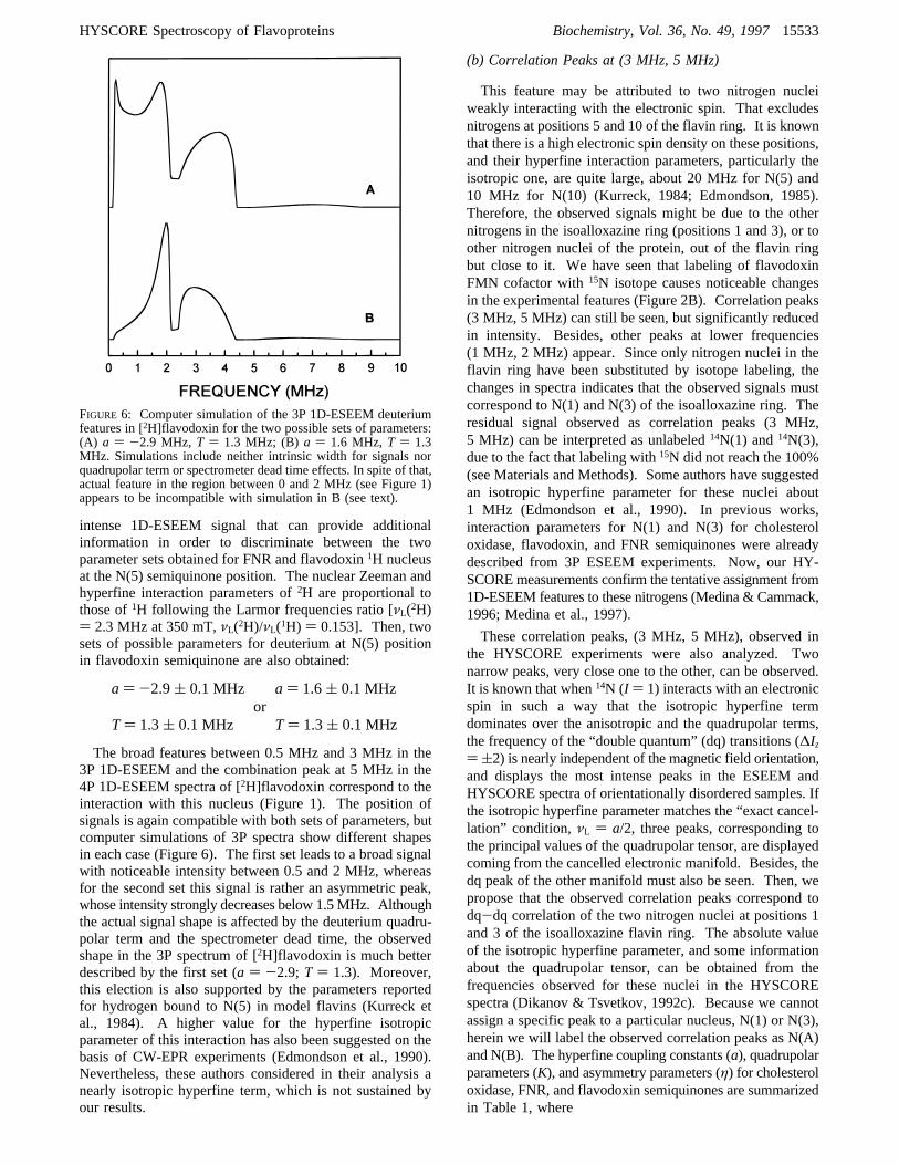
regions were studied, two more peaks, about 15 and 30 MHz,are detected (not shown) in all the samples. These frequencyvalues correspond to the1H Larmor frequency and twice thisfrequency, respectively. Flavodoxin and FNR semiquinone4P experiments also show a less intense peak in the 33 MHzregion.The low-frequency region of [15N]FMN flavodoxin semi-
quinone 3P spectrum shows a dominating broad feature about1 MHz. Moreover, in these spectra the features present inthe unlabeled FMN flavodoxin semiquinone spectra areclearly diminished in comparison with the 1 MHz peak. Itis also noticeable that no combination peaks at the 6-8 MHzregion are detected in the 4P experiment for the [15N]FMNflavodoxin semiquinone and that a peak at 3 MHz is themost prominent in this spectrum.[2H]Flavodoxin semiquinone 3P experiment spectra show
a broad structure between 0.5 and 1.5 MHz and a peak at2.4 MHz. The spectra above the 3 MHz region resemble tothose of unlabeled flavodoxin semiquinone. In the 4Pspectrum an intense (combination) peak at 5 MHz is seen.Two-dimensional HYSCORE spectra for the five different
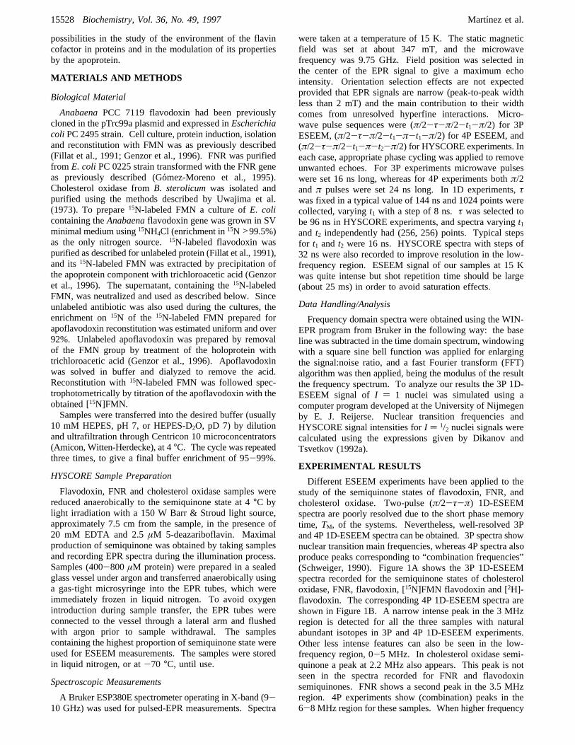
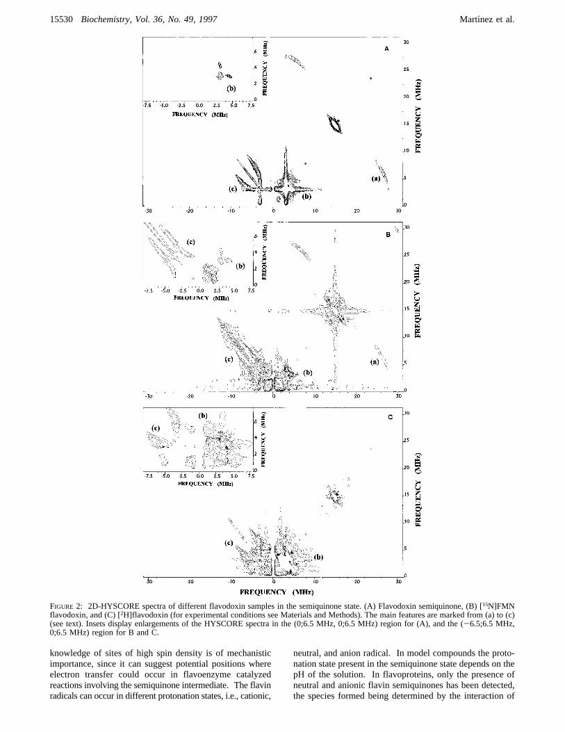
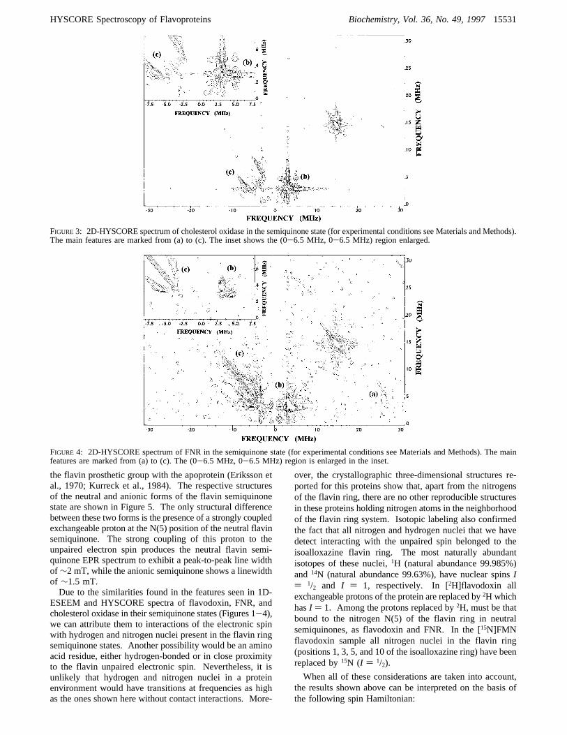
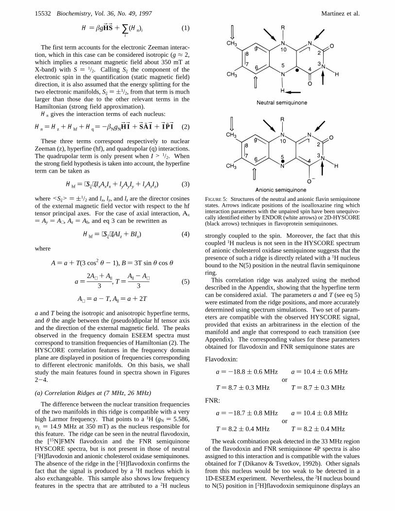
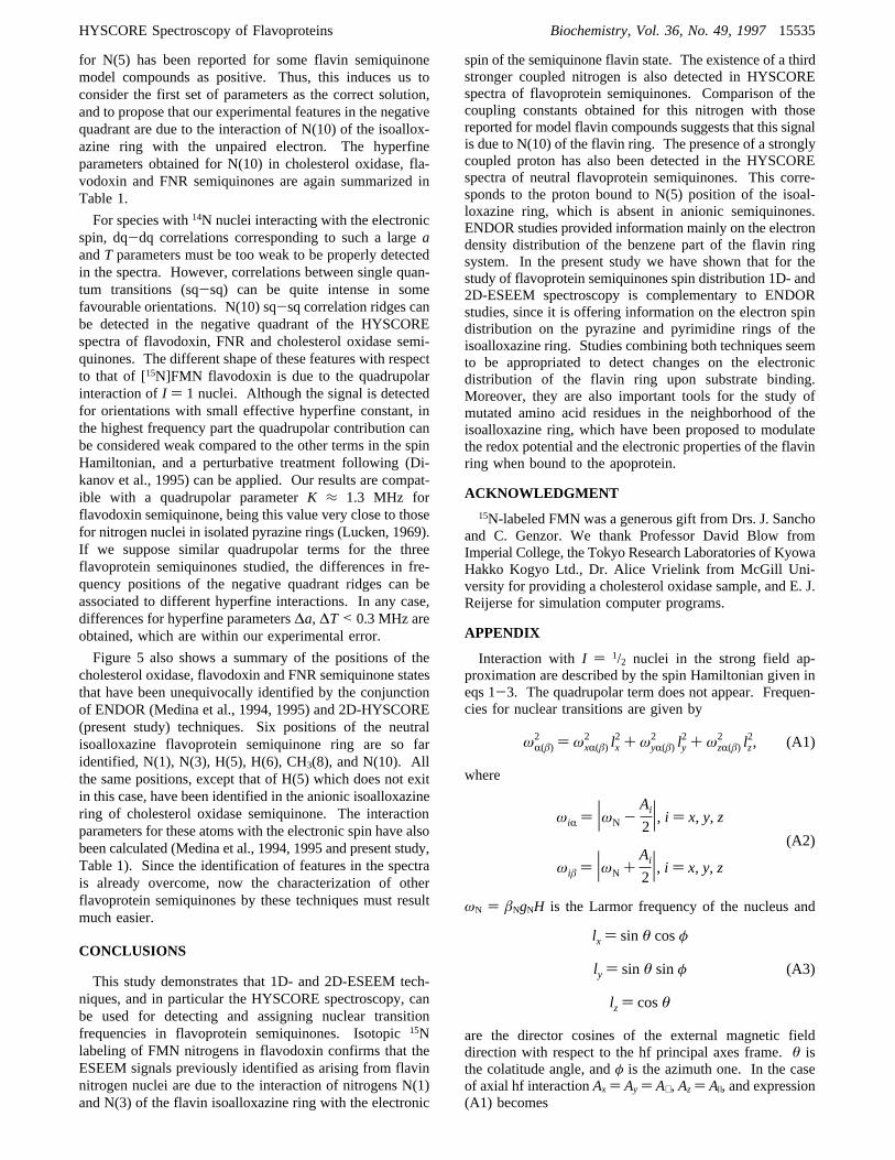
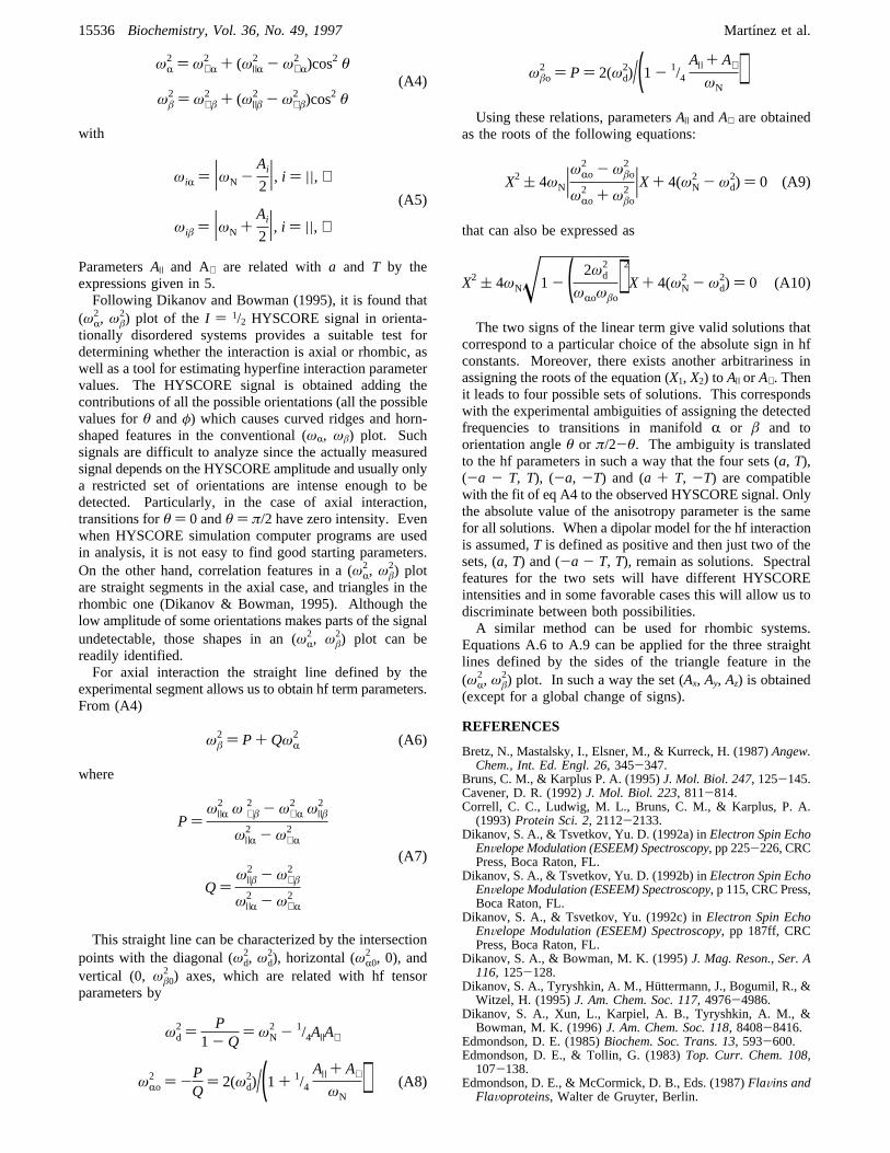
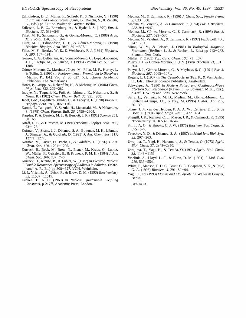
semiquinone samples studied are presented in Figures 2-4.Features shown in the diagonals are due to unwanted echoesor to weakly interacting nuclei. Out-of-diagonal signalscorrespond to correlations between frequencies of transitionsbelonging to different manifolds of the same unpairedelectronic spin. The main correlation features (peaks andridges) detected in the spectra are summarized here:(a) A curved ridge in the positive quadrant, roughly
perpendicular to the diagonal, centered about (7 MHz, 26
MHz). This feature is clearly displayed in the flavodoxinand the [15N]FMN flavodoxin semiquinone HYSCOREspectra, can be distinguish over the noise level in the FNRsemiquinone spectrum, and does not appear at all in thecholesterol oxidase and the [2H]flavodoxin semiquinonespectra.
(b) Two narrow, intense, hardly resolved, peaks in theregion (3 MHz, 5 MHz) (positive quadrant). This featurecan be seen in the spectra of all semiquinone samples, butthe [15N]FMN flavodoxin semiquinone spectrum shows thesepeaks clearly diminished with regard to those shown forunlabeled flavodoxin semiquinone. Besides, for the [15N]-FMN flavodoxin semiquinone sample another pair of cor-relation peaks about (1 MHz, 2 MHz) appears.
(c) A curved correlation ridge in the negative quadrant,approximately parallel to the diagonal. The shape of thisfeature only shows small differences in the spectra obtainedfor flavodoxin, FNR, and cholesterol oxidase semiquinones.No changes were detected for this ridge when studying theHYSCORE spectrum of [2H]flavodoxin semiquinone. Nev-ertheless, the negative quadrant ridge in the HYSCOREspectrum of [15N]FMN flavodoxin semiquinone shows a verydifferent shape and length when compared with the ridgesfound in the rest of flavoprotein semiquinone spectra.
DISCUSSION
The aromatic nature of the heterocyclic flavin ring systemallows a distribution of spin density to be expected. A
FIGURE 1: Fourier transform 3P (A) and 4P (B) 1D-ESEEM spectra of the different flavoprotein samples in the semiquinone state.τ valuewas 144 ns (for experimental conditions see Materials and Methods).
HYSCORE Spectroscopy of Flavoproteins Biochemistry, Vol. 36, No. 49, 199715529
knowledge of sites of high spin density is of mechanisticimportance, since it can suggest potential positions whereelectron transfer could occur in flavoenzyme catalyzedreactions involving the semiquinone intermediate. The flavinradicals can occur in different protonation states, i.e., cationic,
neutral, and anion radical. In model compounds the proto-nation state present in the semiquinone state depends on thepH of the solution. In flavoproteins, only the presence ofneutral and anionic flavin semiquinones has been detected,the species formed being determined by the interaction of
FIGURE 2: 2D-HYSCORE spectra of different flavodoxin samples in the semiquinone state. (A) Flavodoxin semiquinone, (B) [15N]FMNflavodoxin, and (C) [2H]flavodoxin (for experimental conditions see Materials and Methods). The main features are marked from (a) to (c)(see text). Insets display enlargements of the HYSCORE spectra in the (0;6.5 MHz, 0;6.5 MHz) region for (A), and the (-6.5;6.5 MHz,0;6.5 MHz) region for B and C.
15530 Biochemistry, Vol. 36, No. 49, 1997 Martınez et al.
the flavin prosthetic group with the apoprotein (Eriksson etal., 1970; Kurreck et al., 1984). The respective structuresof the neutral and anionic forms of the flavin semiquinonestate are shown in Figure 5. The only structural differencebetween these two forms is the presence of a strongly coupledexchangeable proton at the N(5) position of the neutral flavinsemiquinone. The strong coupling of this proton to theunpaired electron spin produces the neutral flavin semi-quinone EPR spectrum to exhibit a peak-to-peak line widthof ∼2 mT, while the anionic semiquinone shows a linewidthof ∼1.5 mT.Due to the similarities found in the features seen in 1D-
ESEEM and HYSCORE spectra of flavodoxin, FNR, andcholesterol oxidase in their semiquinone states (Figures 1-4),we can attribute them to interactions of the electronic spinwith hydrogen and nitrogen nuclei present in the flavin ringsemiquinone states. Another possibility would be an aminoacid residue, either hydrogen-bonded or in close proximityto the flavin unpaired electronic spin. Nevertheless, it isunlikely that hydrogen and nitrogen nuclei in a proteinenvironment would have transitions at frequencies as highas the ones shown here without contact interactions. More-
over, the crystallographic three-dimensional structures re-ported for this proteins show that, apart from the nitrogensof the flavin ring, there are no other reproducible structuresin these proteins holding nitrogen atoms in the neighborhoodof the flavin ring system. Isotopic labeling also confirmedthe fact that all nitrogen and hydrogen nuclei that we havedetect interacting with the unpaired spin belonged to theisoalloxazine flavin ring. The most naturally abundantisotopes of these nuclei,1H (natural abundance 99.985%)and 14N (natural abundance 99.63%), have nuclear spinsI) 1/2 and I ) 1, respectively. In [2H]flavodoxin allexchangeable protons of the protein are replaced by2H whichhasI ) 1. Among the protons replaced by2H, must be thatbound to the nitrogen N(5) of the flavin ring in neutralsemiquinones, as flavodoxin and FNR. In the [15N]FMNflavodoxin sample all nitrogen nuclei in the flavin ring(positions 1, 3, 5, and 10 of the isoalloxazine ring) have beenreplaced by15N (I ) 1/2).
When all of these considerations are taken into account,the results shown above can be interpreted on the basis ofthe following spin Hamiltonian:
FIGURE3: 2D-HYSCORE spectrum of cholesterol oxidase in the semiquinone state (for experimental conditions see Materials and Methods).The main features are marked from (a) to (c). The inset shows the (0-6.5 MHz, 0-6.5 MHz) region enlarged.
FIGURE 4: 2D-HYSCORE spectrum of FNR in the semiquinone state (for experimental conditions see Materials and Methods). The mainfeatures are marked from (a) to (c). The (0-6.5 MHz, 0-6.5 MHz) region is enlarged in the inset.
HYSCORE Spectroscopy of Flavoproteins Biochemistry, Vol. 36, No. 49, 199715531
The first term accounts for the electronic Zeeman interac-tion, which in this case can be considered isotropic (g ≈ 2,which implies a resonant magnetic field about 350 mT atX-band) with S ) 1/2. Calling Sê the component of theelectronic spin in the quantification (static magnetic field)direction, it is also assumed that the energy splitting for thetwo electronic manifolds,Sê ) (1/2, from that term is muchlarger than those due to the other relevant terms in theHamiltonian (strong field approximation).
Hn gives the interaction terms of each nucleus:
These three terms correspond respectively to nuclearZeeman (z), hyperfine (hf), and quadrupolar (q) interactions.The quadrupolar term is only present whenI > 1/2. Whenthe strong field hypothesis is taken into account, the hyperfineterm can be taken as
where<Sê> ) (1/2 andlx, ly, andlz are the director cosinesof the external magnetic field vector with respect to the hftensor principal axes. For the case of axial interaction,Αx
) Ay ) A⊥, Az ) A|, and eq 3 can be rewritten as
where
a andT being the isotropic and anisotropic hyperfine terms,andθ the angle between the (pseudo)dipolar hf tensor axisand the direction of the external magnetic field. The peaksobserved in the frequency domain ESEEM spectra mustcorrespond to transition frequencies of Hamiltonian (2). TheHYSCORE correlation features in the frequency domainplane are displayed in position of frequencies correspondingto different electronic manifolds. On this basis, we shallstudy the main features found in spectra shown in Figures2-4.
(a) Correlation Ridges at (7 MHz, 26 MHz)
The difference between the nuclear transition frequenciesof the two manifolds in this ridge is compatible with a veryhigh Larmor frequency. That points to a1H (gN ) 5.586,νL ) 14.9 MHz at 350 mT) as the nucleus responsible forthis feature. The ridge can be seen in the neutral flavodoxin,the [15N]FMN flavodoxin and the FNR semiquinoneHYSCORE spectra, but is not present in those of neutral[2H]flavodoxin and anionic cholesterol oxidase semiquinones.The absence of the ridge in the [2H]flavodoxin confirms thefact that the signal is produced by a1H nucleus which isalso exchangeable. This sample also shows low frequencyfeatures in the spectra that are attributed to a2H nucleus
strongly coupled to the spin. Moreover, the fact that thiscoupled1H nucleus is not seen in the HYSCORE spectrumof anionic cholesterol oxidase semiquinone suggests that thepresence of such a ridge is directly related with a1H nucleusbound to the N(5) position in the neutral flavin semiquinonering.This correlation ridge was analyzed using the method
described in the Appendix, showing that the hyperfine termcan be considered axial. The parametersa andT (see eq 5)were estimated from the ridge positions, and more accuratelydetermined using spectrum simulations. Two set of param-eters are compatible with the observed HYSCORE signal,provided that exists an arbitrariness in the election of themanifold and angle that correspond to each transition (seeAppendix). The corresponding values for these parametersobtained for flavodoxin and FNR semiquinone states are
The weak combination peak detected in the 33 MHz regionof the flavodoxin and FNR semiquinone 4P spectra is alsoassigned to this interaction and is compatible with the valuesobtained forT (Dikanov & Tsvetkov, 1992b). Other signalsfrom this nucleus would be too weak to be detected in a1D-ESEEM experiment. Nevertheless, the2H nucleus boundto N(5) position in [2H]flavodoxin semiquinone displays an
FIGURE5: Structures of the neutral and anionic flavin semiquinonestates. Arrows indicate positions of the isoalloxazine ring whichinteraction parameters with the unpaired spin have been unequivo-cally identified either by ENDOR (white arrows) or 2D-HYSCORE(black arrows) techniques in flavoprotein semiquinones.
H ) âgHBSB + ∑i
(Hn)i (1)
Hn ) Hz + Hhf + Hq) -âNgNHB IB + SBA IB + IBPIB (2)
Hhf ) ⟨Sê⟩(lxAxIx + lyAyIy + lzAzIz) (3)
Hhf ) ⟨Sê⟩(AIz + BIx) (4)
A) a+ T(3 cos2 θ - 1),B) 3T sinθ cosθ
a)2A⊥ + A|
3, T)
A| - A⊥
3(5)
A⊥ ) a- T, A| ) a+ 2T
Flavodoxin:
a) -18.8( 0.6 MHz a) 10.4( 0.6 MHzor
T) 8.7( 0.3 MHz T) 8.7( 0.3 MHz
FNR:
a) -18.7( 0.8 MHz a) 10.4( 0.8 MHzor
T) 8.2( 0.4 MHz T) 8.2( 0.4 MHz
15532 Biochemistry, Vol. 36, No. 49, 1997 Martınez et al.
intense 1D-ESEEM signal that can provide additionalinformation in order to discriminate between the twoparameter sets obtained for FNR and flavodoxin1H nucleusat the N(5) semiquinone position. The nuclear Zeeman andhyperfine interaction parameters of2H are proportional tothose of1H following the Larmor frequencies ratio [νL(2H)) 2.3 MHz at 350 mT,νL(2H)/νL(1H) ) 0.153]. Then, twosets of possible parameters for deuterium at N(5) positionin flavodoxin semiquinone are also obtained:
The broad features between 0.5 MHz and 3 MHz in the3P 1D-ESEEM and the combination peak at 5 MHz in the4P 1D-ESEEM spectra of [2H]flavodoxin correspond to theinteraction with this nucleus (Figure 1). The position ofsignals is again compatible with both sets of parameters, butcomputer simulations of 3P spectra show different shapesin each case (Figure 6). The first set leads to a broad signalwith noticeable intensity between 0.5 and 2 MHz, whereasfor the second set this signal is rather an asymmetric peak,whose intensity strongly decreases below 1.5 MHz. Althoughthe actual signal shape is affected by the deuterium quadru-polar term and the spectrometer dead time, the observedshape in the 3P spectrum of [2H]flavodoxin is much betterdescribed by the first set (a ) -2.9; T ) 1.3). Moreover,this election is also supported by the parameters reportedfor hydrogen bound to N(5) in model flavins (Kurreck etal., 1984). A higher value for the hyperfine isotropicparameter of this interaction has also been suggested on thebasis of CW-EPR experiments (Edmondson et al., 1990).Nevertheless, these authors considered in their analysis anearly isotropic hyperfine term, which is not sustained byour results.
(b) Correlation Peaks at (3 MHz, 5 MHz)
This feature may be attributed to two nitrogen nucleiweakly interacting with the electronic spin. That excludesnitrogens at positions 5 and 10 of the flavin ring. It is knownthat there is a high electronic spin density on these positions,and their hyperfine interaction parameters, particularly theisotropic one, are quite large, about 20 MHz for N(5) and10 MHz for N(10) (Kurreck, 1984; Edmondson, 1985).Therefore, the observed signals might be due to the othernitrogens in the isoalloxazine ring (positions 1 and 3), or toother nitrogen nuclei of the protein, out of the flavin ringbut close to it. We have seen that labeling of flavodoxinFMN cofactor with15N isotope causes noticeable changesin the experimental features (Figure 2B). Correlation peaks(3 MHz, 5 MHz) can still be seen, but significantly reducedin intensity. Besides, other peaks at lower frequencies(1 MHz, 2 MHz) appear. Since only nitrogen nuclei in theflavin ring have been substituted by isotope labeling, thechanges in spectra indicates that the observed signals mustcorrespond to N(1) and N(3) of the isoalloxazine ring. Theresidual signal observed as correlation peaks (3 MHz,5 MHz) can be interpreted as unlabeled14N(1) and14N(3),due to the fact that labeling with15N did not reach the 100%(see Materials and Methods). Some authors have suggestedan isotropic hyperfine parameter for these nuclei about1 MHz (Edmondson et al., 1990). In previous works,interaction parameters for N(1) and N(3) for cholesteroloxidase, flavodoxin, and FNR semiquinones were alreadydescribed from 3P ESEEM experiments. Now, our HY-SCOREmeasurements confirm the tentative assignment from1D-ESEEM features to these nitrogens (Medina & Cammack,1996; Medina et al., 1997).
These correlation peaks, (3 MHz, 5 MHz), observed inthe HYSCORE experiments were also analyzed. Twonarrow peaks, very close one to the other, can be observed.It is known that when14N (I ) 1) interacts with an electronicspin in such a way that the isotropic hyperfine termdominates over the anisotropic and the quadrupolar terms,the frequency of the “double quantum” (dq) transitions (∆Iz) (2) is nearly independent of the magnetic field orientation,and displays the most intense peaks in the ESEEM andHYSCORE spectra of orientationally disordered samples. Ifthe isotropic hyperfine parameter matches the “exact cancel-lation” condition,νL ) a/2, three peaks, corresponding tothe principal values of the quadrupolar tensor, are displayedcoming from the cancelled electronic manifold. Besides, thedq peak of the other manifold must also be seen. Then, wepropose that the observed correlation peaks correspond todq-dq correlation of the two nitrogen nuclei at positions 1and 3 of the isoalloxazine flavin ring. The absolute valueof the isotropic hyperfine parameter, and some informationabout the quadrupolar tensor, can be obtained from thefrequencies observed for these nuclei in the HYSCOREspectra (Dikanov & Tsvetkov, 1992c). Because we cannotassign a specific peak to a particular nucleus, N(1) or N(3),herein we will label the observed correlation peaks as N(A)and N(B). The hyperfine coupling constants (a), quadrupolarparameters (K), and asymmetry parameters (η) for cholesteroloxidase, FNR, and flavodoxin semiquinones are summarizedin Table 1, where
FIGURE 6: Computer simulation of the 3P 1D-ESEEM deuteriumfeatures in [2H]flavodoxin for the two possible sets of parameters:(A) a ) -2.9 MHz, T ) 1.3 MHz; (B) a ) 1.6 MHz, T ) 1.3MHz. Simulations include neither intrinsic width for signals norquadrupolar term or spectrometer dead time effects. In spite of that,actual feature in the region between 0 and 2 MHz (see Figure 1)appears to be incompatible with simulation in B (see text).
a) -2.9( 0.1 MHz a) 1.6( 0.1 MHzor
T) 1.3( 0.1 MHz T) 1.3( 0.1 MHz
HYSCORE Spectroscopy of Flavoproteins Biochemistry, Vol. 36, No. 49, 199715533
x′, y′, andz′ being the principal directions of the quadrupolartensor.It is worthy to note that for some of the nitrogen nuclei
observed in these flavoprotein semiquinones the isotropichyperfine parameter cannot be considered dominant withrespect to the quadrupolar term. However, simulations of3P ESEEM spectra demonstrate that, as long as the aniso-tropic hyperfine term is small, the dq feature remains as anarrow, relatively intense peak (at least in one of the twoelectronic manifolds). In these cases the dq-dq correlationis still dominant in the HYSCORE spectrum.For one of the nitrogen nuclei coupled to the spin in
cholesterol oxidase semiquinone, the exact cancellationcondition is nearly verified. In this case, the peak observedin the 3P ESEEM spectrum corresponds to the cancelledmanifold and allows us to obtain the quadrupolar parametersfor nucleus N(A) in cholesterol oxidase semiquinone. Theobtained values for this nucleus areK ) 0.83( 0.2 MHzandη ) 0.5( 0.1. This value for the quadrupolar term (K)is slightly smaller than those reported (Lucken, 1969) fornitrogen in an isolated pyrimidine ring. Since peaks corre-sponding to N(A) in cholesterol oxidase semiquinone arequite narrow, it is likely that the anisotropic hyperfine termfor N(A) in this sample is very small compared to theisotropic one. Just minor changes have been found for allthe parameters calculated for cholesterol oxidase semiquinoneHYSCORE spectra with regard to those already reported in1D-ESEEM (Medina et al., 1997). Nevertheless, the im-provement of the signal:noise ratio achieved by the 2Dtechniques, together with the higher resolution of theHYSCORE experiment as compared with 3P ones, give abetter accuracy for the calculated parameter values. Havinginto account that the quadrupolar term is hardly influencedby changes in the ring far from the corresponding nucleus,values for quadrupolar parameters of N(A) in cholesteroloxidase semiquinone can be also considered a valid estima-tion for flavodoxin and FNR semiquinones.To prove the correctness of our analysis, the frequencies
for peaks corresponding to15N(A) and15N(B) in [15N]FMNflavodoxin have been calculated. The rescaling of param-eters for a15N (I ) 1/2) nucleus gives the following nucleartransitions:
The corresponding peaks can be observed in the [15N]FMNflavodoxin semiquinone spectra. In 1D-ESEEM measure-ments two broad features in the regions of 1 MHz and 2MHz are seen, whereas the HYSCORE spectrum shows abroad correlation at (1 MHz, 2 MHz). Unfortunately, forthe moment we are not in the position to assign each set ofparameters to N(1) or N(3). Nevertheless, the possibility ofassignation of these signals to other nuclei out of the flavinring has been ruled out.
(c) Correlation Ridges in the NegatiVe Quadrant
This feature of the HYSCORE spectra, that appears in allthe flavoprotein semiquinones studied (Figures 2-4), showsa considerable change in shape when [14N]FMN is replacedby [15N]FMN in flavodoxin semiquinone (Figures 2A and2B). This leads us to propose that it can be attributed toone of the flavin nitrogens. Since the analysis ofI ) 1/2signals has been shown to be easier, we will start analyzingthe [15N]FMN flavodoxin semiquinone HYSCORE spectrumfollowing the method described in the Appendix. The ridgeobserved in this spectrum (Figure 2B) corresponds to an axialinteraction with two compatible sets of parameters:
When these values are rescaled for the [14N]FMN flavodoxinsemiquinone the following data are obtained:
Isotropic hyperfine parameters have been determined forseveral flavin model compounds (Kurreck, 1984). Theabsolute value ofa found in our first set of parameters is inthe range of those reported for N(10) of the isoalloxazinering, whereas the absolute value ofa in the second set ofparameters would be consistent with nitrogen N(5) of thesame ring. Nevertheless, the sign of the isotropic parameter
Table 1: 1H and14N Interaction Parameters of the Isoalloxazine Nuclei with the Unpaired Electronic Spin in the Semiquinone State ofDifferent Flavoproteins
flavoprotein semiquinone atom position a (MHz) T (MHz) K (MHz) η K2 (3+ η) (MHz2)
cholesterol oxidase H(5)N(A)a 1.9( 0.2b nd 0.83( 0.2 0.5( 0.1 2.3( 0.2N(B)a 0.7( 0.2b nd nd nd 2.1( 0.1N(10) 11.7( 1.0 9.0( 1.1 1.3e nd nd
FNR H(5) -18.7( 0.8 8.2( 0.4N(A)a 1.2( 0.2b nd 0.83( 0.2c 0.5( 0.1c 2.2( 0.1N(B)a 0.7( 0.3b nd nd nd 2.4( 0.2N(10) 11.7( 1.0 9.0( 1.1 1.3e nd nd
flavodoxin H(5) -18.8( 0.6 8.7( 0.3N(A)a 1.3( 0.3b nd 0.83( 0.2c 0.5( 0.1c 2.2( 0.1N(B)a 0.8( 0.2b nd nd nd 2.1( 0.1N(10) 11.7( 1.0 9.0( 1.1 1.3d nd nd
a These nitrogens correspond to N(1) or N(3) sites in the flavin ring.bOnly the absolute value has been determined.c Estimated from valuesobtained for cholesterol oxidase.d Approximated value.eEstimated from value obtained for flavodoxin.
K )|Pz′|2
) |e2qQ4h |, η ) |Px′ - Py′Pz′| (6) N(A){νR ) 2.5( 0.3 MHz
νâ ) 0.5( 0.2 MHz}N(B){νR ) 2.0( 0.3 MHz
νâ ) 0.9( 0.3 MHz}
a) 15.9( 1.4 MHz a) -28.3( 1.4 MHzor
T) 12.3( 1.1 MHz T) 12.3( 1.1 MHz
a) 11.7( 1.0 MHz a) -20.8( 1.0 MHzor
T) 9.0( 0.8 MHz T) 9.0( 0.8 MHz
15534 Biochemistry, Vol. 36, No. 49, 1997 Martınez et al.
for N(5) has been reported for some flavin semiquinonemodel compounds as positive. Thus, this induces us toconsider the first set of parameters as the correct solution,and to propose that our experimental features in the negativequadrant are due to the interaction of N(10) of the isoallox-azine ring with the unpaired electron. The hyperfineparameters obtained for N(10) in cholesterol oxidase, fla-vodoxin and FNR semiquinones are again summarized inTable 1.
For species with14N nuclei interacting with the electronicspin, dq-dq correlations corresponding to such a largeaandT parameters must be too weak to be properly detectedin the spectra. However, correlations between single quan-tum transitions (sq-sq) can be quite intense in somefavourable orientations. N(10) sq-sq correlation ridges canbe detected in the negative quadrant of the HYSCOREspectra of flavodoxin, FNR and cholesterol oxidase semi-quinones. The different shape of these features with respectto that of [15N]FMN flavodoxin is due to the quadrupolarinteraction ofI ) 1 nuclei. Although the signal is detectedfor orientations with small effective hyperfine constant, inthe highest frequency part the quadrupolar contribution canbe considered weak compared to the other terms in the spinHamiltonian, and a perturbative treatment following (Di-kanov et al., 1995) can be applied. Our results are compat-ible with a quadrupolar parameterK ≈ 1.3 MHz forflavodoxin semiquinone, being this value very close to thosefor nitrogen nuclei in isolated pyrazine rings (Lucken, 1969).If we suppose similar quadrupolar terms for the threeflavoprotein semiquinones studied, the differences in fre-quency positions of the negative quadrant ridges can beassociated to different hyperfine interactions. In any case,differences for hyperfine parameters∆a, ∆T< 0.3 MHz areobtained, which are within our experimental error.
Figure 5 also shows a summary of the positions of thecholesterol oxidase, flavodoxin and FNR semiquinone statesthat have been unequivocally identified by the conjunctionof ENDOR (Medina et al., 1994, 1995) and 2D-HYSCORE(present study) techniques. Six positions of the neutralisoalloxazine flavoprotein semiquinone ring are so faridentified, N(1), N(3), H(5), H(6), CH3(8), and N(10). Allthe same positions, except that of H(5) which does not exitin this case, have been identified in the anionic isoalloxazinering of cholesterol oxidase semiquinone. The interactionparameters for these atoms with the electronic spin have alsobeen calculated (Medina et al., 1994, 1995 and present study,Table 1). Since the identification of features in the spectrais already overcome, now the characterization of otherflavoprotein semiquinones by these techniques must resultmuch easier.
CONCLUSIONS
This study demonstrates that 1D- and 2D-ESEEM tech-niques, and in particular the HYSCORE spectroscopy, canbe used for detecting and assigning nuclear transitionfrequencies in flavoprotein semiquinones. Isotopic15Nlabeling of FMN nitrogens in flavodoxin confirms that theESEEM signals previously identified as arising from flavinnitrogen nuclei are due to the interaction of nitrogens N(1)and N(3) of the flavin isoalloxazine ring with the electronic
spin of the semiquinone flavin state. The existence of a thirdstronger coupled nitrogen is also detected in HYSCOREspectra of flavoprotein semiquinones. Comparison of thecoupling constants obtained for this nitrogen with thosereported for model flavin compounds suggests that this signalis due to N(10) of the flavin ring. The presence of a stronglycoupled proton has also been detected in the HYSCOREspectra of neutral flavoprotein semiquinones. This corre-sponds to the proton bound to N(5) position of the isoal-loxazine ring, which is absent in anionic semiquinones.ENDOR studies provided information mainly on the electrondensity distribution of the benzene part of the flavin ringsystem. In the present study we have shown that for thestudy of flavoprotein semiquinones spin distribution 1D- and2D-ESEEM spectroscopy is complementary to ENDORstudies, since it is offering information on the electron spindistribution on the pyrazine and pyrimidine rings of theisoalloxazine ring. Studies combining both techniques seemto be appropriated to detect changes on the electronicdistribution of the flavin ring upon substrate binding.Moreover, they are also important tools for the study ofmutated amino acid residues in the neighborhood of theisoalloxazine ring, which have been proposed to modulatethe redox potential and the electronic properties of the flavinring when bound to the apoprotein.
ACKNOWLEDGMENT15N-labeled FMN was a generous gift from Drs. J. Sancho
and C. Genzor. We thank Professor David Blow fromImperial College, the Tokyo Research Laboratories of KyowaHakko Kogyo Ltd., Dr. Alice Vrielink from McGill Uni-versity for providing a cholesterol oxidase sample, and E. J.Reijerse for simulation computer programs.
APPENDIX
Interaction with I ) 1/2 nuclei in the strong field ap-proximation are described by the spin Hamiltonian given ineqs 1-3. The quadrupolar term does not appear. Frequen-cies for nuclear transitions are given by
where
ωN ) âNgNH is the Larmor frequency of the nucleus and
are the director cosines of the external magnetic fielddirection with respect to the hf principal axes frame.θ isthe colatitude angle, andφ is the azimuth one. In the caseof axial hf interactionAx ) Ay ) A⊥, Az ) A|, and expression(A1) becomes
ωR(â)2 ) ωxR(â)
2 lx2 + ωyR(â)
2 ly2 + ωzR(â)
2 lz2, (A1)
ωiR ) |ωN -Ai2|, i ) x, y, z
(A2)
ωiâ ) |ωN +Ai2|, i ) x, y, z
lx ) sinθ cosφ
ly ) sinθ sinφ (A3)
lz ) cosθ
HYSCORE Spectroscopy of Flavoproteins Biochemistry, Vol. 36, No. 49, 199715535
with
ParametersA| and A⊥ are related witha and T by theexpressions given in 5.Following Dikanov and Bowman (1995), it is found that
(ωR2, ωâ
2) plot of the I ) 1/2 HYSCORE signal in orienta-tionally disordered systems provides a suitable test fordetermining whether the interaction is axial or rhombic, aswell as a tool for estimating hyperfine interaction parametervalues. The HYSCORE signal is obtained adding thecontributions of all the possible orientations (all the possiblevalues forθ andφ) which causes curved ridges and horn-shaped features in the conventional (ωR, ωâ) plot. Suchsignals are difficult to analyze since the actually measuredsignal depends on the HYSCORE amplitude and usually onlya restricted set of orientations are intense enough to bedetected. Particularly, in the case of axial interaction,transitions forθ ) 0 andθ ) π/2 have zero intensity. Evenwhen HYSCORE simulation computer programs are usedin analysis, it is not easy to find good starting parameters.On the other hand, correlation features in a (ωR
2, ωâ2) plot
are straight segments in the axial case, and triangles in therhombic one (Dikanov & Bowman, 1995). Although thelow amplitude of some orientations makes parts of the signalundetectable, those shapes in an (ωR
2, ωâ2) plot can be
readily identified.For axial interaction the straight line defined by the
experimental segment allows us to obtain hf term parameters.From (A4)
where
This straight line can be characterized by the intersectionpoints with the diagonal (ωd
2, ωd2), horizontal (ωR0
2 , 0), andvertical (0, ωâ0
2 ) axes, which are related with hf tensorparameters by
Using these relations, parametersA| andA⊥ are obtainedas the roots of the following equations:
that can also be expressed as
The two signs of the linear term give valid solutions thatcorrespond to a particular choice of the absolute sign in hfconstants. Moreover, there exists another arbitrariness inassigning the roots of the equation (X1, X2) toA| orA⊥. Thenit leads to four possible sets of solutions. This correspondswith the experimental ambiguities of assigning the detectedfrequencies to transitions in manifoldR or â and toorientation angleθ or π/2-θ. The ambiguity is translatedto the hf parameters in such a way that the four sets (a, T),(-a - T, T), (-a, -T) and (a + T, -T) are compatiblewith the fit of eq A4 to the observed HYSCORE signal. Onlythe absolute value of the anisotropy parameter is the samefor all solutions. When a dipolar model for the hf interactionis assumed,T is defined as positive and then just two of thesets, (a, T) and (-a - T, T), remain as solutions. Spectralfeatures for the two sets will have different HYSCOREintensities and in some favorable cases this will allow us todiscriminate between both possibilities.A similar method can be used for rhombic systems.
Equations A.6 to A.9 can be applied for the three straightlines defined by the sides of the triangle feature in the(ωR
2, ωâ2) plot. In such a way the set (Ax, Ay, Az) is obtained
(except for a global change of signs).
REFERENCES
Bretz, N., Mastalsky, I., Elsner, M., & Kurreck, H. (1987)Angew.Chem., Int. Ed. Engl. 26, 345-347.
Bruns, C. M., & Karplus P. A. (1995)J. Mol. Biol. 247, 125-145.Cavener, D. R. (1992)J. Mol. Biol. 223, 811-814.Correll, C. C., Ludwig, M. L., Bruns, C. M., & Karplus, P. A.(1993)Protein Sci. 2, 2112-2133.
Dikanov, S. A., & Tsvetkov, Yu. D. (1992a) inElectron Spin EchoEnVelope Modulation (ESEEM) Spectroscopy, pp 225-226, CRCPress, Boca Raton, FL.
Dikanov, S. A., & Tsvetkov, Yu. D. (1992b) inElectron Spin EchoEnVelope Modulation (ESEEM) Spectroscopy, p 115, CRC Press,Boca Raton, FL.
Dikanov, S. A., & Tsvetkov, Yu. (1992c) inElectron Spin EchoEnVelope Modulation (ESEEM) Spectroscopy, pp 187ff, CRCPress, Boca Raton, FL.
Dikanov, S. A., & Bowman, M. K. (1995)J. Mag. Reson., Ser. A116,125-128.
Dikanov, S. A., Tyryshkin, A. M., Hu¨ttermann, J., Bogumil, R., &Witzel, H. (1995)J. Am. Chem. Soc. 117, 4976-4986.
Dikanov, S. A., Xun, L., Karpiel, A. B., Tyryshkin, A. M., &Bowman, M. K. (1996)J. Am. Chem. Soc. 118, 8408-8416.
Edmondson, D. E. (1985)Biochem. Soc. Trans. 13, 593-600.Edmondson, D. E., & Tollin, G. (1983)Top. Curr. Chem. 108,107-138.
Edmondson, D. E., & McCormick, D. B., Eds. (1987)FlaVins andFlaVoproteins, Walter de Gruyter, Berlin.
ωR2 ) ω⊥R
2 + (ω|R2 - ω⊥R
2 )cos2 θ(A4)
ωâ2 ) ω⊥â
2 + (ω|â2 - ω⊥â
2 )cos2 θ
ωiR ) |ωN -Ai2|, i ) ||, ⊥
(A5)
ωiâ ) |ωN +Ai2|, i ) ||, ⊥
ωâ2 ) P+ QωR
2 (A6)
P)ω|R2 ω ⊥â
2 - ω⊥R2 ω|â
2
ω|R2 - ω⊥R
2
(A7)
Q)ω|â2 - ω⊥â
2
ω|R2 - ω⊥R
2
ωd2 ) P
1- Q) ωN
2 - 1/4A|A⊥
ωRo2 ) -P
Q) 2(ωd
2)/(1+ 1/4A| + A⊥
ωN) (A8)
ωâo2 ) P) 2(ωd
2)/(1- 1/4A| + A⊥
ωN)
X2 ( 4ωN|ωRo2 - ωâo
2
ωRo2 + ωâo
2 |X+ 4(ωN2 - ωd
2) ) 0 (A9)
X2 ( 4ωNx1- ( 2ωd2
ωRoωâo)2X+ 4(ωN
2 - ωd2) ) 0 (A10)
15536 Biochemistry, Vol. 36, No. 49, 1997 Martınez et al.
Edmondson, D. E., Mu¨ller, F., Schaub, F., & Nisimoto, Y. (1990)in FlaVins and FlaVoproteins(Curti, B., Ronchi, S., & Zanetti,G., Eds.) pp 67-72. Walter de Gruyter, Berlin.
Eriksson, L. E. G., Ehrenberg, A., & Hyde, J. S. (1970)Eur. J.Biochem. 17, 539-543.
Fillat, M. F., Sandmann, G., & Go´mez-Moreno, C. (1988)Arch.Mircrobiol. 150, 160-164.
Fillat, M. F., Edmondson, D. E., & Go´mez-Moreno, C. (1990)Biochim. Biophys. Acta 1040, 301-307.
Fillat, M. F., Borrias, W. E., & Weisbeerk, P. J. (1991)Biochem.J. 280, 187-191.
Genzor, C. G., Belbarrain, A., Go´mez-Moreno, C., Lo´pez-Lacomba,J. L., Cortijo, M., & Sancho, J. (1996)Protein Sci. 5, 1376-1388.
Gomez-Moreno, C., Martı´nez-Ju´lvez, M., Fillat, M. F., Hurley, J.,& Tollin, G. (1995) inPhotosynthesis: From Light to Biosphere(Mathis, P., Ed.) Vol. 2, pp 627-632, Kluwer AcademicPublishers, The Netherlands.
Hofer, P., Grupp, A., Nebenfu¨hr, H., & Mehring, M. (1986)Chem.Phys. Lett. 132, 279-282.
Inouye, Y., Taguchi, K., Fuji, A., Ishimaru, K., Nakamura, S., &Nomi, R. (1982)Chem. Pharm. Bull. 30, 951-958.
Janot, J.-M., Capeillere-Blandin, C., & Labeyrie, F. (1990)Biochem.Biophys. Acta 1016, 165-176.
Kamei, T., Takiguchi, Y. Suzuki, H., Matsuzaki, M., & Nakamura,S. (1978)Chem. Pharm. Bull. 26, 2799-2804.
Karplus, P. A, Daniels, M. J., & Herriott, J. R. (1991)Science251,60-66.
Knaff, D. B., & Hirasawa, M. (1991)Biochim. Biophys. Acta 1056,93-125.
Kofman, V., Shane, J. J., Dikanov, S. A., Bowman, M. K., Libman,J., Shanzer, A., & Goldfarb, D. (1995)J. Am. Chem. Soc. 117,12771-12778.
Kofman, V., Farver, O., Pecht, I., & Goldfarb, D. (1996)J. Am.Chem. Soc. 118, 1201-1206.
Kurreck, H., Bock, M., Bretz, N., Elsner, M., Kraus, G., Lubitz,W., Muller, F., Geissler, H., & Kroneck, P. M. H. (1984)J. Am.Chem. Soc. 106, 737-746.
Kurreck, H., Kirsrte, B., & Lubitz, W. (1987) inElectron NuclearDouble Resonance Spectroscopy of Radicals in Solution.(Marc-hand, A. P., Ed.) pp 308-327. VCH, Weinheim.
Li, J., Vrielink, A., Brick, P., & Blow, D. M. (1993)Biochemistry32, 11507-11515.
Lucken, E. A. C. (1969) inNuclear Quadrupole CouplingConstants, p 217ff, Academic Press, London.
Medina, M., & Cammack, R. (1996)J. Chem. Soc., Perkin Trans.2, 633-638.
Medina, M., Vrielink, A., & Cammack, R. (1994)Eur. J. Biochem.222, 941-947.
Medina, M., Gomez-Moreno, C., & Cammack, R. (1995)Eur. J.Biochem. 227, 529-536.
Medina, M., Vrielink, A., & Cammack, R. (1997)FEBS Lett. 400,247-251.
Mims, W. V., & Peisach, J. (1981) inBiological MagneticResonance(Berliner, L. J., & Reuben, J., Eds.) pp 213-263,Plenum, New York.
Muller, F. (1983)Top. Curr. Chem. 108, 71-107.Pueyo, J. J., & Go´mez-Moreno, C. (1991)Prep. Biochem. 21, 191-204.
Pueyo, J. J., Go´mez-Moreno, C., & Mayhew, S. G. (1991)Eur. J.Biochem. 202, 1065-1071.
Rogers, L. J. (1987) inThe Cyanobacteria(Fay, P., & Van Baslen,C., Eds.) Elsevier Science Publishers, Amsterdam.
Schweiger, A. (1990) inModern Pulsed and Continuous-WaVeElectron Spin Resonance(Kevan, L., & Bowman, M. K., Eds.),p 43ff, J. Wiley and Sons, New York.
Serre, L., Vellieux, F. M. D., Medina, M., Go´mez-Moreno, C.,Fontecilla-Camps, J.C., & Frey, M. (1996)J. Mol. Biol. 263,20-39.
Shane, J. J., van der Heijden, P. A. A. W., Reijerse, E. J., & deBoer, E. (1994)Appl. Magn. Res. 6, 427-454.
Shergill, J. K., Joannou, C. L., Mason, J. R., & Cammack, R. (1995)Biochemistry 34, 16532-16542.
Smith, A. G., & Brooks, C. J. W. (1975)Biochem. Soc. Trans. 3,675-677.
Tsvetkov, Y. D., & Dikanov, S. A., (1987) inMetal Ions Biol. Syst.22, 207-263.
Uwajima, T., Yagi, H., Nakamura, S., & Terada, O. (1973)Agric.Biol. Chem. 37, 2345-2350.
Uwajima, T., Yagi, H., & Terada, O. (1974)Agric. Biol. Chem.38, 1149-1156
Vrielink, A., Lloyd, L. F., & Blow, D. M. (1991) J. Mol. Biol.219, 533-554.
White, P., Manson, F. D. C., Brunt, C. E., Chapman, S. K., & Reid,G. A. (1993)Biochem. J.291, 89-94.
Yagi, K., Ed. (1993)FlaVins and FlaVoproteins, Walter de Gruyter,Berlin.
BI971495G
HYSCORE Spectroscopy of Flavoproteins Biochemistry, Vol. 36, No. 49, 199715537