Embed Size (px)
Citation preview
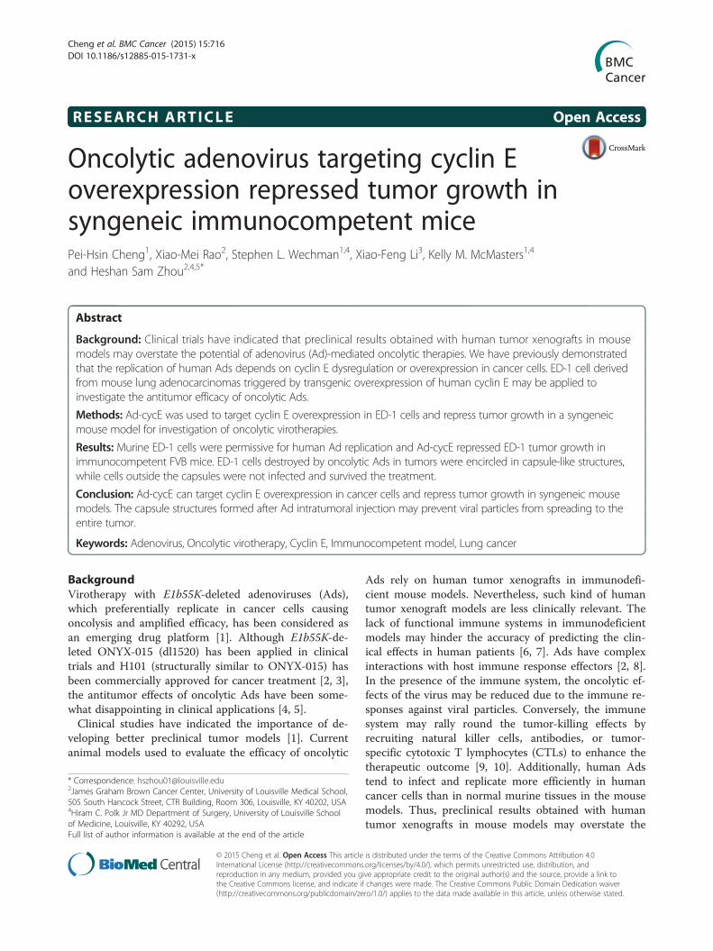
RESEARCH ARTICLE Open Access
Oncolytic adenovirus targeting cyclin Eoverexpression repressed tumor growth insyngeneic immunocompetent micePei-Hsin Cheng1, Xiao-Mei Rao2, Stephen L. Wechman1,4, Xiao-Feng Li3, Kelly M. McMasters1,4
and Heshan Sam Zhou2,4,5*
Abstract
Background: Clinical trials have indicated that preclinical results obtained with human tumor xenografts in mousemodels may overstate the potential of adenovirus (Ad)-mediated oncolytic therapies. We have previously demonstratedthat the replication of human Ads depends on cyclin E dysregulation or overexpression in cancer cells. ED-1 cell derivedfrom mouse lung adenocarcinomas triggered by transgenic overexpression of human cyclin E may be applied toinvestigate the antitumor efficacy of oncolytic Ads.
Methods: Ad-cycE was used to target cyclin E overexpression in ED-1 cells and repress tumor growth in a syngeneicmouse model for investigation of oncolytic virotherapies.
Results: Murine ED-1 cells were permissive for human Ad replication and Ad-cycE repressed ED-1 tumor growth inimmunocompetent FVB mice. ED-1 cells destroyed by oncolytic Ads in tumors were encircled in capsule-like structures,while cells outside the capsules were not infected and survived the treatment.
Conclusion: Ad-cycE can target cyclin E overexpression in cancer cells and repress tumor growth in syngeneic mousemodels. The capsule structures formed after Ad intratumoral injection may prevent viral particles from spreading to theentire tumor.
Keywords: Adenovirus, Oncolytic virotherapy, Cyclin E, Immunocompetent model, Lung cancer
BackgroundVirotherapy with E1b55K-deleted adenoviruses (Ads),which preferentially replicate in cancer cells causingoncolysis and amplified efficacy, has been considered asan emerging drug platform [1]. Although E1b55K-de-leted ONYX-015 (dl1520) has been applied in clinicaltrials and H101 (structurally similar to ONYX-015) hasbeen commercially approved for cancer treatment [2, 3],the antitumor effects of oncolytic Ads have been some-what disappointing in clinical applications [4, 5].Clinical studies have indicated the importance of de-
veloping better preclinical tumor models [1]. Currentanimal models used to evaluate the efficacy of oncolytic
Ads rely on human tumor xenografts in immunodefi-cient mouse models. Nevertheless, such kind of humantumor xenograft models are less clinically relevant. Thelack of functional immune systems in immunodeficientmodels may hinder the accuracy of predicting the clin-ical effects in human patients [6, 7]. Ads have complexinteractions with host immune response effectors [2, 8].In the presence of the immune system, the oncolytic ef-fects of the virus may be reduced due to the immune re-sponses against viral particles. Conversely, the immunesystem may rally round the tumor-killing effects byrecruiting natural killer cells, antibodies, or tumor-specific cytotoxic T lymphocytes (CTLs) to enhance thetherapeutic outcome [9, 10]. Additionally, human Adstend to infect and replicate more efficiently in humancancer cells than in normal murine tissues in the mousemodels. Thus, preclinical results obtained with humantumor xenografts in mouse models may overstate the
* Correspondence: [email protected] Graham Brown Cancer Center, University of Louisville Medical School,505 South Hancock Street, CTR Building, Room 306, Louisville, KY 40202, USA4Hiram C. Polk Jr MD Department of Surgery, University of Louisville Schoolof Medicine, Louisville, KY 40292, USAFull list of author information is available at the end of the article
© 2015 Cheng et al. Open Access This article is distributed under the terms of the Creative Commons Attribution 4.0International License (http://creativecommons.org/licenses/by/4.0/), which permits unrestricted use, distribution, andreproduction in any medium, provided you give appropriate credit to the original author(s) and the source, provide a link tothe Creative Commons license, and indicate if changes were made. The Creative Commons Public Domain Dedication waiver(http://creativecommons.org/publicdomain/zero/1.0/) applies to the data made available in this article, unless otherwise stated.
Cheng et al. BMC Cancer (2015) 15:716 DOI 10.1186/s12885-015-1731-x
therapeutic potential. In fact, results of clinical trialsoften fall short of hopes and expectations based on pre-clinical animal studies. It has become clear that the de-velopment of suitable immunocompetent murine cancermodels for studies of Ad-mediated oncolysis will benefitthe evaluation of virotherapies in more clinically relevantsettings.It was originally proposed that the E1b55K-deleted
Ads could replicate only in p53-deficient tumor cells, asthe E1B55K-mediated degradation of p53 protein wasnot required in those cancer cells [11, 12]. However, theoriginal hypothesis was challenged by several studiesshowing that E1b55K-deleted Ads are able to replicate incells regardless of their p53 status [13–16]. Previously,we have demonstrated that cyclin E dysregulation oroverexpression in cancer cells is an important molecularbasis of selective replication of E1b55K-deleted Ads inhuman cancer cells [17, 18]. Wild-type Ad infection in-duces cyclin E overexpression in normal and cancercells. E1b55K-deleted Ads fail to efficiently induce cyclinE in normal cells, and thus viral replication is restricted;however, E1b55K-deleted Ads can efficiently inducecyclin E in cancer cells with dysregulated cyclin Eand successfully replicate in these cancer cells. Wehave reported that Ad-induced cyclin E activatesCDK2 and targets the transcriptional repressor pRbthat may affect the cellular environment for viral pro-ductive replication [19].Cyclin E is a nuclear protein essential for the cell cycle
progression [20], DNA replication [21, 22], and centro-some duplication [23, 24]. Numerous types of cancersare highly associated with dysregulation of cyclin E [25].Dysregulation of cyclin E occurs in more than 90 % oflung, liver, and gastrointestinal cancers, and in morethan 80 % of glioma/blastoma, bone, and breast cancers[26]. Constitutive overexpression of cyclin E induceschromosome instability [27, 28], impairs normal cellcycle progression, and triggers tumor development intransgenic animal models [29–31]. Human cyclin E over-expression in mouse lungs lead to the development ofpremalignant and malignant lung lesions that resemblethe features found in lung cancer patients [31, 32]. Amurine ED-1 cell line was derived from lung cancers ofcyclin E transgenic mice [32, 33].We have developed a novel E1b-deleted oncolytic Ad
vector, Ad-cycE, in which the E1a gene is under the con-trol of the human cyclin E promoter [34]. With the dele-tion of entire E1b region, Ad-cycE shares the replicationpattern similar to E1b55K-deleted dl1520 which relieson the cyclin E overexpression in cancer cells. As thecyclin E promoter is highly active in multiple types ofcancer cells and would be further stimulated after Ad in-fection, Ad-cycE replication could be enhanced in cancercells. We showed that Ad-cycE elicits efficient antitumor
effects not only in cancer cells reported as permissivefor dl1520 replication but also in those reported as non-permissive for dl1520. Ad-cycE significantly repressedtumor growth in the immunodeficient nude mice bear-ing human lung cancer xenografts. In this study, weaimed to evaluate the impact of Ad-cycE in a more clin-ical relevant model. We compared and characterized thereplication pattern of oncolytic Ads in human and mur-ine lung cancer cells. Our results showed that the ED-1murine cancer cells are permissive for human Ad repli-cation, and that Ad-cycE significantly represses ED-1tumor growth in immunocompetent mice. The availabil-ity of this syngeneic model will allow the opportunity tostudy the interaction between oncolytic viruses and theimmune system. Our model may provide a better pre-clinical system to evaluate virotherapeutic efficacy,safety, pharmacokinetics, and vector biodistribution.
MethodsCell lines and culture conditionsHEK 293 (ATCC no. CRL-1573), human lung cancerA549 (ATCC no. CCL-185), and mouse embryonic fibro-blast NIH/3T3 (ATCC no. CRL-1658) cell lines werepurchased from the American Type Culture Collection(Rockville, MD). The murine ED-1 cell line, a lung can-cer cell line derived from transgenic mice with wild-typehuman cyclin E under control of the human surfactantC (SP-C) promoter [32, 33], was a gift from Dr. EthanDmitrovsky's lab. HEK 293 and A549 cells were culturedin minimal essential medium Alpha. ED-1 cells were cul-tured in RPMI-1640 medium. All media were supple-mented with 10 % fetal bovine serum (FBS) andpenicillin/streptomycin (100 U/ml). Cells were culturedin a 5 % CO2 incubator at 37 °C.
Adenoviral vectorsWild-type Ad type 5 (Adwt, ATCC no. VR-5) was usedas a replication-competent control. AdCMV/GFP, an Advector with E1 deletion carrying a green fluorescent pro-tein (GFP), was used as a replication-defective control.Ad dl1520 is a E1b mutant that contains an 827-bp dele-tion and a point mutation to generate a premature stopcodon in the E1B55K coding region [35]. Ad-cycE is anovel E1b-deleted oncolytic vector carrying a humancyclin E promoter driving an intact E1A expression cas-sette [34]. All of the vectors created and used in thisstudy are based on the backbone of wild-type Ad type 5.
MTT assayCell proliferation was assessed at three days after re-spective treatments by measuring the conversion of the3-(4,5-dimethylthiazol-2-yl)-2,5-diphenyltetrazoliumbromide (MTT) to purple formazan, as previously de-scribed [36]. The experiments were repeated at least
Cheng et al. BMC Cancer (2015) 15:716 Page 2 of 12
three times. The results were expressed as the foldchange relative to the result at day 0. Doubling time wasanalyzed from the cell growth curves on log phase withthe exponential regression analysis provided by http://www.doubling-time.com/compute.php [37, 38].
Cytotoxicity assayCytotoxicity was assessed with crystal violet staining, aspreviously described [39]. The OD values were quanti-tated into the cell viability percentage by the equation:cell viability % = (OD value of experimental group / ODvalue of control group) × 100 %. The mock-control groupwas calculated as 100 % of cell viability in the assay [40].
Southern blot analysisAfter viral infection, cells were collected at different timepoints. The viral DNA synthesis was determined withSouthern blot analyses, as described previously [17, 19].The blot was pre-hybridized for 3 h at 63 °C. Thehybridization and stringency washes were performed at60 °C and followed by the chemiluminescent detection,according to the manufacturer’s protocol. Densitometricvalue for the bands was quantified by Gel-pro Analyzer4.0 software (Media Cybernetics, Bethesda, MD) [41]and expressed as integrated optical density (I.O.D.).
Western blot analysisInfected cells were harvested at indicated time pointsand Western blot analyses were performed as describedpreviously [19, 42]. The primary antibodies used in thisstudy were rabbit anti-cyclin E (M-20), (Santa Cruz Bio-technology, Santa Cruz, CA), mouse anti-Ad type 5 E1A(BD Pharmingen, San Jose, CA), and rabbit anti-Ad type5 antibody (Abcam, Cambridge, MA). The membraneswere then incubated with anti-mouse immunoglobulinG (IgG) or anti-rabbit IgG peroxidase-linked species-specific whole antibody (GE Healthcare, Piscataway, NJ).
Viral titrationTotal infected cells and culture supernatants were col-lected at the indicated time points and lysed to releasevirus particles with three cycles of freezing and thawing.The viral titers were determined by the infective unitmethod, as described previously [43, 44]. Briefly, HEK293 cells were seeded in 96-well plates at a density of103 (cells/well) and then infected with 10-fold serially di-luted viruses. Cytopathic effect (CPE) was recorded andscored after incubation for 7 days.
Burst assayBurst assays were used to determine the replication effi-ciency of human Ads in infected cells [45–47]. Cellswere seeded in 6-well plates at a density of 2 x 10⁵
(cells/well) for 4 h and infected with human Ads at 3.5
(for A549 cells) or 10 multiplicity of infection (MOI)(for ED-1 cells). At 18 h post-infection (p.i.), cell super-natants were removed, and the cell monolayers werewashed twice with phosphate buffered saline (PBS). At18 h and 120 h p.i., cells and supernatants were col-lected. The viral titers were determined by the infectiveunit method. The burst ratio was expressed as the titerof virus at 120 h p.i. (virus output) relative to the titer ofvirus at 18 h p.i. (virus input). An increased ratio in virustiter after 120 h indicates virus replication.
Syngeneic subcutaneous murine lung cancer studyFemale FVB/NCr mice were obtained from NationalCancer Institute (Bethesda, MD). 5 x 10⁶ ED-1 murinelung cancer cells were subcutaneously injected into theflanks of mice (age, 6 weeks). Once tumor volumereached approximately 50 mm3, the mice were random-ized and received 1.5 × 10⁹ IFU of AdGFP or Ad-cycE in50 μL of PBS every 2 days for a total of 4 treatments.The tumors were measured every 3 days; the volumewas determined by externally measuring in 2 dimensionswith a caliper and calculated based on the followingequation: V = (L ×W2) / 2, where L is length and W iswidth of the tumor. Animal experiments were performedaccording to the institutional guidelines approved by theUniversity of Louisville Institutional Animal Care andUse Committee.
Histological and immunohistochemical analysesTumors were harvested one week after the fourth treat-ment, embedded in optimal cutting temperature compound(O.C.T.) (Sakura Finetek, Torrance, CA), and storedat −20 °C. The sections (8 μm) were subjected to ei-ther hematoxylin-eosin (H&E) or immunohistochemi-cal staining (IHC) as described previously [48]. ForIHC staining, the sections were incubated with goat-anti-Ad polyclonal antibody (AB1056, Millipore, Bil-lerica, MA) and diluted (1:800) for 1 h at roomtemperature. The signals were amplified by a biotinyl-ated anti-goat IgG diluted (1:200) in conjunction withVECTASTAIN avidin-biotin complex method kit(Vector Laboratories, Burlingame, CA). Visualizationwas achieved using 3,3-diaminobenzidine tetrahydro-chloride (ImmPACT DAB peroxidase substrate,Vector Laboratories). Hematoxylin was used as acounterstain. Images were acquired at X200 magnifi-cation by using an Olympus BX53 microscope (Olympus,Center Valley, PA).
Statistical analysesQuantitation results were reported as means ± standarddeviation (S.D.). Statistical differences of the combin-ation experiment were assessed with a Student's t-test.Statistical significance of difference was set at p < 0.05.
Cheng et al. BMC Cancer (2015) 15:716 Page 3 of 12
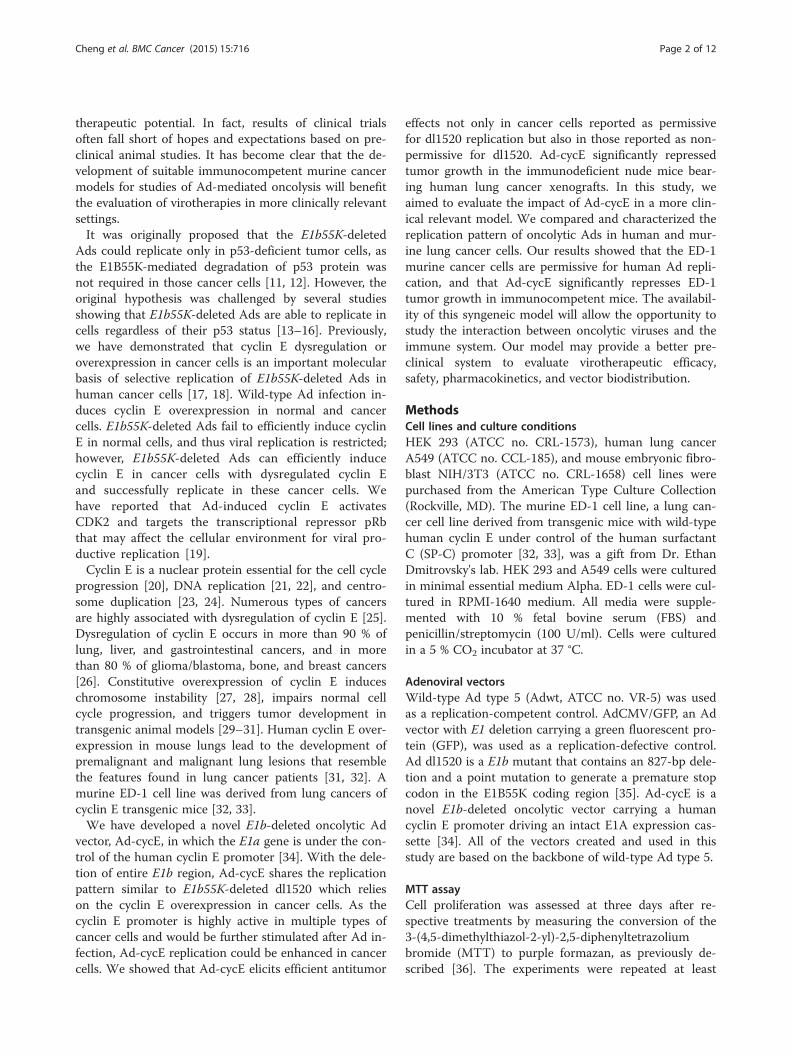
ResultsMurine ED-1 cells show higher growth rate and lowerserum requirement than human A549 cellsThe ED-1 cell line was derived from transgenic micewith wild-type human cyclin E expression in lung can-cers [32, 33]. The human A549 lung cancer cell line,with constitutive cyclin E expression, is highly permis-sive for oncolytic Ad replication [17]. The growth prop-erties of ED-1 and A549 were compared to understandthe difference between the two cell lines. The final num-ber of ED-1 cells increased 10 fold in 3 days, while A549cells increased 5.6 fold (Fig. 1a). The doubling time gen-erated from the ED-1 cell growth data in log phase was18.60 h and A549 was 30.17 h, showing the ED-1 cellgrowth rate was about 1.6-fold faster than that of A549cells. Growth curves of A549 and ED-1 cells in the pres-ence of serum concentrations, ranging from 0 to 10 %,are shown in Fig. 1b. The number of A549 cells culturedin medium with 0 % serum only increased slightly; how-ever, ED-1 cells still increased close to 4 fold under thesame conditions. Thus, ED-1 cells grew significantly fas-ter and exhibited less dependence on serum concentra-tion than A549 cells.
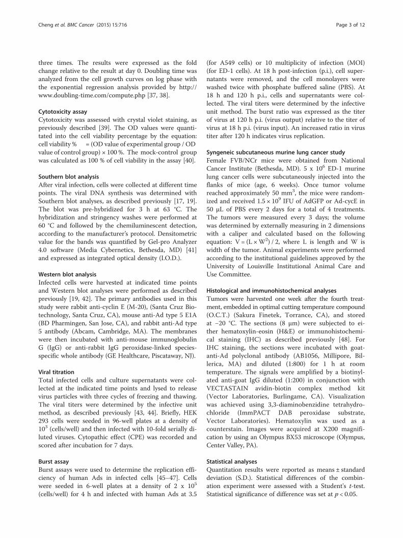
Murine ED-1 cells require 3-fold higher titers of Ads toachieve similar infection as human A549 cellsTo evaluate the efficiency of human Ad infection of hu-man and murine cancer cells under a similar growth rateand a comparable density, A549 and ED-1 cells werecultured in 1 % FBS and 0 % FBS, respectively. We firstevaluated the Ad infection by quantifying the number of
A549 and ED-1 cells expressing GFP after infection withAdGFP (Fig. 2a). Infection with AdGFP at an MOI of 10achieved maximum infection (>90 %) of A549 cells,while an MOI of 20 infected only 80 % of ED-1 cells, in-dicating poor infection of Ads in mouse cells. Consider-ing that the activity of the cytomegalovirus (CMV)promoter used to drive GFP expression in the vectormay differ in different cell lines, we also evaluated theinfection efficiency by quantitating the amount of AdDNA in cells. As AdGFP is a non-replicative virus, theamounts of AdGFP DNA inside cells represent the totalviruses that entered into those cells. With the same MOIof AdGFP infection, the Ad DNA amount in ED-1 cellswas lower than that in A549 cells (Fig. 2b). Yet, increas-ing infection MOI of AdGFP led to a concomitant in-crease of Ad DNA amount in both ED-1 and A549 cells,suggesting that virus entry can be adjusted by alteringthe infection MOI of Ads. To compare the concentra-tion of Ad DNA in ED-1 and in A549 cells, we spe-cifically quantitated the densities of a band of viralDNA in ED-1 cells infected with 10 MOI of AdGFPand in A549 cells infected with AdGFP at MOIs of2.5, 5, and 10 (boxed in Fig. 2b). The algorithmic re-sult revealed that an MOI of 10 of Ads for ED-1 cellsis required to achieve a similar infection of A549 cellsat an MOI of 3.5.
Human oncolytic Ads selectively replicate in murine ED-1cancer cellsTo understand the potential of murine ED-1 cancer cellsfor the study of oncolytic virotherapy, we compared Ad
Fig. 1 Growth properties of human A549 compared with murine ED-1 cells. Cell proliferation and serum sensitivity of A549 and ED-1 cells weredetermined by MTT assay at 0, 24, 48, and 72 h. A549 and ED-1 cells were seeded into 24-well plates at a density of 2.5 x 10⁴ (cells/well) andcultured in (a) 10 % fetal bovine serum (FBS) or (b) A549 and ED-1 cells cultured in 0, 0.5, 1, 2.5, 5, and 10 % FBS, respectively. The results wereexpressed as the fold change relative to the result at 0 h. All values represent the means ± S.D. of triplicate samples
Cheng et al. BMC Cancer (2015) 15:716 Page 4 of 12
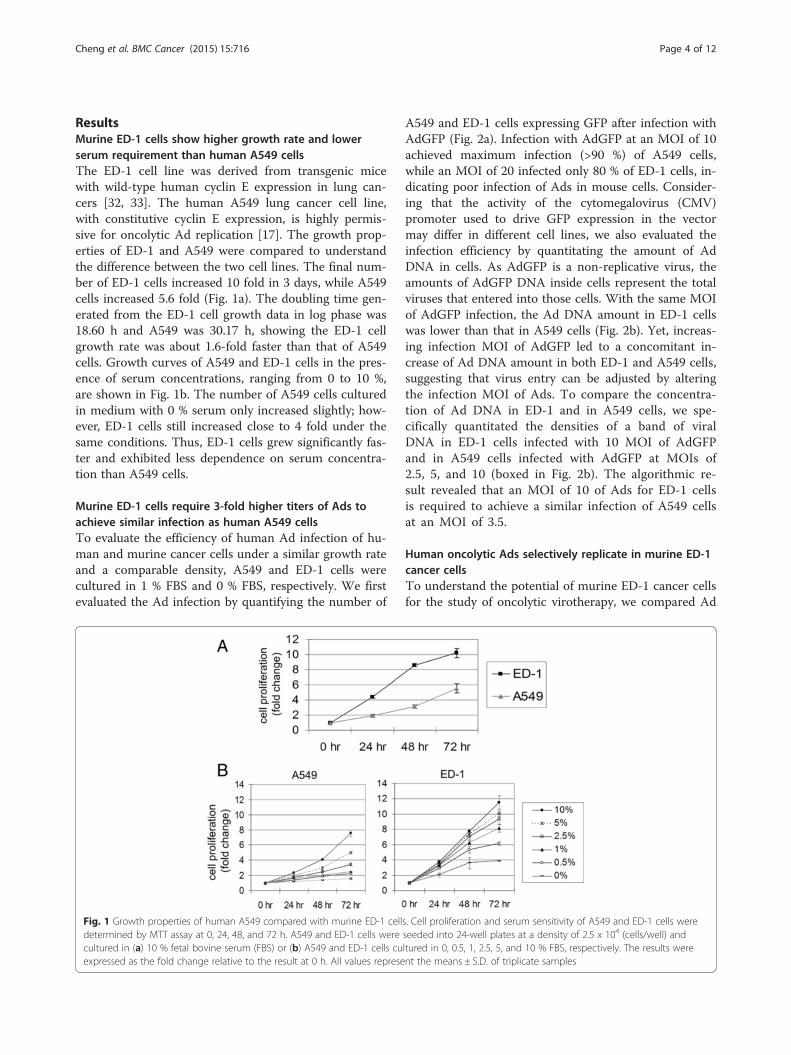
replication in murine and human cancer cells. MurineNIH/3T3 cells generated from NIH Swiss mouse embryofibroblasts [49] were applied here as a non-cancerouscontrol. Relatively higher levels of cyclin E expressionwere detected in human A549 cancer cells, expression ofcyclin E was lower in the murine ED-1 cell, but not inNIH/3T3 cells (Fig. 3a). Replication of wild-type Ad5(Adwt), oncolytic dl1520 (ONYX-015), and Ad-cycEwere evaluated. AdGFP was used as a non-replicativecontrol. Ad dl1520 is an attenuated Ad with E1b
deletion, which has been studied in several clinical trials[2, 35]. Ad-cycE is an E1b-deleted vector with its E1agene controlled by the human cyclin E promoter [34].To achieve equal infections, we chose 3.5 MOI of Ad forinfection of human A549 cells and 10 MOI for murinecells in our in vitro experiments. The photographs andquantitated data of cell viability showed that mock-infection and infection with non-replicative vectorAdGFP did not induce cytotoxicity (Fig. 3b). Adwt in-duced cytotoxicity in all cell lines. However, the two
Fig. 2 Infection efficiency of human adenoviruses on A549 and ED-1 cells. (a) A549 cells were cultured with 1 % FBS, and ED-1 cells were culturedwith 0 % FBS at a density of 105 (cells/well) and infected with increasing MOI of AdGFP after seeding for 4 h. For each infection, three randomfields were taken by EVOS fluorescence microscope (AMG, Bothell, WA) at 72 h post-infection (p.i.). The numbers of GFP cells on each photo werecalculated by ImageJ (US National Institutes of Health, Bethesda, MD). The numbers of GFP-positive cells were divided by total cell numbers oneach photo to determine the infection efficiency. All values represent the means ± S.D. of triplicate samples. (b) A549 cells were cultured with 1 %FBS, and ED-1 cells were cultured with 0 % FBS at a density of 106 (cells/well) in 60-mm dishes. Cells were infected with AdGFP at 0, 1.25, 2.5, 5,10, and 20 MOI, respectively, and harvested at 24 h p.i. The DNA samples were digested with PstI, and then totally loaded into the agarose gel forSouthern blot analyses with Ad DNA fragments. The amounts of AdGFP entering cells were quantitated by Gel-pro Analyzer 4.0 software (MediaCybernetics, Bethesda, MD) and presented as integrated optical density (I.O.D.) values. (Right, magnified view of boxed section.)
Cheng et al. BMC Cancer (2015) 15:716 Page 5 of 12
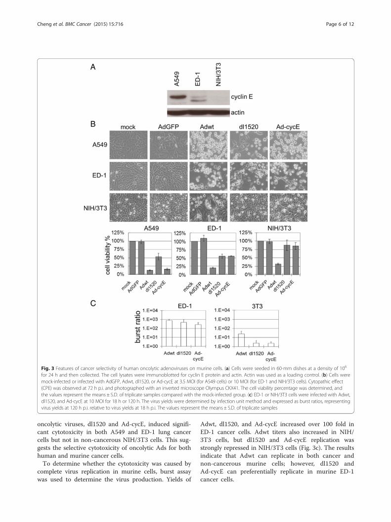
oncolytic viruses, dl1520 and Ad-cycE, induced signifi-cant cytotoxicity in both A549 and ED-1 lung cancercells but not in non-cancerous NIH/3T3 cells. This sug-gests the selective cytotoxicity of oncolytic Ads for bothhuman and murine cancer cells.To determine whether the cytotoxicity was caused by
complete virus replication in murine cells, burst assaywas used to determine the virus production. Yields of
Adwt, dl1520, and Ad-cycE increased over 100 fold inED-1 cancer cells. Adwt titers also increased in NIH/3T3 cells, but dl1520 and Ad-cycE replication wasstrongly repressed in NIH/3T3 cells (Fig. 3c). The resultsindicate that Adwt can replicate in both cancer andnon-cancerous murine cells; however, dl1520 andAd-cycE can preferentially replicate in murine ED-1cancer cells.
Fig. 3 Features of cancer selectivity of human oncolytic adenoviruses on murine cells. (a) Cells were seeded in 60-mm dishes at a density of 106
for 24 h and then collected. The cell lysates were immunoblotted for cyclin E protein and actin. Actin was used as a loading control. (b) Cells weremock-infected or infected with AdGFP, Adwt, dl1520, or Ad-cycE at 3.5 MOI (for A549 cells) or 10 MOI (for ED-1 and NIH/3T3 cells). Cytopathic effect(CPE) was observed at 72 h p.i. and photographed with an inverted microscope Olympus CKX41. The cell viability percentage was determined, andthe values represent the means ± S.D. of triplicate samples compared with the mock-infected group. (c) ED-1 or NIH/3T3 cells were infected with Adwt,dl1520, and Ad-cycE at 10 MOI for 18 h or 120 h. The virus yields were determined by infection unit method and expressed as burst ratios, representingvirus yields at 120 h p.i. relative to virus yields at 18 h p.i. The values represent the means ± S.D. of triplicate samples
Cheng et al. BMC Cancer (2015) 15:716 Page 6 of 12
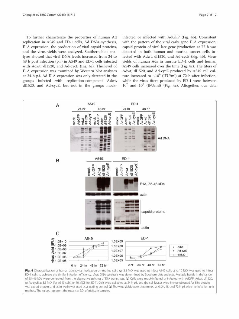
To further characterize the properties of human Adreplication in A549 and ED-1 cells, Ad DNA synthesis,E1A expression, the production of viral capsid proteins,and the virus yields were analyzed. Southern blot ana-lyses showed that viral DNA levels increased from 24 to48 h post infection (p.i.) in A549 and ED-1 cells infectedwith Adwt, dl1520, and Ad-cycE (Fig. 4a). The level ofE1A expression was examined by Western blot analysesat 24-h p.i. Ad E1A expression was only detected in thegroups infected with replication-competent Adwt,dl1520, and Ad-cycE, but not in the groups mock-
infected or infected with AdGFP (Fig. 4b). Consistentwith the pattern of the viral early gene E1A expression,capsid protein of viral late gene production at 72 h wasdetected in both human and murine cancer cells in-fected with Adwt, dl1520, and Ad-cycE (Fig. 4b). Virusyields of human Ads in murine ED-1 cells and humanA549 cells increased over the time (Fig. 4c). The titers ofAdwt, dl1520, and Ad-cycE produced by A549 cell cul-ture increased to ~109 (IFU/ml) at 72 h after infection,while the virus titers produced by ED-1 were between107 and 108 (IFU/ml) (Fig. 4c). Altogether, our data
Fig. 4 Characterization of human adenoviral replication on murine cells. (a) 3.5 MOI was used to infect A549 cells, and 10 MOI was used to infectED-1 cells to achieve the similar infection efficiency. Virus DNA synthesis was determined by Southern blot analyses. Multiple bands in the rangeof 35–46 kDa were generated from the alternative splicing of E1A transcripts. (b) Cells were mock-infected or infected with AdGFP, Adwt, dl1520,or Ad-cycE at 3.5 MOI (for A549 cells) or 10 MOI (for ED-1). Cells were collected at 24 h p.i., and the cell lysates were immunoblotted for E1A protein,viral capsid protein, and actin. Actin was used as a loading control. (c) The virus yields were determined at 0, 24, 48, and 72 h p.i. with the infection unitmethod. The values represent the means ± S.D. of triplicate samples
Cheng et al. BMC Cancer (2015) 15:716 Page 7 of 12
demonstrate that Adwt and oncolytic dl1520 and Ad-cycE can replicate in both human A549 and murine ED-1 lung cancer cells.
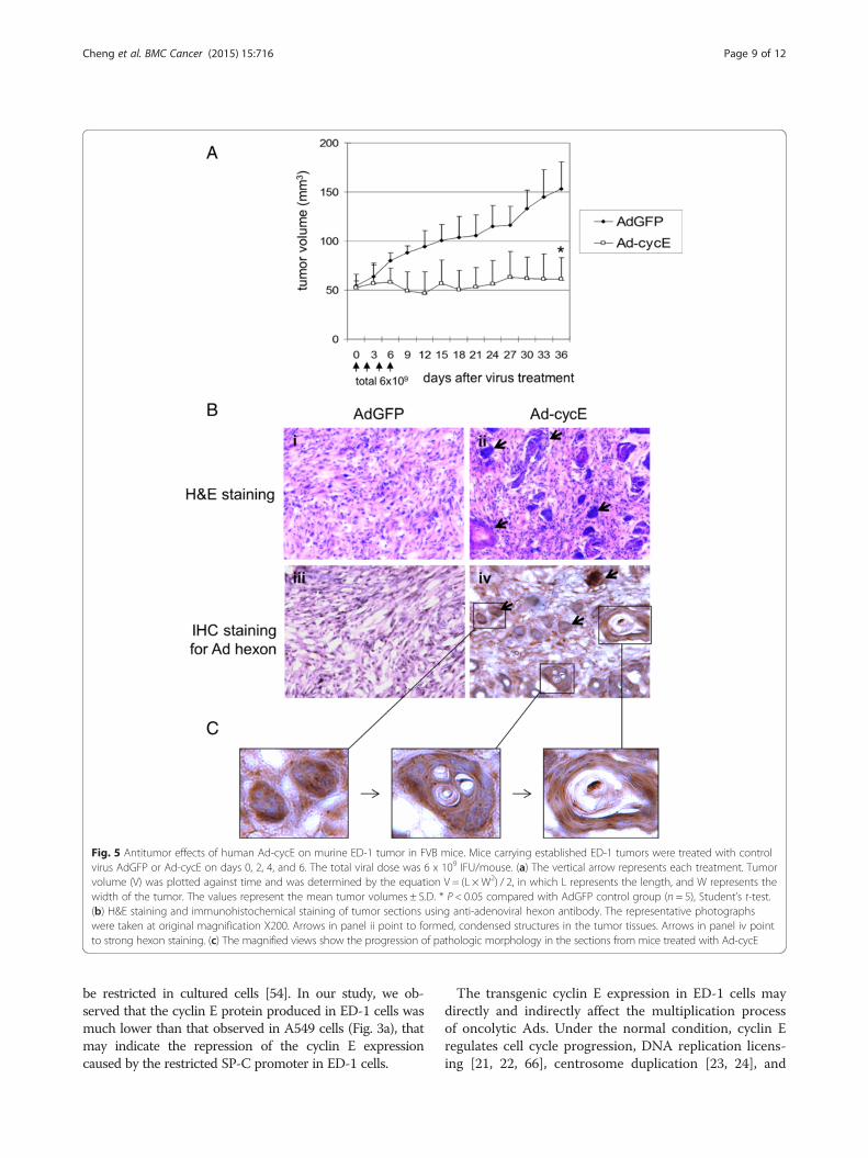
Ad-cycE suppresses murine ED-1 tumor growth inimmunocompetent miceThe expression of the cyclin E gene in murine ED-1 cellsis under control of the human surfactant C promoter(SP-C) promoter [32]. The SP-C promoter has been usedfor lung epithelial cell-specific gene expression in trans-genic models [50–53]. The SP-C promoter may be moreactive in vivo than in vitro [54]. To evaluate the effect ofAd-cycE in vivo, we subcutaneously injected murine ED-1 lung cancer cells into immunocompetent FVB mice.When tumors were approximately 50 mm3, the micewere intratumorally injected for a total of 4 times withtotal 6 × 10⁹ IFU of AdGFP or Ad-cycE. The initialreduction of tumor volumes in the Ad-cycE-treatedgroup was observed at day 9 after the first treatment.Mice treated with Ad-cycE exhibited significant sup-pression of tumor growth, with 60 % reduction in themean tumor volume as compared with mice treatedwith control AdGFP at day 36 after the first treat-ment (P = 0.0002, Fig. 5a).To verify the virus replication in tumor tissue, tu-
mors from the mice were harvested at day 7 and ana-lyzed with histological and immunohistochemicalanalyses. H&E staining showed that some cancer cellsformed condensed structures in the tumor tissuestreated with Ad-cycE (Fig. 5b, panel ii, blue areas in-dicated by arrows). In contrast, such structures didnot occur in the tumors treated with the control vec-tor AdGFP (Fig. 5b, panel i). Immunohistochemicalstudies with the antibody against the Ad hexon pro-tein, the classic marker of virus particles produced incells [55], showed the expression of the viral hexonprotein in tumor tissues treated with Ad-cycE, butnot in the control group treated with AdGFP (Fig. 5b,panels iii and iv), indicating de novo synthesis of Ad-cycE viral late proteins. The strong hexon stainingregions (Fig. 5b, panel iv, brown areas indicated byarrows) are consistent with the condensed structuresshowed by H&E staining (Fig. 5b, panel ii, blue areas).The magnified views further illustrated the possibleprogression of the pathologic morphology change inthe tumor sections after Ad-cycE treatment (Fig. 5c).The results showed that Ad-cycE-infected ED-1tumor cells were encircled in capsule-like structuresand likely killed as a consequence of oncolytic Adreplication indicated by the high level expression ofthe viral hexon protein, forming vacuoles in the cap-sules. However, cells outside of the capsules were notinfected by Ad-cycE and survived the treatment.
DiscussionDevelopment of an immunocompetent murine modelfor oncolytic Ad therapy is critical to accurately evaluateand improve the efficacy and safety of this approach. Inthis study, we have characterized human Ad replicationin murine ED-1 lung cancer cells and studied oncolyticAd therapies with ED-1 tumors developed in immuno-competent mice. Our data revealed that E1b-deleted Adviruses replicated in the ED-1 cells and repressed ED-1tumor growth in syngeneic immunocompetent mice.Murine cells were generally considered not permissive
for human Ad replication [56]. We found in this studythat human Ads can infect ED-1 cells and selectivelyreplicate in and destroy the murine cancer cells. Thecross-species infection of human Ad in ED-1 cells canbe mediated via the homologous Ad receptor CAR onmurine cells [57], coreceptors such as integrins and hep-aran sulfate glycosaminoglycans, or other unknown re-ceptors. We observed that the virions of oncolytic Adswere produced in murine cancer ED-1 cells, but not innon-cancerous murine NIH/3T3 cells. Obviously, theentire human Ad life cycle is completed in ED-1 lungcancer cells, but restricted in NIH/3T3 cells. It has beenreported that human Ads normally undergo abortivereplication in murine cells [58, 59]. Virus yields of Ad2and Ad5 in 3T3-Swiss and BALB/c 3T3 cells were re-duced 3 to 5 logs compared to virus production in hu-man cells [60, 61] and no infectious virus particles ofAd12 were detected in murine 3T3 (embryo) and L(connective tissue) cells [59, 62]. The block of humanAd replication in murine cells may be related to the lowinfectivity [63], unstable and reduced DNA synthesis[59, 61, 64], abortive expression of late proteins, or thedefective assembly and maturation at an later stage ofviral replication cycle [59, 60, 62]. Previously, we ob-served that human Ads induced cytotoxicity in Chinesehamster ovary (CHO) cells; however, there was a lack oflate protein production that prevented infectious virionproduction (Cheng and Zhou, unpublished data). Whilethe exact mechanism(s) supporting the complete multi-plication process of human Ads in murine cancer ED-1cells remains to be investigated, it is tempting to specu-late that the human cyclin E overexpression in the cellmay play an important role. The human cyclin E gene inED-1 cells was validated by polymerase chain reaction(PCR) [33] (Additional file 1: Figure S1A). We also per-formed single-cell cloning and validated the level of cyclinE protein expression in these subcloned ED-1 cells. Thecyclin E proteins were detected at 47 kDa and 37 kDa [32,65] in parent ED-1 and all subclones (Additional file 1: Fig-ure S1B). Any conclusions, though, cannot exclude the pos-sibility that the cyclin E proteins detected in the culturedED-1 cells may be murine proteins, since the human cyclinE gene is under the control of the SP-C promoter that may
Cheng et al. BMC Cancer (2015) 15:716 Page 8 of 12
be restricted in cultured cells [54]. In our study, we ob-served that the cyclin E protein produced in ED-1 cells wasmuch lower than that observed in A549 cells (Fig. 3a), thatmay indicate the repression of the cyclin E expressioncaused by the restricted SP-C promoter in ED-1 cells.
The transgenic cyclin E expression in ED-1 cells maydirectly and indirectly affect the multiplication processof oncolytic Ads. Under the normal condition, cyclin Eregulates cell cycle progression, DNA replication licens-ing [21, 22, 66], centrosome duplication [23, 24], and
Fig. 5 Antitumor effects of human Ad-cycE on murine ED-1 tumor in FVB mice. Mice carrying established ED-1 tumors were treated with controlvirus AdGFP or Ad-cycE on days 0, 2, 4, and 6. The total viral dose was 6 x 10⁹ IFU/mouse. (a) The vertical arrow represents each treatment. Tumorvolume (V) was plotted against time and was determined by the equation V = (L × W2) / 2, in which L represents the length, and W represents thewidth of the tumor. The values represent the mean tumor volumes ± S.D. * P < 0.05 compared with AdGFP control group (n = 5), Student’s t-test.(b) H&E staining and immunohistochemical staining of tumor sections using anti-adenoviral hexon antibody. The representative photographswere taken at original magnification X200. Arrows in panel ii point to formed, condensed structures in the tumor tissues. Arrows in panel iv pointto strong hexon staining. (c) The magnified views show the progression of pathologic morphology in the sections from mice treated with Ad-cycE
Cheng et al. BMC Cancer (2015) 15:716 Page 9 of 12
E2F activation [67]. Cyclin E overexpression may causecell overgrowth and thus increase the accumulation ofmutations associated with tumorigenesis. Consequentlythe alternations of the growth rate and genetic muta-tions may create a suitable environment to loose cellularrestriction to viral propagation. Also, the excess cyclin Emay directly endorse virus replication. We previouslydemonstrated that human Ad replication relies on cyclinE induction in cells after viral infection [17]. Ad-inducedcyclin E turns on the pRb/E2F pathways by activatingCDK2 [19]. It is possible that human Ad replication inmurine ED-1 cells may be associated with cyclin E dys-regulation or cell cycle alterations occurring in the car-cinogenesis caused by the transgenic cyclin E expression.We have investigated the antitumor efficacy of Ad-
cycE with murine tumors in immunocompetent FVBmice. Interestingly, we identified that Ad-cycE-infectedcancer cells located in the specific areas where the clus-ters of cells were encircled in capsule-like structures intumors (Fig. 5c). The capsule formation is likely associ-ated with Ad oncolytic replication because tumorstreated with the control non-replicating AdGFP did notexhibit such structures. We found that cells inside thecapsules died and formed vacuoles, but cells outsidewere not infected and survived the treatment. Thus, thecapsule structures developed in tumors after Ad-cycE in-fection may prevent viruses from spreading to the entiretumor. In a previous clinical study, oncolytic Ads wereobserved in clusters of 5–20 cells after intratumoral ad-ministration, indicating that Ad spread in tumors is re-stricted [68]. Viral spread within solid tumors is limited,and usually is around the site of injection after intratu-moral delivery [69, 70]. The movement of virusesthrough tumors is likely impeded by the dense tumorextracellular matrix [1]. Hyaluronan is a key componentof the tumor extracellular matrix. With an oncolytic Adexpressing hyaluronidase to degrade this kind of import-ant structural element of the ECM, Guedan et al. (2010)showed that the virus distribution could be improved ina human melanoma xenograft model [71]. Our resultsindicate that the capsule structures may be formed as aconsequence of active tumor reactions to Ad replicationto prevent progeny Ad virions from spreading the infec-tion to the rest of the cancer cells in tumors.It is possible that the capsule structures may be also
associated with immune responses of FVB mice to Adreplication. Hallden et al. (2003) reported that Ad5 sig-nificantly induced intratumoral inflammatory cell infil-tration, including macrophage and CD8(+) lymphocytes[9]. The induction of non-specific or specific antitumorimmunity has been reported as one of the mechanismsto mediate tumor cell lysis [72, 73]. The detailed mech-anism by which the capsules formed in tumors requiresfurther study. The immune system of the ED-1 animal
model may have multiple effects on oncolytic virother-apy. Further studies will clarify these immune-mediatedeffects, such as the role of the immune cell infiltrationinto the tumors on Ad spread in tumor. The ED-1 ani-mal model as a preclinical system will also benefit thedevelopment of future strategies to enhance viral pene-tration and spread within solid tumors.
ConclusionOur results showed that murine ED-1 cancer cells arepermissive for human Ad replication, and Ad-cycE sig-nificantly represses ED-1 tumor growth in immunocom-petent FVB mice. Such a model with the uniquebackground of cyclin E overexpression can provide asuitable in vivo environment for researchers to studyoncolytic Ad replication in detail. Moreover, the capsulestructures formed in tumors may prevent viruses fromspreading to the entire tumor. The ED-1 model mayprovide an opportunity to recapitulate clinical phenom-ena and challenges for studies of virus spread in tumorsand the interactions between Ads and immune system.An orthotopic tumor model based on this system can beestablished to look at tumor growth and therapeutic effi-cacy in the context of the lung microenvironment andthus provide a valuable system to study oncolyticvirotherapy.
Additional file
Additional file 1: Figure S1. Characterization of cyclin E background inmurine ED-1 cells. (a) The genomic DNA was isolated from ED-1 cells,and PCR was used to detect cyclin E with sense primer 5'-TTG GCT ATGCTG GAG GAA GTA-3’ and antisense primer 5'-AGT GCT CTT CGG TGGTGT CAT-3’. (b) The cell lysates from parent ED-1 and single-cell cloneswere immunoblotted for cyclin E proteins. (TIFF 289 kb)
AbbreviationsAd: Adenovirus; CTLs: Cytotoxic T lymphocytes; CDK2: Cyclin-dependentkinase 2; FBS: Fetal bovine serum; DMEM: Dulbecco’s Modification of Eagle’sMedium; GFP: Green fluorescent protein; MTT: 3-(4,5-dimethylthiazol-2-yl)-2,5-diphenyltetrazolium bromide; OD: Optical density; I.O.D.: Integrated opticaldensity; CPE: Cytopathic effect; MOI: Multiplicity of infection; p.i.: postinfection; PBS: Phosphate-buffered saline; IFU: Infectious unit; O.C.T.: Optimalcutting temperature compound; H&E: Hematoxylin-eosin;IHC: Immunohistochemical staining; Adwt: Wild-type adenovirus;S.D.: Standard deviation; CAR: Coxsackievirus and adenovirus receptor;CHO: Chinese hamster ovary; pRb: Retinoblastoma protein.
Competing interestsThe authors declare that they have no competing interests.
Authors' contributionsPHC, KMM and HSZ designed the study and drafted the manuscript. PHC,SLW, XFL, KMM, and HSZ participated in the revision of the manuscript. PHCand XMR carried out the experiments. PHC, XFL, KMM, and HSZ participatedin the coordination of the study. All authors read and approved the finalmanuscript.
AcknowledgementsThis work was supported by NIH Grant R01 CA129975 (HSZ), Kentucky LungCancer Research Program GB150463 (HSZ), funding from James Graham
Cheng et al. BMC Cancer (2015) 15:716 Page 10 of 12
Brown Cancer Center and Department of Surgery of University of LouisvilleMedical School. P. H. Cheng is partially supported by the K. C. HuangScholarship from the University of Louisville. We thank the other members inour laboratory for their help in the experiment: Deyi Xiao, Hongying Hao,and Jorge G. Gutierrez. We especially thank Drs. Xi Liu and Ethan Dmitrovskyat Dartmouth Medical School for the gift of transgenic ED-1 cell line andtheir kind suggestions. We also thank Dr. Xiaoxian Duan at University ofLouisville for the assistance in the preparation of tumor sections, and MargaretA. Abby for editing the manuscript.
Author details1Department of Pharmacology and Toxicology, University of Louisville Schoolof Medicine, Louisville, KY 40292, USA. 2James Graham Brown Cancer Center,University of Louisville Medical School, 505 South Hancock Street, CTRBuilding, Room 306, Louisville, KY 40202, USA. 3Department of DiagnosticRadiology, University of Louisville School of Medicine, Louisville, KY 40292,USA. 4Hiram C. Polk Jr MD Department of Surgery, University of LouisvilleSchool of Medicine, Louisville, KY 40292, USA. 5Department of Microbiologyand Immunology, University of Louisville School of Medicine, Louisville, KY40292, USA.
Received: 5 April 2015 Accepted: 8 October 2015
References1. Russell SJ, Peng KW, Bell JC. Oncolytic virotherapy. Nat Biotechnol.
2012;30(7):658–70.2. Kirn D, Martuza RL, Zwiebel J. Replication-selective virotherapy for cancer:
Biological principles, risk management and future directions. Nat Med.2001;7(7):781–7.
3. Yu W, Fang H. Clinical trials with oncolytic adenovirus in China. Curr CancerDrug Targets. 2007;7(2):141–8.
4. Kruyt FA, Curiel DT. Toward a new generation of conditionally replicatingadenoviruses: pairing tumor selectivity with maximal oncolysis. Hum GeneTher. 2002;13(4):485–95.
5. Zhu ZB, Makhija SK, Lu B, Wang M, Rivera AA, Kim-Park S, et al.Incorporating the survivin promoter in an infectivity enhanced CRAd-analysis of oncolysis and anti-tumor effects in vitro and in vivo. Int J Oncol.2005;27(1):237–46.
6. Wang H, Wei F, Zhang J, Wang F, Li H, Chen X, et al. A novelimmunocompetent murine tumor model for the evaluation of RCAd-enhanced RDAd transduction efficacy. Tumour Biol. 2012;33(4):1245–53.
7. Wang Y, Hallden G, Hill R, Anand A, Liu TC, Francis J, et al. E3 genemanipulations affect oncolytic adenovirus activity in immunocompetenttumor models. Nat Biotechnol. 2003;21(11):1328–35.
8. Wold WS, Hermiston TW, Tollefson AE. Adenovirus proteins that subverthost defenses. Trends Microbiol. 1994;2(11):437–43.
9. Hallden G, Hill R, Wang Y, Anand A, Liu TC, Lemoine NR, et al. Novelimmunocompetent murine tumor models for the assessment of replication-competent oncolytic adenovirus efficacy. Mol Ther. 2003;8(3):412–24.
10. Ungerechts G, Springfeld C, Frenzke ME, Lampe J, Parker WB, Sorscher EJ, etal. An immunocompetent murine model for oncolysis with an armed andtargeted measles virus. Mol Ther. 2007;15(11):1991–7.
11. Bischoff JR, Kirn DH, Williams A, Heise C, Horn S, Muna M, et al. Anadenovirus mutant that replicates selectively in p53-deficient human tumorcells. Science. 1996;274(5286):373–6.
12. Rogulski KR, Freytag SO, Zhang K, Gilbert JD, Paielli DL, Kim JH, et al. In vivoantitumor activity of ONYX-015 is influenced by p53 status and isaugmented by radiotherapy. Cancer Res. 2000;60(5):1193–6.
13. Dix BR, Edwards SJ, Braithwaite AW. Does the antitumor adenovirus ONYX-015/dl1520 selectively target cells defective in the p53 pathway? J Virol.2001;75(12):5443–7.
14. Goodrum FD, Ornelles DA. p53 status does not determine outcome of E1B55-kilodalton mutant adenovirus lytic infection. J Virol. 1998;72(12):9479–90.
15. Geoerger B, Grill J, Opolon P, Morizet J, Aubert G, Terrier-Lacombe MJ, et al.Oncolytic activity of the E1B-55 kDa-deleted adenovirus ONYX-015 isindependent of cellular p53 status in human malignant glioma xenografts.Cancer Res. 2002;62(3):764–72.
16. Rothmann T, Hengstermann A, Whitaker NJ, Scheffner M, zur Hausen H.Replication of ONYX-015, a potential anticancer adenovirus, is independentof p53 status in tumor cells. J Virol. 1998;72(12):9470–8.
17. Zheng X, Rao XM, Gomez-Gutierrez JG, Hao H, McMasters KM, Zhou HS.Adenovirus E1B55K region is required to enhance cyclin E expression forefficient viral DNA replication. J Virol. 2008;82(7):3415–27.
18. Rao XM, Zheng X, Waigel S, Zacharias W, McMasters KM, Zhou HS. Geneexpression profiles of normal human lung cells affected by adenoviral E1B.Virology. 2006;350(2):418–28.
19. Cheng PH, Rao XM, McMasters KM, Zhou HS. Molecular basis for viralselective replication in cancer cells: activation of CDK2 by adenovirus-induced cyclin E. PLoS One. 2013;8(2):e57340.
20. Ohtsubo M, Theodoras AM, Schumacher J, Roberts JM, Pagano M. Humancyclin E, a nuclear protein essential for the G1-to-S phase transition. Mol CellBiol. 1995;15(5):2612–24.
21. Coverley D, Laman H, Laskey RA. Distinct roles for cyclins E and A duringDNA replication complex assembly and activation. Nat Cell Biol.2002;4(7):523–8.
22. Furstenthal L, Kaiser BK, Swanson C, Jackson PK. Cyclin E uses Cdc6 as achromatin-associated receptor required for DNA replication. J Cell Biol.2001;152(6):1267–78.
23. Hinchcliffe EH, Li C, Thompson EA, Maller JL, Sluder G. Requirement ofCdk2-cyclin E activity for repeated centrosome reproduction in Xenopusegg extracts. Science. 1999;283(5403):851–4.
24. Matsumoto Y, Hayashi K, Nishida E. Cyclin-dependent kinase 2 (Cdk2) isrequired for centrosome duplication in mammalian cells. Curr Biol.1999;9(8):429–32.
25. Donnellan R, Chetty R. Cyclin E in human cancers. FASEB J. 1999;13(8):773–80.26. Malumbres M, Barbacid M. To cycle or not to cycle: a critical decision in
cancer. Nat Rev Cancer. 2001;1(3):222–31.27. Spruck CH, Won KA, Reed SI. Deregulated cyclin E induces chromosome
instability. Nature. 1999;401(6750):297–300.28. Minella AC, Swanger J, Bryant E, Welcker M, Hwang H, Clurman BE. p53 and
p21 form an inducible barrier that protects cells against cyclin E-cdk2deregulation. Curr Biol. 2002;12(21):1817–27.
29. Bortner DM, Rosenberg MP. Induction of mammary gland hyperplasia andcarcinomas in transgenic mice expressing human cyclin E. Mol Cell Biol.1997;17(1):453–9.
30. Loeb KR, Kostner H, Firpo E, Norwood T, Tsuchiya K D, Clurman BE, et al. Amouse model for cyclin E-dependent genetic instability and tumorigenesis.Cancer Cell. 2005;8(1):35–47.
31. Freemantle SJ, Dmitrovsky E. Cyclin E transgenic mice: discovery tools forlung cancer biology, therapy, and prevention. Cancer Prev Res (Phila).2010;3(12):1513–8.
32. Ma Y, Fiering S, Black C, Liu X, Yuan Z, Memoli VA, et al. Transgenic cyclin Etriggers dysplasia and multiple pulmonary adenocarcinomas. Proc Natl AcadSci U S A. 2007;104(10):4089–94.
33. Liu X, Sempere LF, Galimberti F, Freemantle SJ, Black C, Dragnev KH, et al.Uncovering growth-suppressive MicroRNAs in lung cancer. Clin Canc Res.2009;15(4):1177–83.
34. Cheng PH, Rao XM, Duan X, Li XF, Egger ME, McMasters KM, Zhou HS.Virotherapy targeting cyclin E overexpression in tumors with adenovirus-enhanced cancer-selective promoter. J Mol Med (Berl). 2015;93(2):211–23.
35. Barker DD, Berk AJ. Adenovirus proteins from both E1B reading frames arerequired for transformation of rodent cells by viral infection and DNAtransfection. Virology. 1987;156(1):107–21.
36. Rodriguez-Rocha H, Gomez-Gutierrez JG, Garcia-Garcia A, Rao XM, Chen L,McMasters KM, et al. Adenoviruses induce autophagy to promote virusreplication and oncolysis. Virology. 2011;416(1–2):9–15.
37. Fan W, Lin CS, Potluri P, Procaccio V, Wallace DC. mtDNA lineage analysis ofmouse L-cell lines reveals the accumulation of multiple mtDNA mutantsand intermolecular recombination. Genes Dev. 2012;26(4):384–94.
38. Widera D, Zander C, Heidbreder M, Kasperek Y, Noll T, Seitz O, et al. Adultpalatum as a novel source of neural crest-related stem cells. Stem Cells.2009;27(8):1899–910.
39. Cheng PH, Lian S, Zhao R, Rao XM, McMasters KM, Zhou HS. Combinationof autophagy inducer rapamycin and oncolytic adenovirus improvesantitumor effect in cancer cells. Virol J. 2013;10(1):293.
40. Kwon OJ, Kim PH, Huyn S, Wu L, Kim M, Yun CO. A hypoxia- and {alpha}-fetoprotein-dependent oncolytic adenovirus exhibits specific killing ofhepatocellular carcinomas. Clin Canc Res. 2010;16(24):6071–82.
41. Nguyen MD, Lariviere RC, Julien JP. Reduction of axonal caliber does notalleviate motor neuron disease caused by mutant superoxide dismutase 1.Proc Natl Acad Sci U S A. 2000;97(22):12306–11.
Cheng et al. BMC Cancer (2015) 15:716 Page 11 of 12
42. Zheng X, Rao XM, Snodgrass CL, McMasters KM, Zhou HS. Selectivereplication of E1B55K-deleted adenoviruses depends on enhanced E1Aexpression in cancer cells. Cancer Gene Ther. 2006;13(6):572–83.
43. Sandig V, Youil R, Bett AJ, Franlin LL, Oshima M, Maione D, et al.Optimization of the helper-dependent adenovirus system for productionand potency in vivo. Proc Natl Acad Sci U S A. 2000;97(3):1002–7.
44. Zhao T, Rao XM, Xie X, Li L, Thompson TC, McMasters KM, et al. Adenoviruswith insertion-mutated E1A selectively propagates in liver cancer cells anddestroys tumors in vivo. Cancer Res. 2003;63(12):3073–8.
45. Ganly I, Kim YT, Hann B, Balmain A, Brown R. Replication and cytolysis of anE1B-attenuated adenovirus in drug-resistant ovarian tumour cells isassociated with reduced apoptosis. Gene Ther. 2001;8(5):369–75.
46. Ganly I, Mautner V, Balmain A. Productive replication of humanadenoviruses in mouse epidermal cells. J Virol. 2000;74(6):2895–9.
47. Madara J, Krewet JA, Shah M. Heat shock protein 72 expression allowspermissive replication of oncolytic adenovirus dl1520 (ONYX-015) in ratglioblastoma cells. Mol Cancer. 2005;4(1):12.
48. Huang T, Civelek AC, Zheng H, Ng CK, Duan X, Li J, et al. (18)F-misonidazolePET imaging of hypoxia in micrometastases and macroscopic xenografts ofhuman non-small cell lung cancer: a correlation with autoradiography andhistological findings. Am J Nucl Med Mol Imag. 2013;3(2):142–53.
49. Todaro GJ, Green H. Quantitative studies of the growth of mouse embryocells in culture and their development into established lines. J Cell Biol.1963;17:299–313.
50. Glasser SW, Korfhagen TR, Wert SE, Bruno MD, McWilliams KM, VorbrokerDK, et al. Genetic element from human surfactant protein SP-C geneconfers bronchiolar-alveolar cell specificity in transgenic mice. Am J Physiol.1991;261(4 Pt 1):L349–56.
51. Degryse E, De Santi MM, Dietrich M, Hadji DA, Spetz JF, Villeval D, et al. Ahuman SP-C promoter fragment targets alpha 1-proteinase inhibitor geneexpression to lung alveolar type II cells in transgenic mice. Transgenic Res.1996;5(2):139–43.
52. Glasser SW, Eszterhas SK, Detmer EA, Maxfield MD, Korfhagen TR. Themurine SP-C promoter directs type II cell-specific expression in transgenicmice. Am J Physiol Lung Cell Mol Physiol. 2005;288(4):L625–32.
53. Kerkhoff E, Fedorov LM, Siefken R, Walter AO, Papadopoulos T, Rapp UR.Lung-targeted expression of the c-Raf-1 kinase in transgenic mice exposes anovel oncogenic character of the wild-type protein. Cell Growth Differ.2000;11(4):185–90.
54. Whitsett JA, Glasser SW. Regulation of surfactant protein gene transcription.Biochim Biophys Acta. 1998;1408(2–3):303–11.
55. Wadler S, Yu B, Tan JY, Kaleya R, Rozenblit A, Makower D, et al. Persistentreplication of the modified chimeric adenovirus ONYX-015 in both tumorand stromal cells from a patient with gall bladder carcinoma implants. ClinCanc Res. 2003;9(1):33–43.
56. Oualikene W, Gonin P, Eloit M. Short and long term dissemination ofdeletion mutants of adenovirus in permissive (cotton rat) and non-permissive (mouse) species. J Gen Virol. 1994;75(Pt 10):2765–8.
57. Tomko RP, Xu R, Philipson L. HCAR and MCAR: the human and mousecellular receptors for subgroup C adenoviruses and group Bcoxsackieviruses. Proc Natl Acad Sci U S A. 1997;94(7):3352–6.
58. Duncan SJ, Gordon FC, Gregory DW, McPhie JL, Postlethwaite R, White R, etal. Infection of mouse liver by human adenovirus type 5. J Gen Virol.1978;40(1):45–61.
59. Biron KK, Raska Jr K. Adenovirus type 12 infection of defined mouse-humanhybrid cell clones. Experientia. 1976;32(1):38–40.
60. Eggerding FA, Pierce WC. Molecular biology of adenovirus type 2semipermissive infections. I. Viral growth and expression of viral replicativefunctions during restricted adenovirus infection. Virology. 1986;148(1):97–113.
61. Blair GE, Dixon SC, Griffiths SA, Zajdel ME. Restricted replication of humanadenovirus type 5 in mouse cell lines. Virus Res. 1989;14(4):339–46.
62. Lucher LA. Abortive adenovirus infection and host range determinants. CurrTop Microbiol Immunol. 1995;199(Pt 1):119–52.
63. Seth P, Rosenfeld M, Higginbotham J, Crystal RG. Mechanism ofenhancement of DNA expression consequent to cointernalization of areplication-deficient adenovirus and unmodified plasmid DNA. J Virol.1994;68(2):933–40.
64. Silverstein G, Strohl WA. Restricted replication of adenovirus type 2 inmouse Balb/3T3 cells. Arch Virol. 1986;87(3–4):241–64.
65. Ashraf S, Hompes R, Slater A, Lindsey I, Bach S, Mortensen NJ, et al. A criticalappraisal of endorectal ultrasound and transanal endoscopic microsurgeryand decision-making in early rectal cancer. Colorectal Dis. 2012;14(7):821–6.
66. Zhang H. Life without kinase: cyclin E promotes DNA replication licensingand beyond. Mol Cell. 2007;25(2):175–6.
67. Hwang HC, Clurman BE. Cyclin E in normal and neoplastic cell cycles.Oncogene. 2005;24(17):2776–86.
68. Galanis E, Okuno SH, Nascimento AG, Lewis BD, Lee RA, Oliveira AM, et al.Phase I-II trial of ONYX-015 in combination with MAP chemotherapy inpatients with advanced sarcomas. Gene Ther. 2005;12(5):437–45.
69. Parato KA, Senger D, Forsyth PA, Bell JC. Recent progress in the battlebetween oncolytic viruses and tumours. Nat Rev Cancer. 2005;5(12):965–76.
70. Smith E, Breznik J, Lichty BD. Strategies to enhance viral penetration of solidtumors. Hum Gene Ther. 2011;22(9):1053–60.
71. Guedan S, Rojas JJ, Gros A, Mercade E, Cascallo M, Alemany R.Hyaluronidase expression by an oncolytic adenovirus enhances itsintratumoral spread and suppresses tumor growth. Mol Ther.2010;18(7):1275–83.
72. Naik JD, Twelves CJ, Selby PJ, Vile RG, Chester JD. Immune recruitment andtherapeutic synergy: keys to optimizing oncolytic viral therapy? Clin CancRes. 2011;17(13):4214–24.
73. Mullen JT, Tanabe KK. Viral oncolysis. Oncologist. 2002;7(2):106–19.
Submit your next manuscript to BioMed Centraland take full advantage of:
• Convenient online submission
• Thorough peer review
• No space constraints or color figure charges
• Immediate publication on acceptance
• Inclusion in PubMed, CAS, Scopus and Google Scholar
• Research which is freely available for redistribution
Submit your manuscript at www.biomedcentral.com/submit
Cheng et al. BMC Cancer (2015) 15:716 Page 12 of 12


![Be Prepared [NOT] To Be Financially Repressed](https://img.pdfslide.us/doc/110x75/577cdfec1a28ab9e78b249b8/be-prepared-not-to-be-financially-repressed.jpg)