Embed Size (px)
Citation preview
Oncogenes I and IIOncogenes I and II
Readings for both lectures:
The Biology of Cancer (2The Biology of Cancer (2ndnd edition, 2014) edition, 2014)
by Robert A. Weinbergby Robert A. Weinberg
Chapters 3, 4, 10, and 11Chapters 3, 4, 10, and 11
ExamsExams
take-home take-home vs. vs. in-class examsin-class exams
exam grades are assigned to exam grades are assigned to individualsindividuals
no discussion of course materials with classmates no discussion of course materials with classmates during exam week during exam week
in cases of cheating, the persons who give in cases of cheating, the persons who give information and take information are information and take information are bothboth at fault at fault

Oncogenes IOncogenes I
Organization of todayOrganization of today’’s lectures lecture
mammalian cell culturemammalian cell culture
telomere hypothesistelomere hypothesis
properties of malignantly-transformed cellsproperties of malignantly-transformed cells
retrovirusesretroviruses
mechanisms of retroviral-induced tumorigenesismechanisms of retroviral-induced tumorigenesis

Use of cell culture to study the malignant processUse of cell culture to study the malignant process
advantages:advantages: the properties of cells from normal tissues and from tumors the properties of cells from normal tissues and from tumors
can be compared under controlled conditions.can be compared under controlled conditions.
cells from normal tissues can be experimentally cells from normal tissues can be experimentally ““transformedtransformed”” into cells with a more malignant phenotype. into cells with a more malignant phenotype.
in vitroin vitro cell transformation can be measured using cell transformation can be measured using quantitative and reproducible assays.quantitative and reproducible assays.
the phenotypic differences between normal cells and their the phenotypic differences between normal cells and their transformed counterparts can be correlated with their ability transformed counterparts can be correlated with their ability to form tumors to form tumors in vivoin vivo..
disadvantages:disadvantages: cell culture cannot recapitulate the entire malignant processcell culture cannot recapitulate the entire malignant process

Acquired properties of cancer cellsAcquired properties of cancer cells
Inappropriate cell proliferationInappropriate cell proliferation
Inappropriate resistance to cell deathInappropriate resistance to cell death
Failure of cellular differentiationFailure of cellular differentiation
InvasivenessInvasiveness
Metastatic potentialMetastatic potential
Angiogenic capacityAngiogenic capacity

Culturing normal mammalian cellsCulturing normal mammalian cells to culture mammalian cells to culture mammalian cells in vitroin vitro::
tissue explants are dissociated to single cells (e.g., tissue explants are dissociated to single cells (e.g., with trypsin)with trypsin)
cells are incubated in a petri dish with a sterile cells are incubated in a petri dish with a sterile aqueous medium containing:aqueous medium containing: amino acidsamino acids saltssalts glucose (energy source)glucose (energy source) serum (growth factors, survival factors, transferrin, etc.)serum (growth factors, survival factors, transferrin, etc.)
cells adhere to the plate, and some may proliferate.cells adhere to the plate, and some may proliferate. ““primary cultureprimary culture””

Passaging cells Passaging cells in vitroin vitro
cells are incubated for a defined period.cells are incubated for a defined period. (e.g., 3 days)(e.g., 3 days)
the spent medium is removed, and the adhered cells the spent medium is removed, and the adhered cells are dissociated (trypsin).are dissociated (trypsin).
cells are counted, diluted in fresh media, and re-plated cells are counted, diluted in fresh media, and re-plated at a specific density (e.g., 3 x 10at a specific density (e.g., 3 x 1055 cells/plate). cells/plate).
““secondary culturesecondary culture”” ““3 times 33 times 3”” (3T3) passaging (3T3) passaging cells can be maintained in culture for long periods by cells can be maintained in culture for long periods by
continual passaging.continual passaging.
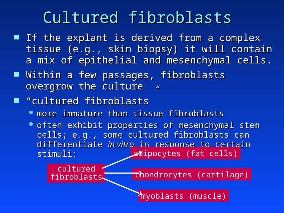
Cultured fibroblastsCultured fibroblasts If the explant is derived from a complex tissue (e.g., If the explant is derived from a complex tissue (e.g.,
skin biopsy) it will contain a mix of epithelial and skin biopsy) it will contain a mix of epithelial and mesenchymal cells.mesenchymal cells.
Within a few passages, fibroblasts overgrow the cultureWithin a few passages, fibroblasts overgrow the culture ““cultured fibroblastscultured fibroblasts””
more immature than tissue fibroblastsmore immature than tissue fibroblasts often exhibit properties of mesenchymal stem cells; e.g., often exhibit properties of mesenchymal stem cells; e.g.,
some cultured fibroblasts can differentiate some cultured fibroblasts can differentiate in vitroin vitro in response in response to certain stimuli:to certain stimuli:
culturedfibroblasts
adipocytes (fat cells)
chondrocytes (cartilage)
myoblasts (muscle)

In vitroIn vitro culture of epithelial cells culture of epithelial cells
need to remove fibroblasts from the cultureneed to remove fibroblasts from the culture
physical isolation of epithelial cellsphysical isolation of epithelial cells dissection of explantdissection of explant partial trypsinization of explantpartial trypsinization of explant
preferential viability of epithelial cells preferential viability of epithelial cells grow in a serum-free mediumgrow in a serum-free medium
remember: serum is especially rich in PDGF and FGFsremember: serum is especially rich in PDGF and FGFs
provide other sources of epithelial growth factors provide other sources of epithelial growth factors (such as recombinant proteins)(such as recombinant proteins)

human mammary epithelial cells (HMECs)human mammary epithelial cells (HMECs)
–– surgical discard from reduction mammoplastiessurgical discard from reduction mammoplastiesexplant
cut away fatty materialcut away fatty material digest with collagenase and hyaluronidasedigest with collagenase and hyaluronidase collect semi-pure epithelial clumps (collect semi-pure epithelial clumps (““organoidsorganoids””) on ) on
a 100-mm filter (filtrate contains mainly fibroblasts) a 100-mm filter (filtrate contains mainly fibroblasts)
“epithelialorganoids”
HMEC culture
seed onto a petri dishseed onto a petri dish incubate in serum-free medium supplemented with:incubate in serum-free medium supplemented with:
rEGF, insulin, hydrocortisone rEGF, insulin, hydrocortisone bovine pituitary extractbovine pituitary extract
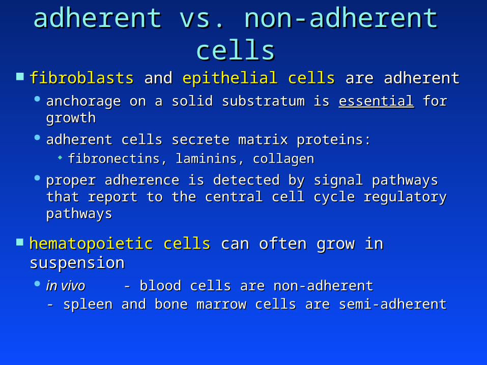
adherent vs. non-adherent cellsadherent vs. non-adherent cells
fibroblasts fibroblasts and and epithelial cells epithelial cells are adherentare adherent anchorage on a solid substratum is anchorage on a solid substratum is essentialessential for growth for growth adherent cells secrete matrix proteins:adherent cells secrete matrix proteins:
fibronectins, laminins, collagenfibronectins, laminins, collagen
proper adherence is detected by signal pathways that proper adherence is detected by signal pathways that report to the central cell cycle regulatory pathwaysreport to the central cell cycle regulatory pathways
hematopoietic cells hematopoietic cells can often grow in suspensioncan often grow in suspension in vivoin vivo - blood cells are non-adherent- blood cells are non-adherent - spleen and bone marrow cells are semi-- spleen and bone marrow cells are semi-
adherentadherent

Life span of human fibroblasts in culture Life span of human fibroblasts in culture human fetal fibroblasts undergo human fetal fibroblasts undergo ““replicativereplicative senescencesenescence””
or or ““early crisisearly crisis”” after ~ 60 population doublings (PDs). after ~ 60 population doublings (PDs). properties of senescent cells:properties of senescent cells:
cells exist in a state of permanent growth arrest.cells exist in a state of permanent growth arrest. resembles but distinct from quiescence (the Gresembles but distinct from quiescence (the G00 state) state)
cells are viable and metabolically active.cells are viable and metabolically active. large cytoplasm (large cytoplasm (““fried eggfried egg”” appearance). appearance). express express -galactosidase (stain blue with Xgal).-galactosidase (stain blue with Xgal). express the p16express the p16INK4aINK4a tumor suppressor (Rb pathway). tumor suppressor (Rb pathway). display shortened telomeres.display shortened telomeres. otherwise, senescent cells have a stable genome (diploid).otherwise, senescent cells have a stable genome (diploid).
mortal cell cultures = mortal cell cultures = ““cells strainscells strains”” immortal cells never emerge spontaneously from cultured immortal cells never emerge spontaneously from cultured
strains of human fibroblasts.strains of human fibroblasts.
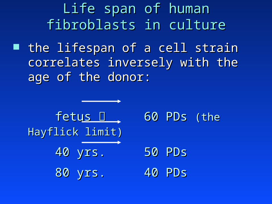
Life span of human fibroblasts in cultureLife span of human fibroblasts in culture
the lifespan of a cell strain correlates the lifespan of a cell strain correlates inversely with the age of the donor:inversely with the age of the donor:
fetus fetus 60 PDs 60 PDs (the Hayflick (the Hayflick
limit)limit)
40 yrs.40 yrs. 50 PDs 50 PDs
80 yrs.80 yrs. 40 PDs 40 PDs

Life span of human fibroblasts in cultureLife span of human fibroblasts in culture
fetal cells
adult (40 yrs.) cells
adult (80 yrs.) cells
PDs
60
40
time in culture

Therefore, the lifespan of a cell strain is not dependent Therefore, the lifespan of a cell strain is not dependent solely on the number of cell divisions in culture.solely on the number of cell divisions in culture. Instead, it reflects the number of cell divisions both Instead, it reflects the number of cell divisions both in vivoin vivo
and and in vitro.in vitro.
Is there a mechanism that Is there a mechanism that ““countscounts”” the number of cell the number of cell divisions from conception ?divisions from conception ?
Life span of human fibroblasts in cultureLife span of human fibroblasts in culture
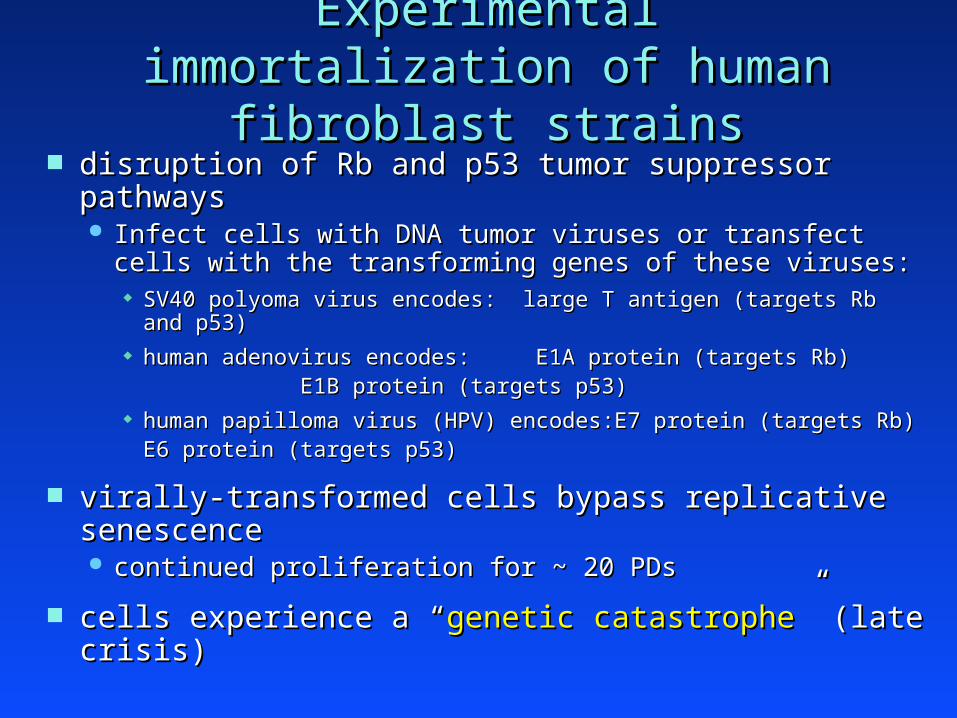
Experimental immortalization of Experimental immortalization of human fibroblast strainshuman fibroblast strains
disruption of Rb and p53 tumor suppressor pathwaysdisruption of Rb and p53 tumor suppressor pathways Infect cells with DNA tumor viruses or transfect cells with Infect cells with DNA tumor viruses or transfect cells with
the transforming genes of these viruses:the transforming genes of these viruses: SV40 polyoma virus encodes: large T antigen (targets Rb and p53)SV40 polyoma virus encodes: large T antigen (targets Rb and p53) human adenovirus encodes:human adenovirus encodes: E1A protein (targets Rb)E1A protein (targets Rb)
E1B protein (targets p53)E1B protein (targets p53) human papilloma virus (HPV) encodes:human papilloma virus (HPV) encodes: E7 protein (targets Rb)E7 protein (targets Rb)
E6 protein E6 protein (targets p53)(targets p53)
virally-transformed cells bypass replicative senescence virally-transformed cells bypass replicative senescence continued proliferation for ~ 20 PDscontinued proliferation for ~ 20 PDs
cells experience a cells experience a ““genetic catastrophegenetic catastrophe”” (late crisis) (late crisis)
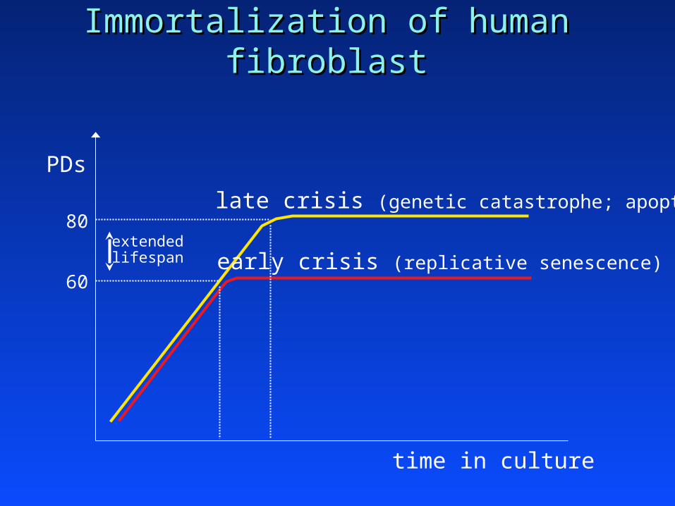
early crisis (replicative senescence)
late crisis (genetic catastrophe; apoptotic)
extendedlifespan
time in culture
PDs
Immortalization of human fibroblastImmortalization of human fibroblast
60
80
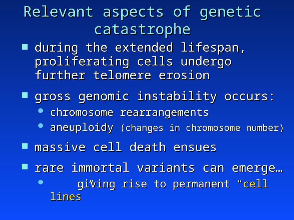
Relevant aspects of genetic catastropheRelevant aspects of genetic catastrophe
during the extended lifespan, proliferating cells during the extended lifespan, proliferating cells undergo further telomere erosionundergo further telomere erosion
gross genomic instability occurs:gross genomic instability occurs: chromosome rearrangementschromosome rearrangements aneuploidy aneuploidy (changes in chromosome number)(changes in chromosome number)
massive cell death ensuesmassive cell death ensues
rare immortal variants can emerge…rare immortal variants can emerge… giving rise to permanent giving rise to permanent ““cell linescell lines””
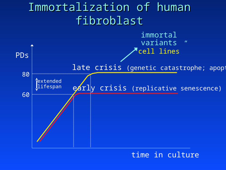
early crisis (replicative senescence)
late crisis (genetic catastrophe; apoptotic)
extendedlifespan
time in culture
PDs
Immortalization of human fibroblastImmortalization of human fibroblast
60
80
immortalvariants
“cell lines”
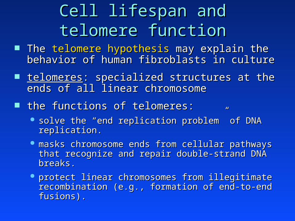
Cell lifespan and telomere functionCell lifespan and telomere function
The The telomere hypothesistelomere hypothesis may explain the behavior of may explain the behavior of human fibroblasts in culturehuman fibroblasts in culture
telomerestelomeres: specialized structures at the ends of all : specialized structures at the ends of all linear chromosomelinear chromosome
the functions of telomeres:the functions of telomeres: solve the solve the ““end replication problemend replication problem”” of DNA replication. of DNA replication. masks chromosome ends from cellular pathways that masks chromosome ends from cellular pathways that
recognize and repair double-strand DNA breaks. recognize and repair double-strand DNA breaks. protect linear chromosomes from illegitimate recombination protect linear chromosomes from illegitimate recombination
(e.g., formation of end-to-end fusions).(e.g., formation of end-to-end fusions).

End replication problemEnd replication problem
5’3’
3’5’
5’
DNA polymerization occurs 5DNA polymerization occurs 5’’33’’ (with respect (with respect to the nascent strand).to the nascent strand).

Leading strand synthesisLeading strand synthesis
5’3’
3’5’
5’
leading strand
lagging strand
leading strand synthesisleading strand synthesis occurs continuously (5occurs continuously (5’’33’’) toward replication fork) toward replication fork
lagging strand synthesislagging strand synthesis cannot occur continuouslycannot occur continuously

Lagging strand synthesisLagging strand synthesis
5’3’
3’5’
5’
leading strand
lagging strand
lagging strand synthesislagging strand synthesis occurs discontinuously in short spurts of 5occurs discontinuously in short spurts of 5’’33’’ synthesis synthesis each spurt is newly primed with an RNA oligonucleotideeach spurt is newly primed with an RNA oligonucleotide generates an generates an ““Okasaki fragmentOkasaki fragment”” of DNA (200-400 bps) of DNA (200-400 bps)
3’

Lagging strand synthesisLagging strand synthesis
5’3’
3’5’
3’5’
leading strand
lagging strand
lagging strand synthesislagging strand synthesis occurs discontinuously in short spurts of 5occurs discontinuously in short spurts of 5’’33’’ synthesis synthesis each spurt is newly primed with an RNA oligonucleotideeach spurt is newly primed with an RNA oligonucleotide generates an generates an ““Okasaki fragmentOkasaki fragment”” of DNA (200-400 bps) of DNA (200-400 bps) as the replication fork progresses, multiple RNA-primed as the replication fork progresses, multiple RNA-primed
Okasaki fragments are produced.Okasaki fragments are produced.

Lagging strand synthesisLagging strand synthesis
5’3’
3’5’
3’5’
leading strand
lagging strand
lagging strand synthesislagging strand synthesis DNA synthesis of new Okasaki fragment displaces the DNA synthesis of new Okasaki fragment displaces the
RNA primer of the previous fragment.RNA primer of the previous fragment.
RNA primer degradedRNA primer degraded
adjacent Okasaki fragments are ligated togetheradjacent Okasaki fragments are ligated together

End replication problemEnd replication problem
5’3’
3’5’
3’5’
leading strand
5’3’
3’5’
leading strand synthesis proceeds to end of template DNAleading strand synthesis proceeds to end of template DNA

End replication problemEnd replication problem
5’3’
3’5’
3’5’
lagging strand
5’
3’
lagging strand synthesis proceeds by consecutive ligation of lagging strand synthesis proceeds by consecutive ligation of Okasaki fragments.Okasaki fragments.
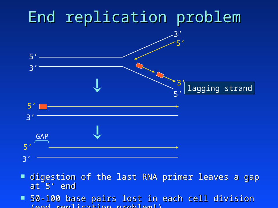
End replication problemEnd replication problem
5’3’
3’5’
3’5’
lagging strand
5’3’
5’3’
digestion of the last RNA primer leaves a gap at 5digestion of the last RNA primer leaves a gap at 5’’ end end 50-100 base pairs lost in each cell division (end replication problem!)50-100 base pairs lost in each cell division (end replication problem!)
GAP

Telomere structureTelomere structure human telomeres contain a tandem repeat sequence (several human telomeres contain a tandem repeat sequence (several
thousand base pairs in length): thousand base pairs in length):
55’’–TTAGGG–3–TTAGGG–3’’ / / 5 5’’–CCCTAA–3–CCCTAA–3’’ telomeres end in a 3telomeres end in a 3’’ single-stranded overhang of the G-rich single-stranded overhang of the G-rich
strand (a few hundred nucleotides in length).strand (a few hundred nucleotides in length). the telomere end folds back on itself to form a the telomere end folds back on itself to form a ““T loopT loop””
protects the 3protects the 3’’ overhang (Figs. 10-17 and 10-19) overhang (Figs. 10-17 and 10-19) renders the chromosome end less recombinogenicrenders the chromosome end less recombinogenic does not elicit a DNA damage response (local inhibition of ATM does not elicit a DNA damage response (local inhibition of ATM
and ATR by and ATR by ““shelterinshelterin”” proteins that bind the T loop) proteins that bind the T loop)
TTAGGGTTAGGGTTAGGGTTAGGGTTAGGGTTAGGGTTAGGGAATCCCAATCCCAATCCCAATCCCAATCCC
3’-ssDNAoverhangdsDNA telomere repeat
– 3’chromosome

Telomere lengthTelomere length note: the terminal DNA sequences lost at each cell note: the terminal DNA sequences lost at each cell
division are comprised entirely of telomere repeatsdivision are comprised entirely of telomere repeats
telomere length shortens during telomere length shortens during in vitroin vitro culture of culture of human fibroblasts: human fibroblasts:
early passage fibroblasts = 18-25 kilobases (kb)early passage fibroblasts = 18-25 kilobases (kb) at replicative senescent = 8-10 kbat replicative senescent = 8-10 kb at genetic catastrophe = 1-2 kbat genetic catastrophe = 1-2 kb
telomere attrition also occurs telomere attrition also occurs in vivo.in vivo. how is telomere length restored in each new generation ?how is telomere length restored in each new generation ?
TelomeraseTelomerase: an enzymatic complex that extends telomeres.: an enzymatic complex that extends telomeres. since telomerase is highly active in germ cells, chromosomes since telomerase is highly active in germ cells, chromosomes
of the germline retain full telomere length.of the germline retain full telomere length.

TelomeraseTelomerase an enzymatic complex consisting of…an enzymatic complex consisting of…
TERT (telomerase reverse transcriptase)TERT (telomerase reverse transcriptase): a catalytic subunit : a catalytic subunit that uses an RNA template to synthesize tandem DNA copies that uses an RNA template to synthesize tandem DNA copies of the telomere repeat sequence.of the telomere repeat sequence.
TR (telomerase RNA)TR (telomerase RNA): ssRNA molecule of 451 nucleotides: ssRNA molecule of 451 nucleotides contains a sequence that is complementary to the telomere repeat:contains a sequence that is complementary to the telomere repeat:
55’’–CUAACCCUAA–3–CUAACCCUAA–3’’ provides the template for synthesis of the telomere repeat.provides the template for synthesis of the telomere repeat.
Telomerase adds telomere repeat units (5Telomerase adds telomere repeat units (5’’–TTAGGG–3–TTAGGG–3’’) to the ) to the 33’’–ssDNA overhang.–ssDNA overhang.
in humans, TERT expression is largely restricted to the germline in humans, TERT expression is largely restricted to the germline and to certain stem cells.and to certain stem cells.

Telomere hypothesisTelomere hypothesis Replicative senescence Replicative senescence (early crisis)…(early crisis)…
is induced by telomeric shortening below a threshold levelis induced by telomeric shortening below a threshold level(8-10 kb in human fibroblasts)(8-10 kb in human fibroblasts)
enforced by the p53 and Rb checkpointsenforced by the p53 and Rb checkpoints Genetic catastrophe Genetic catastrophe (late crisis)…(late crisis)…
occurs when disruption of p53/Rb checkpoint (e.g., by viral occurs when disruption of p53/Rb checkpoint (e.g., by viral oncogenes) allows continued proliferation despite impending oncogenes) allows continued proliferation despite impending telomere dysfunction.telomere dysfunction.
further telomere erosion further telomere erosion telomere dysfunctiontelomere dysfunction loss of telomere function loss of telomere function genetic catastrophegenetic catastrophe
genomic instabilitygenomic instability massive cell deathmassive cell death emergence of immortal variantsemergence of immortal variants

Short telomeres & genetic instabilityShort telomeres & genetic instability
when short telomeres become dysfunctional…when short telomeres become dysfunctional… chromosome ends behave like dsDNA breakschromosome ends behave like dsDNA breaks
highly recombinogenichighly recombinogenic induce the cellular DNA damage responseinduce the cellular DNA damage response
““breakage-fusion-bridgebreakage-fusion-bridge”” cycles are initiated cycles are initiated recombinogenic telomeres of two chromosome ends fuse to recombinogenic telomeres of two chromosome ends fuse to
form a dicentric chromosomeform a dicentric chromosome during metaphase, the same chromatid of a dicentric can attach during metaphase, the same chromatid of a dicentric can attach
to both spindle polesto both spindle poles this chromatid may break during anaphasethis chromatid may break during anaphase
chromosome fragmentation (further rounds of breakage-fusion-bridge)chromosome fragmentation (further rounds of breakage-fusion-bridge) non-reciprocal chromosome translocationsnon-reciprocal chromosome translocations loss of genetic materialloss of genetic material

““breakage-fusion-bridgebreakage-fusion-bridge”” cycles - I cycles - I the recombinogenic telomeres of two chromosomes fuse to the recombinogenic telomeres of two chromosomes fuse to
form a dicentric chromosome…form a dicentric chromosome…
telomere –>
centromere –>
telomere*** –>
fusion
dicentricchromosome
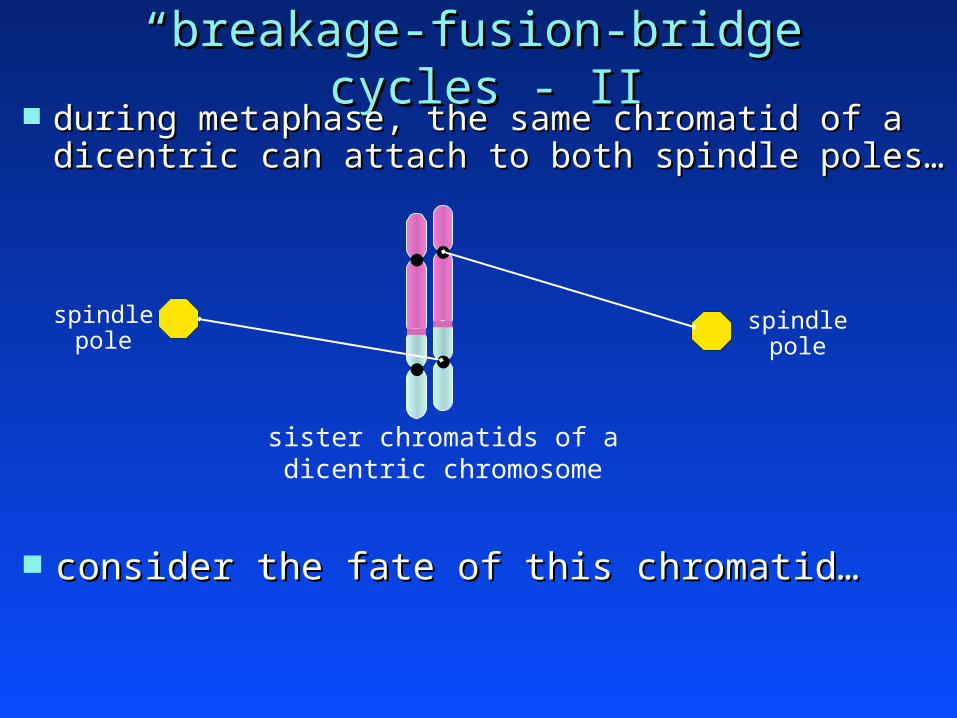
““breakage-fusion-bridgebreakage-fusion-bridge”” cycles - II cycles - II during metaphase, the same chromatid of a dicentric can during metaphase, the same chromatid of a dicentric can
attach to both spindle poles…attach to both spindle poles…
sister chromatids of adicentric chromosome
spindlepole
spindlepole
consider the fate of this chromatid…consider the fate of this chromatid…

““breakage-fusion-bridgebreakage-fusion-bridge”” cycles - II cycles - II during metaphase, the same chromatid of a dicentric can during metaphase, the same chromatid of a dicentric can
attach to both spindle poles …attach to both spindle poles …
dicentric chromatid
spindlepole
spindlepole
As the chromosomes segregate, this chromatid will form an As the chromosomes segregate, this chromatid will form an ““anaphase bridgeanaphase bridge”” (Figure 10.16B), (Figure 10.16B), and will eventually break.and will eventually break.
The break sites of the two resulting broken chromatids will also be The break sites of the two resulting broken chromatids will also be recombinogenic.recombinogenic.

““breakage-fusion-bridgebreakage-fusion-bridge”” cycles - III cycles - III
The previous two slides show a breakage-fusion-bridge (BFB) The previous two slides show a breakage-fusion-bridge (BFB) cycle that is initiated by fusion between the recombinogenic cycle that is initiated by fusion between the recombinogenic telomeres of two different chromosomes.telomeres of two different chromosomes.
Note: BFB cycles can also be initiated by fusion between Note: BFB cycles can also be initiated by fusion between recombinogenic telomeres of the two chromatids of the recombinogenic telomeres of the two chromatids of the samesame chromosome (in G2 phase). This is illustrated in Figure 10.15 of chromosome (in G2 phase). This is illustrated in Figure 10.15 of Weinberg (2007).Weinberg (2007).

ImmortalizationImmortalization immortal variants acquire a genetic change that stabilizes immortal variants acquire a genetic change that stabilizes
telomere length:telomere length: usually by ectopic expression of TERTusually by ectopic expression of TERT sometimes by activation of ALT (Section 10.8)sometimes by activation of ALT (Section 10.8)
once telomere length has been stabilized, telomere once telomere length has been stabilized, telomere function and genomic stability are restoredfunction and genomic stability are restored
yet, these variants have sustained lasting genetic lesionsyet, these variants have sustained lasting genetic lesions karyotypic level: aneuploidy & rearranged chromosomeskaryotypic level: aneuploidy & rearranged chromosomes genetic level: functional alterations of unknown genes ?genetic level: functional alterations of unknown genes ? telomere-induced genomic instability is an important aspect of telomere-induced genomic instability is an important aspect of
human tumorigenesishuman tumorigenesis.. immortal variants of human fibroblasts emerge during immortal variants of human fibroblasts emerge during
genetic catastrophe at a reasonable rate (10genetic catastrophe at a reasonable rate (10-5-5 – 10 – 10-6-6))

Immortalization with TERTImmortalization with TERT stably transfect early passage (stably transfect early passage (““pre-crisispre-crisis””) human ) human
fibroblasts with an expression vector encoding TERT…fibroblasts with an expression vector encoding TERT… telomere length is stabilized.telomere length is stabilized.
p53/Rb checkpoints are not activated.p53/Rb checkpoints are not activated.
replicative senescence and genetic catastrophe are averted.replicative senescence and genetic catastrophe are averted.
cells are rendered immortal.cells are rendered immortal.
TERT immortalized cells have a stable diploid genome.TERT immortalized cells have a stable diploid genome.
Provides strong evidence that, in human fibroblasts, Provides strong evidence that, in human fibroblasts, replicative senescence and genetic catastrophe are both replicative senescence and genetic catastrophe are both telomere-dependent eventstelomere-dependent events

early crisis
late crisis
extendedlifespan
time
PDsTERT-immortalization
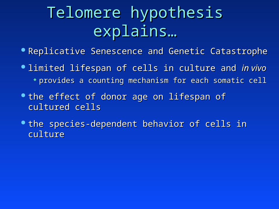
Telomere hypothesis explains…Telomere hypothesis explains…
Replicative Senescence and Genetic CatastropheReplicative Senescence and Genetic Catastrophe
limited lifespan of cells in culture and limited lifespan of cells in culture and in vivoin vivo provides a counting mechanism for each somatic cellprovides a counting mechanism for each somatic cell
the effect of donor age on lifespan of cultured cellsthe effect of donor age on lifespan of cultured cells
the species-dependent behavior of cells in culturethe species-dependent behavior of cells in culture

But, why not express telomerase constitutively ?But, why not express telomerase constitutively ?
telomere-induced replicative senescencetelomere-induced replicative senescence a very useful mechanism to suppress tumor development, a very useful mechanism to suppress tumor development,
especially in large and long-lived animals.especially in large and long-lived animals.
Suppose that…Suppose that… at least 20 cell divisions required to yield an oncogenic mutationat least 20 cell divisions required to yield an oncogenic mutation at least 5 independent mutations are needed for malignancy:at least 5 independent mutations are needed for malignancy:
at least 100 cell divisions necessary to produce a single malignant cell.at least 100 cell divisions necessary to produce a single malignant cell. telomere-induced senescence will severely limit the neoplastic telomere-induced senescence will severely limit the neoplastic
development of pre-malignant cells.development of pre-malignant cells. consequently, telomere-induced senescence is a useful barrier to cancer consequently, telomere-induced senescence is a useful barrier to cancer
development in humans.development in humans.
telomerase activation occurs in almost all human tumorstelomerase activation occurs in almost all human tumors
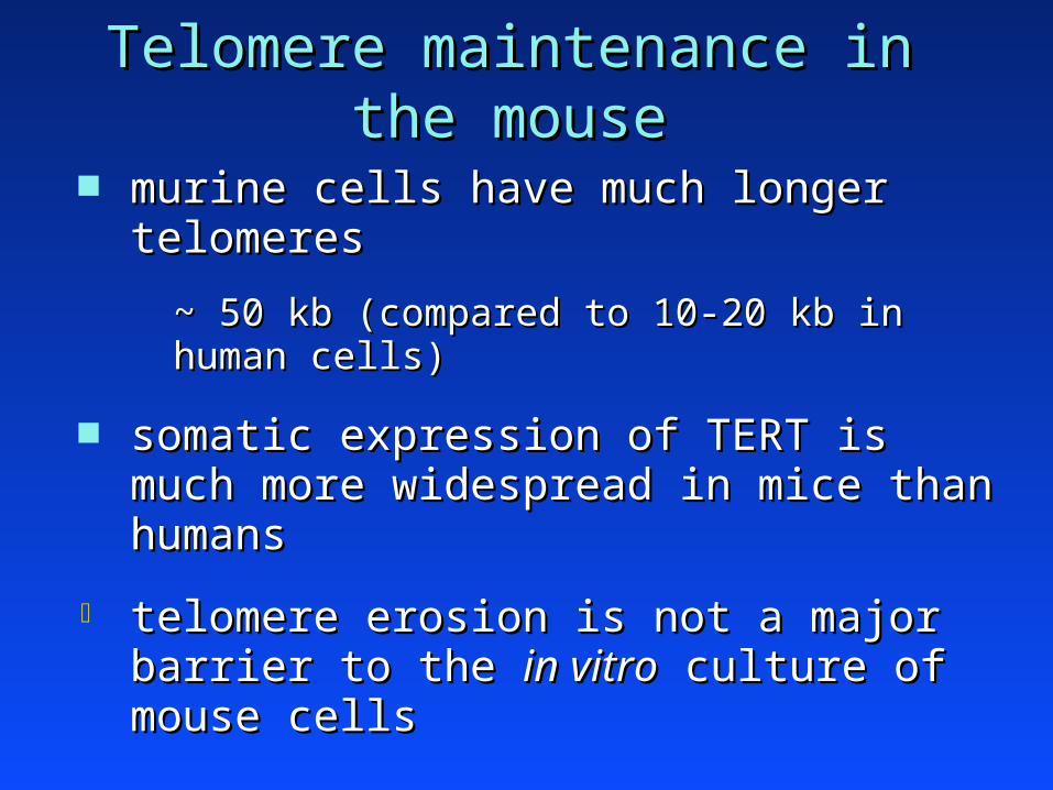
Telomere maintenance in the mouseTelomere maintenance in the mouse
murine cells have much longer telomeresmurine cells have much longer telomeres
~ 50 kb (compared to 10-20 kb in human cells)~ 50 kb (compared to 10-20 kb in human cells)
somatic expression of TERT is much more somatic expression of TERT is much more widespread in mice than humanswidespread in mice than humans
telomere erosion is not a major barrier to the telomere erosion is not a major barrier to the in vitroin vitro culture of mouse cells culture of mouse cells
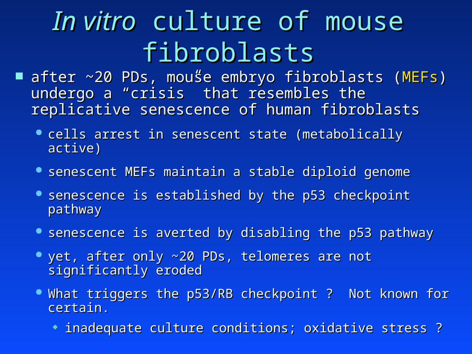
In vitroIn vitro culture of mouse fibroblasts culture of mouse fibroblasts after ~20 PDs, mouse embryo fibroblasts (after ~20 PDs, mouse embryo fibroblasts (MEFsMEFs) )
undergo a undergo a ““crisiscrisis”” that resembles the replicative that resembles the replicative senescence of human fibroblastssenescence of human fibroblasts
cells arrest in senescent state (metabolically active)cells arrest in senescent state (metabolically active)
senescent MEFs maintain a stable diploid genomesenescent MEFs maintain a stable diploid genome
senescence is established by the p53 checkpoint pathwaysenescence is established by the p53 checkpoint pathway
senescence is averted by disabling the p53 pathwaysenescence is averted by disabling the p53 pathway
yet, after only ~20 PDs, telomeres are not significantly erodedyet, after only ~20 PDs, telomeres are not significantly eroded
What triggers the p53/RB checkpoint ? Not known for certain.What triggers the p53/RB checkpoint ? Not known for certain. inadequate culture conditions; oxidative stress ?inadequate culture conditions; oxidative stress ?

mouse fibroblast cell linesmouse fibroblast cell lines
unlike human fibroblasts, MEF cultures routinely unlike human fibroblasts, MEF cultures routinely give rise to immortal variantsgive rise to immortal variants
rare immortal variants arise during crisis (presumably due rare immortal variants arise during crisis (presumably due to mutations in the p53 pathway), and these variants to mutations in the p53 pathway), and these variants overgrow the cultureovergrow the culture
““cell linescell lines””
MEF cell lines are diploid and have reasonably stable MEF cell lines are diploid and have reasonably stable genomesgenomes
the source of the source of ““NIH-3T3NIH-3T3”” cells cells
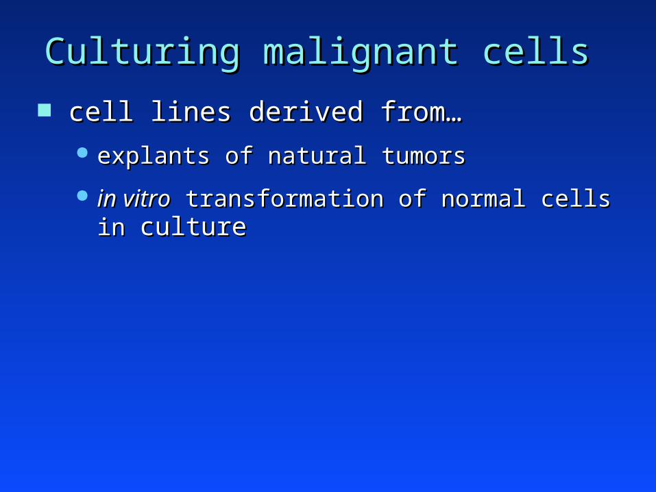
Culturing malignant cellsCulturing malignant cells
cell lines derived from… cell lines derived from… explants of natural tumorsexplants of natural tumors
in vitroin vitro transformation of normal cells in transformation of normal cells in cultureculture

in vitroin vitro properties of malignant cells properties of malignant cells
immortalityimmortality
decreased growth factor requirementsdecreased growth factor requirements
altered morphologyaltered morphology
loss of contact inhibitionloss of contact inhibition
loss of dependence on anchorage for cell growthloss of dependence on anchorage for cell growth

in vitroin vitro properties of malignant cells properties of malignant cells
immortalityimmortality telomerase inductiontelomerase induction inactivation of cell cycle checkpointsinactivation of cell cycle checkpoints

in vitroin vitro properties of malignant cells properties of malignant cells
immortalityimmortality
decreased growth factor requirementsdecreased growth factor requirements

in vitroin vitro properties of malignant cells properties of malignant cells
immortalityimmortality
decreased growth factor requirementsdecreased growth factor requirements
altered morphologyaltered morphology

altered morphology of tumor cellsaltered morphology of tumor cells rounded appearance rounded appearance (vs. the elongated, spindly shape (vs. the elongated, spindly shape
of normal fibroblasts)of normal fibroblasts) increased nuclear volumeincreased nuclear volume
RSV-transformed CEFnormal CEF

in vitroin vitro properties of malignant cells properties of malignant cells
immortalityimmortality
decreased growth factor requirementsdecreased growth factor requirements
altered morphologyaltered morphology
loss of contact inhibitionloss of contact inhibition

loss of contact inhibitionloss of contact inhibition
seed cells onto petri dish at low dilutionseed cells onto petri dish at low dilution cells attach to dish and start to proliferatecells attach to dish and start to proliferate
malignant cellsnormal cells

loss of contact inhibitionloss of contact inhibition
cells remain attached to dish and continue to dividecells remain attached to dish and continue to divide
malignant cellsnormal cells

loss of contact inhibitionloss of contact inhibition
growing culture begins to form a monolayergrowing culture begins to form a monolayer some cells attain cell-cell contacts on all sidessome cells attain cell-cell contacts on all sides these cells normally experience “contact inhibition”these cells normally experience “contact inhibition”
normal cells malignant cells

loss of contact inhibitionloss of contact inhibition
= contact-inhibited cell= contact-inhibited cell
in normal cells, contact inhibition causes cell cycle arrest.in normal cells, contact inhibition causes cell cycle arrest. malignant cells are not affected by contact inhibition.malignant cells are not affected by contact inhibition.
malignant cellsnormal cells

in vitroin vitro properties of malignant cells properties of malignant cells
immortalityimmortality
decreased growth factor requirementsdecreased growth factor requirements
altered morphologyaltered morphology
loss of contact inhibitionloss of contact inhibition
loss of dependence on anchorage for cell growthloss of dependence on anchorage for cell growth
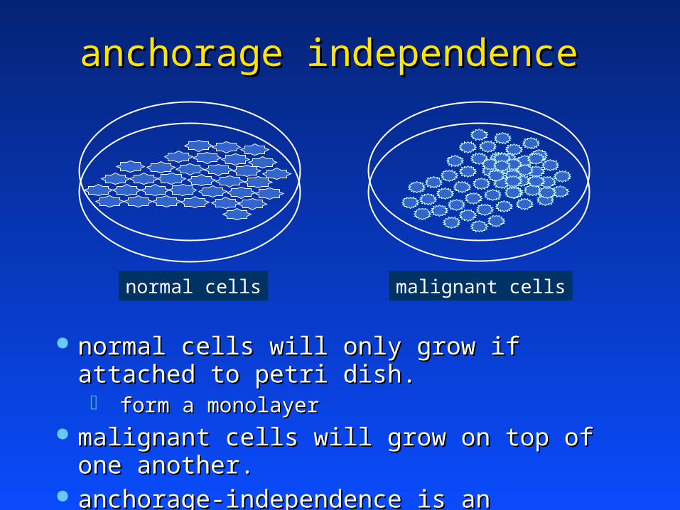
anchorage independenceanchorage independence
normal cells will only grow if attached to petri dish.normal cells will only grow if attached to petri dish. form a monolayerform a monolayer
malignant cells will grow on top of one another.malignant cells will grow on top of one another. anchorage-independence is an especially important anchorage-independence is an especially important
parameter of parameter of in vivoin vivo tumorigenicity tumorigenicity
malignant cellsnormal cells

in vitroin vitro assays of cell transformation assays of cell transformation
what happens to normal cultured cells that are what happens to normal cultured cells that are treated with agents that induce tumors in animals?treated with agents that induce tumors in animals? - e.g., viruses, radiation, chemicals- e.g., viruses, radiation, chemicals
they acquire some properties of malignant cellsthey acquire some properties of malignant cells
““in vitroin vitro cell transformation cell transformation”” can be measured can be measured using quantitative assays:using quantitative assays: focus formationfocus formation
colony formation in soft agarcolony formation in soft agar

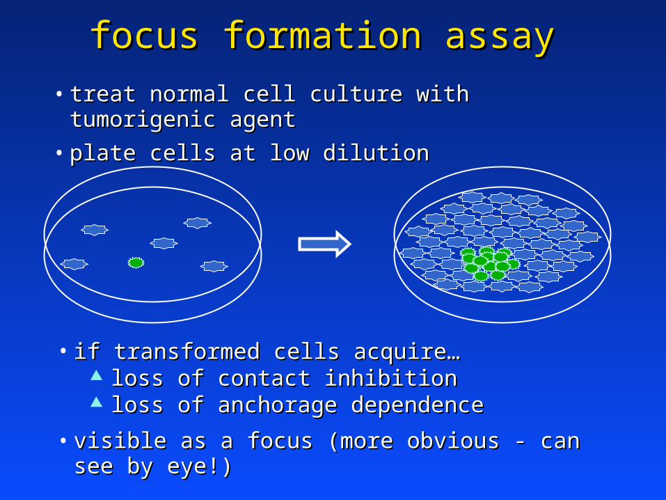
focus formation assayfocus formation assay based on several features of transformed cells:based on several features of transformed cells:
altered morphologyaltered morphology loss of contact-inhibitionloss of contact-inhibition loss of anchorage dependenceloss of anchorage dependence
treat normal cells with oncogenic agenttreat normal cells with oncogenic agent
plate cells at low dilutionplate cells at low dilution
progeny of rare transformed cells give rise to a progeny of rare transformed cells give rise to a ““focusfocus”” that can be visibly distinguished from that can be visibly distinguished from the monolayer of normal cells.the monolayer of normal cells.

focus formation assayfocus formation assay
• treat normal cell culture with tumorigenic agenttreat normal cell culture with tumorigenic agent
• plate cells at low dilutionplate cells at low dilution
= transformed cell= transformed cell
• if transformed cells acquire…if transformed cells acquire…∆ altered morphologyaltered morphology
• visible as a focus (light microscope at low magnification)visible as a focus (light microscope at low magnification)
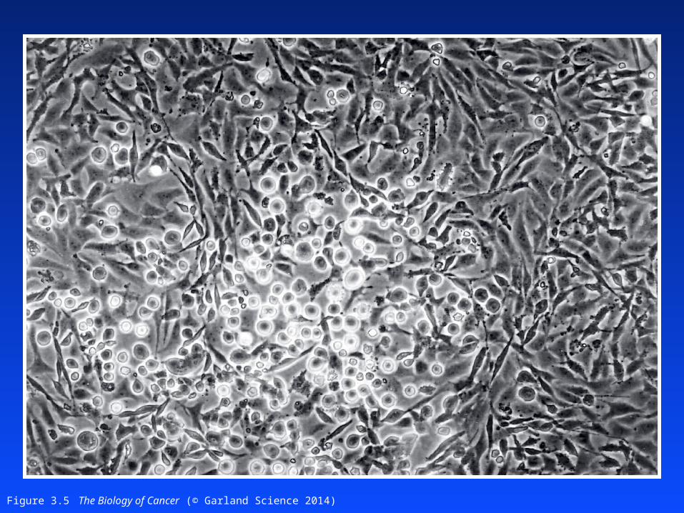
Figure 3.5 The Biology of Cancer (© Garland Science 2014)
focus formation assayfocus formation assay
• treat normal cell culture with tumorigenic agenttreat normal cell culture with tumorigenic agent
• plate cells at low dilutionplate cells at low dilution
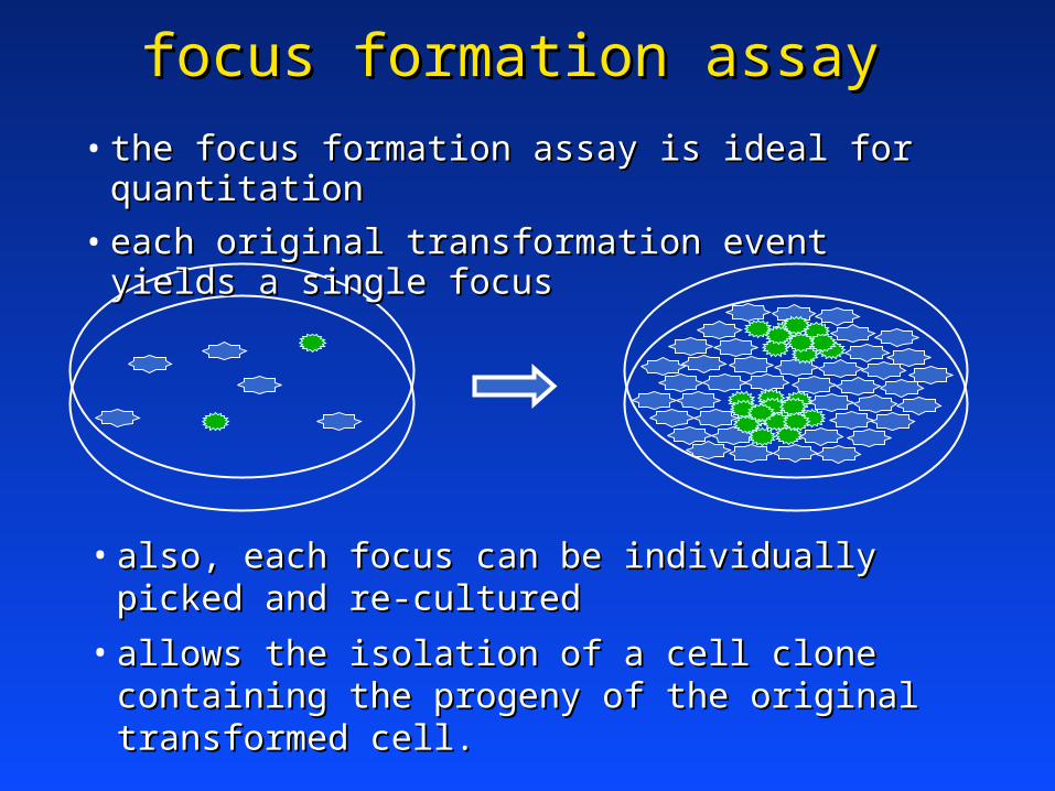
• if transformed cells acquire…if transformed cells acquire… loss of contact inhibitionloss of contact inhibition loss of anchorage dependenceloss of anchorage dependence
• visible as a focus (more obvious - can see by eye!)visible as a focus (more obvious - can see by eye!)
focus formation assayfocus formation assay
• the focus formation assay is ideal for quantitationthe focus formation assay is ideal for quantitation
• each original transformation event yields a single focuseach original transformation event yields a single focus
• also, each focus can be individually picked and re-culturedalso, each focus can be individually picked and re-cultured
• allows the isolation of a cell clone containing the progeny of allows the isolation of a cell clone containing the progeny of the original transformed cell.the original transformed cell.

colony formation assaycolony formation assay based on:based on:
loss of anchorage dependenceloss of anchorage dependence
treat normal cells with oncogenic agenttreat normal cells with oncogenic agent
dilute cells in dilute cells in ““soft agarsoft agar”” (0.3% agar) (0.3% agar)
pour agar onto petri dishpour agar onto petri dish cells are suspended in agarcells are suspended in agar normal cells will not divide (insufficient anchorage)normal cells will not divide (insufficient anchorage)
progeny of rare transformed cells give rise to a progeny of rare transformed cells give rise to a ““colonycolony”” of growing cells of growing cells (visible by eye or under low magnification)(visible by eye or under low magnification)
colonies can be counted and pickedcolonies can be counted and picked

in vivoin vivo tumorigenesis tumorigenesis inject cultured cells into animals…inject cultured cells into animals…
look for and count tumors.look for and count tumors.
common hosts:common hosts: adult miceadult mice
fetalfetal//newborn mice newborn mice immature immune systemimmature immune system
““nudenude”” mice mice no T cell immunity no T cell immunity
SCID miceSCID mice no B or T cell immunity no B or T cell immunity
correlations between correlations between in vitroin vitro properties and properties and in vivoin vivo tumorigenicity of transformed cells are not absolute.tumorigenicity of transformed cells are not absolute.

RetrovirusesRetroviruses
Key features of all retroviruses:Key features of all retroviruses: an enveloped virionan enveloped virion ssRNA genomessRNA genome all retroviral genomes have all retroviral genomes have gaggag, , polpol, and , and envenv genes genes all encode a reverse transcriptaseall encode a reverse transcriptase
Let’s consider Avian Leukosis Virus (ALV) as Let’s consider Avian Leukosis Virus (ALV) as a prototype of an oncogenic retrovirusa prototype of an oncogenic retrovirus
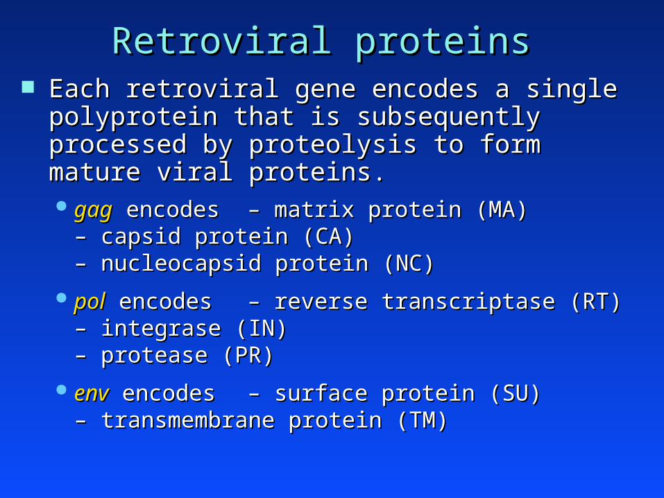
Retroviral proteinsRetroviral proteins Each retroviral gene encodes a single polyprotein Each retroviral gene encodes a single polyprotein
that is subsequently processed by proteolysis to that is subsequently processed by proteolysis to form mature viral proteins.form mature viral proteins. gaggag encodes encodes – matrix protein (MA)– matrix protein (MA)
– – capsid protein (CA)capsid protein (CA)– – nucleocapsid protein (NC)nucleocapsid protein (NC)
polpol encodes encodes – reverse transcriptase (RT)– reverse transcriptase (RT)– – integrase (IN)integrase (IN)– – protease (PR)protease (PR)
envenv encodes encodes – surface protein (SU)– surface protein (SU)– – transmembrane protein (TM)transmembrane protein (TM)

Retroviral virionenvelope
(lipid bilayer)
matrix
capsid
nucleoproteincore
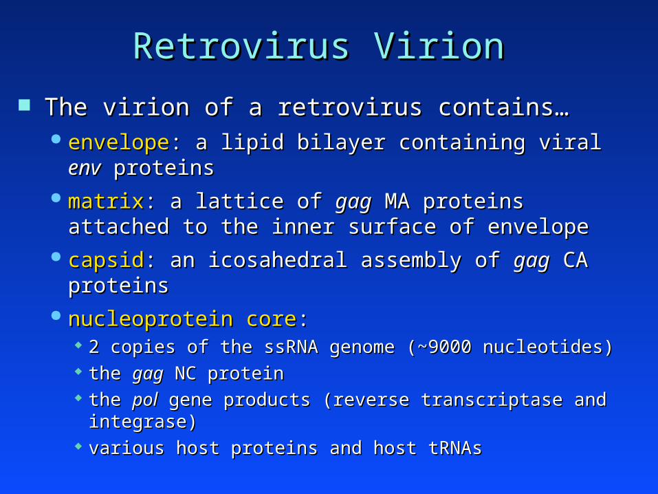
Retrovirus VirionRetrovirus Virion
The virion of a retrovirus contains…The virion of a retrovirus contains… envelopeenvelope: a lipid bilayer containing viral : a lipid bilayer containing viral envenv proteins proteins matrixmatrix: a lattice of : a lattice of gaggag MA proteins attached to the MA proteins attached to the
inner surface of envelopeinner surface of envelope capsidcapsid: an icosahedral assembly of : an icosahedral assembly of gaggag CA proteins CA proteins nucleoprotein corenucleoprotein core::
2 copies of the ssRNA genome (~9000 nucleotides)2 copies of the ssRNA genome (~9000 nucleotides) the the gaggag NC protein NC protein the the polpol gene products (reverse transcriptase and integrase) gene products (reverse transcriptase and integrase) various host proteins and host tRNAsvarious host proteins and host tRNAs

Retroviral virionenvelope
(lipid bilayer)
env SUprotein
env TMprotein
matrix(gag MA)
capsid(gag CA)
nucleoproteincore
- 2 x ssRNA- gag NC- pol proteins

retroviral infectionretroviral infection
host range of a retrovirushost range of a retrovirus determined principally by the determined principally by the envenv protein protein species specificity: ALV infects chickensspecies specificity: ALV infects chickens tissue specificity: ALV infects most cell types tissue specificity: ALV infects most cell types
early stages of infectionearly stages of infection envenv binds specific receptors on host cell membrane binds specific receptors on host cell membrane fusion of viral envelope and host cell membranefusion of viral envelope and host cell membrane viral proteins enter cytoplasmviral proteins enter cytoplasm

Reverse transcriptionReverse transcription Reverse transcription occurs in the cytoplasmReverse transcription occurs in the cytoplasm
generates a dsDNA genomegenerates a dsDNA genome flanked by LTRs (long terminal repeats)flanked by LTRs (long terminal repeats)
ssRNA
viral RT
host DNA pol
ssDNA
dsDNA
5’-cap– AAAAAA
R Rgag pol env
gag pol envLTR LTR

Proviral integrationProviral integration viral dsDNA migrates into the cell nucleus viral dsDNA migrates into the cell nucleus circularization of dsDNAcircularization of dsDNA integration of dsDNA into host genomeintegration of dsDNA into host genome
catalyzed by viral integrase proteincatalyzed by viral integrase protein site-specific with respect to the viral genomesite-specific with respect to the viral genome random with respect to the host genomerandom with respect to the host genome generates a generates a ““provirusprovirus””
gag pol env
host cellchromosome
LTR LTR

Proviral transcriptionProviral transcription the provirus is an active transcription unitthe provirus is an active transcription unit
each LTR containseach LTR contains –– promoter promoter–– enhancerenhancer–– polyA signalpolyA signal
pA enh. pr.
gag pol env
LTR LTR
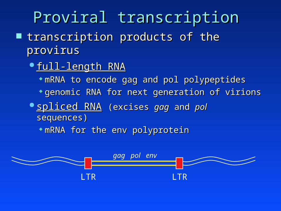
Proviral transcriptionProviral transcription transcription products of the provirustranscription products of the provirus
full-length RNAfull-length RNA mRNA to encode gag and pol polypeptidesmRNA to encode gag and pol polypeptides genomic RNA for next generation of virionsgenomic RNA for next generation of virions
spliced RNAspliced RNA (excises (excises gaggag and and polpol sequences) sequences) mRNA for the env polyproteinmRNA for the env polyprotein
gag pol env
LTR LTR

Proviral replicationProviral replication
During cell division, the provirus is replicated During cell division, the provirus is replicated along with the host genomealong with the host genome all daughter cells inherit the provirusall daughter cells inherit the provirus
Retroviruses can also infect germline cells Retroviruses can also infect germline cells (e.g., oocytes or spermatocytes).(e.g., oocytes or spermatocytes). provirus then becomes part of the genetic material provirus then becomes part of the genetic material
of the speciesof the species
Endogenous retrovirusEndogenous retrovirus constitute > 0.1% of mouse genomeconstitute > 0.1% of mouse genome usually transcriptionally inactiveusually transcriptionally inactive

Virion production - IVirion production - I
envenv proteins proteins processed through the secretory pathway and processed through the secretory pathway and
inserted into host cell membrane.inserted into host cell membrane.
polpol proteins (RT, integrase, protease) proteins (RT, integrase, protease) associate with ssRNA genome to form the associate with ssRNA genome to form the
nucleoprotein core (in cytoplasm). nucleoprotein core (in cytoplasm).
gaggag proteins proteins associate with the nucleoprotein to form a capsid associate with the nucleoprotein to form a capsid
(in cytoplasm). (in cytoplasm).

Virion production - IIVirion production - II the capsid buds from the host cell membranethe capsid buds from the host cell membrane
while the capsid buds out, a segment of the env-while the capsid buds out, a segment of the env-impregnated cell surface encircles it and forms impregnated cell surface encircles it and forms the viral envelope.the viral envelope.
ALV replication is not cytopathicALV replication is not cytopathic Infected cell are difficult to distinguish from Infected cell are difficult to distinguish from
uninfected cells. To do so may require…uninfected cells. To do so may require… serological analysis to detect viral antigensserological analysis to detect viral antigens electron microscopy to visualize intracytoplasmic electron microscopy to visualize intracytoplasmic
capsids and budding virions.capsids and budding virions.
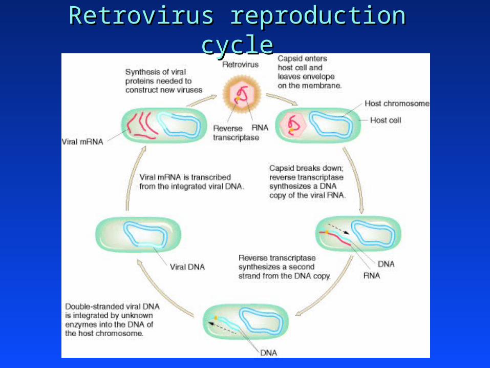
Retrovirus reproduction cycleRetrovirus reproduction cycle

Transmission of retrovirusesTransmission of retroviruses
Horizontal transmission (from another animal)Horizontal transmission (from another animal)
Vertical transmission (from parents)Vertical transmission (from parents) Genetic transmissionGenetic transmission
animal inherits endogenous provirus.animal inherits endogenous provirus. rarely significant in oncogenesis (except in genetically rarely significant in oncogenesis (except in genetically
susceptible strains, such as AKR mice).susceptible strains, such as AKR mice).
Congenital infectionCongenital infection
Experimental transmission (by a scientist)Experimental transmission (by a scientist)

Horizontal transmission (ALV)Horizontal transmission (ALV)
rere. - ALV infects cells from a broad spectrum of . - ALV infects cells from a broad spectrum of chicken tissues.chicken tissues.
if chick is more than a few days old (post-if chick is more than a few days old (post-hatching) at infection:hatching) at infection: transient viremia develops (virions in bloodstream)transient viremia develops (virions in bloodstream) chick produces neutralizing antibodieschick produces neutralizing antibodies viremia clears; chick immune to further infectionviremia clears; chick immune to further infection chick does chick does notnot develop virally-induced lymphoma develop virally-induced lymphoma

Congenital Infection (ALV)Congenital Infection (ALV)
mother infects her offspringmother infects her offspring chickens - infection of egg while in the female chickens - infection of egg while in the female
reproductive organsreproductive organs mammals - transmission via placenta or milkmammals - transmission via placenta or milk
consequences of congenital infection with ALV:consequences of congenital infection with ALV: viremia occurs during embryonic developmentviremia occurs during embryonic development chick develops immunological tolerance to ALVchick develops immunological tolerance to ALV viremia continuesviremia continues lymphomas develop during adulthoodlymphomas develop during adulthood

Acutely Transforming RetrovirusesAcutely Transforming Retroviruses

Acutely Transforming RetrovirusesAcutely Transforming Retroviruses
Peyton Rous (Rockefeller University)Peyton Rous (Rockefeller University) observed a spontaneous sarcoma in a chickenobserved a spontaneous sarcoma in a chicken in retrospect, we know that this chicken was from a in retrospect, we know that this chicken was from a
flock infected with ALVflock infected with ALV
Rous propagated the sarcoma cells by passaging Rous propagated the sarcoma cells by passaging from chicken to chicken.from chicken to chicken.
sarcomaexplant
suspendedtumor cells
newsarcoma
dissociate inoculatechick

Rous Sarcoma Virus (RSV)Rous Sarcoma Virus (RSV) In 1911, Rous prepared a cell-free filtrate from In 1911, Rous prepared a cell-free filtrate from
one of his sarcoma explantsone of his sarcoma explants passed the dissociated explant through filter paper, passed the dissociated explant through filter paper,
removing bacterial & eukaryotic cellsremoving bacterial & eukaryotic cells inoculated chicks with the filtrateinoculated chicks with the filtrate chicks developed sarcomas at site of injection!!chicks developed sarcomas at site of injection!! Rous Sarcoma Virus (RSV)Rous Sarcoma Virus (RSV)
sarcomaexplant
cell-freefiltrate
newsarcomafilter inoculate
chick
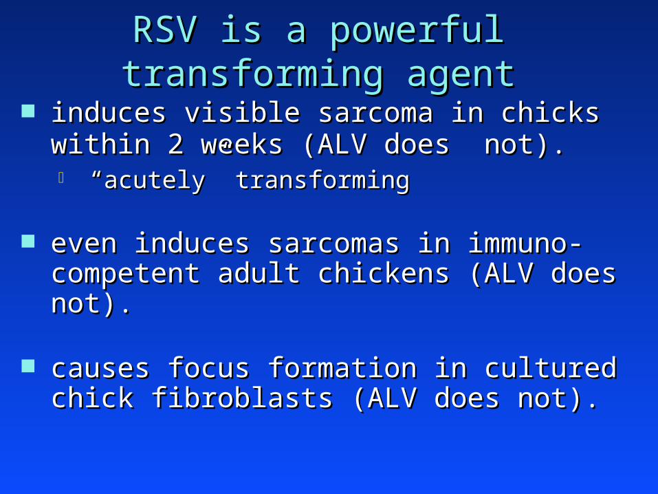
RSV is a powerful transforming agentRSV is a powerful transforming agent
induces visible sarcoma in chicks within 2 weeks induces visible sarcoma in chicks within 2 weeks (ALV does not).(ALV does not). ““acutelyacutely”” transforming transforming
even induces sarcomas in immuno-competent even induces sarcomas in immuno-competent adult chickens (ALV does not).adult chickens (ALV does not).
causes focus formation in cultured chick causes focus formation in cultured chick fibroblasts (ALV does not).fibroblasts (ALV does not).

Why is RSV so tumorigenic ?Why is RSV so tumorigenic ?
especially compared with ALVespecially compared with ALV RSV:RSV: ““acutely-transforming retrovirusacutely-transforming retrovirus”” ALV:ALV: ““slowly-transforming retrovirusslowly-transforming retrovirus””
RSV and ALV: –RSV and ALV: – structurally similarstructurally similar
– – infect similar cellsinfect similar cells

The v-The v-srcsrc gene gene The RSV genome harbors additional sequencesThe RSV genome harbors additional sequences
of ~ 1,500 nucleotides (of ~ 1,500 nucleotides (v-v-src,src, for for ““viral srcviral src””).).
5’-cap– AAAAAA
R Rgag pol envALV9.0 kb
5’-cap– AAAAAA
R Rgag pol env srcRSV10.5 kb
genetic experiments showed that the increased genetic experiments showed that the increased malignant potential of RSV is due to v-malignant potential of RSV is due to v-srcsrc

The origin of v-The origin of v-srcsrc sequences sequences
v-v-srcsrc is absent from almost all other retroviral is absent from almost all other retroviral genomes. Thus, where did it originate ?genomes. Thus, where did it originate ?
in 1977, Bishop & Varmus reported that in 1977, Bishop & Varmus reported that genomic DNA from normal, uninfected genomic DNA from normal, uninfected chicken cells harbors a close homolog of v-chicken cells harbors a close homolog of v-srcsrc c-c-srcsrc (for (for ““cellular srccellular src””)) the c-the c-srcsrc gene is conserved phylogenetically gene is conserved phylogenetically c-c-srcsrc functions in normal animal development functions in normal animal development

Retroviral transductionRetroviral transduction RSV originally appeared in a spontaneous sarcoma RSV originally appeared in a spontaneous sarcoma
from an ALV-infected chickenfrom an ALV-infected chicken thus, it was proposed that RSV arose by incorporation thus, it was proposed that RSV arose by incorporation
of c-of c-srcsrc sequences into ALV sequences into ALV acquisition of c-acquisition of c-srcsrc converted a slowly-transforming converted a slowly-transforming
retrovirus (ALV) into an acutely-transforming retrovirus (ALV) into an acutely-transforming retrovirus (RSV).retrovirus (RSV).
proto-oncogene
“retroviral transduction”
oncogenec-src v-src

other acutely transforming retroviruses have other acutely transforming retroviruses have been identified in chickens, cats, mice, and rats been identified in chickens, cats, mice, and rats (> 100 independent isolates).(> 100 independent isolates).
retrovirus
Y73
MC29
AMV
Harvey
Kirsten
Abelson
tumor
sarcoma
myelocytomatosis
myeloblastosis
sarcoma
sarcoma
leukemia
v-onc
v-yes
v-myc
v-myb
v-H-ras
v-K-ras
v-abl
species
avian
avian
avian
mouse
mouse
mouse
> 40 different v-> 40 different v-onc onc genes identified; all derived from genes identified; all derived from cellular (c-cellular (c-onconc) genes by retroviral transduction.) genes by retroviral transduction.

How are transduced oncogenes How are transduced oncogenes rendered malignant?rendered malignant?
Quantitative effects:Quantitative effects: inappropriate expression - the tranduced gene is inappropriate expression - the tranduced gene is
regulated transcriptionally by the LTR.regulated transcriptionally by the LTR. elevated expression - LTR is a strong promoter.elevated expression - LTR is a strong promoter.
Qualitative effects:Qualitative effects: oncoprotein can be expressed in a truncated or oncoprotein can be expressed in a truncated or
fused form.fused form. transduced oncogenes can acquire point mutations transduced oncogenes can acquire point mutations
during viral replication (due to low fidelity of RT).during viral replication (due to low fidelity of RT).

Slowly-transforming retrovirusesSlowly-transforming retroviruses
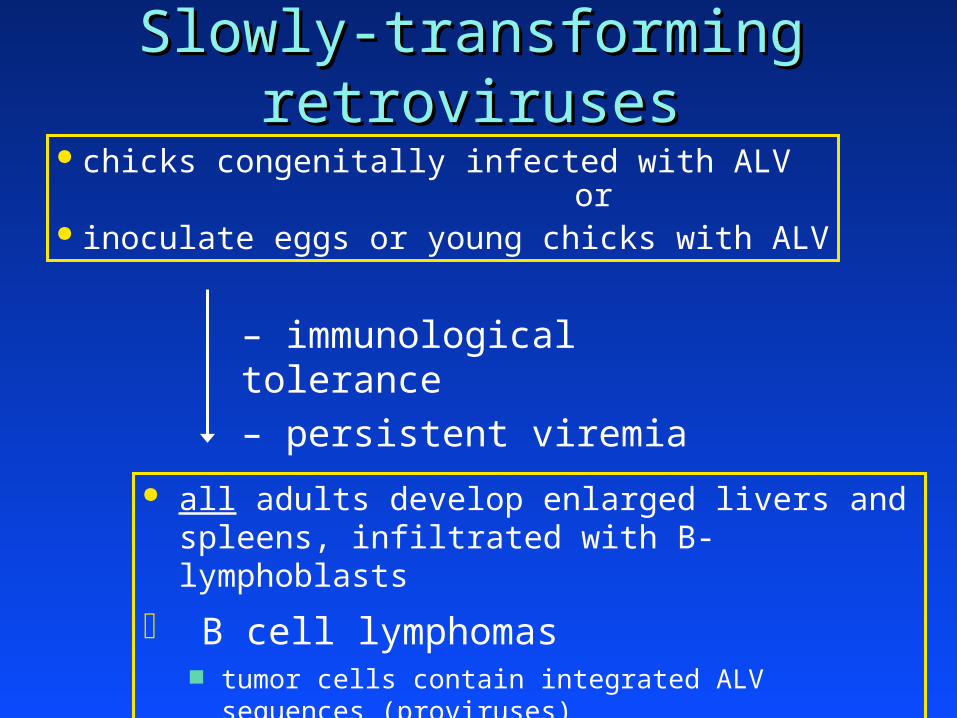
Slowly-transforming retrovirusesSlowly-transforming retroviruses chicks congenitally infected with ALV or inoculate eggs or young chicks with ALV
– immunological tolerance
– persistent viremia
all adults develop enlarged livers and spleens, infiltrated with B-lymphoblasts
B cell lymphomas tumor cells contain integrated ALV sequences (proviruses)

Patterns of ALV proviral integrationPatterns of ALV proviral integration All cells within a particular tumor have the same pattern All cells within a particular tumor have the same pattern
of proviral insertion (i.e., each tumor is monoclonal)of proviral insertion (i.e., each tumor is monoclonal) same # of proviruses insertedsame # of proviruses inserted same integration sites within the chicken genomesame integration sites within the chicken genome
Common integration sites were found in independent Common integration sites were found in independent tumors from different chickens.tumors from different chickens.
HypothesisHypothesis: ALV transforms cells by insertional : ALV transforms cells by insertional mutagenesis of a specific host gene.mutagenesis of a specific host gene.
molecular cloning molecular cloning ALV integration near ALV integration near c-c-mycmyc in in 80% of ALV-induced lymphomas!!80% of ALV-induced lymphomas!! provirally altered c-provirally altered c-mycmyc alleles are transcribed at high rates alleles are transcribed at high rates
(due to integrated LTR sequences).(due to integrated LTR sequences).

Activation of c-Myc by proviral insertion Activation of c-Myc by proviral insertion
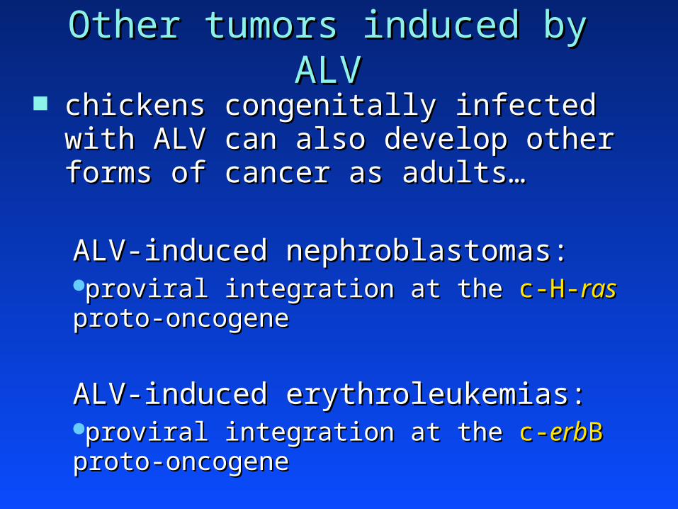
Other tumors induced by ALVOther tumors induced by ALV chickens congenitally infected with ALV can chickens congenitally infected with ALV can
also develop other forms of cancer as adults…also develop other forms of cancer as adults…
ALV-induced nephroblastomas:ALV-induced nephroblastomas:proviral integration at the proviral integration at the c-H-c-H-rasras proto-oncogene proto-oncogene
ALV-induced erythroleukemias:ALV-induced erythroleukemias:proviral integration at the proviral integration at the c-c-erberbBB proto-oncogene proto-oncogene
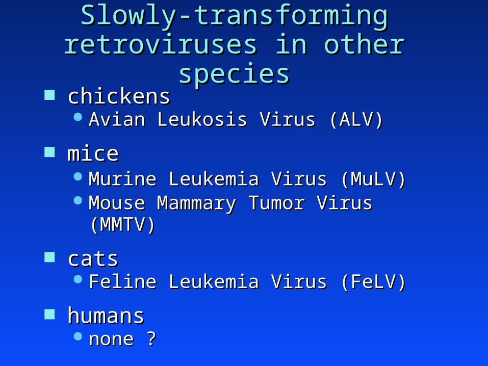
Slowly-transforming retroviruses Slowly-transforming retroviruses in other speciesin other species
chickenschickens Avian Leukosis Virus (ALV)Avian Leukosis Virus (ALV)
micemice Murine Leukemia Virus (MuLV)Murine Leukemia Virus (MuLV) Mouse Mammary Tumor Virus (MMTV)Mouse Mammary Tumor Virus (MMTV)
catscats Feline Leukemia Virus (FeLV)Feline Leukemia Virus (FeLV)
humanshumans none ?none ?
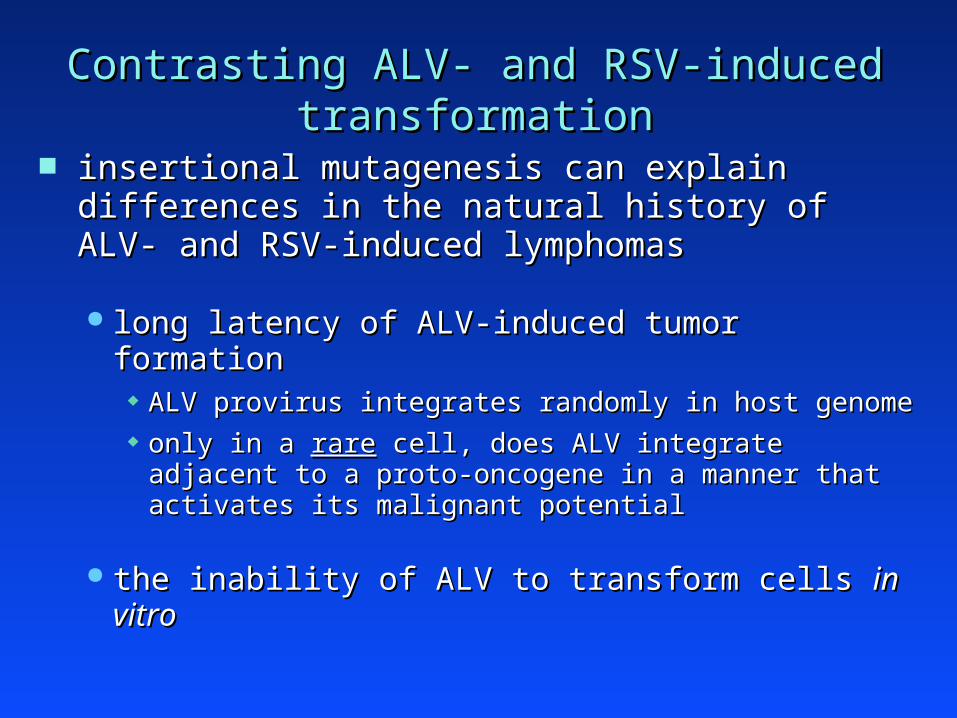
Contrasting ALV- and RSV-induced transformationContrasting ALV- and RSV-induced transformation
insertional mutagenesis can explain differences in insertional mutagenesis can explain differences in the natural history of ALV- and RSV-induced the natural history of ALV- and RSV-induced lymphomaslymphomas
long latency of ALV-induced tumor formationlong latency of ALV-induced tumor formation ALV provirus integrates randomly in host genomeALV provirus integrates randomly in host genome only in a only in a rarerare cell, does ALV integrate adjacent to a proto- cell, does ALV integrate adjacent to a proto-
oncogene in a manner that activates its malignant potentialoncogene in a manner that activates its malignant potential
the inability of ALV to transform cells the inability of ALV to transform cells in vitroin vitro

Retroviral oncogenesisRetroviral oncogenesisTwo major mechanisms identified in animals…Two major mechanisms identified in animals…
Retroviral TransductionRetroviral Transduction mediated by the “acutely-transforming retroviruses”mediated by the “acutely-transforming retroviruses”
Rous Sarcoma Virus (RSV)Rous Sarcoma Virus (RSV) Abelson Murine Leukemia Virus (A-MuLV)Abelson Murine Leukemia Virus (A-MuLV)
these retroviruses carry a transduced oncogenethese retroviruses carry a transduced oncogene
Proviral IntegrationProviral Integration mediated by the “slowly-transforming retroviruses”mediated by the “slowly-transforming retroviruses”
Avian Leukosis Virus (ALV)Avian Leukosis Virus (ALV) Murine Leukemia Virus (MuLV)Murine Leukemia Virus (MuLV) Mouse Mammary Tumor Virus (MMTV)Mouse Mammary Tumor Virus (MMTV)
normal retroviral genome (e.g., gag, pol, env)normal retroviral genome (e.g., gag, pol, env)

Many proto-oncogenes identified in studies of Many proto-oncogenes identified in studies of retroviral tumorigenesis in animals retroviral tumorigenesis in animals
retroviraltransduction H-ras
K-ras
SrcYesAblFosCbl
proviralintegration
EviWntLckPim
MycMyb
➔ Are these proto-oncogenes involved in human cancer?Are these proto-oncogenes involved in human cancer?