Embed Size (px)
Citation preview
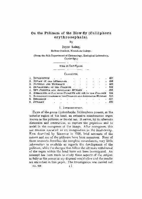
On the Ptilinum of the Blow-fly (Calliphoraerythrocephala).
By
Joyce Laing,B a l f o u r S t u d e n t , N e w n h a m Col l ege .
( F r o m t h e S u b - D e p a r t m e n t of E n t o m o l o g y , Z o o l o g i c a l L a b o r a t o r y ,
C a m b r i d g e . )
W i t h 14 T e x t - f i g u r e s .
C O N T E N T S .
1. I N T R O D U C T I O N . . . . . . . . . 4 9 7
2 . R E V I E W O F T H E L I T E R A T U R E . . . . . . 4 9 8
3 . M A T E R I A L A N D T E C H N I Q U E . . . . . . . 5 0 0
4 . D E V E L O P M E N T O F T H E P T I L I N U M . . . . . . 5 0 2
5 . T H E P T I L I N U M A N D A S S O C I A T E D M U S C L E S . . . . 5 0 6
6. E M E R G E N C E O F F L Y F E O M P U P A B I U M A N D U S E O F T H E P T I L I N U M 5 1 4
7. S U B S E Q U E N T C H A N G E S I N T H E P T I L I N U M A N D A S S O C I A T E D M U S C L E S 5 1 5
8. D I S C U S S I O N . . . . . . . . . . 5 1 7
9. S U M M A R Y . . . . . . . . . . 5 2 0
1. INTRODUCTION.
FLIES of the group Cyclorrhapha Schizophora possess, in theanterior region of the head, an extensive membranous organknown as the ptilinum or frontal sac. It serves, by its alternatedistension and contraction, to rupture the puparium and toassist in the emergence of the imago. After emergence, thissac remains retracted as an invagination in the head-cavity.First described by Eeaumur in 1738, brief accounts of thenature and use of the ptilinum have been numerous. None ofthese accounts describes the complete musculature, very littleinformation is available as regards the development of theptilinum, while the changes that follow the ultimate withdrawalof the organ within the head have not been investigated. Anattempt has been made to study these aspects of the subjectas fully as the means at my disposal would allow and the resultsare embodied in this paper. The investigation was carried out
NO. 308 L 1
498 JOYCE LAING
at the suggestion of Dr. A. D. Imms, F.R.S., who has assistedme throughout the work. I wish to express my sincere thanksto Dr. Imms for his encouragement and help, and to NewnhamCollege for the opportunity of conducting this research.
2. REVIEW OF THE LITERATURE.
The first reference to the ptilinum is that of Reaumur (1738)in his illustrated account of the emergence of the blow-fly.He remarked on the systole and diastole of the 'inuseau',which we now know as the ptilinum, and the associated dilationof the adjacent areas of the head. He pointed out the presenceat the end of the ' museau' of ' un petit enfoncement qui marqueapparemment l'endroit par lequel il est tire quand il rentresous le crane'. It is evident in this observation that he refersto the base of the ptilino-oesophageal muscle. The distensionhe thought to be caused by air-pressure. The obvious purposeof the ' museau' was, he said, to break open the puparium and toassist in the emergence of the imago. He further suggested thatit aided in the circulation of the body-fluids. He showed alsothe importance of the 'museau' in facilitating the escape offlies of another species, living in galls on thistles. The fact thatthe 'museau' did not again appear during the remainder ofthe insect's life did not escape his notice.
In 1911 Knab gave a short review of the literature concerningthe emergence of Cyclorrhaphous flies from their puparia. Promthis account it appears that little had been added, between1738 and 1911, to our knowledge of the ptilinum. Three newpoints, however, emerged. First, Joly in 1846 pointed out thatthe inflation took place by blood-pressure and not by air-pressure.Secondly, Kiinckel d'Herculais (1875) stated that an apparentptilinal mechanism was found in the Syrphids (S y r p h u s,V o l u c e l l a , and E r i s t a l i s ) , but all traces of the ptilinumdisappeared before the insects were ready for flight. Thirdly,this same author described the structure of the ptilinum asbeing, apart from its coloration and transparency, identicalwith that of the surrounding integument. Observations on therate of pulsation of the ptilinum have since been made by
PTILINUM OP CALLIPHORA 499
Graham-Smith (1916) in his description of the emergence ofthe blow-fly.
Little attention has been given to the muscle-supply of theptilinum except by Lowne (1890-5), who attributed the move-ments of the organ to three sets of muscles. Lowne mentioned:(1) two large fan-shaped muscles arising from the lower edgeof the occipital foramen and inserted partly on the genae andpartly on the ptilinum; (2) compressor fibres covering the innersurface of the ptilinum; and (3) two bundles of muscles aris-ing from the ptilinum and inserted on the oesophagus. Thesemuscles, (3), he termed 'retractors of the fulcrum'. The samepaired muscles were described and figured by Graham-Smith(1930) 'retractors of the oesophagus', and he attributed tothem the function of withdrawing the loop of the oesophagusduring retraction of the proboscis. Previous to Graham-Smith,but apparently unknown to him, Mercier and Villeneuve(1925) had announced that muscle-fibres extending from thepharynx to the ptilinum, their 'muscle ptilino-pharyngien',were responsible for retraction of the ptilinum. They held thatthe fibres belonged to the same group of muscles as the fronto-pharyngeal, which were dilators of the pharynx, and that afterinvagination of the ptilinum, the ptilino-pharyngeal muscleplayed only the role of a pharyngeal dilator. Lowne furtherstated that most of the muscles were subsequently absorbed,though the fan-shaped muscles remained for one or more weeksafter emergence.
Although both Lowne and Hewitt (1910) termed the openingof the ptilinal invagination the 'lunula', or 'lunule', the namegenerally adopted is frontal or ptilinal suture. The term lunuleis applied to the small crescentic sclerite between the ptilinalsuture and the bases of the antennae. It is, according to Becher(1882), the well-cuticularized lower border of the ptilinum.The division of the Cyclorrhapha, into Aschiza and Schizophora,was founded by Becher upon the differences in the frontalregion of the head. In the Aschiza there is no ptilinum orassociated suture in the mature fly, while in the Schizophoraboth ptilinum and its suture are present.
As regards the development of the ptilinum, the only direct
500 JOYCE LAING
reference appears to be that by Wahl (1915) who states that'the median hindmost part of the frontal sac comes to theanterior end of the pupa and later becomes again intucked asthe ptilinum'. In this case, 'frontal sac' refers to the vesiclecontaining the cephalic imaginal disks, which, when evaginated,forms the head of the imago. In the account by Perez (1910,p. 176) of the metamorphosis of C a l l i p h o r a is a descriptionof the development of the ptilino-oesophageal muscle. Pereztermed it a dilator muscle of the pharynx, and showed that itdeveloped, chiefly, from the remains of the corresponding larvalpharyngeal dilator.
3. MATERIAL AND TECHNIQUE.
(a) Material .—Larvae of C a l l i p h o r a e r y t h r o c e -p h a l a Meigen were reared, usually on liver, in shallow vesselscontaining a layer of dry sand and kept in an incubator at 25° C.After four days' development at this temperature, the larvaeleft the meat which was then removed from the vessel. Whenpupae of known age were required, the fully fed larvae wereplaced in similar shallow flat-bottomed containers on a layer ofsand, insufficient in depth for them to bury themselves. Byexamining the larvae hourly on their sixth day—when themajority pupated—it was possible to remove those which hadcontracted into the pupal barrel-shape, in order to separatethem, and to label them with their time of pupation. When,9 or 10 days afterwards, the adult flies began to issue they wereremoved from the incubator so that their emergence might beobserved and specimens fixed with the ptilinum expanded.
The adult flies were fed with yeast and sugar; water soakedinto cotton-wool was also provided, together with liver foroviposition.
(b) Methods.—Carnoy's fluid, used cold, gave the mostsatisfactory fixation. Pupae were pierced in the thoracic regionof the puparium, immediately upon immersion in the fixative,and were removed from this shell an hour later. Fixation invacuo was found useful in removing air from the heads of oneclay or older imagines. Of stains, the most satisfactory wasDelafield's haematoxylin counterstained with eosin: iron haema-
PTILINUM OF CALMPHORA 501
toxylin, Mann's methyl-blue-eosin, and Mayer's haemalum werealso used. Paraffin sections were cut 6//. or 10/n in thickness.In order to avoid tearing of the sections of 1, 2, or 3 day-oldflies, double-imbedding in celloidin and paraffin was tried butthis method gave results no better than single imbedding in
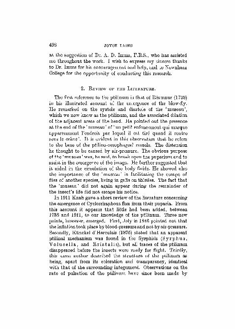
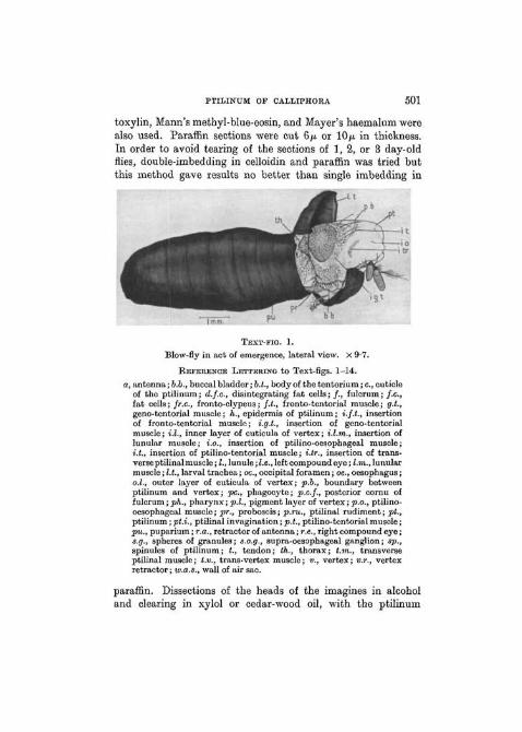
TEXT-FICJ. 1.
Blow-fly in act of emergence, lateral view. X 9-7.
REFERENCE LETTERING to Text-figs. 1-14.
a, antenna; 6.6., buccal bladder; b.l., body of the tentorium; c, cuticleof the ptilinum; d.f.c, disintegrating fat cells; /., fulcrum; / . c ,fat cells; fr.c, fronto-clypeus; f.t., fronto-tentorial muscle; g.L,geno-tentorial muscle; h., epidermis of ptilinum; i.f.t., insertionof fronto-tentorial muscle; i.g.t., insertion of geno-tentorialmuscle; i.l., inner layer of cuticula of vertex; i.l.tn., insertion oflunular muscle; i.o., insertion of ptilino-oesophageal muscle;i.t., insertion of ptilino-tentorial muscle; i.tr., insertion of trans-verse ptilinal muscle; I., lunule; I.e., left compound eye; l.m., lunularmuscle; I.t., larval trachea; oc, occipital foramen; oe., oesophagus;o.l., outer layer of cuticula of vertex; p.b., boundary betweenptilinum and vertex; pc, phagocyte; p.c.f., posterior cornu offulcrum; ph., pharynx; p.l., pigment layer of vertex; p.o., ptilino-oesophageal muscle; pr., proboscis; p.ru., ptilinal rudiment; pt.,ptilinum; pt.i., ptilinal invagination; p.t., ptilino-tentorial muscle;pu., puparium; r.a., retractor of antenna; r.e., right compound eye;s.g., spheres of granules; s.o.g., supra-oesophageal ganglion; sp.,spinules of ptilinum; t, tendon; tk., thorax; t.m., transverseptilinal muscle; t.v., trans-vertex muscle; v., vertex; v.r., vertexretractor; w.a.s., wall of air sac.
paraffin. Dissections of the heads of the imagines in alcoholand clearing in xylol or cedar-wood oil, with the ptilinum
502 JOYCE LAING
inflated, gave preparations very valuable for the study of themusculature.
4. DEVELOPMENT OF THE PTILINUM (Text-figs. 2-8).
During the first day of the pupal period the paired cephalicbuds, bearing antennal and eye rudiments on their walls, fuseinto a single median dorsal cephalic vesicle. Between 24 and36 hours after pupation at 25° C, the cephalic rudiment be-comes evaginated through the opening of the larval pharynxand appears externally as the imaginal head. The most anteriorpart of the head, in front of the eyes and antennae, Avhich beforeevagination was the posterior blind end of the cephalic vesicle,gives rise to the ptilinum. The head membrane is at first thinand distended with blood-plasma, spheres of granules ('Korn-chenkugeln'), and fat cells. The antennal rudiments are lateral,their insertions being separated by more than one-third of thewidth of the head. The epidermis (hypodermis) is not obviouslydemarcated into ptilinum-forming and other areas, although tothe most anterior part are attached fibres destined to form partof the ptilino-oesophageal muscle (Text-fig. 2).
Between \\ and 2 days after pupation, the shape of the headchanges in that the antennal bases move to a more medianposition and the epidermis immediately above them becomesa many-celled layer, which represents the imaginal bud of thefuture ptilinum. Between the cells of this thickening numerousspheres of granules, and fragments of these, are present (Text-figs. 3 and 8).
On the third day after pupation invagination of the ptilinalbud begins (Text-fig. 4). The process of invagination is gradual,being connected with the growth of the sac, and is not per-formed by the still-rudimentary muscles. Insinking occurs firstaround the upper and outer edge of the thickening, the mem-brane extending underneath and parallel to the vertex. As thesurface increases the thickness gradually decreases (Text-figs.5 and 6), the increased area of the membrane accommodatingitself by becoming crumpled as it extends into the head. Beforethe fifth day the ptilinum already bears cuticular spinules,while the developing muscles, attached to its walls, are repre-
PTILINUM OF CALLIPHOBA 503
s o g
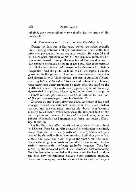
TEXT-FIGS. 2—3.
FIG. 2. Longitudinal section through head of pupa 1£ days afterpupation at 25° C. X 32-9. Pupal integument not shown in thisor other figures of pupae.
FIG. 3. Longitudinal section through head of pupa, 2£ days old.X32-9.
sented by linear chains of nuclei surrounded by a small amountof unstriated cytoplasm (Text-fig. 7). In the completely de-veloped organ the cuticle becomes greatly thickened and the
504 JOYCE LAING
p ru
v?^*&. :r-;;^:
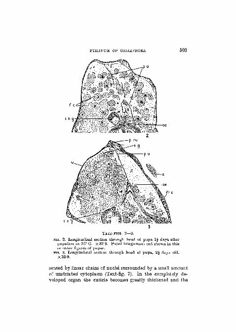
TEXT-MGS. 4—5.
no . 4. Anterior part of head of pupa, longitudinal section, 3J daysold. X78.
FIG. 5. Anterior part of head of pupa, longitudinal section, 4 daysold (early). X 78.
PTILINUM OF CALLIPHOBA 505
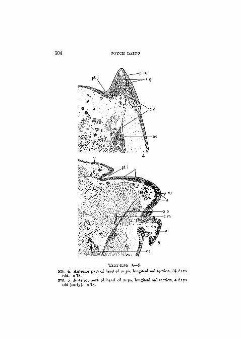
TEXT-FIGS. 6—7.
FIG. 6. Anterior part of head of pupa, longitudinal section, 4 daysold (late). X 78.
FIG. 7. Anterior part of head of pupa, longitudinal section, 5J daysold. x 78.
epidermis reduced to an attenuated layer (Text-fig. 9). Theblood-plasma, on the fourth and fifth days, is laden with athick suspension of the contents of the disintegrated larvalfat cells and spheres of granules, which in the earlier part of thepupal period retained their integrity.
506 JOYCE LAING
5. THE PTILINUM AND ASSOCIATED MUSCLES.The chief surface of the ptilinum lies between the antennae
and vertex. A narrow extension also passes ventrally on eitherside, between the gena and the fronto-clypeus, to about thelevel of the tip of the pendulous third antennal joint. Afteremergence from the puparium, this whole area of spiny mem-brane is invaginated into the head cavity, the external evidence
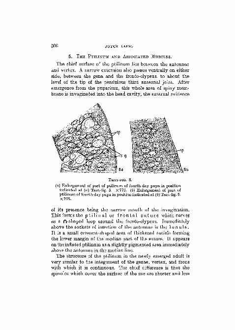
TEXT-FIO. 8.
(a) Enlargement of part of ptilinum of fourth-day pupa in positionindicated at (a) Text-fig. 5. X 775. (6) Enlargement of part ofptilinum of fourth-day pupa in position indicated at (6) Text-fig. 5.X775.
of its presence being the narrow mouth of the invagination.This forms the p t i l i n a l or f r o n t a l s u t u r e which curvesas a n -shaped loop around the fronto-clypeus. Immediatelyabove the sockets of insertion of the antennae is the 1 u n u 1 e.It is a small crescent-shaped area of thickened cuticle formingthe lower margin of the median part of the suture. It appearson the inflated ptilinum as a slightly pigmented area immediatelyabove the antennae in the median line.
The structure of the ptilinum in the newly emerged adult isvery similar to the integument of the genae, vertex, and fronswith which it is continuous. The chief difference is that thespinules which cover the surface of the sac are shorter and less
PTILINUM OF CALLIPHOBA 507
slender than those of the vertex. There are no differences inthe shape of the spines in different parts of the organ such as thosedescribed by Jobling (1932) in the ptilinum of G l o s s i n a .The wall of the ptilinum consists of (a) spinules, regularly-arranged, unstained with haematoxylin, with an average widthat their bases of 20/i and a height of 10/*; (b) a layer of cuticle25-30 JJ. in thickness, without pigment and staining purple withhaematoxylin, and secreted by (c) a syncytial epidermis (Text-fig. 9).
On the inner surface of the wall of the ptilinum are theinsertions of certain muscles, all of which function as retractorsof the organ. These muscles are as follows:
1. T r a n s v e r s e P t i l i n a l Musc le (t.m.) (Lowne's 'com-pressors').—The fibres extend horizontally across the cavity ofthe ptilinum and have a long dorso-ventral attachment to eachside. When the organ is invaginated the muscle forms a com-pact mass over its posterior surface.
2. P t i l i n o - o e s o p h a g e a l Musc le (j>.o.).—This arises onthe oesophagus between the fulcrum and the brain. It has awide base of insertion on the most anterior part of the inflatedptilinum in a narrow band of wide lateral extent. It is thesefibres which Lowne termed 'retractors of the fulcrum', Graham-Smith 'retractors of the oesophagus', and Mercier and Ville-neuve 'muscle ptilino-pharyngien' or 'm. retractor ptilini'. Myown observations corroborate the opinion of Mercier andVilleneuve in indicating that the fibres play an important partin the retraction of the ptilinum. The French authors furthersupposed that, after invagination of the ptilinum, the ptilino-pharyngeal muscle played the role of pharyngeal dilator. Since,however, from dissections and sections of heads of flies, severaldays after emergence, all traces of ptilinal muscles are gone, itseems probable that the only function of the ptilino-pharyngealmuscle is the retraction of the ptilinum.
3. P t i l i n o - t e n t o r i a l Musc le (f.L).—This is a pairedmuscle arising from a tendon which is attached to the strongcuticular body of the tentorium, at its junction with the borderof the occipital foramen. It is inserted on the ptilinum at eachside, dorso-laterally, in a narrow band immediately in front of
508 JOYCE LAING
t V
i L m
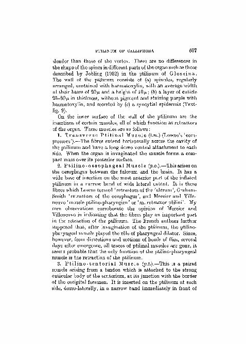
TEXT-FIG. 9.
Vertical section passing through mouth of invaginated ptilinum ofnewly emerged imago. X 163-7.
the line of demarcation between the vertex and the ptilinum.This pair of muscles is evidently homologous with Lowne'slarge fan-shaped retractors.
Although not inserted on the ptilinal membrane there are, inthe head, six other groups of muscle-fibres which appear tobe connected with the distension and contraction of the ptili-
PTILINUM OF CALLIPHORA 509
num, and other parts of the head, during emergence (Text-figs.12 and 13). These are as follows:
(1) G e n o - t e n t o r i a l Musc les (q.t.).—The fibres arisefrom the same tendon as the ptilino-tentorial muscle and fromthe occipital sclerites. They are inserted over a wide area on
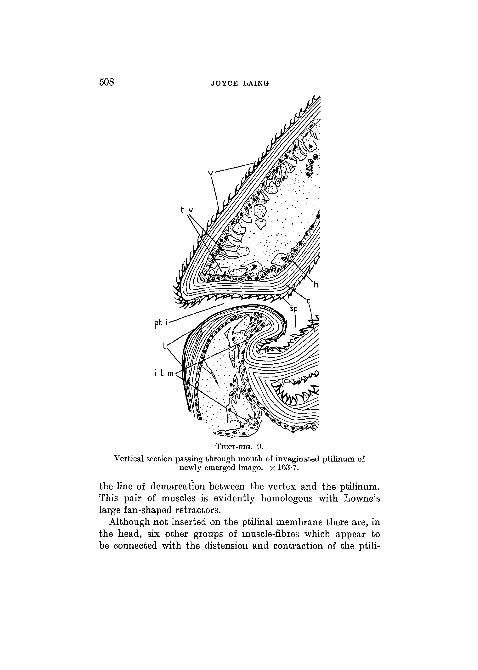
TEXT-FIG. 10.
Diagram of right half of head of adult, with ptilinum inflated,indicating position of intrinsic ptilinal muscles. X 21.
each gena between the eye and the frontal suture. Contractionof the muscles results in the approximation of the gena andocciput and, when blood is being withdrawn into the abdomen,in a return to normal in the shape of the head. When the headis distended with blood, the contraction of these muscles appearsto result merely in the flattening of the genae. Fluid is thus
510 JOYCE LAING
pressed forward into the ptilinum which is thereby renderedmore turgid.
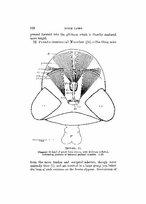
(2) Fronto- ten tor ia l Muscles (/.£.).—The fibres arise
TEXT-FIG. 11.
Diagram of head of adult from above, with ptilinum inflated,indicating position of intrinsic ptilinal muscles. X 22.
from the same tendon and occipital sclerites, though morecentrally than (1), and are inserted in a large group just belowthe base of each antenna on the fronto-clypeus. Contraction of
PTILINUM OF CALLIPHOBA 511
these muscles also results in diminution of the volume of thehead, as in (1).
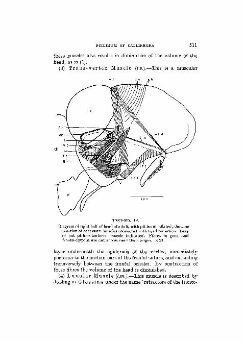
(3) T r a n s - v e r t e x Muscle (t.v.).—This is a muscular
TEXT-FIG. 12.
Diagram of right half of head of adult, with ptilinum inflated, showingposition of accessory muscles connected with head pulsation. Baseof cut ptilino-tentorial muscle indicated. Fibres to gena andfronto-clypeus are cut across near their origin. X 21.
layer underneath the epidermis of the vertex, immediatelyposterior to the median part of the frontal suture, and extendingtransversely between the frontal bristles. By contraction ofthese fibres the volume of the head is diminished.
(4) L u n u l a r Musc le (l.m.).—This muscle is described byJobling in G l o s s i n a under the name 'retractors of the fronto-
512 JOYCE LAING
clypeus'. His account is as follows: 'an unpaired, long, thinmuscle arises from the dorsal edge of the occipital foramen,passes through the canal of the cephalic ganglion towards thefronto-clypeus where it is attached ahove the antennae closeto the ventral edge of the ptilinal suture. It retracts the fronto-clypeus and ptilinum inside the head after hatching. Non-functional in adult insect.' This description applies equally wellto the homologous muscle in C a l l i p h o r a . The fibres areinserted by means of two minute roots on a cone-shaped meshof strands of cytoplasm and nuclei lying beneath the cuticleof the lunule. This muscle was described by Mercier andVilleneuve (1926) in C a l l i p h o r a as 'muscles gubernateursde la lunule'. They held that the origin is on the body of thetentorium. According to these authors the tendonous insertionsof the fibres, just referred to, serve as the framework for a sen-sory organ of the same nature as Johnston's organ in the secondantennal joint.
(5) B e t r a c t o r of t h e A n t e n n a (r.a.).—According toJobling this muscle 'arises from the latero-vertex and passesthrough the scape, being inserted into a short process of thedorsal part of the posterior border of the pedicle'. At the timeof emergence this muscle may assist in the deflation of thehead, by approximating the vertex and fronto-clypeus. Subse-quently it functions only as the antennal retractor.
(6) E e t r a c t o r s of t h e V e r t e x (v.r.).—The fibres arisefrom the hind surface of the posterior cornua of the fulcrumand are inserted on the lateral regions of the vertex, behindthe origin of the retractor of the antenna. The diameter of thefibres is very small. Contraction results in inpulling of thevertex during deflation of the head. These muscles appear tobe the 'muscle fronto-pharyngien' of Mercier and Villeneuve(1925), although they state that the origin of the fibres is on thepharyngeal wall. In their diagrams (p. 883, figs, i and ii) fibresappearing in transverse section underneath the vertex arelabelled as part of the fronto-pharyngeal muscle. The sectionsare actually, however, of the fibres of the trans-vertex muscle.
PTILINUM OF CALLIPHOBA 5J3
pt
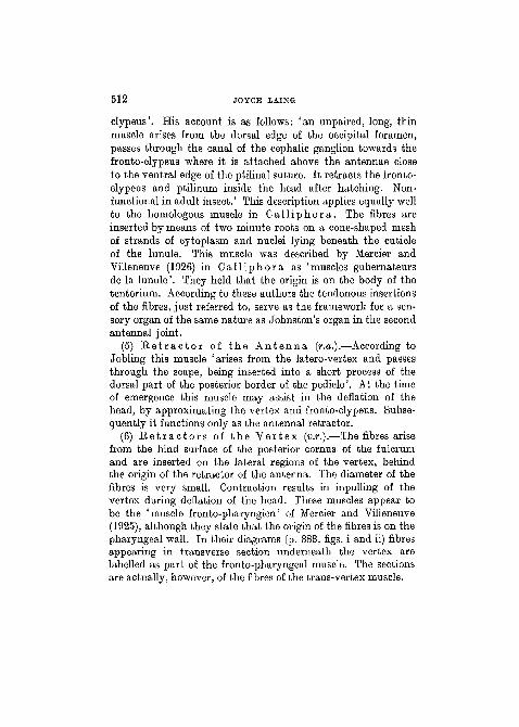
TEXT-JTG. 13.
Diagram drawn from thick vertical section of newly emerged adult;ptilinum invaginated. X 29.
NO. 308
5 1 4 JOYCE LAING
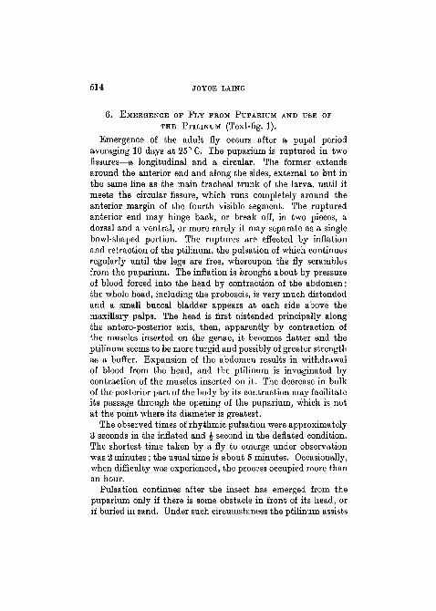
6. EMERGENCE OF FLY FROM PUPARIUM AND USE OF
THE PTILINUM (Text-fig. 1).
Emergence of the adult fly occurs after a pupal periodaveraging 10 days at 25° C. The puparium is ruptured in twofissures—a longitudinal and a circular. The former extendsaround the anterior end and along the sides, external to but inthe same line as the main tracheal trunk of the larva, until itmeets the circular fissure, which runs completely around theanterior margin of the fourth visible segment. The rupturedanterior end may hinge back, or break off, in two pieces, adorsal and a ventral, or more rarely it may separate as a singlebowl-shaped portion. The ruptures are effected by inflationand retraction of the ptilinum, the pulsation of which continuesregularly until the legs are free, whereupon the fly scramblesfrom the puparium. The inflation is brought about by pressureof blood forced into the head by contraction of the abdomen;the whole head, including the proboscis, is very much distendedand a small buccal bladder appears at each side above themaxillary palps. The head is first distended principally alongthe antero-posterior axis, then, apparently by contraction ofthe muscles inserted on the genae, it becomes flatter and theptilinum seems to be more turgid and possibly of greater strengthas a buffer. Expansion of the abdomen results in withdrawalof blood from the head, and the ptilinum is invaginated bycontraction of the muscles inserted on it. The decrease in bulkof the posterior part of the body by its contraction may facilitateits passage through the opening of the puparium, which is notat the point where its diameter is greatest.
The observed times of rhythmic pulsation were approximately3 seconds in the inflated and £ second in the deflated condition.The shortest time taken by a fly to emerge under observationwas 2 minutes; the usual time is about 5 minutes. Occasionally,when difficulty was experienced, the process occupied more thanan hour.
Pulsation continues after the insect has emerged from thepuparium only if there is some obstacle in front of its head, orif buried in sand. Under such circumstances the ptilinum assists
PTILINUM OF CALLIPHORA 515
the insect in making its way to the surface, and irregularpulsations continue so long as the remains of the pupal skincling to the legs. Generally, however, once the fly is free ofthe pupa case, the ptilinum is drawn into the head cavity whereit remains thereafter in a functionless condition.
7. SUBSEQUENT CHANGES IN THE PTILINUM ANDASSOCIATED MUSCLES.
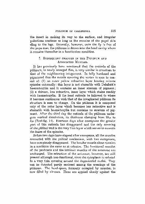
It has previously been mentioned that the cuticula of theptilinum, in newly emerged flies, is very similar in structure tothat of the neighbouring integument. In fully hardened andpigmented flies the cuticle covering the vertex is seen to con-sist of: (1) an outer yellow refractive layer bearing minutespinules externally: this layer is not stainable with Delafield'shaematoxylin and it contains an inner stratum of pigment;(2) a thinner, less refractive, inner layer which stains readilywith haematoxylin. If the head cuticula be followed to whereit becomes continuous with that of the invaginated ptilinum itsstructure is seen to change. On the ptilinum it is composedonly of the outer layer which becomes less refractive and isstainable with haematoxylin but contains no stratum of pig-ment. After the third day the cuticula of the ptilinum under-goes marked dissolution, its thickness changing from 25 ̂ to5/J. (Text-fig. 14). Fourteen days after emergence the greaterpart of this cuticula has disappeared and the only coveringof the ptilinal wall is the very thin layer which serves to connectthe bases of the spinules.
Before two days have elapsed after emergence, all the musclesconnected with the ptilinal mechanism, with two exceptions,have completely disappeared. The lunular muscle alone remainsin a condition the same as at eclosion. The functional musclesof the proboscis and the intrinsic muscles of the antennae areunchanged. The retractors of the antennae, however, are stillpresent although non-functional, since the cytoplasm is reducedto a very thin covering around the degenerated nuclei. Theycan be detected partly enclosed among the creasings of theptilinum. The head-space, formerly occupied by muscles, isnow filled by air-sacs. These are applied closely against the
516 JOYCE LAING
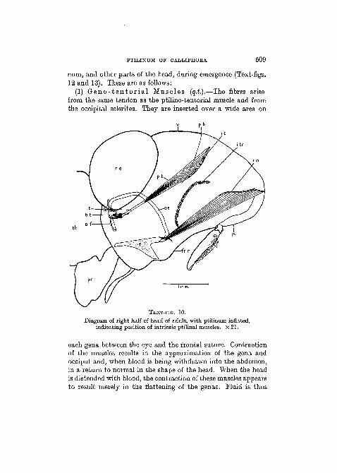
f r c
TEXT-FIG. 14.
Vertical section passing through mouth of invaginated ptilinum ofimago three days after emergence. X 163'7.
contours of the ptilinum; they surround the brain and extendfrom the vertex ventrally to the muscles of the fulcrum. In thefew narrow blood-spaces that still remain are small phagocytesand fat cells. There are no phagocytes distended with ingested
PTILINUM OF CALLIPHOBA 517
muscle debris such as are numerous during histolysis of thelarval muscles.
The degeneration of the muscles seems to proceed mostrapidly during the fly's second day of adult life, since sectionsof the heads of flies 24 hours old show the fibres with but littlechange in their structure. The actual process of histolysis hasnot been followed in any detail, and for this purpose a verycomplete series of sections made at intervals of every few hoursduring the second day after emergence is necessary. The onsetof histolysis appears to manifest itself in loss of staining capacityin the muscles concerned: their striae soon become lost, thecytoplasm becomes spongy, and finally complete breaking-downresults. Although phagocytes are present, both free in the bloodand associated with fat cells, none were found in intimaterelation with degenerating muscles. The actual beginning ofdegeneration is probably associated with disuse of the musclesconcerned after the final retraction of the ptilinum has beenaccomplished. In this connexion the distension of the cephalicair-sacs, which results in cutting off much of the blood-supply,may be significant. If the foregoing observations be correct itwould appear that histolysis of the muscles in question takesplace by some process other than phagocytosis, which is soprominent in the pupa (Perez, 1910). It may also be mentionedthat it is only in relatively few cases that phagocytosis has beenproved to occur as a histolytic process among insects. In othercases it has been much disputed whether phagocytes play anypart at all.
8. DISCUSSION.Prom a comparative study of the head-capsules of Diptera,
Peterson (1916) was not able to determine the origin of theptilinum, although he made the following suggestions: ' It seemsevident that the frontal suture was once a membranous areawhich became invaginated to form a membranous pouch orptilinum. If this is the case the mesal membranous area ofthe fronto-clypeus of S e p s i s , O e c o t h e a , C a l o b a t a , andD e s m o m e t o p a would be very significant. The ptilinummight have originated from some form similar to S c e n o p i n u sin which the ventral margin of the chitinized vertex is located
518 JOYCE LAING
dorsad and laterad of the antennae. It seems quite possiblethat the membrane along this margin became invaginated inthe early stages of the development of the ptilinum.' The' mesal membranous area' here mentioned is that between andbelow the antennae and continuous with the ptilinum. Bothfrom Peterson's views and from the development of the ptilinumfrom the head integument, it seems evident that the origin ofthis sac was by the enlargement of a membranous area in theregion of the antennae. Quite possibly it arose as an extensionof the membrane surrounding the antennal sclerites, similar tobut greater than that found in various members of the Nema-tocera (e.g. E h a b d o p h a g a , M y c e t o p h i l a , Ch i rono-m u s , Mycetobia) . The presence of such a membranous areamay have been of use originally during emergence in allowingdistension of the head with blood from the thorax and abdomen.This area, having become highly flexible, would no longerserve as a rigid base for those of the pharyngeal dilator musclesthat originated from it. The contraction of the fibres of suchmuscles would result not in altering the contour of the pharyn-geal cavity so much as in pulling inwards the membranous areaof the head. It is, therefore, possible that, with the formation ofthe ptilinum from the membranous area mentioned, certainof the bundles of the original dilator muscles of the pharynxhave changed their function so as to constitute the ptilino-oesophageal muscle. In this connexion it is of interest to notethat in some of the Nematocera (Culicidae, Knab, 1911) thepharyngeal dilators are used in the process of emergence. Bythe contraction of these muscles air is sucked into the alimentarycanal and serves to distend and to harden the body. Contraryto what might be expected, a study of the head in a typicalSyrphid fly does not afford any clue to the origin of the ptilinum.
In E r i s t a l i s t e n a x there is a delicate strand of musclepassing from the vertex to the oesophagus in the median line.On account of its relations it may be termed the vertico-oeso-phageal muscle. It is apparently homologous with the ptilino-oesophageal muscle of Ca l l i pho ra , but it is uncertain whetherit is functional. The vertex, to which this muscle is attached,is well-cuticularized and non-distensible, continuing so to the
PTILINUM OF CALLIPHOEA 519
insertions of the antennae. In the median line, immediatelyabove the antennae, is the crescent-shaped lunule to which, asin C a l l i p h o r a , are attached the two minute roots of thelunular muscle. In E r i s t a l i s , however, the fibres do notunite into one median muscle, but diverge and are insertedon the inner marginal area bordering each eye. The vertico-oesophageal and lunular muscles are to be seen in sections anddissections of the heads of pupal and imaginal E r i s t a l i s .Mercier and Villeneuve (1927) drew attention to the lunularmuscle and considered it significant in proving the homologyof the lunule of C a l l i p h o r a and E r i s t a l i s . In theSchizophora the lunule forms the lower median border of theptilinum while in E r i s t a l i s the sclerite is directly continuouswith the vertex, no ptilinal membrane intervening. Even inthe pupa of E r i s t a l i s the integument above the lunuleappears indistensible, while within the upper part of the head-cavity there are no functional muscles. It seems, therefore,highly improbable that eclosion is assisted by a ptilinal mechan-ism, as Kiinckel d'Herculais recorded. Becher denied this,arguing that the lower part and sides of the face of E r i s t a l i swere distended with blood during eclosion, and that it was theiractivity which Kiinckel d'Herculais had mistaken for that ofa ptilinum. Support is lent to Becher's contention by the factthat the lower part of the frons and genae, and the integumentbetween these sclerites and the proboscis, form a crumpledthin membrane covered externally with numerous minute scale-like structures. In C a l l i p h o r a a similar membranous areaswells during emergence as a buccal bladder at each side of themouth frame.
The Aschiza and Schizophora, therefore, as exemplified byE r i s t a l i s and C a l l i p h o r a respectively, possess in commonthe faculty of assisting their eclosion by distending the head,but the process takes place by different methods. By thedevelopment in the Schizophora of a ptilinal membrane, whichis without its counterpart in the Aschiza, the two groups havebecome separated. Prom the foregoing evidence it seems morereasonable to conclude that the Aschiza never possessed aptilinum, rather than to assume that, in common with the
520 JOYCE LAING
Schizophora, they developed such an organ which subsequentlyatrophied.
9. SUMMARY.
1. An account is given of the morphology, development, andchanges following emergence of the ptilinum of the blow-fly.
2. The ptilinum in Ca l l i phora e r y t h r o c e p h a l adevelops from the integument of the anterior part of the head.Differentiation from the surrounding integument begins firstas a local thickening of the epidermis. The structure of thethickened area is similar to that of an epidermal imaginal bud.At the end of the third day after pupation at 25° C. this epi-dermal thickening grows inwards and, as its surface increases,its walls become gradually thinner. Eudiments of the ptilino-oesophageal muscle are obvious even immediately after eversionof the head; other muscles do not appear until the fourth dayafter pupation.
3. In the newly emerged fly the ptilinum is continuous withthe integument of the frons, genae, and vertex. Two unpairedand one pair of retractor muscles are inserted on the ptilinum;six other muscles appear to be accessory in connexion withpulsation of the head during emergence.
4. By rhythmic expansion and contraction of the ptilinumthe puparium is ruptured and emergence of the fly assisted.After final retraction of the ptilinum into the head-cavity, andhardening of the surrounding head integument, no more use ismade of the organ.
5. During the adult life of the fly the ptilinal integument isreduced to a layer of spinules. Before two days have elapsedafter eclosion practically all the ptilinal and accessory muscleshave disappeared. Their disappearance does not appear to bedue to phagocytic activity. From histological evidence the causeand mechanism of muscle degeneration cannot be ascertained.
6. A possible method of origin of the ptilinum from a mem-branous area above the antennae is suggested. E r i s t a l i s ,without ptilinum, is compared with C a l l i p h o r a . From theevidence brought forward it is suggested that the Aschiza andSchizophora represent two independent lines of development.
PTILINUM OF CALLIPHOEA 521
LIST OF EEFERBNCES.
Papers quoted by Knab, and published prior to 1911, are not listed belowexcept where necessary.Becher, E. (1882).—"Zur Kenntniss der Kopfbildung der Dipteren",
'Wiener Ent. Zeitung', 1.Graham-Smith, G. S. (1913).—'Flies and Disease.' Cambridge.
(1916).—-"Observations on the Habits and Parasites of CommonFlies", 'Parasitology', 8.
(1930).—"Anatomy and function of the proboscis of the blow-fly,Calliphora. erythocephala", ibid., 22.
Jobling, B. (1932).—"Revision of the structure of the head, &c, of Glossinapalpalis", ibid. (1932-3), 24.
Knab, F. (1911).—"On ecdysis in Diptera", 'Proc. Ent. Soc. Washington',13.
Kiinckel d'Herculais, J. (1875).—'Reoherches sur l'organisation et ledeVeloppement des Volucelles.' Paris.
Lowne, B. T. (1870).—'Anatomy and Physiology of the Blow-Fly, Muscavomitoria.' London.
(1890-5).—'The Anatomy, Physiology, Morphology and Develop-ment of the Blow-Fly, Calliphora erythroeephala.' London.
Mercier, L., and Villeneuve, J. (1925).—" Anatomie de la tete des Dipteres.Cyclorrhaphes. La ptiline et ie muscle ptilino-pharyngien", 'C.R. Acad.Sci.', 181.
(1926).—"La lunule et ses organes sensoriels", ibid., 182.(1927).—"Les muscles gubernateurs de la lunule chez Eristalis
tenax", ibid., 184.Perez, C. (1910).—"Recherches histologiques sur la metamorphose des
Muscides, Calliphora erythrocephala. Meigen", 'Arch, de Zool. Exp. etG6n.', 4.
Peterson, A. (1916).—"Head Capsule and Mouth Parts of Diptera",'Univ. Illinois Biol. Studies', 3.
Reaumur, R. A. F. de (1738).—'Memoires pour servir a l'histoire desInsectes', torn. iv. Paris.
Wahl, B. (1915).—"t)ber die Kopfbildung cyclorrhapher Dipteren-larvenu. die post-embryonale Entwicklung des Fliegenkopfes", 'Arb. a. d.Zool. Inst. Wien', 20.