Embed Size (px)
Citation preview
Proc. Nat. Acad. Sci. USAVol. 71, No. 11, pp. 4429-4434, November 1974
On the Chemical Nature of Transfer Factor(immune response/leukocyte extract/transfer of immunological information/double-stranded RNA/guinea pig)
DAVID DRESSLER AND STEVEN ROSENFELD
The Biological Laboratories, Harvard University, Cambridge, Massachusetts 02138
Communicated by J. D. Watson, April 3, 1974
ABSTRACT Two transfer factors prepared in an ex-perimental animal model, the guinea pig, have been testedfor their susceptibility to various enzymes of known speci-ficity. The biological activity of these immune responsemediators can be destroyed by RNase III, an enzyme thatdegrades duplex RNA. It, therefore, appears that thesetransfer factors consist entirely or partly of double-stranded RNA.
Transfer factor is a puzzling immunological phenomenon.It appears, to those who have observed the phenomenon, to bea transfer of immunological information from a population of"educated" leukocvtes to a population of "naive" leukocytes.A subcellular, leukocyte component is involved, and it isgenerally believed that information for specific immune re-sponses is transferred.The transfer factor phenomenon can perhaps be most
easily grasped by considering a specific example, such as thecell-mediated immune response mounted against the smallchemical hapten, dinitrochlorobenzene (DNCB). DNCI3 is areactive chemical that permeates into the tissues and con-jugates to the lysine and cysteine residues of various proteins,causing them to appear foreign. A naive individual who isexposed to DNCB will destroy the foreign material in a slowand moderate reaction. During this primary response anotherimportant event occurs: there is a mobilization of a specificpart of the immune system. Thus, if the individual ever en-counters the same antigen again, he will mount a secondaryresponse that is both more rapid and more forceful than theprimary response.The cornerstone of the transfer factor phenomenon, as
contained in the work of Lawrence (1) and of Jeter, Tremaine,and Seebohm (2), is the statement that the leukocytes of theimmunologically experienced individual can be made to yielda subcellular component (the transfer factor) that can trans-mit to a naive individual the immunological informationnecessary for the mobilization of a specific part of the immunesystem. Thus, the naive individual, upon receipt of theleukocyte extract, becomes immediately able to mount astrong secondary response upon his first exposure to the anti-gen.
Since the discovery of transfer factor was an unexpectedaddition to the field of immunology, its very existence hasposed several interesting questions. What are these sub-cellular leukocyte components that can apparently substitutefor antigen in the mobilization of the immune system? Wheredo the transfer factors function in the development of the
full immune response, and why have these intermediate in-formation carriers been designed into the system? Are theyrepresentative of a more general mechanism of informationtransfer between cells?The chemical nature of transfer factor has been a problem
of long-standing interest. Our knowledge in this area is justdeveloping and includes four facts, secured by Lawrenceand his colleagues. The biologically active material is smallenough to pass through a dialysis membrane (3); thus, trans-fer factor is too small to be or to code for any of the proteinsinvolved in a specific immune response. Furthermore, trans-fer factor can withstand treatment with DNase, pancreaticRNase, and trypsin (4).
In this paper we present the results of an enzymologicalanalysis of two transfer factors prepared in the guinea pigexperimental system. The data indicate that the biologicalactivity of these transfer factors resides entirely or partlyin species of low molecular weight, double-stranded RNA.The implications of this result in terms of the possible mode ofaction of transfer factor will be discussed.
Preparation of transfer factors
The experimental system we use is the guinea pig, and theimmune response we have studied is the delayed hypersensi-tivity reaction. In the companion to this paper (5) we de-scribed procedures that allowed us to make 20 preparations ofbiologically active transfer factor, amidst 65 failures. Thesetransfer factors were of two types: one primes an animal togive a secondary response to dinitrochloro-benzene (DNCB)and the other primes the recipient to respond to ortho-chlorobenzoylchloride (OCBC). In brief, the preparation ofthese transfer factors involved (a) sensitizing the donor ani-mals with antigen on the ear, thus provoking a primary re-sponse, (b) waiting 7 days and then challenging the animalswith antigen on the flank, eliciting the delayed hypersensi-tivity response (Fig. 1), (c) sacrificing the donor animals onday 11 so that lymphoid cells from the spleen, lungs, andperitoneal exudate could be obtained, and (d) recovering adialyzable component from these cells which, when injectedinto a naive animal would allow him to immediately mount adelayed hypersensitivity response upon his first exposure tothe antigen (Fig. 2).
Experimental design
The general design of the experiments reported here involvesthe treatment of transfer factor with enzymes of known speci-ficity before the material is injected into a naive recipient.The subsequent response of the recipient upon challenge withantigen will show whether or not the ability of the material
4429
Abbreviation: DNCB, dinitrochlorobenzene.
4430 Immunology: Dressler and Rosenfeld
to transmit the delayed hypersensitivity capacity has beendestroyed by the enzyme.At the outset it may be well to consider the possible pitfalls
of the enzymatic approach. First, when exposing a transferfactor preparation to an enzyme one must explicitly determinethat the enzyme is working; the current methods for prepar-ing transfer factor lead to the accumulation of considerableamounts of salt capable of inhibiting most enzymes. Second,even if the transfer factor preparation were to survive a givenenzyme such as DNase, this does not necessarily mean thattransfer factor is devoid of DNA. For instance, bacteriophagelambda has an essential DNA component, but it would sur-vive DNase digestion because of the protection provided by aprotein coat.
Similarly, resistance to pancreatic RNase does not guar-antee that single-stranded RNA is absent from transfer factor.The RNA might be protected-or it could be exposed but lack-ing in cytidylic acid and uridylic acid residues.
Resistance to protease digestion must also be consideredas a yardstick of uncertain length: some proteins are knownto be resistant to protease digestion (6).
Suppose a transfer factor preparation were to be destroyedby a highly purified enzyme: does this mean that transferfactor has been chemically identified? Not necessarily. Onemust consider the possibility that there are impurities in theenzyme preparation. Furthermore, the transfer factor, al-though inactivated by one enzyme, might still contain addi-tional components that are integral parts of the biologicallyactive compound.
All this notwithstanding, the enzymatic approach is in-valuable for three reasons. First, if successful, it allows one toconclude that the activity of transfer factor resides entirelyor partly in a definable chemical species. Second, one can hopeto reach this conclusion using amounts of material that arewell below the levels required for direct chemical analysis.Third, one is able to study preparations of material that arenot highly purified.
Test substrates
To monitor the effectiveness of the enzyme digestions, radio-active test substrates were prepared. These included tritium-labeled bacteriophage T7 DNA, phosphorus-labeled single-stranded and double-stranded RNA, and sulfur-labeledEscherichia coli proteins, as described below.
Preparation of Radioactive DNA. A thymine-requiringstrain of E. coli was grown in medium containing tritiatedthymidine and infected with bacteriophage T7 until lysis.The phage were precipitated with polyethylene glycol (7),resuspended, and centrifuged to equilibrium in CsCl. Thephage peak was identified by plaque formation. The viralDNA was then extracted by adding sodium lauroyl-sarcosi-nate to 1%, and heating the solution to 500 for 5 min. CsClcentrifugation was used to purify the radioactive viral DNA.
Preparation of Single-Stranded RNA. Purified E. coli RNApolymerase containing the sigma subunit was used to tran-scribe T7 DNA in vitro. [32P]ATP was used to label the prod-uct RNA. The reaction mixture is described in ref. 8.
Preparation of Double-Stranded RNA. An artificial RNAmolecule, available from Miles Laboratories, was used as
template; it consisted of a random sequence of adenylic,cytidylic, and uridylic acid residues. The complementary
strand was synthesized by the QB replicase (9-11), which isable to initiate synthesis on stretches of poly(C) in the tem-plate. [32P]UTP was used to label the product. After 45 min-utes of reaction, about 20% of the single-stranded templatehad been converted to a duplex state; all of the incorporatedradioactivity was located in these duplex portions, as shownby the resistance of this radioactive material to pancreaticRNase, unless the material was first heated to 1000 and thenrapidly cooled.
Preparation of Radioactive Proteins. E. coli was grown in thepresence of [35S]sulfate. The crude cell lysate was fractionatedby ammonium sulfate precipitation, chromatography onDEAE-cellulose, and sedimentation through glycerol (12).The 35S-labeled proteins were a gift of Robert Horvitz.
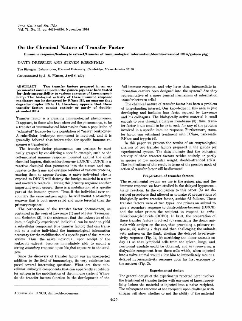
FIGS. 1-7. Transfer factors are subcellular leukocyte com-ponents that appear to be able to transmit information for specificimmune responses from experienced leukocytes to naive leukoeytes.This paper describes the results of an enzymological analysis oftwo transfer factors prepared in the guinea pig experimentalsystem. These transfer factors carry information for cell-mediatedresponses against the antigens dinitrochlorobenzene (DNCB) andortho-chlorobenzoylchloride (OCBC).
FIG. 1. Immune capacity developed in an animal that has beendirectly exposed to antigen. Six days after DNCB or OCBC waspainted on the ear, the back of the animal was shaved and theantigen was applied again. This provoked the secondary re-sponse (a delayed hypersensitivity reaction) shown. The reac-tion is designated +2 in severity; it is characterized by a ho-mogeneous erythema (redness), which represents an increasedblood supply in the area where responding leukocytes are elim-inating the antigen. The lymphoid tissue of animals such as thatshown in Fig. 1 serves as the source of transfer factor (5).
FIG. 2. An example of the immunity acquired by a naive ani-mal that received an injection of transfer factor (about 50%NO ofthe material from a single donor). The transfer factor was in-jected into the peritoneal cavity, and 48 hr later the animal wasshaved and challenged (5) with two concentrations of antigen:35 ll of 50 mM DNCB (on the lower flank) and 35 Il of 20 mMDNCB (on the upper flank). Beginning about 15 hr later, theanimal showed a strong delayed hypersensitivity response.The reaction at the site that received the lower concentrationof antigen was a mild erythema. At the site that received the higherconcentration of antigen, the reaction was more severe (+4) andwas characterized by a patchy necrosis in addition to erythemaand induration (swelling). Necrosis represents a generalized tis-sue destruction in the area where the antigen is being eliminated.
FIG. 3. An example of an animal unable to respond to challengewith antigen because the transfer factor he received had beentreated with RNase III, an enzyme that specifically degradesdouble-stranded RNA.
FIG. 4. Treatment of transfer factor with DNase, RNase, orprotease prior to injection has little effect on the biological ac-tivity of the material. Here, 48 hr after injection of the enzymati-cally treated transfer factor, the test animal was shaved andchallenged with three concentrations of antigen: 35 1A of 50 mMDNCB (upper flank), 25 mM DNCB (middle flank), and 5 mMDNCB (lower flank). The response to the highest concentrationof antigen is +4 in severity, as in Fig. 2. The two vertical blacklines were made by a magic marker to separate the challengesites.
FIGS. 5-7. Transfer factor is heat-sensitive. A solution ofDNCB transfer factor heated to 800 retained full biologicalactivity (Fig. 5); a solution heated to 850 retained partial bio-logical activity (Fig. 6); a solution heated to 900 was inactivated(Fig. 7).
Proc. Nat. Acad. Sci. USA 71 (1974)
Chemical Nature of Transfer Factor 4431
_..~~
Alp'_0. i
_ J_ __ __ _
v |
all f:~~~'d ~
FIGS. 1-7. (Legend appears at bottom of the previous page. )
Proc. Nat. Acad. Sci. USA 71 (1974)
4432 Immunology: Dressler and Rosenfeld
Transfer factor is resistant to deoxyribonuclease
Transfer factor was first tested for its susceptibility to in-activation by DNase. The following experiment was done.An amount of transfer factor capable of transmitting a +4delayed hypersensitivity capacity to a naive animal (gen-erally the material from 1/3 of a donor) was diluted 20-fold,so as to reduce the concentration of salts that might interferewith the enzyme digestion. The dilution buffer was 10 mMTris, pH 8-5 mM MgSO4, and the final NaCl concentrationwas about 0.15 M. Pancreatic DNase (Worthington, CodeDPFF) was then added to 20,ug/ml, and the reaction mixture(10 ml) was incubated for 1 hr at 250.To make sure that the DNase was working, we added as a
test substrate tritium-labeled bacteriophage T7 DNA (finalconcentration: 0.5 ,g/ml of DNA; 63,000 cpm/ml). Virtuallyall of the test DNA was degraded within 60 min but the
Minutes
0103060
cpm/0.1 ml t
6290*134054020
Activity
+4
+4
biological activity of the transfer factor was unaffected andgave responses identical to those shown in Fig. 4. Digestionof the biologically active material by as little as a factor oftwo would be expected to give a markedly weaker response,as shown by dilution experiments (5).
Transfer factor is resistant to pancreaticRNase and Ti RNase
The transfer factor was next tested for its susceptibility topancreatic RNase, which hydrolyzes single-stranded RNAchains -after uridylic acid and cytidylic acid residues. Thetransfer factor solution was diluted 1:10 with 10 mM Tris,pH 8, and 1 mM EDTA to reduce the NaCl concentrationto 0.3 M. The solution was then exposed to 20 ,ug/ml of pan-creatic RNase (Worthington, Code RASE) for 60 min at 37°.To make sure that the pancreatic RNase was working, we
added 26,000 cpm/ml of radioactive T7 mRNA that had beensynthesized in vitro with purified T7 DNA and E. coli RNApolymerase. Almost all of the test RNA was degraded in 60min but the biological activity of the transfer factor remained
Minutes
0103060
cpm/0.1 ml1
2630*110110100
Activity
+4
+4
intact and gave responses like those shown in Fig. 4.In a parallel experiment, Ti ribonuclease, which specifically
cuts single-stranded RNA chains after guanylic acid residues,
* There was virtually no loss of cpm or biological activity in theparallel reaction mixture incubated without enzyme.
t For each enzyme digestion the amount of test substrate stillin macromolecular form was determined by precipitating 11%oof the sample in trichloroacetic acid.
was used. Again the test RNA was degraded in 60 min but
Minutes0
103060
cpm/0.1 mlT1200*190140150
Activity+4
+4
the biological activity of the transfer factor survived, and thedelayed hypersensitivity responses were the same as thoseshown in Fig. 4.
Transfer factor is resistant to protease digestion
The enzymatic approach was next extended to ask whetherthe transfer factor could be destroyed by Pronase. The trans-fer factor solution was diluted 20-fold as before, and digestedfor 60 min with 300 ,g/ml of self-digested (13) Pronase.The test substrate in the reaction mixture consisted of a
collection of radioactive E. coli proteins, at a final concentra-tion of 0.4 ,g/ml; they were totally degraded in 60 min but
Minutes cpm/0.1 ml: Activity0103060
16,760*6,36041040
+4
+3 to +4
the transfer factor solution was virtually undiminished in itsability to transfer delayed hypersensitivity capacity, mediat-ing reactions identical to those shown in Fig 4.
Transfer factor is destroyed by RNase III
In contrast to its resistance to DNase, pancreatic and T1RNase, and Pronase, transfer factor activity is destroyed byRNase III. This enzyme specifically degrades double-strandedRNA (14, 15). The experiment is as follows. The transferfactor solution was diluted 20-fold with 50 mM Tris, pH 8,and the following reagents were added: MgCl2 to 10 mM, 2-mercaptoethanol to 1 mM, and glycerol to 5% (v/v).As a test substrate, radioactive double-stranded RNA was
prepared; this was done by incubating a random ACU poly-mer with the QB replicase and radioactive RNA precursors.The double-stranded product RNA was added to the transferfactor solution (0.2 jig/ml), followed by RNase III. Theenzyme was a highly purified preparation (15) generouslygiven to us by Dr. Robert Crouch of the NIH. The RNase IIInot only degraded the test substrate but also destroyed the
Minutes cpm/0.1 ml Activity0 35, 000* +410 9,7503060
48060 0
transfer factor activity, giving the response shown in Fig. 3.This result has been obtained with two independent prepara-tions of both DNCB and OCBC transfer factor.Can we be certain that the activity that destroyed the
transfer factor was RNase III, and not some impurity in theenzyme preparation-for instance, an unseen enzyme directedagainst glycoproteins or phospholipids? If RNase III is in-deed the enzyme that degrades the transfer factor, then itshould be possible to protect the transfer factor from diges-tion by adding an excess of nonradioactive, double-strandedRNA to the reaction mixture. We have done this experiment,using nonradioactive poly(rA rU) and poly(rI .rC) in a 100-
Proc. Nat. Acad. Sci. USA 71 (1974)
Chemical Nature of Transfer Factor 4433
*-OPERON CONCERNED WITH A SPECIFIC IMMUNE RESPONSE -*
I Im I GENE I GENE 2
OPERATOR-PROMOTER REGION:TRANSFER FACTOR ACTSHERE TO REMOVE A
REPRESSOR OR TO CREATEA SITE FOR MESSENGER RNA
SYNTHESIS
USE NTERNALLY
I GENE 3 - GENE 4 0TF GENE
AAAAAAAAAACCCCTTTTTTTTTTTT TTTTTTTGGGGAAAAAAAAAA
RNAjSYNTHESI S
AAAAAAAAAACCCCUUUUUUUUUU
AAAAAAAA AAFOLDING
uuuuuuuuuu iNG
SECRETED CAAAAAAAAAA1 C
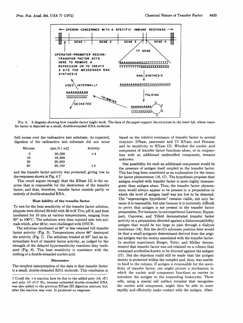
UUUUUUUUUU CuuuuuuuuCFIG. 8. A diagram showing how transfer factor might work. The data of the paper support the structure in the lower left, where trans-
fer factor is depicted as a small, double-stranded RNA molecule.
fold excess over the radioactive test substrate. As expected,digestion of the radioactive test substrate did not occur
Minutes0103060
cpm/0.1 ml$20,02019,80020,60020,100
Activity+4
+4
and the transfer factor activity was protected, giving rise tothe responses shown in Fig. 4. t
This result argues strongly that the RNase III is the en-zyme that is responsible for the destruction of the transferfactor, and that, therefore, transfer factor consists partly orentirely of double-stranded RNA.
Heat lability of the transfer factor
To test for the heat sensitivity of the transfer factor solution,aliquots were diluted 20-fold with 50mM Tris, pH 8, and thenincubated for 10 min at various temperatures, ranging from500 to 100'C. The solutions were then injected into test ani-mals which, after 48 hr, were challenged with DNCB.The solutions incubated at 800 or less retained full transfer
factor activity (Fig. 5). Temperatures above 900 destroyedthe activity (Fig. 7). The solutions treated at 85° had an in-termediate level of transfer factor activity, as judged by thestrength of the delayed hypersensitivity reactions they medi-ated (Fig. 6). This heat sensitivity is consistent with themelting of a double-stranded nucleic acid.
Discussion
The simplest interpretation of the data is that transfer factoris a small, double-stranded RNA molecule. This conclusion is
based on the relative resistance of transfer factor to severalenzymes: DNase, pancreatic and T1 RNase, and Pronase,and its sensitivity to RNase III. Whether the nucleic acidcomponent of transfer factor functions alone, or in conjunc-tion with an additional unidentified component, remainsunknown.One possibility for such an additional component would be
the presence of antigen itself coupled to the transfer factor.This has long been considered as an explanation for the trans-fer factor phenomenon (16, 17). The hypothesis proposes thatantigen coupled with transfer factor is more highly immuno-genic than antigen alone. Thus, the transfer factor phenom-enon would always appear to be present in a preparation inwhich the level of antigen itself was too low to be detected.The "superantigen hypothesis" remains viable, not only be-cause it is reasonable, but also because it is extremely difficultto prove that antigen is not present in the transfer factorpreparation. For instance, in one experiment Lawrence, Rappa-port, Converse, and Tillett demonstrated transfer factoractivity in a preparation directed against a histocompatibilityantigen that would be too large to pass through a dialysismembrane (16). But the devil's advocate position here wouldbe that a small antigenic determinant derived from the origi-nal antigen was the moiety associated with the transfer factor.In another experiment Burger, Vetto, and Malley demon-strated that transfer factor was not retained on a column thatcontained antibodies known to be directed against the antigen(17). But the objection could still be made that the antigenmoiety is protected within the complex and, thus, was unableto bind to the column. If antigen is responsible for the speci-ficity of transfer factor, one might picture a mechanism inwhich the nucleic acid component functions as carrier tointroduce the antigen to the responding leukocytes. Thesecells, using a special cell surface receptor that recognizesthe nucleic acid component, might then be able to morerapidly and efficiently make contact with the antigen. Alter-
t Could the +4 reaction here be due to the added poly (rA-rU)and poly (rI-rC)? No, because unlabeled double-stranded RNAwas also added to the previous RNase III digestion mixture, butafter the reaction was over. It produced no response.
Proc. Nat. Acad. Sci. USA 71 (1974)
I
4434 Immunology: Dressler and Rosenfeld
natively, the transfer factor might stimulate leukocytes thatwere independently interacting with antigen.
If the "transfer factor + antigen = superantigen" formu-lation is not correct, then one must explain how a double-stranded RNA molecule of low molecular weight could by it-self enable a population of leukocytes to develop an immuneresponse against a specific antigen. The transfer factor is toosmall (it can pass through a dialysis membrane) to code for thespecific proteins involved in an immune response. It could,however, stimulate a leukocyte by functioning as a negativecontrol element or depressor, a general idea that draws uponthe studies of gene control in E. coli (23, see also ref 18). Qnemight picture the double-stranded RNA molecule as com-peting with a DNA sequence in the leukocyte for the atten-tion of a specific repressor. If the repressor has a strongeraffinity for the entering transfer factor than for its naturaloperator, the operon would be derepressed and transcribed.This would lead to the production of the components involvedin the immune response.
Alternatively, the transfer factor could be viewed as func-tioning as a positive control element. In this situation, whichcould be more economical, the transfer factor would act di-rectly and positively by matching its nucleic acid sequencewith a specific immune DNA sequence so as to activate theregion for transcription.A characteristic of both of these models for transfer factor
function is that the immune system is essentially prepro-grammed and specific genes need only to be turned on (Fig. 8).
Considering the present data it is also possible to considerthe way in which the transfer factor itself might be produced.Rather than exist as a self-replicating molecule, the transferfactor could be the product of transcription from one elementof the operon (see Fig. 8). We would picture a region in theoperon that contains an inverted and symmetric base se-quence so that its transcript could self-anneal to form ahairpin structure. Removal of the single-stranded region(s)would yield active transfer factor which could then exert itsinfluence internally or upon other leukocytes.The involvement of RNA in the transmission of immuno-
logical information is a problem of general current interest.Fishman and Adler (19) have demonstrated that peritonealexudate cells from rats, incubated in vitro with bacteriophageT2, can yield an RNA extract that elicits antibodies againstT2 from a culture of normal lymphoid cells. A similar phenom-enon has been described by Askonas and Rhodes (20), usinghemocyanin in a mouse system in vitro. They interpreted theirresults as indicating that the RNA functions as a carrier forresidual antigen, greatly enhancing its immunogenicity. How-ever, Dray, Bell, Fishman, and Adler (21, 22) believe thatthis cannot be the full explanation. In extending the phenom-enon to rabbits, they found that the allotype of the anti-bodies elicited by the RNA extract was that of the donor, notthe recipient. From this result, they concluded that the RNA
might include intact messenger RNA molecules which canbe taken up and used by recipient cells. The large size of theRNA involved in these experiments, as well as its sensitivityto pancreatic RNase, distinguish this material from transferfactor.Although the transfer factor phenomenon is described here
in terms of one experimental system, the differentiatingleukocyte, it might have further implications in developmentalbiology. Perhaps other types of cell-cell interactions leadingto differentiation also involve the transmission of informationby a small molecule such as transfer factor.
Drs. Albert Coons and Jhmes Watson provided thoughtfulreadings of the manuscript. We are also indebted to Dr. CharlesDressler who reminded us at a key moment that guinea pigsneed vitamin C. This work was supported by the NationalInstitutes of Health (GM 17088) and the American CancerSociety (NP-57C). D.D. is a recipient of a Public Health ServiceResearch Career Development Award (GM 70440). S.R. is anundergraduate in Harvard College and has received support fromthe Camille and Henry Dreyfus Fund.
1. Lawrence, H. S. (1954) J. Clin. Invest. 33, 951. Lawrence,H. S. (1955) J. Clin. Invest., 34, 219-230.
2. Jeter, W. S., Tremaine, M. M. & Seebohm, P. M. (1954)Proc. Soc. Exp. Biol. Meda 86, 251-253.
3. Lawrence, H. S., Al-Askari, S., David, J., Franklin, E. &Zweiman, B. (1963) Trans. Ass. Amer. Phys., 76, 84-89.
4. Lawrence, H. S. (1959) in Cellular and Humoral Aspects ofthe Hypertensive States, ed. Lawrence, H. S. (Harper, NewYork), pp. 279-319.
5. Rosenfeld, S. & Dressler, D., (1974) Proc. Nat. Acad. Sci.USA 71, 2473-2477.
6. Siegel, S., Brady, A. H. & Awad, W. M. (1972) J. Biol.Chem. 247, 4155-4159.
7. Studier, F. W. (1969) Virology 39, 562-574.8. Minkley, E. & Pribnow, D. (1973) J. Mol. Biol. 77, 255-277.9. Haruna, I. & Spiegelman, S. (1965) Proc. Nat. Acad. Sci.
USA 54, 579-587.10. Kamen, R. (1970) Nature 228, 527-533.11. Kondo, M., Gallerani, R. & Weissmann, C. (1970) Nature
228, 525-527.12. Horvitz, H. R. (1973) Nature New Biol. 244, 137-140.13. Hourcade, D., Dressler, D. & Wolfson, J. (1973) Cold
Spring Harbor Symp. Quant. Biol. 38, 515-527.14. Robertson, H., Webster, R. & Zinder, N. (1968) J. Biol.
Chem. 243, 82-91.15. Crouch, R. (1974) J. Biol. Chem. 249, 1314-1317.16. Lawrence, H. S., Rapaport, F. T., Converse, J. M. &
Tillett, W. S. (1962) J. Clin. Invest. 39, 185-198.17. Burger, D. R., Vetto, R. M. & Malley, A. (1972) Science
175, 1473-1475.18. Lawrence, H. S. (1971) in Immunobiology, eds. Good, R. &
Fisher, D. (Sinaver Associates, Stamford, Conn.), pp. 104-105.
19. Fishman, M. & Adler, F. (1963) J. Exp. Med. 117, 595-602.20. Askonas, B. & Rhodes, J. (1965) Nature 205, 470-474.21. Adler, F., Fishman, M. & Dray, S. (1966) J. Immunol. 97,
554-558.22. Bell, C. & Dray, S. (1969) J. Immunol. 103, 1196-1211.23. Jacob, F. & Monod, J. (1961) J. Mol. Biol. 3, 318-357.
Ptashne, M. & Gilbert, W. (1970) Sci. Amer. 222, 36-44.
Proc. Nat. Acad. Sci. USA 71 (1974)

![[XLS]static.springer.comstatic.springer.com/sgw/documents/1372031/application/... · Web view0 1972 1973 1973 1973 1973 1974 1974 1974 1974 1974 1974 1974 1974 1974 1974 1974 1974](https://img.pdfslide.us/doc/110x75/5ae3d8767f8b9a5d648e7b9b/xls-view0-1972-1973-1973-1973-1973-1974-1974-1974-1974-1974-1974-1974-1974-1974.jpg)