Embed Size (px)
Citation preview

1
On the 120th Anniversary of the Discovery of the Romanowsky Effect
Romanowsky Staining: On the Question of Priority
A.V. Bezrukov, EMCO Ltd. Moscow.
Below is a summary of the methodical part of the works by Ch. I. Chenzinsky and D.L. Romanowsky, which laid the foundation of the techniques for staining of biological substances by means of a mixture of azure B, methylene blue and eosin. It shows that the priority in polychrome stain preparation by means of a mixture of azure B, methylene blue and eosin belongs to D.L. Romanowsky. His works initiated application of "Romanowsky staining, which has been used for 120 years all over the world.
In 2010, it will be 120 years since appearance of the first publication [12] on research of blood
smears stained so that the effect further named as “Romanowsky” was showed.
The end of the nineteenth century, when the article was issued, was a period of rapid
development of morphological methods in medicine due to the following reasons:
1. Applied medicine requirements, in particular, after discovery of the malaria plasmodium by
Laveran, when reliable diagnostics methods were required.
2. Beginning of mass production of advanced microscope models.
3. Beginning of industrial synthesis of the recently discovered aniline dyes.
Our (Russian) compatriots Cheslav Ivanovich Chenzinsky and Dmitry Leonidovich
Romanowsky happened to have made a decisive contribution to development of the thin blood
film staining techniques.
The priority of using a compound dye based on methylene blue and eosin for blood smears and
blood parasites analysis belongs to Cheslav Ivanovich Chenzinsky, who was a medical officer
and Odessa city hospital anatomist.
In 1888, he published an article [20], and in 1889 he obtained the Ph. D. degree with "To the
doctrine on malaria microorganisms" [19] dissertation. In these works, among other issues,
application of methylene blue in combination with eosin for thin blood films and blood parasites
staining was described.
Ch. I. Chenzinsky first used consecutive double staining with saturated water solution of
methylene blue dissolved with half-distilled water and with eosin spirit solution (0,5g eosin, 60g
ethanol and 40g water).Subsequently, he used a mixture of equal volumes of these solutions, and
staining came in 4-5 minutes.The result was bicolor staining: erythrocytes were dyed in pink,
malarial plasmodia became blue and were clearly visible against erythrocytes. Also, leukocyte
nuclei were stained in blue.
Then, a number of outstanding Russian scientists with the international popularity worked in
Odessa, among them I.I. Mechnikov who demonstrated the staining to Laveran.The technique

2
became well-known. Upon publication of works by Cheslav Ivanovich Chenzinsky, a number of
researchers successfully applied this method of thin blood films staining with minor alterations.
However, bicolor staining did not allow the researchers to reveal specifics of the malaria
plasmodium structure, in particular, the nucleus of it and the detailed characteristics of blood cell
morphology did not come to light also. It put D.L.Romanowsky, a doctor and chief of the St.
Petersburg Nikolayevsky Military Hospital eye department, on modernizing the technique
(Chenzinsky's methodology was described and discussed in his dissertation [13]).
The choice of dyes was made on the basis of Ch.I. Chenzinsky 's works.
Here that Dmitry Leonidovich wrote, proving the ratio choice between dyes:
"Having used Ehrlich's theory, his "Farbenanalyse", and noticing that the nuclei of the
majority of cells are being stained mainly with the basic and neutral dyes, we began to search a
neutral combination between methylene blue and eosin.
A priori, it was already possible to expect a neutral dye in the mixture of the
aforementioned solutions, as one of dyes is alkalinous and another one is acidulous.
To eliminate issues that may influence the solubility of mixed dyes and the mixture, we
took water solutions only.
Based on the numerous experiences, we discovered that if we mixed the filtered water
solutions of methylene blue and eosin, then, at some point of time and at surplus of the last, a
sediment which is insoluble in the mixture dropped out, and the mixture got a violet shade.
Such fall-out, possibly, had occurred earlier, but the sediment had dissolved in excess
quantities of methylene blue.
Looking for permanent volumes for certain solutions, we found that the sediment started
to appear clearly in the mixture of one part of the concentrated methylene blue solution and two
parts of 1% water solution of eosin soluble in water.
In this case, the mixture has great staining ability, especially nuclei are well stained, and
the dye does not lose the selective ability; but in addition to the dyes involved, a third dye
appears in the mixture, which has a special color and the greatest affinity to nuclei – or more
exactly, to their chromatin. "
Here, Romanowsky points out the presence (or occurrence) of the third dye in the mixture (as we
know now, the third dye is azure B – an oxidation product of methylene blue, and in combination
with eosin it stains nuclear chromatin in purple [23]).Now it is obvious that "the third dye"
initially contained in the old methylene blue solution.
Further, Romanowsky clarifies how solutions and mixtures were prepared: "The
beginning of falling-out of the sediment – the time of the greatest nucleus staining ability of the

3
mixture – can be found out using the following simple and practical method, as dyes of different
firms are not identical of what we were convinced by experiences.
We pour some methylene blue solution (2 cc) into a graduated cylinder (10 cc)and
carefully add eosin solution to it.
At first, the mixture remains dark blue but when neutralization occurs (e.g., in our case
when adding 4 cc of eosin), eosin ceases mixing up with methylene blue and remains over the
mixture surface in the form of a transparent layer of eosin solution which small surplus,
however, does not influence the coloring. The mixture of dyes thus obtained is carefully stirred
with a glass stick, but is not filtered, similar to all Ehrlich's dyes, because they lose their
selective coloring ability for some reason during filtration.
… Methylene blue starts coloring best of all when mould appears on the solution surface,
seen as a white film on it. We always had a big bottle with the methylene blue solution saturated
to sediments. As it decreases, we add water, shake it up and, after settling, filter for usage.
… Notice that the old methylene blue solution requires less eosin for saturation. So it is
necessary to repeat testing for sediments once per two months, thus in 9 months 1 volume of
methylene blue demanded us to have 2 but 1 Ѕ volume of eosin."
Thus, apparently, during long storage methylene blue in the solution was gradually oxidized with
formation of azure B and other derivative substances that led to polychromatic coloring of
smears. Unfortunately, Romanowsky points out that "dyes from different firms are not identical,
of what we were convinced by experiences", but does not inform us which dyes from what firms
were used. In the book by Romanowsky's contemporary professor M.N.Nikiforov [9], the
following is outlined concerning his technique:
"Good luck of coloring depends on the composition of methylene blue used, and, according to
Gautier, the most reliable method is to use methylene blue from Badisches Soda-Anilin Fabrik, C
and BGN marks, as well as eosin therefrom, A mark."
Below is an abstract by Romanowsky about the obtained tints of staining of different elements
of smear in the first work [13]:
"In my preparations I always get the following picture: red balls are stained in pink, eosinophil
protoplasm – in rich pink, malaria parasites and lymphocyte protoplasm – in light-blue, blood
plates and white ball nuclei – in dark-violet, parasitic nuclei – in purple-violet, leukocyte
protoplasm – in light-violet, in which case it is possible to see transitive colors from light-blue
lymphocyte protoplasm to violet leukocytes. "

4
Dmitry Leonidovich was the first to publish results containing a description of absolutely not
obvious effect of polychrome staining of blood smear and malaria parasites by means of the two
dyes combination. He expressed the assumption that it was connected with some third dye (as we
know now, it is azure B).Moreover, he observed and used the eosin with methylene blue (and
probably with azure B) reaction (sedimentation). Other results in his dissertation are also very
important for medicine and biology: research on the structure of various forms of malaria
plasmodium and research on influence of quinine on plasmodium. Many experts, for example,
[6, 10], consider Dmitry Leonidovich Romanowsky to be the first scientist who proved the
approach to treatment which now is called chemotherapy. In this area, he left Paul Ehrlich
behind for some years.
Unfortunately, in a number of publications [24, 27, 28] since 1978 Romanowsky's priority in
developing the methodology of polychrome staining of blood smears and blood parasites by
means of the compound dye including methylene blue (old solution) and eosin is called into
question. R.D. Lilli in [24] refers that Ernest Malahovsky's (a doctor from Silesia) short article
[25] was published three weeks earlier than Romanowsky's work in German published on
August, 24th (September, 5th) [26].Thus, Romanowsky's publications [12, 13] in Russian are
ignored because they are hardly accessible by western researchers. Meanwhile, even in the first
sentence of the well-known Romanowsky's work in German (D. Romanowsky Zur Frage der
Parasitologie und Therapie der Malaria. St. Petersburger Medicinische Wochensrift № 34 297-
302; № 35, 307-315), the following statement is made: "Vorwort: Vorliegende Arbeit des Dr. D.
Romanowsky ist zuerst im Juni d. J. als Dissertation erschienen." (The preface: The given work
by dr. D.Romanowsky first appeared in this June as a dissertation.)In other words, for anyone
who read this work it should be obvious that before the publication of the dissertation in German,
the dissertation in Russian was published where results had been stated in a fuller
manner.Eventually, in weekly journal VRACH, 1891, №21, p.522 (the end of May or the
beginning of June issue), in section "Chronicle and small news", there was the following
information: "Military-medical Academy Conference recognized Messrs. K.I. Zuev, D.L.
Romanowsky, G.G. Skorichenko and V.G. Slunin as Doctors of Medicine".
Thus, the publication chronology of results of D.L. Romanowsky is as follows:

1. The first publication (the preliminary message on 3 pages) was in the VRACH journal at the end of 1890. [12]. This article contains a clear description of the obtained results and the specific staining of the involved elements, though it is not mentioned that the old methylene blue solution was used.
5

2. The second publication (the fullest one) was the edition of the dissertation by D.L. Romanowsky: "TO THE QUESTION ON PARASITOLOGY AND THERAPY OF THE MARSH FEVER" in Russian [13], it was published not later than the first week of June 1891.
6

3. The third publication – the work referred by the western researchers – was the dissertation statement in German in weekly journal St.Petersburger Medicinische Wochensrift – was published on August, 24th (on September, 5th) 1891. [26].
7

8
The fourth publication [14], which was issued after dissertation defense, was devoted basically to the results of research on influence of quinine on malaria plasmodium. Thus, Malahovsky published the work three weeks prior to the third publication [25], and by the
time his article was issued, Romanowsky had already had the Doctor's degree, and his results
concerning staining techniques had been already published, so his priority is obvious.
What is much more important than formal priority, in our opinion, is that Romanowsky's works
stimulated further research on blood smears and malaria parasites staining, improvement of the
techniques and dye formulations, industrial production of the dyes, in particular, developed
thanks to works by Bernhard Nocht and Gustav Gimzy. For this reason, Gimza named the dye
"Giemsasche Lözung für die Romanowsky färbung" – "Solution by Gimza for Romanowsky
staining" [9, 21, 22].
It is necessary to notice that the International Committee on Standardization in Hematology
(ICSH) is quite reasonably using such terms as "Romanowsky Effect" and "Romanowsky
staining". The ICSH working group of experts on dyes and methods of staining consisting of the
most recognized scientists gives the following definition:
Definition of the Romanowsky stain. The stain consists of a mixture of the cationic dye azure B
and the anionic dye eosin Y, in aqueous solutions, in a ratio ranging from 6.5 to 7.3, with the two
dyes acting separately or in conjunction on appropriately pre-treated biological substrates, to
give a typical staining pattern which depends on the chemical and physical conditions of the
substrate. …
The Romanowsky effect. The unique effect of Romanowsky staining is based on the fact that
with biological substrates the blue cationic dye azure B and the red-orange anionic dye eosin Y
give more colours than just blue and red-orange …. Purple is the most important colour which
characterizes the Romanowsky effect. [23]
In the other words, Romanowsky staining or, as it sometimes is called, by Romanowsky type
staining, is a group of techniques in which the effect with the same name occurs. Close to this
definition, the term "Romanowsky staining" was applied by his contemporaries, in particular,
Bernhard Nocht, Sir William Boog Leishman, James Homer Wright , Gustav Gimza,Mikhail
Nikiforovitch Nikiforov .
Despite 120-year history, “Romanowsky staining is of outstanding importance for the
morphological identification of haemopoietic and other types of cells. [23].Romanowsky effect
mechanisms are still being analyzed, new variants of staining techniques are being developed,
and in particular, which is very important, attempts of practical implementation of the
standardized methodologies are being made [23].Thus, the work begun by Russian scientists
Cheslav Ivanovich Chenzinsky and Dmitry Leonidovich Romanowsky proceeds and benefits
people. Thanks to them, and also thanks to those researchers whose names are not remembered.

9
REFERENCES
1. Алексеев Г.А., Засухин Д.Н. Памяти Дмитрия Леонидовича Романовского к 120-летию со
дня рождения Пробл. Гематологии и переливания крови, 1981, т. 26, №3, с.59-60. 2. Дьяченко С.С. Дмитрий Леонидович Романовский (1861-1921) Врачебное дело, 1952, 4,
367-370. 3. Засухин Д.Н. У истоков отечественной протистологии. Ч.И. Хенцинский. Мед. Паразит и
паразитарн. Бол., 1953, 1, 95-97. 4. Засухин Д.Н. Д.Л. Романовский (80 лет метода окраски крови и паразитов крови). Мед.
Паразитология и паразит. бол. 1971, т. 40, № 6, с.729. 5. Идельчик Х.И., Левит М.М. Выдающиеся работы врачей Одесской городской больницы.
Сов. здравоохр., 1949, 3, 48-54. 6. Кассирский И.А. Проблемы и учёные. М., 1949. 7. Л.Ч. Доктор медицины Ч.И. Хенцинский Зубоврачебный ежемесячник, 1916, 6, 117-118. 8. Мельников-Разведенков Н.Ф. Чеслав Иванович Хенцинский и его значение для научной
медицины. Юбил. Сбор. Одесской окружной больницы1902-1927, Одесса, 1927, 148-162. 9. Никифоров М.Н. Микроскопическая техника. (8-е издание, 1919 г.) 10. Планельс Х.Х. Вклад русских учёных в развитие химиотерапии инфекционных
заболеваний. Журн. Микробиол., эпид., и иммунобиол., 1951, 9, 69-72. 11. Плотников Н.Н. Засухин Д.Н. Из истории борьбы с малярией в СССР. М., 1953, 88 стр.
(стр.27-28). 12. Романовский Д.Л. «К ВОПРОСУ О СТРОЕНИИ ЧУЖЕЯДНЫХ МАЛЯРИИ», ВРАЧЪ,
1890 г., № 52, 1171-1173. 13. Романовский Д.Л. «К ВОПРОСУ О ПАРАЗИТОЛОГИИ И ТЕРАПИИ БОЛОТНОЙ
ЛИХОРАДКИ», СПб, 1891 г. 118с. 14. Романовский Д.Л. «О СПЕЦИФИЧЕСКОМ ДЕЙСТВИИ ХИНИНА ПРИ БОЛОТНОЙ
ЛИХАРАДКЕ.» ВРАЧЪ, 1891 г., № 18, 438-440. 15. Саксонов П.П. Дмитрий Леонидович Романовский /1861-1921/ Фельдшер и акуш., 1950, 10,
41-43. 16. Фикс А.Ф. Приоритет Хенцинского в создании метода двойной окраски крови. Лабор.
Дело, 1963, 4, 59-59. 17. Фридлендер О. Памяти профессора Д.Л. Романовского Врачебная газета, 1922, 3-4, 112-
112. 18. Хаютин Д.М. Ч. И. Хенцинский (К 40-летию со дня смерти) Арх. Патолог. 1956, 18, 2, 121-
123. 19. Хенцинский Ч.И. К учению о микроорганизмах малярии. Дисс. Одесса,1889. 20. Chenzinsky C: Zur lehre von mikroorganismus des malaria-fiebers. Zentralbl Bakteriol 83:457,
1888 21. Fleischer B.100 years ago: Giemsa’s solution for staining of plasmodia Tropical Medicine and
International Health volume 9 no 7 pp 755–756 July 2004. 22. Giemsa G: Färbemethoden für malariaparasiten. Centbl Bakt 31:429, 1902. 23. ICSH reference method for staining of blood and bone marrow films by azure В and eosin Y
(Romanowsky stain). British Journal of Haematology, 1984, 57, 707-710 24. Lillie RD: Romanowsky-Malachowski stains: The so-called Romanowsky stain: Malachowski’s
1891 use of alkali polychromed methylene blue for malaria plasmodia. Stain Technol. 53:23-28, 1978.
25. Malachowski E: Zur morphologie des plasmodium malariae. Centralbl Klin Med 12: 601, 1891 26. Romanowsky, D. L. 1891. Zur Frage der Parasitologie und Therapie der Malaria. St.Petersburg
Med. Wochenschr. 16: №34, 297-302; № 35, 307-316. 27. Woronzoff-Dashkoff K: The Ehrlich-Chenzinsky-Plehn-Malachowski-Romanowsky-Nocht-
Jenner-May-GruЁnwald-Leishman-Reuter-Wright-Giemsa-Lillie-Roe-Wilcox Stain:The mystery unfolds. Clin Lab Med 13:759, 1993.
28. Woronzoff-Dashkoff KK. (2002). THE WRIGHT-GIEMSA STAIN. SecretsRevealed. Clin Lab Med. 22 (1): 15–23.

Addendum 1
Cheslav Ivanovich Chenzinsky
10
Addendum 2
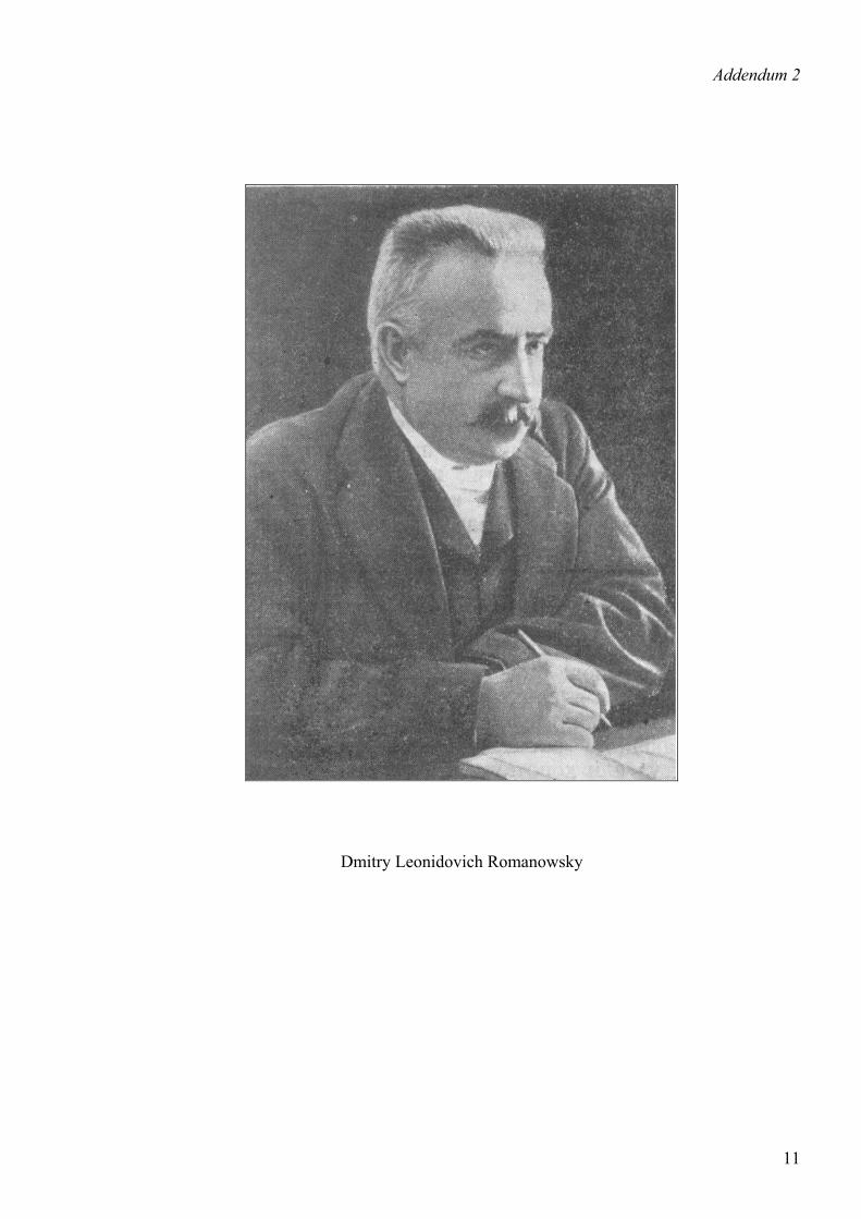
Dmitry Leonidovich Romanowsky
11