Embed Size (px)
Citation preview
3633RESEARCH ARTICLE
INTRODUCTIONFlower formation in plants requires the establishment of four typesof floral organs arranged in concentric whorls: the sepals andpetals, which comprise the sterile perianth; and the stamens andcarpels, which are the male and female reproductive organs. TheABC model, first proposed two decades ago, describes how thecombinatorial interaction of three classes of homeotic genes directsthe development of floral organs (Bowman et al., 1991; Coen andMeyerowitz, 1991). According to this classical model, Arabidopsisthaliana A-class genes APETALA1 (AP1) and AP2 confer sepalidentity in the first floral whorl. Their activity overlaps with B-classgenes APETALA3 (AP3) and PISTILLATA (PI) in the second whorl,which develops into petals. AP3, PI and the C-class geneAGAMOUS (AG) specify stamen identity in whorl three, while AGalone in whorl four promotes carpel development. The ABC modelwas initially deduced from loss-of-function effects. Subsequentcloning of the ABC genes showed that AP1, AP3, PI and AG allencode MADS domain proteins, as do the SEPALLATA (SEP)genes, which encode obligatory co-factors for the homeoticproteins.
An essential postulate of the ABC model is the antagonistic andmutually exclusive action of A and C function genes. In ap2 mutantflowers, expanded AG activity leads to the development of
reproductive organs at the floral periphery. Conversely, ag mutantsshow transformation of reproductive into perianth organs, anexpansion of A function towards the center of the flower. Accordingto the ABC model, A-class function in Arabidopsis is, therefore,required for perianth identity and repression of C-class function.Genes with such dual A function have, however, not yet been foundin any other species, questioning the generality of A-class functionand its role in determining perianth identity (Causier et al., 2010).
In contrast to the highly specific expression of MADS box floralhomeotic genes, it has been reported that AP2 mRNA accumulatesnot only in the perianth, but also in reproductive organ primordia.Three independent groups have suggested that primary AP2expression and promoter activity occur throughout all floral whorls(Jofuku et al., 1994; Würschum et al., 2006; Zhao et al., 2007). Afourth study agreed that AP2 is expressed ubiquitously, but withtransiently stronger mRNA accumulation in different organprimordia (Alvarez-Venegas et al., 2003). Broad expression hasbeen reported for an apparent AP2 ortholog in petunia (Maes et al.,2001), whereas AP2 orthologs in snapdragon and in maize havevery specific expression patterns in inflorescences and floralprimordia (Chuck et al., 1998; Keck et al., 2003).
Apart from its role in specifying floral organ identity, AP2 canpromote ectopic organ formation, an activity that depends at leastin part on the stem cell factor WUSCHEL (WUS) (Chen, 2004;Zhao et al., 2007). In flowers, WUS is a co-activator of AGexpression during early stages of development, while repression ofWUS by AG at later stages is required to produce determinateflowers (Lenhard et al., 2001; Lohmann et al., 2001). Similar towus mutations, a dominant-negative allele of AP2 has beenreported to cause precocious termination of the shoot apicalmeristem, in support of a positive effect of AP2 on WUS that isindependent of its negative role in AG regulation (Würschum et al.,2006).
Development 137, 3633-3642 (2010) doi:10.1242/dev.036673© 2010. Published by The Company of Biologists Ltd
1Department of Molecular Biology, Max Planck Institute for Developmental Biology,72076 Tübingen, Germany. 2Chromatin and Reproduction Group, Temasek LifeSciences Laboratory, 1 Research Link, National University of Singapore, 117604,Singapore. 3Plant Biology Laboratory, The Salk Institute for Biological Studies, LaJolla, CA 92037, USA.
*Present address: Scuola Superiore Sant’Anna, 56127 Pisa, Italy†Author for correspondence ([email protected])
Accepted 30 August 2010
SUMMARYThe ABC model of flower development explains how three classes of homeotic genes confer identity to the four types of floralorgans. In Arabidopsis thaliana, APETALA2 (AP2) and AGAMOUS (AG) represent A- and C-class genes that act in an antagonisticfashion to specify perianth and reproductive organs, respectively. An apparent paradox was the finding that AP2 mRNA issupposedly uniformly distributed throughout young floral primordia. Although miR172 has a role in preventing AP2 proteinaccumulation, miR172 was reported to disappear from the periphery only several days after AG activation in the center of theflower. Here, we resolve the enigmatic behavior of AP2 and its negative regulator miR172 through careful expression analyses.We find that AP2 mRNA accumulates predominantly in the outer floral whorls, as expected for an A-class homeotic gene. Itspattern overlaps only transiently with that of miR172, which we find to be restricted to the center of young floral primordia fromearly stages on. MiR172 also accumulates in the shoot meristem upon floral induction, compatible with its known role inregulating AP2-related genes with a role in flowering. Furthermore, we show that AP2 can cause striking organ proliferationdefects that are not limited to the center of the floral meristem, where its antagonist AG is required for terminating stem cellproliferation. Moreover, AP2 never expands uniformly into the center of ag mutant flowers, while miR172 is largely unaffected byloss of AG activity. We present a model in which the decision whether stamens or petals develop is based on the balance betweenAP2 and AG activities, rather than the two being mutually exclusive.
KEY WORDS: MicroRNA, miRNA, miR172, APETALA2, AGAMOUS, ABC model, Homeotic genes, Arabidopsis
On reconciling the interactions between APETALA2, miR172and AGAMOUS with the ABC model of flower developmentHeike Wollmann1,2, Erica Mica1,*, Marco Todesco1, Jeff A. Long3 and Detlef Weigel1,†
DEVELO
PMENT
3634
AP2 expression is regulated at the post-transcriptional level bya microRNA (miRNA), miR172 (Aukerman and Sakai, 2003;Chen, 2004; Kasschau et al., 2003; Rhoades et al., 2002).Transcript cleavage and translational inhibition both play a role inAP2 regulation by miR172, although assessing the relativeimportance of the two processes is confounded by a negative-feedback loop in which AP2 represses its own transcription(Aukerman and Sakai, 2003; Chen, 2004; Kasschau et al., 2003;Mlotshwa et al., 2006; Schwab et al., 2005). The discovery ofmiR172 as post-transcriptional negative regulator of AP2immediately provided a potential means to solve the apparentparadox of AP2 mRNA being ubiquitously expressed, yetrepressing AG only in the outer two floral whorls. However,miR172 expression was reported to overlap extensively with AP2mRNA throughout young floral primordia, and to disappear fromthe periphery only during stage 7, long after AG is activated (Chen,2004). Thus, miR172-guided regulation alone does not suffice toexplain the paradoxical relationship between AP2 expression andits genetic activity.
Here, we have re-examined not only AP2 mRNA expression, butalso the pattern of miR172 accumulation using in situ hybridizationwith LNA (locked nucleic acid) probes. We find that upon floralinduction, miR172 is strongly upregulated in the shoot meristem,where it has not been observed before (Chen, 2004). In youngfloral primordia, its expression pattern closely resembles that ofAG, being mostly concentrated in the floral center. We also findAP2 to be expressed much more specific, accumulatingpredominantly in the periphery of floral primordia, with onlylimited overlap to miR172. We further show that these expressionpatterns of AP2 and miR172 are required for proper flowerdevelopment.
MATERIALS AND METHODSPlant materialPlants were grown in long-day (16 hours light and 8 hours dark) or short-day(8 hours light and 16 hours dark) conditions at 23°C and 65% humidity.Arabidopsis thaliana Col-0 and Ler-1 plants were used as wild type. ag-1(Bowman et al., 1989), ag-2 (Yanofsky et al., 1990) and dcl1-11 (renamedfrom dcl1-100) (Laubinger et al., 2008) have been described. The ap2 allelewas obtained from the Salk T-DNA collection (Salk_071140) (Alonso et al.,2003) and was named ap2-12 (Yant et al., 2010).
In situ hybridizationTissue was harvested into FAA solution (3.7% formaldehyde, 50% ethanol,5% acetic acid). For embedding, an automated system (Advanced SmartProcessor ASP300, Leica, Wetzlar, Germany) was used. Sections of 8 or 9m thickness were prepared using a rotary microtome (Leica RM2165).Hybridization and detection were carried out as described (Palatnik et al.,2003) with some modifications. After incubation in Histoclear, the sectionswere processed through an ethanol series, treated with Proteinase K (Roche)for 30 minutes at 37°C and post-fixed with FAA. Hybridization was carried
out at 55°C overnight. Slides were blocked with 1% blocking reagent(Roche, Mannheim, Germany) in 1�TBS/0.3% Triton X-100. Forimmunological detection, anti-DIG antibody (Roche) was used in a 1:1259dilution. NBT/BCIP stock solution (Roche) for color reaction was diluted1:50 in 10% polyvinyl alcohol (PVA) in TNM-50. Probes were synthesizedwith the DIG RNA Labeling Kit (Roche) on PCR products of the targetgenes. For the AP2 (At4g36920) 3� end probe, a 634 bp cDNA fragment wasPCR amplified and cloned into pBluescript (pHW083). Oligonucleotidesequences are listed in Table S1 in the supplementary material. The AG(At4g18960) and WUS (At2g17950) probes were based on previouslydescribed plasmids (Leibfried et al., 2005; Yanofsky et al., 1990). ThemiR172 antisense LNA (locked nucleic acid, Exiqon, Vedbaek, Denmark)oligonucleotide with the sequence atgmCagmCatmCatmCaaGatTct (upper case,LNA; lower case: DNA) was end-labeled with the DIG 3�-End Labeling Kit(Roche) and purified with Micro Spin Chromatography Columns (Bio-Rad,Hercules, CA, USA). LNA-based miRNA in situ hybridization was carriedout largely according to the same procedure. Proteinase K incubation wascarried out for 25 minutes at 37°C. For post-fixation, 4% (w/v)paraformaldehyde in 1� PBS was used. After washing, the slides wereincubated in 0.1 M triethanolamine (pH 8.0) and 0.5% acetic anhydride for10 minutes. RNase treatment was carried out after hybridization and slideswere prepared for immunological detection by 45-minute incubation each in0.5% blocking reagent (Roche) and buffer B (1% BSA, 0.3% Triton X-100in 1�TBS); the latter was used also for subsequent washing steps. In anindependent line of experiments, using the protocol of (Long and Barton,1998), an AP2 full-length probe was used to detect AP2 expression in plantsof the Landsberg erecta (Ler-1) background.
Cloning and transgenic plantsThe binary plasmids are listed in Table 1. Oligonucleotide primersequences for PCR amplification and PCR-based mutagenesis are listed inTable S1 in the supplementary material. For the pAP2:AP2::YFP reporter,two copies of the coding sequence of yellow fluorescent protein for energytransfer YPet were fused in frame with the C terminus of AP2 in theJAtY57F17 TAC (transformation-competent artificial chromosome) clone(Liu et al., 1999), which is ~32 kb in length, using a bacterialrecombineering approach (Warming et al., 2005). For the pAP2:AP2::GUSreporter, an ~5 kb upstream fragment and the AP2 transcribed region wereamplified with primers that included sequences for recombination using theGateway technology (Invitrogen, Carlsbad, CA, USA). The AP2 promoterand the AP2 transcribed region from ATG to the stop codon wererecombined into pDONR P4-P1R (Invitrogen) and pDONR/Zeo(Invitrogen), respectively. The b-glucuronidase (GUS) gene was introducedinto pDONR P2R-P3 (Invitrogen). The three inserts were combined into a pALLIGATOR2 binary plasmid (Bensmihen et al., 2004) (http://www.isv.cnrs-gif.fr/jg/alligator/vectors.html) that was modified to allowMultiSite Gateway (Invitrogen) recombination. Primary transgenic plantswere selected based on GFP fluorescence of dry seeds. Other binaryplasmids were based on pGreenII (Hellens et al., 2000) and modified toallow Gateway (Invitrogen) compatible cloning. CaMV35S and AP3promoter sequences were as described (Lohmann et al., 2001). The wild-type and miR172 targeting resistant (rAP2) versions of AP2 have beendescribed (Schwab et al., 2005), and were also introduced into Gatewaycompatible entry plasmids. The artificial target mimicry construct MIM172
RESEARCH ARTICLE Development 137 (21)
Table 1. Plant transformation vectorsPlasmid number Description Purpose
HW075 p35S (empty) Control transgenic plants HW216 pAP3:MIM172 Region-specific miR172 knock-downHW230 pAP3:AP2 Region-specific wild-type AP2 mis-expressionHW245 pAP3:rAP2 Region-specific miR172 resistant AP2 mis-expressionHW235 pAP3:amiR-AP2 Region-specific AP2 knock-downHW210 p35S:amiR-AG-1 Broad AG knock-downHW209 pAP3:amiR-AG-1 Region-specific AG knock-downHW222 p35S:amiR-AG-2 Broad AG knock-downHW319 pAP2:AP2::GUS AP2 genomic reporterJAS100 pAP2:AP2::YFP AP2 genomic reporter D
EVELO
PMENT
was generated by PCR-based mutagenesis of IPS1 (Franco-Zorrilla et al.,2007). Artificial miRNAs were designed using WMD (http://wmd2.weigelworld.org) (Ossowski et al., 2008).
MicroscopyFor scanning electron microscopy, single flowers from T1 transgenic lineswere fixed for 5 minutes in 100% methanol and then transferred to ethanol.After critical point drying and coating with gold and palladium 30 nmparticles, samples were examined using a Hitachi S800 electronmicroscope.
For the pAP2:AP2::YFP reporter, whole inflorescences were embeddedin 3% agarose and placed in a chambered coverglass (NUNC, Rochester,NY) for imaging on a Leica DM IRE2 laser-scanning confocal microscope.
RESULTSPatterns of AP2 and miR172 expression in shootsand flowersAP2 is one of the four genes in the original ABC model of floralorgan specification (Bowman et al., 1991; Coen and Meyerowitz,1991). In contrast to the other three genes, AP3, PI and AG (Drewset al., 1991; Goto and Meyerowitz, 1994; Jack et al., 1994), as wellas the other A function gene AP1 (Mandel et al., 1992), its reportedbroad mRNA expression pattern during early floral developmentdoes not correlate well with its role in conferring specificallyperianth identity. MiR172 negatively regulates AP2, but its reporteddistribution throughout all whorls until floral stage 6 (Chen, 2004)does not satisfactorily explain the discrepancy between AP2mRNA expression and its specific activity. We therefore decided tore-examine the localization of miR172 and AP2 transcriptsspecifically during early flower development.
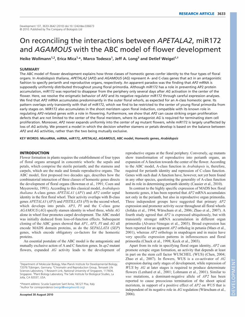
Because the MIR172a-2 precursor has been shown to betranscriptionally upregulated at the shoot apex upon photoperiodicinduction of flowering (Schmid et al., 2003), we chose vegetativeand inflorescence apices during the transition to flowering toestablish locked nucleic acid (LNA)-based in situ hybridization fordetection of miR172. Although the miR172 signal was low invegetative apices of 3-week-old, short-day grown plants, itappeared within 1 day of the transfer to long days, which inducesflowering. It further increased during days 3 and 5, when the firstsigns of inflorescence elongation became apparent (Fig. 1A).
Next we looked at miR172 expression in early floral primordia.Based on a different in situ hybridization approach, it has beenreported that miR172 expression is absent from the shoot meristem,that it is abundant in stage 1 floral primordia and that it persists inall four floral whorls through stage 6 of flower development (Chen,2004). Using the LNA-based method, however, we found miR172expression to be at higher levels in the shoot apical meristem thanin stage 1 and 2 flower primordia (Fig. 1B). From stage 3 onwards,we observed graded miR172 expression that was highest in thecenter of the floral meristem, which gives rise to the fourth whorl(Fig. 1C,D). The miR172 signal persisted in the fourth whorl thelongest, while it was low or absent in the other floral whorls (Fig.1E,F). Expression became restricted to the base of the developinggynoecium, and was subsequently detected in developing ovules(Fig. 1F,G). This last expression pattern might be related to the roleof the miR172 target AP2 in integument development (Léon-Kloosterziel et al., 1994; Modrusan et al., 1994). Because theexpression of miR172 in the center of developing flowers fromstage 3 onwards is similar to that of AG (Drews et al., 1991), weasked whether AG is required for maintenance of the propermiR172 pattern. In ag-2 mutant flowers, early miR172 expressionwas similar to its pattern in wild type (Fig. 1H-J), but persisted inthe indeterminate floral meristem (Fig. 1K,L).
As a negative control, we performed in situ hybridization onplants with a strong hypomorphic allele of DICER LIKE1 (DCL1),the Dicer responsible for miRNA biogenesis in A. thaliana (Parket al., 2002). No miR172 signal was detected (Fig. 1M).
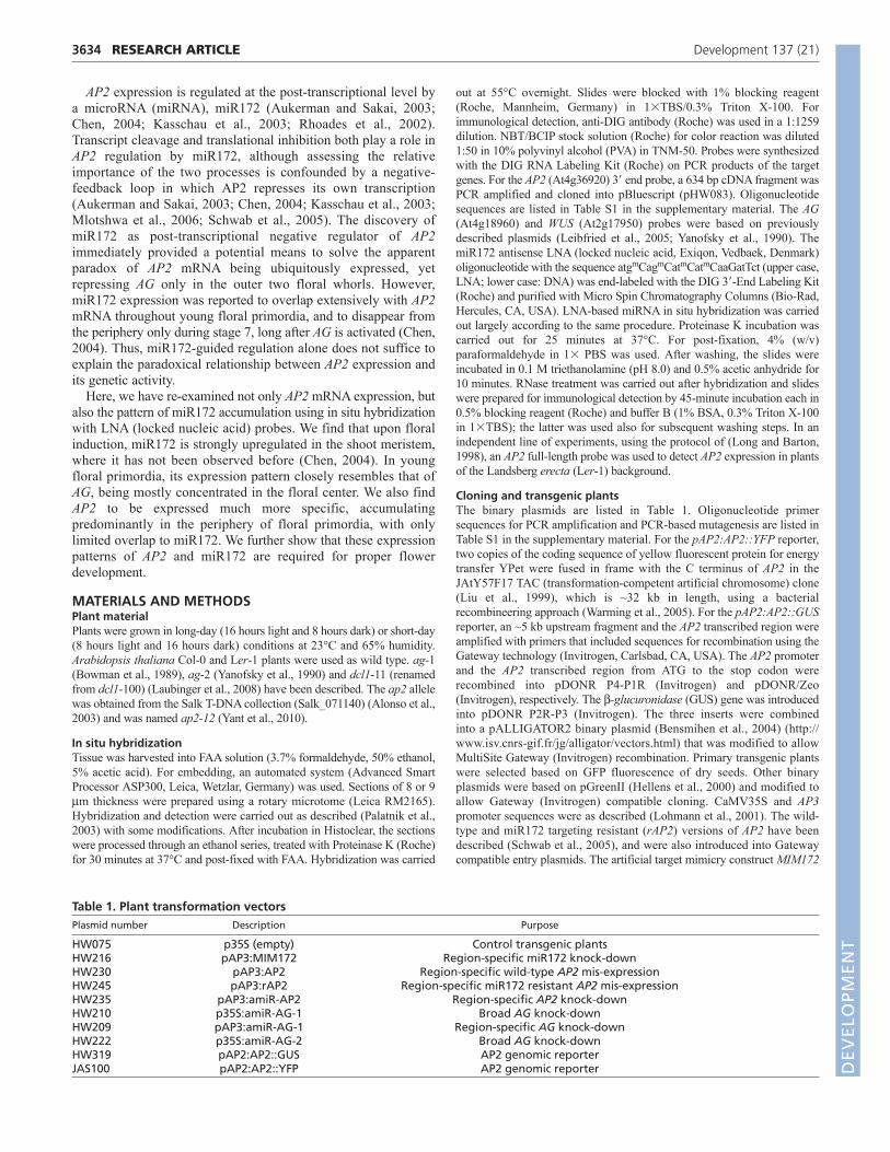
We complemented the in situ hybridization studies of miR172with analyses of its target AP2, using a probe against the 3� regionof the transcript to avoid cross hybridization with homologs. Incontrast to previous reports (Jofuku et al., 1994; Würschum et al.,2006), we found a distinct accumulation pattern of AP2 mRNAthroughout reproductive development (Fig. 2; see Fig. S1 in thesupplementary material). In Col-0 wild-type inflorescences, strongAP2 signal was detected in floral primordia from the earliest stageson. It became rapidly restricted to the periphery from stage 2onwards (Fig. 2A). During stage 3, AP2 signal was abundant insepals emerging on the flanks of the floral primordia (Fig. 2B,C).
3635RESEARCH ARTICLEAPETALA2, miR172 and AGAMOUS
Fig. 1. Expression of miR172. (A-G)Col-0 wild type. (A)Apices fromplants grown in short days and transferred to long days to induceflowering. Days after shift are indicated at the bottom. (B)Inflorescencemeristem (asterisk) with flanking stage 1 and 2 floral primordia.(C)Stage 4 flower. (D)Stage 5 flower. (E)Stage 7 flower. (F)Stage 8flower. (G)Developing ovules with signal in integuments. (H-L)ag-2.(H)Inflorescence meristem (asterisk) with flanking stage 1 and 2 floralprimordia. (I)Stage 5 flower. (J)Stage 6 flower. (K)Approximately stage7 flower. (L)Later stage flower. (M)dcl1-11 inflorescence apex (asterisk).se, sepal; pe, petal; st, stamen; gy, gynoecium; int, integuments. Scalebars: 50m.
DEVELO
PMENT
3636
By comparison, AP2 transcript levels appeared to be low or absentfrom the shoot apical meristem and the center of floral primordiaafter stage 2 (Fig. 2A-C). Subsequently, AP2 signal declined insepals, but appeared in stamen and petal primordia (Fig. 2D-F).Notably, AP2 and miR172 signal transiently overlapped in thethird, and probably also the second, whorl (Fig. 1C,D; Fig. 2D,E).In later stages of flower development, we observed AP2 expressionin developing petals, stamen filaments and the gynoecium,including placenta and developing ovules (Fig. 2G), consistent withthe known role of AP2 in ovule development (Léon-Kloosterziel etal., 1994; Modrusan et al., 1994). Similar results were obtainedwith a probe against the full-length AP2 transcript, which washybridized to Ler-1 inflorescences (Fig. 2I,J).
AP2 is closely related to five other genes that encode AP2-typetranscription factors and that are also targets of miR172. Four ofthese have been shown to act as floral repressors (Aukerman andSakai, 2003; Mathieu et al., 2009; Schmid et al., 2003). A similarrole has recently been described for AP2 (Yant et al., 2010;Mathieu et al., 2009), and vegetative expression of AP2 has been
noted before (Würschum et al., 2006). We used the full-lengthprobe to examine AP2 expression by in situ hybridization invegetative tissue. In 25-day-old, short-day grown Ler-1 apices,AP2 transcripts were abundant in developing leaves, in particularin adaxial regions (Fig. 2K,L). Additionally, AP2 appeared to beexpressed as a ring around the periphery of the vegetative meristemand to be upregulated in the incipient leaf primordia (Fig. 2L). Asa control, we performed in situ hybridization with an ap2 T-DNAinsertion line; much weaker signals were observed with thismaterial (see Fig. S1A-D in the supplementary material).
AP2 levels are regulated by miR172 both through miRNA-guided transcript cleavage and translation inhibition (Aukermanand Sakai, 2003; Chen, 2004), possibly causing AP2 proteinlocalization not to fully overlap with its transcript pattern. Wegenerated two different AP2 reporter constructs that allowed us toinvestigate the localization of AP2 fusion proteins. ApAP2:AP2::GUS (b-glucuronidase) reporter that included ~5 kb ofupstream sequences, and the AP2 transcribed region reproducedseveral aspects of the AP2 transcript pattern (see Fig. S1E,F in the
RESEARCH ARTICLE Development 137 (21)
Fig. 2. Expression of AP2. (A-H)Col-0 wildtype. (A)Inflorescence meristem (asterisk), withflanking stage 1 and 2 floral primordia. (B)Stage3 flower. (C)Late stage 3 flower. (D)Stage 4flower. (E)Stage 5 flower. (F)Stage 6 flower.(G)Stage 9 flower. Expression of AP2 is presentin petals, stamen filaments and placenta withdeveloping ovules. (H)Inflorescence apexhybridized with sense probe. (I,J)Ler-1 wild-typeinflorescence apex (I) and cross section throughan approximately stage 12 flower (J).(K)Longitudinal section of vegetative Ler-1apex. (L)Transverse section. AP2 expression isfound in emerging leaf primordia on the flanksof the shoot apical meristem (asterisk). Indeveloping leaves, AP2 expression is strongestlaterally and adaxially. (M-P)Transgenic plantscarrying a pAP2:AP2::YFP reporter. Entireinflorescence (M), cross-section through anapproximately stage 12 flower (N), and highermagnification of stage 4 (O) and 5 (P) flowers.There is strong YFP signal (yellow) in the sepalsfrom stage 4 flowers onwards (M,O-P) and instamens and petals (M-P), recapitulating the insitu hybridization pattern (J). In M, numbersindicate floral stages, the asterisk indicates theinflorescence meristem. Backgroundfluorescence is red (M-P). (Q-T)ag-2.(Q)Inflorescence meristem, with flanking stage2 and 3 floral primordia. (R)Late stage 4 flower.(S)Approximately stage 7 flower. (T)Late stagewith several extra whorls of organs. Expressionin petals. (U)Cross-section through matureflower of ag-1 mutant, with extensive signal inyounger petals. (V-Y)dcl1-11. (V)Inflorescenceapex (asterisk). (W)Stage 3 flower. (X)Stage 6flower. (Y)Later stage. Interior organs developabnormally. A-H,Q-T,V-Y were hybridized with aprobe against the 3� region of the AP2transcript; I-L,U were hybridized with a full-length probe. Description of floral stagesfollows Smyth et al. (Smyth et al., 1990). se,sepal; p, petal; st, stamen; gy, gynoecium. Scalebars: 50m for A-L,Q-Y.
DEVELO
PMENT
supplementary material), except for the characteristic expression insepals. We also examined a pAP2:AP2::YFP reporter, which wasbased on an ~32 kb TAC clone and which complemented the ap2-2 mutation. This reporter produced strong YFP signal from floralstage 4 onwards in sepal primordia and then in developing sepals,as well as in stamens and petals (Fig. 2M). Later in floraldevelopment, YFP signal was observed in petals and thegynoecium, as well as in stamen filaments (Fig. 2N), recapitulatingthe pattern observed with in situ hybridization (Fig. 2J). Increasingamounts of YFP signal was detected in stamens of stage 4 and 5flowers (Fig. 2O,P), suggesting that miR172 activity at these stagesis not sufficient to fully prevent AP2 protein accumulation. Insummary, AP2 protein appears largely to match its transcriptlocalization. Notably, YFP activity was observed in theinflorescence meristem in a subset of plants analyzed, suggestingtransient expression that is not easily detected by in situhybridization.
A central tenet of the ABC model of floral patterning is themutual antagonism of AP2 and AG (Bowman et al., 1991). Wetherefore analyzed AP2 transcripts in ag mutant flowers. AlthoughAP2 expression appeared in the supernumerary floral primordiaformed in ag-2 mutants, it remained below detection level withinthe meristem itself (Fig. 2Q-T). Similar results were obtained withthe full-length probe, which was hybridized to ag-1 mutantinflorescences (Fig. 2U).
As we did not detect mature miR172 in dcl1 mutant flowers(Fig. 1M), these plants also afforded us an opportunity todetermine the contribution of miR172 to the spatial pattern ofAP2 mRNA accumulation. Similar to other dcl1 mutants(Schauer et al., 2002), dcl1-11 plants have a broad variety ofdevelopmental defects as a result of global reduction in miRNAactivity. Therefore, a specific phenotype caused by increasedAP2 activity might be difficult to pinpoint. As in wild type, AP2was excluded from the center of the floral and inflorescencemeristem (Fig. 2V-Y), but appeared ectopically in thesupernumerary organs that developed in dcl1 mutant flowersduring later stages, similar to what we had observed in agmutants (Fig. 2S,Y). We conclude that the low levels of AP2mRNA in the center of the flower are largely due to negativefactors other than AG and miR172, or to the lack of positivefactors that activate AP2 mRNA expression.
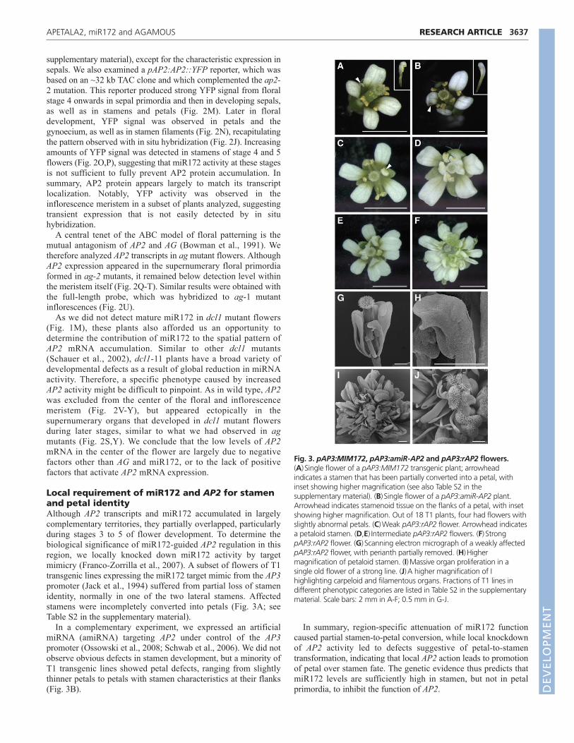
Local requirement of miR172 and AP2 for stamenand petal identityAlthough AP2 transcripts and miR172 accumulated in largelycomplementary territories, they partially overlapped, particularlyduring stages 3 to 5 of flower development. To determine thebiological significance of miR172-guided AP2 regulation in thisregion, we locally knocked down miR172 activity by targetmimicry (Franco-Zorrilla et al., 2007). A subset of flowers of T1transgenic lines expressing the miR172 target mimic from the AP3promoter (Jack et al., 1994) suffered from partial loss of stamenidentity, normally in one of the two lateral stamens. Affectedstamens were incompletely converted into petals (Fig. 3A; seeTable S2 in the supplementary material).
In a complementary experiment, we expressed an artificialmiRNA (amiRNA) targeting AP2 under control of the AP3promoter (Ossowski et al., 2008; Schwab et al., 2006). We did notobserve obvious defects in stamen development, but a minority ofT1 transgenic lines showed petal defects, ranging from slightlythinner petals to petals with stamen characteristics at their flanks(Fig. 3B).
In summary, region-specific attenuation of miR172 functioncaused partial stamen-to-petal conversion, while local knockdownof AP2 activity led to defects suggestive of petal-to-stamentransformation, indicating that local AP2 action leads to promotionof petal over stamen fate. The genetic evidence thus predicts thatmiR172 levels are sufficiently high in stamen, but not in petalprimordia, to inhibit the function of AP2.
3637RESEARCH ARTICLEAPETALA2, miR172 and AGAMOUS
Fig. 3. pAP3:MIM172, pAP3:amiR-AP2 and pAP3:rAP2 flowers.(A)Single flower of a pAP3:MIM172 transgenic plant; arrowheadindicates a stamen that has been partially converted into a petal, withinset showing higher magnification (see also Table S2 in thesupplementary material). (B)Single flower of a pAP3:amiR-AP2 plant.Arrowhead indicates stamenoid tissue on the flanks of a petal, with insetshowing higher magnification. Out of 18 T1 plants, four had flowers withslightly abnormal petals. (C)Weak pAP3:rAP2 flower. Arrowhead indicatesa petaloid stamen. (D,E)Intermediate pAP3:rAP2 flowers. (F)StrongpAP3:rAP2 flower. (G)Scanning electron micrograph of a weakly affectedpAP3:rAP2 flower, with perianth partially removed. (H)Highermagnification of petaloid stamen. (I)Massive organ proliferation in asingle old flower of a strong line. (J)A higher magnification of Ihighlighting carpeloid and filamentous organs. Fractions of T1 lines indifferent phenotypic categories are listed in Table S2 in the supplementarymaterial. Scale bars: 2 mm in A-F; 0.5 mm in G-J.
DEVELO
PMENT
3638
Effects of a non-targeted version of AP2 on organidentity and initiationAn alternative to miRNA target mimicry is the introduction ofmodified targets that escape miRNA regulation because of silentmutations in the miRNA target site. Transgenic expression of amiR172 non-targeted version of AP2 (rAP2) delays floweringand causes indeterminate growth of flowers with either petal orstamen overproliferation (Chen, 2004; Zhao et al., 2007). Tofurther test the importance of miR172 action for floral patterningof second- and third-whorl floral organs, we expressed an rAP2version (Schwab et al., 2005) under the control of the AP3promoter. Plants mis-expressing wild-type AP2 had mostlynormal flowers, whereas those mis-expressing rAP2 often hadpetaloid stamens (Fig. 3C-E,G,H; see Table S2 in thesupplementary material). The most severely affected linesshowed complete conversion of stamens into petals (Fig. 3F).The ectopic organs in these lines had petaloid and carpeloidcharacteristics, the latter forming extensive and partially fusedstructures with ovules and stigmata (Fig. 3I,J; see Fig. S2A,B inthe supplementary material). We also observed filamentousorgans, sometimes with stigmatic papillae at their tip (see Fig.S2C,D in the supplementary material). Organ proliferationappeared to mostly be initiated from multiple meristem-likecenters within such a flower (see Fig. S2E,F in thesupplementary material).
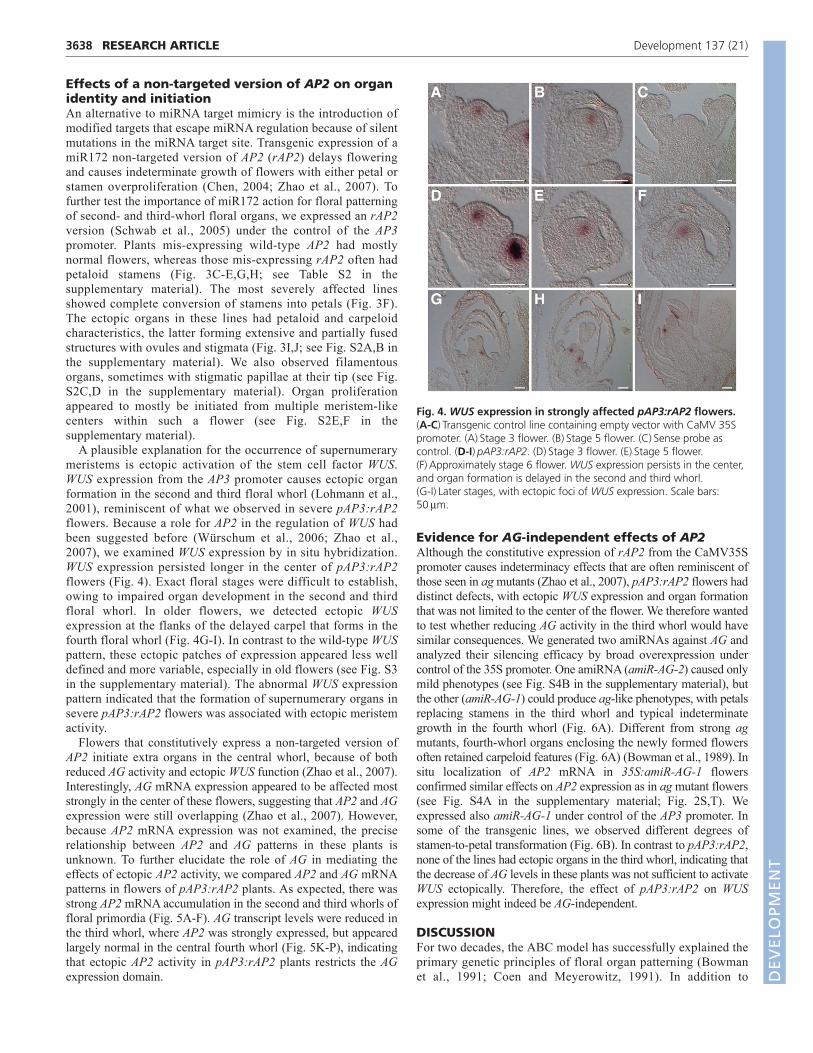
A plausible explanation for the occurrence of supernumerarymeristems is ectopic activation of the stem cell factor WUS.WUS expression from the AP3 promoter causes ectopic organformation in the second and third floral whorl (Lohmann et al.,2001), reminiscent of what we observed in severe pAP3:rAP2flowers. Because a role for AP2 in the regulation of WUS hadbeen suggested before (Würschum et al., 2006; Zhao et al.,2007), we examined WUS expression by in situ hybridization.WUS expression persisted longer in the center of pAP3:rAP2flowers (Fig. 4). Exact floral stages were difficult to establish,owing to impaired organ development in the second and thirdfloral whorl. In older flowers, we detected ectopic WUSexpression at the flanks of the delayed carpel that forms in thefourth floral whorl (Fig. 4G-I). In contrast to the wild-type WUSpattern, these ectopic patches of expression appeared less welldefined and more variable, especially in old flowers (see Fig. S3in the supplementary material). The abnormal WUS expressionpattern indicated that the formation of supernumerary organs insevere pAP3:rAP2 flowers was associated with ectopic meristemactivity.
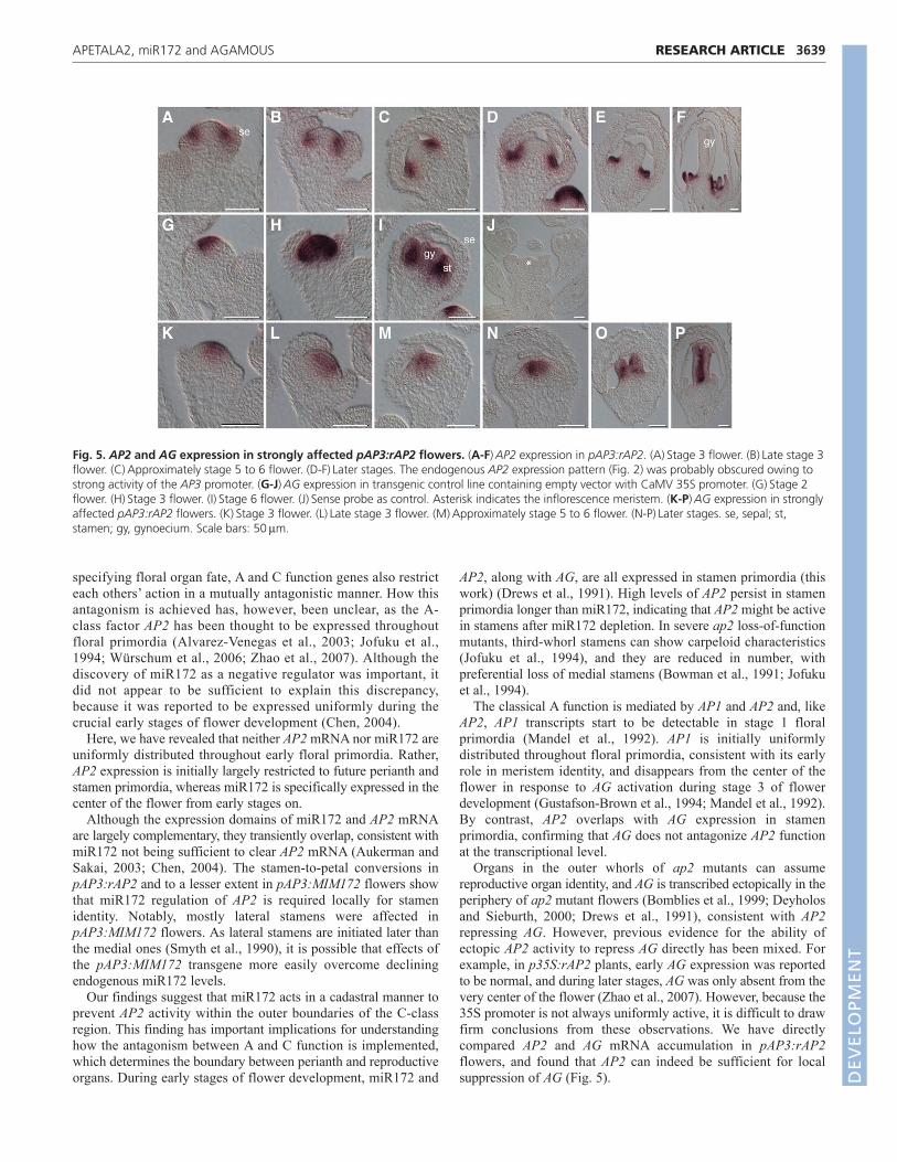
Flowers that constitutively express a non-targeted version ofAP2 initiate extra organs in the central whorl, because of bothreduced AG activity and ectopic WUS function (Zhao et al., 2007).Interestingly, AG mRNA expression appeared to be affected moststrongly in the center of these flowers, suggesting that AP2 and AGexpression were still overlapping (Zhao et al., 2007). However,because AP2 mRNA expression was not examined, the preciserelationship between AP2 and AG patterns in these plants isunknown. To further elucidate the role of AG in mediating theeffects of ectopic AP2 activity, we compared AP2 and AG mRNApatterns in flowers of pAP3:rAP2 plants. As expected, there wasstrong AP2 mRNA accumulation in the second and third whorls offloral primordia (Fig. 5A-F). AG transcript levels were reduced inthe third whorl, where AP2 was strongly expressed, but appearedlargely normal in the central fourth whorl (Fig. 5K-P), indicatingthat ectopic AP2 activity in pAP3:rAP2 plants restricts the AGexpression domain.
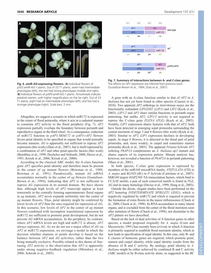
Evidence for AG-independent effects of AP2Although the constitutive expression of rAP2 from the CaMV35Spromoter causes indeterminacy effects that are often reminiscent ofthose seen in ag mutants (Zhao et al., 2007), pAP3:rAP2 flowers haddistinct defects, with ectopic WUS expression and organ formationthat was not limited to the center of the flower. We therefore wantedto test whether reducing AG activity in the third whorl would havesimilar consequences. We generated two amiRNAs against AG andanalyzed their silencing efficacy by broad overexpression undercontrol of the 35S promoter. One amiRNA (amiR-AG-2) caused onlymild phenotypes (see Fig. S4B in the supplementary material), butthe other (amiR-AG-1) could produce ag-like phenotypes, with petalsreplacing stamens in the third whorl and typical indeterminategrowth in the fourth whorl (Fig. 6A). Different from strong agmutants, fourth-whorl organs enclosing the newly formed flowersoften retained carpeloid features (Fig. 6A) (Bowman et al., 1989). Insitu localization of AP2 mRNA in 35S:amiR-AG-1 flowersconfirmed similar effects on AP2 expression as in ag mutant flowers(see Fig. S4A in the supplementary material; Fig. 2S,T). Weexpressed also amiR-AG-1 under control of the AP3 promoter. Insome of the transgenic lines, we observed different degrees ofstamen-to-petal transformation (Fig. 6B). In contrast to pAP3:rAP2,none of the lines had ectopic organs in the third whorl, indicating thatthe decrease of AG levels in these plants was not sufficient to activateWUS ectopically. Therefore, the effect of pAP3:rAP2 on WUSexpression might indeed be AG-independent.
DISCUSSIONFor two decades, the ABC model has successfully explained theprimary genetic principles of floral organ patterning (Bowmanet al., 1991; Coen and Meyerowitz, 1991). In addition to
RESEARCH ARTICLE Development 137 (21)
Fig. 4. WUS expression in strongly affected pAP3:rAP2 flowers.(A-C)Transgenic control line containing empty vector with CaMV 35Spromoter. (A)Stage 3 flower. (B)Stage 5 flower. (C)Sense probe ascontrol. (D-I)pAP3:rAP2. (D)Stage 3 flower. (E)Stage 5 flower.(F)Approximately stage 6 flower. WUS expression persists in the center,and organ formation is delayed in the second and third whorl. (G-I)Later stages, with ectopic foci of WUS expression. Scale bars:50m.
DEVELO
PMENT
specifying floral organ fate, A and C function genes also restricteach others’ action in a mutually antagonistic manner. How thisantagonism is achieved has, however, been unclear, as the A-class factor AP2 has been thought to be expressed throughoutfloral primordia (Alvarez-Venegas et al., 2003; Jofuku et al.,1994; Würschum et al., 2006; Zhao et al., 2007). Although thediscovery of miR172 as a negative regulator was important, itdid not appear to be sufficient to explain this discrepancy,because it was reported to be expressed uniformly during thecrucial early stages of flower development (Chen, 2004).
Here, we have revealed that neither AP2 mRNA nor miR172 areuniformly distributed throughout early floral primordia. Rather,AP2 expression is initially largely restricted to future perianth andstamen primordia, whereas miR172 is specifically expressed in thecenter of the flower from early stages on.
Although the expression domains of miR172 and AP2 mRNAare largely complementary, they transiently overlap, consistent withmiR172 not being sufficient to clear AP2 mRNA (Aukerman andSakai, 2003; Chen, 2004). The stamen-to-petal conversions inpAP3:rAP2 and to a lesser extent in pAP3:MIM172 flowers showthat miR172 regulation of AP2 is required locally for stamenidentity. Notably, mostly lateral stamens were affected inpAP3:MIM172 flowers. As lateral stamens are initiated later thanthe medial ones (Smyth et al., 1990), it is possible that effects ofthe pAP3:MIM172 transgene more easily overcome decliningendogenous miR172 levels.
Our findings suggest that miR172 acts in a cadastral manner toprevent AP2 activity within the outer boundaries of the C-classregion. This finding has important implications for understandinghow the antagonism between A and C function is implemented,which determines the boundary between perianth and reproductiveorgans. During early stages of flower development, miR172 and
AP2, along with AG, are all expressed in stamen primordia (thiswork) (Drews et al., 1991). High levels of AP2 persist in stamenprimordia longer than miR172, indicating that AP2 might be activein stamens after miR172 depletion. In severe ap2 loss-of-functionmutants, third-whorl stamens can show carpeloid characteristics(Jofuku et al., 1994), and they are reduced in number, withpreferential loss of medial stamens (Bowman et al., 1991; Jofukuet al., 1994).
The classical A function is mediated by AP1 and AP2 and, likeAP2, AP1 transcripts start to be detectable in stage 1 floralprimordia (Mandel et al., 1992). AP1 is initially uniformlydistributed throughout floral primordia, consistent with its earlyrole in meristem identity, and disappears from the center of theflower in response to AG activation during stage 3 of flowerdevelopment (Gustafson-Brown et al., 1994; Mandel et al., 1992).By contrast, AP2 overlaps with AG expression in stamenprimordia, confirming that AG does not antagonize AP2 functionat the transcriptional level.
Organs in the outer whorls of ap2 mutants can assumereproductive organ identity, and AG is transcribed ectopically in theperiphery of ap2 mutant flowers (Bomblies et al., 1999; Deyholosand Sieburth, 2000; Drews et al., 1991), consistent with AP2repressing AG. However, previous evidence for the ability ofectopic AP2 activity to repress AG directly has been mixed. Forexample, in p35S:rAP2 plants, early AG expression was reportedto be normal, and during later stages, AG was only absent from thevery center of the flower (Zhao et al., 2007). However, because the35S promoter is not always uniformly active, it is difficult to drawfirm conclusions from these observations. We have directlycompared AP2 and AG mRNA accumulation in pAP3:rAP2flowers, and found that AP2 can indeed be sufficient for localsuppression of AG (Fig. 5).
3639RESEARCH ARTICLEAPETALA2, miR172 and AGAMOUS
Fig. 5. AP2 and AG expression in strongly affected pAP3:rAP2 flowers. (A-F)AP2 expression in pAP3:rAP2. (A)Stage 3 flower. (B)Late stage 3flower. (C)Approximately stage 5 to 6 flower. (D-F)Later stages. The endogenous AP2 expression pattern (Fig. 2) was probably obscured owing tostrong activity of the AP3 promoter. (G-J)AG expression in transgenic control line containing empty vector with CaMV 35S promoter. (G)Stage 2flower. (H)Stage 3 flower. (I)Stage 6 flower. (J)Sense probe as control. Asterisk indicates the inflorescence meristem. (K-P)AG expression in stronglyaffected pAP3:rAP2 flowers. (K)Stage 3 flower. (L)Late stage 3 flower. (M)Approximately stage 5 to 6 flower. (N-P)Later stages. se, sepal; st,stamen; gy, gynoecium. Scale bars: 50m.
DEVELO
PMENT
3640
Altogether, we suggest a scenario in which miR172 is expressedin the center of floral primordia, where it acts in a cadastral mannerto constrain AP2 activity to the floral periphery (Fig. 7). AP2expression partially overlaps the boundary between perianth andreproductive organs in the third whorl. As a consequence, reductionof miR172 function in pAP3:MIM172 or pAP3:rAP2 flowersfavors petal identity to be specified in organs that would normallybecome stamens. AG is apparently not sufficient to repress AP2expression (this work) (Zhao et al., 2007), but is itself repressed bya combination of AP2 and other petal-specific factors (this work)(Bomblies et al., 1999; Deyholos and Sieburth, 2000; Drews et al.,1991; Krizek et al., 2006; Krizek et al., 2000).
According to the classical ABC model, the A-class functiongene AP2 specifies petal identity and therefore must be presentin the center of ag mutant flowers (Bowman et al., 1989;Bowman et al., 1991). Paradoxically, mutant AG mRNAaccumulates normally in the center of ag flowers (Gustafson-Brown et al., 1994), indicating that AP2 is not sufficient torepress AG expression in its normal domain. We have shownthat, although high levels of AP2 transcript appear at leasttransiently in the centrally forming supernumerary floral organsin ag mutants, AP2 never expands uniformly into the center ofag mutant flowers. Thus, petal identity might be conferred bylower levels of AP2 than the ones required for repression of AG.In this scenario, low levels of AP2 in the center of ag mutantflowers combined with incomplete translational repression bymiR172 are sufficient to promote petal development, but do notprevent AG mRNA accumulation. In the periphery, by contrast,where AP2 mRNA levels are high and miR172 is absent, AP2always represses AG. As we do not see a major effect of AG onAP2 or miR172 expression, we envisage a model in which thedecision whether stamens or petals develop is based on thebalance between AP2 and AG activities, rather than the twobeing mutually exclusive. Possibly related to this theme of fine-tuning AP2 activity is the observation that AP2 is apparentlyunder strong negative-feedback regulation (Mlotshwa et al.,2006; Schwab et al., 2005).
A gene with an A-class function similar to that of AP2 in A.thaliana has not yet been found in other species (Causier et al.,2010). Two apparent AP2 orthologs in Antirrhinum majus are thefunctionally redundant LIPLESS1 (LIP1) and LIP2 (Keck et al.,2003). LIP1/2 and AP2 share similar functions in perianth organpatterning, but unlike AP2, LIP1/2 activity is not required torepress the C-class gene PLENA (PLE) (Keck et al., 2003).Notably, LIP1 expression shares features with that of AP2; bothhave been detected in emerging sepal primordia surrounding thecentral meristem of stage 3 and 4 flowers (this work) (Keck et al.,2003). Similar to AP2, LIP1 expression declines in developingsepals. In stage 6 flowers, it is detected in the distal part of petalprimordia, and, more weakly, in carpel and sometimes stamenprimordia (Keck et al., 2003). The apparent Petunia hybrida AP2ortholog PhAP2A complements an A. thaliana ap2 mutant andshares aspects of its expression pattern. Mutant analysis has,however, not revealed a function of PhAP2A in perianth patterning(Maes et al., 2001).
In both species, C-class gene expression is repressed bymembers of the miR169 microRNA family: FISTULATA (FIS) inA. majus and BLIND (BL) in P. hybrida (Cartolano et al., 2007).MiR169 targets HAP2/NF-YA transcription factors, which bind toCCAAT motifs; a pair of such conserved motifs is found in PLE,AG and its many homologs (Davies et al., 1999; Hong et al., 2003).
Outside the dicots, elegant studies have been performed on theAP2 homolog INDETERMINATE SPIKLET1 (IDS1), which isnegatively regulated by miR172 and which is required to preventthe formation of extra florets in the maize inflorescence (Chuck etal., 2008; Chuck et al., 1998). Its RNA accumulates in many lateralorgans, and is excluded from the center of the floral meristem soonafter initiation of florets (Chuck et al., 1998), not dissimilar to theAP2 pattern we have described.
Based on the lack of dual activities of A function genes in otherspecies, a model proposed originally for A. majus (Coen andMeyerowitz, 1991) has recently been revived, in which A functionis primarily required to establish floral meristem identity, which inturn leads to specification of sepal identity. In this case, only B- andC-classes of homeotic genes are required, which promote petal,stamen and carpel identity, while sepal identity results from theabsence of B and C activity. By analogy, petal identity in A.thaliana might be either achieved by combined B and C activity(ABC model) or by B-class activity alone, as suggested in the BC
RESEARCH ARTICLE Development 137 (21)
Fig. 6. amiR-AG-expressing flowers. (A)Individual flowers ofp35S:amiR-AG-1 plants. Out of 23 T1 plants, seven had intermediatephenotypes (left), the rest had strong phenotypes (middle and right).(B)Individual flowers of pAP3:amiR-AG-1 plants. Arrowheads indicatepetaloid stamen, with higher magnification on the far right. Out of 23T1 plants, eight had an intermediate phenotype (left), and five had astronger phenotype (right). Scale bars: 2 mm.
Fig. 7. Summary of interactions between A- and C-class genes.The effects on AP1 expression are inferred from previous work(Gustafson-Brown et al., 1994; Zhao et al., 2007).
DEVELO
PMENT
and (A)BC models (Causier et al., 2010). Perianth identity in thefloral center of ag mutants could similarly be conferred by factorsother than AP2, explaining the largely unaffected expressionpatterns of AP2 and AG in ag mutants (this work) (Gustafson-Brown et al., 1994). Furthermore, if AP2 activity is predominantlyrestricted by miR172, rather than by AG, both would haveprimarily cadastral function, with limited direct contributions tofloral organ specification.
AP2 has previously been shown to affect maintenance ofexpression of the stem cell regulator WUS. In a line carrying anunusual ap2 allele, I28, WUS expression in the shoot apicalmeristem is not maintained, leading to premature termination of theshoot (Würschum et al., 2006). Conversely, expression of rAP2from its own promoter or from the CaMV 35S promoter causes anincrease in the number of floral whorls and, at least in the case ofp35S:rAP2, this is associated with prolonged and expandedexpression of WUS in the center of the flower (Zhao et al., 2007).We have found that region-specific overexpression of rAP2 fromthe AP3 promoter, in pAP3:rAP2 plants, leads to ectopic formationof organs in the third and fourth whorls, apparently arising fromseveral meristem-like centers of proliferation (Fig. 3; see Fig. S2in the supplementary material), and this was associated withectopic WUS expression (Fig. 4; see Fig. S3 in the supplementarymaterial). Similar phenotypes are seen in plants in which WUS isexpressed from the AP3 promoter (Lenhard et al., 2001; Lohmannet al., 2001), but not when AG activity is knocked down in thesame domain (Fig. 6). However, we did observe prolonged WUSexpression in the center of the flower, suggesting the possibilitythat AP2 affects WUS also non-autonomously. Such non-autonomous action might also be the cause of the supernumerarycarpeloid organs in pAP3:rAP2 plants, and might explain theeffects of the I28 allele of AP2 on WUS expression in thevegetative shoot meristem, given that AP2 expression is strongestin emerging leaves (Fig. 2).
In summary, we have shown that while the spatial expressionpatterns of AP2 mRNA and miR172 are largely complementary,there is transient overlap in second and possibly third whorlprimordia. Based on the phenotypes caused by region-specificknockdown of AP2 and miR172, we propose that miR172 is amajor factor of floral organ specification by acting in a cadastralmanner to restrict AP2 activity, and thereby specifying theboundary between perianth and reproductive organs.
AcknowledgementsWe thank Frédéric Berger for critical comments and discussion; Jared Sewellfor constructing the pAP2:AP2::YFP plasmid; Li Jing for engineering theMultiSite Gateway compatible pALLIGATOR plasmid; Felipe Fenselau deFelippes, Frank Küttner and Markus Schmid for Gateway vectors; StephanOssowski for amiR-AP2 design; Christoph Schuster for the WUS probe; SaschaLaubinger and the European Arabidopsis Stock centre for seeds; Jürgen Bergerfor help with scanning electron microscopy; and members of Team MiRNA andRebecca Schwab for discussion. We also thank Frédéric Berger for supportingexperiments by H.W. in his lab, which is funded by Temasek Life SciencesLaboratory. This work was supported by a Boehringer Ingelheim doctoralfellowship (to H.W.), by a NIH grant GM072764 (to J.A.L.), by the Marie CurieResearch Training Network SY-STEM, by European Community FP6 IP SIROCCO(contract LSHG-CT-2006-037900), by a Gottfried Wilhelm Leibniz Award ofthe DFG and by the Max Planck Society (D.W.). Deposited in PMC for releaseafter 12 months.
Competing interests statementThe authors declare no competing financial interests.
Supplementary materialSupplementary material for this article is available athttp://dev.biologists.org/lookup/suppl/doi:10.1242/dev.036673/-/DC1
ReferencesAlonso, J. M., Stepanova, A. N., Leisse, T. J., Kim, C. J., Chen, H., Shinn, P.,
Stevenson, D. K., Zimmerman, J., Barajas, P., Cheuk, R. et al. (2003).Genome-wide insertional mutagenesis of Arabidopsis thaliana. Science 301,653-657.
Alvarez-Venegas, R., Pien, S., Sadder, M., Witmer, X., Grossniklaus, U. andAvramova, Z. (2003). ATX-1, an Arabidopsis homolog of trithorax, activatesflower homeotic genes. Curr. Biol. 13, 627-637.
Aukerman, M. J. and Sakai, H. (2003). Regulation of flowering time and floralorgan identity by a microRNA and its APETALA2-like target genes. Plant Cell 15,2730-2741.
Bensmihen, S., To, A., Lambert, G., Kroj, T., Giraudat, J. and Parcy, F. (2004).Analysis of an activated ABI5 allele using a new selection method for transgenicArabidopsis seeds. FEBS Lett. 561, 127-131.
Bomblies, K., Dagenais, N. and Weigel, D. (1999). Redundant enhancersmediate transcriptional repression of AGAMOUS by APETALA2. Dev. Biol. 216,260-264.
Bowman, J. L., Smyth, D. R. and Meyerowitz, E. M. (1989). Genes directingflower development in Arabidopsis. Plant Cell 1, 37-52.
Bowman, J. L., Smyth, D. R. and Meyerowitz, E. M. (1991). Geneticinteractions among floral homeotic genes of Arabidopsis. Development 112, 1-20.
Cartolano, M., Castillo, R., Efremova, N., Kuckenberg, M., Zethof, J., Gerats,T., Schwarz-Sommer, Z. and Vandenbussche, M. (2007). A conservedmicroRNA module exerts homeotic control over Petunia hybrida andAntirrhinum majus floral organ identity. Nat. Genet. 39, 901-905.
Causier, B., Schwarz-Sommer, Z. and Davies, B. (2010). Floral organ identity:20 years of ABCs. Semin. Cell Dev. Biol. 21, 73-79.
Chen, X. (2004). A microRNA as a translational repressor of APETALA2 inArabidopsis flower development. Science 303, 2022-2025.
Chuck, G., Meeley, R. B. and Hake, S. (1998). The control of maize spikeletmeristem fate by the APETALA2-like gene indeterminate spikelet1. Genes Dev.12, 1145-1154.
Chuck, G., Meeley, R. and Hake, S. (2008). Floral meristem initiation andmeristem cell fate are regulated by the maize AP2 genes ids1 and sid1.Development 135, 3013-3019.
Coen, E. S. and Meyerowitz, E. M. (1991). The war of the whorls: geneticinteractions controlling flower development. Nature 353, 31-37.
Davies, B., Motte, P., Keck, E., Saedler, H., Sommer, H. and Schwarz-Sommer, Z. (1999). PLENA and FARINELLI: redundancy and regulatoryinteractions between two Antirrhinum MADS-box factors controlling flowerdevelopment. EMBO J. 18, 4023-4034.
Deyholos, M. K. and Sieburth, L. E. (2000). Separable whorl-specific expressionand negative regulation by enhancer elements within the AGAMOUS secondintron. Plant Cell 12, 1799-1810.
Drews, G. N., Bowman, J. L. and Meyerowitz, E. M. (1991). Negativeregulation of the Arabidopsis homeotic gene AGAMOUS by the APETALA2product. Cell 65, 991-1002.
Franco-Zorrilla, J. M., Valli, A., Todesco, M., Mateos, I., Puga, M. I., Rubio-Somoza, I., Leyva, A., Weigel, D., García, J. A. and Paz-Ares, J. (2007).Target mimicry provides a new mechanism for regulation of microRNA activity.Nat. Genet. 39, 1033-1037.
Goto, K. and Meyerowitz, E. M. (1994). Function and regulation of theArabidopsis floral homeotic gene PISTILLATA. Genes Dev. 8, 1548-1560.
Gustafson-Brown, C., Savidge, B. and Yanofsky, M. F. (1994). Regulation ofthe Arabidopsis floral homeotic gene APETALA1. Cell 76, 131-143.
Hellens, R. P., Edwards, E. A., Leyland, N. R., Bean, S. and Mullineaux, P. M.(2000). pGreen: a versatile and flexible binary Ti vector for Agrobacterium-mediated plant transformation. Plant Mol. Biol. 42, 819-832.
Hong, R. L., Hamaguchi, L., Busch, M. A. and Weigel, D. (2003). Regulatoryelements of the floral homeotic gene AGAMOUS identified by phylogeneticfootprinting and shadowing. Plant Cell 15, 1296-1309.
Jack, T., Fox, G. L. and Meyerowitz, E. M. (1994). Arabidopsis homeotic geneAPETALA3 ectopic expression: transcriptional and posttranscriptional regulationdetermine organ identity. Cell 76, 703-716.
Jofuku, K. D., den Boer, B. G. W., Van Montagu, M. and Okamuro, J. K.(1994). Control of Arabidopsis flower and seed development by the homeoticgene APETALA2. Plant Cell 6, 1211-1225.
Kasschau, K. D., Xie, Z., Allen, E., Llave, C., Chapman, E. J., Krizan, K. A. andCarrington, J. C. (2003). P1/HC-Pro, a viral suppressor of RNA silencing,interferes with Arabidopsis development and miRNA function. Dev. Cell 4, 205-217.
Keck, E., McSteen, P., Carpenter, R. and Coen, E. (2003). Separation of geneticfunctions controlling organ identity in flowers. EMBO J. 22, 1058-1066.
Krizek, B. A., Prost, V. and Macias, A. (2000). AINTEGUMENTA promotes petalidentity and acts as a negative regulator of AGAMOUS. Plant Cell 12, 1357-1366.
Krizek, B. A., Lewis, M. W. and Fletcher, J. C. (2006). RABBIT EARS is a second-whorl repressor of AGAMOUS that maintains spatial boundaries in Arabidopsisflowers. Plant J. 45, 369-383.
3641RESEARCH ARTICLEAPETALA2, miR172 and AGAMOUS
DEVELO
PMENT
3642
Laubinger, S., Sachsenberg, T., Zeller, G., Busch, W., Lohmann, J. U., Rätsch,G. and Weigel, D. (2008). Dual roles of the nuclear cap-binding complex andSERRATE in pre-mRNA splicing and microRNA processing in Arabidopsis thaliana.Proc. Natl. Acad. Sci. USA 105, 8795-8800.
Leibfried, A., To, J. P., Busch, W., Stehling, S., Kehle, A., Demar, M., Kieber, J.J. and Lohmann, J. U. (2005). WUSCHEL controls meristem function by directregulation of cytokinin-inducible response regulators. Nature 438, 1172-1175.
Lenhard, M., Bohnert, A., Jürgens, G. and Laux, T. (2001). Termination of stemcell maintenance in Arabidopsis floral meristems by interactions betweenWUSCHEL and AGAMOUS. Cell 105, 805-814.
Léon-Kloosterziel, K. M., Keijzer, C. J. and Koornneef, M. (1994). A seedshape mutant of Arabidopsis is affected in integument development. Plant Cell6, 385-392.
Liu, Y. G., Shirano, Y., Fukaki, H., Yanai, Y., Tasaka, M., Tabata, S. andShibata, D. (1999). Complementation of plant mutants with large genomicDNA fragments by a transformation-competent artificial chromosome vectoraccelerates positional cloning. Proc. Natl. Acad. Sci. USA 96, 6535-6540.
Lohmann, J. U., Hong, R., Hobe, M., Busch, M. A., Parcy, F., Simon, R. andWeigel, D. (2001). A molecular link between stem cell regulation and floralpatterning in Arabidopsis. Cell 105, 793-803.
Long, J. A. and Barton, M. K. (1998). The development of apical embryonicpattern in Arabidopsis. Development 125, 3027-3035.
Maes, T., Van de Steene, N., Zethof, J., Karimi, M., D’Hauw, M., Mares, G.,Van Montagu, M. and Gerats, T. (2001). Petunia Ap2-like genes and their rolein flower and seed development. Plant Cell 13, 229-244.
Mandel, M. A., Gustafson-Brown, C., Savidge, B. and Yanofsky, M. F. (1992).Molecular characterization of the Arabidopsis floral homeotic gene APETALA1.Nature 360, 273-277.
Mathieu, J., Yant, L. J., Murdter, F., Kuttner, F. and Schmid, M. (2009).Repression of flowering by the miR172 target SMZ. PLoS Biol. 7, e1000148.
Mlotshwa, S., Yang, Z., Kim, Y. and Chen, X. (2006). Floral patterning defectsinduced by Arabidopsis APETALA2 and microRNA172 expression in Nicotianabenthamiana. Plant Mol. Biol. 61, 781-793.
Modrusan, Z., Reiser, L., Feldmann, K. A., Fischer, R. L. and Haughn, G. W.(1994). Homeotic transformation of ovules into carpel-like structures inArabidopsis. Plant Cell 6, 333-349.
Ossowski, S., Schwab, R. and Weigel, D. (2008). Gene silencing in plants usingartificial microRNAs and other small RNAs. Plant J. 53, 674-690.
Palatnik, J. F., Allen, E., Wu, X., Schommer, C., Schwab, R., Carrington, J. C.and Weigel, D. (2003). Control of leaf morphogenesis by microRNAs. Nature425, 257-263.
Park, W., Li, J., Song, R., Messing, J. and Chen, X. (2002). CARPEL FACTORY, aDicer homolog, and HEN1, a novel protein, act in microRNA metabolism inArabidopsis thaliana. Curr. Biol. 12, 1484-1495.
Rhoades, M. W., Reinhart, B. J., Lim, L. P., Burge, C. B., Bartel, B. and Bartel,D. P. (2002). Prediction of plant microRNA targets. Cell 110, 513-520.
Schauer, S. E., Jacobsen, S. E., Meinke, D. W. and Ray, A. (2002). DICER-LIKE1:blind men and elephants in Arabidopsis development. Trends Plant Sci. 7, 487-491.
Schmid, M., Uhlenhaut, N. H., Godard, F., Demar, M., Bressan, R., Weigel, D.and Lohmann, J. U. (2003). Dissection of floral induction pathways usingglobal expression analysis. Development 130, 6001-6012.
Schwab, R., Palatnik, J. F., Riester, M., Schommer, C., Schmid, M. andWeigel, D. (2005). Specific effects of microRNAs on the plant transcriptome.Dev. Cell 8, 517-527.
Schwab, R., Ossowski, S., Riester, M., Warthmann, N. and Weigel, D. (2006).Highly specific gene silencing by artificial microRNAs in Arabidopsis. Plant Cell18, 1121-1133.
Smyth, D. R., Bowman, J. L. and Meyerowitz, E. M. (1990). Early flowerdevelopment in Arabidopsis. Plant Cell 2, 755-767.
Warming, S., Costantino, N., Court, D. L., Jenkins, N. A. and Copeland, N. G.(2005). Simple and highly efficient BAC recombineering using galK selection.Nucleic Acids Res. 33, e36.
Würschum, T., Gross-Hardt, R. and Laux, T. (2006). APETALA2 regulates thestem cell niche in the Arabidopsis shoot meristem. Plant Cell 18, 295-307.
Yanofsky, M. F., Ma, H., Bowman, J. L., Drews, G. N., Feldmann, K. A. andMeyerowitz, E. M. (1990). The protein encoded by the Arabidopsis homeoticgene agamous resembles transcription factors. Nature 346, 35-39.
Yant, L., Mathieu, J., Dinh, T. T., Ott, F., Lanz, C., Woolmann, H., Chen, X.and Schmid, M. (2010). Orchestration of the floral transition and floraldevelopment in Arabidopsis by the bifunctional transcription factor APETALA2.Plant Cell 7, 2156-2170.
Zhao, L., Kim, Y., Dinh, T. T. and Chen, X. (2007). miR172 regulates stem cellfate and defines the inner boundary of APETALA3 and PISTILLATA expressiondomain in Arabidopsis floral meristems. Plant J. 51, 840-849.
RESEARCH ARTICLE Development 137 (21)
DEVELO
PMENT


![Class B Gene Expression and the Modified ABC Model in ... · Kanno et al: Nongrass Monocots B-class Genes TheScientificWorldJOURNAL (2007) 7, 268-279 269 and APETALA2 [AP2] in A](https://img.pdfslide.us/doc/110x75/60e27fb8a4fb2823bf6f614d/class-b-gene-expression-and-the-modified-abc-model-in-kanno-et-al-nongrass.jpg)