Embed Size (px)
Citation preview

ON COMPARISON OF MANIFOLD LEARNING TECHNIQUES FOR DENDRITIC SPINECLASSIFICATION
Muhammad Usman Ghani1 Ali Özgür Argunsah2 Inbal Israely2 Devrim Ünay3
Tolga Tasdizen4 Müjdat Çetin1
1Faculty of Engineering and Natural Sciences, Sabanci University, Istanbul, Turkey2Champalimaud Neuroscience Programme, Champalimaud Centre for the Unknown, Lisbon, Portugal
3 Faculty of Engineering and Computer Sciences, Izmir University of Economics, Izmir, Turkey4Electrical and Computer Engineering Department, University of Utah, USA
ABSTRACT
Dendritic spines are one of the key functional componentsof neurons. Their morphological changes are correlated withneuronal activity. Neuroscientists study spine shape varia-tions to understand their relation with neuronal activity. Cur-rently this analysis performed manually, the availability of re-liable automated tools would assist neuroscientists and accel-erate this research. Previously, morphological features basedspine analysis has been performed and reported in the litera-ture. In this paper, we explore the idea of using and compar-ing manifold learning techniques for classifying spine shapes.We start with automatically segmented data and construct ourfeature vector by stacking and concatenating the columns ofimages. Further, we apply unsupervised manifold learningalgorithms and compare their performance in the context ofdendritic spine classification. We achieved 85.95% accuracyon a dataset of 242 automatically segmented mushroom andstubby spines. We also observed that ISOMAP implicitlycomputes prominent features suitable for classification pur-poses.
Index Terms— Dendritic Spines, Classification, Mani-fold Learning, ISOMAP, Microscopic Imaging, Neuroimag-ing
1. INTRODUCTION
Ramon y Cajal discovered dendritic spines in the 19th centuryand suggested that spine morphology changes with variationsin neuronal activity[1]. This hypothesis has been supportedby many studies [2]. Consequently, dendritic spine analy-sis has become very important for neurobiological researchand can potentially enable the neuroscientists to decode theunderlying relationship between neuron activity variationsand spine morphology changes [1]. In the literature, den-dritic spines have been classified into four types: mushroom,stubby, filopodia and thin [3]. Examples of these four classesare presented in Figure 1. Quantitative spine analysis is animportant research topic in contemporary neurobiologicalresearch and currently such analysis is performed manually
Fig. 1. Spine Classes: Mushroom, Stubby, Thin, Filopodia(Left to Right)
due to the lack of reliable automated tools. This makes theresearch process slow and subjective. The availability of reli-able automated resources would expedite the research in thisarea.
Manifold learning is an important methodology with ap-plications in a wide range of areas including data compres-sion, pattern recognition, and machine learning [4]. Mani-fold learning can be seen as a dimensionality reduction prob-lem, with the goal of producing a compressed representationof high-dimensional data. It can also be viewed as an algo-rithm to compute degrees of freedom that would be sufficientto reproduce most of the variability in data [4]. Mathemati-cally, we can formulate the dimensionality reduction or man-ifold learning problem as follows: given an N-dimensionalrandom variable x = (x1, x2, ...., xN )T , compute its low di-mensional representation, y = (y1, y2, ...., yD)T such thatD ≤ N , keeping maximum information from original high-dimensional data according to some criterion [5]. Differentalgorithms apply different criterion to reduce dimensional-ity, e.g., principal component analysis (PCA) uses maximumvariance as criteria.
The reason behind their success is the inherent redun-dancy in most natural images and the fact that natural im-ages having high-dimensional data mostly lie near a low-dimensional manifold [4]. To the best of our knowledge,the application of these techniques to spine analysis havenot been reported in the literature.In this study, we use sev-eral manifold learning techniques for spine classification andcompare their performance. Classification results achievedwith various settings are comparable to those of a humanexpert. This analysis is based on two-photon laser scanning
978-1-4799-2349-6/16/$31.00 ©2016 IEEE 339

microscopy (2PLSM) images.The main contributions of this paper are comparison of
unsupervised manifold learning techniques and visual anal-ysis of ISOMAP [6] based extracted features. Analysis ofISOMAP features lead to the conclusion that ISOMAP hasthe capability to implicitly compute the distinguishing fea-tures appropriate for classification.
Rest of this paper is organized as follows: Section 2 con-tains a brief literature review. The data set used in this studyand methodology is described in section 3. Results are pre-sented and discussed in Section 4. Section 5 contains the con-clusion and future research directions.
2. LITERATURE REVIEW
Automated segmentation process of dendritic spines has beenstudied extensively in the literature, but only a few studiesaddress the spine shape classification. Rodriguez et al. [7]computed morphological features and performed classifica-tion using a decision tree. They considered 3D confocal laserscanning microscopy (CLSM) images. Son et al. [8] and Shiet al. [9] also used morphological features and proposed adecision tree based classification system for CLSM images.
Koh et al. [10] proposed a morphological feature basedtechnique applying a rule based classifier for 2PLSM images.A recent study on spine analysis considered morphologicalfeatures to classify 2PLSM spine images and compared theperformance of state-of-the-art classifiers [11].
Most of these studies compute morphological featuresand perform classification using rule based algorithms, alsothere are only a few studies that consider 2PLSM images. Tothe best of authors’ knowledge, manifold learning based spineanalysis is not reported in the literature. In this research, weaim to fill these gaps and apply and compare different mani-fold learning approaches to the spine classification problem.
3. METHODOLOGY
Mice post natal 7 to 10 days old animals are imaged using2PLSM.1 We acquired 15 stacks of 3D images and projectedthem to 2D using maximum intensity projection (MIP) to usefor this study. 15 dendrite branches have been used to ex-tract a data set of 242 spines for this research, 182 spines aremushroom and 60 are stubby.
Before applying manifold learning algorithms, we appliedthe disjunctive normal shape models (DNSM) [12] based al-gorithm to segment spines. This algorithm exploits DNSMbased shape and appearance priors to segment spines withgood accuracy [13]. This algorithm takes a region-of-interest(ROI) as input. We selected the ROI in a way that the spinehead center is positioned almost in the center of the ROI. Fur-ther, we scaled the ROI to 150x150 pixels. Finally, each ROIwas aligned in a way that spine necks are in vertical position.A few images from this dataset are given in Figure2. After
1All animal experiments are carried out in accordance with EuropeanUnion regulations on animal care and use, and with the approval of the Por-tuguese Veterinary Authority (DGV).
Fig. 2. A few images from dataset, without segmentation(above) and segmented images (below). First 2 spines arelabeled as Mushroom and 3rd spine as Stubby.
preparing the dataset, we applied the DNSM based segmen-tation algorithm to segment the spine images. Segmentationresults are not perfect but good enough for shape analysis.It is important to note that classification techniques used inthis paper are sensitive with respect to segmentation, differ-ent segmentation approach could lead to different classifica-tion results.
3.1. Manifold Learning
In this paper we consider several manifold learning tech-niques and compared their performance.
PCA is a widely used classical technique that provides atransformed lower dimensional representation attempting topreserve maximum variance, but it is not very effective in var-ious application due to its global linearity property [14]. Mul-tidimensional scaling (MDS) provides a lower dimensionalrepresentation attempting to preserve the distance betweenpoints, but it suffers from similar problems as PCA [15]. Lo-cally linear embedding (LLE) is a nonlinear dimensionalityreduction approach that finds the low-dimensional represen-tation striving to keep embedding of high-dimensional data[16].
ISOMAP is another non-linear dimensionality reductionapproach that possesses the best features of PCA and MDS[6]. It can be viewed as an extension of MDS by replacing theEuclidean distance metric with geodesic distance. The Lapla-cian eigenmaps method constructs a graph by applying theK-nearest neighbors (KNN) and computes its weights in sucha way that the norm of the gradient is minimized in the leastsquares sense [17]. Local Tangent Space Alignment (LTSA)also constructs the graph using KNN and for dimensionalityreduction it applies an approximation to local tangent spacesfor each neighborhood [18].
Firstly, the segmented spine images were used to con-struct 22,500 dimensional feature vectors by concatenatingthe stacked columns of each spine image. These feature vec-tors were further used to construct the feature matrix. Finally,manifold learning algorithms were applied on this feature ma-trix to produce lower dimensional feature matrices.
340
3.2. Classification
In order to compare the performance of these manifold learn-ing techniques, we selected three different classifiers, supportvector machines (SVM), KNN, and random forests (RF), totest their performance. The linear kernel is used for SVM,K=8 is used for KNN, and 10 decision trees are used for RFclassifier. The idea behind applying different classificationtechniques is to test the performance of these manifold learn-ing approaches irrespective of the classification technique ap-plied.
4. RESULTS
We compared the performance of six manifold learning tech-niques using three different classifiers. Classification resultsand visual analysis of ISOMAP based features are discussedin this section.
4.1. Classification Results
We selected only two features for manifold learning, the rea-son behind selecting two features will become clear later inthis section when we discuss ISOMAP based features. Weapplied three different classifiers to compare the performanceof these techniques to make sure that performance is the resultof feature transformation not because of the classifier.
Classification results using SVM, KNN, and RF are pre-sented in Table 1. It is evident from achieved results thatperformance of these manifold learning approaches is depen-dent upon classifier. It makes sense because just like manifoldlearning techniques, classifiers also use different decision cri-teria. For SVM classifier, Laplacian eigenmaps method per-forms best. However, for KNN classifier ISOMAP gives bestclassification results and for RF classifier, the complete fea-ture set gives best accuracy.
Table 1. Classification Results with SVM, RF, and KNN clas-sifiers
Features SVM KNN RF
Complete Features 84.71% 84.71% 85.12%ISOMAP 85.54% 84.71% 81.41%PCA 82.64% 83.88% 78.51%LLE 85.54% 83.47% 83.06%Laplacian 85.95% 83.47% 80.17%LTSA 77.27% 82.23% 80.58%MDS 84.30% 80.99% 79.75%
These observations imply that one should visually analyzethe produced feature space before making a decision aboutthe classification approach. Another conclusion that can bedrawn from these results is that none of these manifold learn-ing techniques perform best for all scenarios. The best perfor-mance is achieved with Laplacian eigenmaps based featureswith SVM classifier. It even performs better than complete
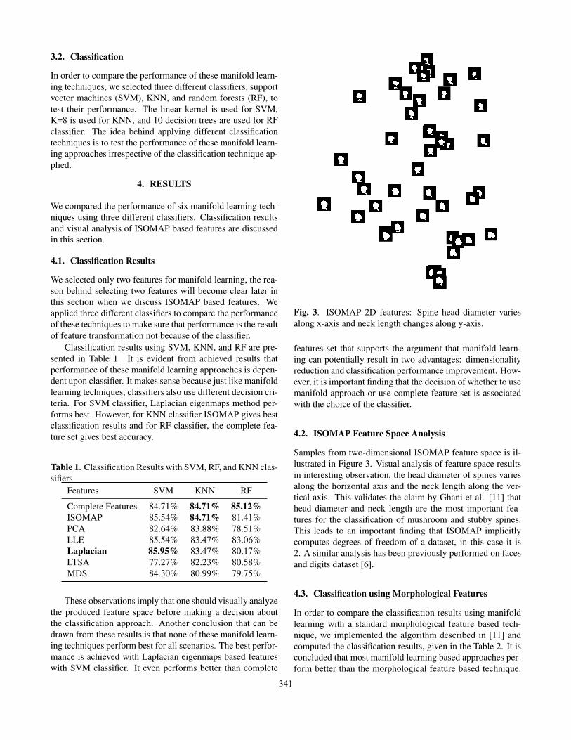
Fig. 3. ISOMAP 2D features: Spine head diameter variesalong x-axis and neck length changes along y-axis.
features set that supports the argument that manifold learn-ing can potentially result in two advantages: dimensionalityreduction and classification performance improvement. How-ever, it is important finding that the decision of whether to usemanifold approach or use complete feature set is associatedwith the choice of the classifier.
4.2. ISOMAP Feature Space Analysis
Samples from two-dimensional ISOMAP feature space is il-lustrated in Figure 3. Visual analysis of feature space resultsin interesting observation, the head diameter of spines variesalong the horizontal axis and the neck length along the ver-tical axis. This validates the claim by Ghani et al. [11] thathead diameter and neck length are the most important fea-tures for the classification of mushroom and stubby spines.This leads to an important finding that ISOMAP implicitlycomputes degrees of freedom of a dataset, in this case it is2. A similar analysis has been previously performed on facesand digits dataset [6].
4.3. Classification using Morphological Features
In order to compare the classification results using manifoldlearning with a standard morphological feature based tech-nique, we implemented the algorithm described in [11] andcomputed the classification results, given in the Table 2. It isconcluded that most manifold learning based approaches per-form better than the morphological feature based technique.
341

Table 2. Classification results using morphological featuresbased approach
Classifier Accuracy
SVM 78.51%KNN 80.17%RF 81.41%
5. CONCLUSION
Six state-of-the-art unsupervised manifold learning tech-niques have been compared in this study for dendritic spineclassification. It is found that the Laplacian eigenmapsmethod results in the best performance. It is also concludedthat most manifold learning techniques result in better per-formance as compared to the baseline morphological featurebased technique. It is also observed that ISOMAP computesdegrees of freedom in a dataset and it is found that for thedendritic spines dataset used in this research, we have twodegrees of freedom. Another interesting observation is, man-ifold learned features perform better than complete featureswith some of the classifiers applied, hence the decision ofapplying manifold learning techniques must be made tak-ing into account the choice of the classifier to be used aswell. Future work could involve larger dataset to preciselycharacterize manifolds.
ACKNOWLEDGEMENTThis work has been supported by the Scientific and Techno-logical Research Council of Turkey (TUBITAK) under Grant113E603 and by a TUBITAK-2221 Fellowship for VisitingScientists and Scientists on Sabbatical Leave.
6. REFERENCES
[1] J. Lippman and A. Dunaevsky, “Dendritic spine morphogene-sis and plasticity,” Journal of neurobiology, vol. 64, no. 1, pp.47–57, 2005.
[2] R. Yuste and T. Bonhoeffer, “Morphological changes in den-dritic spines associated with long-term synaptic plasticity.,”Annu Rev Neurosci, vol. 24, pp. 1071–1089, 2001.
[3] F.L. Chang and W. T. Greenough, “Transient and endur-ing morphological correlates of synaptic activity and efficacychange in the rat hippocampal slice,” Brain Res., vol. 309, pp.35–46, 1984.
[4] A. Ghodsi, “Dimensionality reduction: A short tutorial,” Tech-nical report, Department of Statistics and Actuarial Science,University of Waterloo, 2006.
[5] I. Fodor, “A survey of dimension reduction techniques,” Tech.Rep., Center for Applied Scientific Computing, Lawrence Liv-ermore National Laboratory, 2002.
[6] J. B. Tenenbaum, V. de Silva, and J. C. Langford, “A globalgeometric framework for nonlinear dimensionality reduction,”Science, vol. 290, no. 5500, pp. 2319, 2000.
[7] A. Rodriguez, D. B. Ehlenberger, D. L. Dickstein, P. R. Hof,and S. L. Wearne, “Automated three-dimensional detectionand shape classification of dendritic spines from fluorescencemicroscopy images,” PloS one, vol. 3, no. 4, 2008.
[8] J. Son, S. Song, S. Lee, S. Chang, and M. Kim, “Morpho-logical change tracking of dendritic spines based on structuralfeatures,” Journal of microscopy, vol. 241, no. 3, pp. 261–272,2011.
[9] P. Shi, X. Zhou, Q. Li, M. Baron, M. A. Teylan, Y. Kim,and S. T. Wong, “Online three-dimensional dendritic spinesmophological classification based on semi-supervised learn-ing,” in ISBI’09 IEEE International Symposium on BiomedicalImaging: From Nano to Macro. (pp. 1019-1022), 2009.
[10] I. Y. Koh, W. B. Lindquist, K. Zito, E. A. Nimchinsky, andK. Svoboda, “An image analysis algorithm for dendriticspines,” Neural computation, vol. 14, no. 6, pp. 1283–1310,2002.
[11] M. U. Ghani, S. D. Kanik, A. O. Argunsah, T. Tasdizen,D. Unay, and M. Cetin, “Dendritic spine shape classificationfrom two-photon microscopy images,” in IEEE Signal Pro-cessing and Communications Applications (SIU), 2015.
[12] N. Ramesh, F. Mesadi, M. Cetin, and T. Tasdizen, “Disjunctivenormal shape models,” in Biomedical Imaging (ISBI), 2015IEEE 12th International Symposium on, April 2015, pp. 1535–1539.
[13] F. Mesadi, M. Cetin, and T. Tasdizen, “Disjunctive normalshape and appearance priors with applications to image seg-mentation,” in Medical Image Computing and Computer-Assisted Intervention — MICCAI 2015, N. Navab, J. Horneg-ger, W. M. Wells, and A. F. Frangi, Eds., vol. 9351 of Lec-ture Notes in Computer Science, pp. 703–710. Springer Inter-national Publishing, 2015.
[14] I.T. Jolliffe, Principal Component Analysis, Springer Verlag,1986.
[15] T. F. Cox and M.A.A. Cox, Multidimensional Scaling, SecondEdition, Chapman and Hall/CRC, 2 edition, 2000.
[16] S. T. Roweis and L. K. Saul, “Nonlinear dimensionality re-duction by locally linear embedding,” SCIENCE, vol. 290, pp.2323–2326, 2000.
[17] M. Belkin and P. Niyogi, “Laplacian eigenmaps for dimension-ality reduction and data representation,” Neural Computation,vol. 15, pp. 1373–1396, 2002.
[18] Z. Zhang and H. Zha, “Principal manifolds and nonlinear di-mensionality reduction via tangent space alignment,” SIAM J.Sci. Comput., vol. 26, no. 1, pp. 313–338, Jan. 2005.
342