Embed Size (px)
Citation preview
OMT for the Pregnant Patient
Presented by:
Kristie Petree, DO
Assistant Professor of Neuromusculoskeletal Medicine and Osteopathic Manipulative Medicine
Georgia Campus – Philadelphia College of Osteopathic Medicine
Lecture Objectives
Define somatic dysfunction and osteopathic manipulative treatment (OMT)
Discuss biomechanical changes experienced by the pregnant patient
Review specific techniques that may be helpful for the common musculoskeletal complaints experienced during pregnancy.
Definitions
Osteopathic Manipulative Medicine (OMM)
The application of the osteopathic philosophy, structural diagnosis, and the use of osteopathic manipulative treatment in the diagnosis and management of the patient
Osteopathic Manipulative Treatment (OMT)
The therapeutic application of manually guided forces by an osteopathic physician to improve physiologic function and/or support homeostasis that has been altered by somatic dysfunction.
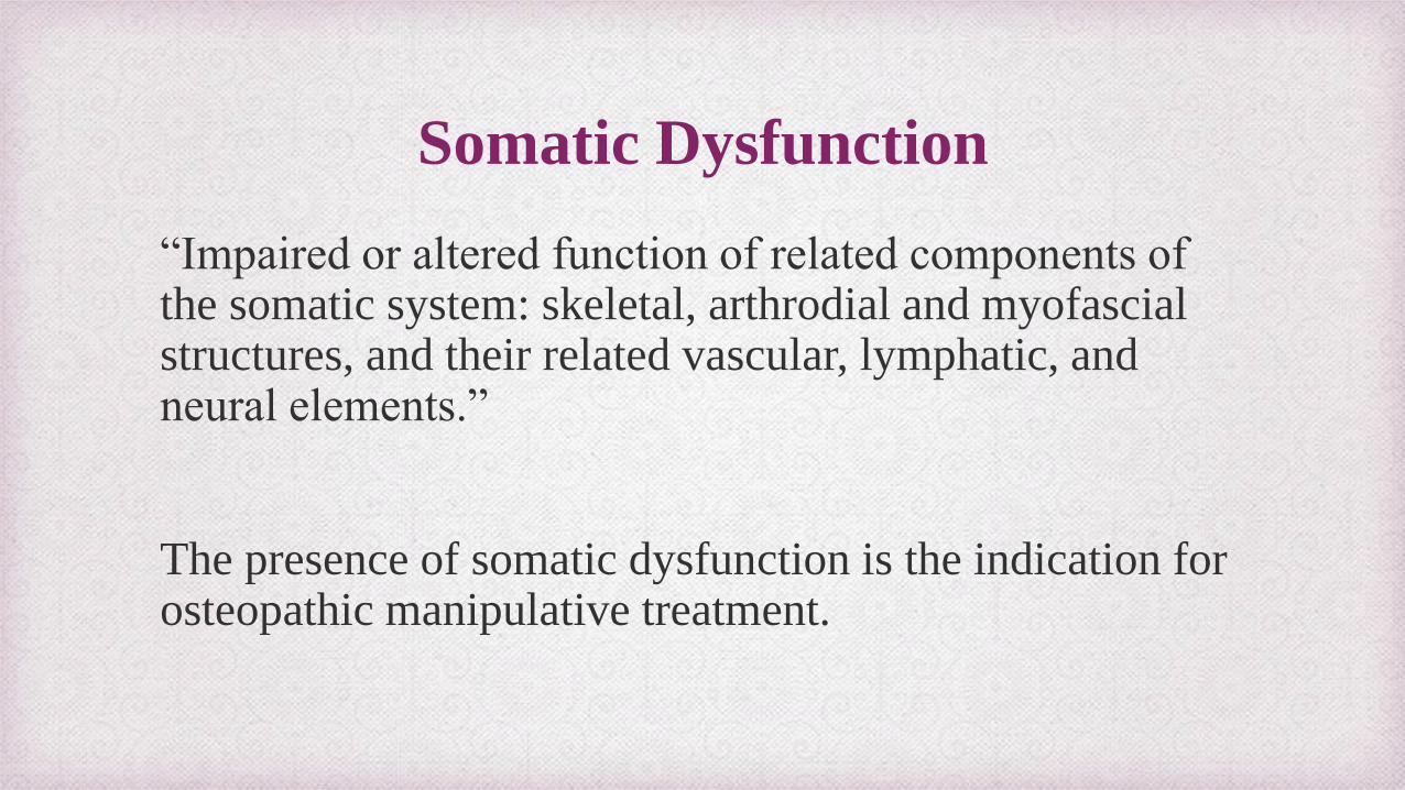
Somatic Dysfunction
“Impaired or altered function of related components of the somatic system: skeletal, arthrodial and myofascial structures, and their related vascular, lymphatic, and neural elements.”
The presence of somatic dysfunction is the indication for osteopathic manipulative treatment.
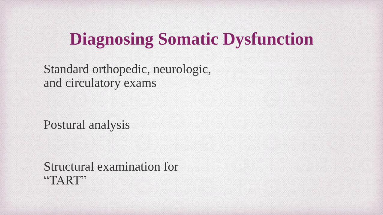
Diagnosing Somatic Dysfunction
Standard orthopedic, neurologic, and circulatory exams
Postural analysis
Structural examination for “TART”
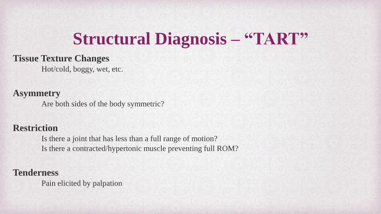
Structural Diagnosis – “TART” Tissue Texture Changes
Hot/cold, boggy, wet, etc.
Asymmetry Are both sides of the body symmetric?
Restriction Is there a joint that has less than a full range of motion?
Is there a contracted/hypertonic muscle preventing full ROM?
Tenderness Pain elicited by palpation
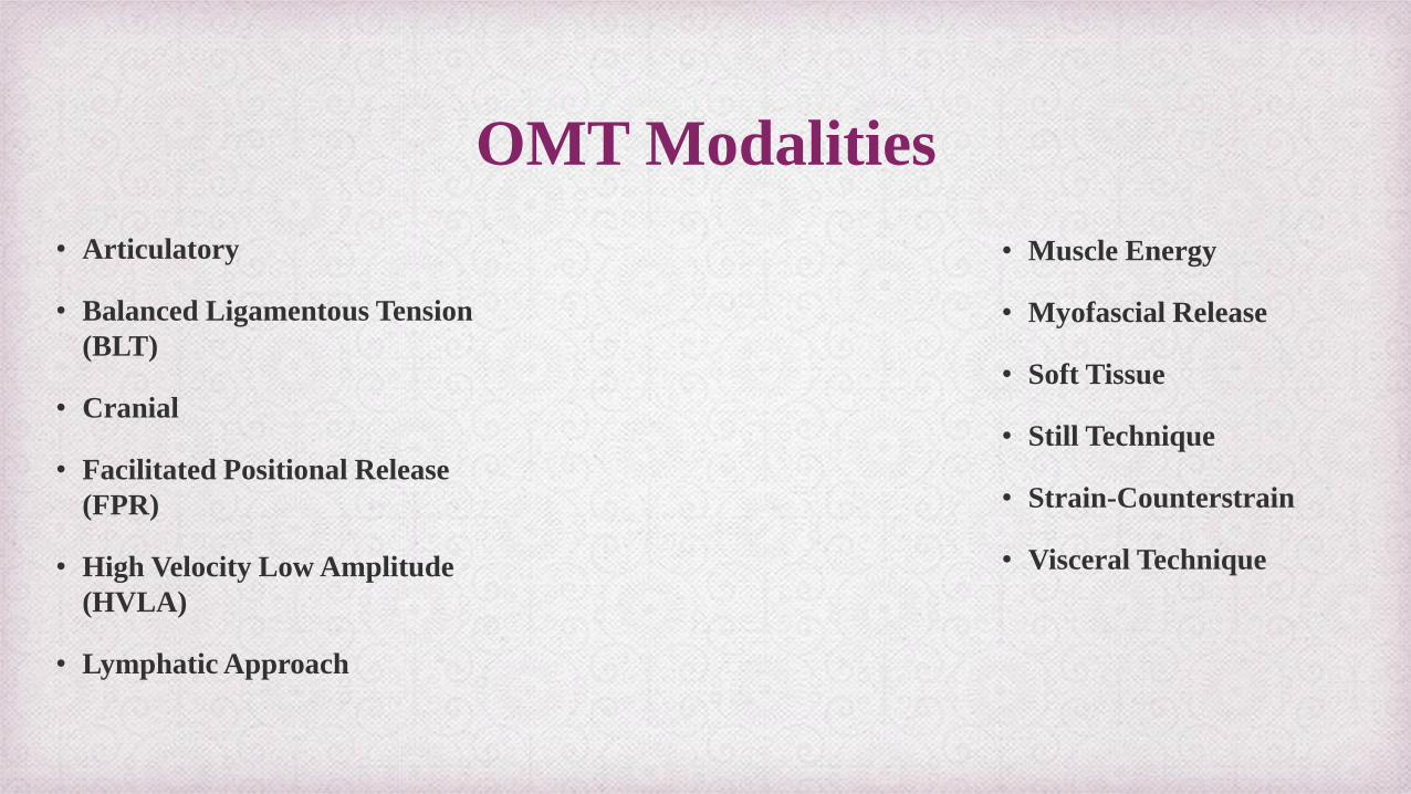
OMT Modalities
• Articulatory
• Balanced Ligamentous Tension
(BLT)
• Cranial
• Facilitated Positional Release
(FPR)
• High Velocity Low Amplitude
(HVLA)
• Lymphatic Approach
• Muscle Energy
• Myofascial Release
• Soft Tissue
• Still Technique
• Strain-Counterstrain
• Visceral Technique
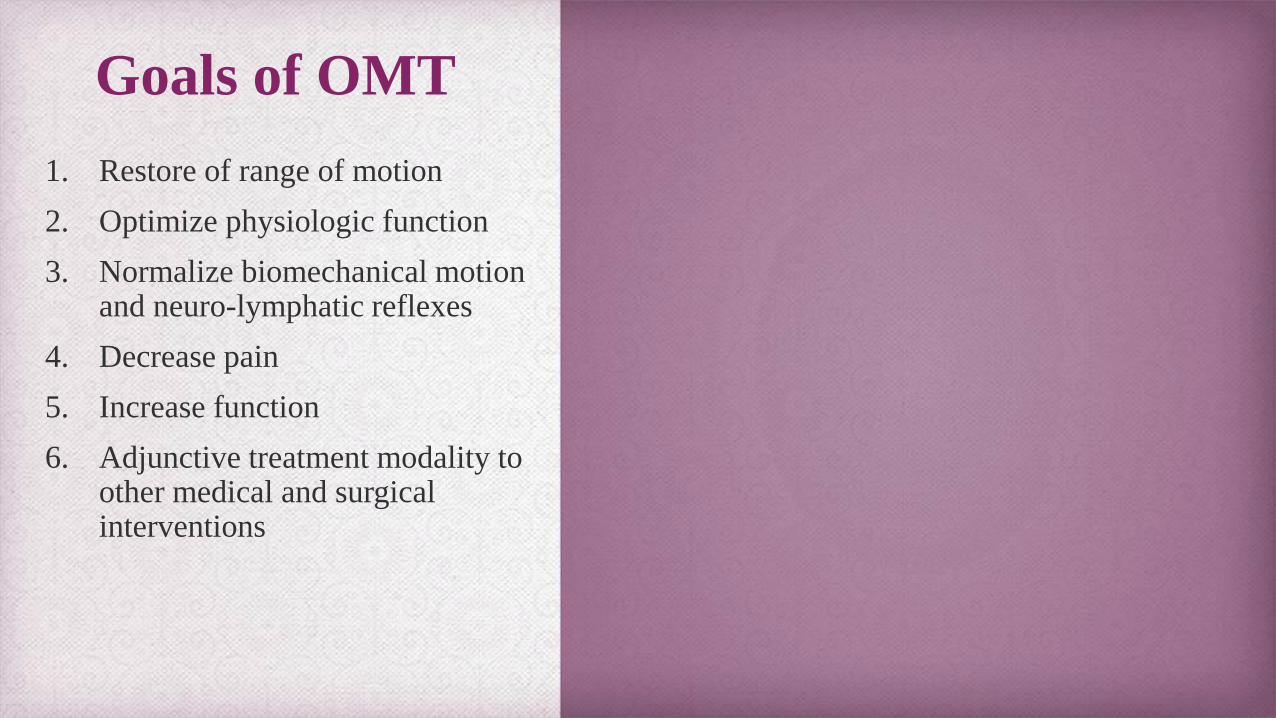
Goals of OMT
1. Restore of range of motion
2. Optimize physiologic function
3. Normalize biomechanical motion and neuro-lymphatic reflexes
4. Decrease pain
5. Increase function
6. Adjunctive treatment modality to other medical and surgical interventions
“An up-to-date osteopath must have a masterful knowledge of anatomy and physiology. He must have brains in osteopathic surgery, osteopathic obstetrics, and osteopathic practice.”
-A.T. Still MD, DO
According to ACOG, more than half of all women report some kind of musculoskeletal pain during pregnancy.
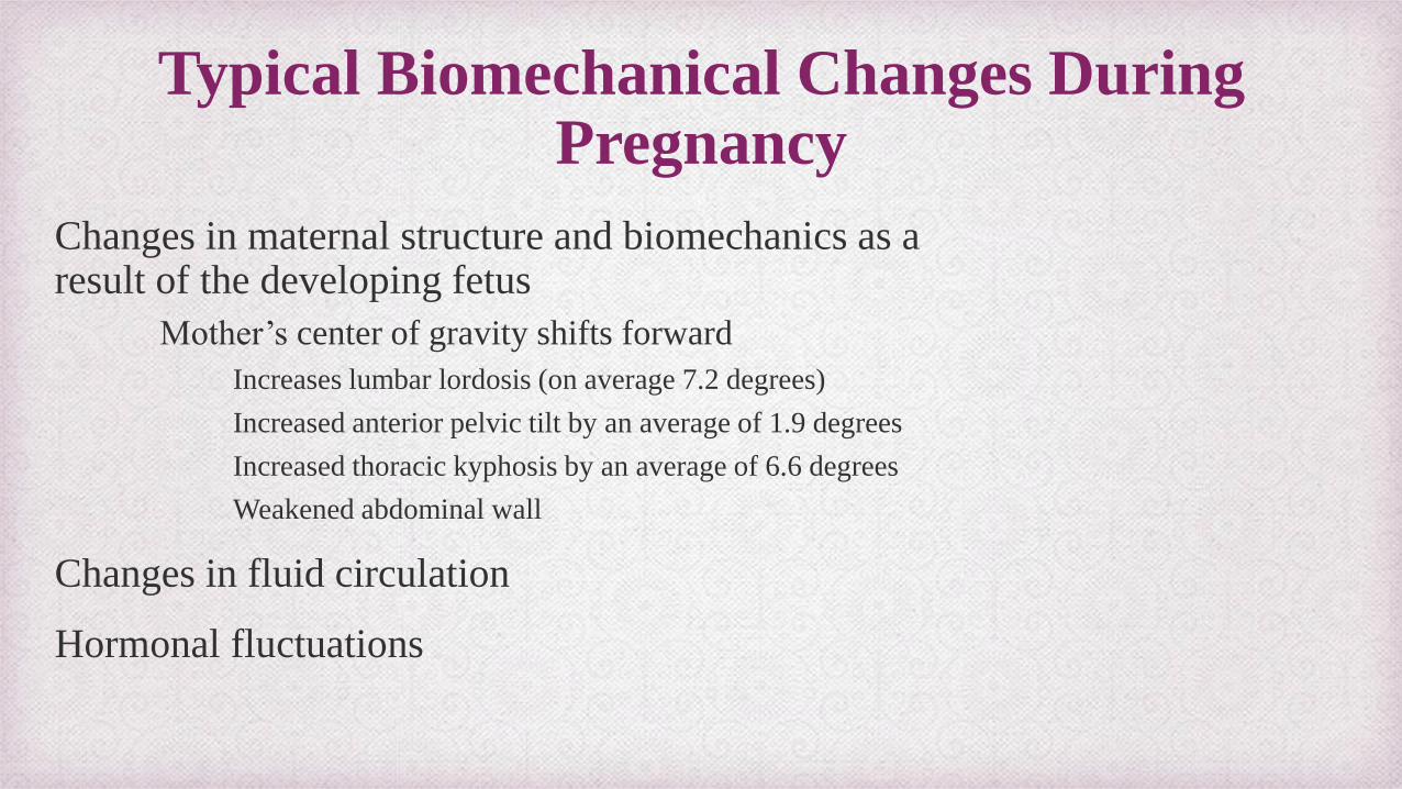
Typical Biomechanical Changes During Pregnancy
Changes in maternal structure and biomechanics as a result of the developing fetus
Mother’s center of gravity shifts forward
Increases lumbar lordosis (on average 7.2 degrees)
Increased anterior pelvic tilt by an average of 1.9 degrees
Increased thoracic kyphosis by an average of 6.6 degrees
Weakened abdominal wall
Changes in fluid circulation
Hormonal fluctuations
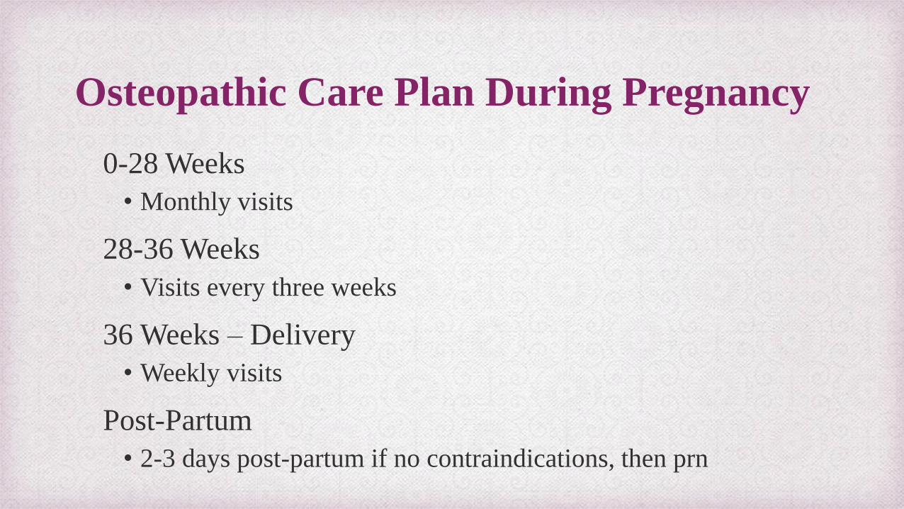
Osteopathic Care Plan During Pregnancy
0-28 Weeks
• Monthly visits
28-36 Weeks
• Visits every three weeks
36 Weeks – Delivery
• Weekly visits
Post-Partum
• 2-3 days post-partum if no contraindications, then prn
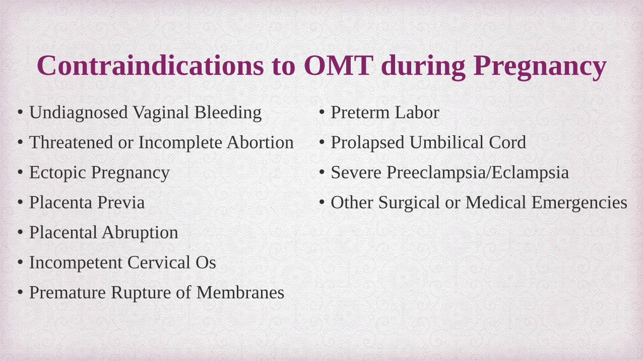
Contraindications to OMT during Pregnancy
• Undiagnosed Vaginal Bleeding
• Threatened or Incomplete Abortion
• Ectopic Pregnancy
• Placenta Previa
• Placental Abruption
• Incompetent Cervical Os
• Premature Rupture of Membranes
• Preterm Labor
• Prolapsed Umbilical Cord
• Severe Preeclampsia/Eclampsia
• Other Surgical or Medical Emergencies
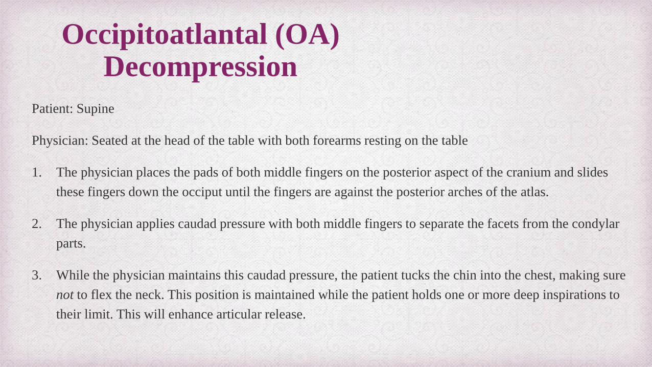
Occipitoatlantal (OA) Decompression
Patient: Supine
Physician: Seated at the head of the table with both forearms resting on the table
1. The physician places the pads of both middle fingers on the posterior aspect of the cranium and slides
these fingers down the occiput until the fingers are against the posterior arches of the atlas.
2. The physician applies caudad pressure with both middle fingers to separate the facets from the condylar
parts.
3. While the physician maintains this caudad pressure, the patient tucks the chin into the chest, making sure
not to flex the neck. This position is maintained while the patient holds one or more deep inspirations to
their limit. This will enhance articular release.
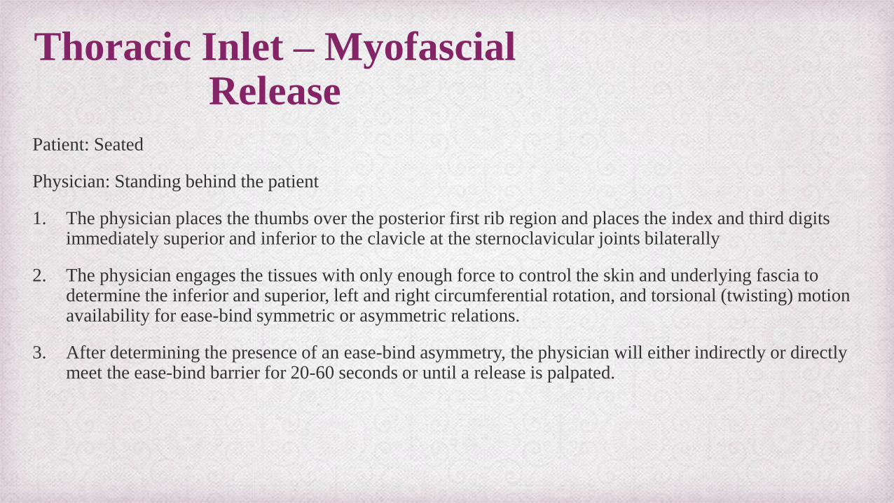
Thoracic Inlet – Myofascial Release
Patient: Seated
Physician: Standing behind the patient
1. The physician places the thumbs over the posterior first rib region and places the index and third digits immediately superior and inferior to the clavicle at the sternoclavicular joints bilaterally
2. The physician engages the tissues with only enough force to control the skin and underlying fascia to determine the inferior and superior, left and right circumferential rotation, and torsional (twisting) motion availability for ease-bind symmetric or asymmetric relations.
3. After determining the presence of an ease-bind asymmetry, the physician will either indirectly or directly meet the ease-bind barrier for 20-60 seconds or until a release is palpated.
Seated Thoracic Articulation
Patient: Seated facing the position
Physician: Standing in front of the patient with one foot in front of the other
1. Patient crosses their arms and lays their head against them. The physician then reaches around the patient and places their finger pads on the costovertebral joints.
2. The physician then gently rocks backward pulling the patient forward into extension.
3. This movement is continued in a gentle, rocking fashion down the patient’s thoracic cage.
Lateral Recumbent Lumbar Soft Tissue
Patient: Lateral recumbent with the restricted side up
Physician: Standing at the side of the patient
1. The patient's knees and hips are flexed, and the physician's thigh is placed against the patient's infrapatellar region, The physician reaches over the patient's back and places the pads of the fingers on the medial aspect of the patient's lumbar paravertebral muscles.
2. To engage the soft tissues, the physician exerts a gentle force ventrally and laterally to create a perpendicular stretch of the lumbar paravertebral musculature.
3. While the physician's thigh against the patient's knees may simply be used for bracing, it may also be flexed to provide a combined bowstring and longitudinal traction force on the paravertebral musculature.
Pelvic Diaphragm Myofascial Release
Patient: Supine
Physician: Seated next to the patient
1. The physician’s hand is placed palm-up under the sacrum, the other forearm and hand over the anterior superior iliac spines of the patient's pelvis.
2. The physician leans down on the elbow of the arm that is contacting the sacrum, keeping the sacral hand relaxed and with the forearm monitors for ease-bind asymmetry in left and right rotation and left and right torsion.
3. After determining the presence of an ease-bind asymmetry, the physician will either indirectly or directly meet the ease-bind barrier with the other hand.
4. This force is held for 20 to 60 seconds or until a release is palpated.
Pubic Symphysis “Shotgun Technique”
Patient: Lying supine with the hips and knees flexed, knees ~18” apart
Physician: Standing at the side of the table
1. The knee closer to the physician is placed against the physician's abdomen, and the physician grasps the lateral
aspect of the other knee with both hands.
2. The physician instructs the patient to pull both knees laterally (abduction) against the physician's abdomen and hands
while the physician applies an equal counterforce.
3. This isometric contraction is maintained for 3 to 5 seconds, and then the patient is instructed to stop and relax.
4. The physician instructs the patient to pull both knees together (adduction) against the physician's hands while the
physician applies an equal counterforce.
5. This isometric contraction is maintained for 3 to 5 seconds, and then the patient is instructed to stop and relax.
6. Steps 2 - 5 are repeated three to seven times.
Research
Osteopathic Manipulative Treatment in Pregnant Women, Lavelle, John, The Journal of the American Osteopathic Association, June 2012, Volume 112, P 343-346.
Pregnancy Research on Osteopathic Manipulation Optimizing Treatment Effects: the PROMOTE Study, Hensel, Kendi L. et al., American Journal of Obstetrics & Gynecology, Volume 212, Issue 1, 108.e1 - 108.e9
References
1. Chila, A. G. (2011). Foundations of Osteopathic Medicine, 3rd edition. Philadelphia: Wolters Kluwer Health/Lippincott Williams & Wilkins.
2. Nicholas A, Nicholas E. Atlas Of Osteopathic Techniques. Philadelphia: Wolters Kluwer Health/Lippincott Williams & Wilkins; 2008
3. Other osteopathic physicians and teachers – Dr. Walter Ehrenfeuchter, DO, FAAO
4. Images from google image search