Embed Size (px)
Citation preview
IET - Used Lab Equipment - Refurbished Analytical Laboratory Instruments
www.IetLtd.com Proudly serving laboratories worldwide since 1979
CALL 001.847.913.0777 for Refurbished & Certified Lab Equipment
Olympus BX51 Microscope & DP70 Digital Camera System The BX51 was voted Microscope Winner by Scientific Computing & Instrumentation
magazine as the 2002 Readers' Choice Award. July 2002.
Olympus BX51 Research Microscope
The BX51 offers increased rigidity coupled with easy-to-operate front controls on a
compact space-efficient frame. The new research microscope is a union of the expanded
range of Olympus infinity corrected optical systems (UIS2 - universal infinity system)
and advanced fluorescence or differential interference contrast DIC (Nomarski)
technology. Based around an enhanced Y-shaped frame, an extended range of accessories
and objectives can be used. The rigid design accepts a variety of heavy attachments
required for today's discerning microscopists and their various techniques and
accessories.
• Bright 12V/100W halogen illumination source is ideal for transmitted light
viewing
• The frame has two built-in neutral density filters (ND6, ND25), one daylight
balancing filter, and one empty slot for an optional filter-fluorescence illuminator
optional
• A light intensity, preset switch ensures reproducible lighting for
photomicrography
• Ergonomic Y-shaped frame for maximum stability and comfort, with low position
focus control knobs located on both the left and right sides of the frame
• Modular design and infinity optics system allows easy attachment of accessories
without any compromise in image
• Large 22mm field of view reduces scanning time (a superwide trinocular head and
eyepiece combination with a 26.5mm field of view is available as an option)
• UIS2 optics deliver bright, sharp, high-contrast images
• The 8-position universal condenser, with either a dry or oil top lens, enables you
to perform brightfield, darkfield, Phase Contrast, DIC, polarization,
andfluorescence with all objectives from 1.25x to 100x
IET - Used Lab Equipment - Refurbished Analytical Laboratory Instruments
Flexible System Format
• Modular design allows easy attachment of accessories without any compromise in
image
• Accepts a variety of video attachments and film cameras, plus the BX-URA2 or
BX-RFA fluorescence attachment
• 100W Halogen for transmitted or reflected light, 100W HBO* (Mercury) and
75W XBO (Xenon) for reflected light and fluorescence.
• Ceramic-coated stage with left-hand or right-hand low drive control, rotating
mechanism and torque adjustment mechanism, optional rubber grips (non-stick
grooved coaxial, plain, rotatable stages and others are also available)
Modularity & Versatility Meet Growing Research Needs
The modular design of the BX51 allows researchers to fully expand the design as their
needs grow. This ensures a flexible and modular configuration even for special high-end
research applications. New enhancements are a 6-position cube turret, 7-position
nosepiece and 8-position universal condenser-not to mention REXBA capability, all-new
mercury* and xenon lamp houses, and a total of three series of DIC prisms.
All components - from the stage, to the eyepieces, to the lightsource - can be customized
and mounted onto the BX51, making it one of Olympus' most versatile microscopes. The
standard UIS2 optics system means there's no compromise when adding intermediate
attachments within the viewing path. For added versatility and flexibility, brightfield,
darkfield, fluorescence, and Nomarski DIC observation all can be performed using a
single set of objectives (U Plan Apochromat or U Plan Fluorite).
Ergonomic Comfort
The six-cube turret rotates in either direction for user convenience and filter set
identification markers are clearly located directly in front of the user. The fluorescence
shutter, illumination controls, and filter slots are all conveniently located within easy
reach.
Advanced Fluorescence Technology Doubles Brightness
Fluorescence applications can be done by attaching a fluorescence illuminator that results
in greater mechanical and thermal stability. The accompanying fluorescence illuminator,
BX-URA2, incorporates a six-cube filter turret including front shutter control. The
newly-developed aspherical lens within the lamp houses allows fluorescence illumination
of up to double the brightness of existing models. To restrict the illumination to the need
of any camera type, the BX2-RFA illuminator allows the substitution of the conventional
circular field stop by a rectangular field diaphragm. As an Olympus accessory, two
excitation balancers can be attached to the BX2-RFA illuminator. Thereby, improved
IET - Used Lab Equipment - Refurbished Analytical Laboratory Instruments
differentiation of multi-labeled fluorescence specimens and better visibility of details is
achieved.
Optimized DIC Capability Increases Resolution or Contrast
New DIC (Nomarski) units provide combinations for all specimens and magnifications
with increased contrast or resolution. There are three series of DIC prisms to generate
three levels of contrast/resolution. In addition to the current set of standard prisms, a
series of high contrast prisms and high resolution prisms have been developed to produce
high-quality images for your most demanding requirements. Within the new universal
condenser, up to 8 optical components can be easily selected.
* Lamp contains mercury. Dispose according to local, state or Federal laws.
Click herefor important information.
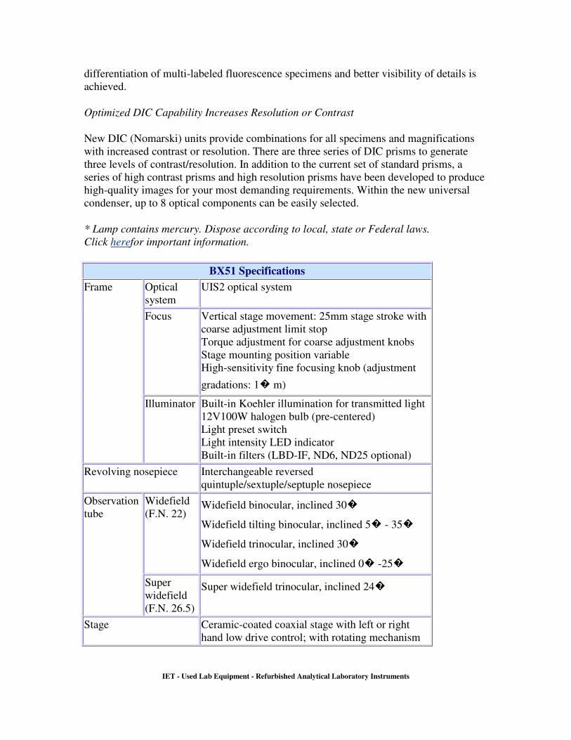
BX51 Specifications
Optical
system
UIS2 optical system
Focus Vertical stage movement: 25mm stage stroke with
coarse adjustment limit stop
Torque adjustment for coarse adjustment knobs
Stage mounting position variable
High-sensitivity fine focusing knob (adjustment
gradations: 1� m)
Frame
Illuminator Built-in Koehler illumination for transmitted light
12V100W halogen bulb (pre-centered)
Light preset switch
Light intensity LED indicator
Built-in filters (LBD-IF, ND6, ND25 optional)
Revolving nosepiece Interchangeable reversed
quintuple/sextuple/septuple nosepiece
Widefield
(F.N. 22) Widefield binocular, inclined 30�
Widefield tilting binocular, inclined 5� - 35�
Widefield trinocular, inclined 30�
Widefield ergo binocular, inclined 0� -25�
Observation
tube
Super
widefield
(F.N. 26.5)
Super widefield trinocular, inclined 24�
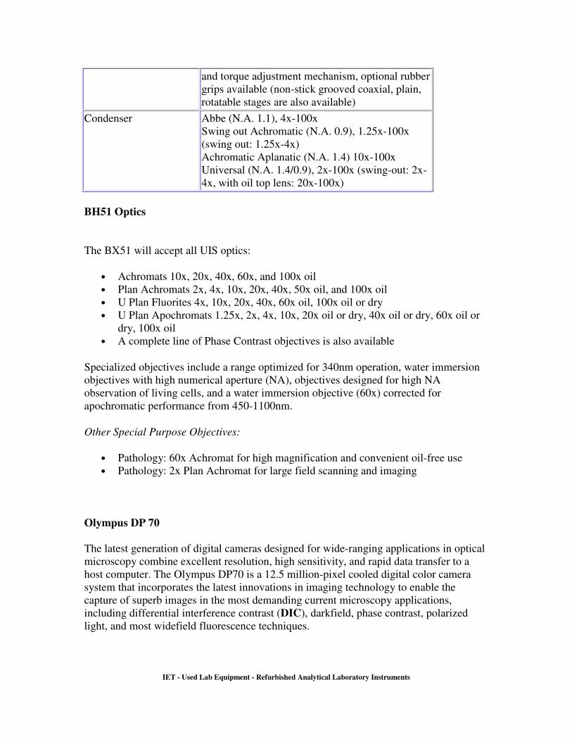
Stage Ceramic-coated coaxial stage with left or right
hand low drive control; with rotating mechanism
IET - Used Lab Equipment - Refurbished Analytical Laboratory Instruments
and torque adjustment mechanism, optional rubber
grips available (non-stick grooved coaxial, plain,
rotatable stages are also available)
Condenser Abbe (N.A. 1.1), 4x-100x
Swing out Achromatic (N.A. 0.9), 1.25x-100x
(swing out: 1.25x-4x)
Achromatic Aplanatic (N.A. 1.4) 10x-100x
Universal (N.A. 1.4/0.9), 2x-100x (swing-out: 2x-
4x, with oil top lens: 20x-100x)
BH51 Optics
The BX51 will accept all UIS optics:
• Achromats 10x, 20x, 40x, 60x, and 100x oil
• Plan Achromats 2x, 4x, 10x, 20x, 40x, 50x oil, and 100x oil
• U Plan Fluorites 4x, 10x, 20x, 40x, 60x oil, 100x oil or dry
• U Plan Apochromats 1.25x, 2x, 4x, 10x, 20x oil or dry, 40x oil or dry, 60x oil or
dry, 100x oil
• A complete line of Phase Contrast objectives is also available
Specialized objectives include a range optimized for 340nm operation, water immersion
objectives with high numerical aperture (NA), objectives designed for high NA
observation of living cells, and a water immersion objective (60x) corrected for
apochromatic performance from 450-1100nm.
Other Special Purpose Objectives:
• Pathology: 60x Achromat for high magnification and convenient oil-free use
• Pathology: 2x Plan Achromat for large field scanning and imaging
Olympus DP 70
The latest generation of digital cameras designed for wide-ranging applications in optical
microscopy combine excellent resolution, high sensitivity, and rapid data transfer to a
host computer. The Olympus DP70 is a 12.5 million-pixel cooled digital color camera
system that incorporates the latest innovations in imaging technology to enable the
capture of superb images in the most demanding current microscopy applications,
including differential interference contrast (DIC), darkfield, phase contrast, polarized
light, and most widefield fluorescence techniques.
IET - Used Lab Equipment - Refurbished Analytical Laboratory Instruments
The latest generation of digital cameras designed for wide-ranging applications in optical
microscopy combine excellent resolution, high sensitivity, and rapid data transfer to a
host computer. The Olympus DP70 is a 12.5 million-pixel cooled digital color camera
system that incorporates the latest innovations in imaging technology to enable the
capture of superb images in the most demanding current microscopy applications,
including differential interference contrast (DIC), darkfield, phase contrast, polarized
light, and most widefield fluorescence techniques.
The control software for the DP70 camera system was developed to provide a multi-
function
photomicrography interface (incorporating several features from traditional film camera
exposure
monitors) that facilitates control of real-time image acquisition, as well as subsequent
image management. In addition to the image acquisition user interface displayed in the
tutorial window above, the DP70 software package includes an image management
program through which stored images, displayed as thumbnails, may be easily viewed
and manipulated by drag and drop procedures. A six-channel image merging function is
provided that enables individual fluorescence channels and differential interference
contrast optical sections, for example, to be merged into a single multi-color image. This
tutorial demonstrates the functionality of the DP70 digital camera image capture
interface. Many, but not all, of the operational features of the actual camera control
software are enabled in the tutorial, and may be explored by the viewer. Upon
initialization, the image window displays a specimen image randomly chosen from a
group of digital images captured utilizing a variety of optical microscopy techniques.
A different specimen can be manually chosen by selecting it from the Choose A
Specimen pull-down menu located beneath the camera control window. To the left of
this menu is a Focus Adjustment slider that allows optimum focus of the image to be
obtained by dragging the slider with the computer mouse. Each specimen name in the
pull-down menu includes, in parentheses, an abbreviation designating the contrast
mechanism employed in obtaining the image. The following nomenclature is used: (FL),
fluorescence; (BF), brightfield; (DF), darkfield; (PC), phase contrast; (DIC), differential
interference contrast (Nomarski); and (POL), polarized light.
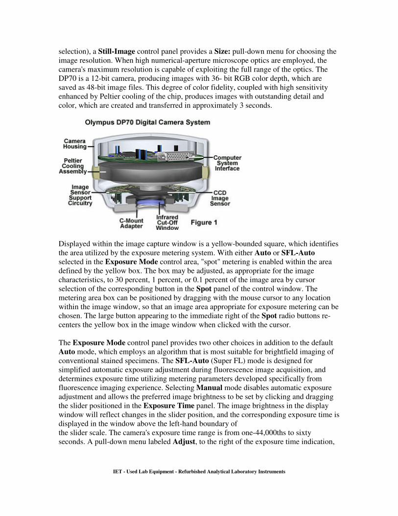
The DP70 camera (illustrated in Figure 1) employs a single-chip charge-coupled device
(CCD) sensor with Bayer RGB primary color filtration. The two-thirds-inch CCD chip
incorporates 1.45 million effective pixels, which can be piezo-shifted during image
acquisition to obtain a maximum effective resolution of 12.5 million pixels. Images may
be acquired at the ultra-high resolution of 4080 x 3072 pixels or at any of three lower-
resolution settings, as dictated by the optical system capabilities or the intended image
use.
With the Capture tab selected from the group appearing below the image window (the
default tab
IET - Used Lab Equipment - Refurbished Analytical Laboratory Instruments
selection), a Still-Image control panel provides a Size: pull-down menu for choosing the
image resolution. When high numerical-aperture microscope optics are employed, the
camera's maximum resolution is capable of exploiting the full range of the optics. The
DP70 is a 12-bit camera, producing images with 36- bit RGB color depth, which are
saved as 48-bit image files. This degree of color fidelity, coupled with high sensitivity
enhanced by Peltier cooling of the chip, produces images with outstanding detail and
color, which are created and transferred in approximately 3 seconds.
Displayed within the image capture window is a yellow-bounded square, which identifies
the area utilized by the exposure metering system. With either Auto or SFL-Auto
selected in the Exposure Mode control area, "spot" metering is enabled within the area
defined by the yellow box. The box may be adjusted, as appropriate for the image
characteristics, to 30 percent, 1 percent, or 0.1 percent of the image area by cursor
selection of the corresponding button in the Spot panel of the control window. The
metering area box can be positioned by dragging with the mouse cursor to any location
within the image window, so that an image area appropriate for exposure metering can be
chosen. The large button appearing to the immediate right of the Spot radio buttons re-
centers the yellow box in the image window when clicked with the cursor.
The Exposure Mode control panel provides two other choices in addition to the default
Auto mode, which employs an algorithm that is most suitable for brightfield imaging of
conventional stained specimens. The SFL-Auto (Super FL) mode is designed for
simplified automatic exposure adjustment during fluorescence image acquisition, and
determines exposure time utilizing metering parameters developed specifically from
fluorescence imaging experience. Selecting Manual mode disables automatic exposure
adjustment and allows the preferred image brightness to be set by clicking and dragging
the slider positioned in the Exposure Time panel. The image brightness in the display
window will reflect changes in the slider position, and the corresponding exposure time is
displayed in the window above the left-hand boundary of
the slider scale. The camera's exposure time range is from one-44,000ths to sixty
seconds. A pull-down menu labeled Adjust, to the right of the exposure time indication,
IET - Used Lab Equipment - Refurbished Analytical Laboratory Instruments
is enabled in either of the auto exposure modes, and allows the metered exposure value to
be compensated, or offset, by a fixed amount ranging from 2 exposure value (EV) units
less (-2) to 2 EV units more (+2) exposure. The compensation chosen is reflected in the
exposure time displayed.
The high-sensitivity CCD was developed specifically for the DP70 camera system, and
with the benefit of Peltier cooling, achieves low-noise performance at sensitivity settings
as high as the equivalent of International Standards Organization designation of ISO
1600. Settings ranging from ISO 200 to ISO 1600 are selected in the tutorial by clicking
the corresponding radio button in the Sensitivity area of the control panel. High
sensitivity is particularly beneficial in that it allows higher frame rates during image
preview, facilitating focusing and framing prior to image capture, even when very faint
fluorescence signals are being collected. The effective chip sensitivity can be further
extended by utilizing pixel binning during image preview. This function can be explored
by selecting the Preview panel tab beneath the image window. The DP70 provides both
2x2 and 4x4 binning options, selectable by clicking the appropriate radio button in the
Binning control panel. When pixels are combined in this fashion, the camera allows live
lower-resolution images (680 x 512 pixels) to be displayed at a rapid frame rate of up to
15 frames per second (depending upon the computer configuration) for preview purposes
of focusing and framing. A frame-averaging feature can be selected by clicking the 2
frame radio button in the Real-time frame
average panel to the right of the binning controls.
Several additional settings panel tabs located beneath the image display window provide
further control possibilities. Selection of the Color balance tab provides several
mechanisms for setting white balance or black balance. Within the White Balance
control panel, two buttons are provided that enable automatic white balancing of the
image, a function primarily employed in brightfield observation to provide accurate color
rendition regardless of illumination variations. Selecting the button labeled One Push
adjusts the image sensor response so that the entire image area is white. In practice, this
setting would be performed with the slide removed from the light path in order to set a
white background. Upon execution, the Manual radio button is enabled and the sliders
for the Red, Green, and Blue color channels can be utilized to make any necessary
manual adjustments to the color balance. For many specimens, better white balance
adjustment may be achieved by manually selecting a white or neutral gray area for
reference within the specimen image area, and the One Touch button can be used to
enable pixel selection by mouse cursor. When the button is selected by mouse, an
eyedropper icon (a white balance setting pointer) replaces the mouse cursor when it is
positioned within the image area. Clicking the icon on a suitable reference area for white
balance adjustment will result in the appropriate color adjustments being conducted, and
the manual adjustment sliders will be enabled for further fine-tuning of the color balance.
In the actual camera software, the cursor may be used to designate a rectangular marquis
area by clicking and dragging from the upper left corner to the lower right corner of the
region to be rendered as white. After either method of automatic white balancing has
been employed, the adjustment made can be cancelled, returning to the
IET - Used Lab Equipment - Refurbished Analytical Laboratory Instruments
original image, by selecting the Off radio button in the white balance control area.
A Black Balance control panel has functional features similar to the white balance
controls, except a single level slider is provided to adjust the image brightness (offset),
instead of the individual color channel sliders. Black balance adjustment is used primarily
in fluorescence observation, in which the background area is dark, and white balance
adjustment would not be appropriate. The One Touch button is utilized in the same
manner as for white balance adjustment, although the eyedropper tool is used to select a
black or dark area in the image for black balance reference. One Push setting is carried
out on the black background with no specimen in place, and adjusts the brightness so that
the entire image area appears black. In either white or black balance adjustment, the
automatic-setting step can be skipped when fully manual control is desired, simply by
selecting the appropriate Manual radio button as the initial step, followed by
manipulation of the corresponding slider(s).
The tab labeled Level adjust may be selected to display histograms for each of the red,
green, and blue color channels, allowing precise adjustment of the image sensor's
response characteristics. This function allows the contrast and gamma of the preview
image to be altered in order to improve both live visualization of the specimen and the
quality of the captured image. Images captured with the DP70 camera will reflect
changes made to the preview image through the level adjustment function. Input and
output intensity values can be altered for the combined RGB signal, or for the individual
color channels, by selection from the Channel pull-down menu. The upper slider
appearing beneath the histogram window enables adjustment of input intensities, and the
lower slider serves the same function for output levels. Modifications are made by
dragging and dropping the set-point triangles along the sliders. Numerical values are
displayed in the Input and Output windows as the setting marks are repositioned, or
alternatively, the desired numerical values may be entered directly through the keyboard.
The left-hand triangle on each slider corresponds to the shadow intensity level, while the
right-hand marker controls the highlight intensity. The center triangle on the input slider
adjusts the gray level, and functions effectively as a gamma adjustment, which should be
varied as necessary by observing the histogram and the effect on the preview image. The
level values for the image may be reset to the default condition by clicking on the Reset
button.
Selection of the Scale tab enables the user to establish up a scale bar that is superimposed
on the image as a reference for making approximate size measurements during specimen
observation. The appropriate scale bar is generated by the DP70 software based on the
objective and relay magnification values entered in the Objective Lens text box and
selected from the Adapter Lens pull-down menu. In the tutorial, a sample scale bar is
displayed, but does not change in response to values entered in the numerical fields, nor
does the displayed objective magnification change in response to clicking the objective
selection buttons. The Display area of the panel contains a check box labeled Show Scale
that determines whether the scale is
displayed or is turned off.
IET - Used Lab Equipment - Refurbished Analytical Laboratory Instruments
A simple time-lapse function is provided in the DP70 capture software, and is accessed
by clicking the Timelapse tab beneath the image window. Although not enabled in the
tutorial, this feature of the camera user interface allows the time and interval variables to
be entered, and a series of images to be captured and converted to an AVI file for
immediate playback.
www.IetLtd.com Proudly serving laboratories worldwide since 1979
CALL 001.847.913.0777 for Refurbished & Certified Lab Equipment