Embed Size (px)
Citation preview
Olympic Ring Formation from Newly Prepared Barium Hexaferrite NanoparticleSuspension
Kurikka V. P. M. Shafi, † Israel Felner,‡ Y. Mastai,§ and Aharon Gedanken*,†
Department of Chemistry, Bar-Ilan UniVersity, Ramat-Gan 52900, Israel, Racah Institute of Physics,Hebrew UniVersity of Jerusalem, Jerusalem, Israel, and Department of Materials and Interface,Weizmann Institute of Science, RehoVot, 76100, Israel
ReceiVed: December 9, 1998; In Final Form: February 23, 1999
We report here, the first observation of features such as theOlympic Ringson transmission electron micrographsof amorphous BaFe12O19 nanoparticles prepared by a sonochemical decomposition technique. The bariumhexaferrite was formed as a colloidal solution without the use of a surfactant. Rings of smaller dimensionstrapped inside the larger ones was another unique observation. The intersection of two rings is amazing, asthis is in direct contradiction to the proposed mechanism for the ring formation, based on the dry hole formationon an evaporating thin film completely wetted to the substrate. The creation of this unique feature is attributedto the interplay of magnetic forces with the regular particle-substrate interactions.
Introduction
Hexagonal magnetic hard ferrites such as BaFe12O19 are ofgreat scientific and technological importance. Owing to theirlarge crystalline anisotropy and high intrinsic coercivity, theyare used in the production of permanent magnets and in thefabrication of certain microwave devices. Fine particles ofBaFe12O19 are currently used in high-density perpendicularmagnetic recording media. A wide range of methods has beenused to prepare ultrafine ferrite particles, including chemicalcoprecipitation,1 combustion,2 pyrosol,3 and sol-gel4 methods,as well as microemulsion5 and glass crystallization6 techniques.Apart from the citrate precursor (sol-gel) method, in all thesemethods, formation of single-phase BaFe12O19 takes place at900 °C or above, and the particles obtained are of relativelylarger size,>50 nm. Control over the size of the particles andtheir uniformity, purity, and good crystallinity is the majordifficulty in these synthetic methods. We have utilized ultrasonicirradiation to achieve, at the atomic level, mixing of theconstituent ions in the required stoichiometric ratio. In this paperwe present a new method for the preparation of BaFe12O19. Theas-prepared material was formed in the amorphous state. Wealso report on the formation of nanorings and intersectingnanorings when the solution of the amorphous as-preparedmaterial was left to dry on a Formavar grid and examined bytransmission electron micrography.
Experimental and Results
(a) Synthesis of Material. A few examples have beenrecently presented for the use of ultrasonic radiation in theformation of colloidal solutions.10-12 In these cases a surfactantsuch as oleic acid was introduced to assist in the formation ofthe colloidal solution. In the current procedure no surfactantwas required to form the colloidal solution.
A solution of Fe(CO)5 and barium ethylhexanoate (Ba-[OOCCH(C2H5)C4H9]2) in decane, in stoichiometric ratio, was
decomposed by high-intensity ultrasonication, employing adirect immersion titanium horn (Vibracell, 20 kHz, 100 W/cm2)in the solution beaker to obtain the product. The as-preparedmaterial was an amorphous BaFe12O19 precursor in colloidalsuspension, where the particles are in the nanometer size regimeand are homogeneously distributed. The precursor is extractedfrom the solution as powder by precipitation or by evaporationand then calcined at low temperature (600°C) to obtain thefinal BaFe12O19 crystalline nanosized powders. The EDX profileof the amorphous as-prepared precursor powder showed thatthe Ba and Fe are in stoichiometric ratio, as in BaFe12O19. TheXRD pattern of the final calcined, nanosized powder confirmedthe presence of single-phase BaFe12O19, with no iron oxideimpurities present.
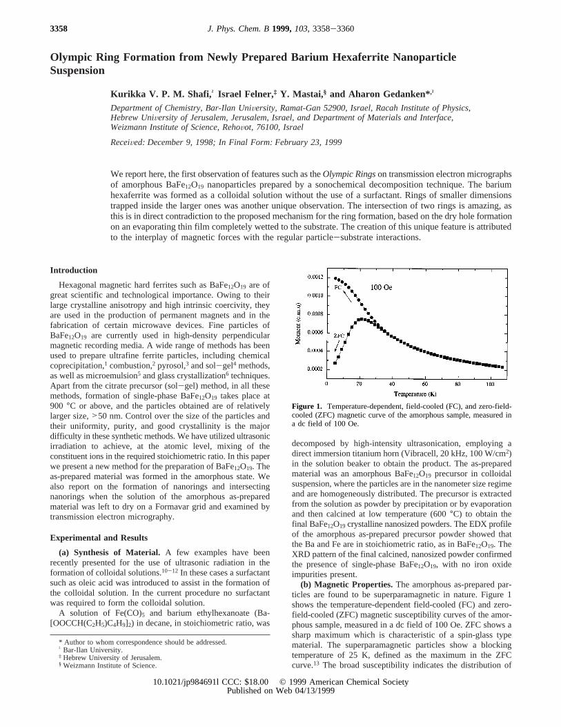
(b) Magnetic Properties. The amorphous as-prepared par-ticles are found to be superparamagnetic in nature. Figure 1shows the temperature-dependent field-cooled (FC) and zero-field-cooled (ZFC) magnetic susceptibility curves of the amor-phous sample, measured in a dc field of 100 Oe. ZFC shows asharp maximum which is characteristic of a spin-glass typematerial. The superparamagnetic particles show a blockingtemperature of 25 K, defined as the maximum in the ZFCcurve.13 The broad susceptibility indicates the distribution of
* Author to whom correspondence should be addressed.† Bar-Ilan University.‡ Hebrew University of Jerusalem.§ Weizmann Institute of Science.
Figure 1. Temperature-dependent, field-cooled (FC), and zero-field-cooled (ZFC) magnetic curve of the amorphous sample, measured ina dc field of 100 Oe.
3358 J. Phys. Chem. B1999,103,3358-3360
10.1021/jp984691l CCC: $18.00 © 1999 American Chemical SocietyPublished on Web 04/13/1999

the particle size. Irreversibility occurs below 35 K. The blockingtemperature and the measurement time constants are consistentwith the magnetic particle diameter of 5 nm.13
(c) Ring Formation. Ring formation of macroscopic particles(e.g., stains from coffee, tomato bits, etc.) is common. However,the formation of macroscopic rings from mesoscopic particles(e.g., polyballs) is not common, and is being studied bothexperimentally and theoretically.7,8 Ohara et al.9 discuss the dryhole formation on the wetting layers of very thin films, formedby the evaporation of a drop of solution of silver nanoparticlein hexane on an amorphous carbon substrate. They observedan array, consisting of a ring (0.9µm diameter) of close packedAg nanoparticles of 2.5 nm size, formed upon the pinning tothe substrate of the perimeter (the contact line between fluidand substrate) of a growing hole that has nucleated in asufficiently thin film of dilute solutions of particles. The con-centration of the particles inside the ring is smaller than theconcentration of particles outside. This was consistent with theirbelief that the rings are formed due to holes opening up in theliquid film on drying, and pushing the particles into the rims.They correlate the formation of rings with the presence ofmonodisperse larger particles, whereas the greater number ofsmaller particles with polydispersity will give compact size-aggregated domains. Ring formation is explained by the size-dependent interaction between the particles and the substrate.
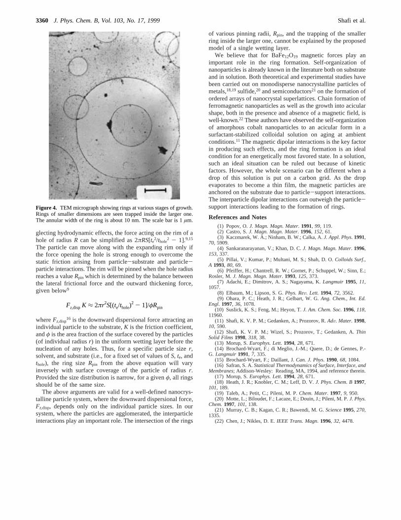
Although our studies are technically similar to those of Oharaet al.,9 the results are different. One drop of a suspension ofamorphous BaFe12O19 precursor in decane was placed on acarbon-coated Formwar copper grid (diam) 3 mm) and allowedto evaporate. The evaporation took place at ambient temperature(24 °C) and at a 30% humidity. A transmission electronmicrograph (Figure 2) revealed an array of rings of diameterabout 600 nm that is much smaller than the ring reported byOhara et al. and other 3D rings reported elsewhere.7 The surfacecoverage of these rings was more than 50% and the annularwidth is about 25 nm. The selected area electron diffraction(SAED) pattern of the annular region shows the particles areindeed amorphous, aggregated, and without any ordered struc-tures. The rings were formed successively on drying the films.On the first day, only the pure rings were observed (Figure 2),while the trapped rings and the intersecting rings (Figure 3)appeared on the next day. A closer look at the TEM pictureshows that the particles inside and outside the rings are inuniform concentration which is very different from what wasobserved by Ohara et al. Moreover, the TEM picture of anotherspecimen (Figure 3) shows interesting features. Here, one cansee the intersecting rings forming the so-calledOlympic Rings.Rings of various sizes at various stages of growth and withdifferent annular width (10 nm) can be seen on anothermicrograph, Figure 4. Also rings of smaller dimensions are seentrapped inside the larger ones. When the grid planes werephotographed at different inclinations, the same TEM imageswere observed. We could not distinguish between pictures takenat different angles between the grid and the electron beam. Thisindicates that the intersection region is aligned in the same plane(within the 25 nm of the annular width). It is amazing to seethe intersection of two rings, as this is in direct contradictionto the proposed mechanism for the ring formation, based onthe dry hole formation on an evaporating thin film completelywetted to the substrate. Hole nucleation occurs innonVolatilewetting fluids, where the film becomes unstable at a thicknesst below a critical valuethole.14 The hole opens up to restore thefilm to its equilibrium thicknesste which is given by (3AH/S)1/2,whereAH ) Asl - All > 0 is the Hamaker constant.10 S, the
positive spreading coefficient, is defined asS ) γsv - (γsl +γ), whereγ is the liquid-vapor interfacial tension andγsv (γsl)is solid-vapor (solid-liquid) interfacial surface tension. Ne-
Figure 2. TEM micrograph showing the self-organization of super-paramagnetic nanoparticles into submicron size (600 nm) rings. Therings are of near uniform size with an annular width of about 25 nm.The scale bar shown is 500 nm.
Figure 3. TEM micrograph showing rings of various sizes. The ringsseem to intersect each other forming the so-calledOlympic Rings. Thescale bar is 0.7µm.
Olympic Ring Formation J. Phys. Chem. B, Vol. 103, No. 17, 19993359
glecting hydrodynamic effects, the force acting on the rim of ahole of radiusR can be simplified as 2πRS[te2/thole
2 - 1].9,15
The particle can move along with the expanding rim only ifthe force opening the hole is strong enough to overcome thestatic friction arising from particle-substrate and particle-particle interactions. The rim will be pinned when the hole radiusreaches a valueRpin which is determined by the balance betweenthe lateral frictional force and the outward thickening force,given below9
whereFz,disp16 is the downward dispersional force attracting an
individual particle to the substrate,K is the friction coefficient,andφ is the area fraction of the surface covered by the particles(of individual radiusr) in the uniform wetting layer before thenucleation of any holes. Thus, for a specific particle sizer,solvent, and substrate (i.e., for a fixed set of values ofS, te, andthole), the ring sizeRpin from the above equation will varyinversely with surface coverage of the particle of radiusr.Provided the size distribution is narrow, for a givenφ, all ringsshould be of the same size.
The above arguments are valid for a well-defined nanocrys-talline particle system, where the downward dispersional force,Fz,disp, depends only on the individual particle sizes. In oursystem, where the particles are agglomerated, the interparticleinteractions play an important role. The intersection of the rings
of various pinning radii,Rpin, and the trapping of the smallerring inside the larger one, cannot be explained by the proposedmodel of a single wetting layer.
We believe that for BaFe12O19 magnetic forces play animportant role in the ring formation. Self-organization ofnanoparticles is already known in the literature both on substrateand in solution. Both theoretical and experimental studies havebeen carried out on monodisperse nanocrystalline particles ofmetals,18,19sulfide,20 and semiconductors21 on the formation ofordered arrays of nanocrystal superlattices. Chain formation offerromagnetic nanoparticles as well as the growth into acicularshape, both in the presence and absence of a magnetic field, iswell-known.22 These authors have observed the self-organizationof amorphous cobalt nanoparticles to an acicular form in asurfactant-stabilized colloidal solution on aging at ambientconditions.11 The magnetic dipolar interactions is the key factorin producing such effects, and the ring formation is an idealcondition for an energetically most favored state. In a solution,such an ideal situation can be ruled out because of kineticfactors. However, the whole scenario can be different when adrop of this solution is put on a carbon grid. As the dropevaporates to become a thin film, the magnetic particles areanchored on the substrate due to particle-support interactions.The interparticle dipolar interactions can outweigh the particle-support interactions leading to the formation of rings.
References and Notes(1) Popov, O.J. Magn. Magn. Mater. 1991, 99, 119.(2) Castro, S.J. Magn. Magn. Mater. 1996, 152, 61.(3) Kaczmarek, W. A.; Ninham, B. W.; Calka, A.J. Appl. Phys. 1991,
70, 5909.(4) Sankaranarayanan, V.; Khan, D. C.J. Magn. Magn. Mater. 1996,
153, 337.(5) Pillai, V.; Kumar, P.; Multani, M. S.; Shah, D. O.Colloids Surf.,
A 1993, 80, 69.(6) Pfeiffer, H.; Chantrell, R. W.; Gornet, P.; Schuppel, W.; Sinn, E.;
Rosler, M.J. Magn. Magn. Mater. 1993, 125, 373.(7) Adachi, E.; Dimitrov, A. S.; Nagayama, K.Langmuir 1995, 11,
1057.(8) Elbaum, M.; Lipson, S. G.Phys. ReV. Lett. 1994, 72, 3562.(9) Ohara, P. C.; Heath, J. R.; Gelbart, W. G.Ang. Chem., Int. Ed.
Engl. 1997, 36, 1078.(10) Suslick, K. S.; Feng, M.; Heyon, T.J. Am. Chem. Soc.1996, 118,
11960.(11) Shafi, K. V. P. M.; Gedanken, A.; Prozorov, R.AdV. Mater.1998,
10, 590.(12) Shafi, K. V. P. M.; Wizel, S.; Prozorov, T.; Gedanken, A.Thin
Solid Films1998, 318, 38.(13) Morup, S.Europhys. Lett. 1994, 28, 671.(14) Brochard-Wyart, F.; di Meglio, J.-M.; Quere, D.; de Gennes, P.-
G. Langmuir1991, 7, 335.(15) Brochard-Wyart, F.; Daillant, J.Can. J. Phys. 1990, 68, 1084.(16) Safran, S. A.Statistical Thermodynamics of Surface, Interface, and
Membranes; Addison-Wesley: Reading, MA, 1994, and reference therein.(17) Morup, S.Europhys. Lett. 1994, 28, 671.(18) Heath, J. R.; Knobler, C. M.; Leff, D. V.J. Phys. Chem. B1997,
101, 189.(19) Taleb, A.; Petit, C.; Pileni, M. P.Chem. Mater. 1997, 9, 950.(20) Motte, L.; Biloudet, F.; Lacaze, E.; Douin, J.; Pileni, M. P.J. Phys.
Chem.1997, 101, 138.(21) Murray, C. B.; Kagan, C. R.; Bawendi, M. G.Science1995, 270,
1335.(22) Chen, J.; Nikles, D. E.IEEE Trans. Magn. 1996, 32, 4478.
Figure 4. TEM micrograph showing rings at various stages of growth.Rings of smaller dimensions are seen trapped inside the larger one.The annular width of the ring is about 10 nm. The scale bar is 1µm.
Fz,disp K ≈ 2πr2S[(te/thole)2 - 1]/φRpin
3360 J. Phys. Chem. B, Vol. 103, No. 17, 1999 Shafi et al.


![Index [ftp.feq.ufu.br]ftp.feq.ufu.br/Luis_Claudio/Segurança/Safety/Double/fire_handbook... · Backdraft Explosion 174 Barium 216 Barium Carbonate 300 Barium Chlorate 300 Barium Nitrate](https://img.pdfslide.us/doc/110x75/5ea2585052451660ed3ed304/index-ftpfequfubrftpfequfubrluisclaudioseguranasafetydoublefirehandbook.jpg)