Embed Size (px)
Citation preview
Oligoribonuclease is the primary degradative enzymefor pGpG in Pseudomonas aeruginosa that is requiredfor cyclic-di-GMP turnoverMona W. Orra,b,c, Gregory P. Donaldsona,c, Geoffrey B. Severind, Jingxin Wange, Herman O. Sintime,Christopher M. Watersf, and Vincent T. Leea,c,1
aDepartment of Cell Biology and Molecular Genetics, University of Maryland, College Park, MD 20742; bBiological Sciences Graduate Program, University ofMaryland, College Park, MD 20742; cMaryland Pathogen Research Institute, University of Maryland, College Park, MD 20742; dDepartment of Biochemistryand Molecular Biology, Michigan State University, East Lansing, MI 48824; eDepartment of Chemistry and Biochemistry, University of Maryland, CollegePark, MD 20742; and fDepartment of Microbiology and Molecular Genetics, Michigan State University, East Lansing, MI 48824
Edited by E. Peter Greenberg, University of Washington, Seattle, WA, and approved July 31, 2015 (received for review April 14, 2015)
The bacterial second messenger cyclic di-GMP (c-di-GMP) controlsbiofilm formation and other phenotypes relevant to pathogenesis.Cyclic-di-GMP is synthesized by diguanylate cyclases (DGCs).Phosphodiesterases (PDE-As) end signaling by linearizing c-di-GMPto 5ʹ-phosphoguanylyl-(3ʹ,5ʹ)-guanosine (pGpG), which is then hy-drolyzed to two GMP molecules by yet unidentified enzymestermed PDE-Bs. We show that pGpG inhibits a PDE-A from Pseu-domonas aeruginosa. In a dual DGC and PDE-A reaction, excesspGpG extends the half-life of c-di-GMP, indicating that removalof pGpG is critical for c-di-GMP homeostasis. Thus, we sought toidentify the PDE-B enzyme(s) responsible for pGpG degradation. Adifferential radial capillary action of ligand assay-based screen forpGpG binding proteins identified oligoribonuclease (Orn), an exo-ribonuclease that hydrolyzes two- to five-nucleotide-long RNAs.Purified Orn rapidly converts pGpG into GMP. To determinewhether Orn is the primary enzyme responsible for degradingpGpG, we assayed cell lysates of WT and Δorn strains of P. aeru-ginosa PA14 for pGpG stability. The lysates from Δorn showed 25-fold decrease in pGpG hydrolysis. Complementation with WT, butnot active site mutants, restored hydrolysis. Accumulation ofpGpG in the Δorn strain could inhibit PDE-As, increasing c-di-GMP concentration. In support, we observed increased transcrip-tion from the c-di-GMP–regulated pel promoter. Additionally, thec-di-GMP–governed auto-aggregation and biofilm phenotypeswere elevated in the Δorn strain in a pel-dependent manner.Finally, we directly detect elevated pGpG and c-di-GMP in the Δornstrain. Thus, we identified that Orn serves as the primary PDE-Benzyme that removes pGpG, which is necessary to complete thefinal step in the c-di-GMP degradation pathway.
cyclic di-GMP | oligoribonuclease | pGpG | PDE-B | nanoRNase
Cyclic-di-GMP (c-di-GMP) is a phylogenetically widely usedbacterial second messenger (1). Cyclic-di-GMP is synthe-
sized by diguanylate cyclase enzymes (DGC) that contain theGGDEF domain (2, 3). Once synthesized, the c-di-GMP bindsto intracellular receptors to decrease motility and increasebiofilm formation, contributing to the virulence of severalpathogens (1, 4–7). Cyclic-di-GMP signaling is terminated byphosphodiesterases (PDE-A) that linearize it into pGpG, whichis then hydrolyzed into GMP by an unknown phosphodiesterasetermed PDE-B (2). Bacteria encode two structurally unrelatedPDE-As: one containing the EAL domain (8–10) and a secondcontaining the HD-GYP domain (11). The overexpression ofGGDEF domain DGCs elevates c-di-GMP and c-di-GMP–regu-lated processes (8, 12); conversely, overexpression of the EAL andHD-GYP domain PDE-As decreases c-di-GMP and c-di-GMP–regulated processes (8, 13–15). These c-di-GMP synthesizing anddegrading domains are commonly linked to sensory and signaltransduction domains (1, 16), thereby allowing synthesis anddegradation of c-di-GMP in response to environmental changes.
In addition to regulation by extracellular signals, c-di-GMPhomeostasis is subject to feedback inhibition. Crystal structures ofthe DGCs PleD from Caulobacter crescentus and WspR fromPseudomonas aeruginosa show that c-di-GMP binds to an RxxDdomain I-site to inhibit c-di-GMP synthesis (17, 18). Additionally,the Xanthomonas campestris XCC4471 DGC lacking an RxxDmotif can also be inhibited by excess c-di-GMP through c-di-GMPbinding to and occluding the active site (19). The linearizedc-di-GMP hydrolysis product, 5ʹ-phosphoguanylyl-(3ʹ,5ʹ)-guanosine(pGpG), also plays an active role in cyclic dinucleotide turnover.Purified YfgF from E. coli, a PDE-A, is inhibited by excesspGpG through an unknown mechanism (20). Here, we showthat the EAL domain PDE-A RocR from the P. aeruginosaPA14 strain is also inhibited by excess pGpG via direct com-petition with c-di-GMP binding in the active site. Accordingly, theaddition of excess pGpG results in an increased c-di-GMP half-lifein vitro, suggesting that removal of pGpG is required for termi-nating c-di-GMP signaling.To elucidate the mechanism of c-di-GMP signal termination,
we sought to identify the PDE-B(s) responsible for cleavingpGpG. Previously, HD-GYP domain-containing proteins were
Significance
Cyclic-di-GMP (c-di-GMP) is a ubiquitous bacterial secondmessenger that regulates complex behaviors such as biofilmformation. These behaviors are changed by altering the in-tracellular concentration of c-di-GMP. Degradation of c-di-GMPoccurs by a two-step process in which one set of phosphodi-esterases (PDE-As) linearize the molecule into 5ʹ-phosphogua-nylyl-(3ʹ,5ʹ)-guanosine (pGpG), followed by hydrolysis byunidentified phosphodiesterases (PDE-Bs) into two GMPs. Highlevels of pGpG inhibit PDE-As, and thus PDE-B activity is im-portant in maintaining c-di-GMP homeostasis. However, theidentity of the PDE-B(s) remained unknown. Using a high-throughput binding screen, we identify oligoribonuclease (Orn)as a putative PDE-B. We demonstrate that Orn is the primarysource of PDE-B activity in Pseudomonas aeruginosa. Identifi-cation of Orn as the primary PDE-B completes the c-di-GMPsignaling pathway.
Author contributions: M.W.O., G.P.D., G.B.S., H.O.S., C.M.W., and V.T.L. designed re-search; M.W.O., G.P.D., G.B.S., and J.W. performed research; G.B.S. contributed newreagents/analytic tools; M.W.O., G.P.D., G.B.S., C.M.W., and V.T.L. analyzed data; andM.W.O., G.B.S., C.M.W., and V.T.L. wrote the paper.
The authors declare no conflict of interest.
This article is a PNAS Direct Submission.
Freely available online through the PNAS open access option.1To whom correspondence should be addressed. Email: [email protected].
This article contains supporting information online at www.pnas.org/lookup/suppl/doi:10.1073/pnas.1507245112/-/DCSupplemental.
E5048–E5057 | PNAS | Published online August 24, 2015 www.pnas.org/cgi/doi/10.1073/pnas.1507245112

proposed to be the PDE-Bs involved in pGpG hydrolysis becausethey bind pGpG with higher affinity than c-di-GMP (21). How-ever, HD-GYP domain proteins are missing in some genomesthat contain other c-di-GMP signaling machinery (1), suggestingthat other enzymes must be responsible for PDE-B activity. Toidentify PDE-B(s), we used a screen based on the differentialradial capillary action of ligand assay (DRaCALA) (22, 23) toprobe an ORF library of Vibrio cholerae El Tor N16961 (24) forproteins that bind pGpG. This screen identified oligoribonuclease(Orn) as a protein that binds pGpG, but not c-di-GMP. Orn is anexoribonuclease that cleaves two- to five-nucleotide-long RNAmolecules (25). We found that purified Orn from both V. choleraeand P. aeruginosa bound and cleaved pGpG. To determinewhether Orn is the primary PDE-B in bacteria, we show thatwhole cell lysates of an orn transposon mutant (orn::tn) from theP. aeruginosa PA14 Non-Redundant Transposon Insertion MutantLibrary (26) and an in-frame deletion mutant of orn (Δorn) weredecreased in pGpG cleaving activity by 25-fold compared with theparental strain. Complementation with WT orn, but not active sitepoint mutants, restored pGpG hydrolysis in cell lysates. Thus, lossof orn is expected to increase pGpG concentration in vivo and mayinhibit PDE-A activity to also elevate c-di-GMP concentrationsin the Δorn strain. In support of elevated c-di-GMP in the Δornstrain, we observed three times more activity from the c-di-GMP–responsive pel promoter FleQ (27). We also demonstrate that twoc-di-GMP–governed phenotypes, biofilm formation (28) and ag-gregation (29), are enhanced in the Δorn strain and are dependenton the c-di-GMP–regulated PEL exopolysaccharides (30). UsingLC-MS/MS, we directly detect higher levels of both pGpG andc-di-GMP in extracts of the Δorn strain compared with WT. Takentogether, these results indicate that Orn is the primary PDE-Bresponsible for degrading pGpG in P. aeruginosa.
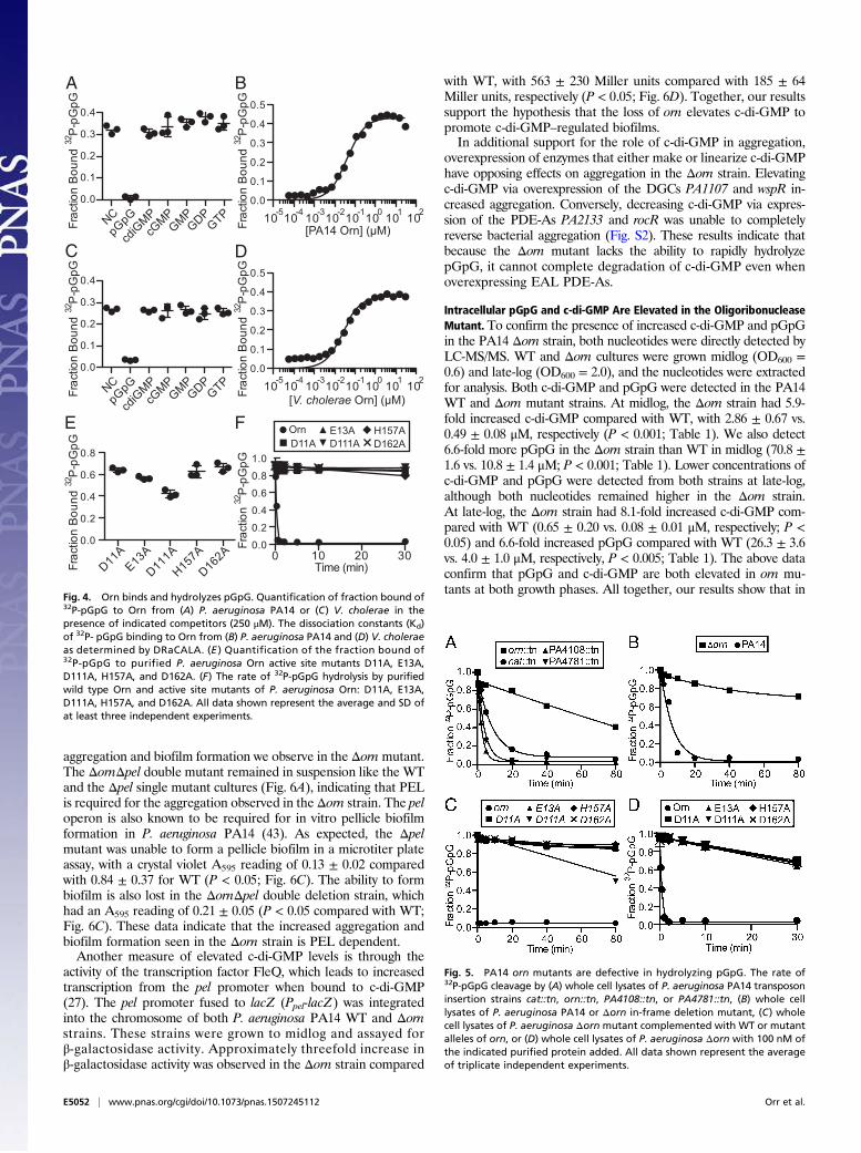
ResultspGpG Inhibits RocR Phosphodiesterase Activity by Binding to the ActiveSite. The PDE-A YfgF from E. coli is inhibited by the addition of100 μM of pGpG (20). We asked whether PDE-A inhibition bypGpG is a generalizable phenomenon. The RocR PDE-A fromP. aeruginosa PA14 was used to determine inhibition by pGpG. Inthe absence of competitor (Fig. 1A), RocR rapidly converts 89% ofthe radiolabeled c-di-GMP to pGpG within 5 min as measuredafter separation by TLC. The addition of 100-fold excess of pGpGnearly completely inhibited c-di-GMP linearization (Fig. 1A).These results demonstrate that pGpG inhibits PDE-A activity. Todetermine whether pGpG is competing with c-di-GMP for activesite binding in RocR, we performed experiments in which binding
of radiolabeled c-di-GMP to purified RocR was assessed in thepresence of different unlabeled nucleotide competitors. RocRbinds c-di-GMP with a fraction bound of 0.54 ± 0.03 in the absenceof competitor, whereas the addition of 50 μM unlabeled pGpGreduced fraction bound to 0.022 ± 0.005 (P < 0.001), indicatingthat pGpG competes for c-di-GMP binding (Fig. 1B, black bars).To see whether binding is mutually exclusive, RocR was incubatedwith radiolabeled pGpG in the presence of the same panel ofunlabeled nucleotide competitors (Fig. 1B, white bars). The addi-tion of 50 μM c-di-GMP reduced fraction bound from 0.48 ± 0.01to 0.13 ± 0.03 (P < 0.001), indicating that c-di-GMP is also able toprevent pGpG binding. No other nucleotide had a significant effecton fraction of pGpG or c-di-GMP bound to RocR (Fig. 1B). Thus,RocR binding to pGpG and c-di-GMP is specific and mutuallyexclusive. Crystal structures of another EAL domain PDE-A FimXshow that pGpG binds in the active site where c-di-GMP cleavageoccurs (31). Because the binding is occurring at the same site, therelative affinity of RocR binding to pGpG and c-di-GMP isimportant to determine whether pGpG competition of c-di-GMP binding could occur in physiological conditions. Thedissociation constant (Kd) of RocR binding to pGpG and c-di-GMP was found to be 3.6 ± 0.4 and 0.60 ± 0.07, μM respectively(Fig. 1C). The c-di-GMP concentration in WT P. aeruginosa hasbeen reported to be up to 11 μM (32). Therefore, rapid turn-over of c-di-GMP can yield pGpG concentrations that exceedthe Kd for RocR if pGpG is not also quickly removed fromthe cell.
Excess pGpG Extends c-di-GMP Half-Life in Vitro. Inhibition of RocRby pGpG should effectively increase the half-life of c-di-GMP. Totest this in vitro, a coupled DGC and PDE-A reaction was per-formed to trace the synthesis and linearization of c-di-GMP in thepresence of different intermediates in the c-di-GMP biosynthesisand degradation pathways. Specifically, the DGCWspR and PDE-ARocR from P. aeruginosa PA14 were incubated with radiola-beled α-32P-GTP in the presence of (i) no competitor, (ii) GMP,(iii) c-di-GMP, or (iv) pGpG. The reactions were stopped atdifferent times by the addition of the divalent metal chelatorEDTA and heat inactivation. The reaction products were sepa-rated by TLC, and intensities of radiolabeled GTP, c-di-GMP,and pGpG were quantified (Fig. 2). In the absence of anycompetitor, the α -32P-GTP is converted to c-di-GMP, which isthen linearized to pGpG. Adding 100-fold excess GMP shows noeffect on enzymatic activity, as expected (Fig. 2 A and B). Addition ofexcess c-di-GMP inhibited the ability of WspR to convert α-32P-GTPto c-di-GMP, which is consistent with inhibition through binding to
0 1 2 3 4 50.0
0.2
0.4
0.6
0.8
1.0
Frac
tion
32P
-pG
pG
Time (min)
A
Frac
tion
Bou
nd
NC
cdiG
MPpG
pG ATPCTP
UTPGTP
GDPGMP
cGMP
cAMP
0.0
0.2
0.4
0.6
0.8B
10-3 10-2 10-1 100 101 1020.0
0.2
0.4
0.6
0.8
[RocR] (μM)
Frac
tion
Bou
nd
C
Fig. 1. pGpG inhibits RocR phosphodiesterase activity by competing for c-di-GMP binding in the active site. (A) The rate of 32P-pGpG formation from 32P-c-di-GMPhydrolysis by RocR in the absence of competitor (NC, circle) or 500 μMpGpG competitor (triangle). (B) The fraction bound of 32P-c-di-GMP (black) or 32P-pGpG (white)to RocR (10 μM) was quantified by DRaCALA in the presence of no competitor (NC) or 500 μMexcess unlabeled nucleotide competitor. (C) The dissociation constants(Kd) for RocR binding to 32P-c-di-GMP (black) and 32P-pGpG (white) as determined by DRaCALA. All data shown represent the average and SD of triplicate in-dependent experiments.
Orr et al. PNAS | Published online August 24, 2015 | E5049
MICRO
BIOLO
GY
PNASPL
US

the I-site (Fig. 2C) (17, 18). Most importantly, the addition of excesspGpG reduced the turnover of c-di-GMP (Fig. 2D), supporting ourhypothesis that elevated concentrations of pGpG can increase thehalf-life of c-di-GMP by reducing PDE-A activity.
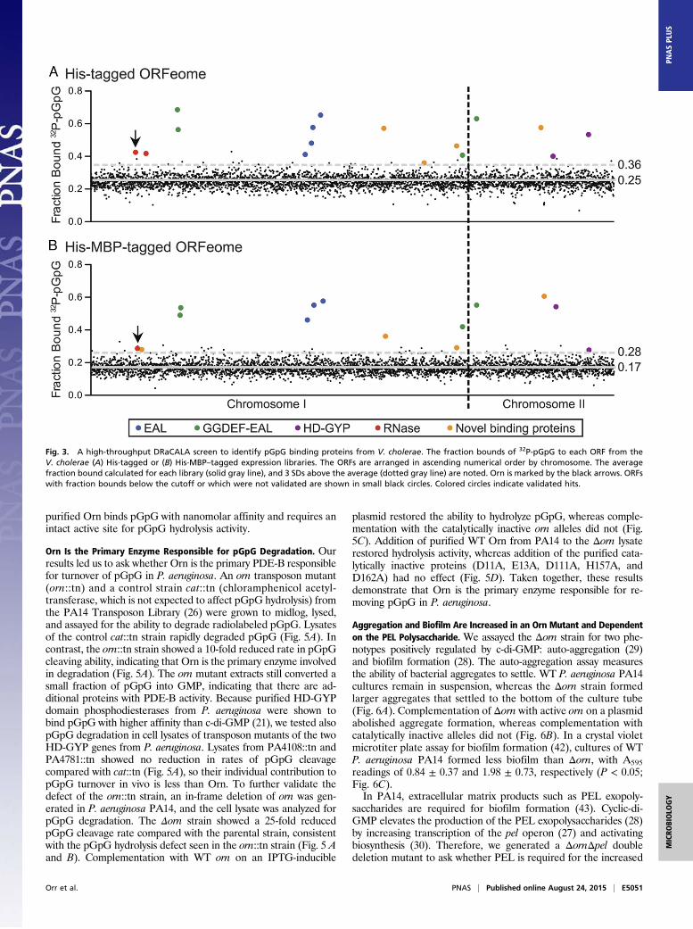
Identification of pGpG Binding Proteins from V. cholerae ORF Library.Based on the above results, the PDE-B responsible for hydro-lyzing pGpG is critical for completing the c-di-GMP degradationpathway. To identify this enzyme, we used a high-throughputDRaCALA-based screen to interrogate pGpG binding to a li-brary of ORFs (22, 23). The V. cholerae El Tor N16961 completegenome ORF library (ORFeome) was used because 97% of theORFs are represented (24), and V. cholerae uses c-di-GMP sig-naling extensively (33, 34). Each ORF was recombined into twodestination vectors with an isopropyl-β-D-1-thiogalactopyrano-side (IPTG)-inducible T7 promoter, one containing a 10×histidine N-terminal tag and a second containing a 10× histidine-maltose binding protein (MBP) N-terminal tag. These two li-braries were individually introduced into an E. coli T7Iq ex-pression strain and arrayed in 96-well plates. Following IPTGinduction, whole cell lysates were generated and assayed forbinding to pGpG using DRaCALA. The fraction bounds foreach ORF tested are listed in Table S1. Positive hits were de-fined as 3 SDs above the mean fraction bound of the entire li-brary. For validation, each positive hit was repicked from theexpression library and reassayed for pGpG binding. The fractionof each ORF bound to pGpG is shown for both ORFeomes withvalidated hits in color (Fig. 3). A list of hits and their known orpredicted functions is provided in Table S2.
The V. cholerae ORFeome library includes known c-di-GMPbinding proteins: 28 of 31 GGDEF domain proteins (33), all 5PilZ domain proteins (35), and all 3 c-di-GMP-binding transcriptionfactors (36–38). None of these proteins were identified in thisscreen, indicating that the assay is specific for pGpG-binding pro-teins. We expected to identify EAL domain and HD-GYP domainproteins, both of which have been shown to bind pGpG (21, 31).The V. cholerae ORFeome library includes 11 of 12 EAL domainproteins encoded in the genome, 9 of 10 dual GGDEF-EAL do-main proteins, and all 9 HD-GYP domain proteins (33). Of these,four EAL domain-containing proteins, VC1592, VC1641, VC1652,and VC1710 (Fig. 3, blue dots); four GGDEF-EAL domain con-taining proteins, VC0653, VC0658, VC2750, and VCA0080 (Fig. 3,green dots); and two HD-GYP domain containing proteins,VCA0681 and VCA0931 (Fig. 3, purple dots), bound pGpG inthe screen. Not all EAL or HD-GYP domain ORF lysatesshowed binding in this system, which could reflect either genuineinability to bind pGpG or low protein expression in E. coli,resulting in concentrations below the Kd.Because pGpG is a two-nucleotide-long RNA, we expected ri-
bonucleases (RNases) to bind as well. The ORFeome library in-cluded all 15 annotated RNases from the V. cholerae genome, ofwhich 2 were found to bind pGpG: VC0210 and VC0341 (Fig. 3,red dots). Additionally, the screen identified five ORFs that werenot previously known to interact with pGpG: VC0371, VC2147,VC2459, VC2708, and VCA0593 (Fig. 3, orange dots). Sequencehomology predictions indicate that four of these five proteins arepredicted to interact with nucleic acids or nucleotides, whereasVCA0593 has no predicted function (Table S2). VC0341 is ahomolog of Orn, which is a known exoribonuclease that cleavestwo- to five-nucleotide-long RNAs (25). Because VC0341 wasshown to bind in both the His-tagged and the His-MBP–taggedORFeomes, it was chosen for further study.
Orn Binds to pGpG Specifically and Can Cleave It into GMP. To fullyvalidate the interaction between Orn and pGpG observed in celllysates, Orn from P. aeruginosa PA14 and V. cholerae wereHis-MBP tagged, purified, and assayed for pGpG binding byDRaCALA. pGpG bound both P. aeruginosa and V. choleraeOrn with fraction bounds of 0.32 ± 0.02 and 0.27 ± 0.01 in theabsence of competitor (NC) (Fig. 4 A and C). Addition of excessunlabeled pGpG effectively competed radiolabeled pGpG bind-ing, reducing the fraction bounds to 0.009 ± 0.008 and 0.03 ±0.003 for P. aeruginosa and V. cholerae Orn (P < 0.05; Fig. 4 Aand C). However, Orn binding to pGpG was not affected by theaddition of excess c-di-GMP or any other guanine-containingnucleotides tested, indicating that Orn binds pGpG specifically(Fig. 4 A and C). Finally, Orn from P. aeruginosa and V. choleraebound to pGpG with high affinity, with a Kd of 40 ± 2 and25 ± 2 nM, respectively (Fig. 4 B and D).Having demonstrated specific and high-affinity binding, we then
investigated the ability of Orn to hydrolyze pGpG. PurifiedP. aeruginosa His-MBP-Orn degraded pGpG (Fig. 4F) into GMPwithin 10 s at room temperature. Orn belongs to the DEDDhsubfamily of 3′ to 5′ exoribonucleases that have a highly conservedactive site motif (39). The locations of the DEDDh active siteresidues were shown in the solved crystal structure of Xanthomonascampestris Orn (40), and an alignment of P. aeruginosa PA14 Ornwith X. campestris Orn revealed the active site residues D11, E13,D111, H157, and D162 (Fig. S1). Single alanine point mutants inthe signature motif of another DEDDh RNase, RNase T of E. coli,had significantly reduced catalytic activity (41). Therefore, we in-troduced single alanine substitutions into each residue of theDEDDh motif (D11A, E13A, D111A, H157A, and D162A) ofP. aeruginosa Orn to assay for pGpG binding and degradation.All of the Orn variants bound pGpG (Fig. 4E) but failed to cleavepGpG (Fig. 4F). Taken together, these results demonstrate that
GMPB
Time (min)
c-di-GMPC
Time (min)
pGpGD
Time (min)
0 2 4 6
NCA
Time (min)
0.0
0.2
0.4
0.6
0.8
1.0
Frac
tion
of32
P s
igna
l
0.0
0.2
0.4
0.6
0.8
1.0
Frac
tion
of32
P s
igna
l
0.0
0.2
0.4
0.6
0.8
1.0
Frac
tion
of32
P s
igna
l
0.0
0.2
0.4
0.6
0.8
1.0
Frac
tion
of32
P s
igna
l
Fig. 2. Excess pGpG extends the half-life of c-di-GMP in an in vitro digua-nylate cyclase and phosphodiesterase activity assay. TLC-based quanti-fication of the conversion of 32P-GTP (gray) to 32P-c-di-GMP (black) and32P-pGpG (white) in a coupled WspR and RocR reaction in (A) the absence ofcompetitor (NC), (B) 300 μM GMP, (C) 300 μM c-di-GMP, or (D) 300 μM pGpG.All data shown represent the average and SD of duplicate independentexperiments.
E5050 | www.pnas.org/cgi/doi/10.1073/pnas.1507245112 Orr et al.
purified Orn binds pGpG with nanomolar affinity and requires anintact active site for pGpG hydrolysis activity.
Orn Is the Primary Enzyme Responsible for pGpG Degradation. Ourresults led us to ask whether Orn is the primary PDE-B responsiblefor turnover of pGpG in P. aeruginosa. An orn transposon mutant(orn::tn) and a control strain cat::tn (chloramphenicol acetyl-transferase, which is not expected to affect pGpG hydrolysis) fromthe PA14 Transposon Library (26) were grown to midlog, lysed,and assayed for the ability to degrade radiolabeled pGpG. Lysatesof the control cat::tn strain rapidly degraded pGpG (Fig. 5A). Incontrast, the orn::tn strain showed a 10-fold reduced rate in pGpGcleaving ability, indicating that Orn is the primary enzyme involvedin degradation (Fig. 5A). The orn mutant extracts still converted asmall fraction of pGpG into GMP, indicating that there are ad-ditional proteins with PDE-B activity. Because purified HD-GYPdomain phosphodiesterases from P. aeruginosa were shown tobind pGpG with higher affinity than c-di-GMP (21), we tested alsopGpG degradation in cell lysates of transposon mutants of the twoHD-GYP genes from P. aeruginosa. Lysates from PA4108::tn andPA4781::tn showed no reduction in rates of pGpG cleavagecompared with cat::tn (Fig. 5A), so their individual contribution topGpG turnover in vivo is less than Orn. To further validate thedefect of the orn::tn strain, an in-frame deletion of orn was gen-erated in P. aeruginosa PA14, and the cell lysate was analyzed forpGpG degradation. The Δorn strain showed a 25-fold reducedpGpG cleavage rate compared with the parental strain, consistentwith the pGpG hydrolysis defect seen in the orn::tn strain (Fig. 5 Aand B). Complementation with WT orn on an IPTG-inducible
plasmid restored the ability to hydrolyze pGpG, whereas comple-mentation with the catalytically inactive orn alleles did not (Fig.5C). Addition of purified WT Orn from PA14 to the Δorn lysaterestored hydrolysis activity, whereas addition of the purified cata-lytically inactive proteins (D11A, E13A, D111A, H157A, andD162A) had no effect (Fig. 5D). Taken together, these resultsdemonstrate that Orn is the primary enzyme responsible for re-moving pGpG in P. aeruginosa.
Aggregation and Biofilm Are Increased in an Orn Mutant and Dependenton the PEL Polysaccharide. We assayed the Δorn strain for two phe-notypes positively regulated by c-di-GMP: auto-aggregation (29)and biofilm formation (28). The auto-aggregation assay measuresthe ability of bacterial aggregates to settle. WT P. aeruginosa PA14cultures remain in suspension, whereas the Δorn strain formedlarger aggregates that settled to the bottom of the culture tube(Fig. 6A). Complementation of Δorn with active orn on a plasmidabolished aggregate formation, whereas complementation withcatalytically inactive alleles did not (Fig. 6B). In a crystal violetmicrotiter plate assay for biofilm formation (42), cultures of WTP. aeruginosa PA14 formed less biofilm than Δorn, with A595readings of 0.84 ± 0.37 and 1.98 ± 0.73, respectively (P < 0.05;Fig. 6C).In PA14, extracellular matrix products such as PEL exopoly-
saccharides are required for biofilm formation (43). Cyclic-di-GMP elevates the production of the PEL exopolysaccharides (28)by increasing transcription of the pel operon (27) and activatingbiosynthesis (30). Therefore, we generated a ΔornΔpel doubledeletion mutant to ask whether PEL is required for the increased
Fig. 3. A high-throughput DRaCALA screen to identify pGpG binding proteins from V. cholerae. The fraction bounds of 32P-pGpG to each ORF from theV. cholerae (A) His-tagged or (B) His-MBP–tagged expression libraries. The ORFs are arranged in ascending numerical order by chromosome. The averagefraction bound calculated for each library (solid gray line), and 3 SDs above the average (dotted gray line) are noted. Orn is marked by the black arrows. ORFswith fraction bounds below the cutoff or which were not validated are shown in small black circles. Colored circles indicate validated hits.
Orr et al. PNAS | Published online August 24, 2015 | E5051
MICRO
BIOLO
GY
PNASPL
US
aggregation and biofilm formation we observe in the Δornmutant.The ΔornΔpel double mutant remained in suspension like the WTand the Δpel single mutant cultures (Fig. 6A), indicating that PELis required for the aggregation observed in the Δorn strain. The peloperon is also known to be required for in vitro pellicle biofilmformation in P. aeruginosa PA14 (43). As expected, the Δpelmutant was unable to form a pellicle biofilm in a microtiter plateassay, with a crystal violet A595 reading of 0.13 ± 0.02 comparedwith 0.84 ± 0.37 for WT (P < 0.05; Fig. 6C). The ability to formbiofilm is also lost in the ΔornΔpel double deletion strain, whichhad an A595 reading of 0.21 ± 0.05 (P < 0.05 compared with WT;Fig. 6C). These data indicate that the increased aggregation andbiofilm formation seen in the Δorn strain is PEL dependent.Another measure of elevated c-di-GMP levels is through the
activity of the transcription factor FleQ, which leads to increasedtranscription from the pel promoter when bound to c-di-GMP(27). The pel promoter fused to lacZ (Ppel-lacZ) was integratedinto the chromosome of both P. aeruginosa PA14 WT and Δornstrains. These strains were grown to midlog and assayed forβ-galactosidase activity. Approximately threefold increase inβ-galactosidase activity was observed in the Δorn strain compared
with WT, with 563 ± 230 Miller units compared with 185 ± 64Miller units, respectively (P < 0.05; Fig. 6D). Together, our resultssupport the hypothesis that the loss of orn elevates c-di-GMP topromote c-di-GMP–regulated biofilms.In additional support for the role of c-di-GMP in aggregation,
overexpression of enzymes that either make or linearize c-di-GMPhave opposing effects on aggregation in the Δorn strain. Elevatingc-di-GMP via overexpression of the DGCs PA1107 and wspR in-creased aggregation. Conversely, decreasing c-di-GMP via expres-sion of the PDE-As PA2133 and rocR was unable to completelyreverse bacterial aggregation (Fig. S2). These results indicate thatbecause the Δorn mutant lacks the ability to rapidly hydrolyzepGpG, it cannot complete degradation of c-di-GMP even whenoverexpressing EAL PDE-As.
Intracellular pGpG and c-di-GMP Are Elevated in the OligoribonucleaseMutant. To confirm the presence of increased c-di-GMP and pGpGin the PA14 Δorn strain, both nucleotides were directly detected byLC-MS/MS. WT and Δorn cultures were grown midlog (OD600 =0.6) and late-log (OD600 = 2.0), and the nucleotides were extractedfor analysis. Both c-di-GMP and pGpG were detected in the PA14WT and Δorn mutant strains. At midlog, the Δorn strain had 5.9-fold increased c-di-GMP compared with WT, with 2.86 ± 0.67 vs.0.49 ± 0.08 μM, respectively (P < 0.001; Table 1). We also detect6.6-fold more pGpG in the Δorn strain than WT in midlog (70.8 ±1.6 vs. 10.8 ± 1.4 μM; P < 0.001; Table 1). Lower concentrations ofc-di-GMP and pGpG were detected from both strains at late-log,although both nucleotides remained higher in the Δorn strain.At late-log, the Δorn strain had 8.1-fold increased c-di-GMP com-pared with WT (0.65 ± 0.20 vs. 0.08 ± 0.01 μM, respectively; P <0.05) and 6.6-fold increased pGpG compared with WT (26.3 ± 3.6vs. 4.0 ± 1.0 μM, respectively, P < 0.005; Table 1). The above dataconfirm that pGpG and c-di-GMP are both elevated in orn mu-tants at both growth phases. All together, our results show that in
Frac
tion
Bou
nd 3
2 P-p
GpG
NCpG
pG
cdiG
MPcG
MPGMP
GDPGTP
0.0
0.1
0.2
0.3
0.4
0.0
0.1
0.2
0.3
0.4
0.5
10-510-4 10-310-210-1100 101 102
Frac
tion
Bou
nd 3
2 P-p
GpG
[PA14 Orn] (μM)
Frac
tion
Bou
nd 3
2 P-p
GpG
NCpG
pG
cdiG
MPcG
MPGMP
GDPGTP
0.0
0.1
0.2
0.3
0.4
0.0
0.1
0.2
0.3
0.4
0.5
10-510-4 10-310-210-1100 101 102
Frac
tion
Bou
nd 3
2 P-p
GpG
[V. cholerae Orn] (μM)
Time (min)0 10 20 30
0.0
0.2
0.4
0.6
0.8
1.0
D11A
E13A
D111A
H157A
D162A
0.0
0.2
0.4
0.6
0.8
Frac
tion
Bou
nd 3
2 P-p
GpG
Frac
tion 3
2 P-p
GpG
H157AD11A D162A
E13AD111A
Orn
Fig. 4. Orn binds and hydrolyzes pGpG. Quantification of fraction bound of32P-pGpG to Orn from (A) P. aeruginosa PA14 or (C) V. cholerae in thepresence of indicated competitors (250 μM). The dissociation constants (Kd)of 32P- pGpG binding to Orn from (B) P. aeruginosa PA14 and (D) V. choleraeas determined by DRaCALA. (E ) Quantification of the fraction bound of32P-pGpG to purified P. aeruginosa Orn active site mutants D11A, E13A,D111A, H157A, and D162A. (F) The rate of 32P-pGpG hydrolysis by purifiedwild type Orn and active site mutants of P. aeruginosa Orn: D11A, E13A,D111A, H157A, and D162A. All data shown represent the average and SD ofat least three independent experiments.
Fig. 5. PA14 orn mutants are defective in hydrolyzing pGpG. The rate of32P-pGpG cleavage by (A) whole cell lysates of P. aeruginosa PA14 transposoninsertion strains cat::tn, orn::tn, PA4108::tn, or PA4781::tn, (B) whole celllysates of P. aeruginosa PA14 or Δorn in-frame deletion mutant, (C) wholecell lysates of P. aeruginosa Δorn mutant complemented with WT or mutantalleles of orn, or (D) whole cell lysates of P. aeruginosa Δorn with 100 nM ofthe indicated purified protein added. All data shown represent the averageof triplicate independent experiments.
E5052 | www.pnas.org/cgi/doi/10.1073/pnas.1507245112 Orr et al.

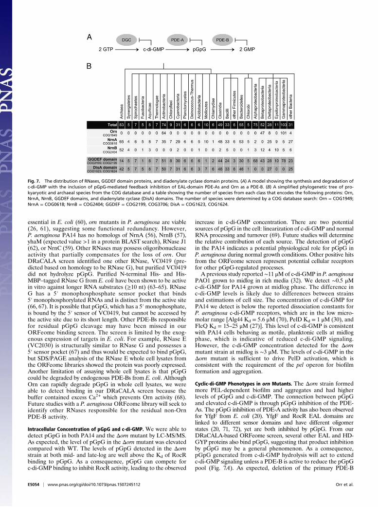
addition to its known role degrading oligoribonucleotides (25),Orn is the major PDE-B that hydrolyzes pGpG in P. aeruginosa.
DiscussionOligoribonucleases as PDE-Bs. Early characterization of c-di-GMPfrom the Benziman laboratory identified that c-di-GMP is line-arized by PDE-As into pGpG, which is then degraded into twoGMPs through the action of a presumed PDE-B (44). Throughnearly three decades of c-di-GMP research that followed, theidentity of the PDE-B remained unknown. We demonstrate herethat the 3′-5′exoribonuclease Orn is the primary PDE-B inP. aeruginosa. The linearized product of c-di-GMP hydrolysis byPDE-As, pGpG, is a two-nucleotide-long RNA that matches theknown substrates of Orn. We present several lines of evidenceindicating that Orn is the primary PDE-B for c-di-GMP in
P. aeruginosa: (i) Orn binds pGpG and not c-di-GMP, (ii) Ornrapidly degrades pGpG in vitro, (iii) lysates of P. aeruginosa ornmutants are greatly reduced for PDE-B activity, (iv) DEDDhcatalytic residues used to degrade oligonucleotides are requiredfor pGpG cleavage, and (v) loss of orn results in enhanced in-tracellular pGpG and c-di-GMP. This additional role for Orn isimportant because we demonstrate that the loss of Orn results inincreased c-di-GMP and c-di-GMP governed phenotypes, likelydue to accumulated pGpG inhibiting PDE-As as shown in ourmodel (Fig. 7A).Orn degrades short oligoRNAs regardless of sequence (45),
raising the interesting possibility that it could degrade all line-arized bacterial cyclic dinucleotides. In addition to c-di-GMP,two other cyclic dinucleotide signaling molecules have thus farbeen identified in bacteria. Cyclic di-AMP (c-di-AMP) is syn-thesized by diadenylate cyclases, which are widely distributedamong bacterial species (46), and a hybrid cAMP-GMP is syn-thesized by DncV in V. cholerae (47) and an unknown cyclasein Geobacter sulfurreducens (48). Cyclic-di-AMP is linearizedby HD domain and DHH-DHHA-1 domain phosphodiester-ases (49, 50), and cAMP-GMP is linearized by HD-GYP pro-teins in V. cholerae (51). The linearized cyclic di-nucleotidescould be hydrolyzed by Orn. However, not all species that usecyclic dinucleotides posses an Orn homolog (Fig. 7B). In a searchof the clusters of orthologous group (COG) database, onlyActinobacteria, Fibrobacteres, Betaproteobacteria, Deltapro-teobacteria, Gammaproteobacteria, and a few unclassified spe-cies are predicted to encode Orn (52) (Fig. 7B). Bacillus subtilishas both c-di-GMP (53) and c-di-AMP signaling systems (54, 55)but lacks orn. Nevertheless, B. subtilis is capable of degradingtwo- to five-nucleotide-long RNAs using the potential Ornfunctional homologs NrnA, NrnB, yhaM, and RNase J1. All ofthese enzymes have oligoribonuclease activity and were able topartially complement an E. coli orn conditional mutant (56, 57).NrnA homologs are found in many bacteria that lack Orn (58).Additionally, another RNase with oligoribonuclease activity,NrnC, was recently identified in Bartonella birtlesii and is widelyfound in Alphaproteobacteria (59). Nearly all bacteria thatencode diguanylate and diadenylate cyclases encode at least oneRNase capable of degrading short oligoRNAs. Based on thisgenomic distribution, we predict these RNases are likely theprimary PDE-Bs for hydrolyzing linearized cyclic dinucleotidesinto mononucleotides.Our observation that residual PDE-B activity is present in
mutants lacking orn indicates that other proteins can cleavepGpG. Proposed PDE-B candidates include the HD-GYP do-main proteins, which were shown to bind pGpG with higher af-finity than c-di-GMP in vitro and were capable of degradingpGpG (21). However, single transposon mutants in both of theHD-GYP domain proteins of P. aeruginosa PA14 did not show adefect in pGpG removal in our system, in contrast to the largedefect observed for the orn mutant. Despite being shown to be
Fig. 6. Increased aggregation and biofilm formation in the orn mutant isPEL dependent. (A) Photograph of auto-aggregation assays of PA14, Δorn,Δpel, and ΔornΔpel mutants. (B) Photograph of auto-aggregation assays ofPA14 and the Δorn strain complemented with WT orn and active site mutantalleles. (C) Quantification of the biofilm formed after 24 h of static cultureby the crystal violet assay by determining absorbance at 595 nm (A595). A595
values results show the average and SD of triplicate independent experi-ments. (D) Miller assay showing β-galactosidase activity of WT and Δornstrains containing a single copy of the pel promoter lacZ (Ppel-lacZ) reporteron the chromosome. *P < 0.05 by Student unpaired two-tailed t test as-suming equal variance for C and D.
Table 1. Quantification of c-di-GMP and pGpG in PA14 and PA14Δorn
OD600 PA14 (μM) Δorn (μM) P value* Δorn/PA14
c-di-GMP0.6 0.49 ± 0.08 2.86 ± 0.67 0.007 5.92.0 0.08 ± 0.01 0.65 ± 0.20 0.02 8.1
pGpG0.6 10.8 ± 1.4 70.8 ± 1.6 2.4 × 10⁻⁶ 6.62.0 4.0 ± 1.0 26.3 ± 3.6 1.3 × 10⁻³ 6.6
Data shown are mean ± SD of three independent samples.*P value was determined by Student unpaired two-tailed t test assumingequal variance.
Orr et al. PNAS | Published online August 24, 2015 | E5053
MICRO
BIOLO
GY
PNASPL
US
essential in E. coli (60), orn mutants in P. aeruginosa are viable(26, 61), suggesting some functional redundancy. However,P. aeruginosa PA14 has no homologs of NrnA (56), NrnB (57),yhaM (expected value >1 in a protein BLAST search), RNase J1(62), or NrnC (59). Other RNases may possess oligoribonucleaseactivity that partially compensates for the loss of orn. OurDRaCALA screen identified one other RNase, VC0419 (pre-dicted based on homology to be RNase G), but purified VC0419did not hydrolyze pGpG. Purified N-terminal His- and His-MBP–tagged RNase G from E. coli have been shown to be activein vitro against longer RNA substrates (≥10 nt) (63–65). RNaseG has a 5′ monophosphosphate sensor pocket that binds5′ monophosphorylated RNAs and is distinct from the active site(66, 67). It is possible that pGpG, which has a 5′monophosphate,is bound by the 5′ sensor of VC0419, but cannot be accessed bythe active site due to its short length. Other PDE-Bs responsiblefor residual pGpG cleavage may have been missed in ourORFeome binding screen. The screen is limited by the exog-enous expression of targets in E. coli. For example, RNase E(VC2030) is structurally similar to RNase G and possesses a5′ sensor pocket (67) and thus would be expected to bind pGpG,but SDS/PAGE analysis of the RNase E whole cell lysates fromthe ORFeome libraries showed the protein was poorly expressed.Another limitation of assaying whole cell lysates is that pGpGcould be degraded by endogenous PDE-Bs from E. coli. AlthoughOrn can rapidly degrade pGpG in whole cell lysates, we wereable to detect binding in our DRaCALA screen because thebuffer contained excess Ca2+ which prevents Orn activity (68).Future studies with a P. aeruginosa ORFeome library will seek toidentify other RNases responsible for the residual non-OrnPDE-B activity.
Intracellular Concentration of pGpG and c-di-GMP. We were able todetect pGpG in both PA14 and the Δorn mutant by LC-MS/MS.As expected, the level of pGpG in the Δorn mutant was elevatedcompared with WT. The levels of pGpG detected in the Δornstrain at both mid- and late-log are well above the Kd of RocRbinding to pGpG. As a consequence, pGpG can compete forc-di-GMP binding to inhibit RocR activity, leading to the observed
increase in c-di-GMP concentration. There are two potentialsources of pGpG in the cell: linearization of c-di-GMP and normalRNA processing and turnover (69). Future studies will determinethe relative contribution of each source. The detection of pGpGin the PA14 indicates a potential physiological role for pGpG inP. aeruginosa during normal growth conditions. Other positive hitsfrom the ORFeome screen represent potential cellular receptorsfor other pGpG-regulated processes.A previous study reported ∼11 μMof c-di-GMP in P. aeruginosa
PAO1 grown to midlog in rich media (32). We detect ∼0.5 μMc-di-GMP for PA14 grown at midlog phase. The difference inc-di-GMP levels is likely due to differences between strainsand estimations of cell size. The concentration of c-di-GMP forPA14 we detect is below the reported dissociation constants forP. aeruginosa c-di-GMP receptors, which are in the low micro-molar range [Alg44 Kd = 5.6 μM (70), PelD Kd = 1 μM (30), andFleQ Kd = 15–25 μM (27)]. This level of c-di-GMP is consistentwith PA14 cells behaving as motile, planktonic cells at midlogphase, which is indicative of reduced c-di-GMP signaling.However, the c-di-GMP concentration detected for the Δornmutant strain at midlog is ∼3 μM. The levels of c-di-GMP in theΔorn mutant is sufficient to drive PelD activation, which isconsistent with the requirement of the pel operon for biofilmformation and aggregation.
Cyclic-di-GMP Phenotypes in orn Mutants. The Δorn strain formedmore PEL-dependent biofilm and aggregates and had higherlevels of pGpG and c-di-GMP. The connection between pGpGand elevated c-di-GMP is through pGpG inhibition of the PDE-As. The pGpG inhibition of PDE-A activity has also been observedfor YfgF from E. coli (20). YfgF and RocR EAL domains arelinked to different sensor domains and have different oligomerstates (20, 71, 72), yet are both inhibited by pGpG. From ourDRaCALA-based ORFeome screen, several other EAL and HD-GYP proteins also bind pGpG, suggesting that product inhibitionby pGpG may be a general phenomenon. As a consequence,pGpG generated from c-di-GMP hydrolysis will act to extendc-di-GMP signaling unless a PDE-B is active to reduce the pGpGpool (Fig. 7A). As expected, deletion of the primary PDE-B
Fig. 7. The distribution of RNases, GGDEF domain proteins, and diadenylate cyclase domain proteins. (A) A model showing the synthesis and degradation ofc-di-GMP with the inclusion of pGpG-mediated feedback inhibition of EAL-domain PDE-As and Orn as a PDE-B. (B) A simplified phylogenetic tree of pro-kyaryotic and archaeal species from the COG database and a table showing the number of species from each class that encodes the following proteins: Orn,NrnA, NrnB, GGDEF domains, and diadenylate cyclase (DisA) domains. The number of species were determined by a COG database search: Orn = COG1949;NrnA = COG0618; NrnB = COG2404; GGDEF = COG2199, COG3706; DisA = COG1623, COG1624.
E5054 | www.pnas.org/cgi/doi/10.1073/pnas.1507245112 Orr et al.
(orn) in P. aeruginosa resulted in the accumulation of c-di-GMPand increased biofilm formation and auto-aggregation. Theseobservations will likely extend to other bacteria that use Orn ho-mologs to degrade pGpG.Orn may also be involved in other phenotypes regulated by
c-di-GMP. Cystic fibrosis patients are chronically infected withmucoid strains of P. aeruginosa, leading to poor patient prog-nosis (73). The mucoid phenotype is due to high production ofthe alginate polysaccharide, which is synthesized by componentsof the alg operon (74). Cyclic-di-GMP regulates productionposttranscriptionally by binding to the PilZ domain of Alg44 toactivate alginate biosynthesis (70). Overexpression of the diguany-late cyclase PA1107 significantly enhanced alginate production inP. aeruginosa (70). In addition, transcription of the operon is reg-ulated by several proteins, including the activator AlgB (75). Amucoid cystic fibrosis isolate FRD of P. aeruginosa becomes non-mucoid on deletion of algB, although the strain is still able toproduce low levels of alginate (75). A genetic screen for suppressormutants that restored alginate production of the algB mutantidentified six transposon insertion mutants. Two of these insertionswere in the orn gene (61). A possible explanation based on ourmodel (Fig. 7A) is that the loss of orn resulted in pGpG accumu-lation, inhibition of PDE-A, and thus elevated c-di-GMP to allowfor increased Alg44 activation and enhanced mucoidy. These ob-servations indicate that the PDE-B must be present and highlyactive during c-di-GMP signaling termination to prevent pGpG-mediated PDE-A inhibition.
Materials and MethodsV. cholerae ORFeome Library pGpG Binding Screen. Gateway destinationvectors were generated from pET-19–derived expression vectors pVL791(N-terminal His10 tag, carbenecillin resistance) and pVL847 (N-terminalHis10-MBP tag, gentamycin resistance). The Gateway destination cassettewas amplified from pRFA and cloned in frame with the N-terminal tags toproduce the gateway adapted vectors pVL791 GW and pVL847 GW. TheV. cholerae O1 biovar El Tor str. N16961 pDONR221 library was obtainedfrom BEI Resources. The library was grown in 1.5 mL lysogeny broth (LB) in2-mL, 96-well plates (Greiner) with kanamycin (50 mg/mL) selection, andthe plasmids were isolated using the 96-well MultiScreenHTS Kit (Millipore).The ORFs were moved into the expression vector using LR-clonase enzymeII (Invitrogen) and introduced into chemically competent E. coli strain T7Iq(NEB) following the manufacturer’s protocols. Recombinants were se-lected on LB agar plates containing either carbenecillin (50 mg/mL) orgentamycin (15 mg/mL). Multiple colonies from individual transformationswere inoculated in LB M9-rich media in 96-well plate format and grownovernight with shaking at 30 °C with the appropriate antibiotic and thenresuspended in 20% (vol/vol) glycerol and frozen at −80 °C.
E. coli T7Iq containing the V. cholerae ORFs were inoculated from frozenstocks in LB M9 rich media with the appropriate antibiotic in 96-well plateformat, grown overnight with shaking at 30 °C, subcultured 1:50 into 1.5-mLfresh LB M9 media with antibiotic, and grown for 4 h at 30 °C with shaking;1 mM IPTG was added to induce protein expression and cultures were grownfor an additional 4 h. The induced culture was pelleted, and cells wereresuspended in 1/10th volume of binding buffer (10 mM Tris, pH 8.0, 100 mMNaCl, 5 mM CaCl2), as well as 10 μg/mL DNase, 250 μg/mL lysozyme, and10 mM PMSF. Cells were lysed by three freeze/thaw cycles at −80 °C/21 °C.ORFeome whole cell lysates were stored at −80 °C until analysis by DRaCALA.
Binding of radiolabeled ligand to the ORFeome whole cell lysates was de-termined by DRaCALA as previously described (22, 23). Briefly, 16 pM 32P-pGpGwas added to the ORFeome whole cell lysate 96-well plates using a MultifloMicroplate Dispenser (BioTek) and then applied to nitrocellulose sheets (GEHealthcare) using a 96-well pin tool (V&P Scientific). The nitrocellulose was airdried and imaged using a Fujifilm FLA-7000 phosphorimager (GE), and theintensity of the DRaCALA spots was quantified using Fujifilm Multi Gaugesoftware v3.0, and the fraction bounds were quantified (22, 23). To confirmthat positive hits were not due to cross-contamination between plate wells,each positive hit was repicked from the expression library, and eight replicatesingle colonies were used to generate eight new whole cell lysates. Thesereplicate lysates were compared with empty vector lysates for 32P-pGpGbinding by DRaCALA.
Strains and Culture Conditions. The primers, plasmids, and strains used in thisstudy are listed in Tables S3–S5, respectively. The in-frame deletion of orn wasgenerated in P. aeruginosa PA14 using a Flp-FRT recombination system (76). The∼1-kb region upstream and downstream were PCR amplified and restrictiondigested to introduced these fragments into a pEX-Gn–based plasmid formaking in-frame deletions as previously described (76). Deletions were verifiedby PCR. The pel promoter-lacZ fusion on a pCTX plasmid wa incorporated intothe genomic att site as previously described (77).
The alanine point mutations were generated by QuikChange using theprimers listed in Table S3. Silent mutations resulting in addition or removalof a restriction site were introduced to facilitate mutagenesis: these werethe addition of an AfeI site in D11A and H157A, addition of an MscI site inD111A, and removal of EcoRV in D162A. The orn alleles from P. aeruginosaPA14 were cloned into pVL847 for purification and pMMB for complemen-tation. The pMMB and pVL847 plasmids were maintained with gentamycin(15 mg/mL) and induced with 1 mM IPTG. Transposon mutants from thePA14 Non-Redundant Transposon Insertion Mutant Library (26) weremaintained with gentamycin (15 mg/mL).
Protein Expression and Purification. His-MBP-RocR, His-MBP-Orn from V. cholerae,and His-MBP-Orn and His-MBP-Orn variants from P. aeruginosa were purified aspreviously described (23). Briefly, E. coli T7Iq strains or E. coli BL21(DE3) con-taining expression plasmids were grown overnight, subcultured in fresh media,and grown to OD600∼1.0 when expression was induced with 1 mM IPTG. Inducedbacteria were pelleted and resuspended in 10 mM Tris, pH 8, 100 mM NaCl, and25 mM imidazole and frozen at −80 °C until purification. Proteins were purifiedover a Ni-NTA column followed by anion exchange on a Q-Sepharose column.Purified proteins were dialyzed twice against 10 mM Tris, pH 8, 100 mM NaCl,and 25% (vol/vol) glycerol, aliquoted, and frozen at −80 °C until use.
Whole Cell Lysate Generation. Overnight cultures of P. aeruginosa PA14 pa-rental, mutant, or complemented strains were subcultured 1:50 into fresh LBmedia with appropriate antibiotic and IPTG conditions, grown to OD600 = 0.4 at37 °C with shaking, resuspended in 1/10th volume of reaction buffer (10 mM Tris,pH 8, 100 mM NaCl, 5 mMMgCl2), 10 μg/mL DNase, 250 μg/mL of lysozyme, and10 mM PMSF, and lysed by sonication.
DRaCALA Measurement of Ligand Binding, Nucleotide Competition, andDissociation Constant. The use of DRaCALA to probe protein–ligand in-teractions has been previously described (23). To assay binding, pure protein inbinding buffer (10 mM Tris, pH 8.0, 100 mM NaCl, 5 mM CaCl2) was mixed withradiolabeled ligand (4 pM 32P-c-di-GMP or 32P-pGpG), applied to nitrocellulosesheets, dried, imaged, and the fraction bound quantified (23). For competitionassays, excess of unlabeled nucleotides were added to radiolabeled ligand andpurified protein before analysis by DRaCALA (23). Tomeasure Kd, twofold serialdilutions of purified His-MBP-Orn from either P. aeruginosa or V. choleraeweremade in binding buffer (10 mM Tris, pH 8, 100 mM NaCl, 5 mM CaCl2) andmixed with radiolabeled ligand and the fraction bound, and Kd was calculatedas previously described (23).
Cell Lysate and Protein Activity Assays. The activity of whole cell lysates andpurified proteins against 32P-labeled substrates was measured by monitoringthe appearance of 32P-labeled products on TLC. The reactions were carriedout at 37 °C in reaction buffer (10 mM Tris, pH 8, 100 mM NaCl, and 5 mMMgCl2). At appropriate times, aliquots were removed and the reactionstopped by adding an equal volume of 0.2 M EDTA, pH 8, and heated at98 °C for 10 min. Samples were spotted on polyethyleneimine-cellulose TLCplates (EMD Chemicals), dried, and developed in mobile phase consisting of1:1.5 (vol:vol) saturated NH4SO4 and 1.5 M KH2PO4, pH 3.60. The TLC platewas dried and imaged using Fujifilm FLA-7000 phosphorimager (GE). Theintensity of the radiolabeled nucleotides was quantified using Fujifilm MultiGauge software v3.0.
Aggregation Assay. Cultures of P. aeruginosa strains were grown in 10 mL LBwith appropriate antibiotic and IPTG conditions for 24 h at 37 °C with shaking.Cultures were imaged after 30 min of aggregate settling at room temperature.
Microtiter Plate Biofilm Assay. P. aeruginosa PA14 WT and mutant strainswere grown overnight in LB at 37 °C with shaking. Overnight cultures werediluted 1:100 in LB and grown as static cultures in a 96-well polystyrene plate(Greiner) at 30 °C inside a humidified chamber for 24 h. The cultures werewashed of planktonic cells and stained with crystal violet as previously de-scribed (42). The A595 was measured on a SpectraMax M5 spectrophotom-eter (Molecular Devices).
Orr et al. PNAS | Published online August 24, 2015 | E5055
MICRO
BIOLO
GY
PNASPL
US

β-Galactosidase Reporter Assay. P. aeruginosa strains containing the reporterwere grown overnight in LB at 37 °C with shaking. Overnight cultures weresubcultured in LB and grown at 37 °C with shaking to midlog (OD600 = 0.5).The β-galactosidase activity was measured according to previously publishedmethods (78).
Quantification of Intracellular c-di-GMP and pGpG. P. aeruginosa PA14 WT andΔorn strains were grown overnight in LB at 37 °C with shaking, subcultured1:100 in 20 mL LB, and grown at 37 °C with shaking to midlog (OD600 = 0.6)and late-log (OD600 = 2); 15 mL was pelleted by centrifugation, resuspendedwith 100 μL ice-cold extraction buffer, 40:40:20 (vol:vol:vol) MeOH, aceto-nitrile, and water with 0.1 N formic acid, incubated 30 min at −20 °C for lysis,and neutralized after a 30-min incubation with 4 μL 15% (wt/vol) NH4NCO3.Cellular debris was pelleted, and the supernatant was removed for desic-cation by a Savant SpeedVac Concentrator (Thermo Scientific). Desiccatedsamples were suspended in 100 μL ultra-pure water, and insoluble materialwas pelleted at 21,000 × g using a table-top microcentrifuge at room tem-perature for 5 min. The resulting supernatant was filtered through a Titansyringe filter (PVDF, 0.45 μm, 4 mm) before quantification of c-di-GMP andpGpG. Quantification of c-di-GMP and pGpG in cellular extracts was per-formed using LC-MS/MS on a Quattro Premier XE mass spectrometer (Wa-ters) coupled with an Acquity Ultra Performance LC system (Waters). Cyclic-di-GMP was detected in 10-μL injections of filtered extracts using previouslydescribed HPLC and MS parameters (79). For the detection of pGpG, filteredextracts were diluted 1:100 in ultra-pure water, and 10-μL injections of thediluted extracts were then analyzed with electrospray ionization multiplereaction monitoring in positive-ion mode at m/z 709.31 → 152.26. The MSparameters were as follows: capillary voltage, 2.8 kV; cone voltage, 34 V;collision energy, 40 V; source temperature, 120 °C; desolvation temper-ature, 350 °C; cone gas flow (nitrogen), 0 L/h; desolvation gas flow(nitrogen), 800 L/h; and collision gas flow (nitrogen), 0.2 mL/min. Chroma-tography separation was normal phase using a Waters BEH Amide 1.7 μm,2.1 × 100-mm column with the following flow rates and gradient of solvent
B (acetonitrile) to solvent A (50 mM ammonium acetate in ultra-pure water,pH 9.28): t = 0.00 min; 0.200mL/min and A-1%:B-99%, t = 1.00 min; 0.300mL/minand A-1%:B-99%, t = 2.00 min; 0.300 mL/min and A-10%:B-90%, t = 4.00 min;0.400 mL/min and A-25%:B-75%, t = 5.01 min; 0.400 mL/min and A-99%:B-1%, t = 5.50 min; 0.400 mL/min and A-99%:B-1%, t = 5.51 min; 0.500 mL/minand A-99%:B-1%, t = 13.00 min; 0.500 mL/min and A-99%:B-1%, t = 13.01 min;0.200 mL/min and A-1%:B-99%, t = 15.00 min; 0.200 mL/min and A-1%:B-99%(end of gradient). Standard curves for calculating c-di-GMP and pGpG con-centrations in cellular extracts were generated by dissolving chemicallysynthesized c-di-GMP (Axxora) in water at concentrations of 250, 125, 62.5,31.25, 15.62, 7.81, 3.91, and 1.95 nM and dissolving chemically synthesizedpGpG (Axxora) in water at concentrations of 125, 62.5, 31.25, 15.62, and 7.81 nM.The intracellular concentrations of c-di-GMP and pGpG were determined byfirst calculating the total number of colony-forming units in each sampleand multiplying this value by the intracellular volume of a single bacterium.The number of cells per sample were enumerated at OD600 = 0.6 and 2.0 foreach strain by plating serial dilutions. The volume of one bacterium was es-timated to be 4.3 × 10⁻¹ fL, assuming the bacterium to be cylindrical in shapewith spherical poles having an average length of 1.5 and 0.65 μm in diameterbased on SEM image analysis (80). The total c-di-GMP and pGpG extracted ineach sample were3 then divided by the total intracellular volume of the cells inthe sample to provide the intracellular concentration of each analyte.
ACKNOWLEDGMENTS. We thank Dr. K. G. Roelofs for preparation of theV. cholerae ORFeome expression library, Dr. W. C. Winkler for critical readingof the manuscript, and Dr. D. J. Wozniak for helpful discussions. We thankDr. M. Y. Galperin and J. R. Goodson for help with database searches forhomologs. We appreciate assistance from Chen Zhang and the MichiganState University Mass Spectrometry Facility. M.W.O. was supported in partby a NIH/NIAID Training Grant in Host-Pathogen Interactions (T32-AI089621).V.T.L. was funded by National Institutes of Health (NIH) NIAID R21AI096083.C.M.W. was funded by National Science Foundation MCB1253684. H.O.S.was funded by National Science Foundation CHE0746446.
1. Römling U, Galperin MY, Gomelsky M (2013) Cyclic di-GMP: The first 25 years of a
universal bacterial second messenger. Microbiol Mol Biol Rev 77(1):1–52.2. Tal R, et al. (1998) Three cdg operons control cellular turnover of cyclic di-GMP in
Acetobacter xylinum: Genetic organization and occurrence of conserved domains in
isoenzymes. J Bacteriol 180(17):4416–4425.3. Ryjenkov DA, Tarutina M, Moskvin OV, Gomelsky M (2005) Cyclic diguanylate is a
ubiquitous signaling molecule in bacteria: Insights into biochemistry of the GGDEF
protein domain. J Bacteriol 187(5):1792–1798.4. Tamayo R, Pratt JT, Camilli A (2007) Roles of cyclic diguanylate in the regulation of
bacterial pathogenesis. Annu Rev Microbiol 61:131–148.5. Hengge R (2009) Principles of c-di-GMP signalling in bacteria. Nat Rev Microbiol 7(4):
263–273.6. Römling U, Simm R (2009) Prevailing concepts of c-di-GMP signaling. Contrib Microbiol
16:161–181.7. Krasteva PV, Giglio KM, Sondermann H (2012) Sensing the messenger: The diverse
ways that bacteria signal through c-di-GMP. Protein Sci 21(7):929–948.8. Simm R, Morr M, Kader A, Nimtz M, Römling U (2004) GGDEF and EAL domains in-
versely regulate cyclic di-GMP levels and transition from sessility to motility. Mol
Microbiol 53(4):1123–1134.9. Tamayo R, Tischler AD, Camilli A (2005) The EAL domain protein VieA is a cyclic di-
guanylate phosphodiesterase. J Biol Chem 280(39):33324–33330.10. Schmidt AJ, Ryjenkov DA, Gomelsky M (2005) The ubiquitous protein domain EAL is a
cyclic diguanylate-specific phosphodiesterase: Enzymatically active and inactive EAL
domains. J Bacteriol 187(14):4774–4781.11. Ryan RP, et al. (2006) Cell-cell signaling in Xanthomonas campestris involves an HD-
GYP domain protein that functions in cyclic di-GMP turnover. Proc Natl Acad Sci USA
103(17):6712–6717.12. Aldridge P, Paul R, Goymer P, Rainey P, Jenal U (2003) Role of the GGDEF regulator
PleD in polar development of Caulobacter crescentus.Mol Microbiol 47(6):1695–1708.13. Bobrov AG, Kirillina O, Perry RD (2005) The phosphodiesterase activity of the HmsP
EAL domain is required for negative regulation of biofilm formation in Yersinia
pestis. FEMS Microbiol Lett 247(2):123–130.14. Tischler AD, Camilli A (2004) Cyclic diguanylate (c-di-GMP) regulates Vibrio cholerae
biofilm formation. Mol Microbiol 53(3):857–869.15. Simm R, Fetherston JD, Kader A, Römling U, Perry RD (2005) Phenotypic convergence
mediated by GGDEF-domain-containing proteins. J Bacteriol 187(19):6816–6823.16. Schirmer T, Jenal U (2009) Structural and mechanistic determinants of c-di-GMP sig-
nalling. Nat Rev Microbiol 7(10):724–735.17. Chan C, et al. (2004) Structural basis of activity and allosteric control of diguanylate
cyclase. Proc Natl Acad Sci USA 101(49):17084–17089.18. De N, Navarro MV, Raghavan RV, Sondermann H (2009) Determinants for the acti-
vation and autoinhibition of the diguanylate cyclase response regulator WspR. J Mol
Biol 393(3):619–633.
19. Yang CY, et al. (2011) The structure and inhibition of a GGDEF diguanylate cyclasecomplexed with (c-di-GMP)(2) at the active site. Acta Crystallogr D Biol Crystallogr67(Pt 12):997–1008.
20. Lacey MM, Partridge JD, Green J (2010) Escherichia coli K-12 YfgF is an anaerobiccyclic di-GMP phosphodiesterase with roles in cell surface remodelling and the oxi-dative stress response. Microbiology 156(Pt 9):2873–2886.
21. Stelitano V, et al. (2013) C-di-GMP hydrolysis by Pseudomonas aeruginosa HD-GYPphosphodiesterases: Analysis of the reaction mechanism and novel roles for pGpG.PLoS One 8(9):e74920.
22. Corrigan RM, et al. (2013) Systematic identification of conserved bacterial c-di-AMPreceptor proteins. Proc Natl Acad Sci USA 110(22):9084–9089.
23. Roelofs KG, Wang J, Sintim HO, Lee VT (2011) Differential radial capillary action ofligand assay for high-throughput detection of protein-metabolite interactions. ProcNatl Acad Sci USA 108(37):15528–15533.
24. Rolfs A, et al. (2008) Production and sequence validation of a complete full lengthORF collection for the pathogenic bacterium Vibrio cholerae. Proc Natl Acad Sci USA105(11):4364–4369.
25. Zhang X, Zhu L, Deutscher MP (1998) Oligoribonuclease is encoded by a highly con-served gene in the 3′-5′ exonuclease superfamily. J Bacteriol 180(10):2779–2781.
26. Liberati NT, et al. (2006) An ordered, nonredundant library of Pseudomonas aerugi-nosa strain PA14 transposon insertion mutants. Proc Natl Acad Sci USA 103(8):2833–2838.
27. Hickman JW, Harwood CS (2008) Identification of FleQ from Pseudomonas aerugi-nosa as a c-di-GMP-responsive transcription factor. Mol Microbiol 69(2):376–389.
28. Hickman JW, Tifrea DF, Harwood CS (2005) A chemosensory system that regulatesbiofilm formation through modulation of cyclic diguanylate levels. Proc Natl Acad SciUSA 102(40):14422–14427.
29. Ueda A, Wood TK (2009) Connecting quorum sensing, c-di-GMP, pel polysaccharide,and biofilm formation in Pseudomonas aeruginosa through tyrosine phosphataseTpbA (PA3885). PLoS Pathog 5(6):e1000483.
30. Lee VT, et al. (2007) A cyclic-di-GMP receptor required for bacterial exopolysaccharideproduction. Mol Microbiol 65(6):1474–1484.
31. Robert-Paganin J, Nonin-Lecomte S, Réty S (2012) Crystal structure of an EAL domainin complex with reaction product 5′-pGpG. PLoS One 7(12):e52424.
32. Irie Y, et al. (2012) Self-produced exopolysaccharide is a signal that stimulates biofilmformation in Pseudomonas aeruginosa. Proc Natl Acad Sci USA 109(50):20632–20636.
33. Galperin MY, Nikolskaya AN, Koonin EV (2001) Novel domains of the prokaryotic two-component signal transduction systems. FEMS Microbiol Lett 203(1):11–21.
34. Beyhan S, Tischler AD, Camilli A, Yildiz FH (2006) Transcriptome and phenotypic re-sponses of Vibrio cholerae to increased cyclic di-GMP level. J Bacteriol 188(10):3600–3613.
35. Pratt JT, Tamayo R, Tischler AD, Camilli A (2007) PilZ domain proteins bind cyclic di-guanylate and regulate diverse processes in Vibrio cholerae. J Biol Chem 282(17):12860–12870.
E5056 | www.pnas.org/cgi/doi/10.1073/pnas.1507245112 Orr et al.

36. Krasteva PV, et al. (2010) Vibrio cholerae VpsT regulates matrix production andmotility by directly sensing cyclic di-GMP. Science 327(5967):866–868.
37. Srivastava D, Hsieh ML, Khataokar A, Neiditch MB, Waters CM (2013) Cyclic di-GMPinhibits Vibrio cholerae motility by repressing induction of transcription and inducingextracellular polysaccharide production. Mol Microbiol 90(6):1262–1276.
38. Srivastava D, Harris RC, Waters CM (2011) Integration of cyclic di-GMP and quorumsensing in the control of vpsT and aphA in Vibrio cholerae. J Bacteriol 193(22):6331–6341.
39. Zuo Y, Deutscher MP (2001) Exoribonuclease superfamilies: Structural analysis andphylogenetic distribution. Nucleic Acids Res 29(5):1017–1026.
40. Chin KH, Yang CY, Chou CC, Wang AH, Chou SH (2006) The crystal structure of XC847from Xanthomonas campestris: A 3′-5′ oligoribonuclease of DnaQ fold family with anovel opposingly shifted helix. Proteins 65(4):1036–1040.
41. Zuo Y, Deutscher MP (2002) Mechanism of action of RNase T. I. Identification ofresidues required for catalysis, substrate binding, and dimerization. J Biol Chem277(51):50155–50159.
42. Merritt JH, Kadouri DE, O’Toole GA (2005) Growing and analyzing static biofilms. CurrProtoc Microbiol Chap 1:Unit 1B.3.1–1B.3.14.
43. Friedman L, Kolter R (2004) Genes involved in matrix formation in Pseudomonasaeruginosa PA14 biofilms. Mol Microbiol 51(3):675–690.
44. Ross P, et al. (1987) Regulation of cellulose synthesis in Acetobacter xylinum by cyclicdiguanylic acid. Nature 325(6101):279–281.
45. Datta AK, Niyogi K (1975) A novel oligoribonuclease of Escherichia coli. II. Mechanismof action. J Biol Chem 250(18):7313–7319.
46. Corrigan RM, Gründling A (2013) Cyclic di-AMP: Another second messenger enters thefray. Nat Rev Microbiol 11(8):513–524.
47. Davies BW, Bogard RW, Young TS, Mekalanos JJ (2012) Coordinated regulation ofaccessory genetic elements produces cyclic di-nucleotides for V. cholerae virulence.Cell 149(2):358–370.
48. Kellenberger CA, et al. (2015) GEMM-I riboswitches from Geobacter sense the bac-terial second messenger cyclic AMP-GMP. Proc Natl Acad Sci USA 112(17):5383–5388.
49. Huynh TN, et al. (2015) An HD-domain phosphodiesterase mediates cooperative hy-drolysis of c-di-AMP to affect bacterial growth and virulence. Proc Natl Acad Sci USA112(7):E747–E756.
50. Rao F, et al. (2010) YybT is a signaling protein that contains a cyclic dinucleotidephosphodiesterase domain and a GGDEF domain with ATPase activity. J Biol Chem285(1):473–482.
51. Gao J, et al. (2015) Identification and characterization of phosphodiesterases thatspecifically degrade 3‘3’-cyclic GMP-AMP. Cell Res 25(5):539–550.
52. Tatusov RL, Galperin MY, Natale DA, Koonin EV (2000) The COG database: A tool forgenome-scale analysis of protein functions and evolution. Nucleic Acids Res 28(1):33–36.
53. Gao X, et al. (2013) Functional characterization of core components of the Bacillussubtilis cyclic-di-GMP signaling pathway. J Bacteriol 195(21):4782–4792.
54. Oppenheimer-Shaanan Y, Wexselblatt E, Katzhendler J, Yavin E, Ben-Yehuda S (2011)c-di-AMP reports DNA integrity during sporulation in Bacillus subtilis. EMBO Rep12(6):594–601.
55. Witte G, Hartung S, Büttner K, Hopfner KP (2008) Structural biochemistry of a bac-terial checkpoint protein reveals diadenylate cyclase activity regulated by DNA re-combination intermediates. Mol Cell 30(2):167–178.
56. Mechold U, Fang G, Ngo S, Ogryzko V, Danchin A (2007) YtqI from Bacillus subtilis hasboth oligoribonuclease and pAp-phosphatase activity. Nucleic Acids Res 35(13):4552–4561.
57. Fang M, et al. (2009) Degradation of nanoRNA is performed by multiple redundantRNases in Bacillus subtilis. Nucleic Acids Res 37(15):5114–5125.
58. Postic G, Danchin A, Mechold U (2012) Characterization of NrnA homologs fromMycobacterium tuberculosis and Mycoplasma pneumoniae. RNA 18(1):155–165.
59. Liu MF, et al. (2012) Identification of a novel nanoRNase in Bartonella. Microbiology158(Pt 4):886–895.
60. Ghosh S, Deutscher MP (1999) Oligoribonuclease is an essential component of themRNA decay pathway. Proc Natl Acad Sci USA 96(8):4372–4377.
61. Woolwine SC, Wozniak DJ (1999) Identification of an Escherichia coli pepA homologand its involvement in suppression of the algB phenotype in mucoid Pseudomonasaeruginosa. J Bacteriol 181(1):107–116.
62. Even S, et al. (2005) Ribonucleases J1 and J2: Two novel endoribonucleases inB.subtilis with functional homology to E.coli RNase E. Nucleic Acids Res 33(7):2141–2152.
63. Tock MR, Walsh AP, Carroll G, McDowall KJ (2000) The CafA protein required for the5′-maturation of 16 S rRNA is a 5′-end-dependent ribonuclease that has context-dependent broad sequence specificity. J Biol Chem 275(12):8726–8732.
64. Jiang X, Belasco JG (2004) Catalytic activation of multimeric RNase E and RNase G by5′-monophosphorylated RNA. Proc Natl Acad Sci USA 101(25):9211–9216.
65. Jourdan SS, Kime L, McDowall KJ (2010) The sequence of sites recognised by amember of the RNase E/G family can control the maximal rate of cleavage, whilea 5′-monophosphorylated end appears to function cooperatively in mediatingRNA binding. Biochem Biophys Res Commun 391(1):879–883.
66. Jourdan SS, McDowall KJ (2008) Sensing of 5′ monophosphate by Escherichia coliRNase G can significantly enhance association with RNA and stimulate the decay offunctional mRNA transcripts in vivo. Mol Microbiol 67(1):102–115.
67. Callaghan AJ, et al. (2005) Structure of Escherichia coli RNase E catalytic domain andimplications for RNA turnover. Nature 437(7062):1187–1191.
68. Niyogi SK, Datta AK (1975) A novel oligoribonuclease of Escherichia coli. I. Isolationand properties. J Biol Chem 250(18):7307–7312.
69. Nickels BE, Dove SL (2011) NanoRNAs: A class of small RNAs that can prime tran-scription initiation in bacteria. J Mol Biol 412(5):772–781.
70. Merighi M, Lee VT, Hyodo M, Hayakawa Y, Lory S (2007) The second messenger bis-(3′-5′)-cyclic-GMP and its PilZ domain-containing receptor Alg44 are required for al-ginate biosynthesis in Pseudomonas aeruginosa. Mol Microbiol 65(4):876–895.
71. Chen MW, et al. (2012) Structural insights into the regulatory mechanism of the re-sponse regulator RocR from Pseudomonas aeruginosa in cyclic Di-GMP signaling.J Bacteriol 194(18):4837–4846.
72. Kulasekara HD, et al. (2005) A novel two-component system controls the expressionof Pseudomonas aeruginosa fimbrial cup genes. Mol Microbiol 55(2):368–380.
73. May TB, et al. (1991) Alginate synthesis by Pseudomonas aeruginosa: A key patho-genic factor in chronic pulmonary infections of cystic fibrosis patients. Clin MicrobiolRev 4(2):191–206.
74. Chitnis CE, Ohman DE (1993) Genetic analysis of the alginate biosynthetic gene clusterof Pseudomonas aeruginosa shows evidence of an operonic structure. Mol Microbiol8(3):583–593.
75. Goldberg JB, Ohman DE (1987) Construction and characterization of Pseudomonasaeruginosa algB mutants: Role of algB in high-level production of alginate. J Bacteriol169(4):1593–1602.
76. Hoang TT, Karkhoff-Schweizer RR, Kutchma AJ, Schweizer HP (1998) A broad-host-range Flp-FRT recombination system for site-specific excision of chromosomally-located DNA sequences: Application for isolation of unmarked Pseudomonasaeruginosa mutants. Gene 212(1):77–86.
77. Hoang TT, Kutchma AJ, Becher A, Schweizer HP (2000) Integration-proficient plasmidsfor Pseudomonas aeruginosa: Site-specific integration and use for engineering ofreporter and expression strains. Plasmid 43(1):59–72.
78. Miller JH (1992) A Short Course in Bacterial Genetics: A Laboratory Manual andHandbook for Escherichia coli and Related Bacteria (Cold Spring Harbor LaboratoryPress, Plainview, NY).
79. Massie JP, et al. (2012) Quantification of high-specificity cyclic diguanylate signaling.Proc Natl Acad Sci USA 109(31):12746–12751.
80. Cole SJ, Records AR, Orr MW, Linden SB, Lee VT (2014) Catheter-associated urinarytract infection by Pseudomonas aeruginosa is mediated by exopolysaccharide-independent biofilms. Infect Immun 82(5):2048–2058.
Orr et al. PNAS | Published online August 24, 2015 | E5057
MICRO
BIOLO
GY
PNASPL
US