Embed Size (px)
Citation preview
8/8/2019 Olfactory
http://slidepdf.com/reader/full/olfactory 1/7
Introduction
The olfactory system represents one of the oldest sensory modalities in the
phylogenetic history of mammals. Olfaction is less developed in humans than in
other mammals such as rodents. As a chemical sensor, the olfactory system detects
food and influences social and sexual behavior. The specialized olfactory epithelial
cells characterize the only group of neurons capable of regeneration. Activation
occurs when odiferous molecules come in contact with specialized processes
known as the olfactory vesicles. Within the nasal cavity, the turbinates or nasal
conchae serve to direct the inspired air toward the olfactory epithelium in the upper
posterior region. This area (only a few centimeters wide) contains more than 100
million olfactory receptor cells. These specialized epithelial cells give rise to the
olfactory vesicles containing kinocilia, which serve as sites of stimulus
transduction.
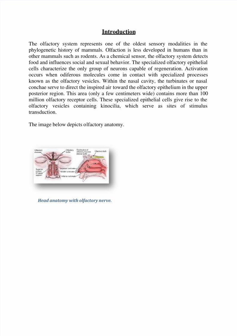
The image below depicts olfactory anatomy.
Head anatomy with olfactory nerve.

8/8/2019 Olfactory
http://slidepdf.com/reader/full/olfactory 2/7
Olfactory Epithelium
The olfactory epithelium consists of 3 cell types, basal, supporting, and olfactory
receptor cells. Basal cells are stem cells that give rise to the olfactory receptor
cells. The continuous turnover and new supply of these neurons are unique to the
olfactory system. In no other location in the mature nervous system do less
differentiated stem cells replace neurons. Supporting cells are scattered among the
receptor cells and have numerous microvilli and secretory granules, which empty
their contents onto the mucosal surface. The receptor cells are actually bipolar
neurons, each possessing a thin dendritic rod that contains specialized cilia
extending from the olfactory vesicle and a long central process that forms the fila
olfactoria. The cilia provide the transduction surface for odorous stimuli.
The vomeronasal organ is a specialized bilateral membranous structure located in
the base of the anterior nasal septum, at the junction of the septal cartilage and thebony septum. It is believed to detect external chemical signals called pheromones.
These signals, which are not detected consciously as odors by the olfactory system,
mediate human autonomic, psychological, and endocrine responses.
The trigeminal nerve innervates the posterior nasal cavity to detect noxious stimuli.
Olfactory Nerve and the Cribriform Plate
The small unmyelinated axons of the olfactory receptor cells form the fine fibers of
the first cranial nerve and travel centrally toward the ipsilateral olfactory bulb to
make contact with the second-order neurons. Conduction velocities are extremely
slow, and support is provided in bundles by a single Schwann cell. As previously
mentioned, the trigeminal nerve (cranial nerve V) sends fibers to the olfactory
epithelium to detect caustic chemicals such as ammonia. The cribriform plate of
the ethmoid bone, separated at the midline by the crista galli, contains multiple
small foramina through which the olfactory nerve fibers, or fila olfactoria, traverse.
Fracture of the cribriform plate in traumatic settings can disrupt these fine fibers
and lead to olfactory dysfunction.

8/8/2019 Olfactory
http://slidepdf.com/reader/full/olfactory 3/7
Olfactory Bulb
The olfactory bulb lies inferior to the basal frontal lobe. The olfactory bulb is a
highly organized structure composed of several distinct layers and synaptic
specializations. The layers (from outside toward the center of the bulb) are
differentiated as follows:
Glomerular layer
External plexiform layer
Mitral cell layer
Internal plexiform layer
Granule cell layer
Mitral cells are second-order neurons contacted by the olfactory nerve fibers at the
glomerular layer of the bulb. The glomerular layer is the most superficial layer,consisting of mitral cell dendritic arborizations (glomeruli), olfactory nerve fibers,
and periglomerular cells. Periglomerular cells contact multiple mitral cell dendrites
within the glomeruli and provide lateral inhibition of neighboring glomeruli while
allowing excitation of a specific mitral cell dendritic tree. Each mitral cell is
contacted by at least 1000 olfactory nerve fibers.
The external plexiform layer contains the passing dendrites of mitral cells and a
few tufted cells, which are similar in size to mitral cells. Some of the granule cell
dendrites in the plexiform layer contact mitral cell dendrites through a specialized
dendrodendritic synapse, which also is termed a reciprocal synapse (vesicles seenwithin both presynaptic and postsynaptic membranes).
Tufted cells also receive granule cell input through dendrodendritic and
dendrosomatic contact. Pyramidal mitral cells are the largest neurons in the bulb
and are located in a narrow band between the external and internal plexiform
layers. The granule cell layer contains multiple small round neurons that lack
axons. Long dendritic processes of the neurons reach the more superficial layers
and inhibit mitral cells and tufted cells. Small distal processes make contacts with
the exiting mitral cell axons.

8/8/2019 Olfactory
http://slidepdf.com/reader/full/olfactory 4/7
Olfactory Tract and Central Pathways
Mitral cell axons project to the olfactory cortex via the olfactory tract. Medial
fibers of the tract contact the anterior olfactory nucleus and the septal area. Some
fibers project to the contralateral olfactory bulb via the anterior commissure.
Lateral fibers contact third-order neurons in the primary olfactory cortex
(prepyriform and entorhinal areas) directly. Third-order neurons send projections
to the dorsomedial nucleus of the thalamus, the basal forebrain, and the limbic
system.
The thalamic connections are thought to serve as a conscious mechanism for odor
perception, while the amygdala and the entorhinal area are limbic system
components and may be involved in the affective components of olfaction.
Investigations of regional cerebral blood flow have demonstrated a significant
increase in the amygdaloid nucleus with the introduction of a highly aversiveodorant stimulus, and this has been associated with subjective perceived
aversiveness.
Central Projections
The pyriform lobe includes the olfactory tract, the uncus, and the anterior part of
the parahippocampal gyrus. The prepyriform and the periamygdaloid areas of the
temporal lobe represent the primary olfactory cortex. The entorhinal area is knownas the secondary olfactory cortex and is included in the pyriform lobe. The
olfactory system is the only sensory system that has direct cortical projections
without a thalamic relay nucleus. The dorsomedial nucleus of the thalamus
receives some olfactory fibers that ultimately reach the orbitofrontal cortex.
The anterior olfactory nucleus receives collateral fibers from the olfactory tract and
projects to the contralateral olfactory bulb and anterior olfactory nucleus via the
anterior commissure.
The region of anterior perforated substance contains cells that receive direct mitral
cell collaterals and input from the anterior olfactory nucleus, amygdaloid nucleus,
and temporal cortex. This area ultimately projects to the stria medullaris and the
medial forebrain bundle.
Using the uncinate fasciculus, the entorhinal area sends projections to the
hippocampal formation, anterior insula, and frontal cortex.

8/8/2019 Olfactory
http://slidepdf.com/reader/full/olfactory 5/7
Clinical Correlation
As many as 2 million people in the United States experience some type of
olfactory dysfunction, causes of which include head trauma, upper respiratory
infections, tumors of the anterior cranial fossa, and exposure to toxic chemicals orinfections. The following terms are used to describe the degree of smell aberration:
Anosmia - Absence of smell sensation
Hyposmia - Decreased sensation
Dysosmia - Distortion of smell sensation
Cacosmia - Sensation of a bad or foul smell
Parosmia - Sensation of smell in the absence of appropriate stimulus
Olfactory dysfunction is a hallmark of certain syndromes such as Kallmann
syndrome (ie, hypogonadism with anosmia) and Foster Kennedy syndrome (ie,papilledema, unilateral anosmia, and optic atrophy usually associated with an
olfactory groove meningioma).
The classic description of partial complex epilepsy with a mesial temporal focus
includes an aura of foul-smelling odors (termed uncinate fits) that occur before
seizure onset, emphasizing presumed origination at the uncus.
Olfactory dysfunction is associated with early Parkinson disease and with other
neurodegenerative disorders such as Alzheimer disease and Huntington chorea.1
An association also exists between abnormal olfactory identification andobsessive-compulsive disorder.
2
Head trauma leading to fracture of the cribriform plate may cause cerebrospinal
fluid (CSF) rhinorrhea and a potential for meningitis. Paranasal sinus endoscopy
may lead to violation of the cribriform plate and potential infectious complications.
Olfactory structures also can be injured during craniotomies involving the anterior
cranial base or from subarachnoid hemorrhage, which may disrupt the fine fibers
of the olfactory nerve.

8/8/2019 Olfactory
http://slidepdf.com/reader/full/olfactory 6/7
Clinical Evaluation
Detection of olfactory dysfunction begins with sampling of a series of common
odors, which can be performed at the bedside with odiferous substances such as
coffee, lemon, and peppermint. Tests, including those developed at the Connecticut
Chemosensory Clinical Research Center (CCCRC), have aided examiners in
identification of abnormalities in odor detection and discrimination. The University
of Pennsylvania Smell Identification Test (UPSIT) is another useful tool; it
consists of 40 items for evaluation of olfactory and trigeminal nerve function in the
nasal cavity.
Central hyposmia may manifest as abnormalities in odor recognition rather than
odor detection. Thorough evaluation of patients who have anosmia includes
imaging of anterior cranial structures. The clinician should always counsel patients
with anosmia regarding sensory loss, including potential risks associated with thelack of smell sensation (eg, inability to detect dangers such as smoke, spoiled
foods, toxins).
Promptly complete evaluation and treatment of clear rhinorrhea in the patient in
whom leakage of CSF is suspected. Initial testing of fluid for glucose suggests CSF
but is not confirmatory. Presence of beta-transferrin is a more sensitive indicator of
CSF rhinorrhea. Computed tomography with cisternography or radionuclide scans
can be used to detect the site of CSF leakage from the anterior cranial fossa. Repair
of leaks at the level of the cribriform plate may be achieved from the intracranial
approach, intranasal (endoscopic) approach, or both, depending on the nature of the defect.
Positron emission tomography (PET) and functional MRI are promising modalities
to assist in making the diagnosis of different types of hyposmia (central vs
peripheral), as well as in delineation of the role of limbic structures as sites of odor
recognition, memory, and integration of multisensory inputs.

8/8/2019 Olfactory
http://slidepdf.com/reader/full/olfactory 7/7
Multimedia