Embed Size (px)
Citation preview

Okay Anatomy
Anatomy I: Lesson 12
Myologia – Practical Tasks 11-14
Objective: Students will examine the practical dissection tasks 11-14 in order to prepare and succeed on the practical final exam.

Practical Tasks:
• 11) muscles of the pelvic girdle
• 12) dorsal muscles of the buttock
• 13) deep muscles of the buttock
• 14) caudal muscles of the buttock

Practical Task 11: Musculature of the Pelvic Girdle
** General Considerations: • They are not as many as in the
shoulder girdle, as in the pelvic girdle the pelvis is firmly connected to the trunk by a „thight joint“. In Ca. powerful, with relatively few tendons.
• These muscles originate from the vertebral column and insert on the pelvis or the femur, thus they are able to move position of the pelvis. These muscles arising partly from the sacrum and caudal vertabrae are important activators of the hip and knee joints as part of the intrinsic trunk-limb muscles.
• They include the following three (3) muscles:

1) M. psoas minor : statically important in fixing the pelvis; covering parts of M. psoas major;
Origin: (Evans, p. 78/52)
– (ventrally) from bodies of last 2 or 3 thoracic vertebrae, and
– from first 4-5 lumbar vertebrae
Insertions: ilum (psoas tubercle)
– (or in Ca): arcuate line
Actions:
– draw pelvis into a deeper position at fixed vertebral column
– to stabilize and arch lumbar vertebral column dorsally when pelvis is fixed
B) Musculature of the Pelvic Girdle

2) M. iliopsoas :
Body: two parts: (in Ca: NOT separated)
– M. psoas major (the lumbar part; lateral to psoas minor on ventral surface of M. quadratus lumborum), and
– M. iliacus (iliac part)
Origin:
– of M. psoas major → ventrolaterally from the bodies and transverse processes of the lumbar vertebrae, and
– from last two thoracic vertebrae, and
– from the proximal ends of their ribs
– of M. iliacus → ventral surface of wing and shaft of ilium
B) Musculature of the Pelvic Girdle
2) M. iliopsoas :
Insertion:
of psoas major and of iliacus → lesser trochanter of femur (together with the iliacus that blends with it)
Actions:
– draw thigh forward by flexing the hip joint and turning stifle joint outwards
– draw pelvis into steeper position
– when limb fixed → stabilizes vertebral column or arches it dorsally
– when limb is drawn backwards → pulls trunk caudally
B) Musculature of the Pelvic Girdle

• 3) M. quadratus lumborum : in Ca: strong muscular sheet, but thinner in other domestic mammals; covered by M. psoas major (Evans p. 78/52); is the most dorsally located one of the sublumbar muscles;
• Origin: attached ventrally to transvere processes of lumbar vertebrae and vertebral extremities of last ribs
• Insertions: transverse processes and wings of the sacrum (Bo, Eq), or on the ilium (Ca, Su)
• Actions: to fix and flex the lumbar vertebral column dorsally
B) Musculature of the Pelvic Girdle

Practical Task 12 & 13:
• Dorsal Muscles of the Buttock
• Deep Muscles of the Buttock

1) M. glutaeus superficialis : it is considerably weaker in animals than in man. It is a separate muscle only in Ca, but in Su and Bo fused with cranial portion of the biceps → glutaeobiceps; in Eq it is connected with caudal portion of the tensor fasciae latae (p. 326/403-36,37)
Body: it is made up by the following four (4) parts (heads), also differing between different species:
1a) cranial superficial
1b) caudofemoral (lies behind „1a“ only in the cat)
1c) vertebral head and the terminal branch of M. biceps femoris (cranial biceps portion)
1d) vertebral head of the M. semitendinosus (in Su and Eq).
C) Intrinsic Musculature of the Pelvic Limbs

Origins:
– in Ca: sacrum and 1st caudal vertebra, and sacrotuberal ligament, and gluteal fascia
– in Eq: coxal tuber, and gluteal fascia
Insertions: greater trochanter (Ca); 3rd trochanter of femur (Eq)
Actions:
– in Ca: extend hip joint, and draw limb backwards
– in Eq, Bo, Su: flex the hip, and abduct and draw limb forwards
C) Intrinsic Musculature of the Pelvic Limbs
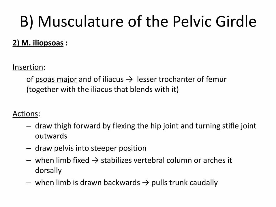
2) M. caudofemoralis : only in the cat; previously known as: M. abductor cruris cranialis; a powerful, elongated muscle, situated between biceps femoris and the superficial gluteal.
Origin: 2nd – 4th caudal vertebrae
Insertions:
– in a thin aponeurosis blending with fascia lata
– laterally on patella
Actions:
– when caudal vertebrae fixed: → draw limb outwards and backwards → helping extend hip joint
– when limb fixed: → pull the tail laterally
C) Intrinsic Musculature of the Pelvic Limbs

3) M. glutaeus medius : one of the strongest of the hip muscles, the strongest extensor of the hip joint; covered by the gluteal and the thoracolumbar fascia, and by the superficial gluteal muscle; it lies on the outer wings of the ilium.
Body:
superficial
deep → also known as: M. glutaeus accessorius
Origins:
in dog (and man): outer surface of the wings of the ilium
other animals:
wings of ilium,
and sacrum, and
(with its lumbar portion): from the covering aponeurosis of the M. longissimus lumborum
C) Intrinsic Musculature of the Pelvic Limbs

3) M. glutaeus medius :
Insertions: greater trochanter
in Eq, Bo, Su: it blends caudally with M. piriformis
Actions:
draw limb backwards and outwards
when limb is weighed → powerfully propelling the trunk forwards
together with the M. longissimus (to which it is connected): one of the main forces in „rearing“ and kicking (esp. In Eq).
C) Intrinsic Musculature of the Pelvic Limbs

• 4) M. piriformis : is an individual muscle only in Ca (and man); in Su, Bo, Eq: fused with the middle gluteal muscle; (Evans p. 67/44)
• Origins: ventrally and laterally from sacrum (behind the middle gluteal)
• Insertions: greater trochanter • Actions:
– extend hip joint – abduct the limb – also: support actions of the
middle gluteal muscle
C) Intrinsic Musculature of the Pelvic Limbs

5) M. glutaeus profundus : small but powerful muscle; directly on the hip joint, covered by the middle gluteal muscle;
Origins:
– ischiatic spine or
– body of ilium
Insertion: greater trochanter
Actions:
– abduct the limb
– support actions of the middle gluteal muscle
C) Intrinsic Musculature of the Pelvic Limbs

d) Deep muscles of the hip joint
A small group of muscles close to the hip joint, covered in main by the muscles already described above. They are mainly doing supination, and their significance is in performing a more delicate influence on the course of the limb's movement.
This group include the following five (5) muscles, of which the first 4 arise from the inner and outer surface of the ischium and insert in the trochanteric fossa of the femur.
Caudal Hip Muscles (hip joint)

d) Deep muscles of the hip joint
• 1) M. obturatorius internus : thin sheet of muscle, only present in Ca and Eq
• Origins: inner surfa of pelvis aroud the obturator foramen; in Eq also from body of ilium
• Insertion: trochanteric fossa
• Actions:
– rotate thigh outwards
– assist in extension of the hip
Caudal Hip Muscles (hip joint)

d) Deep muscles of the hip joint
• 2) M. obturatorius externus : a strong, pyramidal muscle; covered by: Mm. adductores, M. pectineus, and M. semimembranosus;
• Origins: ventral surface of pelvis around the obturator foramen
– in Su, Bo: inner surface of pelvis
– in Su: also from body of ilium and sacrum
• Insertions: trochanteric fossa
• Actions:
– suppinate the thigh
– adduct the limb
– pull the trunk towards the limb when the limb is placed laterally
Caudal Hip Muscles (hip joint)

d) Deep muscles of the hip joint
• 3) Mm. gemelli : (in man: two small muscles → fused into single muscle in domestic mammals) (Evans p. 67/44; p. 74/49)
• Origins:
– ischiatic spine
– body of ischium – its lateral surface caudal to acetabulum, ventral to the lesser ischiatic notch
• Insertion: trochanteric fossa
• Actions:
– rotate thigh outwards
– assist in extension of the hip
Caudal Hip Muscles (hip joint)

d) Deep muscles of the hip joint
• 4) M. quadratus femoris : a short and thick muscular strap, caudolaterally to the Mm. gemelli (Evans p. 67/44)
• Origin: ventral aspect of sacrum / ventral surface of the caudal part of the ischium
• Insertion: intertrochanteric crest near trochanteric fossa
• Actions:
– suppinate the thigh
– extensor of hip joint
– helps in drawing the limb backwards
Caudal Hip Muscles (hip joint)

Practical Task 14
• Caudal Muscles of the Buttock

• This huge mass of muscle lies mainly between the ischium and the crus forming the caudal half of the thigh.
• Besides the pelvic head, buttock muscles can also have vertebral heads (originating from sacrum and 1st caudal vertebra → thus they are also part of the muscels of the croup.
• They activate the hip and the stifle joint, and are also associated with the hock.
• This group includes the following four (4) muscles:
b) Muscles of the buttock = Caudal Thigh Muscles (hip joint)
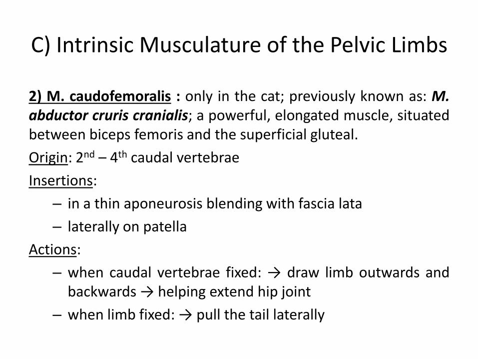
1) M. biceps femoris : it foms the caudal and lateral regions of the thigh and lies immediately underneath of the skin and the fascial planes. In man it is the strongest muscle of the whole body, but the situation is not the same in domestic animals. In the dog it is the widest and longest muscle of the thigh. (Evans p. 65/42)
Body: two parts → intimately connected to each other:
– cranial portion = vertebral- and sacral head (strong) → in Su, Bo: the vertebral head blends with the M. glutaeus superficialis ==> forming the glutaeobiceps
– caudal portion = pelvic head (smaller)
* the muscle divides distally into 2 branches (Ca, Su, Ru) or 3 (Eq)
b) Muscles of the buttock = Caudal Thigh Muscles (hip joint)

1) M. biceps femoris :
Origins:
– vertebral head from: sacrum and the sacrotuberal ligament
– pelvic head from: ischiatic tuber
Insertions: as strong, flat aponeuroses blending with the femoral and crural fascia → inserted on the:
– patellar ligament
– patella
– tibial crest (margo cranialis)
– also on tuber calcanei by means of the calcanean tendon
b) Muscles of the buttock = Caudal Thigh Muscles (hip joint)

1) M. biceps femoris :
Actions: the muscle as a whole: abduct and extend the limb;
– when limb is fixed on the ground: → produce powerful forwards thrust on the trunk
– (cranial partion) → extend hip and stifle joint
– (caudal portion) → flex the stifle joint
b) Muscles of the buttock = Caudal Thigh Muscles (hip joint)

2) M. abductor cruris caudalis :
only in Ca; it lies laterally on the thigh and proximal half of the crus, covered mostly by the M. biceps femoris. It is a thin band of muscle in the dog (p. 412/464-c; Evans p. 67/44), in the cat a rudimentary pale, thin band;
Origin: with a long, delicate tendon from the distal end of the sacrotuberal ligament
Insertions: blending with the crural fascia after having crossed the M. gastrocnemius and fused with the caudal border of the M. biceps femoris;
Action: (insignificant) supporting the caudal portion of the biceps as an abductor
b) Muscles of the buttock = Caudal Thigh Muscles (hip joint)

3) M. semitendinosus : in domestic mammals it is a fleshy and powerful muscle, forming the greater part of the caudal margin of the buttock; (Evans p. 67/44)
Origins:
– ischiatic tuber (Ca, Bo, Eq, Su) and,
– also spinous- and transverse processes of 1st caudal vertebrae (Su, Eq) → vertebral head
Insertions: in stifle region medially on the tibial crest where its extensive tendon blends with the aponeuroses of the gracilis and sartorius muscles
Actions:
– extend the hip-, stifle- and hock joints
– propel trunk forewards
– with the limb off the ground → flexing stifle joint
• drawing the limb backwards and inwards
b) Muscles of the buttock = Caudal Thigh Muscles (hip joint)

4) M. semimembranosus : on medial aspect of the buttock, a fleshy, dark red muscle (p. 412/ 465-9,9')
Body: only in Eq: an additional vertebral head
– (Ca, Su, Bo) → muscle belly divides into two
– (Eq) → only the terminal tendon bifurcates
Origins: ischiatic tuber (its ventral aspect)
– (in Eq → vertebral head) from: sacrosciatic ligament
Insertions:
– medial condyle of femur
– medially on tibia
Actions:
– when supporting the limb: extend hip- and stifle joints, and propel trunk forewards
– in hanging limb: adduct, pronate and pull the limb backwards
b) Muscles of the buttock = Caudal Thigh Muscles (hip joint)


![Dr. Mohammad Nazam Ansari Digestive System Anatomy Practical [PHL 212]](https://img.pdfslide.us/doc/110x75/5697c0031a28abf838cc3c9a/dr-mohammad-nazam-ansari-digestive-system-anatomy-practical-phl-212.jpg)


![Cardiovascular System Anatomy Practical [PHL 212]](https://img.pdfslide.us/doc/110x75/5697c01d1a28abf838cd05f5/cardiovascular-system-anatomy-practical-phl-212.jpg)