Embed Size (px)
Citation preview
Soft tissue conditioning with the Straumann® Bone Level Implant line
Straumann® Bone Level Implant line
15X.533.indd 1 21.04.15 10:09

2
The ITI (International Team for Implantology) is academic partner of Institut Straumann AG in the areas of research and education.
15X.533.indd 2 21.04.15 10:09
1
Surgical intervention(transmucosal healing)
Surgical intervention(submucosal healing)
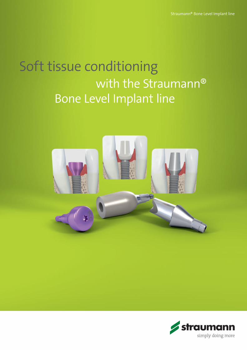
Communication is key in the dental implant treatment flow
Interdisciplinary treatment planning includes the collaboration of different parties such as: ѹ Patient ѹ General practitioner ѹ Surgeon ѹ Prosthodontist ѹ Dental laboratory
In order to reach a successful treatment outcome, communication between all involved is essential. The parties should be aware of the steps and interfaces, and exchange their opinions in order to be able to make the correct decisions.
A general overview of the treatment flow is depicted below, broken down into various treatment steps. Before each decision step (blue in the picture below) communication can be helpful.
Procedure step Decision/communication step
Case analysis andtreatment planning
Treatment typeFlap opening
Implant placement
1st flap opening
Implant placement
Close flap
Flap re-opening
Healing Abutment choice
Healing Abutment placement
Temporization
Temporary placement in order to perform soft
tissue conditioning
Final restoration
15X.533.indd 1 21.04.15 10:09
2
Treatment flow
low
high
low highTreatment time & costs
Esth
etic
com
plex
ity
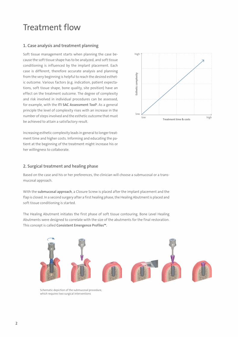
1. Case analysis and treatment planning
Soft tissue management starts when planning the case be-cause the soft tissue shape has to be analyzed, and soft tissue conditioning is influenced by the implant placement. Each case is different, therefore accurate analysis and planning from the very beginning is helpful to reach the desired esthet-ic outcome. Various factors (e.g. indication, patient expecta-tions, soft tissue shape, bone quality, site position) have an effect on the treatment outcome. The degree of complexity and risk involved in individual procedures can be assessed, for example, with the ITI SAC Assessment Tool1. As a general principle the level of complexity rises with an increase in the number of steps involved and the esthetic outcome that must be achieved to attain a satisfactory result.
Increasing esthetic complexity leads in general to longer treat-ment time and higher costs. Informing and educating the pa-tient at the beginning of the treatment might increase his or her willingness to collaborate.
2. Surgical treatment and healing phase
Based on the case and his or her preferences, the clinician will choose a submucosal or a trans-mucosal approach.
With the submucosal approach, a Closure Screw is placed after the implant placement and the flap is closed. In a second surgery after a first healing phase, the Healing Abutment is placed and soft tissue conditioning is started.
The Healing Abutment initiates the first phase of soft tissue contouring. Bone Level Healing Abutments were designed to correlate with the size of the abutments for the final restoration. This concept is called Consistent Emergence Profiles™.
Schematic depiction of the submucosal procedure, which requires two surgical interventions
15X.533.indd 2 21.04.15 10:09
3
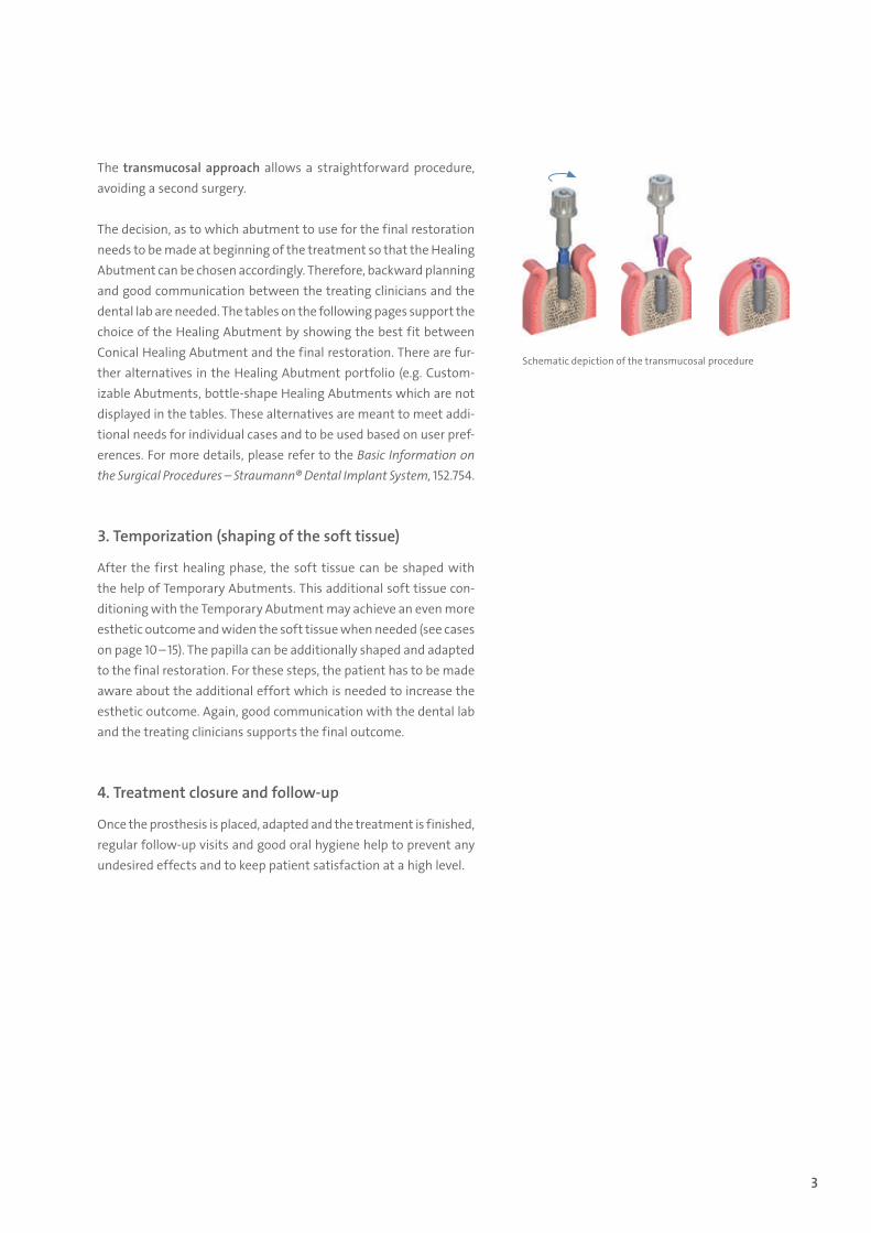
Schematic depiction of the transmucosal procedure
The transmucosal approach allows a straightforward procedure, avoiding a second surgery.
The decision, as to which abutment to use for the final restoration needs to be made at beginning of the treatment so that the Healing Abutment can be chosen accordingly. Therefore, backward planning and good communication between the treating clinicians and the dental lab are needed. The tables on the following pages support the choice of the Healing Abutment by showing the best fit between Conical Healing Abutment and the final restoration. There are fur-ther alternatives in the Healing Abutment portfolio (e.g. Custom-izable Abutments, bottle-shape Healing Abutments which are not displayed in the tables. These alternatives are meant to meet addi-tional needs for individual cases and to be used based on user pref-erences. For more details, please refer to the Basic Information on the Surgical Procedures – Straumann® Dental Implant System, 152.754.
3. Temporization (shaping of the soft tissue)
After the first healing phase, the soft tissue can be shaped with the help of Temporary Abutments. This additional soft tissue con-ditioning with the Temporary Abutment may achieve an even more esthetic outcome and widen the soft tissue when needed (see cases on page 10 – 15). The papilla can be additionally shaped and adapted to the final restoration. For these steps, the patient has to be made aware about the additional effort which is needed to increase the esthetic outcome. Again, good communication with the dental lab and the treating clinicians supports the final outcome.
4. Treatment closure and follow-up
Once the prosthesis is placed, adapted and the treatment is finished, regular follow-up visits and good oral hygiene help to prevent any undesired effects and to keep patient satisfaction at a high level.
15X.533.indd 3 21.04.15 10:09
4
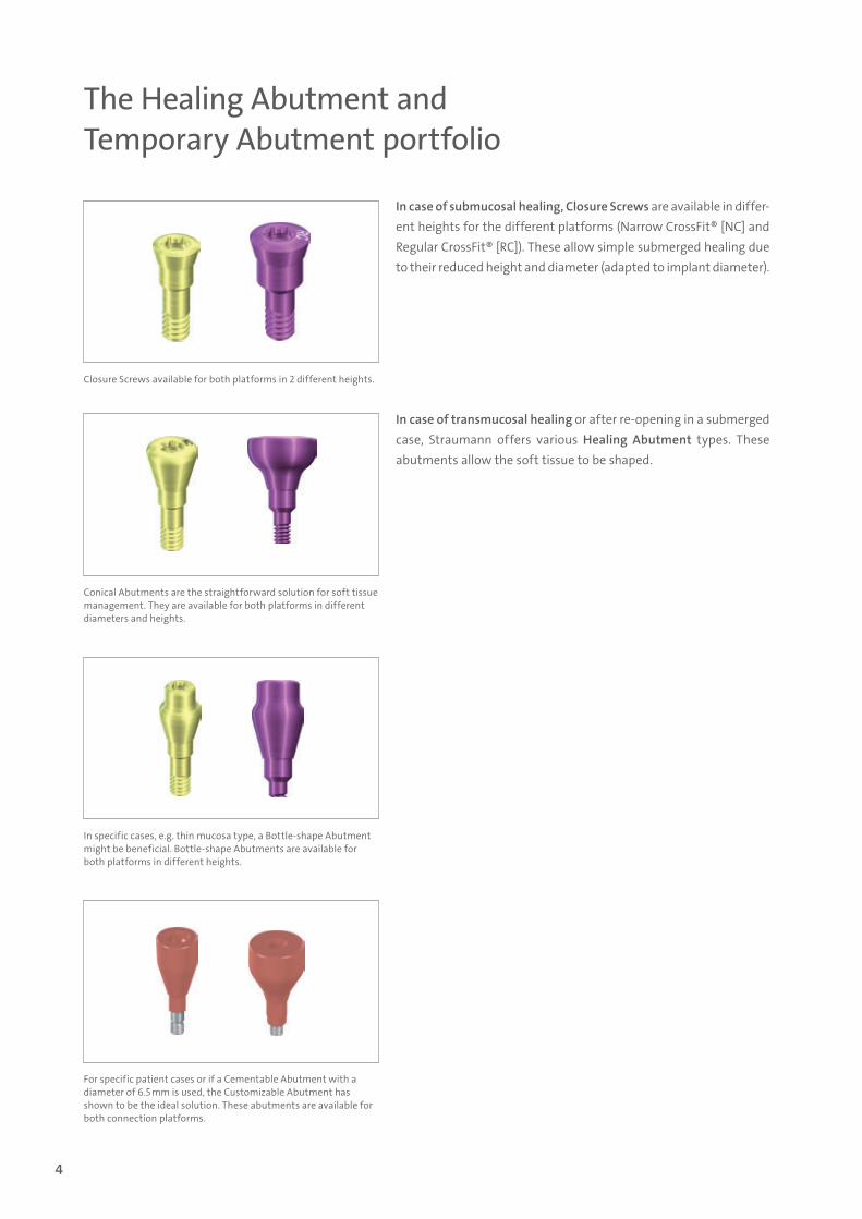
The Healing Abutment and Temporary Abutment portfolio
In case of submucosal healing, Closure Screws are available in differ-ent heights for the different platforms (Narrow CrossFit® [NC] and Regular CrossFit® [RC]). These allow simple submerged healing due to their reduced height and diameter (adapted to implant diameter).
In case of transmucosal healing or after re-opening in a submerged case, Straumann offers various Healing Abutment types. These abutments allow the soft tissue to be shaped.
Closure Screws available for both platforms in 2 different heights.
Conical Abutments are the straightforward solution for soft tissue management. They are available for both platforms in different diameters and heights.
In specific cases, e.g. thin mucosa type, a Bottle-shape Abutment might be beneficial. Bottle-shape Abutments are available for both platforms in different heights.
For specific patient cases or if a Cementable Abutment with a diameter of 6.5 mm is used, the Customizable Abutment has shown to be the ideal solution. These abutments are available for both connection platforms.
15X.533.indd 4 21.04.15 10:09
5
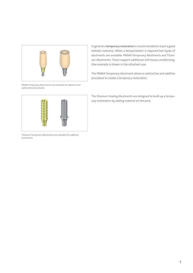
In general a temporary restoration is recommended to reach a good esthetic outcome. When a temporization is required two types of abutments are available: PMMA Temporary Abutments and Titani-um Abutments. These support additional soft tissue conditioning. One example is shown in the attached case.
The PMMA Temporary Abutment allows a subtractive and additive procedure to create a temporary restoration.
The titanium Healing Abutments are designed to build up a tempo-rary restoration by adding material on the post.
PMMA Temporary Abutments are available for additive and subtractive procedures.
Titanium Temporary Abutments are available for additive procedures.
15X.533.indd 5 21.04.15 10:09
6
The Consistent Emergence Profiles™ concept
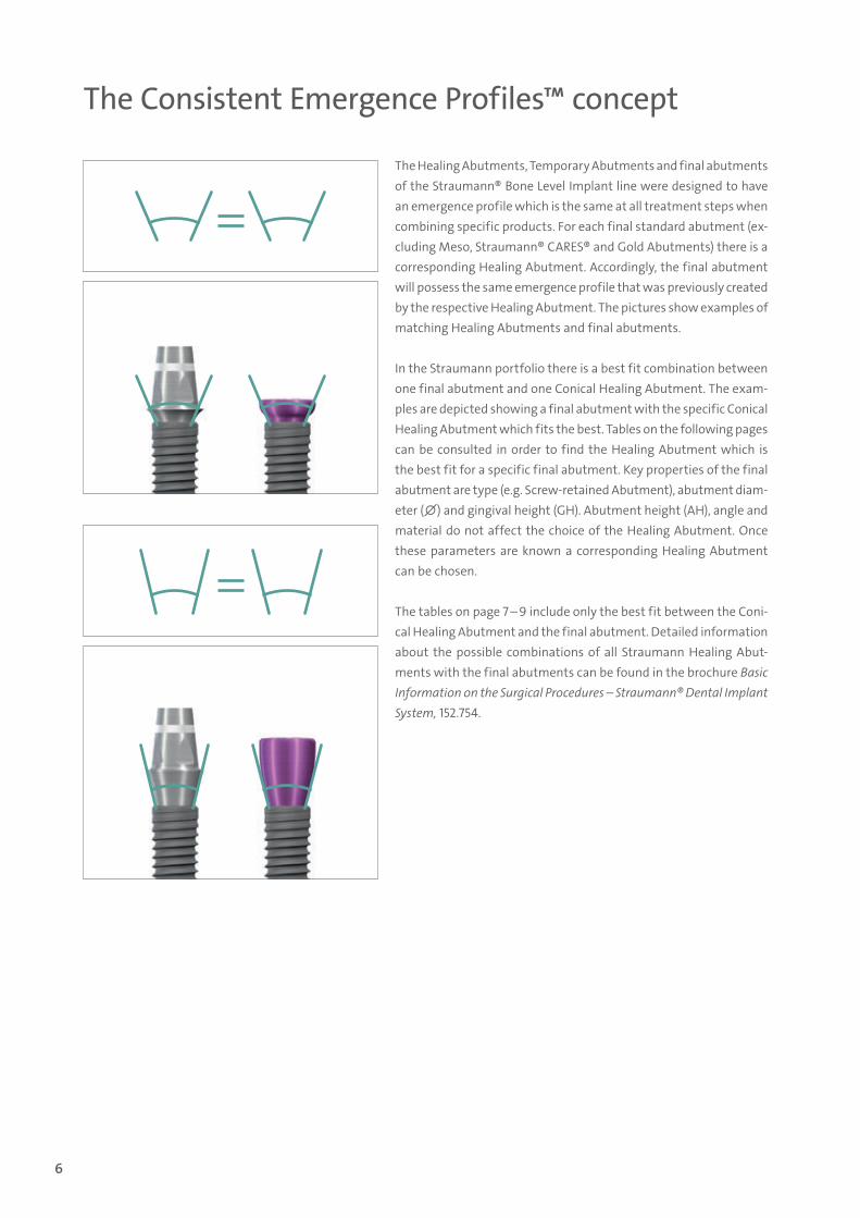
The Healing Abutments, Temporary Abutments and final abutments of the Straumann® Bone Level Implant line were designed to have an emergence profile which is the same at all treatment steps when combining specific products. For each final standard abutment (ex-cluding Meso, Straumann® CARES® and Gold Abutments) there is a corresponding Healing Abutment. Accordingly, the final abutment will possess the same emergence profile that was previously created by the respective Healing Abutment. The pictures show examples of matching Healing Abutments and final abutments.
In the Straumann portfolio there is a best fit combination between one final abutment and one Conical Healing Abutment. The exam-ples are depicted showing a final abutment with the specific Conical Healing Abutment which fits the best. Tables on the following pages can be consulted in order to find the Healing Abutment which is the best fit for a specific final abutment. Key properties of the final abutment are type (e.g. Screw-retained Abutment), abutment diam-eter (∅) and gingival height (GH). Abutment height (AH), angle and material do not affect the choice of the Healing Abutment. Once these parameters are known a corresponding Healing Abutment can be chosen.
The tables on page 7 – 9 include only the best fit between the Coni-cal Healing Abutment and the final abutment. Detailed information about the possible combinations of all Straumann Healing Abut-ments with the final abutments can be found in the brochure Basic Information on the Surgical Procedures – Straumann® Dental Implant System, 152.754.
15X.533.indd 6 21.04.15 10:09
7
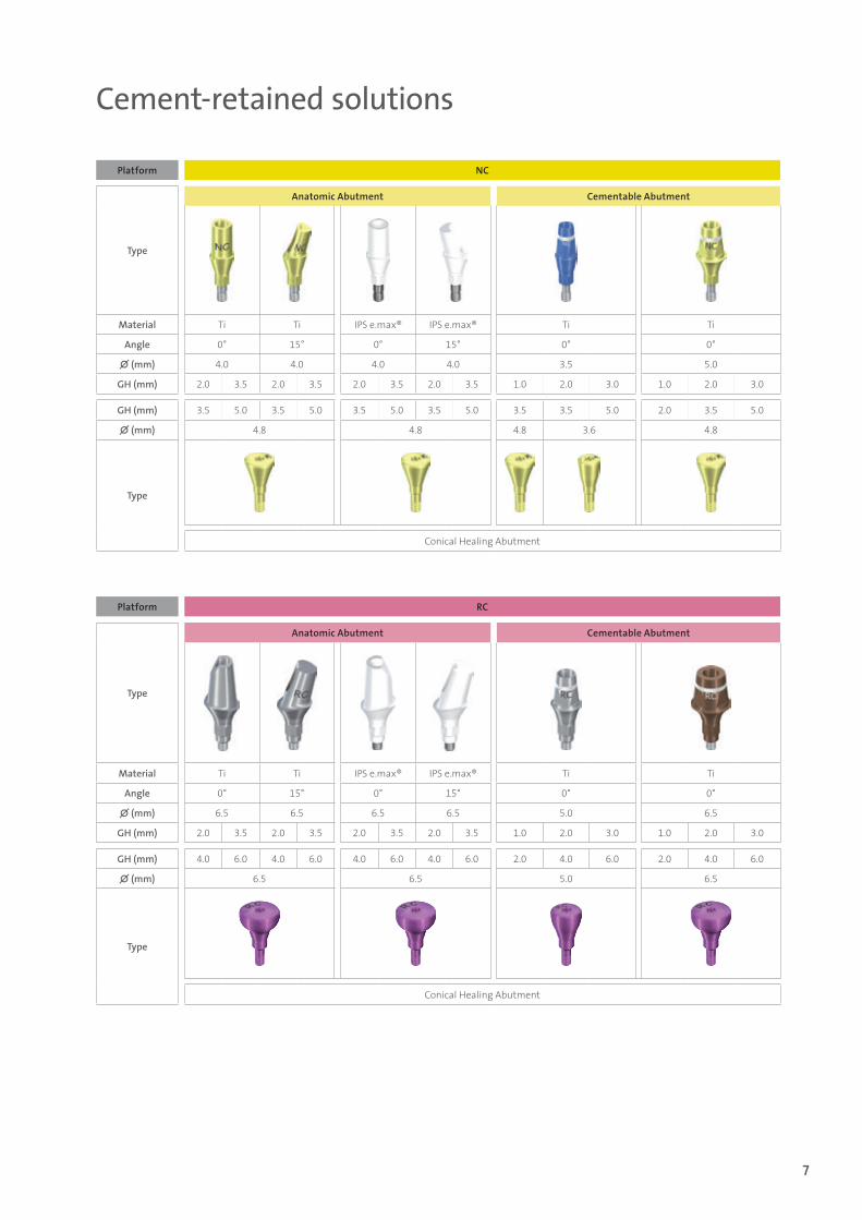
Cement-retained solutions
Platform NC
Type
Anatomic Abutment Cementable Abutment
Material Ti Ti IPS e.max® IPS e.max® Ti Ti
Angle 0° 15° 0° 15° 0° 0°
∅ (mm) 4.0 4.0 4.0 4.0 3.5 5.0
GH (mm) 2.0 3.5 2.0 3.5 2.0 3.5 2.0 3.5 1.0 2.0 3.0 1.0 2.0 3.0
GH (mm) 3.5 5.0 3.5 5.0 3.5 5.0 3.5 5.0 3.5 3.5 5.0 2.0 3.5 5.0
∅ (mm) 4.8 4.8 4.8 3.6 4.8
Type
Conical Healing Abutment
Platform RC
Type
Anatomic Abutment Cementable Abutment
Material Ti Ti IPS e.max® IPS e.max® Ti Ti
Angle 0° 15° 0° 15° 0° 0°
∅ (mm) 6.5 6.5 6.5 6.5 5.0 6.5
GH (mm) 2.0 3.5 2.0 3.5 2.0 3.5 2.0 3.5 1.0 2.0 3.0 1.0 2.0 3.0
GH (mm) 4.0 6.0 4.0 6.0 4.0 6.0 4.0 6.0 2.0 4.0 6.0 2.0 4.0 6.0
∅ (mm) 6.5 6.5 5.0 6.5
Type
Conical Healing Abutment
15X.533.indd 7 21.04.15 10:09
8
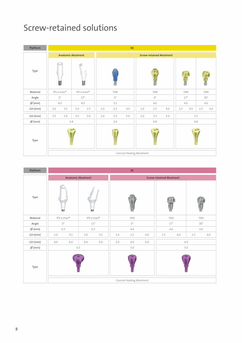
Screw-retained solutions
Platform NC
Type
Anatomic Abutment Screw-retained Abutment
Material IPS e.max® IPS e.max® TAN TAN TAN TAN
Angle 0° 15° 0° 0° 17° 30°
∅ (mm) 4.0 4.0 3.5 4.6 4.6 4.6
GH (mm) 2.0 3.5 2.0 3.5 1.0 2.5 4.0 1.0 2.5 4.0 2.5 4.0 2.5 4.0
GH (mm) 3.5 5.0 3.5 5.0 2.0 3.5 5.0 2.0 3.5 5.0 3.5
∅ (mm) 4.8 3.6 4.8 4.8
Type
Conical Healing Abutment
Platform RC
Type
Anatomic Abutment Screw-retained Abutment
Material IPS e.max® IPS e.max® TAN TAN TAN
Angle 0° 15° 0° 17° 30°
∅ (mm) 6.5 6.5 4.6 4.6 4.6
GH (mm) 2.0 3.5 2.0 3.5 1.0 2.5 4.0 2.5 4.0 2.5 4.0
GH (mm) 4.0 6.0 4.0 6.0 2.0 4.0 6.0 4.0
∅ (mm) 6.5 5.0 5.0
Type
Conical Healing Abutment
15X.533.indd 8 21.04.15 10:09
9
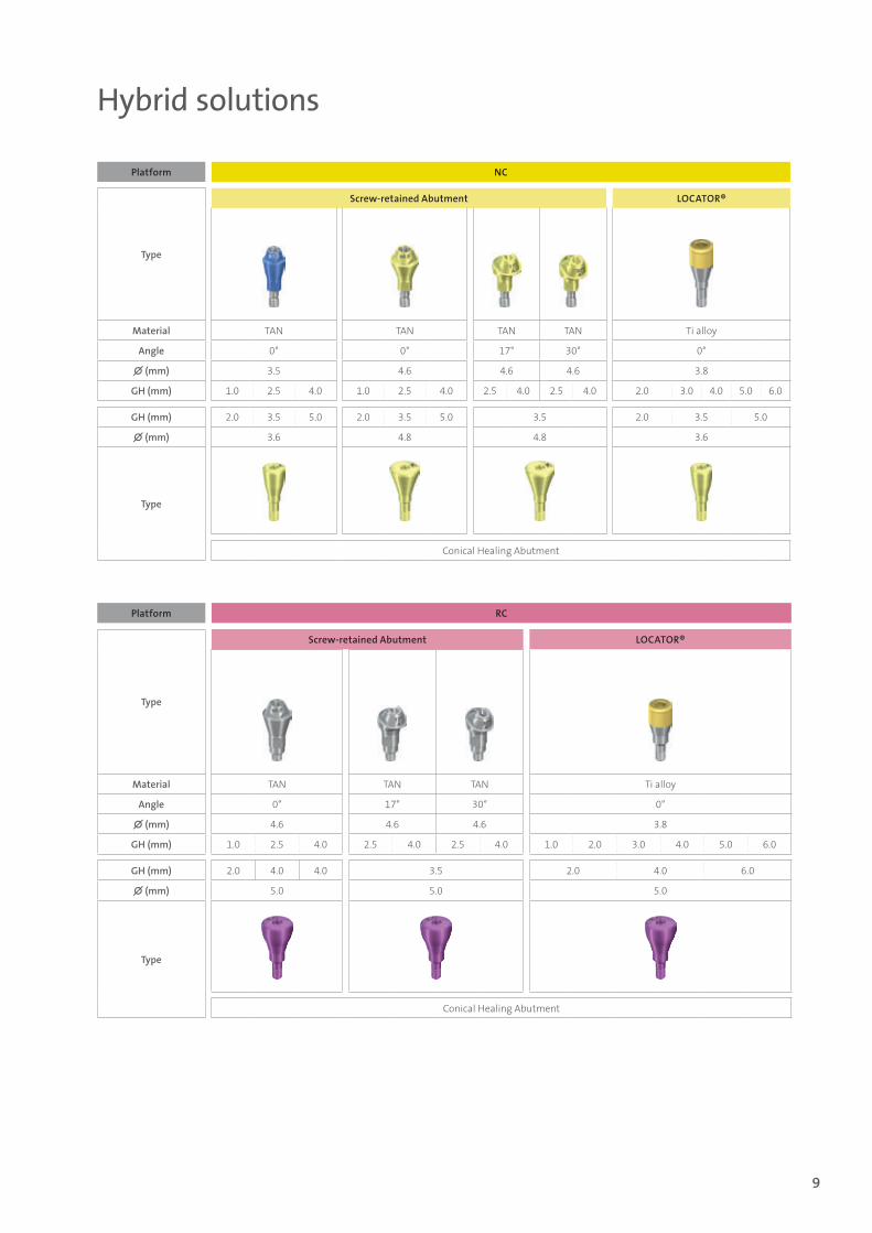
Hybrid solutions
Platform NC
Type
Screw-retained Abutment LOCATOR®
Material TAN TAN TAN TAN Ti alloy
Angle 0° 0° 17° 30° 0°
∅ (mm) 3.5 4.6 4.6 4.6 3.8
GH (mm) 1.0 2.5 4.0 1.0 2.5 4.0 2.5 4.0 2.5 4.0 2.0 3.0 4.0 5.0 6.0
GH (mm) 2.0 3.5 5.0 2.0 3.5 5.0 3.5 2.0 3.5 5.0
∅ (mm) 3.6 4.8 4.8 3.6
Type
Conical Healing Abutment
Platform RC
Type
Screw-retained Abutment LOCATOR®
Material TAN TAN TAN Ti alloy
Angle 0° 17° 30° 0°
∅ (mm) 4.6 4.6 4.6 3.8
GH (mm) 1.0 2.5 4.0 2.5 4.0 2.5 4.0 1.0 2.0 3.0 4.0 5.0 6.0
GH (mm) 2.0 4.0 4.0 3.5 2.0 4.0 6.0
∅ (mm) 5.0 5.0 5.0
Type
Conical Healing Abutment
15X.533.indd 9 21.04.15 10:09
10
Case example for soft tissue conditioning
Case: Soft tissue conditioning with the dynamic compression technique2
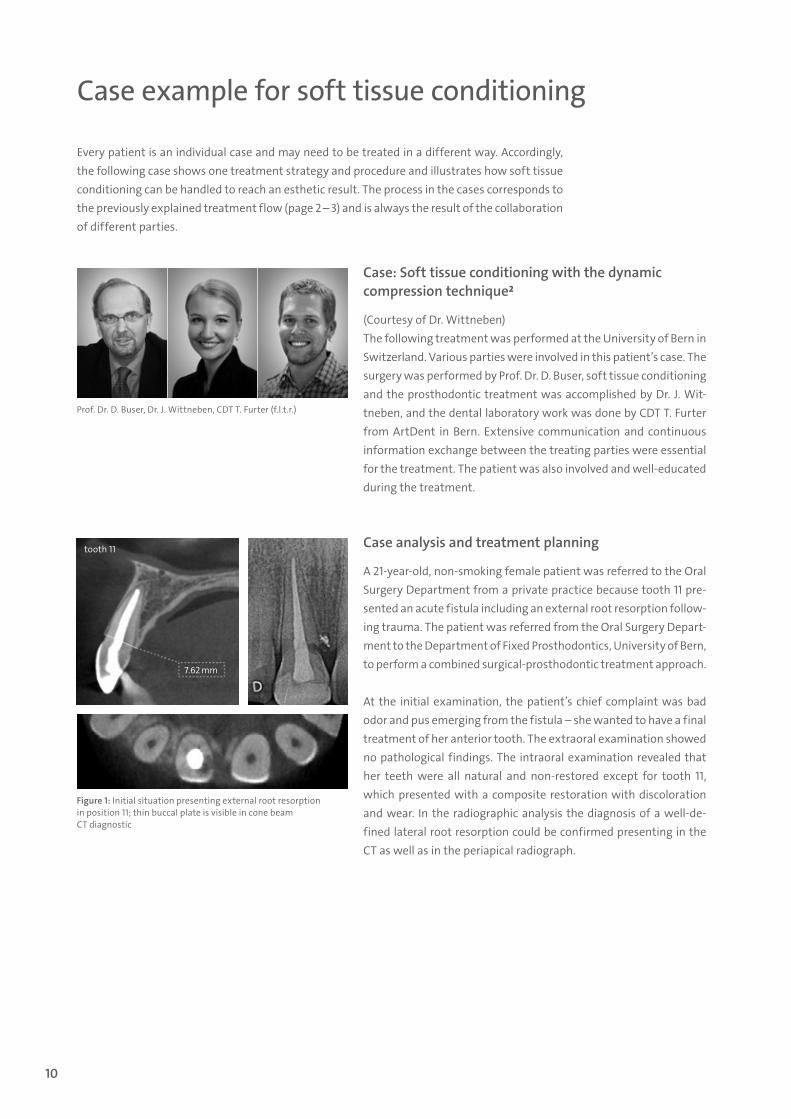
(Courtesy of Dr. Wittneben)The following treatment was performed at the University of Bern in Switzerland. Various parties were involved in this patient’s case. The surgery was performed by Prof. Dr. D. Buser, soft tissue conditioning and the prosthodontic treatment was accomplished by Dr. J. Wit-tneben, and the dental laboratory work was done by CDT T. Furter from ArtDent in Bern. Extensive com munication and continuous information exchange between the treating parties were essential for the treatment. The patient was also involved and well-educated during the treatment.
Case analysis and treatment planning
A 21-year-old, non-smoking female patient was referred to the Oral Surgery Department from a private practice because tooth 11 pre-sented an acute fistula including an external root resorption follow-ing trauma. The patient was referred from the Oral Surgery Depart-ment to the Department of Fixed Prosthodontics, University of Bern, to perform a combined surgical-prosthodontic treatment approach.
At the initial examination, the patient’s chief complaint was bad odor and pus emerging from the fistula – she wanted to have a final treatment of her anterior tooth. The extraoral examination showed no pathological findings. The intraoral examination revealed that her teeth were all natural and non-restored except for tooth 11, which presented with a composite restoration with discoloration and wear. In the radiographic analysis the diagnosis of a well-de-fined lateral root resorption could be confirmed presenting in the CT as well as in the periapical radiograph.
Prof. Dr. D. Buser, Dr. J. Wittneben, CDT T. Furter (f.l.t.r.)
Figure 1: Initial situation presenting external root resorption in position 11; thin buccal plate is visible in cone beam CT diagnostic
Every patient is an individual case and may need to be treated in a different way. Accordingly, the following case shows one treatment strategy and procedure and illustrates how soft tissue conditioning can be handled to reach an esthetic result. The process in the cases corresponds to the previously explained treatment flow (page 2 – 3) and is always the result of the collaboration of different parties.
7.62 mm
tooth 11
15X.533.indd 10 21.04.15 10:09
11
Therefore, tooth 11 was considered hopeless and implant therapy was recommended as the neighboring teeth were non-restored.
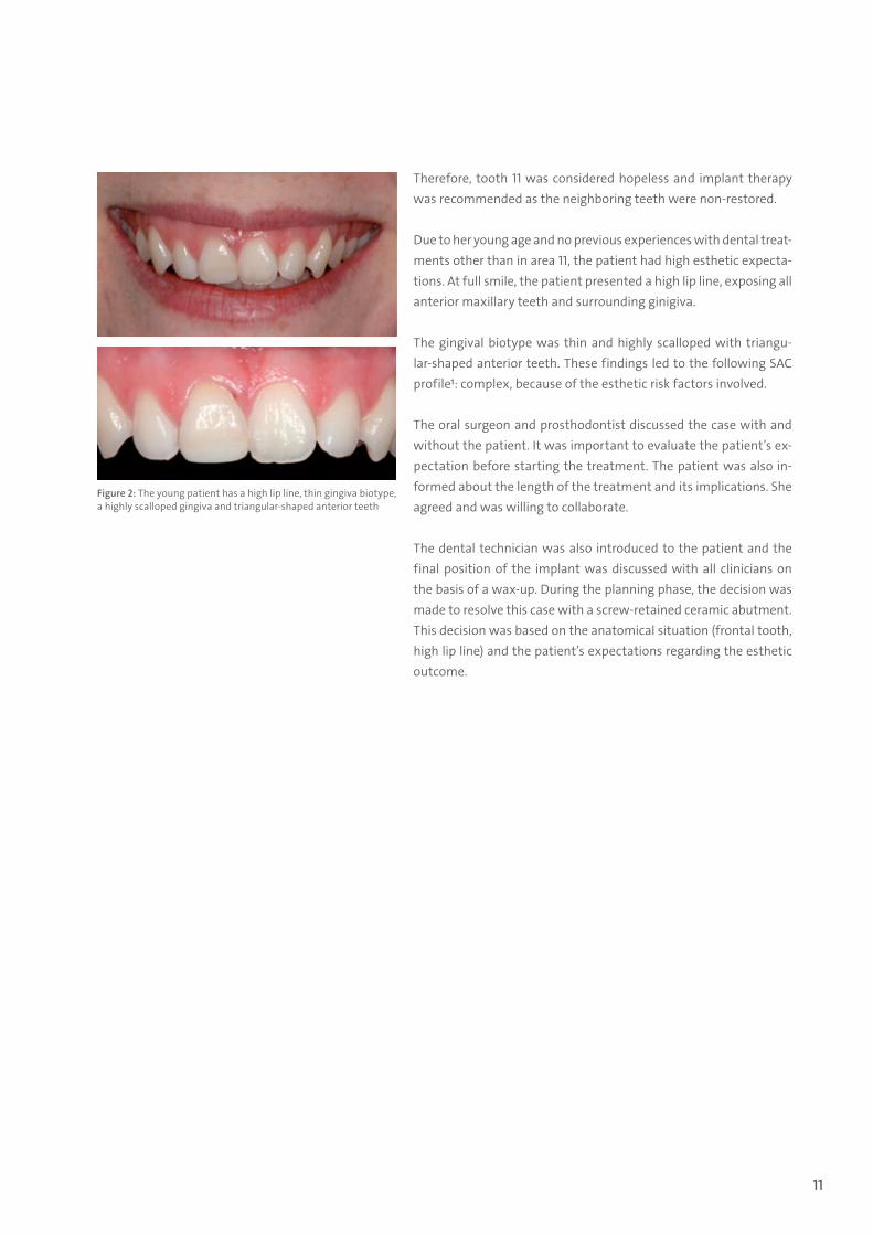
Due to her young age and no previous experiences with dental treat-ments other than in area 11, the patient had high esthetic expecta-tions. At full smile, the patient presented a high lip line, exposing all anterior maxillary teeth and surrounding ginigiva.
The gingival biotype was thin and highly scalloped with triangu-lar-shaped anterior teeth. These findings led to the following SAC profile1: complex, because of the esthetic risk factors involved.
The oral surgeon and prosthodontist discussed the case with and without the patient. It was important to evaluate the patient’s ex-pectation before starting the treatment. The patient was also in-formed about the length of the treatment and its implications. She agreed and was willing to collaborate.
The dental technician was also introduced to the patient and the final position of the implant was discussed with all clinicians on the basis of a wax-up. During the planning phase, the decision was made to resolve this case with a screw-retained ceramic abutment. This decision was based on the anatomical situation (frontal tooth, high lip line) and the patient’s expectations regarding the esthetic outcome.
Figure 2: The young patient has a high lip line, thin gingiva biotype, a highly scalloped gingiva and triangular-shaped anterior teeth
15X.533.indd 11 21.04.15 10:09
12
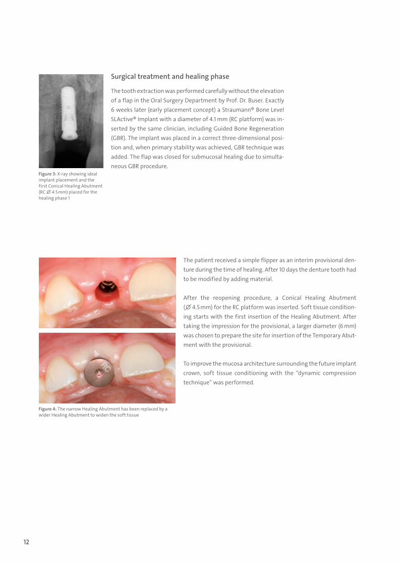
The patient received a simple flipper as an interim provisional den-ture during the time of healing. After 10 days the denture tooth had to be modified by adding material.
After the reopening procedure, a Conical Healing Abutment (∅ 4.5 mm) for the RC platform was inserted. Soft tissue condition-ing starts with the first insertion of the Healing Abutment. After taking the impression for the provisional, a larger diameter (6 mm) was chosen to prepare the site for insertion of the Temporary Abut-ment with the provisional.
To improve the mucosa architecture surrounding the future implant crown, soft tissue conditioning with the “dynamic compression technique” was performed.
Figure 3: X-ray showing ideal implant placement and the first Conical Healing Abutment (RC ∅ 4.5 mm) placed for the healing phase 1
Figure 4: The narrow Healing Abutment has been replaced by a wider Healing Abutment to widen the soft tissue
Surgical treatment and healing phase
The tooth extraction was performed carefully without the elevation of a flap in the Oral Surgery Department by Prof. Dr. Buser. Exactly 6 weeks later (early placement concept) a Straumann® Bone Level SLActive® Implant with a diameter of 4.1 mm (RC platform) was in-serted by the same clinician, including Guided Bone Regeneration (GBR). The implant was placed in a correct three-dimensional posi-tion and, when primary stability was achieved, GBR technique was added. The flap was closed for submucosal healing due to simulta-neous GBR procedure.
15X.533.indd 12 21.04.15 10:09
13
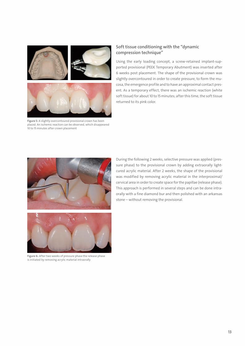
Figure 5: A slightly overcontoured provisional crown has been placed. An ischemic reaction can be observed, which disappeared 10 to 15 minutes after crown placement
Figure 6: After two weeks of pressure phase the release phase is initiated by removing acrylic material intraorally
Soft tissue conditioning with the “dynamic compression technique”
Using the early loading concept, a screw-retained implant-sup-ported provisional (PEEK Temporary Abutment) was inserted after 6 weeks post placement. The shape of the provisional crown was slightly overcontoured in order to create pressure, to form the mu-cosa, the emergence profile and to have an approximal contact pres-ent. As a temporary effect, there was an ischemic reaction (white soft tissue) for about 10 to 15 minutes; after this time, the soft tissue returned to its pink color.
During the following 2 weeks, selective pressure was applied (pres-sure phase) to the provisional crown by adding extraorally light-cured acrylic material. After 2 weeks, the shape of the provisional was modified by removing acrylic material in the interproximal/cervical area in order to create space for the papillae (release phase). This approach is performed in several steps and can be done intra-orally with a fine diamond bur and then polished with an arkansas stone – without removing the provisional.
15X.533.indd 13 21.04.15 10:09
14
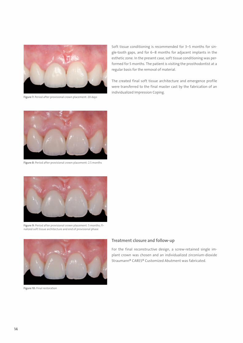
Soft tissue conditioning is recommended for 3–5 months for sin-gle-tooth gaps, and for 6–8 months for adjacent implants in the esthetic zone. In the present case, soft tissue conditioning was per-formed for 5 months. The patient is visiting the prosthodontist at a regular basis for the removal of material.
The created final soft tissue architecture and emergence profile were transferred to the final master cast by the fabrication of an individualized Impression Coping.
Treatment closure and follow-up
For the final reconstructive design, a screw-retained single im-plant crown was chosen and an individualized zirconium -dioxide Straumann® CARES® Customized Abutment was fabricated.
Figure 7: Period after provisional crown placement: 20 days
Figure 8: Period after provisional crown placement: 2.5 months
Figure 9: Period after provisional crown placement: 5 months; fi-nalized soft tissue architecture and end of provisional phase
Figure 10: Final restoration
15X.533.indd 14 21.04.15 10:09
15
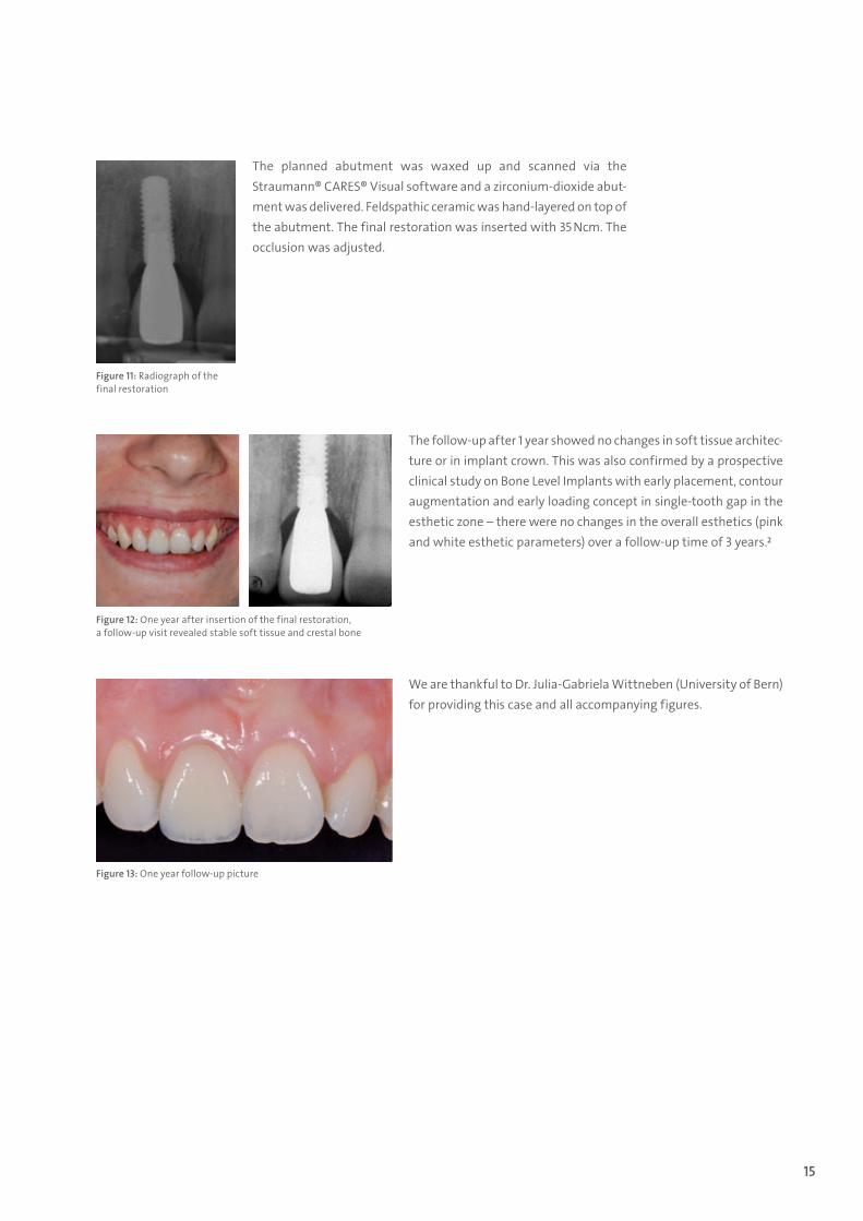
The follow-up after 1 year showed no changes in soft tissue architec-ture or in implant crown. This was also confirmed by a prospective clinical study on Bone Level Implants with early placement, contour augmentation and early loading concept in single-tooth gap in the esthetic zone – there were no changes in the overall esthetics (pink and white esthetic parameters) over a follow-up time of 3 years.2
We are thankful to Dr. Julia-Gabriela Wittneben (University of Bern) for providing this case and all accompanying figures.
Figure 12: One year after insertion of the final restoration, a follow-up visit revealed stable soft tissue and crestal bone
Figure 13: One year follow-up picture
Figure 11: Radiograph of the final restoration
The planned abutment was waxed up and scanned via the Straumann® CARES® Visual software and a zirconium-dioxide abut-ment was delivered. Feldspathic ceramic was hand-layered on top of the abutment. The final restoration was inserted with 35 Ncm. The occlusion was adjusted.
15X.533.indd 15 21.04.15 10:09

16
REFERENCES
1 Dawson A, Chen S, Buser D, Cordaro L, Martin W, Belser U. The SAC Classification in Implant Dentistry: Straightforward – Advanced – Complex. In Dawson A. and Chen S (eds):2009:Quintessence Publishing Group.
2 Buser D, Wittneben J, Bornstein MM, Grütter L, Chappuis V, Belser UC. Stability of contour augmentation and esthetic outcomes of implant-supported single crowns in the esthetic zone: 3-year results of a prospective study with early implant placement postextraction. J. Periodontol. 2011;82(3):342–9.
15X.533.indd 16 21.04.15 10:09

15X.533.indd 17 21.04.15 10:09

International Headquarters Institut Straumann AG Peter Merian-Weg 12 CH-4002 Basel, Switzerland Phone +41 (0)61 965 11 11 Fax +41 (0)61 965 11 01 www.straumann.com
IPS e.max® is a registered trademark of Ivoclar Vivadent AG, Liechtenstein.LOCATOR® is a registered trademark of Zest Anchors, Inc., USA.
© Institut Straumann AG, 2015. All rights reserved.Straumann® and/or other trademarks and logos from Straumann® mentioned herein are the trademarks or registered trademarks of Straumann Holding AG and/or its affiliates.
15
2.53
3/en
/A/0
0 02
/15
15X.533.indd 18 21.04.15 10:09