Embed Size (px)
Citation preview

Plant Physiol. (1997) 114: 539-547
Characterization of the Cell Wall Microdomain Surrounding Plasmodesmata in Apple Fruit
Stéphane Roy*, Alley E. Watada, and William P. Wergin
Horticultural Crops Quality Laboratory, United States Department of Agriculture, Agricultura1 Research Service, Beltsville, Maryland 20705 (S.R., A.E.W.); and Electron Microscopy Laboratory, United States Department of
Agriculture, Agricultural Research Service, Beltsville, Maryland 20705 (S.R., W.P.W.)
In fleshy fruits ripening i s generally associated with a loss in tissue firmness resulting from depolymerization of wall components and separation of adjacent cells. I n the regions of the wall that contain plasmodesmata, the usual sequences of ripening events, i.e. depo- lymerization of the middle lamellae and splitting of the walls, are not observed. I n the present study we attempted to characterize in apple (Malus domestica Borkh.) fruit the structural microdomain of the cell wall that surrounds the plasmodesmata by in muro visual- ization of the cell wall components. Anionic sites of galacturonic acids were labeled with cationic gold. Low-esterified homogalactu- ronans were labeled with the monoclonal antibody JIM 5. In addi- tion, a polyclonal antibody directed toward ~(1+3)-glucopyranose was used to target callose in situ. l h e results indicated that the plasmodesmata-wall complexes were surrounded by a pectic mi- crodomain. This domain was composed of low-esterified homoga- lacturonans that were not involved in calcium cross-bridging but were probably surrounded by a cationic environment. These struc- tural features may result in the prevention of normal cell wall separation in regions containing plasmodesmata. However, obser- vations by low-temperature scanning electron microscopy sug- gested that splitting of these walls ruptured the plasmodesmata and ultimately resulted in the spatial separation of adjacent cells.
In higher plants different developmental processes are associated with a regulated cell separation, which results in loosely attached cells that are surrounded by extensive intercellular spaces (Knox, 1992). For example, fruit soften- ing, which occurs during ripening, results from the loss of cell cohesion (Labavitch, 1981; Brady, 1987; Poovaiah et al., 1988; Van Buren, 1991). Pectolytic enzymes and changes in pectin composition are responsible for the decreased adhe- sion between cells and have been the focus of numerous studies of fruit ripening (for a review, see Fischer and Bennett, 1991) as well as investigations to evaluate the commercial potential of genetically modified fruits (Brady, 1992; Grierson and Schuch, 1993).
Ultrastructural studies show that fruit softening involves a disruption of the middle lamella and cell wall autolysis to different extents, depending on the degree of juiciness of the fruits (Mohr and Stein, 1969; Ben Arie et al., 1979; Platt-Aloia et al., 1980; Crookes and Grierson, 1983). The development of affinity methods, which use specific
* Corresponding author; e-mail [email protected]; fax 1-413- 545-3243.
539
probes, has enabled investigators to use new approaches to understand the microheterogeneity of the cell wall (Carpita and Gibeaut, 1993; Roberts, 1994). Targeting the hydrolytic enzymes (Dallman et al., 1989; Morre, 1989; Pogson et al., 1992) and visualizing in muro the subtle changes that the substrates undergo (Roy et al., 1992; ODonoghue et al., 1994) have led to the conclusion that fruit tissue is a mosaic of microdomains where minute changes can occur during the ripening process (Roy et al., 1992).
In the cell wall regions that contain plasmodesmata, the normal sequence of ripening events, i.e. depolymerization of the middle lamella and swelling and splitting of the cell wall, is usually not observed (Ben-Arie et al., 1979; Hallett et al., 1992; Martin-Cabrejas et al., 1994). Plasmodesmata form an important symplasmic pathway for cell-to-cell transport in higher plants (for a review, see Robards and Lucas, 1990; Lucas et al., 1993). Their ultrastructural fea- tures are becoming more clearly defined because of im- provements in the preservation of the tissues (Ding et al., 1992; Badelt et al., 1994; Turner et al., 1994). However, because it is difficult to isolate plasmodesmata, the cell walls that surround these structures are difficult to char- acterize. Investigators have found that, in many tissues, regions of the cell wall that contain plasmodesmata stain differently and have denser fibrillar structures (Vian and Rougier, 1974; Ben-Arie et al., 1979; Hallett et al., 1992; Martin-Cabrejas et al., 1994). These observations suggest that the cell wall regions around the plasmodesmata may differ in composition.
In the present investigation we attempted to characterize the cell wall microdomain that surrounds plasmodesmata to understand its behavior during cell separation. To achieve this objective severa1 probes were used to visualize the cell wall components: (a) a cationic POIY-L-LYS colloidal gold complex was used to localize anionic sites in muro (Roy et al., 1994a), (b) a previously characterized JIM 5 monoclonal antibody against low-esterified homogalactu- ronans (Knox et al., 1990) was used to characterize the differentiation pattern of the pectic matrix components, and (c) a polyclonal antibody directed toward /3(1-+3)- glucopyranose (Northcote et al., 1989) was used to visual- ize callose in situ. These probes can be used either alone or after dissecting treatments. The results suggest that a
Abbreviations: LTSEM, low-temperature scanning electron mi- croscopy; PG, polygalacturonase; PME, pectin methyl esterase.
www.plantphysiol.orgon May 8, 2018 - Published by Downloaded from Copyright © 1997 American Society of Plant Biologists. All rights reserved.
Plant Physiol. Vol. 114, 1997 540 Roy et al.
clearly defined cell wall microdomain surrounds the plas- modesmata and may be involved in the modulation of cell wall autolysis in this area.
MATERIALS A N D METHODS
Plant Material and Specimen Preparation
Golden delicious apples (Malus domestica Borkh.) were harvested from a commercial orchard in Pennsylvania and stored at 0°C for a few days. Small pieces of the pericarp were cut and chemically fixed with 2.5% glutaraldehyde in 0.1 M cacodylate buffer at pH 7.4 for 3 h, then washed in cacodylate buffer, and postfixed overnight in 1% osmium tetroxide. After dehydration in an alcohol series, the sam- ples were embedded in London Resin White' methacrylate resin (Polysciences, Warrington, PA), as described by Ro- land and Vian (1991).
Labeling with Cationic Colloidal Poly-~-Lys Cold Complexes
The labeling was performed as a one-step procedure by the direct use of the gold complex. Ultrathin sections were incubated in 3% acetic acid at pH 2.6 for 20 min. The sections were treated for 1 h at room temperature with a cationic PO~Y-L-LYS colloidal gold complex consisting of gold particles of either 10 nm (CGC10, British BioCell In- ternational, Cardiff, UK) or 5 nm (CGC5, British BioCell International), diluted 1/60 (v/v) in 3% acetic acid. The sections were then rinsed thoroughly in 3% acetic acid, followed by distilled water. A control experiment was per- formed by preincubating sections with a 1 mg mL-l solu- tion of PO~Y-L-LYS (molecular weight > 350,000; Sigma) prior to the labeling.
lmmunolabeling of Homogalacturonan Sequences
Ultrathin sections were incubated for 30 min in normal goat serum diluted 1 /30 in 0.05 M TBS (pH 7.4) containing 0.5% BSA. The sections were treated overnight at 4"C, with the cell culture supernatant of JIM 5, a monoclonal anti- body directed toward low-esterified homogalacturonans (Knox et al., 1990), diluted 1 /5 (v/v) in TBS plus BSA 0.5%. Following treatment, the specimens were rinsed thor- oughly in TBS. Sections were treated for 1 h with a 10-nm colloidal gold goat anti-rat immunoglobulin complex (Brit- ish BioCell International) diluted 1/30 in TBS. They were then washed thoroughly in TBS and distilled water. To assess the specificity of the labeling, the cell culture super- natant JIM 5 was incubated with polygalacturonic acid from citrus fruit (Sigma).
Cytochemical Dissection
In some cases sections were subjected to chemical or enzymatic treatments prior to labeling. For in situ chemical
' Mention of a trademark or proprietary product does not con- stitute a guarantee or warranty of the product by the U.S. Depart- ment of Agriculture and does not imply its approval to the exclu- sion of other products that may also be suitable.
de-esterification of pectins, sections were preincubated with 0.1 M Na,CO, at 4°C for 16 h (Fry, 1989). For in situ enzymatic de-esterification of pectins, sections were prein- cubated with a 0.1 mg mL-' solution of PME from orange peel (Sigma) in 0.1 M NaCl at pH 7.5 (Vreeland et al., 1989). The control consisted of preincubation with NaCl solution only. For in situ chelation of calcium, sections were prein- cubated in 0.2 M EDTA (Sigma), pH 8.0, at 60°C for 1 h (Wick and Hepler, 1980).
lmmunolabeling of p(1+3)-Clucopyranose
Ultrathin sections were incubated for 30 min in normal goat serum diluted 1 / 30 in 0.05 M TBS, pH 7.5, containing 0.5% BSA. The sections were treated overnight at 4°C with a polyclonal antibody directed toward p(l+3)-glucopyranose (Euromedex, Souffelweyersheim, France), diluted 1 /25 (v/v) in TBS plus BSA 0.5% (Northcote et al., 1989). The specimens were rinsed thoroughly in TBS. Sections were treated for 1 h with a 10-nm colloidal gold goat anti-rabbit immunoglobulin complex (British BioCell International) di- luted 1 I30 in TBS; they were then washed thoroughly in TBS and distilled water.
LTSEM
A field emission scanning electron microscope (S-4100, Hitachi, Tokyo, Japan) equipped with an Oxford CT-1500HF Cryotrans system (Oxford Instruments, Eynsham, UK) was used for LTSEM observations. Specimen preparation con- sisted of removing 1-cm2 segments of the pericarp and mounting them onto a complementary holder with a cryoadhesive (Tissue Tek, Miles Scientific, Naperville, IL). The holder was rapidly plunge-frozen in liquid nitrogen and cryotransferred under vacuum to a cold stage in the pre- chamber of the cryosystem. The frozen specimen was etched in the prechamber by raising the temperature of the stage to -95"C, sputter-coated with Pt, and then transferred to the cryostage in the SEM for observations. An accelerating volt- age of 10 kV was used to view the specimens.
RESULTS
The general ultrastructural features of the parenchyma cells in apple fruits are similar to those previously de- scribed (Ben-Arie et al., 1979; Roy et al., 1994a, 1995). The fruit pericarp is composed of (a) superficial layers of tightly cemented isodiametric cells having thick cell walls and numerous plasmodesmata that are concentrated in discrete areas, and (b) in deeper areas of the pericarp, large paren- chyma cells that are separated from one another along a portion of their thin cell walls, thereby forming intercellu- lar spaces (data not shown). Plasmodesmata were rarely observed within the latter area. Further observations were focused on the small, isodiametric cells region, and second- ary plasmodesmata, as described by Ding and Lucas (1996), were mainly observed. Figure 1 illustrates an "H- shaped" secondary plasmodesmata with the development of a central cavity within the middle lamella (arrows, Fig. 1). In sections that were stained with lead citrate, the cell wall domains that surround plasmodesmata appeared
www.plantphysiol.orgon May 8, 2018 - Published by Downloaded from Copyright © 1997 American Society of Plant Biologists. All rights reserved.

Plasmodesmata-Wall Complexes in Apple Fruit 541
m+r- v v
Figure 1. Structure of secondary plasmodesmata in cell wall fromapple pericarp. The secondary plasmodesmata is characterized byprotoplasmic branches and a central cavity within the middle lamellaregion (arrows). Bar = 0.2 /nm.
more electron-opaque than other areas of the wall (data notshown).
Distribution of the Pectic Matrix Component
To characterize the molecular environment of the cellwall surrounding the plasmodesmata, cationic colloidalgold was used to label the anionic sites of the wall (Fig. 2a).Cross-sections through the cell wall showed that the cat-ionic gold labeling was dense and evenly distributedthroughout most of the wall; however, the areas that con-sisted of plasmodesmata-wall complexes were not labeledwith cationic gold (arrowheads, Fig. 2a), except along asmall zone of the cell wall that bordered the plasmalemma.Control sections that were preincubated with a solution ofunlabeled poly-L-Lys only rarely exhibited cationic goldlabeling (data not shown).
Following immunolabeling, the low-esterified homoga-lacturonans epitope recognized by JIM 5 was intensivelylocalized in the plasmodesmata-wall complexes but wasonly weakly seen in the primary cell wall (Fig. 2b). Preab-sorption of the monoclonal antibody JIM 5 with a solutionof polygalacturonic acid greatly reduced the labeling (datanot shown).
Influence of PreincubationTreatment prior to the Labeling
Specific enzymatic or chemical treatments were used todiscriminate the various forms in which the homogalactu-ronans may exist, i.e. low-esterified, highly esterified, orbound with calcium ions. Neither the calcium-chelatortreatment with EDTA (Fig. 3a) nor hydrolysis of the methylgroups (Fig. 3b) resulted in labeling with cationic gold inthe plasmodesmata-wall area. Conversely, chemical de-
esterification (Fig. 3c), enzymatic de-esterification (data notshown), or calcium-chelator treatment (Fig. 4) prior to la-beling dramatically modified the distribution pattern oflow-esterified homogalacturonans epitopes recognized byJIM 5. JIM 5 labeling was abundant throughout the primarycell wall adjacent to the plasmodesmata-wall complexesafter de-esterification (compare Figs. 2b and 3c) and aftercalcium-chelator treatment (compare Figs. 2b and 4). Theresults are summarized in Table I.
LTSEM Observation and Callose Distribution
To further characterize the wall regions that were richin plasmodesmata, immunogold labeling with j3(l^>3)-glucopyranose was carried out on sectioned material. Theresults clearly show the association of callose with proto-plasmic branches or strands (Fig. 5).
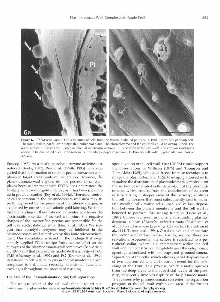
LTSEM allowed us to observe the frozen, fully hydratedcells of the pericarp. The frozen tissues did not exhibit thestructural artifacts that are usually associated with chem-ical fixation, dehydration, and critical point drying (Wer-gin and Erbe, 1991). This technique enabled us to visualizesmall, round protuberances on the surfaces of walls fromcells that had separated, i.e. cells found in the deep areasof the pericarp (Fig. 6a). The protuberances are believed toconsist of cytoplasmic material that is surrounded by alayer of cell wall (Fig. 6b). They probably represent theplasmodesmata that were mechanically ruptured duringcell separation.
DISCUSSION
Although apple fruits never reach the degree of juicinessfound in tomatoes or peaches, Ben-Arie et al. (1979) haveshown a progressive splitting and dissolution of the pectin-rich apple middle lamella. We assume that the pattern ofpectin dissolution in apple fruits follows the sequence vi-sualized in tomato fruits (Roy et al., 1992, 1994b). By meansof in situ visualization of cell wall components, we haveclearly illustrated that there are differences in the cell wallthat surrounds the plasmodesmata. Because during soften-ing of juicy fruits, plasmodesmata-wall complexes do notshow swelling and splitting of the cell wall (Ben-Arie et al.,1979; Hallett et al., 1992; Martin-Cabrejas et al., 1994), theoccurrence of a cell wall microdomain suggests modulationof cell separation in these areas.
A Cell Wall Microenvironment
The specificity of the cationic colloidal gold probe hasbeen previously characterized (Roy et al., 1994a). Cationicgold recognizes the COO- groups of galacturonic acid ofunesterified pectins and glucuronic acid in xylans. Be-cause large biochemical quantities of the former and smallamounts of the latter are present in the primary cell wallsof all dicotyledons analyzed so far (Carpita and Gibeaut,1993), we assume that cationic gold probes the carboxylicgroups of galacturonic acids (Roy et al., 1994a). Threepossible reasons account for the low amount of goldlabeling detected in the plasmodesmata-wall complexesafter probing the sections with cationic gold (Fig. 2a): (a) www.plantphysiol.orgon May 8, 2018 - Published by Downloaded from
Copyright © 1997 American Society of Plant Biologists. All rights reserved.

542 Roy et al. Plant Physiol. Vol. 114, 1997
Figure 2. Labeling of the pectic network aroundthe plasmodesmata. a, In muro visualization ofanionic sites labeled with cationic colloidalgold. Labeling is intense and evenly distributedin the cell wall with the exception of the plas-modesmata area (arrowheads), b, Distribution oflow-esterified homogalacturonic sequences byimmunolabeling with JIM 5. Intense labeling isrestricted to the area where the cell wall narrowsand surrounds the plasmodesmata. In the pri-mary cell wall, the labeling is less intense. C,Primary cell wall; E, ER; M, mitochondrion; Ml,middle lamella; P, plastid; Pd, plasmodesmata;and V, vacuole. Bars = 0.5 /j,m.
2a
19
the cell wall components that were targeted were notabundant, (b) methyl groups or calcium ions may havemasked the anionic sites, or (c) a cationic environmentprevented the area from being labeled. To investigatethese possibilities, immunogold labeling and cytochemi-cal dissection were used.
JIM 5 was used to recognize low-esterified homogalactu-ronans. The intense reaction that we observed indicated thatplasmodesmata-wall complexes were probably composed oflow-esterified homogalacturonans (Fig. 2b). The segregationof the distribution for cationic gold and JIM 5 labeling wassurprising because these two probes should target the samecomponent, i.e. low-esterified galacturonic acid chains. Thismay suggest that the sensitivity of the cationic gold, whichrecognizes a single unit of galacturonic acid in pectic chains,is higher than that of JIM 5. However, these data do notexplain why the plasmodesmata-wall complexes were voidof cationic gold labeling.
In the plasmodesmata-wall area, anionic sites may havebeen masked by either methyl groups or calcium ions. Wehave shown that the homogalacturonans are not esterified,because de-esterification prior to probing does not restorethe cationic gold labeling in the plasmodesmata-wall com-plexes (Fig. 3b), as it does in the primary cell wall with JIM5 labeling (Fig. 3c). Similarly, pretreatment with a calciumchelator does not enhance the cationic gold labeling in theplasmodesmata-wall regions, indicating that calcium cross-bridges do not exist (Fig. 3a), as they do in the primary cellwall (Fig. 4).
In the plasmodesmata-wall complexes anionic sitesseem to be masked in some way that is different frommethyl groups or calcium ions. Cationic gold labeling,which is based on electrostatic interactions, may havebeen prevented by the presence of a cationic environment.This may reflect pectin interaction with other cell wallcomponents. Although we do not yet have direct evi- www.plantphysiol.orgon May 8, 2018 - Published by Downloaded from
Copyright © 1997 American Society of Plant Biologists. All rights reserved.
Plasmodesmata-Wall Complexes in Apple Fruit 543
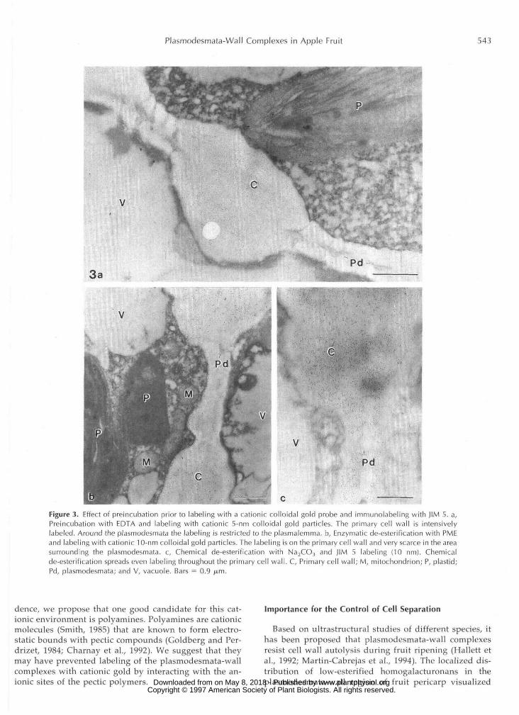
Figure 3. Effect of preincubation prior to labeling with a cationic colloidal gold probe and immunolabeling with JIM 5. a,Preincubation with EDTA and labeling with cationic 5-nm colloidal gold particles. The primary cell wall is intensivelylabeled. Around the plasmodesmata the labeling is restricted to the plasmalemma. b, Enzymatic de-esterification with PMEand labeling with cationic 10-nm colloidal gold particles. The labeling is on the primary cell wall and very scarce in the areasurrounding the plasmodesmata. c, Chemical de-esterification with Na2CO3 and JIM 5 labeling (10 nm). Chemicalde-esterification spreads even labeling throughout the primary cell wall. C, Primary cell wall; M, mitochondrion; P, plastid;Pd, plasmodesmata; and V, vacuole. Bars = 0.9 /j.m.
dence, we propose that one good candidate for this cat-ionic environment is polyamines. Polyamines are cationicmolecules (Smith, 1985) that are known to form electro-static bounds with pectic compounds (Goldberg and Per-drizet, 1984; Charnay et al., 1992). We suggest that theymay have prevented labeling of the plasmodesmata-wallcomplexes with cationic gold by interacting with the an-ionic sites of the pectic polymers.
Importance for the Control of Cell Separation
Based on ultrastructural studies of different species, ithas been proposed that plasmodesmata-wall complexesresist cell wall autolysis during fruit ripening (Hallett etal., 1992; Martin-Cabrejas et al., 1994). The localized dis-tribution of low-esterified homogalacturonans in theplasmodesmata-wall region of fruit pericarp visualized www.plantphysiol.orgon May 8, 2018 - Published by Downloaded from
Copyright © 1997 American Society of Plant Biologists. All rights reserved.
544 Roy et al. Plant Physiol. Vol. 114, 1997
N
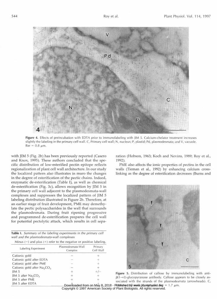
Figure 4. Effects of preincubation with EDTA prior to immunolabeling with JIM 5. Calcium-chelator treatment increasesslightly the labeling in the primary cell wall. C, Primary cell wall; N, nucleus; P, plastid; Pd, plasmodesmata; and V, vacuole.Bar = 0.8 /xm.
with JIM 5 (Fig. 2b) has been previously reported (Caseroand Knox, 1995). These authors concluded that the spe-cific distribution of low-esterified pectin epitope reflectsregionalization of plant cell wall architecture. In our studythe localized pattern also illustrates in muro the changesin the degree of esterification of the pectic chains. Indeed,enzymatic de-esterification (Table I), as well as chemicalde-esterification (Fig. 3c), allows recognition by JIM 5 inthe primary cell wall adjacent to the plasmodesmata-wallcomplexes and suppresses the localized pattern of JIM 5labeling distribution illustrated in Figure 2b. Therefore, atan earlier stage of fruit development, PME may demethy-late the pectic polysaccharides in the wall that surroundsthe plasmodesmata. During fruit ripening progressiveand programmed de-esterification prepares the cell wallfor potential pectolytic attack, which results in cell sepa-
Table I. Summary of the labeling experiments in the primary cellwall and the plasmodesmata-wall complexes
Minus (-) and plus ( + ) refer to the negative or positive labeling.. . . . ,. .Labeling Experiment Plasmodesmata-Wall_ ,Complex
Primary^ ,,,.,,,Cell Wall
Cationic goldCationic gold after EDTACationic gold after PMECationic gold after Na2CO3
JIM 5JIM 5 after Na2CO3
JIM 5 after PMEJIM 5 after EDTA
ration (Hobson, 1963; Koch and Nevins, 1989; Roy et al.,1992).
PME also affects the ionic properties of pectins in the cellwalls (Tieman et al., 1992) by enhancing calcium cross-linking as the degree of esterification decreases (Burns and
Figure 5. Distribution of callose by immunolabeling with anti-(3(1—»3)-glucopyranose antibody. Callose appears to be closely as-sociated with the strands of the plasmodesmata (arrowheads). C,Primary cell wall; V, vacuole. Bar = 1.7 jam. www.plantphysiol.orgon May 8, 2018 - Published by Downloaded from
Copyright © 1997 American Society of Plant Biologists. All rights reserved.
Plasmodesmata-Wall Complexes in Apple Fruit 545
Figure 6. LTSEM observation. Cross-fractures of cells from the frozen, hydrated pericarp, a, Profile view of a pericarp cell.The fracture does not follow a single flat, horizontal plane. The plasmalemma and the cell wall could be distinguished. Theouter surface of the cell wall contains circular extensions (arrows), b, Face view of the cell wall. The circular extensionsappear to be composed of cell wall material surrounding cytoplasm (arrows). C, Primary cell wall; PI, plasmalemma. Bars =4.5 fj,m.
Pressey, 1987). As a result, pectolytic enzyme activities arereduced (Brady, 1987). Roy et al. (1994b, 1995) have sug-gested that the formation of calcium-pectin interaction com-plexes in target areas limits cell separation. However, theplasmodesmata-wall regions do not possess these com-plexes because treatment with EDTA does not restore thelabeling with cationic gold (Fig. 3a), as it has been shown todo in previous studies (Roy et al., 1994a). Therefore, controlof cell separation in the plasmodesmata-wall area may bepartly explained by the presence of the cationic charges, asevidenced by our results of cationic gold labeling. We thinkthat the binding of these cationic molecules will lower theelectrostatic potential of the cell wall, since the negativecharges of the unesterified pectins are responsible for thecell wall electrostatic potential (Nari et al., 1986). We sug-gest that pectolytic enzymes may be inhibited in theplasmodesmata-wall complexes by this ionic microenviron-ment. Our speculation is based on the findings that exog-enously applied PG in unripe fruits has no effect on theautolysis of the plasmodesmata-wall complexes (Ben-Arie etal., 1979) and that polyamines modulate the activity of bothPME (Charnay et al., 1992) and PG (Kramer et al., 1989).Resistance to cell wall autolysis in the plasmadesmata-wallcomplexes is a dramatic necessity to maintain functionalexchanges throughout the process of ripening.
The Fate of the Plasmodesmata during Cell Separation
The unique collar of the cell wall that is found sur-rounding the plasmodesmata is an example of structural
specialization of the cell wall. Our LTSEM results supportthe observations of Willison (1976) and Thomson andPlatt-Aloia (1985), who used freeze-fracture techniques toimage the plasmodesmata. LTSEM imaging allowed us tovisualize the distribution of plasmodesmata complexes onthe surface of separated cells. Separation of the plasmod-esmata, which results from the detachment of adjacentcells occurring in deeper areas of the pericarp, rupturesthe cell membranes that must subsequently seal to main-tain metabolically viable cells. Localized callose deposi-tion between the plasma membrane and the cell wall isbelieved to perform this sealing function (Lucas et al.,1993). Callose is present in the ring surrounding plasmo-desmata in bean (Phaseolus vulgaris) roots (Northcote etal., 1989) and in maize (Lea mays L.) root tips (Balestrini etal., 1994; Turner et al., 1994). Our data, which demonstratethe presence of callose in fruit tissues, support these ob-servations. Apparently, the callose is restricted to a pe-ripheral collar, where it is interspersed within the cellwall and can constrict or completely seal the cytoplasmicopenings associated with the ruptured plasmodesmata.Separation of the cells, which allows spatial displacementof two adjacent cells, is an important event for the soft-ening of the fruit. This process, which usually spreadsfrom the deep areas to the superficial layers of the peri-carp, apparently involves rupture of the plasmodesmata.The reasons why plasmodesmata can enter the separationprogram of the cell wall within one area of the fruit isunknown. www.plantphysiol.orgon May 8, 2018 - Published by Downloaded from
Copyright © 1997 American Society of Plant Biologists. All rights reserved.
546 Roy et al. Plant Physiol. Vol. 114, 1997
In conclusion, we have illustrated another cell wall mi- crodomain within the fruit tissue. We continue to support the idea that the existence of microdomains within the cell wall allows the modulation of autolysis during cell sepa- ration. Any substance, i.e. methyl groups, calcium, or cat- ionic molecules, that is able to modify the cell wall charge density plays an important role in the localized control of pectolytic enzyme activity.
ACKNOWLEDCMENTS
The authors express their gratitude to Dr. J. Paul Knox (Univer- sity of Leeds, UK) for the generous gift of JIM 5. We are grateful to Gudrun Siegert for her photographic assistance in preparing the final plates.
Received December 9, 1996; accepted March 11, 1997. Copyright Clearance Center: 0032-0889/97/ 114/0539/09.
LITERATURE CITED
Badelt K, White RG, Overall RL, Vesk M (1994) Ultrastructural specialization of the cell wall sleeve around plasmodesmata. Am J Bot 81: 1422-1427
Balestrini R, Romera C, Puigdomenech P, Bonfante P (1994) Location of a cell-wall hydroxyproline-rich glycoprotein, cellu- lose and @-1,3-glucans in apical and differentiated regions of maize mycorrhizal roots. Planta 195: 201-209
Ben-Arie R, Kislev N, Frenkel C (1979) Ultrastructural changes in the cell walls of ripening apple and pear fruit. Plant Physiol64
Brady CJ (1987) Fruit ripening. Annu Rev Plant Physiol 38:
Brady CJ (1992) Molecular approaches to understanding fruit ripening. NZ J Crop Hortic Sci 20: 107-117
Burns JK, Pressey R (1987) Ca2+ in cell wall of ripening tomato and peach. J Am SOC Hortic Sci 112: 783-787
Carpita NC, Gibeaut DM (1993) Structural models of primary cell walls in flowering plants: consistency of molecular structure with the physical properties of the wall during growth. Plant J
Casero PJ, Knox JP (1995) The monoclonal antibody JIM 5 indi- cates patterns of pectin deposition in relation to pit fields at the plasma-membrane face of tomato pericarp cell walls. Proto- plasma 188: 133-137
Chamay D, Nari J, Noat G (1992) Regulation of plant cell-wall pectin methyl esterase by polyamines-interactions with the effects of metal ions. Eur J Biochem 205: 711-714
Crookes PR, Grierson D (1983) Ultrastructure of tomato fruit ripening and the role of polygalacturonase isoenzymes in cell wall degradation. Plant Physiol 72: 1088-1093
Dallman TF, Thomson WW, Eaks IL, Nothnagel EA (1989) Ex- pression and transport of cellulase in avocado mesocarp during ripening. Protoplasma 151: 3346
Ding B, Lucas WJ (1996) Secondary plasmodesmata: biogenesis, special functions and evolution. In M Smallwood, JP Knox, DJ Bowles, eds, Membranes: Specialized Functions in Plants. Bios Scientific, Oxford, UK, pp 489-506
Ding B, Turgeon R, Parthasarathy MV (1992) Substructure of freeze-substituted plasmodesmata. Protoplasma 169: 28-41
Fischer RL, Bennett AB (1991) Role of the cell wall hydrolases in fruit ripening. Annu Rev Plant Physiol Plant Mo1 Biol 42:
Fry SC (1989) Analysis of cross-links in the growing cell walls of higher plants. In HS Linskens, JS Jackson, eds, Plant Fibers: Modern Methods in Plant Analysis, New Series, Vol 10. Springer Verlag, Heidelberg, Germany, pp 12-36
'
197-202
155-178
3: 1-30
675-703
Goldberg R, Perdrizet E (1984) Ratio of free to bound polyamines during maturation of seedlings in mung bean hypocotyl cells. Planta 161: 531-535
Grierson D, Schuch W (1993) Control of ripening. Philos Trans R SOC Lond-Biol Sci,342: 241-250
Hallett IC, Mac Rae EA, Wegrzyn TF (1992) Cell packing and cell wall ultrastructure during ripening in kiwi fruit. Int J Plant Sci
Hobson GE (1963) Pectinesterase in normal and abnormal tomato fruit. Biochem J 86: 358-365
Knox JP (1992) Cell adhesion, cell separation and plant morpho- genesis. Plant J 2: 137-141
Knox JP, Linstead PJ, King J, Cooper C, Roberts K (1990) Pectin esterification is spatially regulated both within cell walls and between developing tissues of root apices. Planta 181: 512-521
Koch JL, Nevins DJ (1989) Tomato fruit cell wall. I. Use of purified tomato polygalacturonase and pectin methyl esterase to identify developmental changes in pectins. Plant Physiol91: 816-822
Kramer GF, Wang CY, Conway WS (1989) Correlation of reduced softening and increased polyamine levels during low-oxygen storage of 'Mc Intosh' apples. J Am SOC Hortic Sci 114: 942-946
Labavitch JM (1981) Cell wall turnover in plant development. Annu Rev Plant Physiol 32: 385406
Lucas WJ, Ding B, Van der Schoot C (1993) Plasmodesmata and the supracellular nature of plants. New Phytol 125: 435476
Martin-Cabrejas MA, Waldron KW, Selvendran RR, Parker ML, Moates GK (1994) Ripening-related changes in the cell walls of Spanish pear (Pyrus communis). Physiol Plant 91: 671-679
Mohr WP, Stein M (1969) Fine structure of fruit development in tomato. Can J Plant Sci 49: 549-553
Morre DJ (1989) Sorting signals and trafficking of lysosomal and extracellular hydrolases of cell separation. NATO AS1 Adv Sci Inst Ser Ser H Cell Biol 3 5 81-99
Nari J, Noat G, Diamantidis G, Woudstra M, Ricard J (1986) Electrostatic effects and the dynamic of enzyme reactions at the surface of plant cells. 3. Interplay between limited cell-wall autolysis, pectin methyl esterase activity and electrostatic effects in soybean cell walls. Eur J Biochem 155: 199-202
Northcote DH, Davey R, Lay J (1989) Use of antisera to localize callose, xylan and arabinogalactan in the cell-plate, primary and secondary walls of plant cells. Planta 178: 353-366
ODonoghue EM, Huber DJ, Timpa JD, Erdos GW, Brecht JK (1994) Influence of avocado (Peusea americana) Cx-cellulase on the structural features of avocado cellulose. Planta 194: 573-584
Platt-Aloia KA, Thomson WW, Young RE (1980) Ultrastructural changes in the walls of ripening avocados: transmission, scan- ning and freeze fracture microscopy. Bot Gaz 141: 366-373
Pogson BJ, Seymour GB, Brady CJ, Jones M, Goodchild D (1992) Immunolocalisation of pectinases in tomato fruit. NZ J Crop .Hortic Sci 20: 137-146
Poovaiah BW, Glenn GM, Reddy ASN (1988) Calcium and fruit softening: physiology and biochemistry. Hortic Rev 10: 107-151
Robards AW, Lucas WJ (1990) Plasmodesmata. Annu Rev Plant Physiol Plant MOI Biol 41: 369419
Roberts K (1994) The plant extracellular matrix: in a new expan- sive mood. Curr Opin Cell Biol 6 688-694
Roland JC, Vian B (1991) General preparation and staining of thin sections. In JL Hall, C Hawes, eds, Electron Microscopy of Plant Cells. Academic Press, London, pp 1-66
Roy S, Conway WS, Watada AE, Sams CE, Pooley CD, Wergin WP (1994a) Distribution of the anionic sites in the cell wall of apple fruit after calcium treatment: quantitation and visualiza- tion by a cationic colloidal gold probe. Protoplasma 178: 156-167
Roy S, Gillen G, Conway WS, Watada AE, Wergin WP (1995) Use of secondary ion mass spectrometry to image 44calcium uptake by the cell walls of apple fruit. Protoplasma 189: 163-172
Roy S, Jauneau A, Vian B (199413) Analytical detection of calcium ions and immunocytochemical visualization of homogalactu- ronic sequences in the ripe cherry tomato. Plant Physiol Biochem
Roy S, Vian B, Roland JC (1992) Immunocytochemical study of the deesterification patterns during cell wall autolysis in the ripening of cherry tomato. Plant Physiol Biochem 30: 139-146
153: 49-60
32: 1-5
www.plantphysiol.orgon May 8, 2018 - Published by Downloaded from Copyright © 1997 American Society of Plant Biologists. All rights reserved.

Plasmodesmata-Wall Complexes in Apple Fruit 547
Smith TA (1985) Polyamines. Annu Rev Plant Physiol36: 117-143 Thomson WW, Platt-Aloia K (1985) The ultrastructure of the
plasmodesmata of the salt glands of Tamarix as revealed by transmission and freeze-fracture electron microscopy. Proto- plasma 125: 13-23
Tieman DM, Harriman RW, Ramamohan G, Handa AK (1992) An antisense pectin methylesterase gene alters pectin chemistry and soluble solids in tomato fruit. Plant Cell 4: 667-679
Turner A, Wells B, Roberts K (1994) Plasmodesmata of maize root tips: structure and composition. J Cell Sci 107 3351-3361
Van Buren JP (1991) Functions of pectin in plant tissue structure and firmness. In RH Walter, ed, The Chemistry and Technology of Pectin. Academic Press, San Diego, CA, pp 1-22
Vian B, Rougier M (1974) Ultrastructure des plasmodesmes après cryo-ultramicrotomie. J Microsc 20: 307-312
Vreeland V, Morse SR, Robichaux RH, Miller KL, Hua SST, Laetsch WM (1989) Pectate distribution and esterification in Dubautia leaves and soybean nodules, studied with a fluores- cent hybridization probe. Planta 177: 435446
Wergin WP, Erbe EF (1991) Introduction to the advantages and problems associated with low temperature scanning electron microscopy. Scanning 13: 24-26
Wick SM, Hepler PK (1980) Localisation of C a i + containing antimonate precipitates during mitosis. J Cell Biol 86: 500-508
Willison JHM (1976) Plasmodesmata: a freeze-fracture view. Can J Bot 54: 2842-2847
www.plantphysiol.orgon May 8, 2018 - Published by Downloaded from Copyright © 1997 American Society of Plant Biologists. All rights reserved.