Embed Size (px)
Citation preview

Ziqing Yuan
Biosecurity Tasmania
Plant Biosecurity and Diagnostics Branch
Depar tment of Pr imar y Industr ies, Par ks, Water and Environment
Pictor ial At las of
PLANT DISEASESDiagnosed in Tasmania
Pictorial A
tlas o
f PL
AN
T D
ISE
AS
ES
Dia
gno
sed
in Ta
sma
nia
Ziqing Yuan
Pictorial Atlas of Plant Diseases Diagnosed in Tasmania
First edition, 2017 100 printed copies
Printed publication ISBN: 978-1-74380-019-5 Online publication ISBN: 978-1-74380-020-1
Publisher: Department of Primary Industries, Parks, Water & Environment GPO Box 44, Hobart 7001 www.dpipwe.tas.gov.au
Book design: Land Tasmania Design Unit Department of Primary Industries, Parks, Water & Environment
Printer : Foot and Playsted Pty Ltd
Cover photos: Ziqing Yuan
© 2017 Department of Primary Industries, Parks, Water & Environment
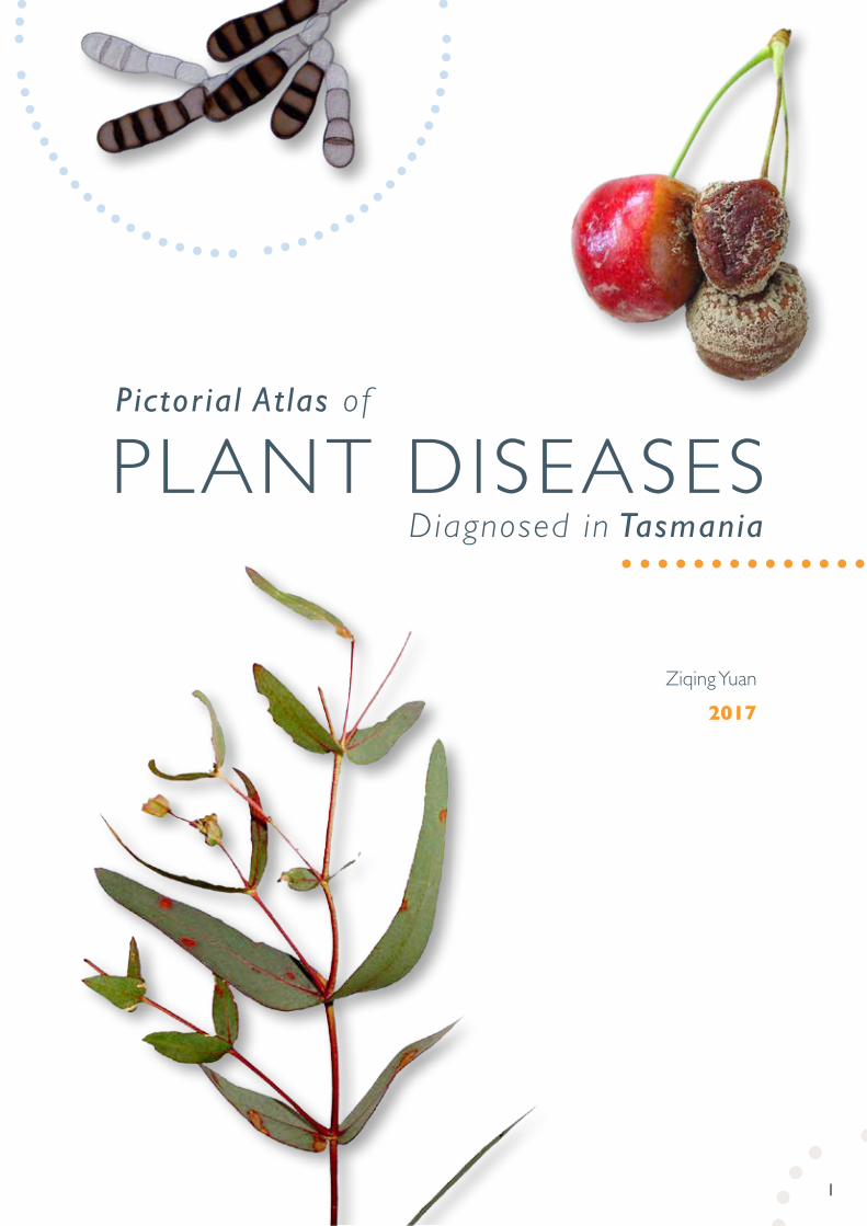
Ziqing Yuan2017
1
Pictor ial At las of
PLANT DISEASESDiagnosed in Tasmania

2
FOREWORD 3
ACKNOWLEDGEMENT 4
INTRODUCTION 5
LIST OF PLANT PATHOGENS TREATED IN THE ATLAS 7
IMAGE PLATES - FUNGI 11
IMAGE PLATES - NEMATODES 103
HOST PLANT INDEX 112
TABLE OF CONTENTS
3
Tasmania as Australia’s only island State has significant biosecurity advantages over mainland Australia with relatively low pest and disease pressures, geographical borders enhancing the management of a biosecurity status, and a reputation for high quality agricultural and horticultural produce exported overseas. The importance of biosecurity to Tasmania is fully recognised by the Tasmanian Government with Biosecurity Tasmania (located in the Department of Primary Industries, Parks, Water and Environment) having lead responsibility for managing the biosecurity system in conjunction with a wide range of partners. With a focus on risk management off-shore, at the border, and post border, activities are undertaken across the entire biosecurity system.
The Tasmanian Plant Health Laboratories are an essential part of the State’s biosecurity system providing routine diagnostic testing as well as testing for potential and suspect organisms exotic to Australia and/or Tasmania. The Plant Pathology laboratories, located at New Town, in the south of the State have for many years provided a critical and at times unnoticed contribution to protecting the State from disease incursions through early detection and diagnosis. The work carried out by the staff in these laboratories literally maintains critical functions of the State both protecting our primary industries and our environment. They really are unsung Tasmanian heroes.
This publication is the result of the significant efforts of Biosecurity Tasmania’s plant pathology team and in particular its senior plant pathologist, Dr Ziqing Yuan. The images provided of specimens photographed during the course of the pathologists work are not only informative and constructive they are each very much an opportunity to look into a fascinating world of interaction between disease organisms and the plants they attack. I would like to extend my sincere thanks to all staff in Biosecurity Tasmania for the work they do in ensuring Tasmania’s biosecurity status is maintained, my own staff located in the plant pathology team and in particular to the very talented Dr Ziqing Yuan.
Andrew Bishop Chief Plant Health Manager (Tasmania)
Biosecurity Tasmania
FOREWORD

4
The author has worked with the Department of Primary Industries, Parks, Water and Environment for more than ten years, as a senior plant pathologist, diagnosing a broad range of plant diseases from local and interstate clients as well as quarantine interceptions. During this time, the Department has provided support to undergo research activities, and the laboratory facilities to undertake these activities. My personal thanks are extended to Andrew Bishop, Manager Plant Biosecurity and Diagnostics Branch, Biosecurity Tasmania and Chief Plant Health Manager Tasmania, for his encouragement in preparing this publication, including kindly writing the Foreword.
Thanks are due to Peter Cross, Team Leader - Plant Pathology, for his support in the preparation of this atlas, and to Dr Alison Dann for conducting molecular tests to confirm some of the identifications based on morphological characters to species level.
Thanks are also due to Dr Roger Shivas, Principal Plant Pathologist, Biosecurity Queensland, Department of Agriculture and Fisheries, and Dr Jacqueline Edwards, Research Leader Plant Pathology, Agriculture Victoria, Department of Economic Development, Jobs, Transport and Resources for critical review of the manuscript, and to Dr John Wainer, Nematologist, Agriculture Victoria, Department of Economic Development, Jobs, Transport and Resources for checking the nematode section.
The author is very grateful to Changyou Pan, Technical Officer (Plant Pathology), for her skillful technical assistance in fungal isolation and culturing. It would not have been possible to complete this atlas without her excellent assistance.
Ziqing Yuan, PhD Senior Plant Pathologist
December 2016, Hobart
ACKNOWLEDGEMENT
5
The diagnosis of plant diseases caused by fungi and nematodes, based on disease symptoms and signs, is an essential task for all plant pathologists. It can be challenging. Identification using morphological characteristics requires sound taxonomic knowledge of fungi and nematodes together with experience and good microscopy skills.
The plant health services within Biosecurity Tasmania provide a written report for all diagnostic tests. In many cases, supporting photographic illustrations are submitted together with the written diagnostic report.
The pathogenic fungi and nematodes illustrated in this atlas were identified by their morphological characteristics, with the exception of Plate 38 (Melampsoridium betulinum) and Plate 42 (Neofusicoccum luteum) which were confirmed to the species level by molecular tests completed by Dr Alison Dann.
In this atlas, 92 species of fungi and 8 nematodes are illustrated. In most cases the disease symptoms are shown from photographs taken in the Plant Pathology Laboratory. Each heading of the image plates consists of the botanical name of the pathogen and common name of the host plant, with the associated disease name shown in brackets where applicable.
Unless otherwise referenced, all images used in the atlas were taken by the author from diagnostic samples submitted to the Plant Pathology Laboratory from local and interstate farmers, horticulturalists and Biosecurity Tasmania staff over the past ten years. Several entries were taken from samples obtained via the Australian National Quality Assurance Program for the National Plant Health Proficiency Testing Program since its inception in 2012. The diseases presented in this atlas do not necessarily occur in Tasmania. Those from mainland Australia are indicated as such with their origins in the “List of plant pathogens treated in the atlas” and listed below:
Acrospeira mirabilis Berk. & Br. on chestnut from Victoria (Shell Mould)
Bipolaris hawaiiensis (M.B. Ellis) J.Y. Uchida & Aragaki on sorghum from Queensland (Seed Mould)
Colletotrichum dematium (Pers. Ex Fr.) Grove on cotton from New South Wales (Anthracnose)
Cylindrocladium pauciramosum CL Schoch & Crous on acacia from Victoria (Blight)
Ditylenchus dipsaci (Kühn) Filipjev extracted from garlic from Victoria (Nematode)
Gabarnaudia betae (Delacr.) Samson & W. Gams on onion from New South Wales
Hirschmanniella mucronata (Das) Luc & Goodey, extracted from rice from North East Asia (Nematode)
Phoma tropica R. Schneid. & Boerema on poinsettia from New South Wales (Basal Stem Canker)
Puccinia psidii Winter on Chilean guava from New South Wales (Myrtle Rust)
Pythium helicoides Drechsler on strawberry from Queensland (Crown Rot)
Rotylenchus robustus (de Man) Filipjev extracted from lavenders from Victoria (Nematode)
Pratylenchus penetrans (Cobb) Filipjev & S. Stekhoven extracted from lily from Victoria (Nematode)
Thekopsora minima P. Syd. & Syd. on blueberry from New South Wales (Rust)
Ustilago maydis (DC.) Corda on maize from New South Wales (Boil Smut)
Vizella grevilleae Swart on grevillea from Victoria (Leaf Spot)
continued over page
INTRODUCTION
6
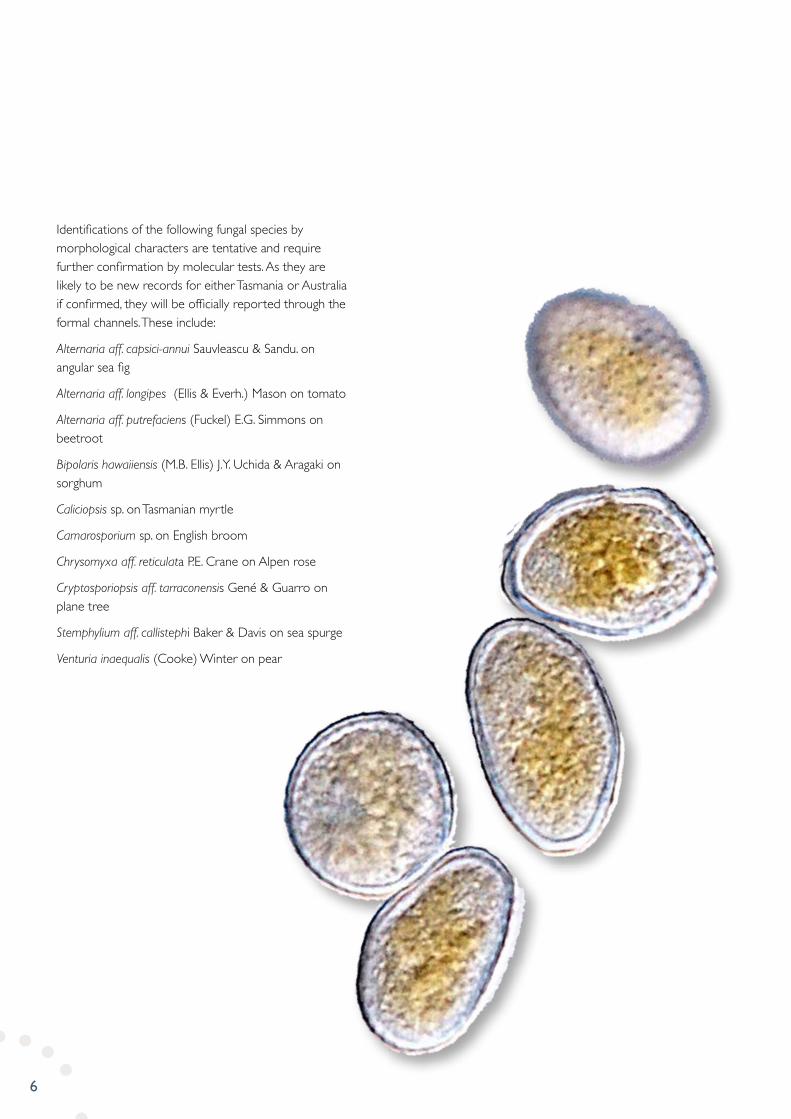
Identifications of the following fungal species by morphological characters are tentative and require further confirmation by molecular tests. As they are likely to be new records for either Tasmania or Australia if confirmed, they will be officially reported through the formal channels. These include:
Alternaria aff. capsici-annui Sauvleascu & Sandu. on angular sea fig
Alternaria aff. longipes (Ellis & Everh.) Mason on tomato
Alternaria aff. putrefaciens (Fuckel) E.G. Simmons on beetroot
Bipolaris hawaiiensis (M.B. Ellis) J.Y. Uchida & Aragaki on sorghum
Caliciopsis sp. on Tasmanian myrtle
Camarosporium sp. on English broom
Chrysomyxa aff. reticulata P.E. Crane on Alpen rose
Cryptosporiopsis aff. tarraconensis Gené & Guarro on plane tree
Stemphylium aff. callistephi Baker & Davis on sea spurge
Venturia inaequalis (Cooke) Winter on pear
7
FUNGI
AAcrospeira mirabilis Berk. & Br. on chestnut from Victoria (Shell Mould)
Albugo candida (Pers.) O. Kuntze – on cabbage (White Rust)
Albugo lepidii A.N.S. Rao on springy peppercress (White Rust)
Alternaria alternata (Fr.) Keissler on pear (Fruit Rot)
Alternaria brassicae (Berk.) Sacc. on cabbage (Leaf Spot)
Alternaria aff. capsici-annui Sauvleascu & Sandu. on angular sea fig (Leaf Spot)
Alternaria citri Ellis & Pierce on lemon (Fruit Rot)
Alternaria embellisia Woudenberg & Crous on garlic (Skin Blotch and Bulb Rot)
Alternaria aff. longipes (Ellis & Everh.) Mason on tomato (Fruit Rot)
Alternaria aff. putrefaciens (Fuckel) E.G. Simmons on beetroot (Leaf Spot)
Alternaria tenuissima (Kunze : Pers.) Wiltshire on pear (Leaf Spot)
BBipolaris hawaiiensis (M.B. Ellis) J.Y. Uchida & Aragaki on sorghum from Queensland (Seed Mould)
Botrytis allii Munn. on onion (Neck Rot)
Botrytis cinerea Pers. on cherry and capsicum (Grey Mould)
Bremia lactucae Regel on lettuce (Downy Mildew)
CCaliciopsis sp. on Tasmanian myrtle (Leaf Spot)
Camarosporium sp. on English broom (Shoot Blight)
Ceratocystis paradoxa (Dade) C. Moreau on banana (Fruit Rot)
Chrysomyxa aff. reticulata P.E. Crane on Alpen rose (Leaf Rust)
Cladosporium macrocarpum Preuss on apple (Leaf Spot)
Colletotrichum acutatum Simmonds on hazelnut (Anthracnose)
Colletotrichum dematium (Pers. Ex Fr.) Grove on cotton from New South Wales (Anthracnose)
Colletotrichum sp. on blueberry (Seedling Dieback)
Cryptosporiopsis aff. tarraconensis Gené & Guarro on plane tree (Leaf Spot)
Cylindrocladium pauciramosum CL Schoch & Crous on acacia from Victoria (Blight)
DDidymella applanata (Niessl) Sacc. on raspberry (Stem Canker & Spur Blight)
Diplodia seriata De Not. on grapevine (Internal Discoloration)
FFusarium dimerum Penzig on tomato (Stem Canker)
Fusarium oxysporum Schle. Emend. Snyder & Hansen on broccoli (Basal Stem Rot)
Fusarium spp. on grapevine (Internal Discoloration)
Fusicladium radiosum var. letiferum (Peck) Ritschel & U. Braun on aspen (Leaf Spot)
LIST OF PLANT PATHOGENS TREATED IN THE ATLAS
8
GGabarnaudia betae (Delacr.) Samson & W. Gams on onion from New South Wales
Gabarnaudia betae (Delacr.) Samson & W. Gams on raspberry
Geotrichum candidum Link on squash (Sour Rot)
IIdiocercus aff. australis (Cooke) Swart on birch (Leaf Blight)
LLeptosphaerulina sp. on ryegrass (Endophyte)
MMelampsora euphorbiae (Ficinus & C Schub.) Castagne on petty spurge (Rust)
Melampsora laricis-populina Kleb. on white poplar (Rust)
Melampsoridium betulinium (Pers.) Kleb. on alders (Rust)
Microdochium panattonianum (Berl.) Sutton Galea & Price on lettuce (Anthracnose)
Monilinia laxa (Aderh. & Ruhl.) Honey on apricot and cherry (Brown Rot)
Myxosporium aff. rosae Fuckel on loganberry (Stem Canker)
NNeofusicoccum luteum (Pennycook & Samuels) Crous, Slippers & A.J.L. Phillips on golden ash (Stem Canker)
Neofusicoccum ribis (Slippers, Crous & MJ Wingf.) Crous, Slippers & AJL Philllips on blueberry (Twig Blight)
PParaconiothyrium fuckelii (Sacc.) Verkley & Gruyter on loganberry (Cane Blight)
Passalora fulva (Cooke) U. Braun & Crous on tomato (Leaf Mould)
Penicillium digitatum Sacc. on orange (Fruit Rot)
Penicillium hirsutum Dierckx on tulip (Bulb Rot)
Peronospora destructor (Berk.) Casp. ex Berk. on onion (Downy Mildew)
Peronospora parasitica (Pers. : Fr.) Fr. on cabbage (Downy Mildew)
Phloeospora mori (Lev.) Sacc. on mulberry (Leaf Spot)
Phoma tropica R. Schneid. & Boerema on poinsettia from New South Wales (Basal Stem Canker)
Phragmidium rubi-idaei (DC.) Karst. on raspberry (Leaf Rust)
Phragmidium violaceum (C F Schultz) Winter. on blackberry (Leaf Rust)
Phyllactinia corylea (Pers.) Karst. on plane tree (Powdery Mildew)
Phytophthora cactorum (Leb. & Cohn) Schroeter on horse chestnut (Bleeding Canker)
Phytophthora drechsleri Tucker on raspberry (Root Rot)
Phytophthora megasperma Drechsler on azalea (Crown Rot)
Plasmopara obducens (J. Schröt.) J. Schröt. on impatiens (Downy Mildew)
Podosphaera clandestina (Wallr.) Lev. on hawthorn (Powdery Mildew)
Puccinia allii Rud. on onion (Rust)
Puccinia graminis Pers. subsp. graminicola on grass (Leaf Rust)
Puccinia iridis Rabenhorst on iris (Leaf Rust)
Puccinia malvacearum Bertero ex. Mont. on hollyhock (Leaf Rust)

9
Puccinia psidii Winter on Chilean guava from New South Wales (Myrtle Rust)
Puccinia rhei-undulati Hirats. on rhubarb (Leaf Rust)
Puccinia saccardoi Ludw. on goodenia (Rust)
Pythium helicoides Drechsler on strawberry from Queensland (Crown Rot)
RRamularia collo-cygni on barley (Leaf Spot)
Rhizoctonia sp. on beardtongue (Crown Rot)
SSeimatosporium lichenicola (Corda) Shoemaker & E. Müll. on raspberry (Shoot Blight)
Seiridium cupressi (Guba) Boesew.on cedar (Stem Canker)
Septoria citri Pass. on lemon (Septoria Spot)
Septoria hydrocotylicola Speg. on arthritis herb (Leaf Spot)
Septoria slaptonensis D. Hawksw. & Punith. on gorse (Leaf Blight)
Sordaria fimicola (Roberge ex Desm.) Ces. & De Not. on ryegrass (Endophyte)
Spilocaea photiniicola (McClain) M.B. Ellis on photinia (Scab)
Stemphylium aff. callistephi Baker & Davis on sea spurge (Shoot Blight)
Stemphylium vesicarium (Wallr.) Simmons on Brussels sprouts (Black Peppery Spot)
TTeratosphaeria eucalypti (Cooke & Massee) Crous on eucalypt (Leaf Spot)
Thekopsora minima P. Syd. & Syd. on blueberry from New South Wales (Rust)
Thielaviopsis basicola (Berk. & Broome) Ferraris on carrot (Black Root Rot)
Tranzschelia discolor (Fuckel) Tranz. & Litv. on apricot (Leaf Rust)
Trichothecium roseum Link on hazelnut (Mould)
UUlocladium atrum Preuss on wheat (Grain Mould)
Uromyces beticola (Bellynck) Boerema on beetroot (Leaf Rust)
Uromyces dianthi (Pers.) Niessl on carnation (Rust)
Uromyces viciae-fabae (Pers.) Schroet. on broad bean (Leaf Rust)
Ustilago bullata Berk. on grass (Ear Smut)
Ustilago maydis (DC.) Corda on maize from New South Wales (Boil Smut)
VVenturia inaequalis (Cooke) Winter on apple and pear (Scab)
Venturia pirina Aderh. on pear (Scab)
Vizella grevilleae Swart on grevillea from Victoria (Leaf Spot)
continued over page
10
NEMATODES
Colbranium truncatum (Colbran) Andrassy extracted from fern bark
Ditylenchus dipsaci (Kühn) Filipjev extracted from garlic from Victoria
Dorylaimus sp. extracted from carrot field
Heterodera avenae Wollenweber extracted from soil
Hirschmanniella mucronata (Das) Luc & Goodey, extracted from rice from North East Asia
Pratylenchoides sp. extracted from peppermint plants and soil
Pratylenchus penetrans (Cobb) Filipjev & S. Stekhoven extracted from lily from Victoria
Rotylenchus robustus (de Man) Filipjev extracted from lavenders from Victoria

11
IMAGE PLATESFUNGI

12
Fig. 1: Chestnuts showing shell moulds due to a range of fungal species; Fig. 2: 5-day old colony of A. mirabilis on PDA (Top and reverse view); Fig. 3: Three to four-celled conidia of A. mirabilis from specimen; Fig. 4: Conidia produced in cultures (Scale Bars = 20 µm)
Plate 1: Acrospeira mirabilis on chestnut (Shell Mould)
1
3 4
2

13
Fig. 1: Symptoms; Fig. 2: Close-up view of sori; Fig. 3: Longitudinal section of a partial sorus; Fig. 4: sporangia in chain; Fig. 5: Sporangia (Scale bar = 50 µm for Fig. 3; = 10 µm for Figs 4-5)
Plate 2: Albugo candida on cabbage (White Rust)
1
5
3 4
2
14
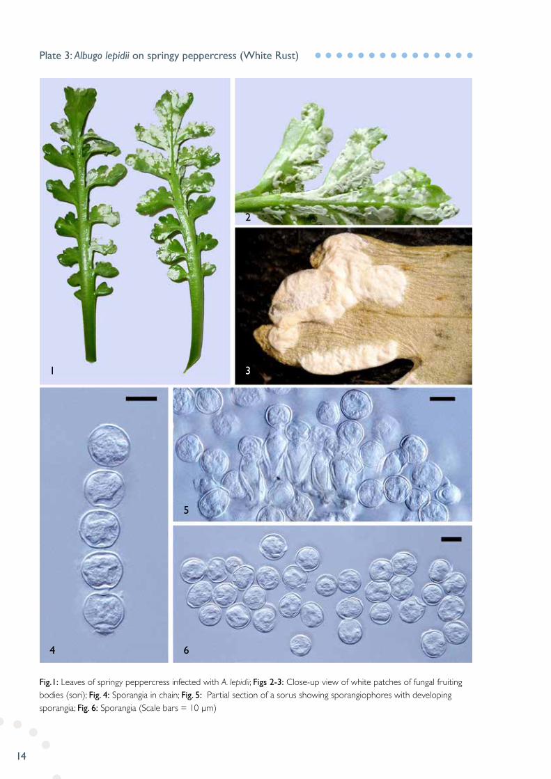
Fig.1: Leaves of springy peppercress infected with A. lepidii; Figs 2-3: Close-up view of white patches of fungal fruiting bodies (sori); Fig. 4: Sporangia in chain; Fig. 5: Partial section of a sorus showing sporangiophores with developing sporangia; Fig. 6: Sporangia (Scale bars = 10 µm)
Plate 3: Albugo lepidii on springy peppercress (White Rust)
1
4 6
5
2
3
15
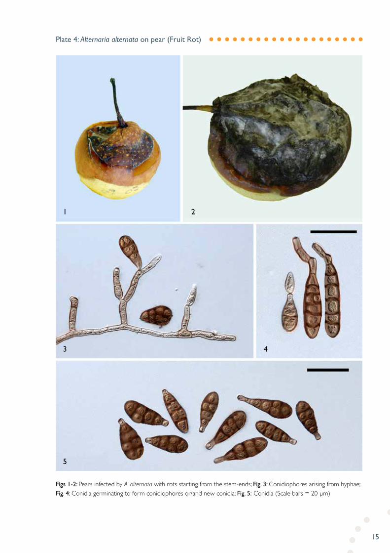
Figs 1-2: Pears infected by A. alternata with rots starting from the stem-ends; Fig. 3: Conidiophores arising from hyphae; Fig. 4: Conidia germinating to form conidiophores or/and new conidia; Fig. 5: Conidia (Scale bars = 20 µm)
Plate 4: Alternaria alternata on pear (Fruit Rot)
1
5
3 4
2

16
Figs 1-2: Irregular-shaped black spots caused by A. brassicae on cabbage leaves with the lesions of sporulation circled; Figs 3-4: Short unbranched conidiophores forming in clusters; Fig. 5: Conidia (Scale bar = 50 µm)
Plate 5: Alternaria brassicae on cabbage (Leaf Spot)
1
5
2
3 4
17
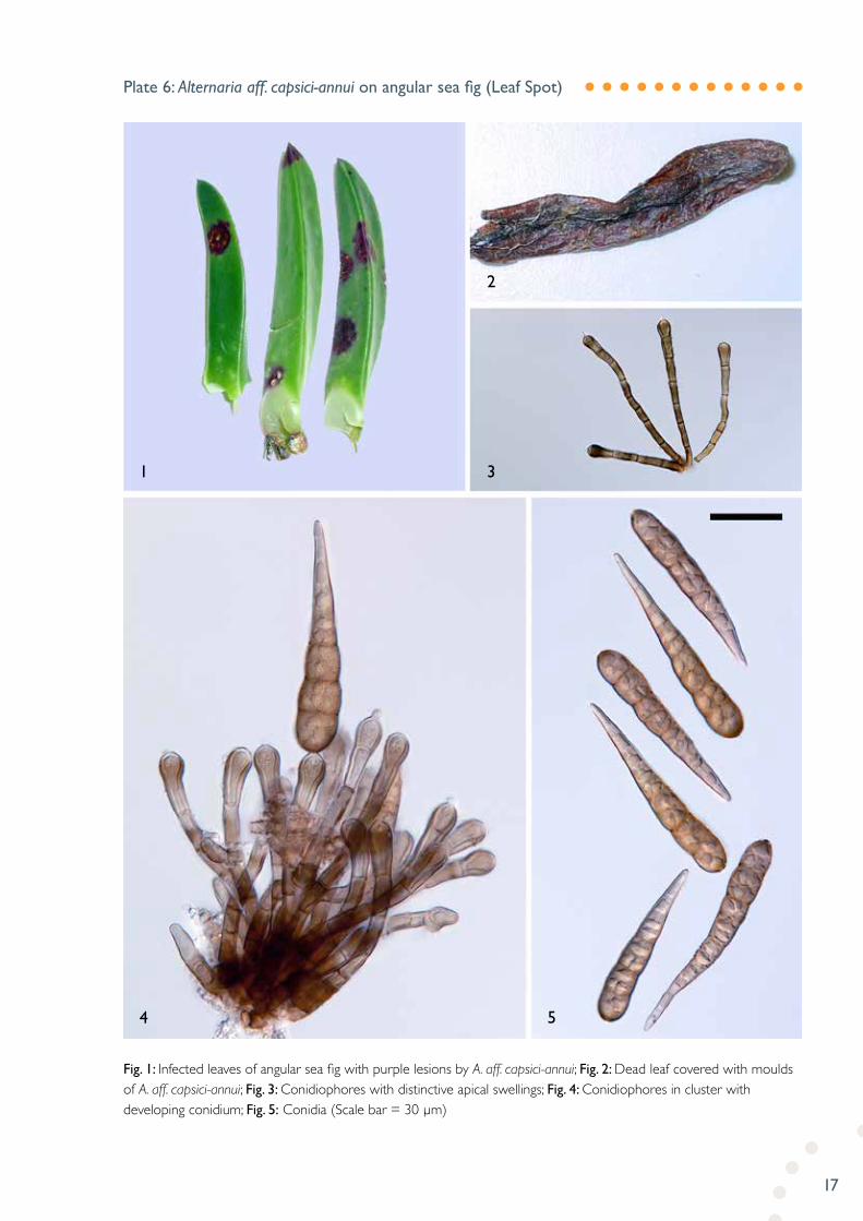
Fig. 1: Infected leaves of angular sea fig with purple lesions by A. aff. capsici-annui; Fig. 2: Dead leaf covered with moulds of A. aff. capsici-annui; Fig. 3: Conidiophores with distinctive apical swellings; Fig. 4: Conidiophores in cluster with developing conidium; Fig. 5: Conidia (Scale bar = 30 µm)
Plate 6: Alternaria aff. capsici-annui on angular sea fig (Leaf Spot)
1
4 5
3
2
18
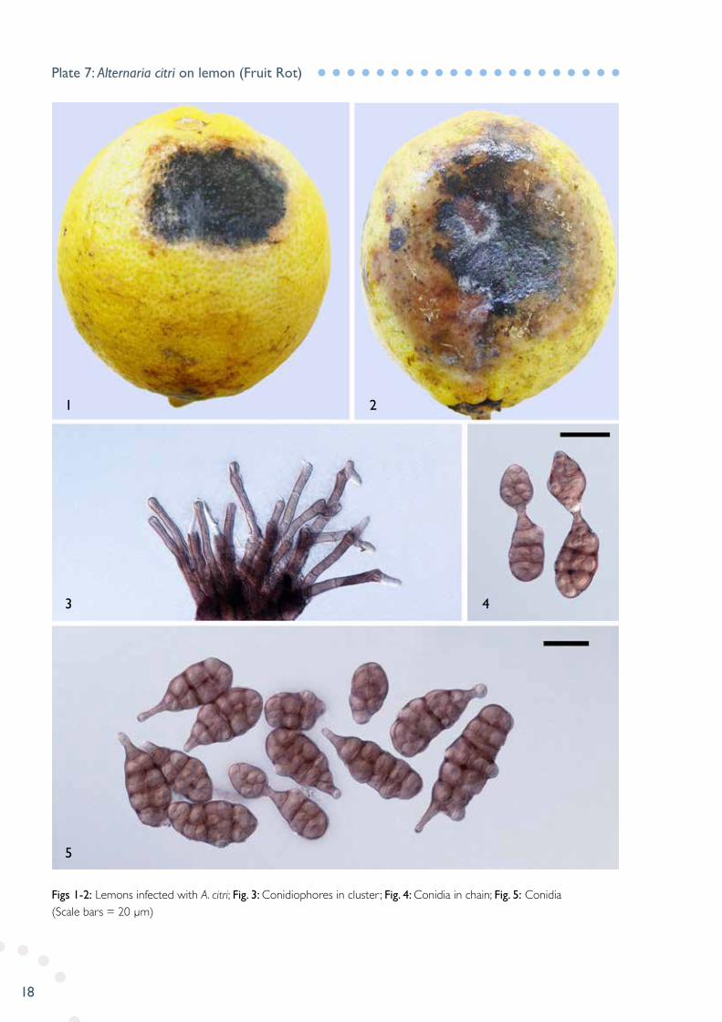
Figs 1-2: Lemons infected with A. citri; Fig. 3: Conidiophores in cluster ; Fig. 4: Conidia in chain; Fig. 5: Conidia (Scale bars = 20 µm)
Plate 7: Alternaria citri on lemon (Fruit Rot)
1
5
2
3 4

19
Fig. 1: Infected garlic cloves with characteristic symptoms of clove tip infection under the scales; Fig. 2: Lesion covered with black mould (conidial masses) as seen when the scales are removed; Figs 3-4: Conidiophores arising from hyphae; Figs 5-6: Conidiophores arising directly from conidial bodies; Fig. 7: Conidia with transverse septa (up to 10) and occasionally 1-2 oblique or longitudinal septa (Scale bars = 30 µm)
Plate 8: Alternaria embellisia on garlic (Skin Blotch and Bulb Rot)
1
3 4 5 6
7
2
20
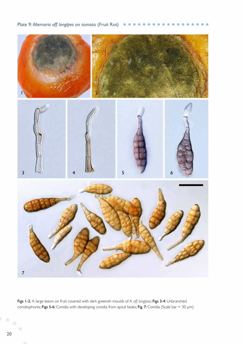
Figs 1-2: A large lesion on fruit covered with dark greenish moulds of A. aff. longipes; Figs 3-4: Unbranched conidiophores; Figs 5-6: Conidia with developing conidia from apical beaks; Fig. 7: Conidia (Scale bar = 30 µm)
Plate 9: Alternaria aff. longipes on tomato (Fruit Rot)
1 2
3 4 5 6
7
4
21
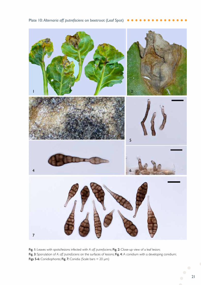
Fig. 1: Leaves with spots/lesions infected with A aff. putrefaciens; Fig. 2: Close-up view of a leaf lesion; Fig. 3: Sporulation of A. aff. putrefaciens on the surfaces of lesions; Fig. 4: A conidium with a developing conidium; Figs 5-6: Conidiophores; Fig. 7: Conidia (Scale bars = 20 µm)
Plate 10: Alternaria aff. putrefaciens on beetroot (Leaf Spot)
1
3
4
5
6
7
2

22
Fig. 1: Pear leaves with spots occurring mainly at margin areas, Fig. 2: A leaf with large irregularly-shaped lesion; Fig 3: Conidiophores and conidia in clusters on the surface of lesions; Fig. 4: Conidiophores with pores; Figs 5-6: Conidia with short to long beaks (Scale bar = 30 µm)
Plate 11: Alternaria tenuissima on pear (Leaf Spot)
1 3
2
6
4 6
5

23
Fig. 1: Sorghum seeds colonised by B. hawaiiensis; Fig. 2: Colonies of subcultures of B. hawaiiensis on PDA; Figs 3-4: Conidiophores; Fig. 5: Conidia with 2-7 (8) distosepta (Scale bars = 10 µm)
Plate 12: Bipolaris hawaiiensis on sorghum (Seed Mould)
1
3 4
5
2

24
Figs 1-3: Grey mould and black sclerotia developing on brown and white onions; Fig. 4: Internal infections of neck rot- initial symptoms; Figs 5-6: Laterally branched conidiophores; Fig. 7: Conidia (Scale bar = 10 µm)
Plate 13: Botrytis allii on onion (Neck Rot)
1 2
5
3 4
6 7

25
Figs 1-2: Grey mould symptoms on cherries and capsicum; Figs 3-4: Conidiophores with developing conidia of B. cinerea (images taken with dark field and normal light); Fig. 5: Mature conidia (Scale bar = 10 µm)
Plate 14: Botrytis cinerea on cherry and capsicum (Grey Mould)
1
5
3 4
2
26
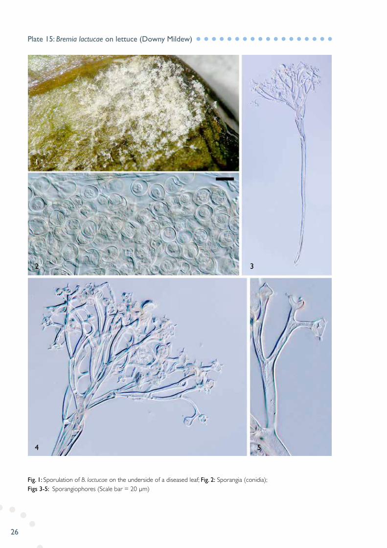
Fig. 1: Sporulation of B. lactucae on the underside of a diseased leaf; Fig. 2: Sporangia (conidia); Figs 3-5: Sporangiophores (Scale bar = 20 µm)
Plate 15: Bremia lactucae on lettuce (Downy Mildew)
1
2
4
3
5
27
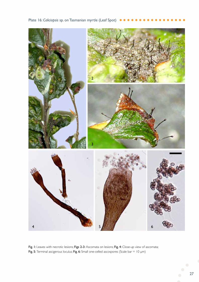
Fig. 1: Leaves with necrotic lesions; Figs 2-3: Ascomata on lesions; Fig. 4: Close-up view of ascomata; Fig. 5: Terminal ascigerous loculus; Fig. 6: Small one-celled ascospores (Scale bar = 10 µm)
Plate 16: Caliciopsis sp. on Tasmanian myrtle (Leaf Spot)
1
4 5 6
3
2

28
Fig. 1: Infected broom plant with symptoms of shoot blight; Fig. 2: Close-up view of pycnidia of Camarosporium sp. on shoots; Fig. 3: An longitudinal section of pycnidium; Fig. 4: Partial section of pycnidium showing wall cells and developing conidia; Fig. 5: Mature conidia (Scale bar = 20 µm)
Plate 17: Camarosporium sp. on English broom (Shoot Blight)
1 2
3 4
5

29
Fig. 1: Affected fruits; Fig. 2: Close-up view of rotting area covered with white fungal growth; Figs 3-4: Conidiophores of C. paradoxa; Fig. 5: Conidia; Fig. 6: Chlamydospores (Scale Bars = 10 µm)
Plate 18: Ceratocystis paradoxa on banana (Fruit Rot)
1
3 4 6
5
2

30
Figs 1-2: Infected leaves of alpenrose (upper and under side view); Figs 3-4: Lesions covered with orange-coloured pustules (uredia); Fig. 5: Vertical section of an uredium; Fig. 6: Urediniospores; Figs 7-8: Urediniospores showing wall surface ornamentations (warts). (Scale bar = 50 µm for Fig. 5, 15 µm for Fig. 6; 10 µm for Figs 7-8)
Plate 19: Chrysomyxa aff. reticulata on Alpen rose (Leaf Rust)
1 3
2
4
5
7 8
6

31
Figs 1-3: Symptoms of leaf spots; Fig. 4: Close-up view of a lesion surface covered with conidiophores and conidial masses; Figs 5-6: Sympodial conidiophores; Figs 7-8: 1-3-septate conidia (Scale bar = 10 µm)
Plate 20: Cladosporium macrocarpum on apple (Leaf Spot)
1
3
7 8
5 6
4
2
32
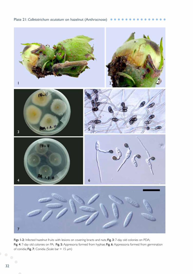
Figs 1-2: Infected hazelnut fruits with lesions on covering bracts and nuts; Fig. 3: 7-day old colonies on PDA; Fig. 4: 7-day old colonies on PA; Fig. 5: Appresoria formed from hyphae; Fig. 6: Appressoria formed from germination of conidia; Fig. 7: Conidia (Scale bar = 15 µm)
Plate 21: Colletotrichum acutatum on hazelnut (Anthracnose)
3
4 6
5
21
7

33
Fig. 1: Infected fruit; Figs.2-3: Close-up view of the lesion with fruiting bodies; Fig.4: A conidioma with setae; Fig. 5: Conidia (Scale bar = 20 µm)
Plate 22: Colletotrichum dematium on cotton (Anthracnose)
1 3
2
4 5
34
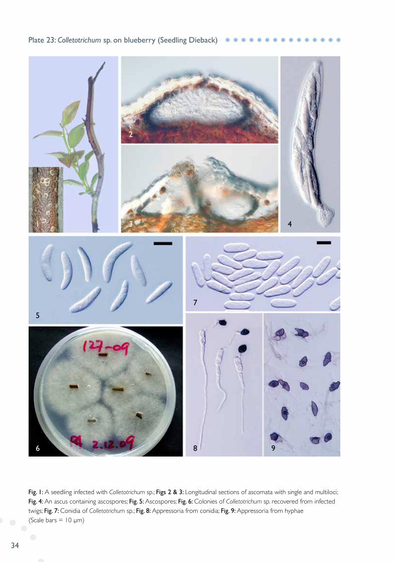
Fig. 1: A seedling infected with Colletotrichum sp.; Figs 2 & 3: Longitudinal sections of ascomata with single and multiloci; Fig. 4: An ascus containing ascospores; Fig. 5: Ascospores; Fig. 6: Colonies of Colletotrichum sp. recovered from infected twigs; Fig. 7: Conidia of Colletotrichum sp.; Fig. 8: Appressoria from conidia; Fig. 9: Appressoria from hyphae (Scale bars = 10 µm)
Plate 23: Colletotrichum sp. on blueberry (Seedling Dieback)
1
1 3
2
4
5
6 8
7
9

35
Figs 1-2: A leaf of plane tree infected with C. aff. tarraconensis; Fig. 3: Close-up view of fungal fruiting bodies (acervuli) on the leaf lesions; Fig. 4: Longitudinal section of an acervulus; Fig. 5: Conidia (Scale bar = 10 µm)
Plate 24: Cryptosporiopsis aff. tarraconensis on plane tree (Leaf Spot)
1
2
4
5
3

36
Figs 1-2: 12-day-old colony on PDA (top and reverse view); Figs 3-4: Formation of conidia on PDA; Fig.5: Conidiophores terminating in vesicles and conidia formatted in bundles; Fig. 6: A conidiophore with densely branched phialides and terminal vesicle; Fig. 7: 0-1-septate conidia; Figs 8-9: Chlamydospores produced in aerial hyphae in chains (Fig. 8) or single (Fig. 9). (Scale bar = 20 µm)
Plate 25: Cylindrocladium pauciramosum on acacia (Blight)
6
2
1
4
3
5
8
7
9
37
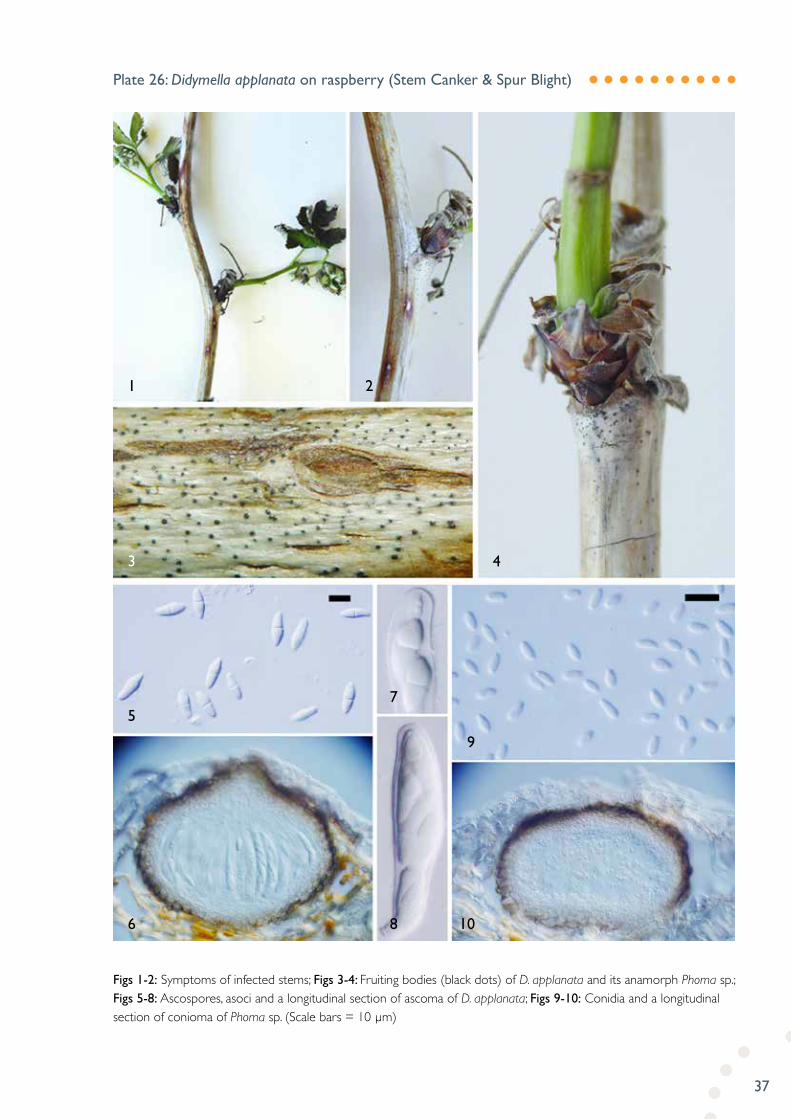
Figs 1-2: Symptoms of infected stems; Figs 3-4: Fruiting bodies (black dots) of D. applanata and its anamorph Phoma sp.; Figs 5-8: Ascospores, asoci and a longitudinal section of ascoma of D. applanata; Figs 9-10: Conidia and a longitudinal section of conioma of Phoma sp. (Scale bars = 10 µm)
Plate 26: Didymella applanata on raspberry (Stem Canker & Spur Blight)
1 2
3
5
6 8 10
9
7
4
38
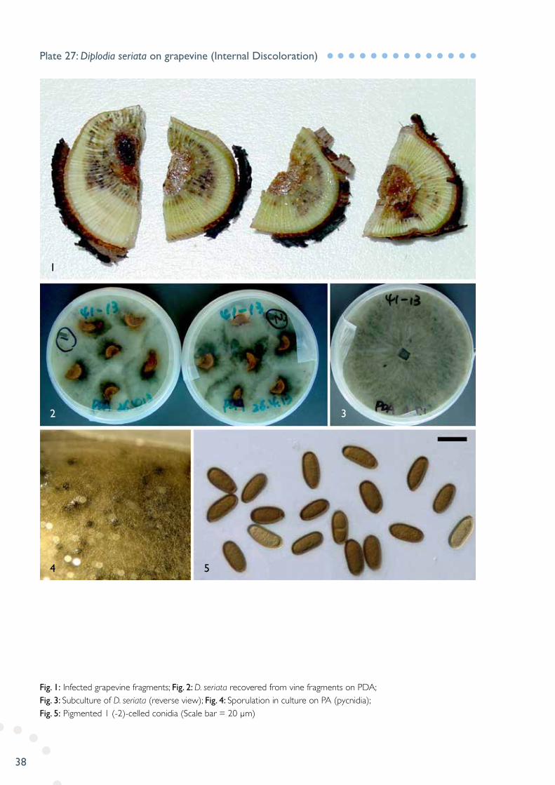
Fig. 1: Infected grapevine fragments; Fig. 2: D. seriata recovered from vine fragments on PDA; Fig. 3: Subculture of D. seriata (reverse view); Fig. 4: Sporulation in culture on PA (pycnidia); Fig. 5: Pigmented 1 (-2)-celled conidia (Scale bar = 20 µm)
Plate 27: Diplodia seriata on grapevine (Internal Discoloration)
4
2 3
1
5

39
Fig. 1: Tomato stem infected with F. dimerum; Fig. 2: Canker lesions on stem covered with yellowish sporulation; Fig. 3: Conidiophores with developing conidia; Fig. 4: Macroconidia (under dark field); Fig. 5: Two-celled macroconidia (Scale bar = 10 µm)
Plate 28: Fusarium dimerum on tomato (Stem Canker)
1 2
3 4
5

40
Fig. 1: Broccoli seedlings infected with F. oxysporum at the basal stems; Fig. 2: Infected plants with leaf and flower turning purple in colour; Fig. 3: Close-up view of basal stem rot; Fig. 4: Macroconidia (Scale bar = 20 µm)
Plate 29: Fusarium oxysporum on broccoli (Basal Stem Rot)
1
3 4
2
41
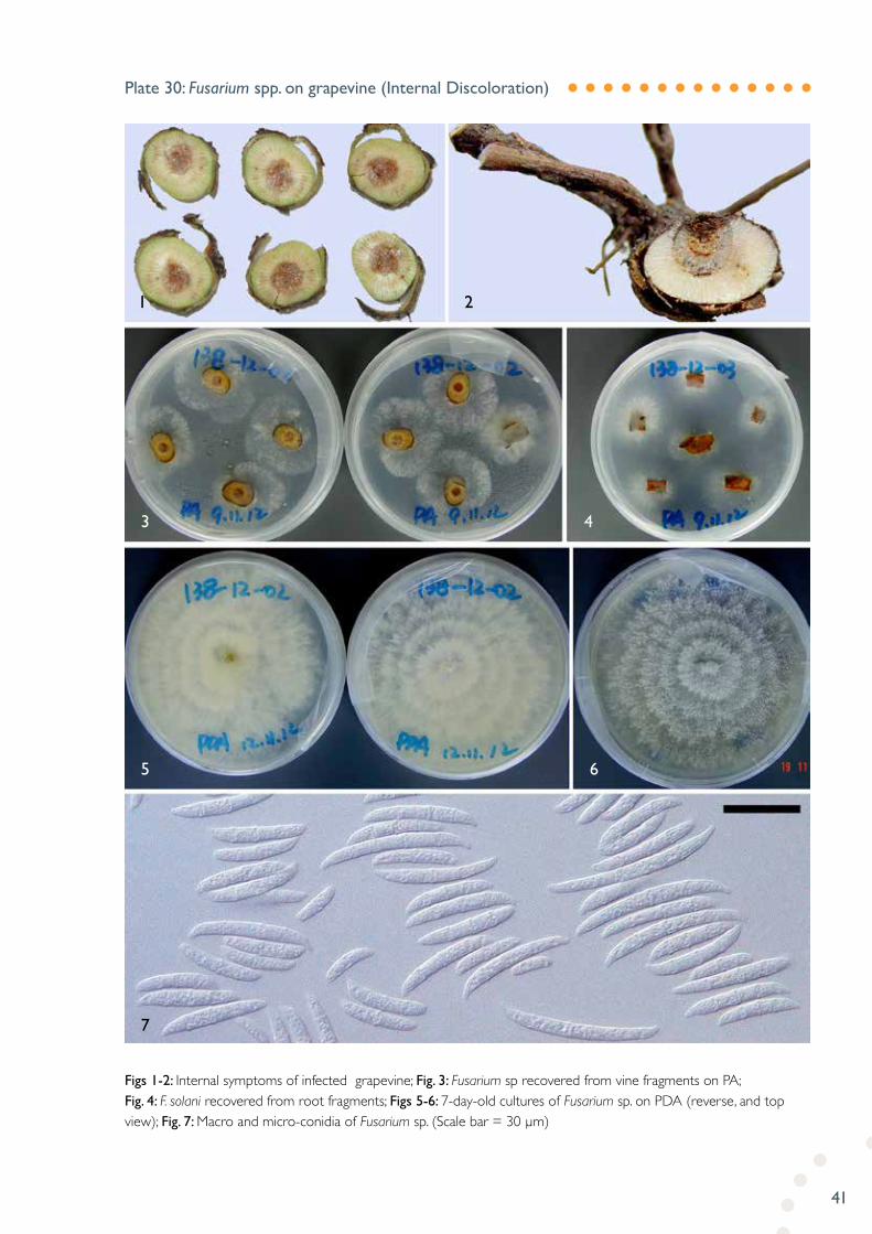
Figs 1-2: Internal symptoms of infected grapevine; Fig. 3: Fusarium sp recovered from vine fragments on PA; Fig. 4: F. solani recovered from root fragments; Figs 5-6: 7-day-old cultures of Fusarium sp. on PDA (reverse, and top view); Fig. 7: Macro and micro-conidia of Fusarium sp. (Scale bar = 30 µm)
Plate 30: Fusarium spp. on grapevine (Internal Discoloration)
1
3
5
7
6
4
2
42
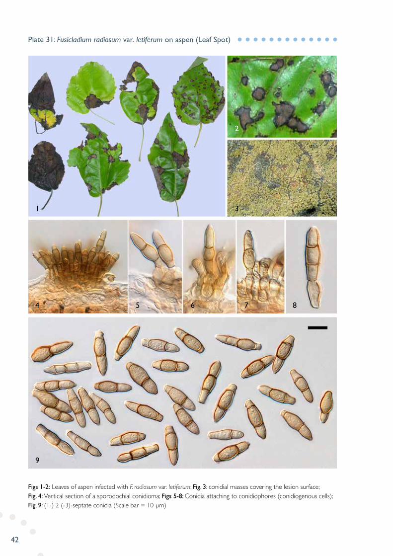
Figs 1-2: Leaves of aspen infected with F. radiosum var. letiferum; Fig. 3: conidial masses covering the lesion surface; Fig. 4: Vertical section of a sporodochial conidioma; Figs 5-8: Conidia attaching to conidiophores (conidiogenous cells); Fig. 9: (1-) 2 (-3)-septate conidia (Scale bar = 10 µm)
Plate 31: Fusicladium radiosum var. letiferum on aspen (Leaf Spot)
1
4
9
5 6 7 8
3
2
43
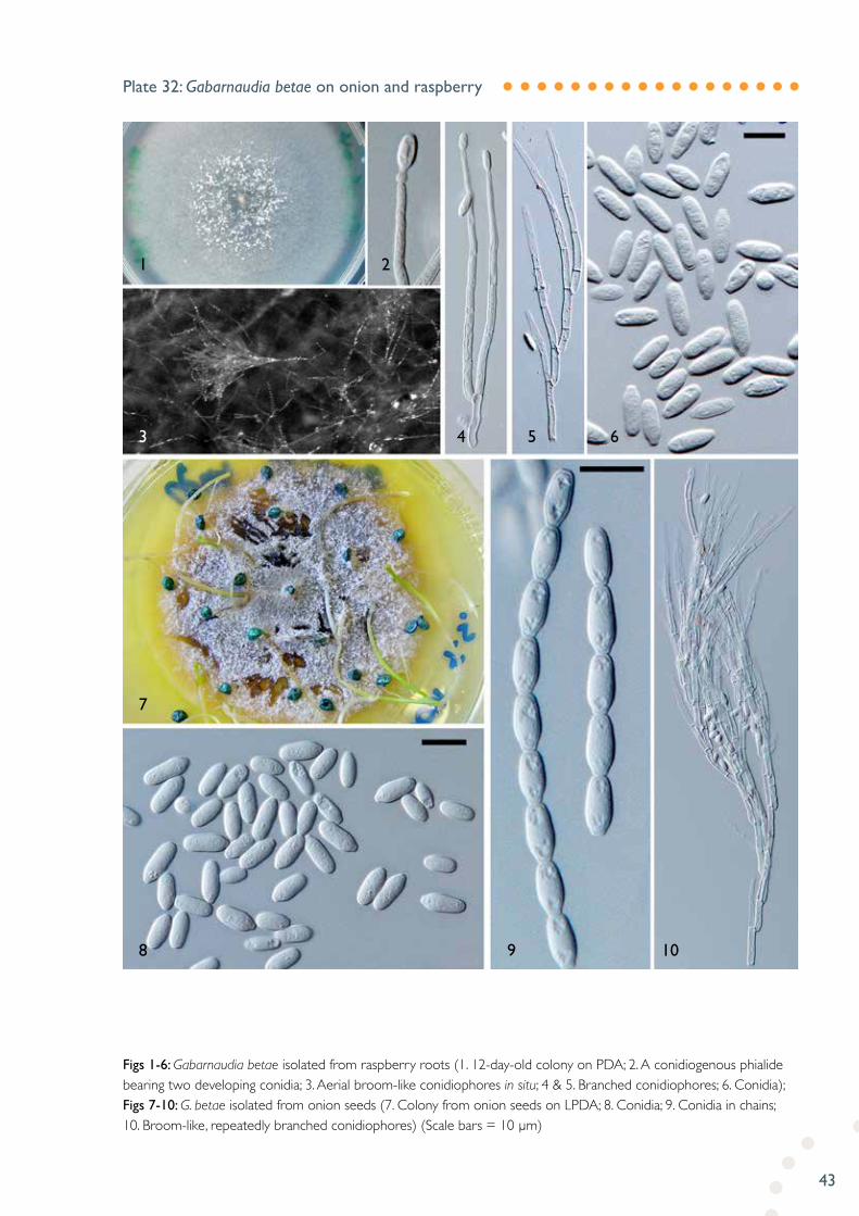
Figs 1-6: Gabarnaudia betae isolated from raspberry roots (1. 12-day-old colony on PDA; 2. A conidiogenous phialide bearing two developing conidia; 3. Aerial broom-like conidiophores in situ; 4 & 5. Branched conidiophores; 6. Conidia); Figs 7-10: G. betae isolated from onion seeds (7. Colony from onion seeds on LPDA; 8. Conidia; 9. Conidia in chains; 10. Broom-like, repeatedly branched conidiophores) (Scale bars = 10 µm)
Plate 32: Gabarnaudia betae on onion and raspberry
1
3
7
8 9 10
4 5 6
2

44
Fig. 1: Sour rot of squash infected by G. candidum via stem-end; Fig. 2: Cut squash showing internal soft rot; Fig. 3: Hyphal conidiophores (anthric) of G. candidum; Fig. 4: Conidia (Scale bar = 10 µm)
Plate 33: Geotrichum candidum on squash (Sour Rot)
1
3
4
2

45
Figs 1-2: Birch leaves with large necrotic lesions developing from the tip; Fig. 3: Close-up view of the fungal fruiting bodies (pycnidia) on the surface of the lesion; Fig. 4: Longitudinal section of a pycnidium; Fig. 5: Conidia (Scale bar = 10 µm)
Plate 34: Idiocercus aff. australis on birch (Leaf Blight)
1 3
2
4
5
46
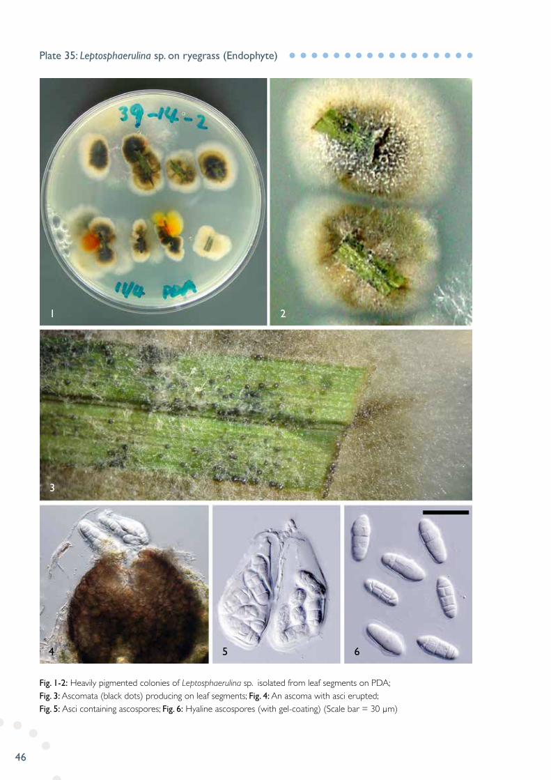
Fig. 1-2: Heavily pigmented colonies of Leptosphaerulina sp. isolated from leaf segments on PDA; Fig. 3: Ascomata (black dots) producing on leaf segments; Fig. 4: An ascoma with asci erupted; Fig. 5: Asci containing ascospores; Fig. 6: Hyaline ascospores (with gel-coating) (Scale bar = 30 µm)
Plate 35: Leptosphaerulina sp. on ryegrass (Endophyte)
4
3
1 2
5 6

47
Fig. 1: Plant infected with M. euphorbiae; Fig. 2: Uredinia of M. euphorbiae; Fig. 3: A longitudinal section of uredinia; Fig. 4: Partial uredinia showing developing urediniospores and paraphyses; Fig. 5: Urediniospores (Scale bar = 20 µm)
Plate 36: Melampsora euphorbiae on petty spurge (Rust)
1 2
3
4 5

48
Fig. 1: Symptoms of rust on both upper and lower sides of the leaves; Fig. 2: Close-up view of uredinia; Fig. 3: Longitudinal section of an uredinium; Fig. 4: Partial section of uredinium with non-pigmented paraphyses; Fig. 5: Urediniospores (Scale bar = 30 µm)
Plate 37: Melampsora laricis-populina on white poplar (Rust)
5
3 4
1 2

49
Figs 1-2: Symptoms on stems (stem blisters) of Mexican alder (photo in Fig.1, courtesy of Deborah Combes); Figs 3-4: An infected leaf of common alder (Upper and lower side view); Figs 5-6: Close-up view of rust pustules (uredinia) on the leaf; Fig. 7: Urediniospores (Scale bar = 20 µm)
Plate 38: Melampsoridium betulinum on alders (Rust)
7
5
1
6
3 4
2

50
Fig. 1: Infected seedlings of lettuce with M. panattonianum; Fig. 2: Water-soaked spots (later appearing as “shot-hole”); Fig. 3: Sporulation on the surfaces of lesions; Fig. 4: Slightly curved, 1-2-celled conidia (Scale bar = 10 µm)
Plate 39: Microdochium panattonianum on lettuce (Anthracnose)
4
2
1
3
51
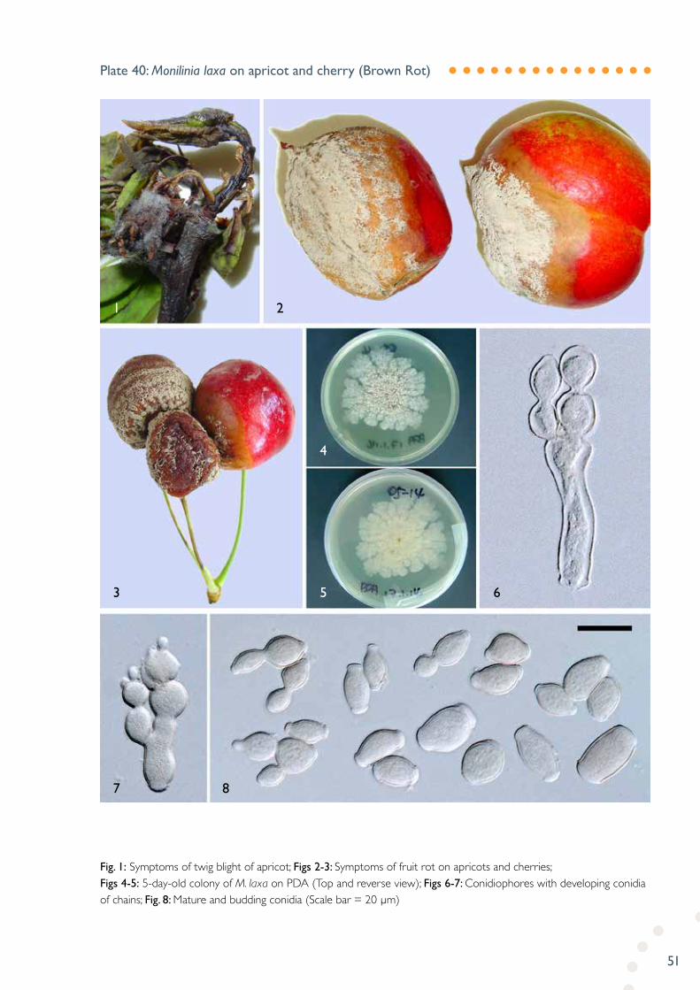
Fig. 1: Symptoms of twig blight of apricot; Figs 2-3: Symptoms of fruit rot on apricots and cherries; Figs 4-5: 5-day-old colony of M. laxa on PDA (Top and reverse view); Figs 6-7: Conidiophores with developing conidia of chains; Fig. 8: Mature and budding conidia (Scale bar = 20 µm)
Plate 40: Monilinia laxa on apricot and cherry (Brown Rot)
7
3
1 2
5
4
6
8

52
Fig. 1: Stem of loganberry with superficial acervuli of M. aff. rosae; Fig. 2: Longitudinal section of an acervulus; Fig. 3: Slightly curved, one-celled conidia (Scale bar = 5 µm)
Plate 41: Myxosporium aff. rosae on loganberry (Stem Canker)
2
3
1

53
Fig. 1: Branches of ash infected with N. luteum.; Fig. 2: Sunken canker developing downwards to healthy tissue; Fig. 3: Close-up view of stromata embedded in the canker area; Fig. 4: Longitudinal section of a stroma with three locule; Fig. 5: Partial section of a stroma with the loculus containing young asci; Fig. 6: Ascospores (Scale bar = 10 µm)
Plate 42: Neofusicoccum luteum on golden ash (Stem Canker)
5
4
1 2 3
6
54
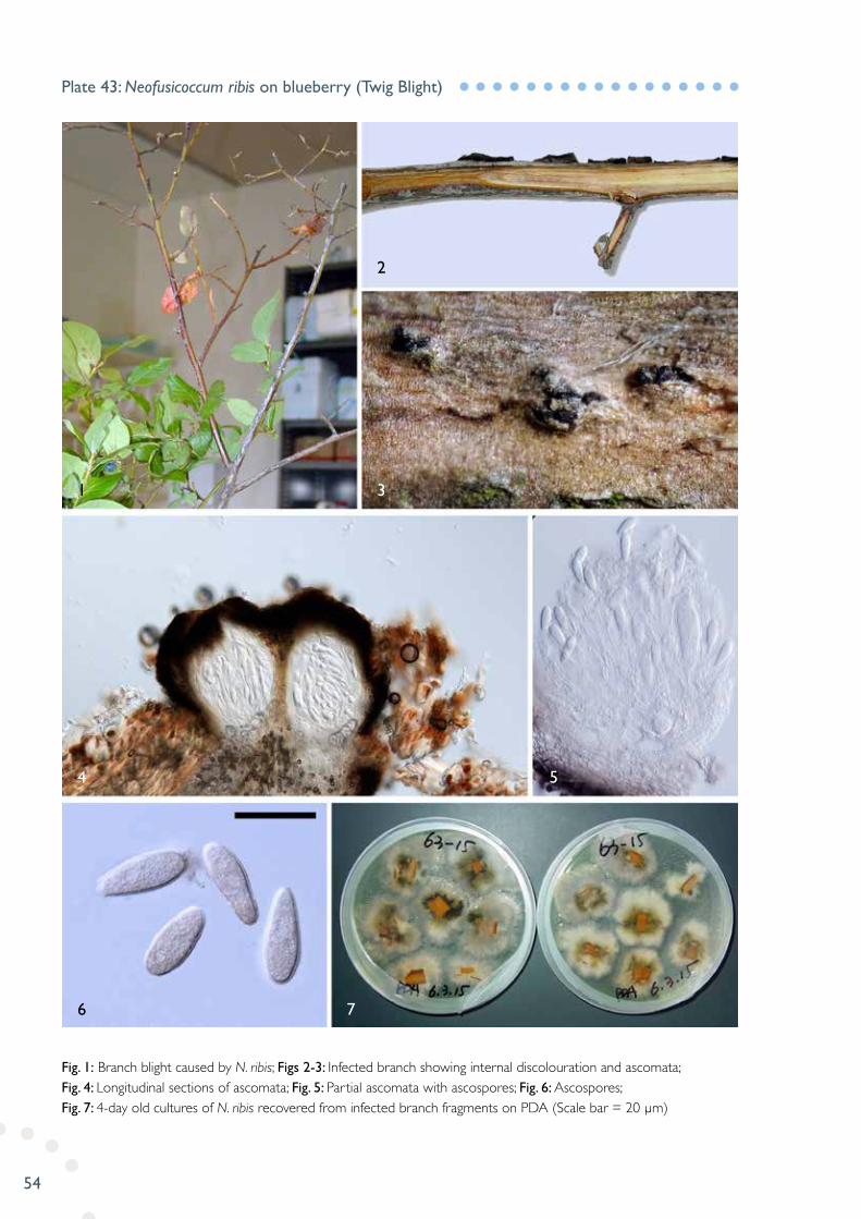
Fig. 1: Branch blight caused by N. ribis; Figs 2-3: Infected branch showing internal discolouration and ascomata; Fig. 4: Longitudinal sections of ascomata; Fig. 5: Partial ascomata with ascospores; Fig. 6: Ascospores; Fig. 7: 4-day old cultures of N. ribis recovered from infected branch fragments on PDA (Scale bar = 20 µm)
Plate 43: Neofusicoccum ribis on blueberry (Twig Blight)
4
1 3
2
2
6 7
5

55
Fig. 1: Cane of loganberry infected with P. fuckelii; Fig. 2: Fruiting bodies of P. fuckelii imbedded in the bark; Fig. 3: Longitudinal sections of pycnidia; Fig. 4: Conidia (Scale bar = 5 µm)
Plate 44: Paraconiothyrium fuckelii on loganberry (Cane Blight)
1
3
4
2
56
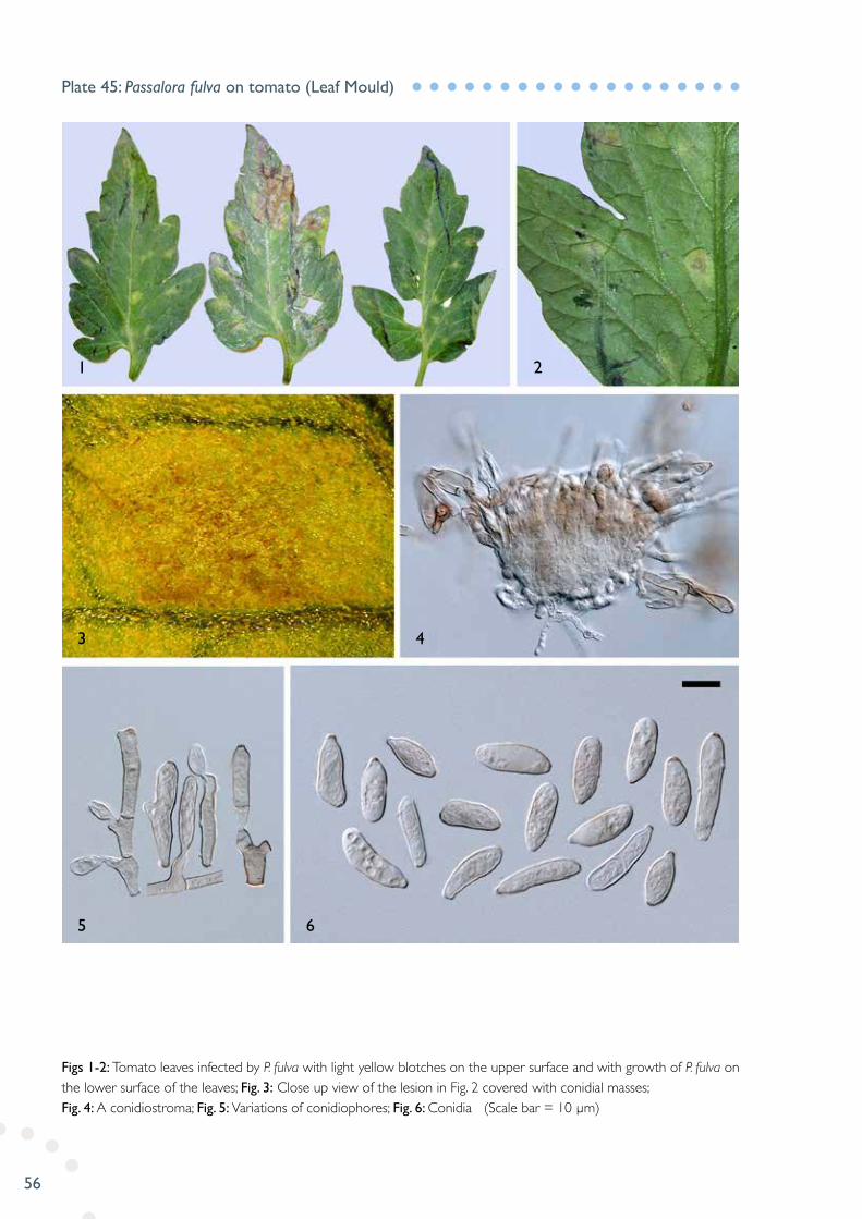
Figs 1-2: Tomato leaves infected by P. fulva with light yellow blotches on the upper surface and with growth of P. fulva on the lower surface of the leaves; Fig. 3: Close up view of the lesion in Fig. 2 covered with conidial masses; Fig. 4: A conidiostroma; Fig. 5: Variations of conidiophores; Fig. 6: Conidia (Scale bar = 10 µm)
Plate 45: Passalora fulva on tomato (Leaf Mould)
1
3
5 6
4
2

57
Fig. 1: Orange infected with P. digitatum; Figs 2-3: Conidiophores; Fig. 4: Hyaline to slightly pigmented conidia in chains (Scale bar = 10 µm)
Plate 46: Penicillium digitatum on orange (Fruit Rot)
1
3 4
2
58
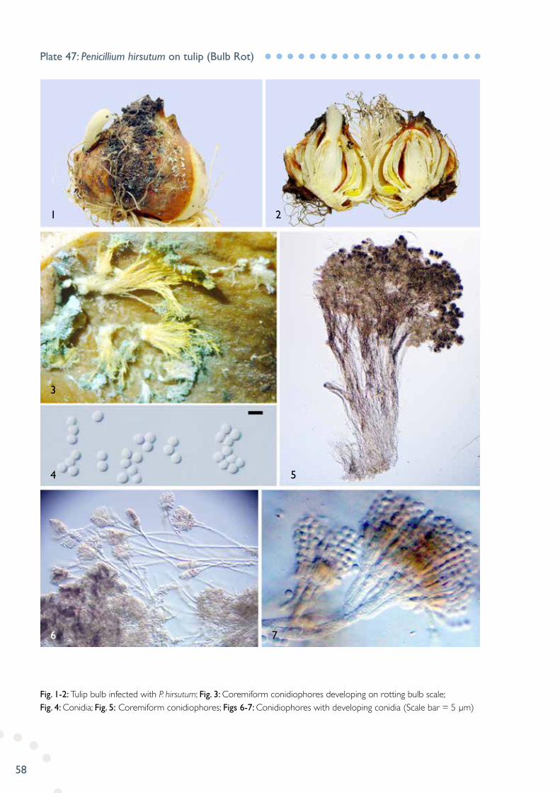
Fig. 1-2: Tulip bulb infected with P. hirsutum; Fig. 3: Coremiform conidiophores developing on rotting bulb scale; Fig. 4: Conidia; Fig. 5: Coremiform conidiophores; Figs 6-7: Conidiophores with developing conidia (Scale bar = 5 µm)
Plate 47: Penicillium hirsutum on tulip (Bulb Rot)
1
3
4
6 7
5
2

59
Fig. 1: Onion leaves infected with P. destructor; Figs 2-3: Close-up view of lesions and white fungal sporulation covering on the lesion surfaces; Fig. 4: Sporangiophore; Fig. 5: Apical part of sporangiophore; Fig. 6: Sporangia (Scale bar = 30 µm)
Plate 48: Peronospora destructor on onion (Downy Mildew)
1
4 6
5
3
2
60
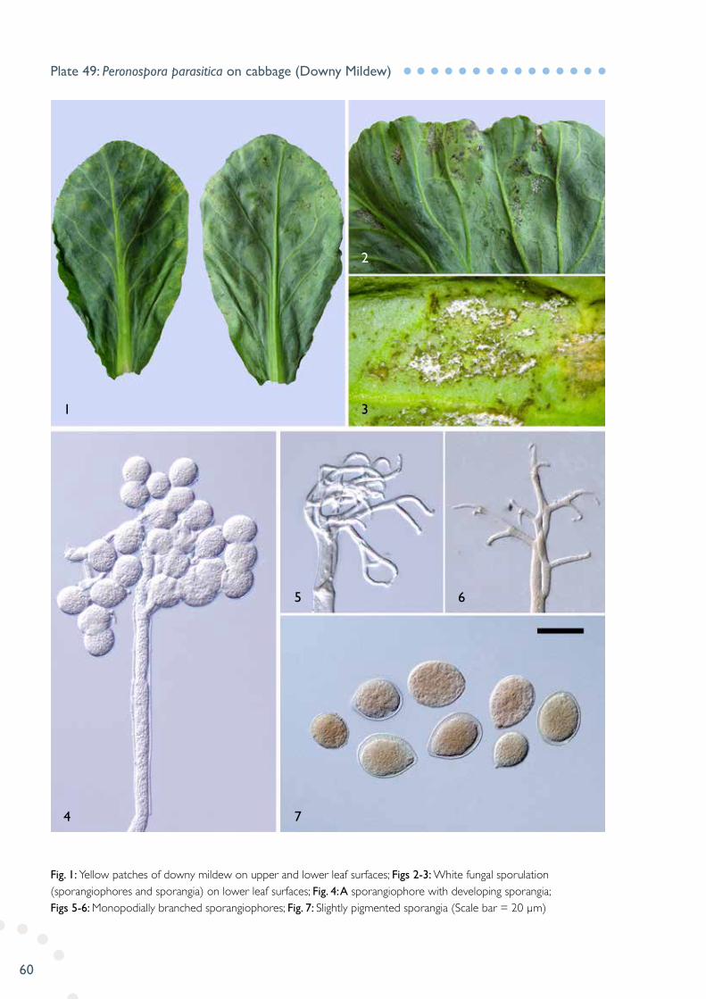
Fig. 1: Yellow patches of downy mildew on upper and lower leaf surfaces; Figs 2-3: White fungal sporulation (sporangiophores and sporangia) on lower leaf surfaces; Fig. 4: A sporangiophore with developing sporangia; Figs 5-6: Monopodially branched sporangiophores; Fig. 7: Slightly pigmented sporangia (Scale bar = 20 µm)
Plate 49: Peronospora parasitica on cabbage (Downy Mildew)
1
4 7
5 6
3
2
61
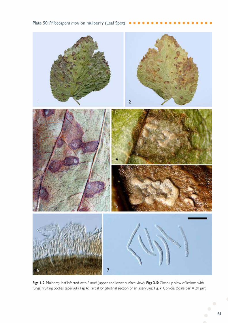
Figs 1-2: Mulberry leaf infected with P. mori (upper and lower surface view); Figs 3-5: Close-up view of lesions with fungal fruiting bodies (acervuli); Fig. 6: Partial longitudinal section of an acervulus; Fig. 7: Conidia (Scale bar = 20 µm)
Plate 50: Phloeospora mori on mulberry (Leaf Spot)
1
3
6 7
5
4
2
62
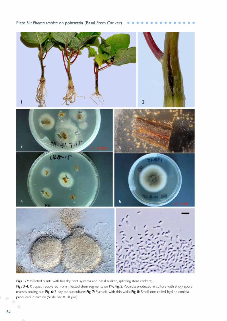
Figs 1-2: Infected plants with healthy root systems and basal sunken, splinting stem cankers; Figs 3-4: P. tropica recovered from infected stem segments on PA; Fig. 5: Pycnidia produced in culture with sticky spore masses oozing out; Fig. 6: 5-day old subculture; Fig. 7: Pycnidia with thin walls; Fig. 8: Small, one-celled, hyaline conidia produced in culture (Scale bar = 10 µm)
Plate 51: Phoma tropica on poinsettia (Basal Stem Canker)
1
3
4
7 8
6
5
2

63
Fig. 1: Symptoms of raspberry leaves infected with P. rubi-idaei; Fig. 2: Infected leaves with brown necrotic lesions on upper surfaces; Figs 3-4: Lower leaf surface view of rusty and black mould-like sporulation; Fig. 5: A longitudinal section of an uredinium/telium; Fig. 6: Urediniospores; Fig. 7: Teliospores (Scale bar = 20 µm for Fig. 6; 50 µm for Fig. 7)
Plate 52: Phragmidium rubi-idaei on raspberry (Leaf Rust)
1
4
6 7
5
3
2
64
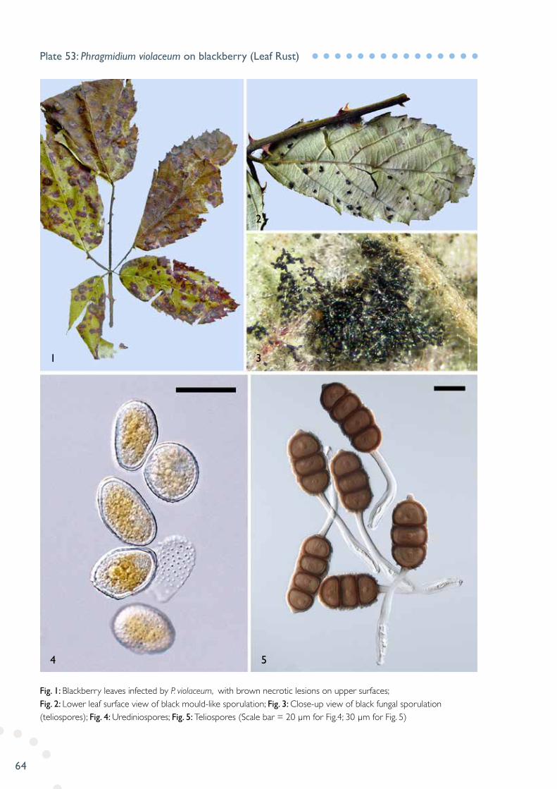
Fig. 1: Blackberry leaves infected by P. violaceum, with brown necrotic lesions on upper surfaces; Fig. 2: Lower leaf surface view of black mould-like sporulation; Fig. 3: Close-up view of black fungal sporulation (teliospores); Fig. 4: Urediniospores; Fig. 5: Teliospores (Scale bar = 20 µm for Fig.4; 30 µm for Fig. 5)
Plate 53: Phragmidium violaceum on blackberry (Leaf Rust)
1
4 5
3
2

65
Figs 1-2: Symptoms of Infected leaves and buds; Fig. 3: Conidia; Fig. 4: Conidiophores; Fig. 5: Germinated conidia; Fig. 6: Conidia in chain (Scale bars = 20 µm)
Plate 54: Phyllactinia corylea on plane tree (Powdery Mildew)
1
3
4 5 6
2
66
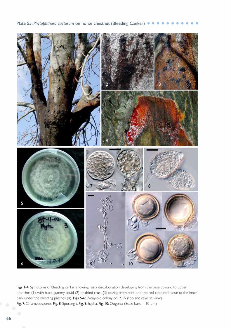
Figs 1-4: Symptoms of bleeding canker showing rusty discolouration developing from the base upward to upper branches (1), with black gummy liquid (2) or dried crust (3) oozing from bark, and the red-coloured tissue of the inner bark under the bleeding patches (4). Figs 5-6: 7-day-old colony on PDA (top and reverse view). Fig. 7: Chlamydospores. Fig. 8: Sporangia. Fig. 9: hypha. Fig. 10: Oogonia (Scale bars = 10 µm)
Plate 55: Phytophthora cactorum on horse chestnut (Bleeding Canker)
1
5
6 9 10
7 8
4
2 3

67
Fig. 1: Infected Root segments; Fig. 2: P. drechsleri recovered from roots; Figs 3-4: A 5-day old colony on PDA (reverse and top view): Fig. 5: Sporangia; Fig. 6: Hyphal swellings (Scale bar = 20 µm)
Plate 56: Phytophthora drechsleri on raspberry (Root Rot)
1
3
5 6
4
2

68
Fig. 1: A dying azalea plant; Figs 2-3: Sections of basal stems showing internal discolouration; Fig. 4: Isolation of Phytophthora megasperma from infected stem tissue (segments) on P10ARP; Figs 5-7: Oogonia of P. megasperma with amphigynous (Fig. 5) and paragynous (Figs 6-7) antheridia (Scale bar = 30 µm)
Plate 57: Phytophthora megasperma on azalea (Crown Rot)
1
3
5 6 7
4
2

69
Fig. 1: Symptoms of downy mildew on upper leaf surfaces; Figs 2-4: Symptoms of downy mildew on lower leaf surfaces; Fig. 5: A lateral branch of sporangiophore bearing one sporangium; Fig. 6: A sporangiophore with lateral branches diverging at right angles; Fig. 7: Sporangia (Scale bars = 10 µm)
Plate 58: Plasmopara obducens on impatiens (Downy Mildew)
6
4
1 3
2
5
7
70
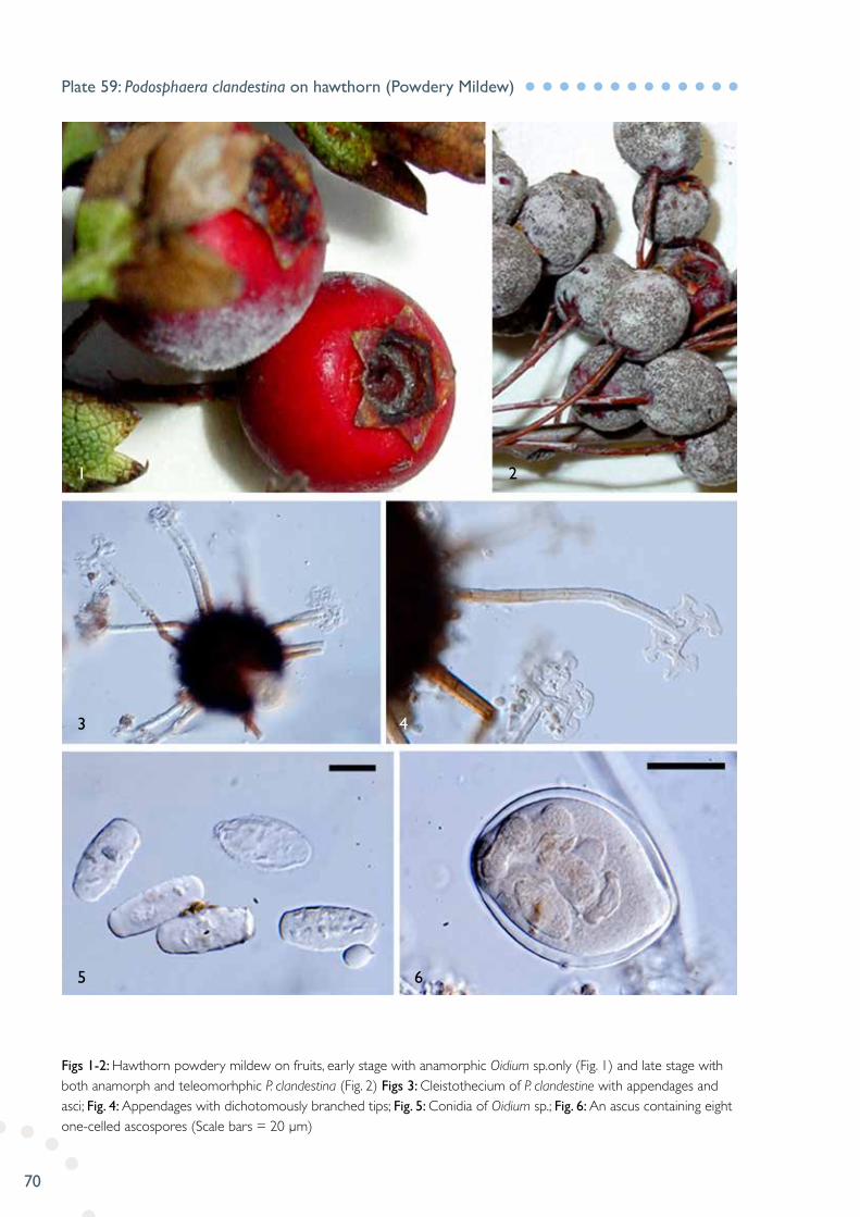
Figs 1-2: Hawthorn powdery mildew on fruits, early stage with anamorphic Oidium sp.only (Fig. 1) and late stage with both anamorph and teleomorhphic P. clandestina (Fig. 2) Figs 3: Cleistothecium of P. clandestine with appendages and asci; Fig. 4: Appendages with dichotomously branched tips; Fig. 5: Conidia of Oidium sp.; Fig. 6: An ascus containing eight one-celled ascospores (Scale bars = 20 µm)
Plate 59: Podosphaera clandestina on hawthorn (Powdery Mildew)
5
3
1 2
4
6

71
Fig. 1: Onion plant infected with P. allii on leaf and stem; Fig. 2: Close-up view of uredinia on leaves; Fig. 3: Close-up view of telia on stems; Fig. 4: Urediniospores; Fig. 5: Teliospores developing within a telium; Fig. 6: Longitudinal section of telia; Fig. 7: Partial longitudinal section of a leaf with stromatic telia covered by epidermis; Fig. 8: Immature and mature teliospores (Scale bars = 20 µm)
Plate 60: Puccinia allii on onion (Rust)
7
6
1 4
3
2
5
8
72
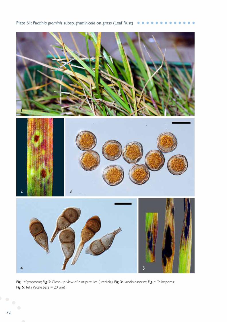
Fig. 1: Symptoms; Fig. 2: Close-up view of rust pustules (uredinia); Fig. 3: Urediniospores; Fig. 4: Teliospores; Fig. 5: Telia (Scale bars = 20 µm)
Plate 61: Puccinia graminis subsp. graminicola on grass (Leaf Rust)
4
2
1
3
5

73
Fig. 1: Infections on leaves of iris; Fig. 2: Close-up view of telia; Figs 3-4: Longitudinal section of a telium; Fig. 5: Teliospores with round and slightly acute apex; Fig. 6: Teliospores with thicker and flater apex; Fig. 7: Urediniospores (Scale bar = 100 µm for Fig. 3; 50 µm for Fig. 4; 20 µm for Figs 5-7)
Plate 62: Puccinia iridis on iris (Leaf Rust)
5
4
1 3
2
7
6
74
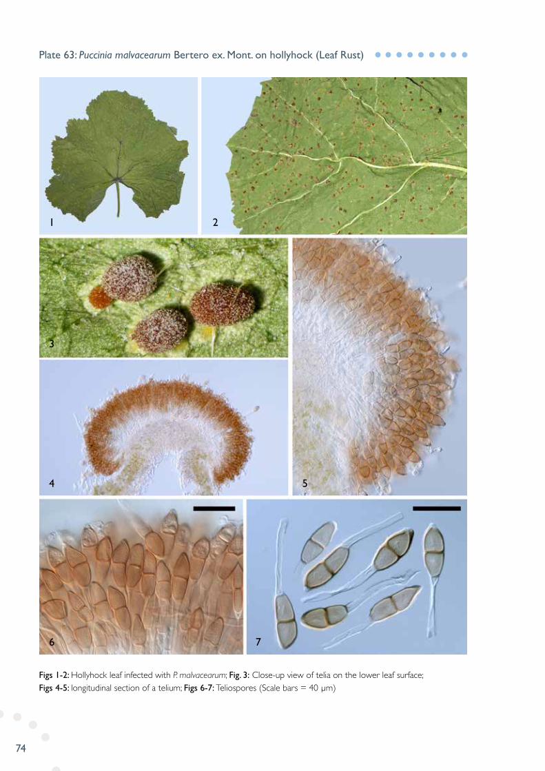
Figs 1-2: Hollyhock leaf infected with P. malvacearum; Fig. 3: Close-up view of telia on the lower leaf surface; Figs 4-5: longitudinal section of a telium; Figs 6-7: Teliospores (Scale bars = 40 µm)
Plate 63: Puccinia malvacearum Bertero ex. Mont. on hollyhock (Leaf Rust)
6
4 5
3
1 2
7
75
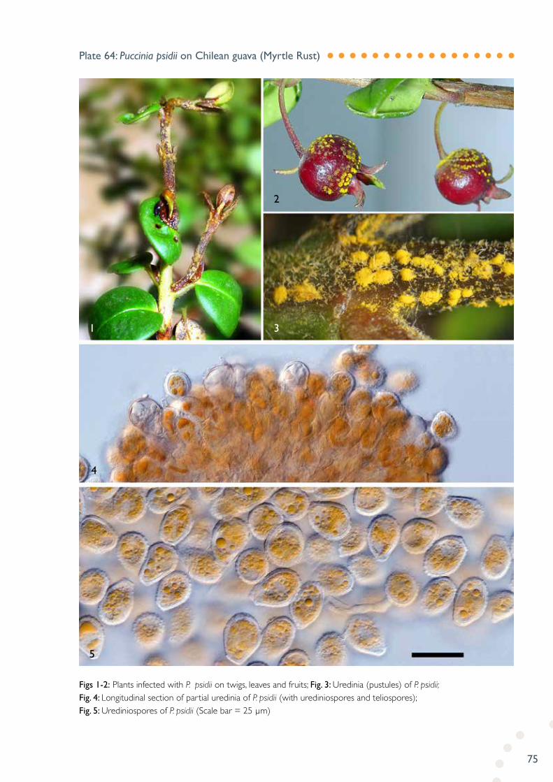
Figs 1-2: Plants infected with P. psidii on twigs, leaves and fruits; Fig. 3: Uredinia (pustules) of P. psidii; Fig. 4: Longitudinal section of partial uredinia of P. psidii (with urediniospores and teliospores); Fig. 5: Urediniospores of P. psidii (Scale bar = 25 µm)
Plate 64: Puccinia psidii on Chilean guava (Myrtle Rust)
5
4
1 3
2

76
Figs 1-2: Rhubarb leaf infected with P. rhei-undulati (upper and lower surface view); Fig. 3: Lesions on the upper leaf surface; Fig. 4: Lesions with fungal fruiting bodies (uredinia) on lower leaf surface; Fig. 5: Partial longitudinal section of an uredinium; Fig. 6: Urediniospores (Scale bar = 20 µm)
Plate 65: Puccinia rhei-undulati on rhubarb (Leaf Rust)
6
5
3 4
1 2

77
Figs 1-2: A plant infected with P. saccardoi on leaves and stems; Fig. 3: Aecia of P. saccardoi; Fig. 4: A longitudinal section of an aecium; Fig. 5: One-celled aeciospores in chains; Fig. 6: Two-celled teliospores (Scale bars = 20 µm)
Plate 66: Puccinia saccardoi on goodenia (Rust)
5
4
1 3
2
6

78
Fig. 1: Strawberry plants with crown rot (photo courtesy of Dr Hoong Pung); Fig. 2: Five-day old colony on PDA (top and reverse view); Figs 3-4: Appressoria produced in cultures; Fig. 5: An oogonium with diclinous antheridium; Figs 6-8: Oogonia showing antheridia with helical turns (Scale bars = 20 µm)
Plate 67: Pythium helicoides on strawberry (Crown Rot)
6
2
1
4
3
5
7 8

79
Figs 1-3: Symptoms; Fig. 4: White conidial masses of R. collo-cygni on the surfaces of leaf lesions; Fig. 5: A “swan-neck” like conidiophore with a developing conidium attached; Fig. 6: Apex of conidiogenous cell; Fig. 7: Conidia (Scale bar = 10 µm)
Plate 68: Ramularia collo-cygni on barley (Leaf Spot)
7
4
2
1
3
5 6
80
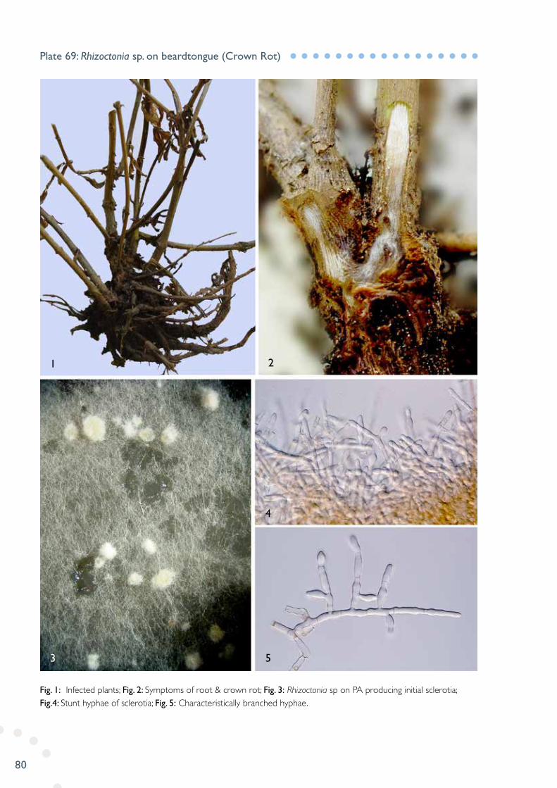
Fig. 1: Infected plants; Fig. 2: Symptoms of root & crown rot; Fig. 3: Rhizoctonia sp on PA producing initial sclerotia; Fig.4: Stunt hyphae of sclerotia; Fig. 5: Characteristically branched hyphae.
Plate 69: Rhizoctonia sp. on beardtongue (Crown Rot)
3
1
5
4
2

81
Figs 1-3: Symptoms of raspberry spur blight with dying budding leaves and stems; Fig. 4: Close-up view of conidiomata imbedded in the bark of stems infected with S. lichenicola (black conidial masses); Fig. 5: Reverse view of colonies of S. lichenicola recovered from infected raspberry bud tissues on PDA; Fig. 6: Longitudinal section of a conidioma; Fig. 7: Mature conidia with periclinal walls collapsed (Scale bar = 10 µm)
Plate 70: Seimatosporium lichenicola on raspberry (Spur Blight)
7
5
1 3
2
4
6

82
Figs 1-3: Characterised symptoms of cankers; Fig. 4: Longitudinal section of cankered stem; Fig. 5: Conidia masses (black dots) of S. cupressi; Fig. 6: Longitudinal section of conidiomata; Fig. 7: Conidia; Fig.8: A conidium with the conidiophore attached (Scale bar = 20 µm)
Plate 71: Seiridium cupressi on cedar (Stem Canker)
1
4
7 8
6
5
2 3

83
Fig. 1: Lemons infected with S. citri; Fig. 2: Close-up view of the lesions (pits): Figs 3-4: Longitudinal sections of pycnidia; Fig. 5: Conidia (Scale bar = 10 µm)
Plate 72: Septoria citri on lemon (Septoria Spot)
4
1
2 3
5
84
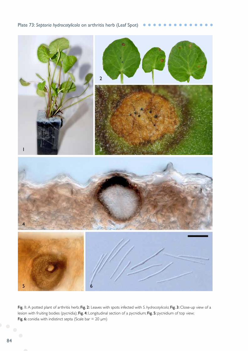
Fig. 1: A potted plant of arthritis herb; Fig. 2: Leaves with spots infected with S. hydrocotylicola; Fig. 3: Close-up view of a lesion with fruiting bodies (pycnidia); Fig. 4: Longitudinal section of a pycnidium; Fig. 5: pycnidium of top view; Fig. 6: conidia with indistinct septa (Scale bar = 20 µm)
Plate 73: Septoria hydrocotylicola on arthritis herb (Leaf Spot)
1
4
5 6
3
2

85
Fig. 1: Symptoms of needle infections; Fig. 2: Close-up view of needles with apical infections; Fig. 3: Infected needle with embedded pycnidia of S. slaptonensis; Fig 4: Longitudinal-sections of pycnidia; Fig. 5: Conidia (Scale bar =10 µm)
Plate 74: Septoria slaptonensis on gorse (Leaf Blight)
1 3
2
4 5
86
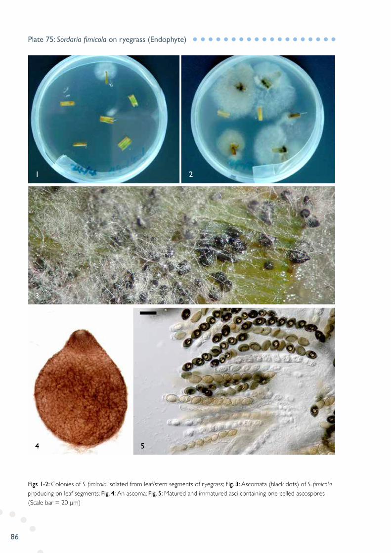
Figs 1-2: Colonies of S. fimicola isolated from leaf/stem segments of ryegrass; Fig. 3: Ascomata (black dots) of S. fimicola producing on leaf segments; Fig. 4: An ascoma; Fig. 5: Matured and immatured asci containing one-celled ascospores (Scale bar = 20 µm)
Plate 75: Sordaria fimicola on ryegrass (Endophyte)
1
3
4 5
2
87
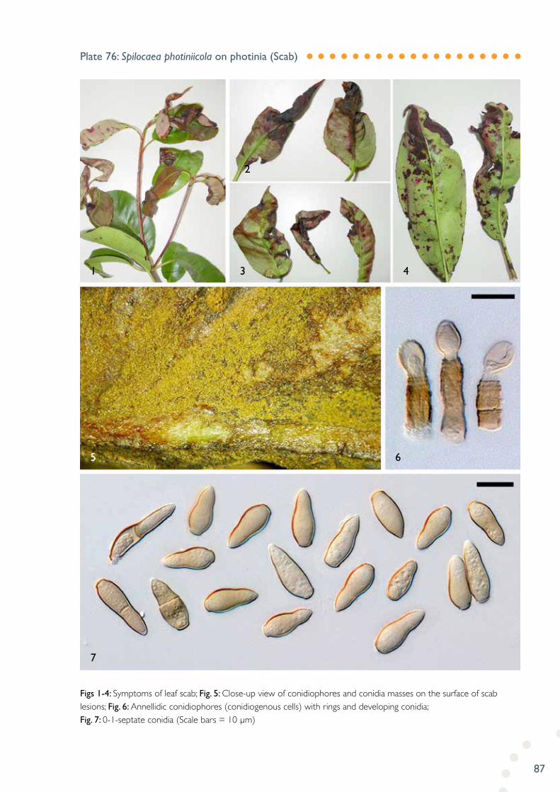
Figs 1-4: Symptoms of leaf scab; Fig. 5: Close-up view of conidiophores and conidia masses on the surface of scab lesions; Fig. 6: Annellidic conidiophores (conidiogenous cells) with rings and developing conidia; Fig. 7: 0-1-septate conidia (Scale bars = 10 µm)
Plate 76: Spilocaea photiniicola on photinia (Scab)
1
5
7
6
3 4
2
88
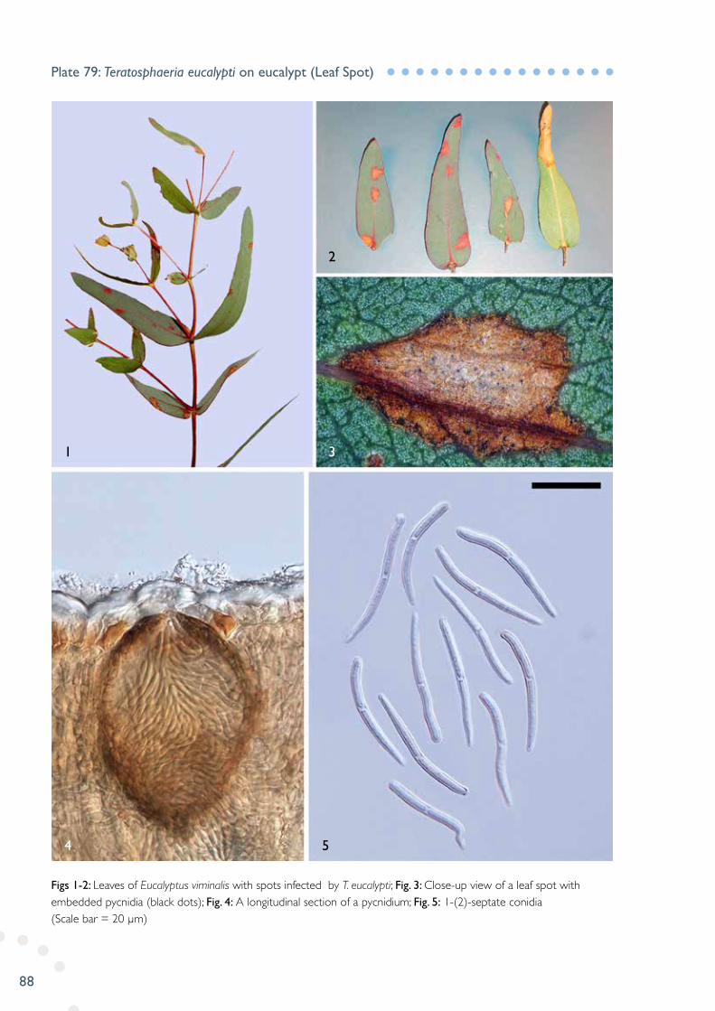
Figs 1-2: Leaves of Eucalyptus viminalis with spots infected by T. eucalypti; Fig. 3: Close-up view of a leaf spot with embedded pycnidia (black dots); Fig. 4: A longitudinal section of a pycnidium; Fig. 5: 1-(2)-septate conidia (Scale bar = 20 µm)
Plate 79: Teratosphaeria eucalypti on eucalypt (Leaf Spot)
4
1 3
2
5

89
Fig. 1: Blueberry fruits infected with T. minima on crown scar areas; Figs 2-3: Close-up view of rust pustules on crown scar areas; Figs 4-5: Blueberry leaves infected with T. minima; Fig. 6: Close-up view of rust pustules on leaves; Fig. 7: Urediniospores (Scale bar = 15 µm)
Plate 80: Thekopsora minima on blueberry (Rust)
7
4
1 2 3
5 6

90
Figs 1-2: Symptoms on carrots; Figs 3-4: Chains of chlamydospores; Fig. 5: Conidiophores (phialides) and chlamydospores developing from the same hypha; Fig. 6: conidia from cultures on carrot agar (Scale Bars = 20 µm)
Plate 81: Thielaviopsis basicola on carrot (Black Root Rot)
1
2 4
3
5 6

91
Figs 1-2: Apricot leaves infected with T. discolor (lower and upper surface view); Fig. 3: Uredinia at lower leaf surface; Fig. 4: Longitudinal section of an uredinium; Fig. 5: Urediniospores (Scale bar = 20 µm)
Plate 82: Tranzschelia discolor on apricot (Leaf Rust)
1
3
5
4
2
92
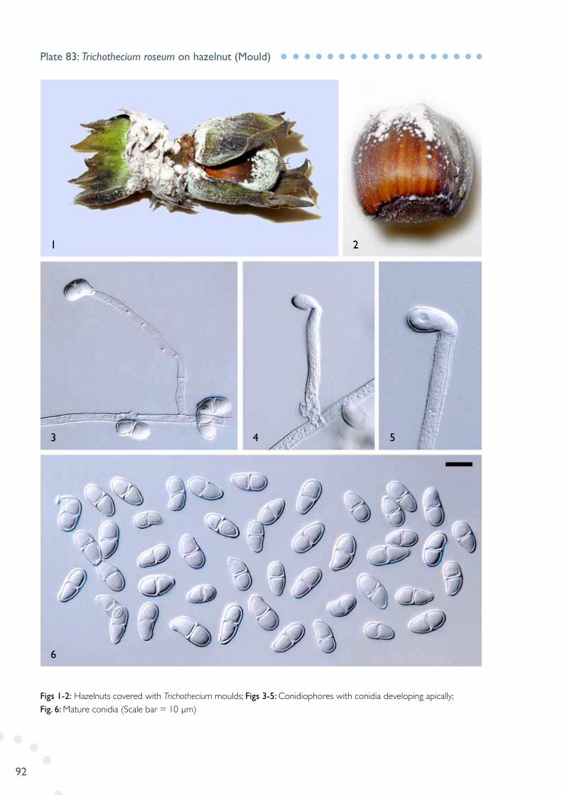
Figs 1-2: Hazelnuts covered with Trichothecium moulds; Figs 3-5: Conidiophores with conidia developing apically; Fig. 6: Mature conidia (Scale bar = 10 µm)
Plate 83: Trichothecium roseum on hazelnut (Mould)
6
3
1 2
4 5

93
Figs 1-3: Wheat grains infected with U. atrum; Fig. 4: Conidiophores; Fig. 5: Conidia (Scale bar = 10 µm)
Plate 84: Ulocladium atrum on wheat (Grain Mould)
4
2
1
3
5

94
Fig. 1: Beetroot leaf infected with U. betae; Figs 2-3: Close-up view of uredinia and telia with mycoparasitic fungus Sphaerolopsis filum (black dots embedded within the uredinia/telia); Fig. 4: Longitudinal section of uredinium and pycnidium of S.filum; Fig. 5: Partial section of uredinium/telium; Fig. 6: Urediniospores; Fig. 7: Teliospores (Scale bars = 20 µm)
Plate 85: Uromyces beticola on beetroot (Leaf Rust)
6
4
1 3
2
5
7

95
Figs 1-2: Infected leaves with rust pustules (uredinia); Fig. 3: Longitudinal section of an uredinium; Fig. 4: Close-up view of partial uredinium with developing urediniospores; Fig. 5: Urediniospores (Scale bar = 30 µm)
Plate 86: Uromyces dianthi on carnation (Rust)
4
1 3
2
5
96
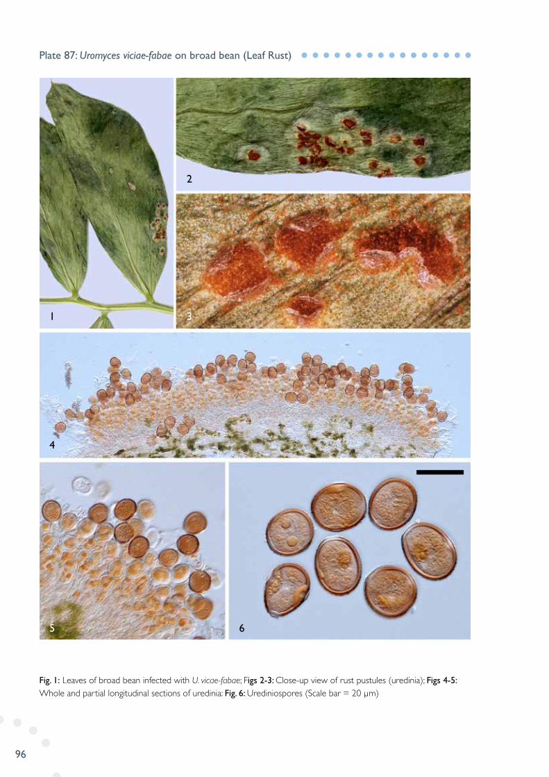
Fig. 1: Leaves of broad bean infected with U. vicae-fabae; Figs 2-3: Close-up view of rust pustules (uredinia); Figs 4-5: Whole and partial longitudinal sections of uredinia: Fig. 6: Urediniospores (Scale bar = 20 µm)
Plate 87: Uromyces viciae-fabae on broad bean (Leaf Rust)
5
4
1 3
2
6

97
Figs 1-2: Grass heads infected with ear smut; Fig. 3: Germinating teliospores with spindle-shaped promycelia; Fig. 4: Teliospores in optical section; Fig. 5: Teliospores in surface view (Scale bars = 5 µm)
Plate 88: Ustilago bullata on grass (Ear Smut)
3
1 2
4
5
98
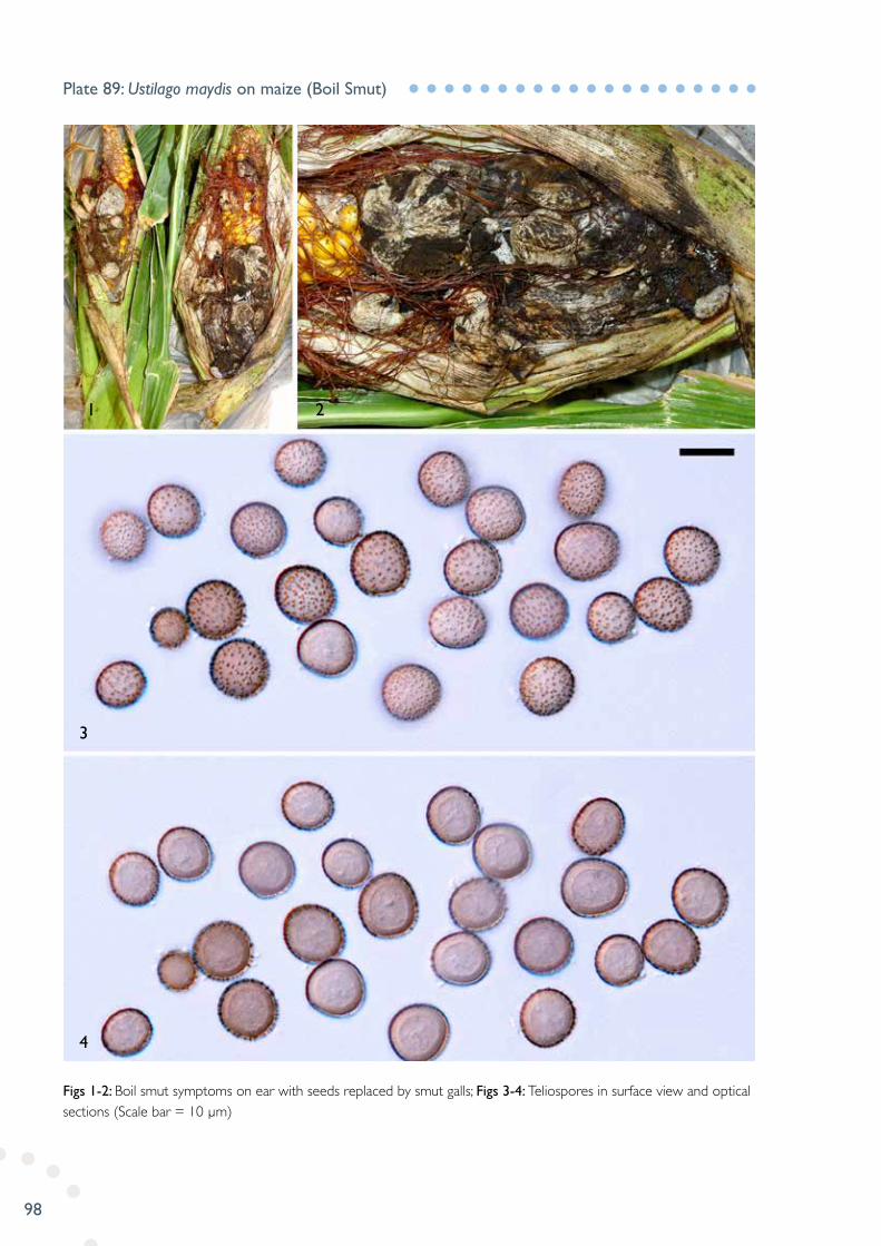
Figs 1-2: Boil smut symptoms on ear with seeds replaced by smut galls; Figs 3-4: Teliospores in surface view and optical sections (Scale bar = 10 µm)
Plate 89: Ustilago maydis on maize (Boil Smut)
3
4
1 2
99
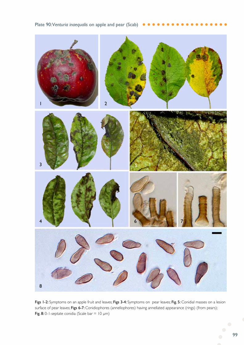
Figs 1-2: Symptoms on an apple fruit and leaves; Figs 3-4: Symptoms on pear leaves; Fig. 5: Conidial masses on a lesion surface of pear leaves; Figs 6-7: Conidiophores (annellophores) having annellated appearance (rings) (from pears); Fig. 8: 0-1-septate conidia (Scale bar = 10 µm)
Plate 90: Venturia inaequalis on apple and pear (Scab)
1
3
4
8
6 7
5
2

100
Fig. 1: Infected fruits with scab lesions and cracks; Fig. 2: Vertical section of lesion tissue showing subcuticular conidiophores (solitary or in loose fascicles); Fig. 3: Close-up view of scab lesions covered with dark brown to black mould (conidial masses); Fig. 4: Geniculate-subnodulose conidiophores with pores (porosporic rings or scars); Fig. 5: 0-1-septate conidia (Scale bars = 20 µm)
Plate 91: Venturia pirina on pear (Scab)
5
1 3
2
4
101
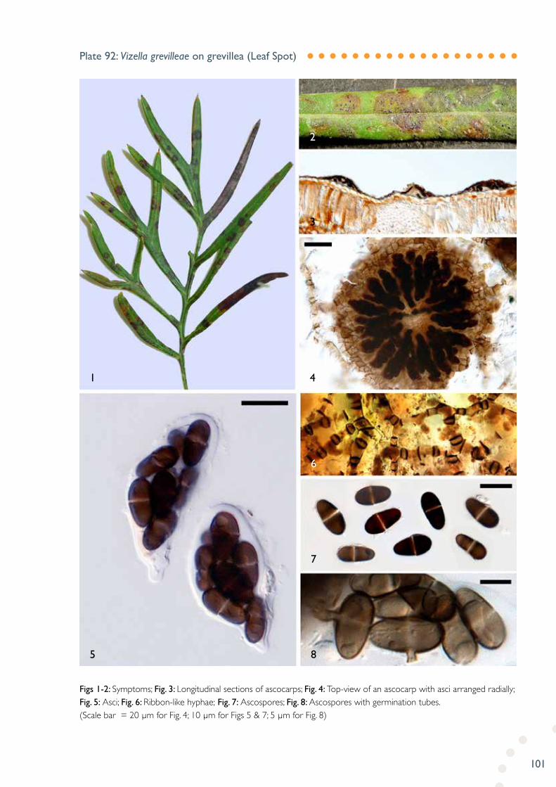
Figs 1-2: Symptoms; Fig. 3: Longitudinal sections of ascocarps; Fig. 4: Top-view of an ascocarp with asci arranged radially; Fig. 5: Asci; Fig. 6: Ribbon-like hyphae; Fig. 7: Ascospores; Fig. 8: Ascospores with germination tubes. (Scale bar = 20 µm for Fig. 4; 10 µm for Figs 5 & 7; 5 µm for Fig. 8)
Plate 92: Vizella grevilleae on grevillea (Leaf Spot)
5
1
8
7
6
4
3
2
102
103
IMAGE PLATESNEMATODES

104
Fig. 1: Extraction of C. truncatum using a tray method from bark and stem debris of ferns; Fig. 2: A female body; Fig. 3 & 6: Head of female; Fig. 4: Head end of female; Fig. 5: Tail end of female (Scale bar = 0.1mm for Fig. 2; 10 µm for Figs 3-4)
Plate 93: Colbranium truncatum extracted from fern
6
1
2 4 5
3

105
Fig. 1: Garlic plant infected with D. dipsaci; Fig. 2: Extraction of D. dipsaci from garlic sample; Fig. 3: Male and female bodies; Fig. 4: Female head; Fig. 5: Male oesophageal region; Fig. 6: Female partial reproductive system (vulva and post-uterine sac); Fig. 7: Female head; Fig. 8: Spiculum of male; Fig. 9: Posterior portion of male (Scale bars = 10 µm)
Plate 94: Ditylenchus dipsaci extracted from garlic
1
2
5 8 9
7
6
3 4
106
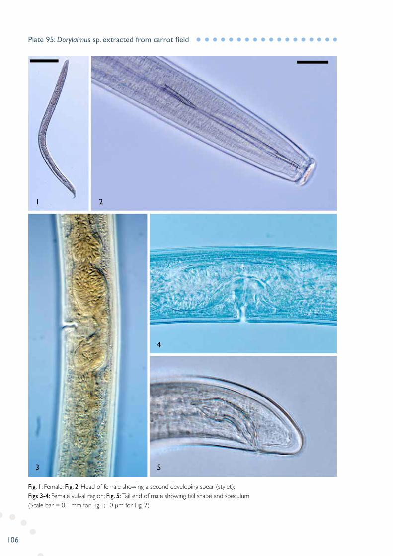
Fig. 1: Female; Fig. 2: Head of female showing a second developing spear (stylet); Figs 3-4: Female vulval region; Fig. 5: Tail end of male showing tail shape and speculum (Scale bar = 0.1 mm for Fig.1; 10 µm for Fig. 2)
Plate 95: Dorylaimus sp. extracted from carrot field
1
3 5
4
2

107
Figs 1-2: Cysts; Fig. 3: Bullae; Fig. 4: Perivulval; Fig. 5: Fenestra; Fig. 6: Egg; Fig. 7: Second-stage juvenile; Fig. 8: Head of juvenile; Fig. 9: Tail of juvenile (Scale bar = 0.1 mm for Fig. 7; 10 µm for Fig. 8)
Plate 96: Heterodera avenae extracted from soil
1
2 3 5
4
6
7 8
9
108
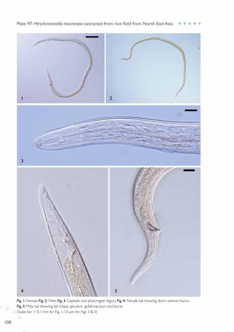
Fig. 1: Female; Fig. 2: Male; Fig. 3: Cephalic and pharyngeal region; Fig. 4: Female tail showing short ventral mucro; Fig. 5: Male tail showing tail shape, spiculum, gubernaculum and bursa (Scale bar = 0.1 mm for Fig. 1; 10 µm for Figs 3 & 5)
Plate 97: Hirschmanniella mucronata extracted from rice field from North East Asia
1
3
4 5
2
109
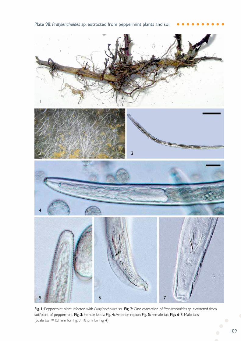
Fig. 1: Peppermint plant infected with Pratylenchoides sp.; Fig. 2: One extraction of Pratylenchoides sp. extracted from soil/plant of peppermint; Fig. 3: Female body; Fig. 4: Anterior region; Fig. 5: Female tail; Figs 6-7: Male tails (Scale bar = 0.1mm for Fig. 3; 10 µm for Fig. 4)
Plate 98: Pratylenchoides sp. extracted from peppermint plants and soil
4
2
1
3
5 6 7
110
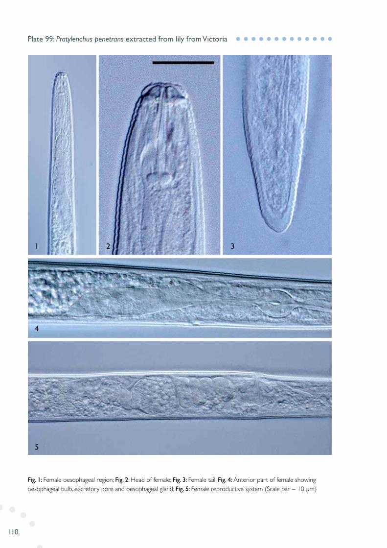
Fig. 1: Female oesophageal region; Fig. 2: Head of female; Fig. 3: Female tail; Fig. 4: Anterior part of female showing oesophageal bulb, excretory pore and oesophageal gland; Fig. 5: Female reproductive system (Scale bar = 10 µm)
Plate 99: Pratylenchus penetrans extracted from lily from Victoria
1
4
5
2 3

111
Fig. 1: Single-spiralled female bodies; Fig. 2: C-shaped male bodies; Fig. 3: Head of male; Fig. 4: Anterior part of male showing oesophageal bulb, excretory pore and oesophageal gland; Fig. 5: Tail end of male showing tail shape, spiculum, gubernaculum and bursa; Figs 6-7: Female tail showing anus and pore-like phasmid; Fig. 8: Near-tail end of male showing partially areolated lateral field; Fig. 9: Female vulval region (Scale Bar = 150 µm for Figs 1-2; 10 µm for Fig. 3)
Plate 100: Rotylenchus robustus extracted from lavenders from Victoria
1
4
8 9
5 7
6
2 3

112
Alphabetic in common names Acacia - Acacia sp. Cylindrocladium pauciramosum
Alder - Alnus spp. Melampsoridium betulinum
Alpen rose - Rhododendron ferrugineum L. Chrysomyxa aff. reticulata
Angular Sea-Fig - Carpobrotus glaucescens (Haw.) Schwantes Alternaria aff. capsici-annui
Apple - Malus domestica Borkh. Cladosporium macrocarpum Venturia inaequalis
Apricot - Prunus armeniaca L. Monilinia laxa Tranzschelia discolor
Arthritis Herb - Centella asiatica (L.) Urban Septoria hydrocotylicola
Aspen - Populus tremula L. Fusicladium radiosum var. letiferum
Azalea - Azaleastrum sect. tsutsusi Phytophthora megasperma
Banana - Musa spp. Ceratocystis paradoxa
Barley - Hordeum vulgare L. Ramularia collo-cygni
Beardtongue - Penstemon sp. Rhizoctonia sp.
Beetroot - Beta vulgaris L. Alternaria aff. putrefaciens Uromyces beticola
Birch - Betula sp. Idiocercus aff. australis
Blackberry - Rubus allegheniensis Porter Phragmidium violaceum
Blueberry - Vaccinium corymbosum L. Neofusicoccum ribis Colletotrichum sp. Thekopsora minima
Broad Bean - Vicia faba L. Uromyces viciae-fabae
Broccoli - Brassica oleracea L. var. italic Plenck Fusarium oxysporum
Brussels Sprouts - Brassica oleracea L. var. gemmmifera Stemphylium vesicarium
Cabbage - Brassica oleracea L. Albugo candida Alternaria brassicae Peronospora parasitica
Carnation - Dianthus caryophyllus L. Uromyces dianthi
Carrot - Daucus carota L. subsp. sativus (Hoffm.) Arcan. Dorylaimus sp.(nematode) Thielaviopsis basicola
Cedar - Calocedrus decurrens (Torr.) Florin Seiridium cupressi
Cherry - Prunus avium L. Botrytis cinerea Monilinia laxa
Chestnut - Castanea sativa Mill. Acrospeira mirabilis
Chilean Guava - Ugni molinae Turcz. Puccinia psidii
Cotton - Gossypium sp. Colletotrichum dematium
English Broom - Cytisus scoparius (L.) Link. Camarosporium sp.
Eucalypt – Eucalyptus viminalis Labill. Teratosphaeria eucalypti
Fern – Dicksonia sp. Colbranium truncatum (nematode)
HOST PLANT INDEX
113
Garlic - Allium sativum L. Alternaria embellisia Ditylenchus dipsaci (nematode)
Golden Ash - Fraxinus excelsior L. “Aurea” Neofusicoccum luteum
Goodenia - Goodenia sp Puccinia saccardoi
Gorse - Ulex europaeus L. Septoria slaptonensis
Grapevine - Vitis vinifera L. Diplodia seriata Fusarium spp.
Grass - Poa spp. Puccinia graminis Pers. subsp. graminicola Ustilago bullata
Grevillea - Grevillea sp. Vizella grevilleae
Hawthorn - Crataegus sp. Podosphaera clandestina
Hazelnut - Corylus avellana L. Colletotrichum acutatum Trichothecium roseum
Hollyhock - Alcea rosea L. Puccinia malvacearum
Horse Chestnut - Aesculus hippocastanum L. Phytophthora cactorum
Impatiens - Impatiens walleriana Hook. f. Plasmopara obducens
Iris - Iris sp. Puccinia iridis
Lemon - Citrus lemon (L.) Burm. f. Alternaria citri Septoria citri
Lettuce - Lactuca sativa L. Bremia lactucae Microdochium panattonianum
Lavender - Lavandula spp. Rotylenchus robustus (nematode)
Lily - Lilium sp. Pratylenchus penetrans (nematode)
Loganberry - Rubus × loganobaccus L.H. Bailey Paraconiothyrium fuckelii Myxosporium aff. rosae
Maize - Zea mays L. Ustilago maydis
Mulberry - Morus sp. Phloeospora mori
Onion - Allium cepa L. Botrytis allii Munn. Gabarnaudia betae Peronospora destructor Puccinia allii
Orange - Citrus × sinensis (L.) Osbeck Penicillium digitatum
Pear - Pyrus spp. Alternaria alternata Alternaria tenuissima Venturia inaequalis Venturia pirina
Pepper - Capsicum annuum L. Botrytis cinerea
Peppermint - Mentha × piperita L. Pratylenchoides sp. (nematode)
Petty Spurge - Euphorbia peplus L. Melampsora euphorbiae
Photinia - Photinia spp. Spilocaea photiniicola
Plane tree - Platanus x acerifolia (Aiton) Willd. Cryptosporiopsis aff. tarraconensis Phyllactinia corylea
Poinsettia - Euphorbia pulcherrima Willd. ex Klotzsch Phoma tropica

114
Raspberry - Rubus spp. Didymella applanata Gabarnaudia betae Phragmidium rubi-idaei Phytophthora drechsleri Seimatosporium lichenicola
Rhubarb - Rheum rhaponticum L. Puccinia rhei-undulati
Rice - Oryza sativa L. Hirschmanniella mucronata (nematode)
Ryegrass - Lolium spp. Leptosphaerulina sp. Sordaria fimicola
Sea Spurge - Euphorbia paralias L. Stemphylium aff. callistephi
Sorghum - Sorghum bicolor (L.) Conrad Moench Bipolaris hawaiiensis
Springy Peppercress - Lepidium flexicaule Kirk Albugo lepidii
Squash - Cucurbita maxima Duchesne Geotrichum candidum
Strawberry - Fragaria × ananassa Duchesne Pythium helicoides
Tasmanian Myrtle - Nothofagus cunninghamii Hook.f. Caliciopsis sp.
Tomato - Solanum lycopersicum L. Alternaria aff. longipes Fusarium dimerum Passalora fulva
Tulip - Tulipa sp. Penicillium hirsutum
Wheat - Triticum spp. Ulocladium atrum
White Poplar - Populus alba L. Melampsora laricis-populina

Pictorial A
tlas o
f PL
AN
T D
ISE
AS
ES
Dia
gno
sed
in Ta
sma
nia
Ziqing Yuan