Embed Size (px)
Citation preview
Neuronal Connections and the Function of the Corpora Pedunculata in the Brain of the American Cockroach, Periplaneta americana (L.)
MITCHELL J. WEISSZ D e p a r t m e n t of Zoology, The Uiz7ueisity of Mlchzgan , Ann Arbor, Michzgan, 481 04
ABSTRACT The fiber constituents and connections of the calyces - the in- put-receiving regions - of the corpora pedunculata (“mushroom bodies”) were studied in reduced silver preparations from the American cockroach, Periplaneta americana (L.). In the outer synaptic layer of the calyces five fiber classes were distinguished, the first three of which arise outside the mushroom body. (1) Four highly similar neurons with somata near the optic lobe branch into different parts of the ipsilateral protocerebrum, including both calyces. Their fibers are highly constant in arrangement and position and contain small nucleus-like bodies. (2) The tractus olfactorio-globularis (sensu Into) emits fiber groups which course along the calycal walls as “calycal tracts” before ultimately dissipating into the synaptic layer. Variability within these tracts is described. (3 ) Fibers of undetermined origin outside the mushroom body radiate from the calycal center outwards through the synaptic layer. (4) From the inner calycal layer of neurites belonging to intrinsic mushroom-body neurons, perpendicular collaterals enter the synaptic layer. (5) Intrinsic-neuron somata near the calycal rim emit fibers which course tangentially within the synaptic layer from calycal rim to center. These fibers form a special peripheral zone in the pedunculus.
The predominant presumably afferent calycal fiber class is that derived from the tractus olfactorio-globularis. No evidence was found for tracts from optic lobe to calyces. On this basis, and in light of the experimental and comparative ana- tomical literature, it is suggested that the corpora pedunculata of P. americanu and other pterygotes are fundamentally second-order antenna1 sensory process- ing centers.
Conflicting observations in earlier reports are critically discussed.
Few structures within the nervous sys- tems of invertebrates have attracted as long-lasting an interest as have the cor- portr pcdiincxltiin or “mushroom bodies” of the insect brain. What biological role do these often strikingly prominent centers play? This question has been asked re- peatedly since Dujardin (1850) discovered the corpora pedunculata in a range of in- sects and related their degree of develop- ment to the level of “intelligence” (for extensive bibliographies see Hanstrom, ’28; Horridge, ’65; Gouin, ’65). Yet a con- vincing answer has remained elusive. The question will be approached here through structural examination of the kinds of in- put the corpora pedunculata receive.
The principal inputs of the corpora pe- dunculata are believed to arrive through
J. MORPH , 142 21-70.
tracts to their calyces (fig. 1): Maynard’s work (‘56, ’67) supplies the strongest elec- trophysiological support for this view. Al- though numerous papers describe calycal fiber connections in various insects (see above bibliographies), for no one species is a sufficiently full, adequately documented account available. This situation stems from limitations in the techniques used. Most workers have used common dyes such as haematoxylin, but because these ordinarily show individual nerve fibers poorly their employment usually leads to erroneous or at best unconvincing asser-
~
Based on portions of a dissertation submitted to the Department of Zoology, The University of Michigan, In partial fulfillment of the requirements for the degree of Doctor of Philosophy (Weiss, ’70).
Present address: Department of Zoology, University of Iowa, Iowa City, Iowa 52242.
21
22 MITCHELL J. WEISS
tions. On the other hand, two of the three fragments, so that the extent to which the major types of neurological technique ca- information they provide is either repre- pable of showing nerve fibers clearly, Golgi sentative or complete remains uncertain. impregnation and inira nitam methylene The best hope for obtaining a convincing blue, display only an unknown and possi- cataloging of at least all major calycal bly biased proportion of neurons present connections, with knowledge of relative in a preparation and often only neuronal fiber numbers, lies with the “total stain-
r / optic lobe
Fig. 1 Diagram of left half of the brain of adult male P. nmericana, viewed from the front. Most nerves are omitted. Proto-, deuto-, tritocerebrum, the three traditionally recognized “divisions” of the brain; protocerebral bridge, central body, special neuropilar formations of the protocerebrum; pi, pars inter- cerebralis, a medial zone of the protocerebrum; medial calyx, lateral calyx, pedunculus, a lobe, p lobe, the different neuropilar divisions of the corpus pedunculatum. Note large antennal sensory nerve and correspondingly large antenna1 sensory lobe of the deutocerebrum. Within the corpus pedunculatum the basic arrangement of a typical globuli cell is illustrated; the number and pattern of its collaterals are shown highly schematically. Conduction within the globuli cells is believed (see Maynard, ’56, ’67) to extend from the calyces through the pedunculus and into the a and p lobes, whose connections pre- sumably are essentially efferent. (Details of globuli cell combined from various published sources.)
CORPORA PEDUNCULATA OF
ing” method of reduced silver impregna- tion.
A thorough description of calycal con- nections from reduced silver preparations has never been published. Vowles (’55) offers a list of connections in a bee and an ant but does not provide detailed documen- tation. Pearson’s (’71) admirable study of lepidopteran corpora pedunculata involved reduced silver as well as Golgi-silver prep- arations; her treatment of the calycal con- nections, however, relies primarily on the latter.
In this study, based on reduced silver preparations from the American cockroach, Perzplaneta amerrcana (L,), I have at- tempted to elucidate the calycal fiber con- nections by distinguishing all visible fiber classes within the calyces and determining as fully as possible their courses and ori- gins inside or outside the corpus peduncu- latum. The calyces of P. amerzcana, while particularly large, are in basic form and arrangement typical of many pterygotes. The study has been made possible by de- velopment of a reduced silver procedure (Weiss, ’72a) which permits fine ramifica- tions of entering fibers and tracts to be visualized: in the best impregnations fibers of 0.2- to 0.3-p diameter are visible within the calycal neuropile. The results provide evidence that the calyces of P. amerzcanu serve primarily as second-order antennal centers and suggest that in this insect and others the chief function of the corpus pedunculatum may profitably be viewed as the processing of antennal sensory infor- mation.
An abstract of this study has previously appeared (Weiss, ’71).
MATERIALS A N D METHODS
Specimens of Pmplnnetcz umerzcana were taken from a laboratory culture kind- ly furnished originally by Dr. Louis M. Roth. The brains of 41 adult males were prepared according to an improved version (Weiss, ’72a) of Blest’s collidine modifica- tion of Holmes’ reduced silver impregna- tion method, with occasional technical var- iations which proved either unessential or unsuccessful. The procedure includes fix- ation in Bodian’s (‘37) fixative No. 2 and paraffin embedding. In 24 cases the entire brain was sectioned serially, ordinarily at 10 w . In 17 cases 10-p serial sections were
THE AMERICAN COCKROACH 23
prepared of half or slightly more of the brain (these originally served as “test” slides). All three major sectioning planes were represented. While many preparations proved useful, only a few exhibit impreg- nations suitable for the more demanding observations. These include whole brains sectioned transversely and frontally (see terminology below). A few similarly stained preparations from both sexes of Periplanetu sp .3 have been available for reference; these include a beau tifully impregnated sagittal series through the complete brain. Unless otherwise indicated, the observa- tions deal exclusively with adult male P. americana.
The terms fionttrl and tra,nsverse will refer respectively to section planes approx- imately parallel to (1) the surface of the protocerebrum (see fig. 1) facing the frons and (2) the plane dividing the brain into dorsal and ventral halves. Usage of terms such as dol-sir1 and anterior assumes the head to be held in a generally vertical posi- tion. The apex of the protocerebral lobe is considered dorsal, the protocerebral sur- face facing the frons, anterior, and so forth.
Unless otherwise noted, serial tracing of fiber bundles and tracts has been car- ried out under 1000 X magnification, with careful attention to establishing continu- ity between fiber groups in adjacent sec- tions. Most of the photomicrographs neces- sarily show objects at lower magnification than that used for the original observa- tions.
OBSERVATIONS
The microscopic anatomy of the corpora pedunculata and their fiber connections with other regions have been examined in various genera of cockroaches (Dictyop- tera: Blattaria) (Flogel, 1876,* 1878; New-
Obtained commercially several years ago under the name “P. americana” but later identified in retrospect as almost certainly P. fuliginosa.
On 21 September 1876, in Hamburg, Flogel deliv- ered before one of the sections of the Gesellschaft deutscher Naturforscher und Aerzte a remarkable lec- ture, filled with detail, concerning the inner structure of the brain of insects and other arthropods as visible i n microtome sections, thus formally inaugurating the mod- ern era of the study of the insect brain. The published text (Flogel, 1876) has remained in obscurity, only Holste (’231, of later authors, even alluding vaguely to its existence. Many specific findings on insect corpora pedunculata, as well as the introduction of various stan- dard names for structures within the insect brain (Bal- ken, Centralharper, etc.), date not from Flogel’s well- known study of 1878 but from this 1876 publication.

24 MITCHELL J. WEISS
ton, 1879; Haller, '05; Holmgren, '09; Bretschneider, '13a,b, '14; Hanstrom, '28, '40; Sanchez y Sanchez, '33; JawAowski, '48, '53; Day, '50; Lhoste and Roche, '56; Arnold, '60; Drescher, '60; Barbier, '61; Willey, '61, p. 235; Satija and Singla, '67; Prigent, '68; Brousse-Gaury, '68, '71). ln- formation is also available concerning re- lated aspects of cockroach mushroom bod- ies: growth and development (Panov, '57; Neder, '59; Khan, '62; Khan and Fraser, '62; Malzacher, '68; Levi-Montalcini and Chen, '69, fig. 1; Chen and Levi-Montal- cini, '70; Weiss, '72b; see also Ratzerdorf- er, '52), histochemistry (Shao and Dixon, '64, fig. 1 ; Frontali and Norberg, '66; Frontali, '68, '71a,b; Mancini and Fron- tali, '70; Frontali, Piazza and Scopelliti, '71; Hess, '72; Frontali and Pierantoni, '73), and ultrastructural anatomy and cytochemistry (Arnold, '64; Mancini and Frontali, '67, '68, '70; Frontali and Man- cini, '70; Frontali, '71a,b; Weiss, '71; fig. 15 in Maynard, '67).
Beginning with Flogel (1876, 1878), nearly all authors have attributed the same basic construction (fig. 1) to the corpus pedunculatum of different cockroaches. Dorsally there are two calyces, sometimes but not invariably truly concave, which are overlain by numerous globuli cell somata. The calyces give rise ventrally to a stalk- like pedunculus which bifurcates at its distal (ventral) end into two elongate lobes (named here following Vowles, '55). The medial p lobe extends to the midline, while the dorsal (Y lobe ascends for some dis- tance near the front surface of the brain. Haller's ('05) divergent account clearly resulted from misunderstanding of both Newton's (1879) work and his own prep- arations (note the self-contradictory figure labels), while Sanchez y Sanchez's ('33) incorrect interpretations seem to have originated largely in Haller's account. It is clear from the fiber arrangement of the typical globuli cell shown in figure 1 that the gross form of the mushroom body re- flects the branching pattern of its globuli cells. These may be termed i.ntrinsic cells and their fibers intrinsic fibers of the mush- room body, while fibers entering the mush- room body from outside, with cell bodies elsewhere in the nervous system, may be termed extrinsic. Periplaneta nmericnna (e.g., Hanstrom, '28, '40; JawAowski, '48) is
one of those cockroaches with large, deeply concave calyces.
My observations reconfirm this basic plan of construction in P. cimericnnn (figs. 5, 6, 13, 20), although it remains possible that a small proportion of intrinsic fibers may leave the (Y or p lobe, for example in the manner Drescher ('60) describes. In one specimen a striking departure from the usual conditions was found, in that the two p lobes were medially fused. Drescher ('60) found such fusion in about 2% of his specimens of P. amerzcana, and Haller ('05) saw it in Blatta orientalis.
As Flogel (1876) originally reported in Blatta orientalis and Hanstrom ('28) and others have since confirmed for P. ameri- c a m , the calycal walls in this species com- prise two distinct layers (fig. 7). That ad- joining the calycal concavity, though variable in width, is always the thinner, and will be designated zonu interna in recogni- tion of its internal position. It is believed to consist (e.g., Hanstrom, '28) of globuli cell neurites which extend tangentially through it into the pedunculus, meanwhile emitting dendritic processes into the subjacent layer. The latter, here termed zonn externa in ac- cordance with its position, is believed $0 consist (e.g., Hanstrom, '28) of intermin- gled terminal ramifications of globuli cell dendrites and afferent extrinsic fibers, and thus is regarded as the synaptic layer. Where the vertical walls of both calyces are in apposition, their zonae externae lie in contact (fig. 41) and contrary to a pre- vious report (Hanstrom, '40) appear contin- uous in some regions. In such places an extrinsic fiber has occasionally seemed to pass between them.
In the silver preparations studied, the zona interna is characterized by the pres- ence of numerous very narrow fibers ori- ented tangentially to the calycal wall. These commonly have been seen emanat- ing, typically in small bundles, and in only certain areas, from the layer of globuli cell somata adjacent to the internal calycal surface (fig. 7). In addition, small bundles of processes from within the zona interna pass perpendicularly into the zona externa (fig. 8). Although they have been seen most clearly in the ventral wall of the medial calyx, they nearly certainly occur also in the ventral wall of the lateral calyx and the apposed vertical walls of both calyces. Thus

CORPORA PEDUNCULATA OF
they are widely distributed over the calyces, but whether their distribution is uniform remains undetermined.
The zona interna of each calyx resem- bles a misshapen finnel which gives rise to a solid stem. Within each zona interna the fibers converge ventrally to leave the calyx as one of the two “roots” of the pedunculus (figs. 13, 40); firther ventrad, these “roots” shortly unite to produce the pedun- culus proper. There has been much incon- sistency among different authors in de- noting these “peduncular roots,” some even calling each of them a “pedunculus.” I shall refer to them as the two peduncular columns.
My observations have furnished no con- vincing evidence for the normal presence of extrinsic nerve fibers within the zona interna.
The zona externa appears very different in texture from the zona interna. It is char- acterized by a matrix of fairly homoge- neous appearance, typically moderately stained, through which course distinctly impregnated nerve fibers of widely varied diameters. Some of these plunge directly into its midst from outside it; others, ei- ther singly or in the form of well-defined bundles or tracts, first travel along its ex- ternal margin (in which I include, through- out this paper, the portion of the calycal margin at the region of calycal apposi- tion), sometimes emitting branches or bi- furcating during their course, and finally become lost in the interior of the layer. Within the zona externa I have distin- guished several nerve fiber classes, some representing extrinsic and others intrinsic fibers. These classes will now be treated in turn, including, where known, their origin and specific topographical arrangement within or beyond the corpus pedunculatum.
Quartet tzeurons On each side of the brain is a group of
four similar neurons which connect the calyces with other brain regions. These neurons will be designated quartet cells.
The quartet cells were first discovered and examined in detail in several prepara- tions from Periplaneta s p . In serial sections cut in the three major planes, i t proved possible, starting at the calyces, to follow the various groups of branches of these neurons until, inevitably, one group led to
THE AMERICAN COCKROACH 25
the cell bodies. Continued work established the constancy of the number and position of cell bodies and of the position and ar- rangement of the groups of major fiber branches. These neurons have subsequent- ly been studied in P. americuna. In each of six hemispheres (four frontally, one trans- versely, and one sagittally sectioned) they have been carefully traced from the so- mata as far distad as possible along all ob- served branches; in addition, less critical observations at 400 x have been made on most or all major branches in each of over a dozen other hemispheres. This work on P. cimericnna has confirmed the existence of the same bundles of major fiber branch- es established earlier in Periplanetci s p . Figure 2 indicates the numbers assigned for purposes of discussion to the various fiber regions of these neurons in P. (imer- icana .
The four somata of the quartet cells of one side (fig. 9) lie adjacent to one another in the protocerebral cortex, at the point where the postero-ventral surface of the optic lobe (see fig. 1) joins the protocere- brum. The cell bodies are large: measure- ments of greatest diameter in a plane transverse to the axis of the stem process for two sets of cells from different brains gave a range of 30 to 43 p, with a mean of 35 p.5 Each nucleus, usually slightly ovoidal in section, contains a single prom- inent nucleolus which often appears pitch black in the silver preparations (fig. 10).
The stem processes of these unipolar so- mata are oriented approximately in the transverse plane and give rise to thick fi- bers (figs. l l , 37) which enter the posterior protocerebral neuropile in this plane at an angle of usually more than 45” towards the midline (figs. 10, 12). The fibers im- mediately turn more anteriad and some- what dorsad and travel dorso-medio-an- teriad toward their main branching region. En route, each of the four fibers gives rise to branch 2 (fig. 13), which extends pos-
All measurements given should be recognized as applying strictly to preserved brains processed as indi- cated above. The values for large distances or sizable structures may be expected to correspond roughly to those obtaining in the living animal (erring on the low side), but in the case of individual nerve fibers, shrink- age can be so appreciable (as indicated both by the pres- ence of shrinkage spaces surrounding individual fibers and by some comparisons with electron micrographs) that the diameter in the silver preparations may repre- sent half or even less of the actual diameter in the living tissue.

26 MITCHELL J. WEISS
tero-dorsad for some tens of microns and then may fork. The further course of these branches is unknown.
Having given rise to branch 2, each
main fiber continues to the most prominent branching region of the neuron (fig. 14). This region, whose characteristic appear- ance usually renders it easily identifiable
calyces
Fig. 2 Schematized representation of the branching pattern of the quartet neuron fiber bundles, shown as though projected onto a frontal plane and viewed from the front. Dorsal is upwards. The outer boundaries of the brain surface and of parts of the corpus pedunculatum are also shown. The numbers indicate various fiber regions; mbr, main branching region of neurons. In this projection certain portions of the neuronal tree (notably regions 2, 4, and 5A) appear foreshortened. Drawn with the aid of a serial- section reconstruction.
CORPORA PEDUNCULATA OF T H E AMERICAN COCKROACH 27
even at 100 x , lies beneath the lateral calyx and lateral to the anterior border of the pedunculus. Here each fiber gives rise to an anterior continuation, designated 4, a dorsal branch, designated 5, and a very short medial stem. This stem immediately gives rise to a branch 7 and in at least three of the neurons (through bifurcation) to a branch 6. Branches 5, 6 and 7 supply the calyces.
The four branch 4’s continue medio- anteriad in approximately the transverse plane until passing about the posterior one-third of the adjacent portion of the 01
lobe. Then they curve directly ventrad and disperse into the neuropile in this region. The bifurcation of a branch 4 in this area has been clearly observed twice and strong- ly suggested in other instances. Conse- quently these four fibers probably do not merely separate but actually terminate here, at least partly.
The four branch 5’s extend directly dor- sad from their point of origin and then curve postero-mediad toward the base of the apposed calycal walls (figs. 14, 15). While passing adjacent to the lower border of the lateral calyx, before reaching the region of calycal apposition, each fiber gives off a dorso-postero-lateral branch 5A. The 5A branches immediately enter the lower border of the lateral calyx and ex- tend along this border (fig. 16) posteriad and slightly laterad. In two preparations proof of the intracalycal branching of one of these fibers has been obtained, and in a few other cases images indicating a prob- able branching have been observed. In the clearest example, a branch 5A bifurcates -85 p from its point of origin into an- tero-lateral and postero-lateral forks; the former curves anteriad and then antero- mediad, being joined by at least two wide- ly-spaced parallel fibers which may well derive from other 5A fibers. In the other case in which a 5A branch has been clearly seen to branch (likewise 85 p from its origin), a similar widely-spaced group of four fibers emanates in the adjacent sec- tion in close proximity to this branching point. These four fibers thus probably rep- resent forks of the 5A fibers. They sweep along the calycal wall first latero-anteriad and finally medio-anteriad, the largest, lateral-most fiber emitting one branch and bifurcating once (fig. 28). In addition to
dispatching these anterior forks, which are probably consistently present, the 5A branches extend far posteriad: in two prep- arations at least some were traceable, per- haps after additional bifurcation, to within -90 p of the posterior calycal border.
Having given rise to branch 5A, each branch 5 continues to the base of the re- gion of calycal apposition, where it forks into branches 5B and 5C. The four 5B branches pass between the apposed cal- yces, maintaining the postero-medio-dor- sal course of their parent branch 5’s (fig. 17). Upon reaching a level a few tens of microns from the dorsal border of this “in- tercalycal region” they assume a trans- verse course, and at least some of them or their branches continue postero-medio- nentrad. The 5B fibers eventually become lost in the intercalycal region, presumably branching into one or both apposed verti- cal calycal walls. Twice a 5B fiber has been observed giving off a branch or bifur- cating and in some other instances this has seemed to be the case, but these prod- ucts could not be traced satisfactorily.
The four 5C branches (fig. 17, inset) ex- tend antero-mediad into the lower wall of the medial calyx and travel along the low- er border of the wall for some distance. Whereas at first they maintain their ante- ro-medial direction, toward the anterior end of the calyx they curve mediad and at least some but possibly all of them finally assume a postero-medial and then posterior course, along the ventro-medial calycal border. Whether the 5C fibers fork during this course is unclear.
The branch 6 s are less well understood than the other main branches, owing to the small size of some of them. In nearly every case only three could be distinguished, but in two preparations four such fibers are clearly visible and this may well be the normal number. From their point of origin (fig. 14) these branches extend mediad, in a curve directed first anteriad and slightly dorsad and later dorsad and somewhat pos- teriad, to a point behind the 01 lobe. Here, just anterior to the top of the pedunculus and below the lower walls of the calyces, the largest fiber and very possibly also the smaller ones branch. Branches of the larg- est fiber with particular courses have each been observed several times although it is uncertain whether they always arise in a

28 MITCHELL J. WEISS
consistent arrangement from this branch- ing area. These branches comprise: (i) a medial fiber which seems to enter either the ventral wall of the medial calyx or the ascending span of the anterior division of the tractus olfuctorio-globularis (described below) or, through forking, both; (ii) a branch which passes into the intercalycal region dorso-medio-posteriorly ; and (iii) a dorso-lateral branch which immediately penetrates the lower wall of the lateral calyx. This latter branch has often been observed to give rise to a fork immediately upon entering the calycal neuropile. In any case it does not travel along the external border of the calycal wall but rather ex- tends directly through the midst of the zona externa (fig. 18). In favorable prep- arations it can eventually be observed to branch once or twice and dissipate into this layer.
The four branch 7’s, from their point of origin in the main branching region of the neurons, take a nearly transverse, postero- medial course to a point behind the top of the pedunculus (figs. 31, 32). Here, at the base of the region of calycal apposition, at least two but probably all bifurcate into dorsal 7A branches and medial 7B branch- es. The four 7B branches enter the lower wall of the medial calyx and extend along its lower border in an arc curving mediad and then anteriad. In addition to their an- terior progress, at least two and perhaps all extend as far mediad as the medial bor- der of the medial calyx. The 7A branches, of which only two are known certainly to exist, extend upwards into the region of calycal apposition and continue in a gen- erally dorsal, perhaps slightly posterior direction. A branch 7A has been found to extend at least 110 p in one case and has been followed for- 150 p in another. In one instance, a branch ?A has been clearly observed to emit a sizable, short perpendic- ular branch into the vertical wall of the medial calyx, the latter branch immediate- ly ramifying into the zona externa. In ad- dition, there is strong evidence in a single case for the emission of a large lateral branch to the vertical wall of the lateral calyx.
Each numbered group of quartet cell branches discussed above has been ob- served consistently in over a dozen hemis- pheres of P. americana. In addition to these
established branches, however, others, usu- ally much narrower, have occasionally been observed in certain regions, generally in particularly good preparations. Some have been found in two or more brains, which suggests that they are constantly present but ordinarily too poorly preserved or stained to be apparent. Outside the calycal area, all these branches have been confined to region 1 and the base of regions 3 and probably 2. Within the calycal area, such branches, seemingly destined at least in some cases for the calycal neuropile, have been found arising from region 7. In addi- tion, there is virtual proof in one prepara- tion and highly suggestive evidence in two others for the presence of a single branch in region 5 , between the 5-5A and 5B-5C branching points, which immediately en- ters the lateral calyx and there branches.
Although the preparations cannot reli- ably provide accurate reflections of fiber diameters in the living state, some general conclusions are possible, based on approx- imately 80 measurements from the various regions of the quartet neurons. A notice- able diameter range is usual among fibers in single bundles (see, for example, fig. 17, inset) and is sometimes so striking and con- sistent (for example in region 6) as to ex- clude artifact as the primary basis. Fur- thermore, the diameters of the largest fibers within bundles from different numbered regions fall within the same order of mag- nitude, typically assuming values of N 3 to 7 p. The question of whether the quar- tet neurons may be individually unique, and may differ among themselves quali- tatively or quantitatively in a coizsiste~? t manner, requires further study.
As the above description indicates, most quartet cell fibers destined for the calyces extend up to and then travel within the external margin of the zona externa, in- cluding the region where the zonae exter- nae of both calyces are apposed. In such locations the fibers have shown diameters up to 6 p. Here at the external calycal margin these fibers, having only rarely emitted readily visible offshoots into the calycal interior, eventually become lost to tracing. Presumably these externally lo- cated fibers do not actually end at the calycal margin but finally turn into and disperse within the interior of the zona ex- terna, along with the one or more quartet
CORPORA PEDUNCULATA OF
cell fibers known to pass directly into the midst of this layer from outside the corpus pedunculatum. Fibers in the interior of the zona externa established certainly to belong to quartet neurons have shown diameters up to 3.5 p. I have perceived no single pre- ferred orientation which such fibers or their ramifications share.
Curious bodies resembling tiny nuclei occur embedded within quartet cell fibers. Observations have been made on over 30 such bodies distributed among seven silver- impregnated brains of P. americana.
The bodies (fig. 19) are typically ellip- soidal in shape and average about 3.2 ,.L
in length and 2.1 ,.L in diameter. Usually they are surrounded by a shrinkage space, whose volume may exceed theirs. In favor- able cases they exhibit a distinct, darkly stained peripheral membrane and one or two (rarely more) darkly stained internal particles. They have also been observed a few times in identified quartet cell fibers of P. americann in dyed, 1- to 2 - p Epon sections of tissue fixed in glutaraldehyde and post-fixed in osmic acid; in this mate- rial they stain similarly to typical nuclei. Although in the silver preparations the bodies sometimes lie against the peripher- ies of their fibers or even, where fiber shrinkage is very pronounced, protrude as a “blister,” they frequently lie well within the fibers. Nevertheless, in neither type of material above have I found signs of either a thin cytoplasmic layer applied to their membranes or a connection with the out- side of their fibers.
These inclusions are interestingly dis- tributed. Of the more than 30 discovered in the silver preparations while scanning with equal attention the different regions of the quartet neurons, about a third each occurred in regions 5, 6 and 7 and their respective branches, while a single pre- sumed example was present at the very proximal end of region 4. No bodies were observed in the soma or in region 1, 2 , or 3. Furthermore, within regions 5, 6 and 7 they were invariably situated in the distal half. Within the calycal zone they have been observed in or next to the 5-5B-5C and 7-7A-7B branching points; further distad within the 5B and 7A regions; in branches from region 6 located within the intercalycal region and probably (in one case) within the lateral calyx; and in re-
T H E AMERICAN COCKROACH 29
gion 7B. Thus, with the single exception noted they have been found only in those parts of the quartet neurons which are close to, in contact with, or actually with- in the calyces. This conclusion agrees with that reached earlier after careful exam- ination of the somata and all major branch- es of the quartet neurons in several silver- impregnated brains of Perzplaneta sp .
Two or more bodies have sometimes been seen in one neuron, but never more than about two or three have been found closely approximated within one branch or branch- ing point.
Within bundles of quartet cell branches the bodies have been seen in only the larg- er fibers, and they may be limited to one or two of these neurons. But because they are often poorly preserved in the silver preparations and consequently only sporad- ically discovered, the present observations cannot indicate the degree of constancy of their presence or distribution. Previous ob- servations on silver preparations of Per)- planetn s p . , in which the bodies are better preserved, suggest that in that species their presence in close association with at least the 5-5B-5C and 7-7A-7B branch- ing points and initial branching area of region 6 is indeed consistent, and I sus- pect this to be the case also in P. ameri- curia.
The nature and significance of these curious bodies remain obscure. In insects and other animal groups neurons are some- times invaginated by other cells (see Bul- lock and Horridge, ’65), and the bodies may represent invaginating nuclei whose continuity with glial or other cytoplasm outside the nerve fiber is so tenuous as to be easily overlooked or invisible with light microscopy. Other explanations are possi- ble. Either ultrastructural or developmen- tal information could shed light on this problem.
T h e tractus olfnctorio-globzilaris and its emmil t ions to the cnlijces
In addition to the quartet-cell fiber bun- dles, the external calycal margins exhibit a second series of fiber groups variously containing from one or two dozen to several score visible fibers. In recognition of their larger fiber complements these groups will be referred to as calycal tracts as distin- guished from bundles. All are believed to
30 MITCHELL J. WEISS
represent emanations from the tractus ol- factorio-globularis (senszi lato).
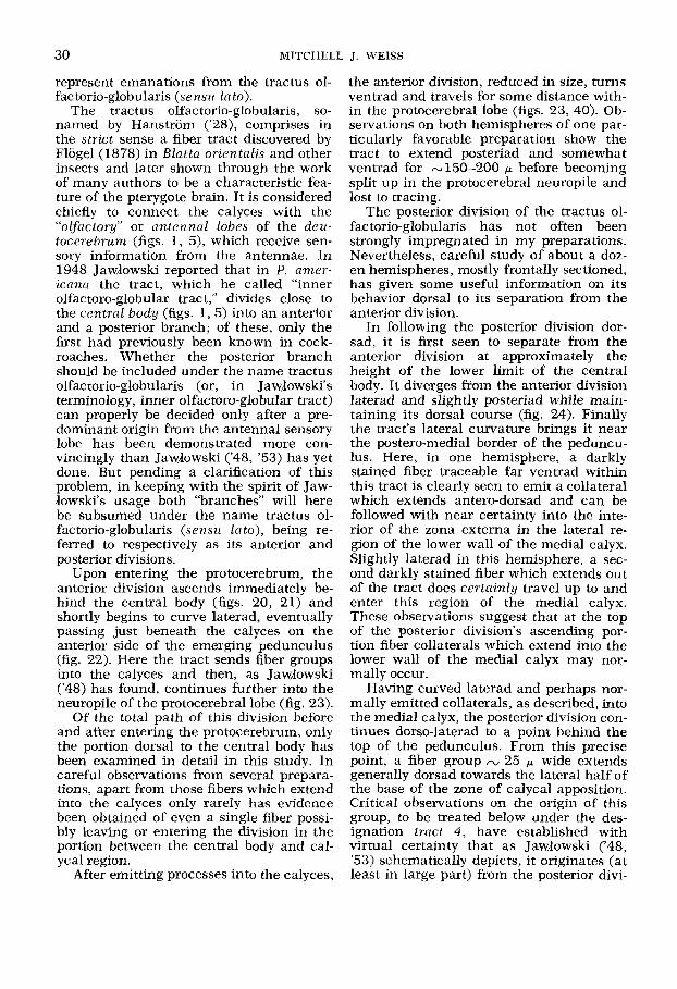
The tractus olfactorio-globularis, so- named by Hanstrom (’28), comprises in the strict sense a fiber tract discovered by Flogel (1878) in Blatta orientulis and other insects and later shown through the work of many authors to be a characteristic fea- ture of the pterygote brain. It is considered chiefly to connect the calyces with the “olfactory” or antennal lobes of the deu- tocerebrum (figs. 1 , 5) , which receive sen- sory information from the antennae. In 1948 JawJowski reported that in P. amer- iccrna the tract, which he called “inner olfactoro-globular tract,” divides close to the central body (figs. 1,5) into an anterior and a posterior branch; of these, only the first had previously been known in cock- roaches. Whether the posterior branch should be included under the name tractus olfactorio-globularis (or, in Jawlowski’s terminology, inner olfac toro-globular tract) can properly be decided only after a pre- dominant origin from the antennal sensory lobe has been demonstrated more con- vincingly than JawJowski (‘48, ’53) has yet done. But pending a clarification of this problem, in keeping with the spirit of Jaw- dowski’s usage both “branches” will here be subsumed under the name tractus ol- fac torio-globularis (sensu la to), being re- ferred to respectively as its anterior and posterior divisions.
Upon entering the protocerebrum, the anterior division ascends immediately be- hind the central body (figs. 20, 21) and shortly begins to curve laterad, eventually passing just beneath the calyces on the anterior side of the emerging pedunculus (fig. 22). Here the tract sends fiber groups into the calyces and then, as JawJowski (‘48) has found, continues further into the neuropile of the protocerebral lobe (fig. 23).
Of the total path of this division before and after entering the protocerebrum, only the portion dorsal to the central body has been examined in detail in this study. In careful observations from several prepara- tions, apart from those fibers which extend into the calyces only rarely has evidence been obtained of even a single fiber possi- bly leaving or entering the division in the portion between the central body and cal- ycal region.
After emitting processes into the calyces,
the anterior division, reduced in size, turns ventrad and travels for some distance with- in the protocerebral lobe (figs. 23, 40). Ob- servations on both hemispheres of one par- ticularly favorable preparation show the tract to extend posteriad and somewhat ventrad for ~150-200 p before becoming split up in the protocerebral neuropile and lost to tracing.
The posterior division of the tractus ol- factorio-globularis has not often been strongly impregnated in my preparations. Nevertheless, careful study of about a doz- en hemispheres, mostly frontally sectioned, has given some useful information on its behavior dorsal to its separation from the anterior division.
In following the posterior division dor- sad, it is first seen to separate from the anterior division at approximately the height of the lower limit of the central body. It diverges ffom the anterior division laterad and slightly posteriad while main- taining its dorsal course (fig. 24). Finally the tract’s lateral curvature brings it near the postero-medial border of the peduncu- lus. Here, in one hemisphere, a darkly stained fiber traceable far ventrad within this tract is clearly seen to emit a collateral which extends antero-dorsad and can be followed with near certainty into the inte- rior of the zona externa in the lateral re- gion of the lower wall of the medial calyx. Slightly laterad in this hemisphere, a sec- ond darkly stained fiber which extends out of the tract does certainly travel up to and enter this region of the medial calyx. These observations suggest that at the top of the posterior division’s ascending por- tion fiber collaterals which extend into the lower wall of the medial calyx may nor- mally occur.
Having curved laterad and perhaps nor- mally emitted collaterals, as described, into the medial calyx, the posterior division con- tinues dorso-laterad to a point behind the top of the pedunculus. From this precise point, a fiber group n, 25 p wide extends generally dorsad towards the lateral half of the base of the zone of calycal apposition. Critical observations on the origin of this group, to be treated below under the des- ignation tract 4, have established with virtual certainty that as Jawlowski (‘48, ’53) schematically depicts, it originates (at least in large part) from the posterior divi-

CORPORA PEDUNCULATA OF
sion. Whether whole fibers or simply collat- erals leave the latter to form tract 4 is unknown. Concomitantly, it is uncertain whether some or even all fibers of this divi- sion continue laterad beyond the point of origin of tract 4, as JawJowski ('48, '53) depicts schematically but does not actually prove. Fibers have been seen leaving this region laterally and these might represent a lateral continuation of the division.
1. Topography of the calycal tracts The calycal fiber tracts emanating from
the tractus olfactorio-globularis ( sensn Into) exhibit some variability which makes their topographical study difficult. Consequently my efforts have been directed toward a de- tailed understanding of tracts in a limited region, namely the lateral calyx excluding its medial, vertical wall. First, with two types of exceptions, all defined groups of
THE AMERICAN COCKROACH 31
extrinsic fibers discovered in this portion of the lateral calyx will be described as they typically appear when present. The numbering scheme for these tracts is shown in figure 3 . These descriptions will be followed by a treatment of observed var- iability. The exceptions mentioned comprise bundles of quartet cell fibers and any oth- er, comparatively minor groups of extrinsic fibers which encroach slightly into the region of interest following passage up- wards between the apposed vertical calycal walls. Finally, fiber tracts in the remainder of the calyces will be treated briefly. It should be kept in mind during the follow- ing account that not every fiber necessar- ily extends the whole length of its tract before passing into the interior of the zona externa.
( a ) Typical conditions in portinn of lat- eral calyx. Tract 1 . This tract originates
Fig. 3 Diagram showing numbering scheme of calycal tracts in the lateral calyx, repre- sented as though projected onto a transverse plane. Anterior is upwards, while medial is to the left. Abbreviations: ped, pedunculus; ant. div., anterior division of tractus olfactorio- globularis; aii, anterior intercalycal fiber influx; 1 , 2 , 2 a , 2b, 3 , 4 , 4 a , 5 ,5a , 5b, various tracts.
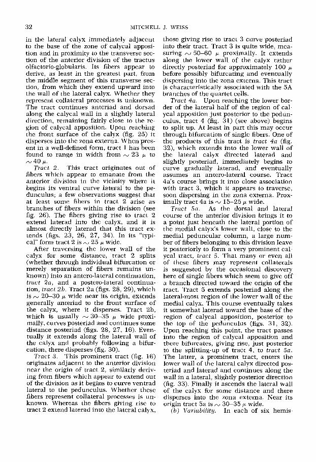
32 MITCHELL J. WEISS
in the lateral calyx immediately adjacent to the base of the zone of calycal apposi- tion and in proximity to the transverse sec- tion of the anterior division of the tractus olfactorio-globularis. Its fibers appear to derive, as least in the greatest part, from the middle segment of this transverse sec- tion, from which they extend upward into the wall of the lateral calyx. Whether they represent collateral processes is unknown. The tract continues anteriad and dorsad along the calycal wall in a slightly lateral direction, remaining fairly close to the re- gion of calycal apposition. Upon reaching the front surface of the calyx (fig. 25) i t disperses into the zona externa. When pres- ent in a well-defined form, tract 1 has been found to range in width from /V 23 p to IV 40 p.
Tract 2 . This tract originates out of fibers which appear to emanate from the anterior division in the vicinity where i t begins its ventral curve lateral to the pe- dunculus; a few observations suggest that at least some fibers in tract 2 arise as branches of fibers within the division (see fig. 26). The fibers giving rise to tract 2 extend laterad into the calyx, and it is almost directly laterad that this tract ex- tends (figs. 23, 26, 27, 34). In its “typi- cal” form tract 2 is - 25 p wide.
After traversing the lower wall of the calyx for some distance, tract 2 splits (whether through individual bifurcation or merely separation of fibers remains un- known) into an antero-lateral continuation, tract 2a, and a postero-lateral continua- tion, tract 2b. Tract 2a (figs. 28, 29), which is N 20-30 p wide near its origin, extends generally anteriad to the front surface of the calyx, where it disperses. Tract 2b, which is usually N 30-35 p wide proxi- mally, curves posteriad and continues some distance posteriad (figs. 28, 27, 16). Even- tually it extends along the lateral wall of the calyx and probably following a bifur- cation, there disperses (fig. 30).
This prominent tract (fig. 16) originates adjacent to the anterior division near the origin of tract 2, similarly deriv- ing from fibers which appear to extend out of the division as i t begins to curve ventrad lateral to the pedunculus. Whether these fibers represent collateral processes is un- known. Whereas the fibers giving rise to tract 2 extend laterad into the lateral calyx,
Tract 3 .
those giving rise to tract 3 curve posteriad into their tract. Tract 3 is quite wide, mea- suring N 50-60 p proximally. It extends along the lower wall of the calyx rather directly posteriad for approximately 100 p before possibly bifurcating and eventually dispersing into the zona externa. This tract is characteristically associated with the 5A branches of the quartet cells.
Upon reaching the lower bor- der of the lateral half of the region of cal- ycal apposition just posterior to the pedun- culus, tract 4 (fig. 31) (see above) begins to split up. At least in part this may occur through bifurcation of single fibers. One of the products of this tract is tract 4a (fig. 32), which extends into the lower wall of the lateral calyx directed laterad and slightly posteriad, immediately begins to curve gradually laterad, and eventually assumes an antero-lateral course. Tract 4a’s course brings it into close association with tract 3, which it appears to traverse, soon dispersing in the zona externa. Prox- imally tract 4a is AJ 15-25 p wide.
Tract 5a. As the dorsal and lateral course of the anterior division brings it to a point just beneath the lateral portion of the medial calyx’s lower wall, close to the medial peduncular column, a large num- ber of fibers belonging to this division leave it posteriorly to form a very prominent cal- ycal tract, tract 5. That many or even all of these fibers may represent collaterals is suggested by the occasional discovery here of single fibers which seem to give off a branch directed toward the origin of the tract. Tract 5 extends posteriad along the lateral-most region of the lower wall of the medial calyx. This course eventually takes it somewhat laterad toward the base of the region of calycal apposition, posterior to the top of the pedunculus (figs. 31, 32). Upon reaching this point, the tract passes into the region of calycal apposition and there bifurcates, giving rise, just posterior to the splitting-up of tract 4, to tract 5a. The latter, a prominent tract, enters the lower wall of the lateral calyx directed pos- teriad and laterad and continues along the wall in a lateral, slightly posterior direction (fig. 33). Finally it ascends the lateral wall of the calyx for some distance and there disperses into the zona externa. Near its origin tract 5a is
(b) Vuriability. In each of six hemis-
Tract 4a.
30-35 p wide.

CORPORA PEDUNCULATA OF THE AMERICAN COCKROACH 33
pheres examined, the presence of normal tracts 3 and 4a could be confirmed. Simi- larly, in each of five hemispheres exam- ined, the presence of a normal tract 5a could be confirmed. The remaining tracts, however, exhibited some interesting var- iability, which was studied in ten hemis- pheres. One of these will be excluded from consideration in view of uncertainty in the observations due to appreciable shrinkage of extrinsic fibers. Because metallic im- pregnation is a notoriously variable tech- nique, i t may be emphasized here that the considerable reliability of staining of given fiber types within successfully impregnated regions and the nature itself of some of the observed variability both serve to exclude variable staining of an unvarying set of tracts as a sufficient explanation for the conditions to be described.
Of the nine hemispheres, only seven show a “typical” tract 2. In all seven cases, tract 2 gives rise to tract 2b. In only five of the seven cases, however, does a tract 2a as such appear to be present: in the sixth case (fig. 34) tract 2a is presumably represented by a wide band of scattered fibers which is not as massive as a “typi- cal” tract 2a, while in the seventh case the presence of even such a band of scattered fibers is uncertain.
In these seven hemispheres with a “typ- ical” tract 2, tract 1 can be observed in a well-defined form in only three. In another two of the seven, tract 1 appears to be rep- resented by a number of fibers not grouped into a very well-defined tract, while in the two remaining cases the presence of even such fibers remains uncertain. Moreover, there are suggestions of a negative corre- lation between the presence of a well-de- fined tract 1 and that of a well-defined tract 2a: in one of the seven hemispheres both tracts seem to be present, but in each of the other six hemispheres only the one or the other occurs in a well-defined form (in four tract 2a, in two tract 1).
The two hemispheres not showing a “typical” tract 2 exhibit some interesting features which will be considered in detail.
1. The first hemisphere exhibits good fixation and impregnation, with only aver- age fiber shrinkage. A comparatively nar- row fiber bundle nearly identical in position and orientation to a typical tract 2 is pres- ent in the lateral calyx. These fibers can
be observed laterally to curve posteriad and continue posteriad for some distance as a very loose bundle occupying a position sim- ilar to that of a typical tract 2b. However, there is no evidence whatever for the pres- ence of a tract 2a or even a diffuse repre- sentative of one, although this calycal re- giofl is well impregnated and tract 1 can be observed quite clearly in an adjacent area. But more surprising is the existence of an extra tract on the lateral side of tract 3. It resembles in size and orientation a “typical” tract 2b but is located much further mediad. Its fibers seem to originate in the anterior division of the tractus olfac- torio-globularis, in the general vicinity in which tracts 2 and 3 normally originate. Interestingly, the 5A branches of the quar- tet cells, rather than being associated with tract 3 , are located on the lateral side of the extra tract. Finally, a transverse tract ~ 2 4 p wide, which has not been observed in any other hemisphere, is located at the level of the posterior side of the peduncu- lus (fig. 35). This tract, whose exact origin medially is uncertain, can be traced later- ad from the region of tract 3 and the other “extra” tract and appears to disperse among the fibers meagerly representing the typical tract 2b. The other hemisphere of this brain is normal with respect to tracts 1, 2, 2a, 2b, and 3 , except that tract 2a is poorly developed.
2. In the second hemisphere of inter- est, the fibers are sufficiently well fixed and very intensely stained. There is only unconvincing evidence for the presence of even an extremely reduced tract 2 and no sign of tracts 2a and 2b. Tract 1, how- ever, is well defined and fairly massive. Re- markably, at the lower surface of the lat- eral calyx near its anterior end, tract 1 appears to give rise to a lateral group of fibers (whether these are collaterals is un- known) which turn posteriad a little lat- erally and extend posteriad, following the normal course of tract 2a. These fibers subsequently continue on along the normal course of tract 2b to disperse terminally not far from the level of the posterior surface of the top of the pedunculus. This extra tract can be observed clearly and unquestionably arises at the front of the calyx rather than in the usual vicinity of tract 2. The corre- sponding tracts in the opposite hemisphere are normal except that tract 1 does not
34 MJTCHELL J. WEISS
occur in a well-defined form and may be absent.
Different interpretations of the observed variability are possible. On the one hand, fibers reaching a given area may always belong to the same invariable set of neu- rons but simply arise from variable points along their parent fibers and (or) reach their destination following variable routes. The mechanism by which the same destina- tion could be consistently reached would be of some interest. On the other hand, fibers reaching a given area may not al- ways belong to the same invariable set of neurons. In this case the nature of the ob- served variability suggests that some mech- anism may nevertheless insure a constant innervation density for a given calycal area. Two lines of evidence can support such a conclusion: first, the finding that although tracts 1 and 2a each terminate distally in the same section of the calyx they tend not to appear in a well-developed form simul- taneously , and second, the seeming rela- tionship between the courses or areas of dispersion of extra tracts and those of the normal tracts which in their hemispheres are lacking or reduced. In any event, fail- ure of the highly atypical conditions in the two hemispheres showing extra tracts to occur simultaneously in the contralateral hemispheres suggests that the observed variability may not be genetic in origin.
( c ) The remaining calycal ureas. Fiber tracts comparable to those described for the major portion of the lateral calyx also occur over the entire medial calyx and the remainder of the lateral calyx. Figure 36 shows a typical example from the medial calyx. Since, aside from quartet cell branches, the only groups of extrinsic fi- bers I have ever seen passing into either calyx appear to extend directly from the tractus olfactorio-globularis ( sensu Zato), all tracts in these remaining areas pre- sumably represent merely additional ema- nations of the tractus olfactorio-globularis. Due to the expected variability in these re- maining tracts, a detailed study of each has been impracticable. Nevertheless, that the tracts in these areas are simply coun- terparts of the emanations already de- scribed in the lateral calyx is supported by the similarity between both calyces in their ultrastructurally distinguishable nerve fi- ber classes (Weiss, unpublished observa-
tions) and by the following brief descrip- tions of representative examples of tracts in these remaining areas.
As previously mentioned, just posterior to the pedunculus tract 4 begins to split up, one product, tract 4a, passing into the lower wall of the lateral calyx. A second product, however, extends generally dorsad between the apposed vertical calycal walls. There it disperses, presumably, as one particularly favorable hemisphere strongly suggests, into both apposed walls. In ad- dition there are suggestions, requiring con- firmation, of fibers which extend posteriad within the base of the region of calycal ap- position to tract 5, with which they seem to become associated, and of fibers which ex- tend across the base of the appositional re- gion to the medial calyx.
During its course, described above, along the lateral-most region of the lower wall of the medial calyx, tract 5 sends at least one fiber group into this wall. In bifurcat- ing posterior to the pedunculus, tract 5 gives rise, in addition to tract 5a, to a prominent tract 5b which extends more or less dorsad between the apposed calycal walls (fig. 37) and ultimately disperses within both calyces.
Finally, as the anterior division of the tractus olfactorio-globularis passes be- neath the base of the region of calycal ap- position just anterior to the origin of the pedunculus, a large number of fibers leave the division more or less perpendicularly to enter the region of calycal apposition di- rected postero-dorsad. After coursing be- tween the apposed calycal walls, these fibers ultimately disperse into both calyces. That many if not all originate as collater- als is strongly suggested by several appear- ances in the preparations. Since I am not yet certain that this prominent fiber infiow becomes organized into only one coherent tract, it will not be given a number but will simply be designated the anterior in- tercalycal fiber inf lux (fig. 23).
The intercalycal fiber groups now de- scribed are believed to account for most or all fiber tracts situated between the ap- posed vertical calycal walls.
Scattered fibers similar in size to calycal tract fibers are commonly observed at the external margin of the zona externa. They sometimes run parallel and in proximity to the tracts, making the latters’ strict
CORPORA PEDUNCULATA O F
delimitation partly arbitrary. Presumably most of these fibers similarly derive from the tractus olfactorio-globularis (sensu lato), perhaps by way of calycal tracts, al- though some may represent branches of quartet cell fibers or of extrinsic fibers of unknown origin to be described below. I assume that these scattered fibers eventual- ly terminate in the midst of the zona ex- terna.
2. General comments o n the origin of the calycal tracts
In the preceding section the origin of in- dividual calycal tracts has been described with caution. Caution is necessary because of the difficulty here of tracing individual fibers clearly over a sufficient distance and because of the absence of proof that every fiber within a tract shares an identical origin. Evidence of the latter kind repre- sents an ideal which in practice is prob- ably unachievable with light microscopy. The best that can be wished for is estab- lishment of the immediate origin of some fibers in a calycal tract and confirmation for the remaining fibers that their behavior is consistent with such an origin and that any alternate origin is unlikely. This repre- sents the level of evidence I have sought to obtain.
Every calycal tract studied has been found to originate in intimate association with the tractus olfactorio-globularis (sen- su lato) (e.g., figs. 23, 26). In some in- stances at least a predominant origin from it (e.g., calycal tract 5 and the anterior intercalycal fiber influx) has been grossly apparent. In the case of calycal tracts 1, 2, 3 , and 5 and the anterior intercalycal influx, individual fibers have been seen extending directly out of the anterior divi- sion toward or into the originating calycal fiber group. Suggestions that such fibers arise as collaterals (e.g., figs. 26, 38, inset) have found confirmation in ultrastructural study of the anterior division. This has es- tablished for several fibers the presence of an oblique or perpendicular collateral which extends toward the calycal surface (fig. 38).
Apart from quartet cell fibers and those emanating from the divisions of the tractus olfactorio-globularis, only one or two addi- tional extrinsic fibers, of uncertain origin, have ever been found entering the cal-
THE AMERICAN COCKROACH 35
yces. Moreover, I have been unable to dis- cover any other fiber groups near the cal- yces which might be suspected of elusively passing into them either directly or after becoming mixed with either division of the tractus olfactorio-globularis. Thus the pos- sibility that any significant proportion of calycal tract fibers might elusively stem from a source other than the tractus olfac- torio-globularis (sensu lato) can be reason- ably excluded.
On these grounds I consider the observa- tions to warrant the following conclusion: the calycal tract fibers taken collectively originate exclusively, or almost exclusively, in the tractus olfactorio-globzilctris (sensu lato).
3. Teiminations of the calycal tracts Regardless of the individual courses of
the calycal tracts, their fibers all eventually disperse into the zona externa. In my prep- arations, fibers derived from these tracts typically constitute the most prominent fiber class in the zona externa. Although distributed within this layer throughout all areas of both calyces, they are partic- ularly conspicuous where a tract turns into the midst of the zona externa to dis- perse (figs. 30, 39). The fibers course in various directions without any apparent common orientation. Bifurcations have been observed.
The diameter of fibers within calycal tracts mostly does not exceed 2.5 p and commonly falls far below this; it has never been found to exceed 3.5 p. Fibers enter- ing the interior of the zona externa from calycal tracts rarely if ever exceed 2 p in diameter and ordinarily measure much less.
Extrznsic calycal $fibers of unknown origin
In several preparations individual, sharp- ly stained fibers within the zona externa have been observed extending over an ap- preciable distance in one direction, parallel to the zona interna. Such fibers are distrib- uted throughout each calyx. A dozen rep- resentative examples selected from two preparations and given detailed study range in maximum diameter from 1 p to nearly 3 p and invariably lie (at least prox- imal to their first branching point) in the internal half of the zona externa, often
36 MITCHELL J. WEISS
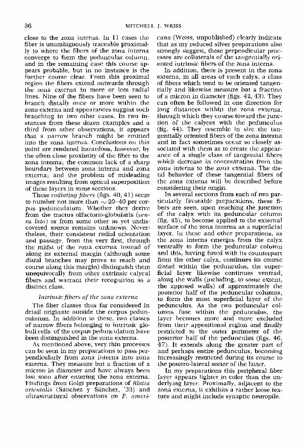
close to the zona interna. In 11 cases the fiber is unambiguously traceable proximal- ly to where the fibers of the zona interna converge to form the peduncular column, and in the remaining case this course ap- pears probable, but in no instance is the further course clear. From this proximal region the fibers extend outwards through the zona externa in more or less radial lines. Nine of the fibers have been seen to branch distally once or more within the zona externa and appearances suggest such branching in two other cases. In two in- stances from these dozen examples and a third from other observations, it appears that a narrow branch might be emitted into the zona interna. Conclusions on this point are rendered hazardous, however, by the often close proximity of the fiber to the zona interna; the common lack of a sharp boundary between zona interna and zona externa; and the problem of misleading images resulting from optical superposition of these layers in some sections.
These radiating fibers (figs. 40,41) seem to number not more than N 20-40 per cor- pus pedunculatum. Whether they derive from the tractus olfactorio-globularis (sen- su lato) or from some other as yet undis- covered source remains unknown. Never- theless, their consistent radial orientation and passage, from the very first, through the midst of the zona externa instead of along its external margin (although some distal branches may prove to reach and course along this margin) distinguish them unequivocally from other extrinsic calycal fibers and warrant their recognition as a distinct class.
Intrinsic fibers of the zona externa The fiber classes thus far considered in
detail originate outside the corpus pedun- culatum. In addition to these, two classes of narrow fibers belonging to intrinsic glo- buli cells of the corpus pedunculatum have been distinguished in the zona externa.
As mentioned above, very thin processes can be seen in my preparations to pass per- pendicularly from zona interna into zona externa. They measure but a fraction of a micron in diameter and have always been lost soon after entering the zona externa. Findings from Golgi preparations of Blatta orientalis (Sanchez y Sanchez, '33) and ultrastructural observations on P. ameri-
c a m (Weiss, unpublished) clearly indicate that as my reduced silver preparations also strongly suggest, these perpendicular proc- esses are collaterals of the tangentially ori- ented intrinsic fibers of the zona interna.
In addition, there is present in the zona externa, in all areas of each calyx, a class of fibers which tend to be oriented tangen- tially and likewise measure but a fraction of a micron in diameter (figs. 42,43). They can often be followed in one direction for long distances within the zona externa, through which they course toward the junc- tion of the calyces with the pedunculus (fig. 44). They resemble in size the tan- gentially oriented fibers of the zona interna and in fact sometimes occur so closely as- sociated with them as to create the appear- ance of a single class of tangential fibers which decrease in concentration from the zona interna to the zona externa. The dis- tal behavior of these tangential fibers of the zoiia externa will be described before considering their origin.
In several sections from each of two par- ticularly favorable preparations, these fi- bers are seen, upon reaching the junction of the calyx with its peduncular column (fig. 45), to become applied to the external surface of the zona interna as a superficial layer. In these and other preparations, as the zona interna emerges from the calyx ventrally to form the peduncular column and this, having fused with its counterpart from the other calyx, continues its course distad within the pedunculus, the super- ficial layer likewise continues ventrad, along the walls (including, to some extent, the apposed walls) of approximately the posterior half of the peduncular columns, to form the most superficial layer of the pedunculus. As the two peduncular col- umns fuse within the pedunculus, the layer becomes more and more excluded from their appositional region and finally restricted to the outer perimeter of the posterior half of the pedunculus (figs. 46, 47). It extends along the greater part of and perhaps entire pedunculus, becoming increasingly restricted during its course to the postero-lateral sector of the latter.
In my preparations this peripheral fiber layer appears lighter in color than the un- derlying layer. Proximally, adjacent to the zona externa, it exhibits a rather loose tex- ture and might include synaptic neuropile.

CORPORA PEDUNCULATA OF
Immediately underlying the peripheral layer is a more darkly stained layer. There are strong indications that this layer too derives from narrow tangential fibers with- in the zona externa which have become applied to the zona interna at the junction of the calyx with its peduncular column. This derivation is not certain, however, for it is difficult to exclude the possibility that it derives at least partly from the external- most fibers of the zona interna. The second layer, from its origin at the top of the pe- duncular columns, follows the same course as the overlying lighter layer: it is restrict- ed to the posterior half of the peduncular columns (except proximally where i t ex- tends forward somewhat between them) and eventually becomes localized in the postero-lateral sector of the pedunculus (figs. 46, 47). I have established definitely that this second layer extends the entire length of the pedunculus. The behavior of both layers further distad remains to be clarified.
THE AMERICAN COCKROACH 37
Establishment of the entry of narrow tangential fibers from the zona externa into the special posterior zone of the pe- duncular columns has proven possible only in generally posterior areas within certain favorable sections. Nevertheless, such fibers from all areas of the calyces have been observed to approach the pedun- cular columns. This suggests that all these fibers, even those from strictly anterior calycal areas, pass into the same special posterior zone. Some views have in fact seemed to show fibers from calycal areas anterior to the columns becoming directed towards and passing into the second, com- paratively darkly stained layer in the pos- terior region of the latter. Although it is difficult to establish in the available prep- arations, I suspect that all or virtually all fibers of this class do enter the special pos- terior zone of the columns rather than another sector of their perimeter. In any event it is clear that the fibers do leave the calyces along with the zona interna, to
“ordinary“ globuli cell somata
4 1 1
1 1 / I
1 1
somata f , 1 -
extrinsic radiating ,‘ calycal quartet f iber I
I ce l l f ibers I tract
narrow tangential f iber
Fig. 4 Diagram of a generalized section through the wall of a calyx of P. amen’cana, summarizing known fiber constituents. The pedunculus would be to the right and the calycal rim is seen at the left. The fiber arrangement of the “ordinary” globuli cell is shown in part after the findings of Sknchez y Shchez (‘33) on Blatta orientalis and those of Weiss (unpublished ultrastructural observations) on P. americana. Although the narrow tangential fibers of the zona externa presumably possess calycal col- laterals these remain to be demonstrated and consequently are not shown here.

38 MITCHELL J. WEISS
which they have become applied, to con- stitute a component part of the peduncular columns.
It has previously been known (e.g., Han- strom, ’28; Arnold, ’60) that in histological preparations the pedunculus and Q: and p lobes of P. americana display longitudinal layers of alternately lighter and darker ap- pearance (see fig. 20). The two posterior fiber layers described here seem to corre- spond to the numerous remaining longitu- dinal layers. Some interesting implica- tions arising from such a correspondence are suggested elsewhere (Weiss, ’72b).
In numerous sections mostly from two preparations, the narrow tangential fibers of the zona externa are seen to extend to and sometimes turn along the margin of the neuropile at the peripheral rim of the calycal walls (fig. 48). Now it is an inter- esting fact, first recognized in a cockroach by Flogel (1876, 1878) in Blatta orientalis, that the globuli cell somata in at least some species are not confined to the con- cavities of the calyces. In P. americana the somata “overflow” each calyx around its entire circumference and lie adjacent to the rim of the zona externa; sometimes they extend far along the external surface of the zona externa (figs. 7, 48, 49) (cf. Brousse-Gaury, ’71). In several sections from preparations showing least shrinkage, these somata appear larger than most if not all more central ones (fig. 50) (for ap- proximately comparable observations on Blatta orientalis see Sanchez y Sanchez, ’33). Fiber bundles from these “external” somata have been found several times to extend to (fig. 49) and travel along the pe- ripheral rim of the zona externa, the very region to which narrow tangential fibers of the zona externa have been traced. I have not been able to obtain an unambig- uous view of any single narrow tangential fiber within the zona externa indisputably both passing into the layer of external so- mata via the calycal rim and joining the soma of a globuli cell. Nevertheless, all sequential portions of such a course have been clearly viewed enough times as to leave little doubt that this occurs. Fiber bundles from external globuli cell somata have also been observed in a few instances to extend to points on the surface of the zona externa which lie beyond the calycal rim. It remains unclear whether such fi-
bers all travel along the external surface to enter the calycal wall at its rim or wheth- er they may enter the zona externa (and give rise to “narrow tangential fibers”) at points lying beyond the rim.
Consequently, in consideration of their similarity to and sometimes close associa- tion with the comparably oriented fibers of the zona interna; their extension into the pedunculus, of which they form an in- tegral part; and their presumable origin as direct continuations of fibers belonging to the external globuli cell somata, the nar- row tangential fibers of the zona externa are concluded to belong to intrinsic globuli cells of the corpus pedunculatum, specifi- cally those whose cell bodies “overflow” the calyces.
DISCUSSION
Fiber connections and constituents of the calyces
In sum I have distinguished five fiber classes within the zona externa of the cal- yces in P. americana. Three consist of ex- trinsic and two of intrinsic fibers (fig. 4). In the interior of the zona externa, fibers from the calycal tracts are typically the most ubiquitously apparent in my prepara- tions, although “radiating fibers” as well as quartet cell branches can also appear conspicuous when met. The perpendicular- ly and tangentially oriented intrinsic fibers are typically comparatively inconspicuous.
Whereas the constitution and fiber deri- vation of the zona interna of cockroaches have been correctly understood since the time of Flogel (1876, 1878), previous re- ports concerning the organization and fi- ber connections of the zona externa have been highly contradictory. Of the five fiber classes distinguished here within this layer, only the class of narrow fibers which pass into i t perpendicularly from the zona interna has been recognized and correctly described in previous accounts of cock- roaches (see particularly Bretschneider, ’13b, ’14; Hanstrom, ’28; Sanchez y San- chez, ’33). Scrutiny of the descriptions and figures of Sanchez y Sanchez (‘33) reveals in addition that some fibers depicted in Blatta orientalis as “centripetal” with re- spect to the calyces (fibers here termed “extrinsic”) may represent intrinsic tan- gential fibers of the zona externa. More- over, figure 5 in the same work includes &

CORPORA PEDUNCULATA OF
portion of a fiber (fiber “B, H”) which may perhaps correspond to an extrinsic radiat- ing fiber of P. americana. Prior studies of cockroach calyces include no hints of quartet cell fibers or calycal tracts and their terminations, although reports that the tractus olfactorio-globularis enters the calyces naturally imply the presence there of terminations of some sort.
Of the two main sets of extrinsic fibers followed in the present work from within the zona externa to regions outside the cor- pus pedunculatum, the tractus olfactorio- globularis (sensu lato), as indicated above, has been described by JawJowski (‘48, ’53) in a form closely resembling that observed here (for a particularly well documented account of a similarly arranged tractus ol- factorio-globularis, sensu stricto, in Lepi- doptera see Pearson, ’71). Apart from the naming by Brousse-Gaury (’68, ’71) of a “nucleus” which apparently includes quartet cell somata, and the report of a connection between this and the nerous corporis cardiaci I1 (see below), previous accounts of cockroaches contain no indi- cation of the existence of the quartet neu- rons or any fibers attributable to them.
In other insect groups intrinsic fibers comparable to those in cockroaches which pass perpendicularly from zona interna into zona externa are well known (e.g., Hymenoptera: Kenyon, 1896; Hemiptera: Pflugfelder, ’3W37). The presence in these groups of a fiber class homologous with the intrinsic tangential fibers of the zona externa of P. americana is discussed else- where (Weiss, ’70). Although a tractus ol- factorio-globularis (sensu stricto) has been widely reported throughout the Pterygota, little is known concerning calycal termi- nations explicitly attributed to it (Hemip- tera: Pflugfelder, ’3&’37; Formica: Goll, ’67; Lepidoptera: Pearson, ’71); none of these authors mentions an arrangement of fibers into calycal tracts such as occur in P. americann. Extrinsic calycal fibers oriented radially were discovered by Ken- yon (1896) in Apis mellifica and have been briefly reported in Orthoptera (Strausfeld, ’70). Beyond this similar orientation, in- sufficient information is available for pos- tulating homologies with the radiating extrinsic fibers described here. The liter- ature dealing with insects other than cock- roaches has furnished no indications of
T H E AMERICAN COCKROACH 39
neurons seemingly homologous with the quartet cells.
Numerous reports concerning calycal fi- ber connections in cockroaches, some wide- ly accepted in current publications (e.g., Horridge, ’65; Guthrie and Tindall, ’68), have not been confirmed in the present study. These reports are summarized be- low.
Flogel (1878) first described the anterior division of the tractus olfactorio-globularis in a cockroach but was unable to make out its distal course. Consequently, Haller’s (‘05) account of Blatla orientalis contains the first report of a calycal fiber connec- tion in a cockroach. Haller states (pp. 211-212) that just beneath the calyces a portion of the pedunculus carries fibers from one calyx into the other. The support offered is limited to a drawing (fig. 6,B) on which images (of unspecified origin) of these fibers appear to be schematically superimposed.
Bretschneider (‘13b, ’14) also studied Blatta (“Periplaneta”) orienkzliis.(i He em- ployed Mallory’s mixture in connection with other ordinary stains, as well as a gold impregnation method (though appar- ently without added advantage) (‘14, p. 271). Horridge’s attribution of his findings to Golgi preparations (Horridge, ’65, fig. 16.19) is in error.
Bretschneider mentions various fiber connections of the calyces. One, the Riech- strung, is the anterior division of the trac- tus olfactorio-globularis, as detailed draw- ings clearly show (‘14). The neurons of this tract are reported to belong partly to the deutocerebrum but mostly (’14, p. 296) to the pars intercerebralis anterior; converse- ly, the large cells of the pars intercerebra- lis anterior are said to belong chiefly to this tract (‘14, p. 291). Evidence for a con- nection between somata of the pars inter- cerebralis and fibers within the tract is presented in a drawing (‘14, Tafelfig. Fq) which, in my opinion, provides insuffi- cient support for this relationship.
6Text figure 10 of the 1914 work consists of a sche- matic representation of some neuronal connections of the calyces and other regions in a cockroach brain, “ [n] ach Ziegler.” This figure derives, however, from neither of Ziegler’s cited works, which are his only works known to treat the insect brain. The figure may merely represent the conception which Ziegler, Bretschneider’s teacher, held of his student’s findings, although it de- picts, for example, a cell body (belonging to an optic- calycal connection) whose location was unknown to Bretschneider.
40 MITCHELL J. WEISS
A second calycal connection is with the ipsilateral optic lobe, the location of the somata being unknown. Finally, two con- nections are described between the calyces and central body, one with somata at the lateral border of the protocerebrum (‘14, p. 294) and the other with somata of un- known location (‘14, p. 297). As the first of these two tracts passes between the 01
lobe and calyces, fibers are said to branch off perpendicularly into the calycal walls. Bretschneider may here be describing the anterior division of the tractus olfactorio- globularis as it passes beneath the calyces. Unfortunately, his detailed drawings do not illustrate this section of the anterior division or the reported calycal connections with the central body or optic lobe, al- though all reported calycal connections but one are shown in a schematic figure. Bretschneider could not confirm Haller’s report of a fiber passage between the cal- yces and considered it incorrect; similarly, Sanchez y Sanchez (‘33) failed to see it.
Hanstrom (‘28) briefly mentions two cal- ycal connections of P. americana which were examined using Golgi impregnation and vital staining with methylene blue. A “tractus optico-globularis” with large so- mata in the optic lobe is said to connect it with the calyces. An illustration of the tract is lacking. The tractus olfactorio-glo- bularis (sensu stricto) is said to connect the antennal glomeruli with both central body and calyces and is figured diagrammati- cally without indication of a lateral contin- uation beneath the calyces. Hanstrom was unable to confirm Bretschneider’s report that this tract belongs partly to somata in the pars intercerebralis. In turn, JawAow- ski (‘48) subsequently noted the disagree- ment between Hanstrom’s representation of the tracts of the antennal lobes in P. americana and his own observations, a disagreement which includes the tractus olfac torio-globularis.
Arnold (’60) likewise deals with calycal connections in P. americana and makes statements which I have not confirmed. These observations have remained unpub- lished and will not be detailed here.
In a study involving ordinary stains as well as reduced silver (which does not, however, appear to have produced selective fiber impregnation), Drescher (‘60) briefly reports observations on the tractus olfac-
torio-globularis (sensu stricto) of P. amer- icana. He states that the tract penetrates into the medial calyx and belongs not only to somata lying dorsal to the olfactory lobe but also partly to large somata located un- der the calyces at the back side of the brain. The tract is shown photographically but evidence is not provided for its rela- tionships.
Willey (‘61, p. 235) briefly alludes in an incidental manner to certain fibers with somata in the tritocerebrum which are said to enter the calyces in P. americana.
Barbier (‘61), in a study based on ordi- nary staining methods, attributes three “outside” fiber connections to the calyces of Blatta orientalis. The medial calyx is said to receive a horizontal fiber bundle whose somata lie above the protocerebral lobe and to give off from its own globuli cell somata a vertical bundle which unites it with the central body. In addition, nu- merous very short fibers believed to arise in calycal globuli cell somata are reported to pass from both calyces into the 01 lobe. The vertical bundle is represented schemati- cally in Barbier’s figure 2 and probably corresponds to a portion of the anterior division of the tractus olfactorio-globularis as well as the initial section of calycal tract 5. Beyond this, evidence substantiating the reported calycal connections is lacking. Al- though neither remaining connection is ex- plicitly indicated in a figure, the author’s figure 8 depicts schematically what could represent the fibers said to pass between calyces and (Y lobe. My own sections of P. americana have sometimes presented im- ages of short segments of sharply impreg- nated nerve fibers lying between the 01 lobe and adjacent calycal surface (fig. 51). These fibers have never shown signs of connecting with a calyx but may represent the “very short fibers” which Barbier re- ports observing here in Blatta.
In a paper on P. americana, Satija and Singla (‘67) mention extrinsic fiber con- nections of the corpora pedunculata. Their study utilized iron haematoxylin, methyl- ene blue (apparently applied to sectioned material), and Holmes’ reduced silver meth- od. The illustrations are schematic. No tract is explicitly said to pass into the cal- yces, but the “antenno-cerebral tract,” originating from the antennal lobe (p. 301), is implied (p. 298) to connect with

CORPORA PEDUNCULATA OF THE AMERICAN COCKROACH 41
the corpus pedunculatum, and in the au- thors’ figure 2 this connection is depicted as being with the medial calyx, without in- dication of a lateral continuation.
In a study, based on non-selective stain- ing methods, of deutocerebral fiber con- nections in P. americana, Prigent (’68) presents observations on the tractus olfac- torio-globularis. During its ascending course this is reported to divide into a %a- meau superieur” and “rameau inf6rieur.” Prigent appears to indicate that the former connects with both calyces and seems to imply that the latter connects with the initial portion of the pedunculus. These two branches of the tractus olfactorio- globularis are depicted schematically in Prigent’s figure 1 and indicated in his photomicrograph 6, although in this photo- micrograph I am unable to recognize them as discrete structures relatable to my own observations. The author does not mention any equivalence between his two branches and those earlier described and illustrated by JawJowski (‘48), whose paper he cites.
Finally, Brousse-Gaury (‘71) describes observations based on reduced silver prep- arations concerning fibers said to enter the tractus olfactorio-globularis in cockroach- es. With reference to P. americana, fibers from three sources are reported. One group of neurons, with somata in the ocellar center of the anterior pars intercerebralis, is indicated to send axons into the tractus olfactorio-globularis, directed downwards. A second group, with somata in the poste- rior pars intercerebralis, is reported to send axons into the nervus corporis cardi- aci I1 and dendrites into the tractus olfac- torio-globularis, directed downwards. A third group, with somata also within the pars intercerebralis, is indicated (fig. 94) to send into this tract fibers which bifur- cate and send a branch in each direction. While none of these three neuronal types is stated to send fibers into the calyces, Brousse-Gaury does remark that the third group might correspond to neuron type 16 of Bretschneider (’14), which was sup- posed to represent a calycal connection. In my opinion the evidence for these fiber passages into the tractus olfactorio-globu- laris is insufficiently convincing.
Brousse-Gaury’s (’68) earlier report on Blabera fusca and P. americana, also based on reduced silver impregnation, includes
mention and an illustration of a ventro- posterior area of the optic lobe from which fibers are said to enter the nervus corporis cardiaci 11. The drawings of this “nucleus” (“noyau infero-posterieur du lobe optigue”) in B. fusca in Brousse-Gaury’s later (‘71) work, in which this fiber course is again reported, virtually convince me that the nucleus corresponds to or at least includes the quartet neuron somata (note particu- larly her fig. 36). But its stated emission of fibers into the nervus corporis cardiaci 11, for which critical evidence is lacking, does not agree with the branching pattern of the quartet neurons as determined here.
The above reports are typical of litera- ture concerning fiber connections in the in- sect brain. Rarely do the preparations studied allow visualization of the disper- sion of fibers into the regions they are re- ported to connect, and it is apparently common for authors to report as “con- nections” tracts seen to contact but not actually verified to enter structures. The habits of illustrating “connections” in schematic figures, and of omitting detailed descriptions, then minimize the ability of later workers to repeat observations and potentially disprove them. The result of these practices has been an accumulation in different insect groups of contradictory reports which may bear little relation to reality.
How complete are the present observa- tions? It seems unlikely that at least hints of any coherent fiber group of appreciable size which enters the calyces would have escaped detection. Scattered entering fi- bers, however, could perhaps have been missed; especially the calycal area imme- diately behind the pedunculus deserves further examination for this possibility, for here one or two fibers of uncertain origin have been observed. If the extrinsic “radi- ating fibers” of the zona externa do not stem from the tractus olfactorio-globularis (sensu lato) then they probably derive from fairly narrow fibers which pass into the calyces in a dispersed manner.
The conclusion reached above that the calycal tract fibers derive exclusively, or almost exclusively, from the divisions of the tractus olfactorio-globularis does not necessarily imply that they all synapse in the antenna1 lobe. First, the origin of the posterior division remains to be established

42 MITCHELL J. WEISS
conclusively. Secondly, although the ante- rior division (i.e., tractus olfactorio-globu- laris in its usual usage) has repeatedly been reported in different groups to ascend from the antennal lobe, it may well receive some fibers from other regions. Jawlowski (‘53), for example, has reported that two small fiber bundles extending respectively out of the tritocerebrum and subesopha- geal ganglion become included in the divi- sions of the tractus olfactorio-globularis in P. americana. Still, the widely reported ventral connection with the antennal lobe can be accepted for at least the bulk of the anterior division, and this implies that in P. ame.l-zcnna calycal fibers extending f r o m the antennal lobe (or perhaps from both of them: see Hanstrom, ’28; Jawlowski, ’53) far outnumber extrinsic calycal fibers f r o m any other single source and probably com- prise the preponderant majority of extrin- sic fibers in the calyces. Corresponding well to this conclusion is the finding from ultrastructural study (Weiss, unpublished) that most extrinsic calycal fibers belong to a single class which derives from calycal tracts.
Functional implications Despite over a century of interest, the
role of insect corpora pedunculata remains unclear. Traditionally these structures have been regarded as “higher centers,” centers of sensory association, “the seat of complex behavior,” and so forth. These views originated in anatomical studies, particularly of the massive, complex cor- pora pedunculata of some Hymenoptera. Although recent experimental studies have yielded many interesting observations, I believe with Howse and Williams (‘69) that they have failed to provide an incisive ex- planation of the special role of the corpora pedunculata. Representative of the varied experimental approaches employed are pa- pers by Maynard (‘56, ’67), Voskresenskaya (‘57), Burtt and Catton (‘59), Drescher (‘60), Rowel1 (‘63), Huber (several works, mostly reviewed in Huber, ’65), Horridge, Scholes, Shaw and Tunstall (’65), Dingle and Fox (‘66), Vowles (’67 and earlier works), Steiner and Pieri (’69), Bernstein and Bernstein (‘69), Otto (‘71), and Turan- skaya (‘72).
The corpora pedunculata of P. ameri- c a m , while large, are more typical of pter-
ygotes than are the highly elaborated cen- ters of some Hymenoptera, and their study can accordingly provide a proper perspec- tive. The present findings do in fact sug- gest a way of looking at these centers which may prove fruitful.
Studies entailing electrical stimulation of the corpora pedunculata in various in- sects have demonstrated that different be- havioral acts start or stop following stim- ulation. Yet significantly, there is a lack of experimental evidence that any partic- ular segments of the behavioral reper- toire are uniquely associated with the cor- pora pedunculata throughout the insects. Moreover, despite the impressive elabora- tion of the corpora pedunculata in some social insects, comparisons within the full range of other insects does not suggest any consistent correlation between the nature or complexity of the overt behavioral rep- ertoire and the degree of development of these brain centers. Thus the special sig- nificance of the corpora pedunculata may not relate to their output at all. Then it would not be surprising that complete ex- tirpation of all four calyces and their cell bodies in P. americana has so little obvi- ous effect on motor capabilities (Drescher, ’60).
If the special role of the corpora pedun- culata does not concern their output, might it concern their input and its processing? What is the nature of this input? So many tracts have been claimed to connect the calyces with other brain regions in differ- ent insects that one prevalent notion re- gards the corpora pedunculata as the paramount collectors and integrators of information from throughout the brain. Another view, advocated notably by Han- strom (e.g., ’28, ’40), maintains that in the original as well as most present-day pter- ygotes the corpora pedunculata serve pri- marily to combine optic with antennal in- formation. A very few authors, however, have noticed a particular comparative ana- tomical relationship between antennal in- put and the size of corpora pedunculata or their calycal region (Pflugfelder, ’3-37; Jawlowski, ’63; Prigent, ’68; see also Howse and Williams, ’69). Jawlowski especially has emphasized the dependence of the level of calycal development on the degree of de- velopment of the antennal lobes but not compound eyes. My own observations pro-

CORPORA PEDUNCULATA OF
vide immediate support for the view that antennal but not optic input is all-impor- tant to the corpora pedunculata, and they suggest that the corpora pedunculata of pterygotes are fundamentully second-order antennal sensory processing centers.
The anatomical literature offers wide- spread evidence in pterygotes for an insis- tent dependence of the calycal region on the antennal lobes. The first three of the authors just cited provide examples. In an extensive examination of the literature for each order since 1850, I have found no case in which a moderately developed calycal region occurs simultaneously with rudi- mentary antennal lobes. Moreover, it ap- pears that wherever the calycal region at- tains its most prominent development, as in some cockroaches, acridid grasshoppers, and hymenopterans, unquestionably well- developed antennal lobes are invariably present (note their prominence, for exam- ple, in fig. 5).
The literature does not appear to hold evidence for a similar dependency of the calycal region on any other central nervous structure or sense organ, including the op- tic lobes or compound eyes. It is noteworthy that instances are available from widely different orders in which a fully developed calycal region occurs in a species or caste simultaneously with extremely reduced compound eyes (ants: Pandazis, '30; ter- mites: Hecker, '66; a blind species of the apterygote group Lepismatidae: Hanstrom, '40); thus the combination of optic with antennal information could hardly be the primary task of the corpora pedunculata. Although tracts have frequently been re- ported to connect the optic lobe and calycal region in various pterygotes, little substan- tiation for these claims has ever been of- fered. Such connections may occur in those Hymenoptera with highly developed cor- pora pedunculata, where their existence has been reported by workers successfully applying silver impregnation methods. But in other pterygotes, none of the few authors who have obtained demonstrable success with specific neurological methods reports finding a tract connecting the optic lobe and calycal region, although Hanstrom ('28), who used specific methods on P. americuna, states without substantiation that such a tract occurs in this species. The closest thing to such a connection to
THE AMERICAN COCKROACH 43
receive substantiation by these authors is described by Pearson ('71) in the butterfly Pieris brassicae: here she found a tract which extends into the calyx and is said to arise not in the optic lobe but in an area of the protocerebrum in which optic fibers end. In the moth Sphinx ligustri she found corresponding fibers but no definite tract. In the lepidopterans which she studied she believed the visual input into the calyx to be probably less than one quarter as large as the antennal input. On the basis of his own observations in different orders, al- beit founded on the use of iron haematox- ylin, Jawlowski ('63) too has questioned the occurrence, except in some Hymenoptera, of distinct, well-differentiated optic-calycal connections.
A distal continuation of the tractus olfac- torio-globularis (sensu stricto) lateral to the pedunculus, such as occurs in P. amer- icana, is known in several other orders (e.g., Power, '46; Jawlowski, '53, '63; Pear- son, '71). I suspect that the widely men- tioned "tractus optico-globularis" of Han- strom, or at least certain tracts given this name by later authors, may actually repre- sent nothing more than this distal contin- uation (cf. Power, '46).
Certainly, even outside the Hymenop- tera, the calycal region may receive strong non-antenna1 input. Perhaps an increased processing of such input is ordinarily tied to the elaboration of accessory calycal neuropiles: thus in Orthoptera, a thick tract believed to derive from the tritocere- brum connects with the substantial acces- sory calyx (Jawlowski, '53). Evidence may indeed eventually appear in groups outside the Hymenoptera for direct optic lobe-calyx connections. But in any case comparative anatomy shows that throughout the pter- ygotes the calycal region has remained overridingly dependent on antennal input, implying that its role as a processor of an- tennal information has remained predom- inant.
Whether the size of the entire corpus pe- dunculatum is as obligately dependent on antennal input as is the size of the calycal region is presently unclear, although there does seem to be some dependency. Perhaps in addition to the neurologically complex role of processing information, the corpus pedunculatum serves some simpler role such as supplying tonic neural output, a
44 MITCHELL J. WEISS
role which would remain when the calycal region, with its informational processing function, is reduced.
In sum, I suggest that attempts to under- stand the corpora pedunculata principally in terms of the nature of their output may be basically misdirected. Probably they serve as only two of many sources of “in- structions” which ultimately influence mo- tor behavior. What seems to distinguish these centers is that their instructions de- rive principally from analysis of antennal information. Indeed their significance for the higher-level processing of antennal in- formation may compare to that of the me- dulla interna and medulla externa of the optic lobe for the higher-level processing of optic information. But what the relative importance of different sensory modalities such as olfaction and mechanoreception may be in their input, remains to be learned.
In a discussion of the corpora peduncu- lata of Hymenoptera, Horridge (‘65, p. 1244) has suggested that the calyces are hemispherical charts onto which antennal and optic information is spatially project- ed. While my observations cannot strictly exclude such a mapping of antennal infor- mation in P. americana, they provide no visible support for it, unless the compara- tively few “radiating fibers” should turn out to represent an ordered antennal input. The possibility is open, then, that widely branching fibers from the tractus olfac- torio-globularis (sensu Zato) provide an es- sentially identical combination of input over each entire calyx or at least within large areas. Still, at every given locus, dif- ferent dendritic trees of the globuli cells (which number some 2 X 10% Neder, ’59) would be expected to experience numerous subtly different combinations of input from the extrinsic fibers, whose parent neurons number in the hundreds. We may speculate that globuli-cell dendritic trees happening to be responsive to identical combinations of input might at any one instant be found scattered statistically over the calycal wall, or at least over large areas within it; re- sponsiveness would presumably depend on both physiological state and anatomical conditions, either of which might have been modified by developmental changes, impor- tantly including any related to “learning.” We can then imagine that particular stim-
uli patterns, notably those of biological importance, would evoke a response in the dendritic trees of an appropriate, statisti- cally scattered collection of globuli cells, and that i t is a statistical average of the particular temporal and spatial distribu- tion of firing axons across a cross-section of the CY and p lobes which evokes the ap- propriate postsynaptic response in the ex- trinsic fibers of these lobes. Interestingly, in my reduced silver preparations of P. americana the comparatively few extrinsic neurons of the CY and p lobes do indeed seem arranged so as to sum activity over a wide cross-sectional area of the lobes.
Yamada’s (‘71) recent study of extracel- lularly recorded responses to odors in sin- gle fibers of the antennal lobe of P. ameri- cana bears directly on these ideas. This author concludes that odor encoding at the level of the antennal lobe may involve a “unique across-lobe pattern of discharges (‘odour code pattern’) for each particular odorant.” Since many or even most of Ya- mada’s recordings were presumably from originating tractus olfactorio-globularis fi- bers his conclusion would almost neces- sarily imply, for each particular odorant, a unique “across-tract” discharge pattern within one or both divisions of the tractus olfactorio-globularis. While olfactory infor- mation undoubtedly constitutes only one component of the antennal input into the calyces, i t is nevertheless unique multi- fiber input patterns of precisely this kind to which I suggest appropriate sets of glo- buli cells are responsive, and which I sug- gest the corpora pedunculata can thus discriminate. It is most important that elec- trophysiological work along these lines be continued, and that responses to various natural stimuli be followed into the calyces and ultimately to the extrinsic fibers which leave the LY and p lobes.
ACKNOWLEDGMENTS
I am indeb1:d to Dr. Richard D. Alexan- der for his p.itience and encouragement throughout tki-: course of this study. I would like to thank him and Drs. Bruce Oakley, John S. Edwards, and R. K. Mur- phey for valuable comments on various drafts of the manuscript. The final draw- ings were carefully prepared by Mrs. Carol C. Jerome.
The study was aided by Graduate Fel-

CORPORA PEDUNCULAI‘A OF
lowships from the National Science Foun- dation. Preparation of the manuscript was supported at the University of Washington by Postdoctoral Research Fellowships from the National Institute of Neurological Dis- eases and Stroke.
LITERATURE CITED
Arnold, W. J. 1960 The anatomy of the cephalic nervous system of the cockroach, Prriplniiettr trnzrricimtr (L.), with histological and histochem- ical studies of the brain. Ph.D. Thesis, University of California, Berkeley, 81pp.
Structural organization in the neu- ropile of the Prwpltcrrettr brain. J. Cell B i d , 23: 6 A (abstract).
Barbier, R. 1961 Contribution a l’etude des cen- tres cerebroides chez les Insectes. Les corps pe- doncules et le corps ccntral de la Blatte, Blntttr (Peripltri iettrj oricwttr[ is. Bull. Sac. Zool. Fr., 86: 737-753.
Bernstein, S., and R. A. Bernstein 1969 Relation- ships between foraging efficiency and the size of the head and component brain and sensory struc- tures in the red wood ant. Brain Res., 16: 85- 104.
Bodian, D. 1937 The staining of paraffin sections of nervous tissues with activated protargol. Thc role of fixatives. Anat. Kec., 69: 153-162.
Bretschneider, F. 1913a Uber das Gehirn und das Gedachtnis der Kuchenschabc. Naturw. Wochschr., N.F., 1 2 : 154-156.
Der Centralkorper und die pilz- formigen Korper im Gehirn der Insekten. Zool. Anz., 41 : 56C569.
1914 Uber die Gehirne der Kuchenschabe und des Mehlkafers. Jena. Z. Naturw., 52: 269- 362.
Brousse-Gaury, P. 1968 Localisation des noyaux- origines des nerfs paracardiaques, de Dictyop- teres Bloheridrrr et Blrrttidrrr. C. R. Hebd. Seanc. Acad. Sci., Paris (D), 266: 1972-1975.
Influence de stimuli externes sur le comportement neuro-endocrinien de Blattes. I. Les organes sensoriels cephaliques, point de de- part de reflexes neuro-endocriniens. 11. Histo- physiologie des voies reflexes neuro-endocrini- ennes. Ann. Sci. Nat., Zool., (12) 13: 181-450.
Bullock, T. H., and G. A. Horridge 1965 Struc- ture and Function in the Nervous Systems of Invertebrates. W. H. Freeman and Company, San Francisco and London, 2 vols.
Burtt, E. T., and W. T. Catton 1959 Transmis- sion of visual responses in the nervous system of the locust. J. Physiol., Land., 146: 492-515.
Chen, J. S . , and R. Levi-Montalcini 1970 Long term cultures of dissociated nerve cells from the embryonic nervous system of the cockroach Prr- ipltrirettr omericir irtr . Arch. Ital. Biol., 108 : 503- 537.
The histology of a very large in- sect, Mtwroptrnrsthitr r k i n o c t ~ i ~ t s Sauss. (Blatti- dae). Aust. J. Scient. Res., Ser. B, 3 : 61-75.
Dingle, H., and S. S. Fox 1966 Microelectrode analysis of light responses in the brain of the cricket (Gry l l i i s domes t i cus ) . J. Cell. Physiol., 68: 4 5 5 9 .
1964
1913b
1971
Day, M. F. 1950
THE AMERICAN COCKROACH 45
Drescher, W. 1960 Rcgenerationsvrrsuclie a m Gehirne van Periplrrxf,ttr u i ? z e r , i r r r i i ( r unter Be- rucksichtigung von Verhaltensanderung und Neurosekretion. Z. Morph. Okol. Tiere, 48: 5 7 6 649.
Dujardin, F. 1850 Memoire sur le systkme ner- veux des Insectes. Ann. Sci. Nat., 2001.. (3) 14:
Flogel, J. H. L. 1876 Ueber den feineren Bau des Arthropodengchirns. Tagrblatt der 49. Vcrsamm- lung deutscher Naturforscher und Aerzte, Bei- lage, pp. 115-120.
1878 Ueber den einheitlichen Bau des Gehirns in den verschiedenen Insecten-Ordnun- gen. Z. wiss. Zool., Suppl., 30: 556-592.
Frontali, N. 1968 Histochemical localization of catecholamines in the brain of normal arid drug- treated cockroaches. J. Insect Physiol., 14: 881- 886.
1971a On the organization of the cor- pora pedunculata: a histochemical and untra- structural [ultrastructural] study. Proc. XI11 Intern. Congr. Ent., Moscow, 1968, Val. I, pp. 244-245.
On the organization of the corpora pedunculata: a histochemical and ultrastruc- t u r d study. Proc. XI11 Intern. Congr. Ent., Mos- cow, 1968, Val. I, p. 377.
Frontdli, N., and G . Mancini 1970 Studies on the neuronal organization of cockroach corpora pe- dunculata. J. Insect Physiol., 16: 2293-2301.
Frontali, N., and K.-A. Norberg 1966 Catechola- mine containing neurons in the cockroach brain. Acta Physiol. Scand., 66: 243-244.
Frontali, N., R. Piazza and R. Scopelliti 1971 Lo- calization of acetyl-cholinesterase in the brain of l’eripltriwttr iirnertctt~in. J. lnsect Physiol., 17: 1833-1842.
Frontali, N., and R. Pierantoni 1973 Autoradio- graphic localization of :$H-GABA in the cockroach brain. Cornp. Biochem. Physiol., 44A: 1369- 1372.
Goll, W. 1967 Strukturuntcrsuchungen am Ge- hirn van Foniiirti. Z . Morph. Okol. Tiere, 5 9 : 143-2 10.
Gouin, F. J. 1965 Morpbologic, Histologie und Entwicklungsgeschichte der Myriapoden und Insekten. 111. Das Nervensystem und die neuro- crinen Systeme. Fortschr. Zool., 17: 189-237.
Guthrie, D. M., and A. R. Tindall 1968 The Biol- ogy of the Cockroach. St. Martin’s Press, New York.
Haller, B. 1905 Uber den allgemeinen Bauplan des Tracheatensyncerebrums. Arch. mikrosk. Anat., 65: 181-279.
Hanstrom, B. 1928 Vergleichende Anatomie des Nervensystems der wirbellosen Tiere unter Beriicksichtigung seiner Funktion. J. Springer, Berlin.
1940 Inkretorische Organe, Sinnesorgane und Nervensystem des Kopfes einiger niederen Insektenordnungen. K. Svenska VetenskAkad. Handl., (3) 18(8) : 1-266.
Hecker, H. 1966 Das Zentralnervensystem des Kopfes und seine postembryonale Entwicklung bei Bellicositermes bcllicoszta (Smeath.) (Isop- tera). Acta Trap., 23: 297-352.
Hess, A. 1972 Histochemical localizatiun of cho- linesterase in the brain of the cockroach (Pcr i - pln??etri t i rn t , r i r t i rw) . Brain Res., 46: 287-295.
195-206.
1971b

46 MITCHELL J. WEISS
Holmgren, N. 1909 Termitenstudien I. Anatom- ische Untersuchungen. K. Svenska VetenskAkad. Handl., 44(3): 1-215.
Holste, G. 1923 Das Gehirn von Dytiscus murgi- nctlis L. Z. wiss. Zool., 120: 251-280.
Horridge, G. A. 1965 The Arthropoda, Part 111. In: Structure and Function in the Nervous Sys- tems of Invertebrates. T. H. Bullock and G. A. Horridge, eds. W. H. Freeman and Company, San Francisco and London, pp. 801-1270.
Horridge, G. A,, J. H. Scholes, S. Shaw and J. Tun- stall 1965 Extracellular recordings from single neurones in the optic lobe and brain of the locust. In: The Physiology of the Insect Central Nervous System. J. E. Treherne and J. W. L. Beament, eds. Academic Press. London and New York. pp. 165-202.
Howse, P. E., and J. L. D. Williams 1969 The brains of social insects in relation to behaviour. Proc. VI Congr. Intern. Union for the Study of Social Insects, Bern, pp. 59-64.
Huber, F. 1965 Neural integration (central ner- vous system). In : The Physiology of Insecta. M. Rockstein, ed. Academic Press, New York and London, Vol. 11, pp. 333-406.
Jawjowski, H. 1948 Studies on the insects brain. Ann. Univ. Mariae Curie-Sldodowska, Lublin (C), 3 : 1-37.
Uber die Struktur des Gehirnes bei Strltatoria. Ann. Univ. Mariae Curie-Sklodowska, Lublin (C), 8: 403434 .
__ 1963 On the origin of corpora peduncu- lata and the structure of the tuberculum opticum (Insecttr). Acta Anat., 53: 346-3559.
The brain of the bee. A pre- liminary contribution to the morphology of the nervous system of the Arthropoda. J. Comp. Neur., 6: 133-210.
Khan, T. R. 1962 Histological studies on the brain and retrocerebral complex of the cock- roaches, Pri'iplirnrtrr cinzericnvn L. and Blnttci orienttrlis L. (Dictyoptera : Blattaria : Blattidae). Ph.D. Thesis, University of Glasgow, pp. iii + 222.
Khan, T. R., and A. Fraser 1962 Neurosecretion in the embryo and later stages of the cockroach ( P r r t p h z e t a nmrricrrizrr L.). In: Neurosecretion. Mem. SOC. Endocr., No. 12. H. Heller and R. B. Clark, eds. Academic Press, London and New York, pp. 349-369.
Levi-Montalcini, R., and J. S. Chen 1969 Iir vitro studies of the insect embryonic nervous system. In: Cellular Dynamics of the Neuron. Symp. In- tern. SOC. Cell Biol., Vol. 8. s. H. Barondes, ed. Academic Press, New York and London, pp. 2 77-298.
Lhoste, J., and A. Roche 1956 Action de l'hepta- chlore sur les ganglions ckrkbroides de la blatte Blrrbertr f7isctr Br. Arch. Anat. Microsc. Morph.
Malzacher, P. 1968 Die Embryogenese des Ge- hirns paurometaboler Insekten. Untersuchungen an Cnrnirsizrs morosi is und Prripltriref(i r r m r r i - cnntr. Z. Morph. Tiere, 62: 103-161.
Mancini, G., and N. Frontali 1967 Fine structure of the mushroom body neuropile of the brain of the roach, Pvriplniretn trmericrr~ztr. Z . Zellforsch. mikrosk. Anat., 83: 334-343. - 1968 On the ultrastructural localization of catecholamines in the brain of the cockroach,
1953
Kenyon, F. C . 1896
ExP., 45: 302-310.
Periplnnetn nmericctncr. In: Electron Microscopy: 1968. Pre-Congr. Abstr. Fourth Europ. Reg. Conf., Rome. Vol. 11. D. S. Bocciarelli, ed. Tipo- grafia Poliglotta Vaticana, Rome, pp. 543-544.
1970 On the ultrastructural localization of catecholamines i n the beta lobes (corpora pedunculata) of P ~ r i p k ~ r r r t c r trmrrirtrntr. Z. Zell- forsch. mikrosk. Anat., 103: 341-350.
Maynard, D. M. 1956 Electrical activity in the cockroach cerebrum. Nature, Lond., 177: 529- 530.
1967 Organization of central ganglia. In: Invertebrate Nervous Systems: Their Signifi- cance for Mammalian Neurophysiology. C. A. G. Wiersma, ed. The Univ. of Chicago Press, Chi- cago and London, pp. 231-255.
Neder, R. 1959 Allometrisches Wachstum von Hirnteilen bei drei verschieden grossen Schabe- narten. Zool. Jahrb., Anat., 77: 411-464.
Newton. E. T. 1879 On the brain of the cock- roach, Blultci orientnlzs. Quart. J. Microsc. Sci.,
Otto, D. 1971 Untersuchungen zur zentralner- N.S., 19: 340-356.
vosen Kontrolle der Lauterzeugung voii Grillen. 2. vergl. Physiol., 7 4 : 227-271.
Pandazis, G. 1930 Uber die relative Ausbildung der Gehirnzentren bei biologisch verschiedenen Ameisenarten. 2. Morph. Okol. Tiere, 18: 114- 169.
Panov, A. A. 1957 Bau des Insektengehirns wahrend der postembryonalen Entwicklung. Entomologich. Obozr., 36: 26g284 . (In Rus- sian, with German summary.)
Pearson, L. 1971 The corpora pedunculata of Sphinx ligustri L. and other Lepidoptera: a n anatomical study. Phil. Trans. Roy. SOC. Lond.,
Pflugfelder, 0. 1936-1937 Vergleichend-anatom- ische, experimentelle und embryologische Un- tersuchungen uber das Nervcnsystem und die Sinnesorgane der Rhynchoten. Zoologica, Stutt- gart, 34(931: 1-102.
Power, M. E. 1946 The antenna1 centers and their connections within the brain of Drosophiltr melnwogcrster. J. Comp. Neur., 85: 485-517.
Prigent, J:P, 1968 Les connexions du deuto- cerebron de l'adulte de Peripltrnrttr anzeric.trirtr L, Bull. SOC. Zool. Fr., 93: 149-157.
Ratzerdorfer, C. 1952 Volumetric indices for the parts of the insect brain. A comparative study i n cerebralization of insects. J. New York Ent. Soc.,
B, 259: 477-516.
60: 129-152. Rowell, C. H. F. 1963 A method for chronically
implanting stimulating electrodes into the brains of locusts, and some results of stimula- tion. J. Exp. Biol., 40: 271-284.
Sanchez y Sanchez, D. 1933 Contribution a la connaissance de la structure des corps fongi- formes (calices) et de leurs pedicules chez l a blatte commune (Stylopyyrr (Blatta) orieutn2is). Trab. Lab. Invest. Biol. Univ. Madrid, 28: 149- 185.
-~
The following virtually unknown work, of which a copy was obtained through the kindness of Prof. Dr. A. Carrato Ihifiez of the Instituto Cajal, is merely a Span- ish translation of Sinchez y SLnchez's 1933 paper: S in- chez y Sinchez, D. 1937 Datos para el conocimiento de l a estructura de 10s cuerpos fungiformes (cilices) y sus pediculos en lacucaracha comun (Stylopyga ( B l a t t a ) orientalis L.), Arch. Neurohiol., 16 (1-2): 1-37.
Satija, R. C., and 0. P. Singla 1967 Structure of the brain of Periplmieter cirrzeric.~ t r i r L. (Dicty- optcra). Res. Bull. (N.S.) Panjab Univ., 18: 297- 302.
Shao, Y. Y., and S. E. Dixon 1964 Hydrolyzing enzyme activity in the brain and the retroccrc- bra1 system of the American cockroach (Pcr ip l t r - nettr cimc~rictrrttr (L)) with special reference to the neurosecretory materials. Can. J. Zool., 42 : 281-293.
Steiner, F. A,, and L. Pieri 1969 Comparative microelectrophoretic studies of invertebrate and vertebrate neurones. In: Mechanisms of Synap- tic Transmission. Prog. Brain Res., Vol. 31. K. Akert and P. G. Waser, eds. Elsevier Publ. Co., Amsterdam, London, and New York, pp. 191- 199.
Strausfeld, N. J. 1970 Variations and invariants of cell arrangements in the nervous systems of insects. (A rcview of neuronal arrangements i n the visual system and corpora pedunculata). Verh. Dtsch. 2001. Ges., 64. Tag., pp. 97-108.
Turanskaya, V. M. 1972 The role of corpora pedunculata in conditional reflexes of honey bees. (In Ukrainian, with English summary.) Dopov. Akad. Nauk Ukr. RSR, B, 1972: 562- 564.
Voskresenskaya, A. K. 1957 On the role played by the fungoid bodies of the supraesophageal ganglion in conditioned reflexes of the honeybee. Dokl. Akad. Nauk SSSR (Biol. Sci. Sect. Transl.). 1 1 2 : 166-169.
Vowles, D. M. 1955 The structure and connex-
ions of the corpora pedunculata in bees and ants. Quart. J. Microsc. Sci., 96: 239-255.
1967 Interocular transfer, brain lcsions. and maze learning in the wood ant, Formicti rri,f{r. In: Chemistry of Learning. W. C. Corning arid S. C. Ratner, eds. Plenum Press, New York, pp. 42-47,
Weiss, M. J. 1970 Structure and fiber connec- tions of the calyces in the brain of the American cockroach, Periplnueln ~ ~ m e r ~ c ' t r ? z t ~ (L.), with comparative observations on an acridid grass- hopper, M ~ l u n o p 1 m frmur-rcibrzim f.-r. (DeGeer). Ph.D. Dissertation, University of Michigan, pp. vi + 182. No. 71-4768, University Microfilms, Inc., Ann Arbor, Mich., U.S.A. Dissert. Abstr. Intern., 31 : 5085-B.
From soma to synapse: the tracing of fiber inputs of a complex brain center in the American cockroach, Prriplrrncttr crnicrictr J I ( I
(L.). Am. Zool., 11 : 696-697 (abstract). A reduced silver staining method
applicable to dcnse neuropiles, neuroendocrine organs, and other structures in insects. Brain Res., 39: 268-273.
"Growth rings" and their evolu- tion in the brain of insects. Am. Zool., 12: 717- 718 (abstract).
Willey, R. B. 1961 The morphology of the sto- modeal nervous system in Pwipltr irrtri rrmrrrcir 1111
(L.) and other Blattaria. J. Morph., 108: 219-261. Yamada, M. 1971 A search for odour encoding
i n the olfactory lobe. J. Physiol., Lond., 214: 127-143.
1971
1972a
1972b
All photomicrographs are from 10-p sections of the brain of adult male P. rtmericnna, prepared as indicated in MATERIALS AND METHODS. Unless otherwise noted, in each photograph stated to represent either a frontal, transverse, or sagittal section, it will respectively hold true that dorsal will be upwards, anterior will be upwards, or anterior will be to the left.
PLATE 1
EXPLANATION O F FIGURES
Frontal view of brain (cf. fig. 1 ) . Note the dorsal calyces, whose con- cavities are filled with innumerable globuli cell somata; the massive /3 lobes, adjoining in the midline just beneath the central body; and, sit- uated ventrally, the large glomeruli-filled antennal sensory lobes, each connected laterally with its correspondingly large nerve. The section is located anterior to the optic lobes and to the connection of each pedun- culus with its pair of calyces. Approximately X 57.
Sagittal view of brain. The calyces are sectioned in the region of their apposed vertical walls. Note the pedunculus, which descends from the calyces above the center of the picture, and the massive LY lobe, which ascends vertically, near the front of the brain, from the lower end of the pedunculus. Below the 01 lobe several glomeruli of the antennal sensory lobe are apparent. X 87.
Frontal view of lower wall of medial calyx. The two layers of the calycal wall are clearly visible: the zona interna (zi), adjacent to the somata- filled cavity of the calyx and in one region seen to be receiving fiber bundles from these somata; and the zona externa (ze), along the exter- nal surface of which the layer of globuli cell somata can be seen to ex- tend downward for some distance. X 350.
Frontal view of a small portion of lower wall of medial calyx, with glo- buli cell somata of the calycal concavity seen at upper right. Arrows indicate some of the narrow fibers or fiber bundles which pass per- pendicularly from the zona interna into the zona externa. X 900.
48

CORPORA FEDUNCULATA OF THE AMERICAN COCKROACH Mitchell J. W e i s s
PLATE 1
49

PLATE 2
EXPLANATION OF FIGURES
9 Frontal view of a section just glancing posteriorly the connection be- tween optic lobe (upper left) and brain. The arrow points to the four large cell bodies of the quartet neurons. X 87.
10 Transverse view of posterior border of protocerebrum, showing cell bodies of quartet neurons (the two large cell bodies with prominent nucleoli below center of picture) as well as two quartet neuron fibers (arrows) just entering the protocerebral neuropile from the cortical layer of cell bodies. Medial is to the right. X 350.
Frontal view showing the fibers of the four quartet neurons while still within the layer of cell bodies (arrows). Medial is to the right. x 350.
Frontal view showing the fibers of the quartet neurons sectioned in region 1 , not far from the point of entrance into the protocerebral neu- ropile (the four large fibers arranged in a row in center of picture). Medial is to the left. X 900.
Frontal view. The branch 2’s (upper arrow) and fiber 1’s (lower arrow) of the quartet neurons are sectioned just posterior to their junction. Note the origin and presence of two columns in the pedunculus of the corpus pedunculatum. The field shown in figure 40 is included within this photograph. X 87.
11
12
13
14 Frontal view showing main branching region of the auartet neurons. with the various branches labeled (cf. fig. 2). Medial’is to the right: X 350.
50
CORPORA PEDUNCULATA OF T H E AMERICAN COCKROACH Mitchell J. Weiss
PLATE 2
51

PLATE 3
EXPLANATION OF FIGURES
15 Frontal view showing passage of the four large, parallel branch 5's of the quartet neurons into the intercalycal region of the corpus peduncu- latum (just above center of picture). Medial is to the left. X 350.
Frontal view of lower wall of lateral calyx. Medial is to the right. Along the external margin of the zona externa are seen, from left to right, calycal tract 2b (black arrows), two large representatives of the 5 A branches of the quartet neurons (white arrows), and calycal tract 3 (black arrows). X 350.
Frontal view of the apposed vertical walls of the two calyces of a corpus pedunculatum. The fiber indicated by the arrow and three large, sim- ilarly oriented fibers immediately below it represent the four 5B branches of the quartet neurons. Medial is to the right. Approximately X 440. hzset: Frontal view of the four 5C branches of'the quartet neurons, sec- tioned not far from their origin. Note the significant range in size. Medial is to theleft. X 900.
Frontal view of lower wall of lateral calyx. The large, darkly stained fiber within the zona externa, just below the center of the picture, is traceable to the branch 6 of a quartet neuron. Medial is to the right. X 350.
The 5B branch of a quartet neuron with a typical example of a nucleus- like body embedded within it (arrow). X 1700.
Frontal view showing the ascent of the anterior division of the tractus olfactorio-globularis (arrow) to the level of the medial calyx. The lamel- lated nature of the pedunculus is apparent at the right edge of the pic- ture. The black line indicates the approximate plane of section of figure 21. X 87.
16
17
18
19
20
52
CORPORA I’EDUNCULATA OF THE AMERICAN COCKROACH Mitchell J Weiss
PLATE 3
53
PL
AT
E 4
EX
PL
AN
AT
ION
O
F
FIG
UR
ES
21
Tra
nsve
rse
view
sho
win
g (i
mm
edia
tely
to
rig
ht o
f ce
nter
) th
e an
teri
or
divi
sion
of
the
trac
tus
olfa
ctor
io-g
lohu
lari
s se
ctio
ned
at th
e ap
prox
imat
e le
vel
indi
cate
d in
fig
ure
20. M
edia
l is
to t
he
left
. X
46
0.
Fro
ntal
vie
w
show
ing
pass
age
of
the
ante
rior
div
isio
n of
th
e tr
actu
s ol
fact
orio
-glo
hula
ris
up t
o an
d t
hen
ben
eath
the
cal
yces
. P
art
of
the
med
ial
caly
x is
see
n at
the
upp
er r
ight
, an
d a
tin
y po
rtio
n of
the
lat
eral
ca
lyx
is i
nclu
ded
in t
he
extr
eme
uppe
r le
ft c
orne
r. T
he t
hree
lar
ge,
para
llel
fib
ers
at t
he u
pper
lef
t ar
e b
ran
ch 5
’s o
f th
e q
uar
tet
neur
ons.
X
35
0.
Fro
nta
l vi
ew
show
ing
pass
age
of
the
ante
rior
div
isio
n of
the
tra
ctu
s ol
fact
orio
-glo
bula
ris
bene
ath
and
the
n (l
ater
ally
) be
yond
th
e ca
lyce
s;
med
ial
is t
o th
e ri
ght.
The
lef
t ar
row
in
dic
ates
th
e lo
cati
on o
f ca
lyca
l tr
act
2, w
hich
is
mor
e cl
earl
y vi
sibl
e in
fig
ure
27. T
he r
ight
arr
ow p
oint
s to
th
e an
teri
or i
nter
caly
cal f
iber
inf
lux,
who
se o
rigi
n in
in
tim
ate
asso
ci-
atio
n w
ith
the
ante
rior
div
isio
n of
the
tra
ctu
s ol
fact
orio
-glo
hula
ris
is a
p-
par
ent
here
. X
35
0.
Fro
nta
l se
ctio
n. T
he
arro
w i
ndic
ates
th
e po
int
of s
epar
atio
n of
the
pos
- te
rior
div
isio
n of
th
e tr
actu
s ol
fact
orio
-glo
hula
ris
(rig
ht, s
een
asce
ndin
g to
war
d th
e ca
lyce
s) f
rom
the
ant
erio
r di
visi
on (
left
). X
11
4.
22
23
24

CO
RP
OR
A P
ED
UN
CU
LA
TA
OF
TH
E A
ME
RIC
AN
CO
CK
RO
AC
H
Mit
chel
l J.
Wei
ss
PL
AT
E 4

PLA
TE
5
EX
PL
AN
AT
ION
O
F
FIG
UR
ES
25
T
rans
vers
e se
ctio
n sh
owin
g th
e an
teri
or p
orti
on o
f th
e la
tera
l cal
yx w
ith
caly
cal
trac
t 1
(arr
ows)
. M
edia
l is
to
the
left
. X
35
0.
Fro
ntal
vie
w s
how
ing
at h
ighe
r m
agni
fica
tion
a p
orti
on o
f th
e fi
eld
in-
clud
ed i
n fi
gure
23.
Th
e ri
gh
t ar
row
po
ints
to
two
fibe
rs w
hic
h s
eem
to
aris
e a
s co
llat
eral
s fr
om t
he
ante
rior
div
isio
n of
th
e tr
actu
s ol
fact
orio
- gl
obul
aris
(no
te t
heir
ex
pan
ded
ven
tral
ext
rem
itie
s).
Th
e fi
bers
ext
end
tow
ard
the
very
re
gion
wh
ere
caly
cal
trac
t 2
(lef
t ar
row
) or
igin
ates
. x
900.
Tra
nsve
rse
view
of
low
er w
all
of l
ater
al c
alyx
. T
he
arro
ws
deli
mit
cal
- yc
al t
ract
2. A
sho
rt s
ecti
on o
f ca
lyca
l tr
act
2b
is
seen
in
th
e lo
wer
rig
ht
corn
er.
The
an
teri
or d
ivis
ion
of t
he
trac
tus
olfa
ctor
io-g
lobu
lari
s is
vis
ible
at
th
e up
per
left
(as
teri
sk).
Med
ial
is to
the
left
. X
35
0.
Tra
nsve
rse
view
sho
win
g lo
wer
wal
l of
th
e la
tera
l ca
lyx.
Th
e pa
ired
ar-
ro
ws
deli
mit
, re
spec
tive
ly,
caly
cal
trac
ts 2
a (a
bove
) an
d 2
b (b
elow
).
Th
e th
ree
larg
e, m
ore
or l
ess
para
llel
fib
ers
at
the
righ
t ar
e be
liev
ed
(see
te
xt)
to
deri
ve f
rom
5A
fib
ers
of
the
qu
arte
t n
euro
ns;
not
e th
e b
ran
ch f
rom
an
d d
ista
l bif
urca
tion
of
the
late
ral-
mos
t fi
ber.
Th
e an
teri
or
divi
sion
of
the
trac
tus
olfa
ctor
io-g
lobu
lari
s is
ind
icat
ed b
y an
ast
eris
k.
Med
ial
is to
th
e le
ft.
X 350.
26
27
28

57

PLATE 6
EXPLANATION OF FIGURES
29 Transverse view of field comparable to part of that shown in figure 28, but from the adjacent (more dorsal) section. Arrows delimit calycal tract 2a. The anterior division of the tractus olfactorio-globularis is in- dicated by an asterisk. Medial is to the left. X 460.
Transverse view of lower wall of lateral calyx, showing (just below cen- ter) the distal dispersion of calycal tract 2b. Medial is to the left. X 350.
Frontal view of apposed calyces, sectioned posterior to the pedunculus. Medial is to the left. The paired arrows at the left delimit calycal tract 5 , while those further to the right delimit tract 4. The single short arrow at the far right points to branch 7’s of the quartet neurons. X 350.
Frontal view of field comparable to that shown in figure 31 but from a more posterior section through the same hemisphere. Medial is to the left. The arrows, from left to right, point respectively to calycal tract 5, one of the branch 7’s of the quartet neurons, and calycal tract 4a (addi- tional products of the splitting-up of tract 4 are seen just to the upper left of the branch 7’s). X 460.
30
31
32
58

CORPORA PEDUNCULATA OF THE AMERICAN COCKROACII Mitchell J . Weiss
PLATE 6

PLATE 7
EXPLANATION O F FIGURES
33 Frontal view of lateral calyx, showing calycal tract 5a (dark band at lower margin of’ calyx). Medial is to the left. X 450.
Transverse view showing lower wall of lateral calyx, sectioned at a level at which calycal tract 2a, if present as such, should be visible laterally at the anterior margin of the zona externa (medial is here to the left). Note the presence there, however, of only scattered fibers, of which the larger ones are believed to derive from 5A branches of the quartet neu- rons. Compare figure 29. The arrow points to the proximal section of calycal tract 2 . The anterior division of the tractus olfactorio-globularis is indicated by an asterisk. X 460.
Transverse view showing lower wall of the lateral calyx. A section of the transversely oriented “extra” calycal tract described in the text is de- limited by arrows. The anterior division of the tractus olfactorio-globu- laris is indicated by a n asterisk. Medial is to the right. X 450.
34
35
60

CORPORA PEDUNCUTATA OF T H E AMERICAN COCKROACH Mitchell J. Wciss
PLATE 7

PLATE 8
EXPLANATION O F FIGURES
36 Transverse view of an anterior portion of medial calyx showing pres- ence of a calycal tract (arrow) at the external margin of the zona ex- terna. Medial is to the right. X 350.
Frontal view of posterior section through the brain. The vertical fiber mass situated in the intercalycal region includcs calycal tract 5b as well as branches of the quartet neurons. The arrow points to fibers of the quartet neurons sectioned posterior to their entrance into the proto- cerebral neuropile. X 113.
Periplanetu umericana, adult male. Electron micrograph of frontal sec- tion through a portion of the anterior division of the tractus olfactorio- globularis; some synaptic neuropile of the zona externa of an adjacent calyx is visible at the upper edge of the picture (asterisks). The arrow, which is directed approximately dorsad, indicates a collateral process which arises perpendicularly from a fiber within the anterior division and which presumably is destined for one or another calycal tract. The micrograph is from material fixed in glutaraldehyde, post-fixed in osmic acid, embedded in Epon 812, and stained with Reynold’s lead citrate. Approximately X 2700. Inset: Frontal section through a por- tion of the anterior division of the tractus olfactorio-globularis (photo- micrograph). The arrow indicates point at which a vertically oriented fiber seems to originate as a perpendicular collateral (compare electron micrograph). Medial is to the right. X 1570.
37
38
62
CORPORA PEDUNCULATA OF THE AMERICAN COCKROACfI Mitchell J. W e i s s
PLATE 8
63

PL
AT
E 9
EX
PL
AN
AT
ION
O
F
FIG
UR
ES
39
T
rans
vers
e vi
ew o
f a
n a
nter
ior
port
ion
of t
he m
edia
l ca
lyx.
The
fib
er
tang
le i
ndic
ated
by
the
arro
w r
epre
sent
s th
e di
stal
dis
pers
ion
of a
cal
- yc
al t
ract
int
o th
e zo
na e
xter
na.
Med
ial
is to
the
left
. X
46
0.
Fro
ntal
vie
w s
how
ing
the
low
er w
all
of
the
late
ral
caly
x an
d u
nd
er-
lyin
g po
rtio
n of
the
pro
toce
rebr
al lo
be.
The
whi
te a
rrow
s po
int
to o
ne
of
the
extr
insi
c ra
dia
tin
g f
iber
s of
the
zon
a ex
tern
a; t
he m
issi
ng s
egm
ent
of t
his
fibe
r is
incl
uded
in
an
ad
jace
nt
sect
ion.
The
bla
ck a
rrow
s de
lim
it
the
ante
rior
di
visi
on o
f th
e tr
ac tu
s ol
fact
orio
-glo
bula
ris
in i
ts c
ours
e di
stal
to
its
pass
age
bene
ath
the
caly
ces.
Not
e th
e ty
pica
l pr
esen
ce o
f ex
trin
sic
fibe
rs a
t th
e ex
tern
al m
arg
in o
f th
e zo
na e
xte
rna
and
the
con
- fl
uenc
e of
the
zon
a in
tern
a of
th
e ca
lyx
into
its
cor
resp
ondi
ng p
edu
n-
cula
r co
lum
n (c
f. fi
g. 1
3). M
edia
l is
to
the
left
. x
350.
Fro
ntal
vie
w o
f th
e ap
pose
d ve
rtic
al w
alls
of
the
caly
ces,
sho
win
g o
ne
of t
he e
xtri
nsic
rad
iati
ng
fib
ers
(arr
ow)
in t
he v
erti
cal
wal
l of
the
med
ial
caly
x. x
350
.
Fro
ntal
vie
w o
f a
port
ion
of t
he
appo
sed
vert
ical
wal
ls o
f th
e ca
lyce
s.
Tow
ard
the
left
th
e zo
na i
nte
rna
of t
he m
edia
l ca
lyx
is s
een,
whi
le i
n
the
uppe
r ri
ght
corn
er a
sm
all
area
of
the
zona
ex
tern
a of
th
e la
tera
l ca
lyx
is v
isib
le.
The
arr
ows
ind
icat
e so
me
narr
ow t
ang
enti
al f
iber
s of
th
e zo
na e
xter
na i
n th
e m
edia
l ca
lyx.
X
900.
40
41
42

m W b
a s
Lr, 0
65
PLATE 10
EXPLANATION OF FIGURES
43 Frontal view of lower wall of the lateral calyx. Toward the upper right the zona interna is seen; toward the lower left the external margin of the zona externa is visible. The arrows indicate some narrow tangen- tial fibers of the zona externa. Medial is to the right. X 900.
Frontal view showing medial portion of the lower wall of the lateral calyx; medial is to the left. The curvature downward of the zona interna to form the peduncular column is seen at the upper and left borders of the picture. The arrow points to one of the narrow tangential fibers of the zona externa: note the direction of its course. X 900.
Frontal view of a field within the lower wall of the lateral calyx compar- able to that seen in the left half of figure 44; medial is to the left. The arrow points to a bundle of narrow tangential fibers of the zona externa in that portion of their course just proximal to their application onto the zona interna (above and left). X 900.
Transverse sections through the same pedunculus, figure 47 represent- ing the more ventral level. Medial is to the left. Black arrows delimit some points along the border (din'icult to distinguish in these black- and-white photographs) between the lightly stained peripheral layer within the special posterior zone of the pedunculus and the surrounding neuropile. White arrows point to that portion of the underlying darkly stained layer which proximally extends forward between the two pe- duncular columns but distally becomes progressively excluded from this region. Both X 320.
44
45
4 6 4 7
66

CORPORA PEDUNCULATA OF THE AMERICAN COCKROACH Mitchell J. Weiss
PLATE 10
67
PLATE 11
EXPLANATION OF FIGURES
48 Frontal view showing rim of the lower wall of the medial calyx. Medial is to the right. The arrows point to two narrow tangential fibers of the zona externa seen extending up to its margin. Note the extension of the layer of globuli cell somata down along the external surface of the zona externa (right). X 900.
Frontal view showing the lower walls of the medial calyces on either side of the midline. On each side the layer of globuli cell somata can be seen to extend far along the external surface of the zona externa. The arrow points to a fiber bundle which extends out of the layer of somata onto this surface i n the direction of the calycal rim. X 450.
Frontal view showing the larger size of the most peripheral globuli cell somata ofthe calyces (arrow) in comparison with the remaining somata. Medial is to the right. Approximately X 280.
Frontal view showing presence of short segments of nerve fibers (arrow) between the 01 lobe (left) and the adjacent surface of the medial calyx (right). Medial is to the right. X 350.
49
50
51
68

CORPORA PEDUNCULATA O F THE AMERICAN COCKROACH Mitchell J. Weiss
PLATE 11
69